Embed Size (px)
Citation preview

NANO EXPRESS
A Nonaqueous Approach to the Preparation of Iron PhosphideNanowires
Houde She • Yuanzhi Chen • Ruitao Wen •
Kui Zhang • Guang-Hui Yue • Dong-Liang Peng
Received: 7 January 2010 / Accepted: 30 January 2010 / Published online: 16 February 2010
� The Author(s) 2010. This article is published with open access at Springerlink.com
Abstract Previous preparation of iron phosphide nano-
wires usually employed toxic and unstable iron carbonyl
compounds as precursor. In this study, we demonstrate that
iron phosphide nanowires can be synthesized via a facile
nonaqueous chemical route that utilizes a commonly
available iron precursor, iron (III) acetylacetonate. In the
synthesis, trioctylphosphine (TOP) and trioctylphosphine
oxide (TOPO) have been used as surfactants, and oleyl-
amine has been used as solvent. The crystalline structure
and morphology of the as-synthesized products were
characterized by powder X-ray diffraction (XRD) and
transmission electron microscopy (TEM). The obtained
iron phosphide nanowires have a typical width of *16 nm
and a length of several hundred nanometers. Structural and
compositional characterization reveals a hexagonal Fe2P
crystalline phase. The morphology of as-synthesized
products is greatly influenced by the ratio of TOP/TOPO.
The presence of TOPO has been found to be essential for
the growth of high-quality iron phosphide nanowires.
Magnetic measurements reveal ferromagnetic characteris-
tics, and hysteresis behaviors below the blocking temper-
ature have been observed.
Keywords Iron phosphides � Nanowire � Synthesis �Magnetic properties
Introduction
Nanoscale building blocks have attracted much interest due
to their potential applications in replacement of traditional
materials in the coming next few decades. However, most
of the work in the synthesis of nanocrystals using solution-
phase chemical method is focused on VI-II semiconductor
systems and noble metals [1]. Transition metal phosphides
exhibit unique catalytic, optical, electronic, and magnetic
properties depending on their phases [2]. For example,
Fe3P is ferromagnet that has a high transition temperature
(Tc = 716 K) [3], while FeP2 is a small-bandgap semi-
conductor with a Tc of 15 K. Some preparation methods
have been developed for the interest of studying the size-
property relations of nanoscale iron phosphides. For
example, Gu et al. [4] prepared 200 nm orthorhombic FeP
via solvothermal synthesis employing FeCl3 and Na3P at
180�C. Luo et al. [5] synthesized nanocrystalline Fe2P in a
similar way at 180�C. Hu et al. [6] obtained a mixture of
iron oxide nanoshells and hollow iron phosphide nano-
particles using sonichemistry method. Brock et al. [3]
prepared FeP nanoparticles via a de-silylation route using a
highly reactive phosphine source, P(SiMe3)3. Later, Brock
[7] found that trioctylphosphine (TOP), which is a less-
reactive reagent compared with P(SiMe3)3, can be used as
phosphine source as well as surfactant and solvent in the
H. She � Y. Chen (&) � R. Wen � K. Zhang � G.-H. Yue �D.-L. Peng (&)
Department of Materials Science and Engineering, College of
Materials, Research Center for Materials Design & Application,
Xiamen University, 361005 Xiamen, People’s Republic of China
e-mail: [email protected]
D.-L. Peng
e-mail: [email protected]
H. She
e-mail: [email protected]
R. Wen
e-mail: [email protected]
K. Zhang
e-mail: [email protected]
G.-H. Yue
e-mail: [email protected]
123
Nanoscale Res Lett (2010) 5:786–790
DOI 10.1007/s11671-010-9559-4
preparation of MnP and FeP using metal carbonyls as metal
source. Liu et al. [8] synthesized FeP nanorods by injecting
Fe(CO)5/TOP into hot solution of TOP and trioctylphos-
phine oxide (TOPO). Hyeon et al. [9] studied the relation
between the injection rate and length of Fe2P nanorods
synthesized using a syringe pump. Fe2P nanorods can also
be prepared without using a syringe pump as shown in the
work of Whitmire et al. [10] who developed a single-source
organometallic method to control morphology by varying
the ratio of tri-n-octylamine (TOA) to oleic acid (OA) in a
one-pot reaction.
We recently reported the synthesis of Ni2P nanowires in
the system of TOA/OA via an injection approach [11]. In
this work, we present a primary result on the synthesis of
iron phosphide nanowires using a solution-phase chemical
approach. Instead of using toxic and unstable iron carbonyl
compounds that were usually employed in most of previous
work on the synthesis of iron phosphide nanowires, we
developed a approach that uses a metal precursor of iron
(III) acetylacetonate (Fe(acac)3),which is low toxicity,
inexpensive and rather stable at ambient condition. The
structure and magnetic properties of as-synthesized nano-
wires are also presented in this work.
Experimental Section
Synthesis
In a typical synthesis, 0.5 mmol of Fe(acac)3 (96%, Acros)
was added in 10 mL of oleylamine (OM, 80–90%, Acros)
and heated to 130�C. The resulting solution was continu-
ously injected into a stock solution of 5 ml of OM, 5 mmol
of TOP (97%, Acros), and 2.5 mmol of TOPO (90%,
Acros) at 340�C using a syringe pump. The whole injection
time varied from 3 to 6 h. A one-pot reaction was also
attempted by reacting a mixture of 0.5 mmol of Fe(acac)3,
4 mmol of TOP, 2 mmol of TOPO, and 6 mL of OM
directly at 350�C for 1 h. The obtained product precipitates
were washed by hexane and acetone, and separated from
solution by centrifugation.
Characterization
Powder X-ray diffraction (XRD) data were collected on a
Panalytical X’pert PRO X-ray diffractometer using Cu Ka
radiation. Transmission electron microscopy (TEM) was
performed on a TECNAI F-30 transmission electron
microscope. Samples for TEM analyses were prepared by
sonicating the as-synthesized powders in hexane and
dropping a small volume onto a carbon-coated copper
TEM grid followed by solvent evaporation. Magnetic
analysis was performed using a superconducting quantum
interference device (SQUID) magnetometer (MPMS-5).
Results and Discussion
Figure 1a shows the low-resolution TEM image of the iron
phosphide wires prepared using injection method. The
nanowires typically have a width of *16 nm and a length
of several hundred nanometers. The selected area diffrac-
tion (SAED) pattern recorded from bundles of nanowires
can be indexed to hexagonal Fe2P. High-resolution TEM
(HRTEM) images were taken on individual nanowires to
observe their microstructures. As shown in Fig. 1b, the
single nanowire exhibits single-crystalline characteristics.
The measured lattice fringes (0.344 nm) that are perpen-
dicular to the growth direction correspond well to the lat-
tice spacing of (001) planes, indicating a growth direction
of [001]. Park et al. [9] also reported a similar result for the
growth direction of Fe2P nanowires. Typical XRD pattern
of the as-prepared iron phosphide samples is shown in
Fig. 1c. The observed diffraction peaks also can be indexed
with hexagonal Fe2P structure, agreeing well with the
SAED analytic result. Energy-dispersive X-ray spectros-
copy (EDS) analysis on individual nanowires (Fig. 1d)
reveals Fe and P peaks, confirming the formation of iron
phosphide. Further quantitative analysis reveals a Fe: P
ratio of *1.93:1, which is close to the stoichiometric ratio
of Fe2P.
It can be inferred from the above results that the
anisotropic shape of nanowires comes mainly from the
intrinsically anisotropic nature of hexagonal crystalline
structure. The monomer concentration of the injection
route is relatively low, which favors the growth of existing
seeds formed in the early stage of continuous delivery.
Another important prerequisite for the formation of
anisotropic nanostructures is related to the surfactants used.
Usually, a multi-surfactant system favors the formation of
an anisotropic nanostructure. In our case, the employed
TOP and TOPO both can act as surfactants to cover dif-
ferent crystallographic surfaces of newly formed nano-
crystal seeds, and eventually lead to the anisotropic growth
toward c axis, forming a one-dimensional nanostructure.
The formation of iron phosphide is closely related to the
reaction temperature. Our studies found that if the experi-
ments were conducted below 320�C, the as-synthesized
products were mainly iron oxide nanoparticles, which
indicates that necessary thermal energy is needed to
decompose TOP to form active phosphorus atoms to take
part in the phosphide formation. TOP concentration is also
an important factor that influences the synthesized prod-
ucts. For example, iron nanoparticles were obtained when
TOP concentration was relatively low (less than 2 mmol).
Nanoscale Res Lett (2010) 5:786–790 787
123

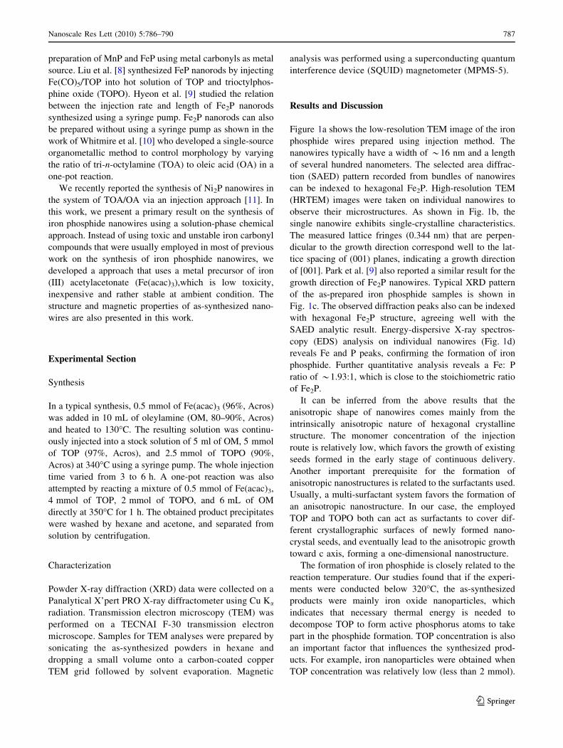
However, when high concentration of TOP (e.g. 12 mmol)
was used, the synthesized nanowires would have a curved
morphology (see Fig. 2a), reflecting the insufficient capa-
bility of TOP for the protection of nanowires’ sidewalls. A
small variation of TOP amount (from 3 to 5 mmol) did not
make apparent difference in the products, showing the
robustness of our synthetic approach. The employed sur-
factant TOPO also plays an important role in control-
ling the product’s morphology. If no TOPO is added, the
synthesized products mainly exhibit a nanoblock-like
morphology resulting from the agglomeration of many
soft-warped nanowires. This indicates that the existence of
TOPO may function as a sidewall protector that can keep
iron phosphide nanowires grow straightly over hundreds of
nanometers. However, high concentration of TOPO on the
contrary will reduce the quality of nanowires and lead to
the formation of some big nanoblocks in the products.
The formation process of Fe2P nanowires may be as
follows. At first, the complex of Fe(acac)3-OM decom-
poses to iron clusters once added to hot solvent of OM.
Then, these iron clusters will be attached to TOP mole-
cules, and the P–C bonds located at the particle surface
may break and cause phosphorus to react with iron,
forming iron phosphide. Once Fe2P seeds with an aniso-
tropic structure forms, the growth along [001] direction
will be favored due to the cooperation of anisotropic nature
and the side-protection effect of surfactant molecules. This
eventually leads to the formation of a one-dimensional
nanostructure.
We also explored one-pot reaction for the synthesis of
iron phosphide nanowires. As shown in Fig. 2b, the
obtained products via one-pot reaction consist of nanowires
plus nanosheets or nanoblocks. The shape singularity of the
products from the one-pot reaction is not superior to the
continuous-injection approach. Considering the robustness
of the reaction, this difference should not come from the
small difference in TOP amount. It is certain that contin-
uous-injection route is crucial to keep the singularity of the
products. A possible reason is that one-pot synthesis incurs
a high concentration of nuclei, some of which are unable to
develop into nanowires due to insufficient protections of
surfactant molecules, whereas for the injection approach,
the nuclei concentration at the beginning stage is restricted
by the delivering rate, and the relatively low concentration
of nuclei plus the continuous delivery of source materials
will allow nuclei to fully grow into nanowires.
The hysteresis loop of as-prepared Fe2P nanowires
measured at 5 K is shown in Fig. 3a. It is clear that the
magnetization curve is difficult to be saturated and the
unsaturated magnetization moment is 45.5 emu/g (1.16
lB), which is much lower than the saturation magnetization
(Ms) value of bulk Fe2P (2.81 lB) [12]. Meanwhile, the
Fig. 1 Normal TEM image
along with SAED pattern (a),
HRTEM image (b), XRD
pattern (c) and EDS spectrum
(d) of the as-prepared iron
phosphide nanowires
788 Nanoscale Res Lett (2010) 5:786–790
123
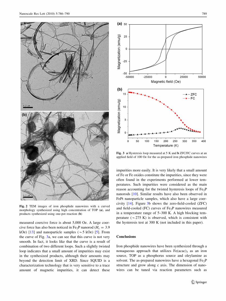
measured coercive force is about 5,000 Oe. A large coer-
cive force has also been noticed in Fe2P nanorod (Hc = 3.9
kOe) [13] and nanoparticle samples (*5 kOe) [5]. From
the curve of Fig. 3a, we can see that this curve is not very
smooth. In fact, it looks like that the curve is a result of
combination of two different loops. Such a slightly twisted
loop indicates that a small amount of impurities may exist
in the synthesized products, although their amounts may
beyond the detection limit of XRD. Since SQUID is a
characterization technology that is very sensitive to a trace
amount of magnetic impurities, it can detect these
impurities more easily. It is very likely that a small amount
of Fe or Fe oxides constitute the impurities, since they were
often found in the experiments performed at lower tem-
peratures. Such impurities were considered as the main
reason accounting for the twisted hysteresis loops of Fe2P
nanorods [10]. Similar results have also been observed in
FePt nanoparticle samples, which also have a large coer-
civity [14]. Figure 3b shows the zero-field-cooled (ZFC)
and field-cooled (FC) curves of Fe2P nanowires measured
in a temperature range of 5–300 K. A high blocking tem-
perature (*275 K) is observed, which is consistent with
the hysteresis test at 300 K (not included in this paper).
Conclusions
Iron phosphide nanowires have been synthesized through a
nonaqueous approach that utilizes Fe(acac)3 as an iron
source, TOP as a phosphorus source and oleylamine as
solvent. The as-prepared nanowires have a hexagonal Fe2P
structure and grow along c axis. The dimension of nano-
wires can be tuned via reaction parameters such as
Fig. 2 TEM images of iron phosphide nanowires with a curved
morphology synthesized using high concentration of TOP (a), and
products synthesized using one-pot reaction (b)
Fig. 3 a Hysteresis loop measured at 5 K and b ZFC/FC curves at an
applied field of 100 Oe for the as-prepared iron phosphide nanowires
Nanoscale Res Lett (2010) 5:786–790 789
123

injection rate, reaction temperature, and TOP/TOPO ratio.
Both injection method and one-pot reaction can produce
iron phosphide nanowires, although the former can gener-
ate products with much better uniformity and yield. The
magnetic tests reveal ferromagnetic characteristics below
the blocking temperature, and a large coercive force
of *5,000 Oe has been observed. The reported synthetic
approach does not use commonly used toxic iron carbonyl
compounds as iron precursor, which provides a convenient
access to further studies of their special physical and
chemical properties.
Acknowledgments This work was partially supported by the
National Natural Science Foundation of China (Grant nos. 50701036
and 50671087) and the National Outstanding Youth Science Foun-
dation of China (Grant no. 50825101).
Open Access This article is distributed under the terms of the
Creative Commons Attribution Noncommercial License which per-
mits any noncommercial use, distribution, and reproduction in any
medium, provided the original author(s) and source are credited.
References
1. T. Hyeon, S.S. Lee, J. Park, Y. Chung, H.B. Na, J. Am. Chem.
Soc. 123, 12798 (2001)
2. S.L. Brock, K. Senevirathne, J. Solid State Chem 181, 1552
(2008)
3. S.C. Perera, P.S. Fodor, G.M. Tsoi, L.E. Wenger, S.L. Brock,
Chem. Mater 15, 4034 (2003)
4. Y. Gu, G. Fan, Y. Qian, H. Zheng, Z. Yang, Mater. Res. Bull 37,
1101 (2002)
5. F. Luo, H.-L. Su, W. Song, Z.-M. Wang, Z.-G. Yan, C.-H. Yan, J.
Mater. Chem 14, 111 (2004)
6. C.G. Hu, Y. Li, J.P. Liu, Y.Y. Zhang, G. Bao, B. Buchine, Z.L.
Wang, Chem. Phys. Lett. 428, 343 (2006)
7. S.L. Brock, S.C. Perera, K.L. Stamm, Chem. Eur. J 10, 3364
(2004)
8. C. Qian, F. Kim, L. Ma, F. Tsui, P. Yang, J. Liu, J. Am. Chem.
Soc. 126, 1195 (2004)
9. J. Park, B. Koo, Y. Hwang, C. Bae, K. An, J.G. Park, H.M. Park,
T. Hyeon, Angew. Chem. Int. Ed. Eng 43, 2282 (2004)
10. A.T. Kelly, I. Rusakova, T. Ould-Ely, C. Hofmann, A. Luttge,
K.H. Whitmire, Nano Lett 7, 2920 (2007)
11. Y. Chen, H. She, X. Luo, G.-H. Yue, D.-L. Peng, J. Cryst.
Growth 311, 1229 (2009)
12. A. Koumina, M. Bacmann, D. Fruchart, J.L. Soubeyroux, P.
Wolfers, J. Tobola, S. Kaprzyk, S. Niziol, M. Mesnaoui, R. Zach,
Ann. Chimie Sci. Materiaux 23, 177 (1998)
13. J. Park, B. Koo, K.Y. Yoon, Y. Hwang, M. Kang, J.-G. Park, T.
Hyeon, J. Am. Chem. Soc. 127, 8433 (2005)
14. T. Iwaki, Y. Kakihara, T. Toda, M. Abdullah, K. Okuyama, J.
Appl. Phys 94, 6807 (2003)
790 Nanoscale Res Lett (2010) 5:786–790
123


![A Nonaqueous Approach to the Preparation of Iron Phosphide ...€¦ · preparation of MnP and FeP using metal carbonyls as metal source. Liu et al. [8] synthesized FeP nanorods by](https://img.pdfslide.us/doc/110x75/60d78e4e1379346cfc1eb676/a-nonaqueous-approach-to-the-preparation-of-iron-phosphide-preparation-of-mnp.jpg)