Embed Size (px)
Citation preview
A new Miocene penguin from Patagonia andits phylogenetic relationships
CAROLINA ACOSTA HOSPITALECHE, CLAUDIA TAMBUSSI, MARIANO DONATO,
and MARIO COZZUOL
Acosta Hospitaleche, C., Tambussi, C., Donato, M., and Cozzuol, M. 2007. A new Miocene penguin from Patagonia andits phylogenetic relationships. Acta Palaeontologica Polonica 52 (2): 299–314.
We describe a new medium−sized penguin, Madrynornis mirandus gen. et sp. nov., from the early late Miocene PuertoMadryn Formation, Chubut Province, Argentina. Although it is evident that extant and fossil penguins form a remarkablyhomogeneous family of birds, Spheniscidae, their within−group phylogenetic relationships are less obvious. In order toidentify the phylogenetic position of the new taxon, we conducted a phylogenetic analysis using 44 osteological charac−ters sampled from 14 representative species of all living genera and five fossil species of Spheniscidae. The family isclearly monophyletic and Madrynornis mirandus is closely related to living taxa. Our phylogenetic interpretation is con−gruent with biostratigraphic data, with Paraptenodytes from the early Miocene (about 20 Ma) located at the base of theSpheniscidae. Classically, two basic tarsometatarsal types were recognized (one for pre−Miocene and the other for thepost−Miocene penguins) based on the pattern of the proximal foramina and the hypotarsus. Madrynornis mirandus exhib−its an arrangement of the proximal foramina and a degree of metatarsals fusion similar to that in the living forms, althoughits elongation index (total length/proximal width) is reminiscent of the extinct Paraptenodytes (a penguin historically rec−ognized as a pre−Miocene form, coming from the early Miocene of Argentina). Madrynornis reveals that the two basictarsometatarsal types co−existed among Miocene penguins.
Key words: Aves, Spheniscidae, penguins, Miocene, Puerto Madryn Formation, Chubut Province, Argentina.
Carolina Acosta Hospitaleche [[email protected]] and Claudia P. Tambussi [[email protected]], Conicet, División Paleontología Vertebrados, Museo de La Plata, Paseo del Bosque s/n, 1900Ciudad de La Plata, Argentina;Mariano Donato [[email protected]], Conicet, Laboratorio de Sistemática y Biología Evolutiva(LASBE), Museo de La Plata, Paseo del Bosque s/n, 1900 Ciudad de La Plata, Argentina;Mario Cozzuol [[email protected]], Departamento de Zoologia, Instituto de Ciências Biológicas, Av. AntônioCarlos, 6627 Pampulha, 31270−910, Belo Horizonte, RS, Brazil.
Introduction
Penguins are obligate marine wing−propelled diving birdswith an exclusively austral history that extends back 55 mil−lion years (Tambussi et al. 2005). The group displays a mo−saic of ancient and derived characters making it a very inter−esting subject from a phylogenetic viewpoint. Various au−thors have studied the extant penguin species, but there is noconsensus about their phylogenetic relationships (e.g., Zusi1975; O’Hara 1989; Sibley and Monroe 1990; Grant et al.1994) and these hypotheses have not been properly con−fronted with those obtained from fossil data.
There are several localities from which spheniscids havebeen recovered. All of them are restricted to Cenozoic sites inthe Southern Hemisphere, including Antarctica (Myrcha etal. 2002; Tambussi et al. 2006), Africa (Simpson 1971), Aus−tralia and New Zealand (Fordyce and Jones 1990 and litera−ture cited therein), and South America. Particularly, in thelast area there are at least four Miocene to Pliocene penguinspecies known form Peru (Stucchi 2002; Stucchi et al. 2003;
Acosta Hospitaleche and Stucchi 2005; Göhlich 2007; seealso Muizon and DeVries 1985), at least five recovered fromMiocene to Pliocene sediments in Chile (Walsh and Hume2001; Acosta Hospitaleche et al. 2002; Emslie and Correa2003), and finally many species from the Paleogene andNeogene of Patagonia (Simpson 1946, 1972, 1981; Clarke etal. 2003). The Patagonian fossil penguins include a wide ar−ray of taxa erected mainly on the basis of differences in limbmorphology (Simpson 1946, 1972, 1981). These penguinsare late middle Eocene–late Miocene in age (Tonni 1980;Cione and Tonni 1981; Clarke et al. 2003) and thereforesome of them are contemporary with the Chilean and Peru−vian species.
The rich collections of other Argentinean fossil penguinsare almost completely restricted to the latest Oligocene–earlyMiocene Leonian Marine Stage and consist mainly of vari−ous isolated bones, with the single exception of Parapteno−dytes antarcticus (Moreno and Mercerat, 1891).
Here we report on a complete skeleton of a new Pata−gonian species of penguin from the lower part of the upper
http://app.pan.pl/acta52/app52−299.pdfActa Palaeontol. Pol. 52 (2): 299–314, 2007
Miocene Puerto Madryn Formation, Chubut Province,Argentina. These remains were previously mentioned byCozzuol et al. (1993). We provide a thorough description ofthe skull and postcranial remains in order to diagnose a newgenus and species, and to discuss its phylogenetic positionand taxonomic status.
Institutional abbreviations.—AMNH, American Museum ofNatural History, New York, USA; IAA, Instituto AntárticoArgentino, Ciudad Autónoma de Buenos Aires, Argentina;MEF, Museo Paleontológico Egidio Feruglio, Trelew, Ar−gentina; MLP, Museo de La Plata, Ciudad de La Plata, Ar−gentina.
Material and methods
Comparative materials used in this study are listed in Appen−dix 1. A phylogenetic analysis was conducted including 22species (14 representative species of all living genera andfive fossil species of Spheniscidae, as well as three out−groups) using 44 morphological characters (Appendix 2) todetermine the phylogenetic position of the new taxon. Thesources for the morphological characters used here wereO'Hara (1989), Acosta Hospitaleche (2004), and Bertelli andGiannini (2005); we modified these characters as needed(Appendix 2). Outgroup comparison was used for the deter−mination of character polarity and rooting (Nixon and Car−penter 1993). The selected outgroups were Diomedea exu−lans Linnaeus, 1758, Fregata magnificens Mathews, 1914,and Gavia immer (Brunnich, 1764), according to phylo−genetic hypotheses proposed by various authors (e.g., VanTuinen et al. 2001; Mayr 2004). G.immer was used as root.
Phylogenetic analysis of our data matrix (Appendix 3) wasdone using TNT v.1.0 (Goloboff et al. 2003) and Wincladaversion 1.00.08 (Nixon 2002). The characters were consideredequally weighted and coded as non−additive. Heuristic sear−ches were performed, with 10,000 replications. Consistencyand retention indices were obtained from Winclada, whichwas also used to manipulate the tree, prepare figures, and ana−lyze character distribution.
Osteological terminology with English equivalents of theLatin names follows Baumel and Witmer (1993) and, wherenecessary, Simpson (1946), O'Hara (1989), and Kanfeder(1994). Description style is according to Pycraft (1898). Wefollow the systematics proposed by Martínez (1992) for themodern species, and by Simpson (1946, 1972) and AcostaHospitaleche (2004) for the fossil penguins. Measurementswere taken with Vernier calipers with 0.01 mm increments.
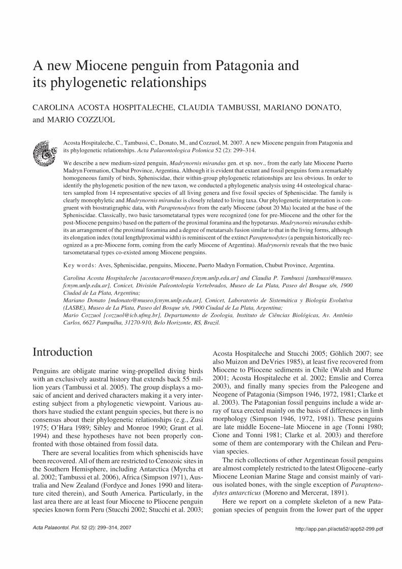
Geologic settingThe specimen was found in an outcrop on the southern coastof Golfo San José, Península Valdés, Chubut Province, Ar−gentina, close to Punta Tehuelche (Fig. 1). In addition to thespecimen described here (Fig. 2), remains of mollusks, com−plete and articulated remains of crabs, sharks, bony fishes,seals, and baleen whales were found.
The outcrop is a small cliff that reaches about two metersabove sea level and forms a platform around the base of themain cliff. It is part of the lower levels of the Puerto MadrynFormation (Haller 1978), known informally as “Entrerriense”because it is considered to be a correlate of marine beds crop−ping out in the Entre Ríos Province, northeastern Argentina(Frenguelli 1926; Scasso and del Río 1987; Scasso et al.
300 ACTA PALAEONTOLOGICA POLONICA 52 (2), 2007
20 km
Fig. 1. Map of Península Valdés, Chubut Province (Argentina) indicating Playa Villarino, Puerto Madryn Formation, early late Miocene, the locality fromwhich the holotype of Madrynornis mirandus gen. et sp. nov., MEF−PV 100, was collected. The penguin locality is indicated by an arrow.
2001). The age of the Puerto Madryn Formation is early lateMiocene based on radiometric data (9.4 Ma, Zinsmeister et al.1981; 10.0 ± 0.3 Ma, Scasso et al. 2001).
The base of the Tortonian is characterized worldwide by asea level drop of about 100 m below the present level (Haq etal. 1987). Since the Puerto Madryn Formation environmentis of relative shallow waters, close to the coast, changing to aterrestrial environment at the top (Scasso and del Río 1987;Dozo et al. 2002), it seems reasonable to infer that the base ofthis unit may be late Serravalian in age, when episodes ofmoderate sea level rise occurred. High sea levels above thepresent one are observed again at the base of the Messinian,which is, however, in disagreement with the age indicated bythe faunal association.
The extraordinary preservation of vertebrates and inver−tebrates (Cozzuol 1993, 2001; Riva Rossi et al. 2000; del Ríoet al. 2001) may in part be due to rapid burial after a storm,which deposited a large volume of sediment in a short time.Articulated vertebrate remains, complete crabs and molluskaggregation in life position indicates a rapid burial event withvery little or no exposure after the event. A series of stormdeposits along the sequence was described by Scasso and delRío (1987), Scasso et al. (2001), and del Río et al. (2001).The best preserved specimens are normally found at the topof massive layers of fine, silty sandstones of a less than a me−ter thick. No evidence of post−burial predation or scavengingwas observed.
Systematic paleontology
Order Sphenisciformes Sharpe, 1891Family Spheniscidae Bonaparte, 1831Genus Madrynornis nov.Type species: Madrynornis mirandus sp. nov., monotypic.
Derivation of the name: From Madryn, for its stratigraphic provenance,
the Puerto Madryn Formation, and ornis, Greek, referring to a “bird ofMadryn”. Gender is masculine.
Diagnosis.—Differs from all other known spheniscids by thefollowing combination of characters: transverse occipital crestexpanded into posterolaterally directed wings (not developedin Pygoscelis Wagler, 1832); temporal fossa more triangularand deeper than in Paraptenodytes Ameghino, 1891; post−orbital process slender and longer than in Spheniscus Brisson,1760; jugal arch only slightly curved compared to that inEudyptes Vieillot, 1816 and Pygoscelis; interorbital narrowerthan in Spheniscus and Eudyptes; nasal fossa without an exter−nal edge as in Spheniscus and Paraptenodytes (edge present inPygoscelis); parasphenoidal plate broader than in all livingspecies. Mandibular ramus straight with the retroarticular pro−cess longer than in Paraptenodytes and Spheniscus, and ex−tending beyond the articular fossa. Humeral diaphysis straight(slightly curved in Palaeospheniscus Moreno and Mercerat,1891 and Spheniscus); proximal and distal subequal and pre−axial angle smaller than in Spheniscus and Pygoscelis; shaft−trochlear angle (ca. 38�) smaller than in Aptenodytes Miller,1778 and Palaeospheniscus. Large rounded and bipartite trici−pital fossa with ventral part smaller and deeper than the dorsalportion (undivided in Paraptenodytes). Foramen ilioischia−dicum smaller than the foramen acetabular (unlike EudyptulaBonaparte, 1856 and Palaeospheniscus). Femur with trochan−ter much higher than the head, unlike the living species;trochanter crest broad but poorly developed compared withextant species; intercondylar groove deeper and wider than inParaptenodytes; intercnemial groove reaches proximal endwith uniform depth along its length (irregular depth in Pygo−scelis); supratendinosus bridge oblique and broader than inParaptenodytes, Pygoscelis, and Spheniscus; fossa flexoriashallower than in Pygoscelis and Spheniscus; medial epi−condyle single (double in Spheniscus) and rounded (elongatein Pygoscelis). Fibular crest with sharp edges as in Para−ptenodytes, Spheniscus, and Pygoscelis adeliae (rounded inthe remaining species). Elongation index (total length/proxi−
http://app.pan.pl/acta52/app52−299.pdf
ACOSTA HOSPITALECHE ET AL.—MIOCENE PENGUIN FROM PATAGONIA 301
100 mm
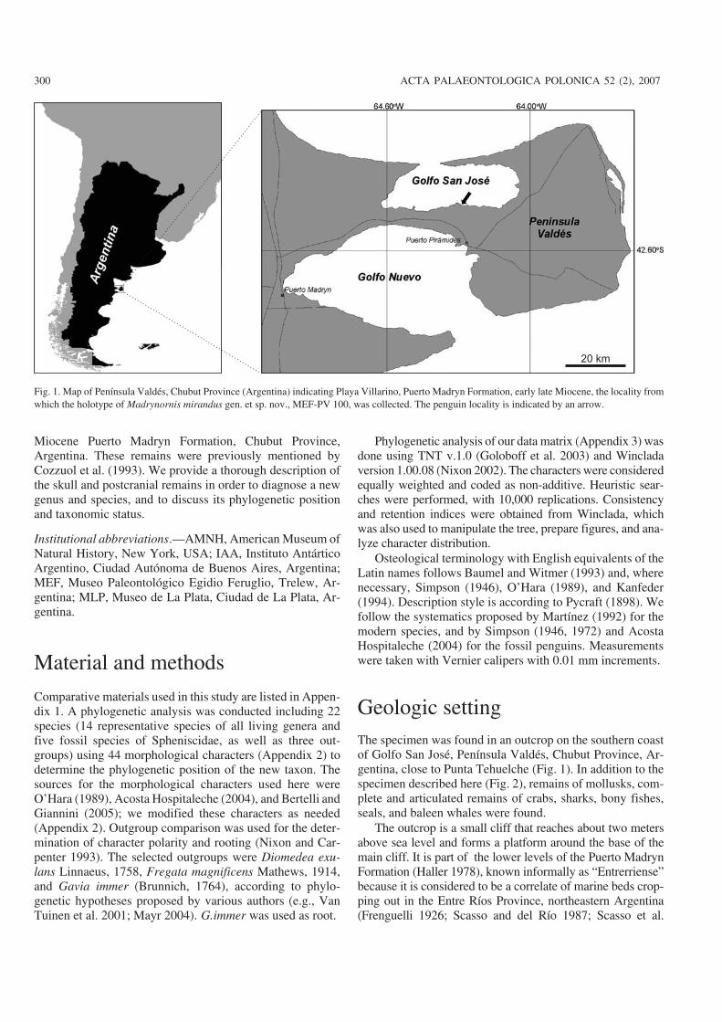
Fig. 2. Miocene penguin Madrynornis mirandus gen. et sp. nov., MEF−PV 100 (holotype), Puerto Madryn Formation, early late Miocene, Playa Villarino,Península Valdés, Chubut Province, Argentina. Photo of the articulated skeleton before preparation.
mal width) of tarsometatarsus 1.79 (smaller than in Palaeo−spheniscus). Medial proximal vascular foramen smaller thanthe lateral (subequal in Pygoscelis, Eudyptes, Eudyptula, andParaptenodytes); only the lateral proximal vascular foramenopens directly on plantar side (both of them open on the plan−tar surface in Pygoscelis and Paraptenodytes); trochlear edgessturdier than in Spheniscus.
Temporal and geographic distribution.—Early late Miocene,Puerto Madryn Formation, Argentina. Playa Villarino (S 42�
25’, W 64�16’), Golfo San José, Chubut Province (Fig. 1).
Comments.—Simpson (1946) established five subfamilies ofSpheniscidae based on characters of the humerus and tarso−metatarsus: Palaeospheniscinae, Paraptenodytinae, Anthro−pornithinae, Palaeeudyptinae, and Spheniscinae (includingall living forms).
Following the criticism of Marples (1952), Simpson(1971) abandoned his subfamilial division of the Sphenisci−dae after examining the fossil penguins from New Zealand.
Although we do not have enough evidence to propose theadoption of a suprageneric arrangement, our current studies onthe penguin faunas from Antarctica and South America allowus (CAH and CT) to identify groups that are partially equiva−lent to those of Simpson’s classification (Acosta Hospitaleche2003, 2004; Tambussi et al. 2006; but see Ksepka et al. 2006).
Madrynornis differs from the Palaeospheniscus speciesand Eretiscus tonnii in having a shorter tarsometatarsus,smaller shaft−trochlear angle, and both proximal vascular fo−ramina well developed. A bipartite tricipital fossa and asmaller shaft−trochlear angle differentiate Madrynornis fromParaptenodytes sp. and Arthrodytes andrewsi, the largestPatagonian penguin even discovered. Madrynornis is distin−guished from Anthropornis grandis and A. nordenskjoeldi byhaving a shorter tarsometatarsus, a weakly curved rather thansigmoid humerus, and a larger and bipartite tricipital fossa. Itdiffers from Palaeeudyptes species by the presence of bothproximal vascular foramina and a larger and bipartite trici−pital fossa. Finally, although the metatarsal fusion is strongerthan in living species, a bipartite tricipital fossa, short tarso−metatarsus, small shaft−trochlear angle and similar develop−ment of the proximal vascular foramina are shared with theseforms.
Madrynornis mirandus sp. nov.Figs. 2–7.
Holotype: Madrynornis mirandus, a nearly complete and articulatedskeleton, MEF− PV 100, collected by one of the authors (MC). It in−cludes: skull with partially preserved rostrum, mandible missing distalportion, 27 vertebrae, pygostyle, pelvis, left and right femora, right pa−tella, left and right tibiotarsi and fibulae, left and right tarsometatarsi, 16pedal phalanges (2 and ungual of second digit, 1, 2, and 3 of third digit,1, 3, and 4 of fourth digit of the left side; 1 of second digit, 1, 2, 3, andungual of third digit, and 2, 3, and 4 of fourth digit of the right side), ster−num, 13 ribs, right scapula, left and right coracoids, furcula, left andright humeri, right ulna, right radius, right carpometacarpus.
Derivation of the name: From Latin, mirandus, wonderful. From the ex−cellent preservation of the skeleton.
Measurements.—Skull: total length, ca.145 mm; postorbitalwidth, 51 mm; mandible length, ca. 111 mm; posterior heightof mandible, 9.7 mm; height at level of mandibular angle,15.5 mm. Sternum: total length, 136 mm; width at posteriormost costal facet, 66 mm. Coracoid: length, 8.48 mm; distalwidth, 31 mm. Scapula: length, 114.2 mm; width of articularend, 23.9 mm. Humerus: length, 79.1 mm; distal width, 20.5mm. Ulna: length, 59.1 mm. Radius: length, 57.8 mm. Car−pometacarpus: length, 48.6 mm; proximal width, 15.8 mm.Pelvis: length among midline, 99 mm. Femur: length, 85.9mm. Tibiotarsus: maximum length, 134.6 mm; distal width,16.9 mm. Tarsometatarsus: length, 36.5 mm; proximalwidth, 18.1 mm; distal width, 19.1 mm. Pygostyle: length, 44mm. Left phalanges (length) of the second digit: 2 (19.4 mm)and ungual (15.6 mm); of the third digit: 1 (25.4 mm), 2 (20.7mm), and 3 (20.2 mm); and of the fourth digit: 1 (20.2 mm), 3(14.4 mm), and 4 (14.8 mm). Right phalanges (length) of thesecond digit: 1 (22.7 mm); of the third digit: 2 (25.6 mm), 2(21.7 mm), 3 (19 mm), and ungual (18.1 mm); and of thefourth digit: 2 (15.5 mm), 3 (14.4 mm), and 4 (14.8 mm).
Diagnosis.—As for the genus.
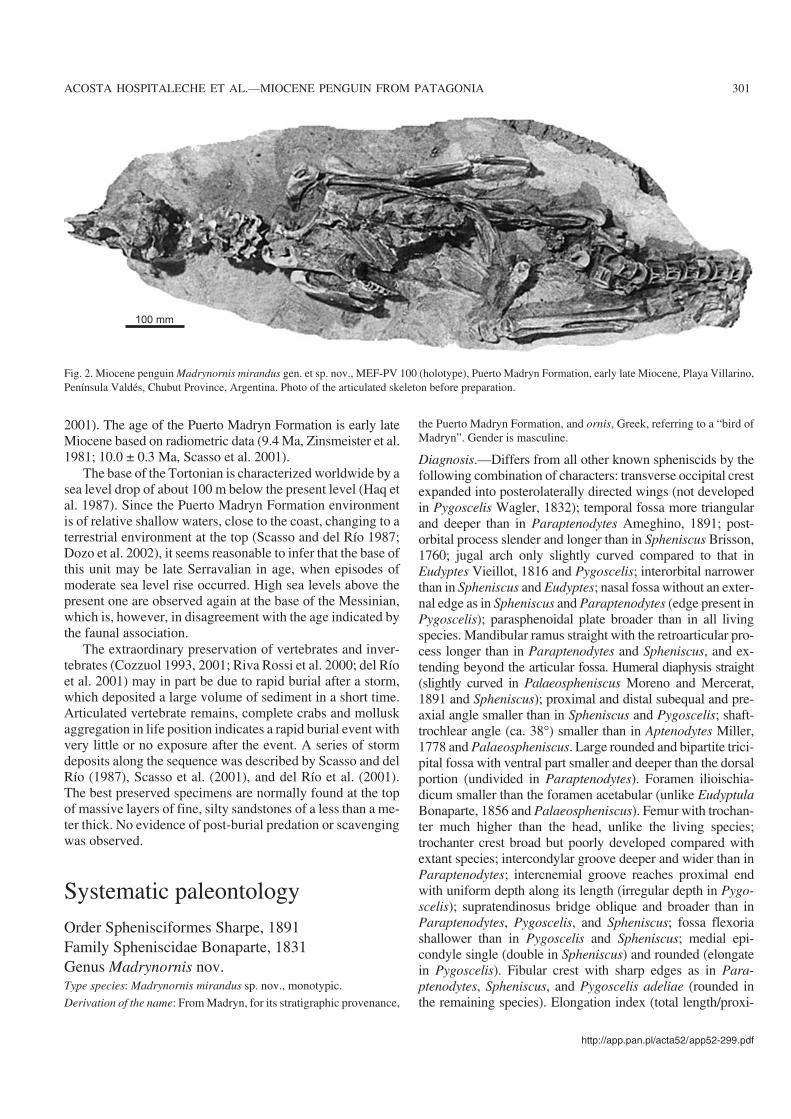
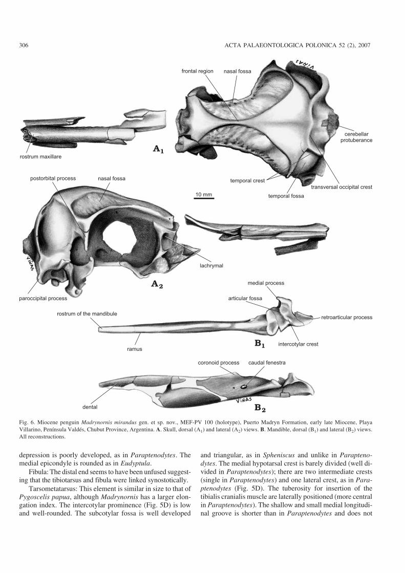
Description and comparisons.—Skull: Similar in size to theAdelie Penguin, Pygoscelis adeliae (Hombron and Jacquinot,1841). The cerebellar protuberance projects more distally thanthe paroccipital process (Figs. 3A, 6A), while in Pygoscelisantarctica (Forster, 1781) they project slightly further posteri−orly. The paroccipital processes are ventrally directed as in theliving species, whereas in Paraptenodytes they are bifid andcaudally projected. The transverse occipital crest is expandedposterolaterally and wing−like, similar to Spheniscus, Eudyp−tes, and Paraptenodytes. The sagital crest (apparent in Para−ptenodytes) is absent in Madrynornis. The occipital region istrapezoidal in shape, but is subcircular in Paraptenodytes andquadrangular in Eudyptes, Eudyptula, and Spheniscus. Thetemporal fossa is deep and triangular with its dorsal tip notreaching the position of the sagittal crest. The postorbital pro−cesses are thin, long (Figs. 3A, 6A), and project ventrally,whereas in Paraptenodytes they are directed posteriorly. Thefrontals form a medial crest much narrower than in the livingspecies, and which is absent in Paraptenodytes. The entirejugal arch is gently curved, a characteristic shared with Sphe−niscus. Extreme curvature of the anterior portion of the arch is afeature of Eudyptes, Pygoscelis adeliae, and P. antarctica, andto a lesser degree of P. papua Forster, 1781, MegadyptesMilne−Edwards, 1880, and Aptenodytes (Zusi 1975). The basi−temporal plate is broader than in all the compared species. Thepterygoid (narrow and rod−like in Paraptenodytes) is expandedto form a thin, horizontal plate similar to that seen in extant spe−cies. The tomial crest is above the level of the parasphenoidalplate, as in Aptenodytes and Pygoscelis, but is not parallel as inAptenodytes forsteri Gray, 1844.
The mandible is straight. The medial process, larger thanin Paraptenodytes, bears a medially directed hook−like pro−jection. The caudal fenestra is oval and the rostral fenestra isabsent. On the medial surface of the mandible, extending
302 ACTA PALAEONTOLOGICA POLONICA 52 (2), 2007
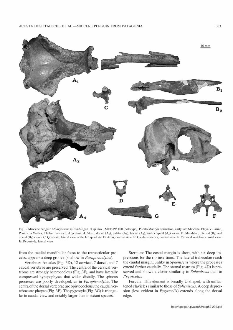
from the medial mandibular fossa to the retroarticular pro−cess, appears a deep groove (shallow in Paraptenodytes).
Vertebrae: An atlas (Fig. 3D), 12 cervical, 7 dorsal, and 7caudal vertebrae are preserved. The centra of the cervical ver−tebrae are strongly heterocoelous (Fig. 3F), and have laterallycompressed hypapophyses that widen distally. The spinousprocesses are poorly developed, as in Paraptenodytes. Thecentra of the dorsal vertebrae are opistocoelous; the caudal ver−tebrae are platyan (Fig. 3E). The pygostyle (Fig. 3G) is triangu−lar in caudal view and notably larger than in extant species.
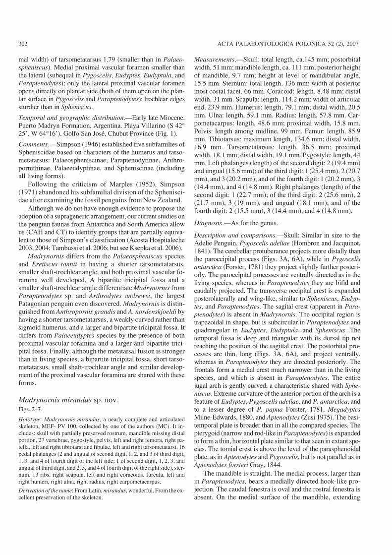
Sternum: The costal margin is short, with six deep im−pressions for the rib insertions. The lateral trabeculae reachthe caudal margin, unlike in Spheniscus where the processesextend farther caudally. The sternal rostrum (Fig. 4D) is pre−served and shows a closer similarity to Spheniscus than toPygoscelis.
Furcula: This element is broadly U−shaped, with unflat−tened clavicles similar to those of Spheniscus. A deep depres−sion (less evident in Pygoscelis) extends along the dorsaledge.
http://app.pan.pl/acta52/app52−299.pdf
ACOSTA HOSPITALECHE ET AL.—MIOCENE PENGUIN FROM PATAGONIA 303
Fig. 3. Miocene penguin Madrynornis mirandus gen. et sp. nov., MEF−PV 100 (holotype), Puerto Madryn Formation, early late Miocene, Playa Villarino,Península Valdés, Chubut Province, Argentina. A. Skull, dorsal (A1), palatal (A2), lateral (A3), and occipital (A4) views. B. Mandible, internal (B1) anddorsal (B2) views. C. Quadrate, lateral view of the left quadrate. D. Atlas, cranial view. E. Caudal vertebra, cranial view. F. Cervical vertebra, cranial view.G. Pygostyle, lateral view.
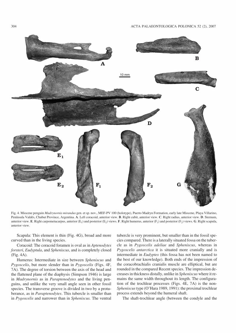
Scapula: This element is thin (Fig. 4G), broad and morecurved than in the living species.
Coracoid: The coracoid foramen is oval as in Aptenodytesforsteri, Eudyptula, and Spheniscus, and is completely closed(Fig. 4A).
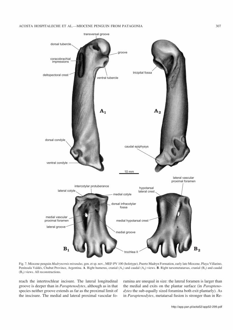
Humerus: Intermediate in size between Spheniscus andPygoscelis, but more slender than in Pygoscelis (Figs. 4F,7A). The degree of torsion between the axis of the head andthe flattened plane of the diaphysis (Simpson 1946) is largein Madrynornis as in Paraptenodytes and the living pen−guins, and unlike the very small angle seen in other fossilspecies. The transverse groove is divided in two by a protu−berance, as in Paraptenodytes. This tubercle is smaller thanin Pygoscelis and narrower than in Spheniscus. The ventral
tubercle is very prominent, but smaller than in the fossil spe−cies compared. There is a laterally situated fossa on the tuber−cle as in Pygoscelis adeliae and Spheniscus, whereas inPygoscelis antarctica it is situated more cranially and isintermediate in Eudyptes (this fossa has not been named tothe best of our knowledge). Both ends of the impression ofthe coracobrachialis cranialis muscle are elliptical, but arerounded in the compared Recent species. The impression de−creases in thickness distally, unlike in Spheniscus where it re−mains the same width throughout its length. The configura−tion of the trochlear processes (Figs. 4E, 7A) is the non−Spheniscus type (O’Hara 1989, 1991): the proximal trochlearprocess extends beyond the humeral shaft.
The shaft−trochlear angle (between the condyle and the
304 ACTA PALAEONTOLOGICA POLONICA 52 (2), 2007
Fig. 4. Miocene penguin Madrynornis mirandus gen. et sp. nov., MEF−PV 100 (holotype), Puerto Madryn Formation, early late Miocene, Playa Villarino,Península Valdés, Chubut Province, Argentina. A. Left coracoid, anterior view. B. Right cubit, anterior view. C. Right radius, anterior view. D. Sternum,anterior view. E. Right carpometacarpus, anterior (E1) and posterior (E2) views. F. Right humerus, anterior (F1) and posterior (F2) views. G. Right scapula,anterior view.
axis of the diaphysis, sensu Simpson 1946) is small (ca. 38�),but bigger than in Pygoscelis. The impression of the supra−coracoideus muscle is oblique to the axis, as in Parapteno−dytes, Spheniscus, Eudyptes, and Aptenodytes, although thisscar is shorter in the Recent species.
Ulna: This element is similar in size to Pygoscelis. Theventral edge is curved, being straight in Spheniscus.
Radius: The bicipital tubercle is well developed, forminga sharp anteriorly directed projection (Fig. 4C).
Carpometacarpus: The distal end is widest as in Sphenis−cus, but narrower than in Pygoscelis (Fig. 4E). The dorsaledge is concave as in Eudyptula. In Madrynornis, Sphenis−cus, and Pygoscelis, the metacarpal major bone is more dis−tally extended than the minor, whereas they are extendedequally in Aptenodytes. Both metacarpal bones are morestrongly fused and the intermetacarpal distance is much nar−rower in Eudyptes.
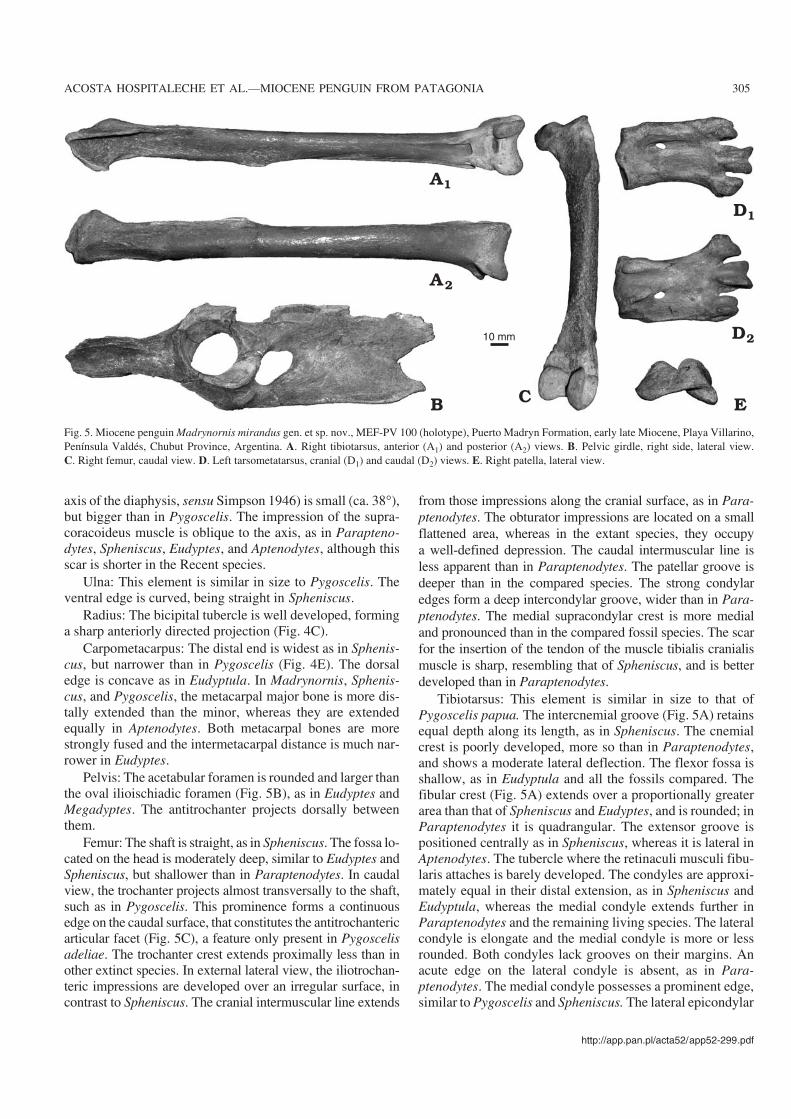
Pelvis: The acetabular foramen is rounded and larger thanthe oval ilioischiadic foramen (Fig. 5B), as in Eudyptes andMegadyptes. The antitrochanter projects dorsally betweenthem.
Femur: The shaft is straight, as in Spheniscus. The fossa lo−cated on the head is moderately deep, similar to Eudyptes andSpheniscus, but shallower than in Paraptenodytes. In caudalview, the trochanter projects almost transversally to the shaft,such as in Pygoscelis. This prominence forms a continuousedge on the caudal surface, that constitutes the antitrochantericarticular facet (Fig. 5C), a feature only present in Pygoscelisadeliae. The trochanter crest extends proximally less than inother extinct species. In external lateral view, the iliotrochan−teric impressions are developed over an irregular surface, incontrast to Spheniscus. The cranial intermuscular line extends
from those impressions along the cranial surface, as in Para−ptenodytes. The obturator impressions are located on a smallflattened area, whereas in the extant species, they occupya well−defined depression. The caudal intermuscular line isless apparent than in Paraptenodytes. The patellar groove isdeeper than in the compared species. The strong condylaredges form a deep intercondylar groove, wider than in Para−ptenodytes. The medial supracondylar crest is more medialand pronounced than in the compared fossil species. The scarfor the insertion of the tendon of the muscle tibialis cranialismuscle is sharp, resembling that of Spheniscus, and is betterdeveloped than in Paraptenodytes.
Tibiotarsus: This element is similar in size to that ofPygoscelis papua. The intercnemial groove (Fig. 5A) retainsequal depth along its length, as in Spheniscus. The cnemialcrest is poorly developed, more so than in Paraptenodytes,and shows a moderate lateral deflection. The flexor fossa isshallow, as in Eudyptula and all the fossils compared. Thefibular crest (Fig. 5A) extends over a proportionally greaterarea than that of Spheniscus and Eudyptes, and is rounded; inParaptenodytes it is quadrangular. The extensor groove ispositioned centrally as in Spheniscus, whereas it is lateral inAptenodytes. The tubercle where the retinaculi musculi fibu−laris attaches is barely developed. The condyles are approxi−mately equal in their distal extension, as in Spheniscus andEudyptula, whereas the medial condyle extends further inParaptenodytes and the remaining living species. The lateralcondyle is elongate and the medial condyle is more or lessrounded. Both condyles lack grooves on their margins. Anacute edge on the lateral condyle is absent, as in Para−ptenodytes. The medial condyle possesses a prominent edge,similar to Pygoscelis and Spheniscus. The lateral epicondylar
http://app.pan.pl/acta52/app52−299.pdf
ACOSTA HOSPITALECHE ET AL.—MIOCENE PENGUIN FROM PATAGONIA 305
Fig. 5. Miocene penguin Madrynornis mirandus gen. et sp. nov., MEF−PV 100 (holotype), Puerto Madryn Formation, early late Miocene, Playa Villarino,Península Valdés, Chubut Province, Argentina. A. Right tibiotarsus, anterior (A1) and posterior (A2) views. B. Pelvic girdle, right side, lateral view.C. Right femur, caudal view. D. Left tarsometatarsus, cranial (D1) and caudal (D2) views. E. Right patella, lateral view.
depression is poorly developed, as in Paraptenodytes. Themedial epicondyle is rounded as in Eudyptula.
Fibula: The distal end seems to have been unfused suggest−ing that the tibiotarsus and fibula were linked synostotically.
Tarsometatarsus: This element is similar in size to that ofPygoscelis papua, although Madrynornis has a larger elon−gation index. The intercotylar prominence (Fig. 5D) is lowand well−rounded. The subcotylar fossa is well developed
and triangular, as in Spheniscus and unlike in Parapteno−dytes. The medial hypotarsal crest is barely divided (well di−vided in Paraptenodytes); there are two intermediate crests(single in Paraptenodytes) and one lateral crest, as in Para−ptenodytes (Fig. 5D). The tuberosity for insertion of thetibialis cranialis muscle are laterally positioned (more centralin Paraptenodytes). The shallow and small medial longitudi−nal groove is shorter than in Paraptenodytes and does not
306 ACTA PALAEONTOLOGICA POLONICA 52 (2), 2007
10 mm
rostrum maxillare
frontal region nasal fossa
temporal crest
cerebellar
protuberance
transversal occipital crest
temporal fossa
nasal fossapostorbital process
paroccipital process
medial process
lachrymal
articular fossa
retroarticular process
intercotylar crest
caudal fenestracoronoid process
ramus
rostrum of the mandibule
dental
Fig. 6. Miocene penguin Madrynornis mirandus gen. et sp. nov., MEF−PV 100 (holotype), Puerto Madryn Formation, early late Miocene, PlayaVillarino, Península Valdés, Chubut Province, Argentina. A. Skull, dorsal (A1) and lateral (A2) views. B. Mandible, dorsal (B1) and lateral (B2) views.All reconstructions.
reach the intertrochlear incisure. The lateral longitudinalgroove is deeper than in Paraptenodytes, although as in thatspecies neither groove extends as far as the proximal limit ofthe inscisure. The medial and lateral proximal vascular fo−
ramina are unequal in size: the lateral foramen is larger thanthe medial and exits on the plantar surface (in Parapteno−dytes the sub−equally sized foramina both exit plantarly). Asin Paraptenodytes, metatarsal fusion is stronger than in Re−
http://app.pan.pl/acta52/app52−299.pdf
ACOSTA HOSPITALECHE ET AL.—MIOCENE PENGUIN FROM PATAGONIA 307
dorsal tubercle
transversal groove
groove
ventral tubercle
ventral condyle
dorsal condyle
deltopectoral crest
coracobrachial
impressions
tricipital fossa
caudal epiphysys
10 mm
lateral vascular
proximal foramen
hypotarsal
lateral crest
medial cotyle
intercotylar protuberance
dorsal infracotylar
fossa
medial hypotarsal crest
medial groove
trochlea II
lateral groove
medial vascular
proximal foramen
lateral cotyle
Fig. 7. Miocene penguin Madrynornis mirandus, gen. et sp. nov., MEF−PV 100 (holotype), Puerto Madryn Formation, early late Miocene, Playa Villarino,Península Valdés, Chubut Province, Argentina. A. Right humerus, cranial (A1) and caudal (A2) views. B. Right tarsometatarsus, cranial (B1) and caudal(B2) views. All reconstructions.
cent species; Eudyptula has the extreme condition with themetatarsals separated by deep furrows. The trochlea of meta−tarsal II exhibits stronger lateral divergence than in Pygo−scelis, Eudyptes, and Spheniscus; the trochlea of metatarsalIII possesses strong, distally divergent trochlear edges, andthe trochlea of metatarsal IV is approximately straight andhas less distal elongation than Spheniscus.
Phalanges: There are no fossil phalanges available forcomparison with Madrynornis. The 16 phalanges of Madry−nornis are similar in shape to those of the living species; theyare more robust than those of Spheniscus magellanicus.
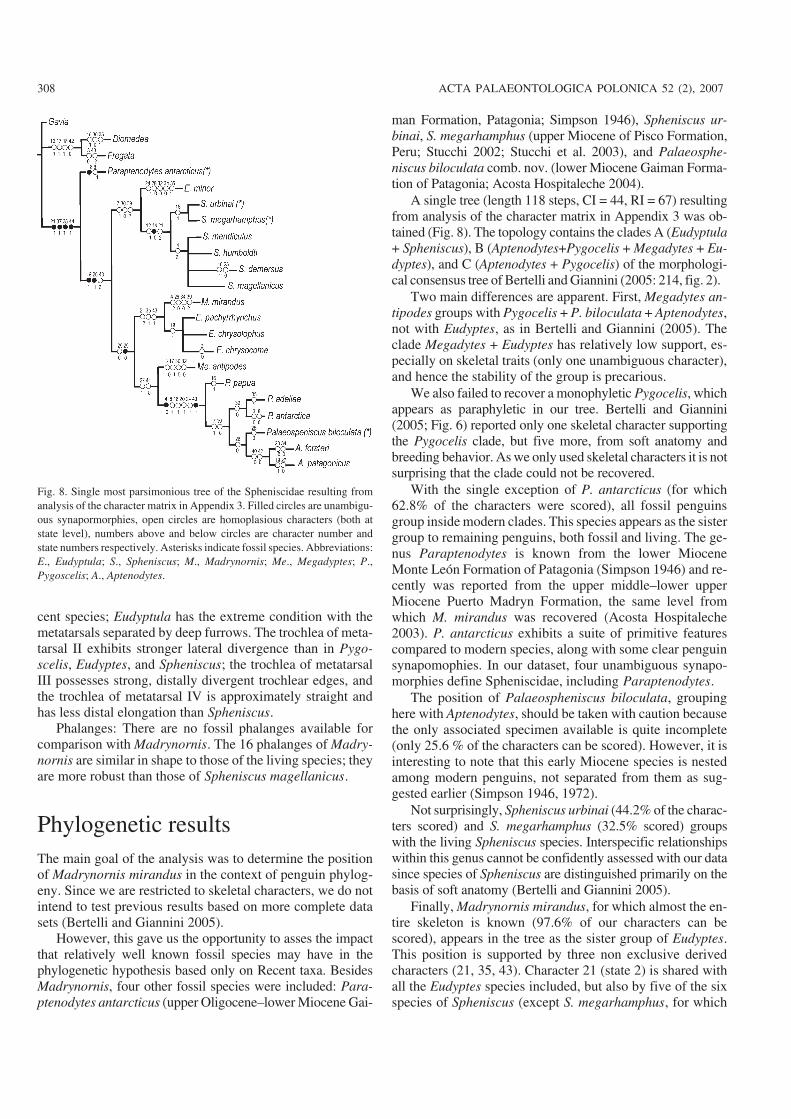
Phylogenetic resultsThe main goal of the analysis was to determine the positionof Madrynornis mirandus in the context of penguin phylog−eny. Since we are restricted to skeletal characters, we do notintend to test previous results based on more complete datasets (Bertelli and Giannini 2005).
However, this gave us the opportunity to asses the impactthat relatively well known fossil species may have in thephylogenetic hypothesis based only on Recent taxa. BesidesMadrynornis, four other fossil species were included: Para−ptenodytes antarcticus (upper Oligocene–lower Miocene Gai−
man Formation, Patagonia; Simpson 1946), Spheniscus ur−binai, S. megarhamphus (upper Miocene of Pisco Formation,Peru; Stucchi 2002; Stucchi et al. 2003), and Palaeosphe−niscus biloculata comb. nov. (lower Miocene Gaiman Forma−tion of Patagonia; Acosta Hospitaleche 2004).
A single tree (length 118 steps, CI = 44, RI = 67) resultingfrom analysis of the character matrix in Appendix 3 was ob−tained (Fig. 8). The topology contains the clades A (Eudyptula+ Spheniscus), B (Aptenodytes+Pygocelis + Megadytes + Eu−dyptes), and C (Aptenodytes + Pygocelis) of the morphologi−cal consensus tree of Bertelli and Giannini (2005: 214, fig. 2).
Two main differences are apparent. First, Megadytes an−tipodes groups with Pygocelis + P. biloculata + Aptenodytes,not with Eudyptes, as in Bertelli and Giannini (2005). Theclade Megadytes + Eudyptes has relatively low support, es−pecially on skeletal traits (only one unambiguous character),and hence the stability of the group is precarious.
We also failed to recover a monophyletic Pygocelis, whichappears as paraphyletic in our tree. Bertelli and Giannini(2005; Fig. 6) reported only one skeletal character supportingthe Pygocelis clade, but five more, from soft anatomy andbreeding behavior. As we only used skeletal characters it is notsurprising that the clade could not be recovered.
With the single exception of P. antarcticus (for which62.8% of the characters were scored), all fossil penguinsgroup inside modern clades. This species appears as the sistergroup to remaining penguins, both fossil and living. The ge−nus Paraptenodytes is known from the lower MioceneMonte León Formation of Patagonia (Simpson 1946) and re−cently was reported from the upper middle–lower upperMiocene Puerto Madryn Formation, the same level fromwhich M. mirandus was recovered (Acosta Hospitaleche2003). P. antarcticus exhibits a suite of primitive featurescompared to modern species, along with some clear penguinsynapomophies. In our dataset, four unambiguous synapo−morphies define Spheniscidae, including Paraptenodytes.
The position of Palaeospheniscus biloculata, groupinghere with Aptenodytes, should be taken with caution becausethe only associated specimen available is quite incomplete(only 25.6 % of the characters can be scored). However, it isinteresting to note that this early Miocene species is nestedamong modern penguins, not separated from them as sug−gested earlier (Simpson 1946, 1972).
Not surprisingly, Spheniscus urbinai (44.2% of the charac−ters scored) and S. megarhamphus (32.5% scored) groupswith the living Spheniscus species. Interspecific relationshipswithin this genus cannot be confidently assessed with our datasince species of Spheniscus are distinguished primarily on thebasis of soft anatomy (Bertelli and Giannini 2005).
Finally, Madrynornis mirandus, for which almost the en−tire skeleton is known (97.6% of our characters can bescored), appears in the tree as the sister group of Eudyptes.This position is supported by three non exclusive derivedcharacters (21, 35, 43). Character 21 (state 2) is shared withall the Eudyptes species included, but also by five of the sixspecies of Spheniscus (except S. megarhamphus, for which
308 ACTA PALAEONTOLOGICA POLONICA 52 (2), 2007
Fig. 8. Single most parsimonious tree of the Spheniscidae resulting fromanalysis of the character matrix in Appendix 3. Filled circles are unambigu−ous synapormorphies, open circles are homoplasious characters (both atstate level), numbers above and below circles are character number andstate numbers respectively. Asterisks indicate fossil species. Abbreviations:E., Eudyptula; S., Spheniscus; M., Madrynornis; Me., Megadyptes; P.,Pygoscelis; A., Aptenodytes.
the condition is unknown). Character 35 (state 1) is sharedwith Eudyptes chrysocome (unknown in the other two Eu−dyptes species) but also with Eudyptes minor and Pygoscelisadeliae. Finally, character 43, in its state 1, is shared with E.chrysocome (unknown in the other two Eudyptes species)and with P. biloculata.
Discussion and conclusions
Simpson (1946) provided the first suprageneric systematics ofthe Spheniscidae, and his study has remained the basis for allother analyses of penguin relationships. Giannini and Bertelli(2004) made a phylogenetic arrangement of the living speciesbased on integumentary characters. Subsequently, Bertelli andGiannini (2005) tested their proposal using a morphologicaland molecular data set. Our analysis is the first to employcladistic methods to a data matrix 179–183.
Since the referral of isolated elements to a given species isalways problematic we have preferred to base our phylogen−etic analysis on articulated or associated specimens whereverpossible. In essence, we used the near complete skeleton ofParaptenodytes antarcticus, the unpublished associated ma−terial of Palaeospheniscus biloculata comb. nov. (AcostaHospitaleche 2004) and skeletons of all the modern genera(including two fossil species of Spheniscus) as a basis for es−tablishing the phylogenetic relationships of Madrynornis.
Although it is evident from observations and compari−sons that extant and fossil penguins constitute a remarkablyhomogeneous family of birds, their within−group phylogen−etic relationships are less obvious. In our analysis, Sphenis−cidae is easily recognized as a monophyletic group based onosteological characters alone (characters 21[1], 37[1], 38[1],44[1]), in agreement with Mayr (2004) and Bertelli andGiannini (2005). Madrynornis mirandus sp. nov. is the firstspecies described from Argentinean rocks that is closely re−lated to the living forms.
According to this analysis, Palaeospheniscus biloculatais the sister taxon of Aptenodytes, but we regard its locationas questionable and suggest that its placement may be a con−sequence of the large amount of missing data.
Simpson (1946) stated that pre−Miocene penguins have amore elongate tarsometatarsus with more completely fusedmetatarsals than the Recent forms. Zusi (1975) recognizedtwo variable tarsometatarsal types in extant species based onthe pattern of the proximal foramina in relationship to thehypotarsus. Madrynornis resembles the Recent species in thedegree of fusion of the metatarsals and the location of theproximal foramina, but the elongation index is similar to thepre−Miocene forms.
As one might expect, Paraptenodytes from the early Mio−cene (about 20 Ma) is located at the base of the Spheniscidae.This outcome further supports the possibility that modernforms would have appeared in the Miocene.
AknowledgementsEduardo Ruigómez (MEF) allowed CAH access to new material and lentus the specimen described herein. Pablo Ljumberg (Trelew, Argentina)collected MEF−PV 1729. José Laza further prepared the specimen to al−low detailed study and Agustín Viñas (Museo Argentino de CienciasNatuales Bernardino Rivadavia, Ciudad de Buenos Aires, Argentina)drew the figures. Stig Walsh (Natural History Museum, London, UK)improved considerably our English and an earlier draft of this manu−script. Ewan Fordyce (Otago University, Department of Geology, NewZealand) and an anonymous reviewer provided helpful comments on themanuscript. Fieldwork and preparation of the new specimen was sup−ported by National Geographic Grant 4044−89 to Mario A. Cozzuol, theFrank Chapman Memorial Fund and the CONICET PIP 5694 to CT.
AddendumSome phylogenetic studies were published shortly after thepreparation of this manuscript, however, they could not beincorporated into this project. See for example: Slack et al.(2006) and Ksepka et al. (2006).
ReferencesAcosta Hospitaleche, C. 2003. Paraptenodytes antarcticus (Aves: Sphenisci−
formes) en la Formación Puerto Madryn (Mioceno tardío temprano),provincia de Chubut, Argentina. Revista Española de Paleontología 18:179–183.
Acosta Hospitaleche, C. 2004. Los pingüinos (Aves, Sphenisciformes) fósilesde Argentina. Sistemática, biogeografía y evolución. Unpublished Ph.D.thesis. 321 pp. Facultad de Ciencias Naturales y Museo, UniversidadNacional de La Plata, Ciudad de La Plata.
Acosta Hospitaleche, C. 2005. Systematic revision of Arthrodytes Ameghino,1905 (Aves, Spheniscidae) and its assignment to the Paraptenodytinae.Neues Jahrbuch für Geologie und Paläontologie 7: 404–414.
Acosta Hospitaleche, C. and Stucchi, M. 2005. Nuevos restos terciarios deSpheniscidae (Aves, Sphenisciformes) procedentes de la costa del Perú.Revista de la Sociedad Española de Paleontología 20: 1–5.
Acosta Hospitaleche, C., Fritis, O., Tambussi, C., and Quinzio, A. 2002.Nuevos restos de pingüinos (Aves: Spheniscidae) en la FormaciónBahía Inglesa (Mioceno superior–Plioceno inferior) de Chile. Actas delPrimer Congreso Latinoamericano de Paleontología de Vertebrados,16. Santiago de Chile.
Ameghino, F. 1891. Enumeración de las aves fósiles de la República Argen−tina. Revista Argentina de Historia Natural 1: 441–453.
Baumel, J. and Witmer, L.M. 1993. Osteologia. In: J.J. Baumel, A.S. King,A.M. Lucas, J.E. Breazile, and H.E. Evans (eds.), Handbook of AvianAnatomy: Nomina Anatomica Avium, 45–132. Publications of the NuttallOrnithological Club, Cambridge, Massachusetts.
Bertelli, S. and Giannini, N. 2005. A phylogeny of extant penguins (Aves:Sphenisciformes) combining morphology and mitochondrial sequences.Cladistics 21: 209–239.
Bonaparte, C.L. 1831. Saggio di una distribuzione metodica degli animalivertebrati. Giornale Arcadico di Scienze Lettere ed Arti 52: 155–189.
Bonaparte, C.L. 1856. Espéces nouvelles d'oiseaux d'Asie et d'Amérique ettableaux para lléliques des Pélagiens ou Gavae. Comptes Rendus heb−domadaires des Séances de l'Académie des Sciences 42: 764–776.
Brisson, M. 1760. Ornithologie ou méthode contenant la division desoiseaux en ordres, sections, genres, espèces et leurs varietes. 632 pp.Bauche, Paris
Brünnich, M.T. 1764. Ornithologia borealis, sistens collectionem, avium ex
http://app.pan.pl/acta52/app52−299.pdf
ACOSTA HOSPITALECHE ET AL.—MIOCENE PENGUIN FROM PATAGONIA 309
omnibus, imperio Danico subjectis, provinciis insulisque borealibusHafni? factam, cum descriptionibus novarum, nominibus incolarum,locis natalium et icone. 80 pp. Kall & Godiche, Kopenhagen.
Cione, A.L. and Tonni, E.P. 1981. Un pingüino de la Formación PuertoMadryn (Mioceno tardío) de Chubut, Argentina. Comentarios acercadel origen, la paleoecología y zoogeografía de los Spheniscidae. Analesdel Congreso Latinoamericano de Paleontología 2: 591–604.
Clarke, J.A., Olivero, E.B., and Puerta, P. 2003. Description of the earliest fos−sil penguin from South America and first Palaeogene vertebrate locality ofTierra del Fuego, Argentina. American Museum Novitates 3423: 1–18.
Cozzuol, M.A. 1993. Mamíferos acuáticos del Mioceno medio y tardío deArgentina. Sistemática, Evolución y Biogeografía. Unpublished Ph.D.thesis. 148 pp. Facultad de Ciencias Naturales y Museo de La Plata.
Cozzuol, M.A. 2001. A fossil “northern” seal, from Argentina. Implicationsfor phylogeny and biogeography. Journal of Vertebrate Paleontology21: 415–421.
Cozzuol, M.A., Tambussi, C.P., and Noriega, J.I. 1993. Un pingüino (Aves,Spheniscidae) de la Formación Puerto Madryn (Mioceno medio) enPenínsula Valdés, Chubut, Argentina, con importantes implicanciasfilogenéticas. Ameghiniana 30: 327–328.
del Río, C.J., Martínez, S., and Scasso, R.A. 2001. Nature and origin of spec−tacular marine Miocene beds of northeastern Patagonia (Argentina):paleoecological and bathymetric significance. Palaios 16: 135–159.
Dozo, M.T., Monti, A., Bouza, P., Vucetich, M.G., Cione, A., Tonni, E., andScillato−Yané, G. 2002. Geología y vertebrados continentales en cer−canías de Punta Delgada (Neógeno de Península Valdés, Chubut, Ar−gentina). Actas XV Congreso Geológico Argentino 1: 536–541.
Emslie S.D. and Correa, C.G. 2003. A new species of penguin (Spheniscidae:Spheniscus) and other birds from the late Pliocene of Chile. Proceedingsof the Biological Society of Washington 116: 308–316.
Fordyce R.E. and Jones, C.M. 1990. Penguin history and new fossil materialfrom New Zealand. In: L.S. Davis and J.T. Darby (eds.), Penguin Biol−ogy, 419–446. Academic Press, San Diego.
Forster, J.R. 1781. Historia Aptenodytae. Generis avivm orbi avstrali proprii.Commentationes Societatis Regiae Scientiarum Gottingensis 3: 121–148.
Frenguelli, J. 1926. El Entrerriense del golfo Nuevo en el Chubut. Boletín dela Academia Nacional de Ciencias de Córdoba 29: 191–270.
Giannini, N.P. and Bertelli, S. 2004. Phylogeny of extant penguins based onintegumentary and breeding characters. Auk 121: 421–434.
Göhlich, U.B. 2007. The oldest fossil record of the extant penguin genusSpheniscus—a new species from the Miocene of Peru. Acta Palaeonto−logica Polonica 52: 285–298.
Goloboff, P.A., Farris, J.S., Källersjö, M., Oxelman, B., Ramírez, M., andSzumik, C.A. 2003. Improvements to resampling measures of groupsupport. Cladistics 19: 324–332.
Grant, W.S., Duffy, D.C., and Leslie, R.W. 1994. Allozyme phylogeny ofSpheniscus penguins. Auk 111: 716–720.
Gray, G.R. 1844. The Zoology of the Voyage of H.M.S. Erebus and Terror,During the Years 1839–43. 17 pp. E.W. Janson, London.
Haller, M. 1978. Estratigrafía de la región al poniente de Puerto Madryn,Chubut, Argentina. Actas VII Congreso Geológico Argentino 1: 285–297.
Haq, B.U., Hardenbol, J., and Vail, P. 1987. Chronology of fluctuating sealevels since the Triassic. Science 235: 1156–1167.
Hombron, J.B. and Jacquinot, H. 1841. Description de plusieurs oiseauxnouveaux ou peu connus, provenant de l’expédition autour du mondefaite sur les corvettes l’Astrolabe et la Zélée. Annales Des SciencesNaturelles, Zoologie 16: 312–320.
Kanfeder, K.M. 1994. Różnorodność fauny pingwinów kopalnych antarktycz−nej Wyspy Seymour w oparciu o analizę humeri z kolekcji Instytutu Biolo−gii Filii Uniwersytetu Warszawskiego w Białymstoku. Unpublished Ph.D.thesis. 124 pp. Instytut Biologii, Uniwersytet Warszawski, Białystok.
Ksepka, D., Bertelli, S., and Giannini, N. 2006. The phylogeny of the livingand fossil Sphenisciformes (penguins). Cladistics 22: 412–441.
Linnaeus, C. 1758. Systema naturae per regna tria naturae, secundumclasses, ordines, genera, species, cum characteribus, differentiis, syno−nymis, locis. Tomus I. Editio decima, reformata. 824 pp. LaurentiusSalvius, Stockholm.
Lowe, P. 1933. On the primitive characters of the penguins and their bearingon the phylogeny of the ostrich and its allies. Proceedings of the Zoolog−ical Society of London 1: 185–247.
Marples, B.J. 1952. Early Tertiary penguins of New Zeland. GeologicalSurvey Palaeontological Bulletin 20: 1–66.
Martínez, I. 1992. Order Sphenisciformes. In: J. del Hoyo, A. Elliott, andJ. Sargatal (eds.), Handbook of the Birds of the World, Vol. 1: Ostrich toDucks, 140–160. Lynxs Editions, Barcelona.
Mathews, G.M. 1914. On the species and subspecies of the genus Fregata.Australian Avian Record 2: 117–121.
Mayr, G. 2004. Tertiary plotopterids (Aves, Plotopteridae) and a novel hypoth−esis on the phylogenetic relationships of penguins (Spheniscidae). Journalof Zoological Systematics and Evolutionary Research 43: 61–71.
Miller, J.F. 1778. Various Subjects of Natural History, Wherein Are Delin−eated Birds, Animals and Many Curious Plants. 10 pp. London.
Milne−Edwards, A. 1880. Expéditions scientifiques du Travailleur. Annalesdes Sciences Naturelle 6: 1–56.
Moreno, F.P. and Mercerat, A. 1891. Catálogo de los pájaros fósiles de laRepública Argentina conservados en el Museo de La Plata. Anales delMuseo de La Plata, Paleontología Argentina 1: 7–71.
Muizon, C. de and DeVries, T.J. 1985. Geology and paleontology of late Ce−nozoic marine deposits in the Sacaco area (Peru). Geologische Rundschau74: 547–563.
Myrcha, A., Jadwiszczak, P., Tambussi, C.P., Noriega, J.I., Gaździcki, A.,Tatur, A., and del Valle, R. 2002. Taxonomic revision of Eocene Ant−arctic penguins based on tarsometatarsal morphology. Polish Polar Re−search 23: 5–46.
Nixon, K.C. 2002. WinClada ver. 1.0000. Published by the author, Ithaca,New York.
Nixon, K.C. and Carpenter, J.M. 1993. On outgroups. Cladistics 9: 413–426.O’ Hara, R. 1989. Systematics and the Study of Natural History, with an Es−
timate of the Phylogeny of the Living Penguins (Aves: Spheniscidae).Unpublished Ph.D. thesis. 171 pp. Harvard University, Cambridge,Massachusetts.
O’Hara, R. 1991. An estimate of the phylogeny of the living penguins (Aves:Spheniscidae). American Zoology 29: 11a.
Pycraft, W.P. 1898. Contributions to the osteology of birds, part. II. Impennes.Proceedings of the Zoological Societu of London 1898: 958–989.
Riva−Rossi, C.R., Gosztonyi, A.E., and Cozzuol, M.A. 2000. A Miocenecusk−eel (Ophidiiformes: Ophidiidae) from Península Valdés, Argen−tina. Journal of Vertebrate Paleontology 20: 645–650.
Scasso, R.A. and del Río, C.J. 1987. Ambientes de sedimentación, estratigrafíay proveniencia de la secuencia marina del Terciario superior de la regiónde Península Valdés, Chubut. Revista de la Asociación Geológica Argen−tina 42: 291–321.
Scasso, R.A., McArthur, J.M., del Río, C.J., Martínez, S., and Thirwall, M.F.2001. 87Sr/86Sr late Miocene age of fossil molluscs in the “Entrerriense” ofthe Valdés Península (Chubut, Argentina). Journal of South AmericanEarth Sciences 14: 319–329.
Sharpe, R.B. 1891. A review of recent attempts to classify birds. Proceedingsof the Second International Ornithological Congress, 90. Budapest.
Sibley, C.G. and Monroe. B.1990. Distribution and Taxonomy of the Birdsof the World. 1111 pp. Yale University Press, New Haven.
Simpson, G.G. 1946. Fossil penguins. Bulletin of the American Museum ofNatural History 87:1–100.
Simpson, G.G. 1971. A review of the pre−Pliocene penguins of New Zealand.Bulletin of the American Museum of Natural History 144: 321–378.
Simpson, G.G. 1972. Conspectus of Patagonian fossil penguins. AmericanMuseum Novitates 2488: 1–37.
Simpson, G.G. 1981. Notes on some fossil penguins, including a new genusfrom Patagonia. Ameghiniana 18: 266–272.
Slack, K., Jones, C., Ando, T., Harrison, G. Fordyce, E., Arnason, U., andPenny, D. 2006. Early penguin fossils, plus mitochondrial genomes, cali−brate avian evolution. Molecular Biology and Evolution 23: 1144–1155.
Stucchi, M. 2002. Una nueva especie de Spheniscus (Aves: Spheniscidae)de la Formación Pisco, Perú. Boletín de la Sociedad Geológica del Perú94: 19–26.
310 ACTA PALAEONTOLOGICA POLONICA 52 (2), 2007

Stucchi, M., Urbina, M., and Giraldo, A. 2003. Una nueva especie deSpheniscidae del Mioceno tardío de la Formación Pisco, Perú. Bulletinde l’Institut Français d’Etudes Andines 32: 361–375.
Tambussi, C.P., Reguero, M., Marenssi, S., and S. Santillana. 2005. Cross−vallia unienwillia, a new Spheniscidae (Sphenisciformes, Aves) fromthe late Paleocene of Antarctica. Geobios 38: 667–675.
Tambussi, C.P., Acosta Hospitaleche, C., Reguero, M., and Marensi, S. 2006.Late Eocene penguins from West Antarctica: systematics and biostrati−graphy. In: D. Pirrie, J.E. Francis, and J.A. Crame (eds.), Cretaceous–Ter−tiary High−Latitude Palaeoenvironments, James Ross Basin, Antarctica.Geological Society of London. Special Publications 258: 145–161.
Tonni, E.P. 1980. The present state of knowledge of the Cenozoic birds ofArgentina. Natural History Museum, Los Angeles, Contributions in Sci−ence 330: 105–114.
Van Tuinen, M., Butvill, D.B., Kirsh, J.A., and Hedges, S.B. 2001. Conver−gence and divergence in the evolution of aquatic birds. Proceedings ofthe Royal Society of London B 268: 1345–1350.
Vieillot, L. 1816. Analyse d'une nouvelle ornithologuie élémentaire. 70 pp.d'Éterville, Paris.
Wagler, J.G. 1832. Mittheilungen über einige merkwürdige Thiere. IIVögel. Isis, oder Encyclopaedische Zeitung, von Oken 25: 1– 281.
Walsh, S.A. and Hume, J.P. 2001. A new Neogene marine avian assemblagefrom north−centralChile. JournalofVertebrate Paleontology 21:484–491.
Zinsmeister, W., Marshall, L.G., Drake, R., and Curtis, R. 1981. First radio−isotope (Potassium−Argon) age of marine Neogene Río Negro beds innortheastern Patagonia, Argentina. Science 212: 440.
Zusi, R.L. 1975. An interpretation of skull structure in penguins. In: B. Stone−house (ed.), The Biology of Penguins, 59–84. Macmillan Press, London.
Appendix 1Catalogue numbers for museum specimens examined in the construction of the character dataset.
Comparative material used in this study included the follow−ing specimens: Spheniscus magellanicus: MLP A2 (skull),MLP A3 (skull), MLP A31 (skull, tarsometatarsus), MLP A34(skull, humerus, femur); MLP A35 (skull, humerus, tarso−metatarsus), MLP A466 (humerus, femur), IAA5 (skull),IAA6 (skull), IAA7 (skull), IAA23 (skull), MACN 52767(skull, humerus, femur, tibiotarsus, tarsometatarsus), MACN54681 (complete skeleton), MACN 54682 (complete skele−ton), MACN 54683 (skull), MACN 54685 (skull), MEF 0063(complete skeleton), AMNH 8823 (complete skeleton).Spheniscus humboldti AMNH 4920 (complete skeleton).Spheniscus demersus AMNH 22678 (complete skeleton).Pygoscelis adeliae: MLP A36 (humerus, tibiotarsus, femur),MLP A414 (humerus, femur, tibiotarsus, tarsometatarsus),MLP A415 (skull), MLP A416 (skull), MLP A417 (skull),MLP A418 (skull), MLP A419 (skull), MLP A420 (skull),MLP A421 (skull), MLP A422 (skull), MLP A423 (skull),MLP A424 (skull), MLP A425 (skull), MLP A426 (skull),MLP A427 (skull), MLP A428 (skull), MLP A429 (skull),MLP A430 (skull), MLP A431 (skull), MLP A432 (skull),MLP A433 (skull), MLP A434 (skull), MLP A464 (humerus,femur, tibiotarsus, tarsometatarsus), MLP A469 (humerus, fe−mur, tibiotarsus, tarsometatarsus), MLP A471 (humerus),IAA18 (skull), IAA21 (skull), MACN 52363 (humerus, fe−mur, tibiotarsus, tarsometatarsus). Pygoscelis papua: MLPA38 (humerus, femur, tibiotarsus, tarsometatarsus), MLPA449 (skull), MLP A450 (skull), MLP A451 (skull), MLPA452 (femur, tibiotarsus, tarsometatarsus), MLP A463 (com−plete skeleton), MLP A468 (humerus, femur, tibiotarsus,tarsometatarsus), IAA13 (humerus), IAA14 (humerus),IAA15 (humerus), MACN 52364 (humerus, femur, tibio−tarsus, tarsometatarsus), AMNH 5973 (complete skeleton).Pygoscelis antarctica: MLP A37 (humerus, femur, tibiotar−sus, tarsometatarsus), MLP A435 (skull), MLP A436 (skull),
MLP A437 (skull), MLP A438 (skull), MLP A439 (skull),MLP A440 (skull), MLP 441 (skull), MLP A442 (skull), MLPA443 (skull), MLP A444 (skull), MLP A445 (skull), MLPA446 (skull), MLP A447 (skull), MLP A448 (complete skele−ton), MLP A465 (humerus, femur, tibiotarsus, tarsometatar−sus), MLP A470 (humerus, femur, tibiotarsus), IAA8 (skull),IAA10 (femur), IAA11 (femur), IAA12 (humerus), IAA16(tibiotarsus), IAA17 (tibiotarsus), IAA19 (skull), IAA20(skull), IAA22 (skull). Eudyptes crestatus: MLP A39 (com−plete skeleton), IAA 1 (skull), IAA 2 (skull), IAA 3 (skull),IAA 4 (skull), MACN 53556 (skull), AMNH 5912 (completeskeleton). Aptenodytes patagonicus: IAA 9 (skull), AMNH2611 (complete skeleton). Aptenodytes forsteri: AMNH 3745.Eudyptula minor AMNH 6257 (complete skeleton). Completeskeletons of Diomedea exulans AMNH 5109, Podilymbuspodiceps AMNH 1050, Fregata magnificens AMNH 21469,and Gavia immer AMNH 10759 were used in the characterstate polarization.
The fossils Paraptenodytes antarcticus MLP 20−2, 4, 6 (fe−mur, tibiotarsus and tarsometatarsus; holotype), AMNH 3338(near complete skeleton), Paraptenodyptes biloculata AMNH3341 and AMNH 3346 (humerus), and MPEF−PV 1729 (par−tial skeleton) were also included in the matrix. The holotypesof the following species were examined: Arthrodytes grandis(= A. andrewsi) MLP M−606 (cast of humerus), MLP M−607(cast of coracoid), MLP M−608 (cast of scapula), Paleo−spheniscus gracilis MLP M−611 (cast of tarsometatarsus), P.bergi MLP 20−81 (tarsometatarsus), P. patagonicus MLP 20−34(tarsometatarsus), P. wimani (= P. menzbieri) MLP 20−62(tarsometatarsus), Paraptenodytes brodkorbi (= Isotremornisnordenskjoeldi) MLP M−601 (cast of femur) and MLP M−603(cast of humerus), Eretiscus tonii MLP 81−VI−26−1 (tarso−metatarsus). Characters of Paraptenodytes robustus BM (NH)A/591 (humerus) were taken from Simpson (1972).
http://app.pan.pl/acta52/app52−299.pdf
ACOSTA HOSPITALECHE ET AL.—MIOCENE PENGUIN FROM PATAGONIA 311
Appendix 2Character list and character states employed in the phylogenetic analysis.
Mandible1. Shape of the mandibles: decurved (0); recurved (1). After O'Hara
(1989, character 7). Shape of outgroup Fregata and Gavia mandiblesare no comparable (Acosta Hospitaleche 2004, character 0).
2. Cranial fenestra of the mandible: small or imperforate (0); well de−veloped (1). Modified from O'Hara (1989, character 8) and follow−ing Acosta Hospitaleche (2004, character 1) and Bertelli and Gian−nini (2005, character 99).
3. Caudal fenestra of the mandible: closed (0); open (1). After Bertelliand Giannini (2005, character 100) coded close as 1 and open as 0.O'Hara (1989, character 9) and Acosta Hospitaleche (2004, charac−ter 2) included three states, closed, polymorphic or open indeed ourtwo states
4. Retroarticular process compared to the articular fossa in dorsal view:subequal broad (0); narrower and moderately long (1); slender andvery long (2). After Bertelli and Giannini (2005, character 105). Theout groups are not comparable.
5. Medial process of the mandible: not hooked (0); hooked (1). AfterBertelli and Giannini (2005, character 104). The presence of a con−spicuous hook in the medial process is a synapomorphy of the pen−guins.
Skull
6. Postorbital process: ventrolaterally projected (0); ventrolaterallyand posteriorlly projected (1). Simpson (1946: 11) described this as adistinctive feature for Paraptenodytes.
7. Supraoccipital with paired grooves for the exit of the venae occi−pitalis externae: poorly developed (0); deeply excavated (1). AfterBertelli and Giannini (2005, character 74).
8. Foramen for the external ophthalmic artery (rami occipitalis) on thesquamosal: small or vestigial (0); well−developed (1). After Bertelliand Giannini (2005, character 78).
9. Pterygoid shape: narrow and rod−like (0); expanded as a thin hori−zontal plate (1). O'Hara (1989) considered no variation of thepterygoid within penguins; Bertelli and Giannini (2005, character89) coded this character considering it like a well known synapo−morphy of the Sphenisciformes, but they only included living spe−cies in the in−group. Simpson (1946: 12) recognized a great dispar−ity in the shape of this bone in Paraptenodytes; for that reason wedecide to include this character in our analysis.
10. Lacrimal (= prefrontal): perforated (0); unperforate (1). After O'Hara(1989, character 11), Acosta Hospitaleche (2004, character 5) andBertelli and Giannini (2005, character 81)
11. Shape of processes of the nasal and premaxillary bones: narrowenough to see the nasal fossa in dorsal view (0); wide and hiding thenasal fossa (1). After O'Hara (1989, character 6), Acosta Hospi−taleche (2004, character 6) and Bertelli and Giannini (2005, charac−ter 85).
12. Location of the caudal end of the nasal aperture: over the orbito−nasal foramen (0); rostral to the orbitonasal foramen (1). AfterO'Hara (1989, character 5), Acosta Hospitaleche (2004, character7) and Bertelli and Giannini (2005, character 84).
13. Configuration of the temporal fossa: deeper posteriorly and joined tothe opposite one in the sagittal line (0); does not reach the sagittal line(1). After Zusi (1975) and Acosta Hospitaleche (2004, character 8).States (0) and (1) in Bertelli and Giannini (2005, character 76) corre−spond to states (1) and (0) of the present work, respectively.
14. Temporal crest: well developed (0); poorly developed or absent (1).Pycraft (1898: 963 discussed this character in relationship with thedepth of the posterior region of the temporal fossa. After AcostaHospitaleche (2004, character 13) Depth is coded by Bertelli andGiannini (2005, character 77).
15. Jugal arch: strongly curved (0); weakly curved or straight (1). AfterAcosta Hospitaleche (2004, character 10). We do not follow Bertelliand Giannini (2005, character 93) that identified four states of thischaracter, neither Zusi (1975) that distinguished only three becausewe accommodate all variation into two states.
16. Supraorbital edge: narrower near to the nasal bones (0); constant inwidth (1); absent (2). Modified from O'Hara (1989, character 10)and Acosta Hospitaleche (2004, character 10). We do not agreewith Bertelli and Giannini (2005, character 75) who did not con−sider the observed variation in the degree of development of thesupraorbital edge to be justification for an additional state.
17. Width of temporal crests respect to the nasal bones: wider (0); simi−lar (1). After Acosta Hospitaleche (2004, character 12).
18. Transversal occipital crest: well−developed (0); poorly developed (1).
19. Plane that contains the basitemporal plate and the tomial crests: par−allel (0); non−parallel (1). Character described by Zusi (1975) andcoded by Acosta Hospitaleche (2004, character 14).
20. Disposition of tomial crests: approximately at the same level of thebasitemporal plate (0); dorsal at the level of the basitemporal plate(1). This two distinctive morphologies were recognized by Zusi(1975: 69) and coded by Bertelli and Giannini (2005, character 97).
21. Ventral border of the otic process of the quadrate: absent (0); pres−ent as a ridge (1); present as a tubercle (2). After Bertelli andGiannini, 2005 (character 96).
Postcranium
22. Atlas hypapophyses: absent or sligthly developed (0); well devel−oped, with a prominent ridge (1). After Bertelli and Giannini (2005,character 108). The condition of Madrynornis is like the Sphe−niscus. The condition of Paraptenodytes is described by Simpson(1946: 13): “ventral longitudinal ridge terminating posteriorly in asmall but distinct hypapophysis”.
23. Sternal rostrum: present (0); absent (1). After O'Hara (1989, char−acter 13), Acosta Hospitaleche (2004, character 16) and Bertelliand Giannini (2005, character 15).
24. Medial margin of the coracoid: complete (0); incomplete (1); absent(2). After Acosta Hospitaleche (2004, character 17) and Bertelliand Giannini (2005, character 119). This feature was described byZusi (1975) and followed by O’Hara (1989, character 14) who con−sidered only two states.
Humerus
25. Configuration of the trochlear processes of the humerus: non−Spheniscus type (0); Spheniscus type (1). We follow O'Hara (1989,character 15) and Acosta Hospitaleche (2004, character 18). Ber−telli and Giannini (2005, character 128) identified similar states inthis character. Mayr (2004, character 37) coded the presence or ab−sence of the trochlear processes in a very different context, analyz−ing the relationships of the penguin with another birds, e.g., Ploto−pteridae.
26. Tricipital fossa of the humerus: bipartite with a small septum (0);
312 ACTA PALAEONTOLOGICA POLONICA 52 (2), 2007
bipartite with a high septum (1); no bipartite (2). After AcostaHospitaleche (2004, character 19).
27. Preaxial angle of the humerus: smaller than 20� (0); equal to orlarger than 20� (1). After Acosta Hospitaleche (2004, character 20).
28. Shaft−trochlear angle of the humerus: large (0); small (1). AfterAcosta Hospitaleche (2004, character 21).
Pelvis29. Ilioschiatic fenestra in relation to the acetabulum: smaller (0); simi−
lar to larger (1). After O'Hara (1989 character 16), Acosta Hospi−taleche (2004, character 38) and Bertelli and Giannini (2005, char−acter 132).
Femur30. More proximal extension of the femur: corresponding to the head
(0); corresponding to the trochanter (1). After Acosta Hospitaleche(2004, character 22)
31. Patella: shallow groove (0); deep groove (1), perforated (2). AfterBertelli and Giannini (2005, character 135) who recognized thesethree states. O'Hara (1989, character 18), Acosta Hospitaleche(2004, character 39) and Mayr (2004, character 43) recognized onlytwo states.
Tibiotarsus32. Tibiotarsal tuberosity for insertion of the popliteus tendon: present
(0); absent (1). After Acosta Hospitaleche (2004, character 23) andBertelli and Giannini (2005, character 137).
33. Flexor fossa of the tibiotarsus: present (0); absent (1). After AcostaHospitaleche (2004, character 24).
34. Tibiatarsus fibular crest: rounded (0); quadrangular (1). After AcostaHospitaleche (2004, character 25).
35. Supratendinous bridge: narrow (0); wide (1). After Acosta Hospi−taleche (2004, character 26).
36. Tubercle where the retinaculi musculi fibularis attaches: present(0); absent (1). After Acosta Hospitaleche (2004, character 27).
37. Shape of the lateral condyle of the tibiotarsus: circular (0); elongate(1). After Acosta Hospitaleche (2004, character 28).
38. Indentation of edge of medial condyle of the tibiotarsus: present (0);absent (1). After Acosta Hospitaleche (2004, character 29).
39. Relative distal extension of the condyles of the tibiotarsus (incaudal view): lateral condyle more extended than the medial (0);
medial condyle more expanded than the lateral (1); similar exten−sion in both of them (2). After Acosta Hospitaleche (2004, char−acter 32).
Tarsometatarsus
40. Medial proximal vascular foramen: present (0); absent (1). AfterAcosta Hospitaleche (2004, character 33) and modified fromO'Hara (1989, character 20) and Bertelli and Giannini (2005, char−acter 139).
41. Lateral proximal vascular foramen in posterior view: present (0);absent (1). After Acosta Hospitaleche (2004, character 34). Modi−fied from O'Hara (1989, character 19) and Bertelli and Giannini(2005, character 140).
42. Tibial tuberosities of the tarsometatarsus: flat (0); mounted (1). Af−ter Bertelli and Giannini (2005, character 142).
43. Number of hypotarsal crests: two (0); three (1); four (2). AfterAcosta Hospitaleche (2004, character 36).
44. Hypotarsal channel: present (0); absent (1). After Acosta Hospi−taleche (2004, character 37) and Bertelli and Giannini (2005, char−acter 141).
Comments.—We discarded character 73 proposed byBertelli and Giannini (2005) because this character does notshow any variation among the species here analyzed (seeO'Hara 1989). According to O'Hara (1989), we believe thatcharacter 79 from Bertelli and Giannini (2005) exhibits vari−ation in the population and we do not use it in our analysis.
Characters 80, 82, 83, 88, 90–92, 94, 95, 98, and 103 ofBertelli and Giannini (2005) cannot be seen in Madrynornisand were therefore omitted. We also did not include their char−acter 106 because we think it is redundant with their character105 (number 3 in our matrix). Characters 109−125 and 129(Bertelli and Giannini 2005) were not included because of theimpossibility to score them on the fossil materials. Characters86, 87, 126, 130, 131, 133, 134, and 138 of Bertelli andGiannini (2005) were discarded because we observed no vari−ation among penguins, while character 127 was not taken intoaccount due to its redundancy with character 128.
http://app.pan.pl/acta52/app52−299.pdf
ACOSTA HOSPITALECHE ET AL.—MIOCENE PENGUIN FROM PATAGONIA 313
314 ACTA PALAEONTOLOGICA POLONICA 52 (2), 2007
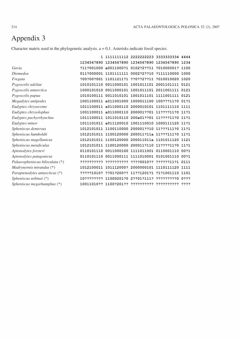
Appendix 3Character matrix used in the phylogenetic analysis. a = 0,1. Asterisks indicate fossil species.
1 1111111112 2222222223 3333333334 4444
1234567890 1234567890 1234567890 1234567890 1234
Gavia ?11?001000 a0011000?1 0102?2??11 ?01000001? 1100
Diomedea 011?000001 1101111111 0002?2??10 ?111110000 1000
Fregata ?00?00?001 11011211?1 ??0??2??11 ?010010020 1020
Pygoscelis adeliae 1010101110 0011000101 1001011101 2001101111 0121
Pygoscelis antarctica 1000101010 0011000101 1001011101 2011001111 0121
Pygoscelis papua 1010100111 0011010101 1001011101 1111001111 0121
Megadytes antipodes 1001100011 a011001000 1000011100 100???11?0 01?1
Eudyptes chrysocome 1011100011 a011000110 2000010101 1101111110 1111
Eudyptes chrysolophus 1001100011 a011000110 200001??01 11????11?0 11?1
Eudyptes pachyrrhynchus 1011100011 1011010110 200a01??01 11????11?0 11?1
Eudyptes minor 1011101011 a011120010 1001110010 1000111120 11?1
Spheniscus demersus 1012101011 1100110000 200001??10 11???111?0 11?1
Spheniscus humboldti 1012101011 1100120000 200011?11a 11???111?0 11?1
Spheniscus magellanicus 1012101011 1100120000 200011011a 1101011120 1121
Spheniscus mendiculus 1012101011 1100120000 200011?110 11????11?0 11?1
Aptenodytes forsteri 0110101110 0011000100 1111011001 0110001110 00?1
Aptenodytes patagonicus 0110101110 0011000111 1111010001 0101001110 00?1
Palaeospheniscus biloculata (*) ?????????? ?????????? ????0010?? ??????11?1 0111
Madrynornis mirandus (*) 1012100011 101112000? 2000000101 1110111120 1111
Paraptenodytes antarcticus (*) ?????1010? ??01?200?? 11??1201?1 ?1?1001110 1101
Spheniscus urbinai (*) 10???????? 11000201?0 2??01?111? ?????????0 0???
Spheniscus megarhamphus (*) 10011010?? 1100?201?? ?????????? ?????????? ????