Embed Size (px)
Citation preview
Pak. J. Bot., 45(4): 1431-1436, 2013.
A NEW METHOD FOR THE ISOLATION AND PURIFICATION OF LAWSONE FROM LAWSONIA INERMIS AND ITS ROS INHIBITORY ACTIVITY
SYED MUHAMMAD GHUFRAN SAEED1*, SYED ASAD SAYEED1, SEEMA ASHRAF1, SHAHINA NAZ1,
RAHMANULLAH SIDDIQI1, RASHIDA ALI1, 2, 4 AND M. AHMED MESAIK3
1Department of Food Science and Technology, University of Karachi, Karachi, 75270, Pakistan 2Division of Food Research, HEJ Research Institute of Chemistry, International Center for Chemical and Biological
Sciences, University of Karachi, Karachi, 75270, Pakistan 3Dr. Panjwani Center for Molecular Medicine and Drug Research, International Center for Chemical and Biological
Sciences, University of Karachi, Karachi, 75270, Pakistan 4English Biscuit Manufacturers (Pvt.) Limited, Plot 1-4, Sector 23, Industrial Area, Karachi-749 00, Pakistan.
*Corresponding author’s e-mail: [email protected], [email protected]
Abstract
Lawsone a coloring component of the Henna (Lawsonia inermis) leaves was found to bind with proteins. Present study has suggested a new rapid method for the isolation of lawsone from L. inermis through the calcium ion using the Flash and Disc Counter Current Chromatography (DCC). The purified pigment structurally characterized by 2D, TLC and NMR spectroscopy, was found to be active against reactive oxygen species (ROS) by Chemeluminiscence assay using lymphocytes. Purified lawsone showed remarkable inhibitory activity on the oxidative burst response of the whole blood, polymorphonuclear cells (PMNCs), Mononuclear cells (MNCs) with IC50 as 13 ±1.6 μg/ml, 11±2.7 μg/ml. and 10±4.2 μg/ml respectively.
Introduction
Lawsone (2-hydroxy-1, 4-naphthaquinone) a natural pigment present in the leaves of Lawsonia inermis has been used as a skin and hair dye since 1400 BC (Petkewich, 2006). The concentration of lawsone in leaves varies from place to place depending upon many of the environmental factors and the highest quantity reported so far is about 1% of the dry mass. Colored compounds often interact with proteins and form covalent or noncovalent complexes. Earlier, it has been reported that natural colored extracts and isolate from Henna leaves bind with a number of electrophoretically resolved proteins on PAGE (Ali & Sayeed, 1988; 1990, Ali et al., 1995b). SDS-PAGE (Sodium dodecyl sulphate polyacrylamide gel Electrophoresis) is highly useful technique to analyze the protein in short period of time (Akbar et al., 2012). SDS-PAGE analysis is reliable method for seed and plant storage proteins because these proteins are not influenced by environmental conditions during analysis (Zada et al., 2013). This technique not only provides protein profiles but a useful marker in the studies of genetic diversity and genetic relationships for genera and species (Tamkoc & Arsalan, 2011).
In the last few decades a variety of biological activities related to lawsone have been reported such as the anticomplementary activity (Handa et al., 1997) dihydroorotate dehydrogenase inhibitory activity (Knecht et al., 2000), macrophage-stimulating activity as a result of the movement of granulocyte macrophage colony stimulating factor. (Wagner et al., 1988), antimicrobial activity (Malekzadeh, 1968; Abd-el-Malek et al., 1973), anti-sickling activity (Chang & Suzuka, 1982; Clarke et al., 1986), hepatoprotective activity (Anaad et al., 1992), cytotoxic activity (Ali & Grever, 1998), anti-inflammatory, antipyretic and analgesic activities (Ali et al., 1995a). In view of the vast therapeutic application of lawsone has gain high importance in pharmaceutical uses.
As there is no reference in literature to investigate the lawsone for its ROS inhibitory effects on the whole blood, PMNCs and MNCs etc. The present study therefore evaluated the immune capacity (IC) of the pure lawsone related to the whole blood, polymorph nuclear cells (PMNCs) and mononuclear cells (MNCs) using chemilluminence technique (CT). Material and Methods Materials: All the chemicals were used of analytical reagent grade and the solvents were redistilled before use, lawsone was purchased from Fluka No. 55 900.
The Hanks balance salts solution [HBSS- -] in Ca and Mg-free buffers was purchased from Flow Lab Sydney, Australia; phorbol myristate acetate [PMA], and dextrans were supplied by MP Biomedical Inc. USA; ficoll paque was purchased from American Bioscience AB, Sweden; zymosan–A; 3-aminopthalhydrazide (luminol); bis-N-methlacridinium nitrate (lucignin) and RPMI 1640 media were obtained from Sigma Chemical Co. USA; fetal bovine serum was purchased from MP Biomedicals Inc. Germany. Isolation of lawsone Extraction of the crude dye: The leaves of Henna (1 kg) in 2.5 L of 1.5 M calcium hydroxide solution (pH 10), were chopped to fine slurry in an electric blender, stirred for about five hours at room temperature using a magnetic stirrer and a plastic coated magnetic rod to avoid the oxidation in presence of metal. The mixture was transferred to a round bottom jar with diameter 12mm and opening at the top (Henna jar).The liquid was heated to 60°C for 15 min. using an immersion heater that helped the solid particles to float on top of the liquid. The clear solution containing the natural dyes was collected through the tap at the bottom leaving the suspended solids behind.
S.M.GHUFRAN SAEEDET AL. 1432
The small particles of insoluble solids, present in the liquid were further removed by centrifugation at 1000g for 10 min., and filtered under vacuum through Whatman filter paper No.1. The extract was concentrated to double strength using the freeze drier, model OSK 6972, Ovawa, Seiki Co. Ltd. Alternatively the mixture was concentrated using rotary vacuum evaporator at 37°C to avoid the
undesirable decomposition of the extract. The concentrated mixtures were extracted separately thrice with chloroform in a separating funnel, filtered and dried on a rotary evaporator. The sample was kept in freezer till its utilization. The comparison of novel method and old method for the quantitative isolation and distribution of lawsone are comparatively described in Table 1.
Table 1. A comparison of novel method and old method of quantitative isolation and distribution of lawsone. Parts of the Plant % On the dry basis Season Place References
Leaves 0.97-0.95 Summer Edfo & Assuit Karawya et al., 1969 Leaves 0.55-0.63 Cold Cairo & Fayed Karawya et al., 1969
Present work Leaves 1-1.2 Summer Karachi Leaves 0.78-0.88 Winter Karachi Bark 0.21-0.24 Summer Karachi
Pericarp 0.13-0.25 Summer Karachi Leaves 0.92-1.37 Summer Multan
Present work
Effect of pH on the yield: In order to evaluate the optimum pH for extraction, the above experiment was repeated by taking leaves (200g) separately in five conical flasks of 250ml at pH 4, 6, 8, 10 and 12 and extraction was carried out following the same procedure as described above. Purification of pigment by chromatography: The residue was shaken in a mixture of chloroform, pet ether and acetic acid (4:6:0.5) referred as 'Solvent A'. The precipitated lawsone was collected on filter paper and was purified by using a column (50 x 2.5cm) containing silica gel of mesh size 60. The silica gel was shaken in a beaker in solvent “A" for about 1-2 hrs before packing and the column was left for 12-15 hrs for equilibration. A sample of 0.5g in 1ml of solvent (A) was subjected to the column and 0.5 ml effluents were collected with a flow rate of 1 ml/min, dyes (orange, red) were separated automatically using Flash Chromatography apparatus. The dye was identified by 2 dimension TLC in solvents A and B (BuOH: AcOH: H2O: 4:1:5) by comparing with known lawsone.
The seventh fraction from the column was collected after 10 hours; the effluent was immediately dried under
vacuum at 35°C using the rotary evaporator. The Flash Chromatography technique was used for further purification of the isolated colored bands. The colored compound in the same mobile phase A was resolved on Flash Chromatography column of dimension 20 x 2.5cm using silica gel of mesh size 60 as the solid support. The flow rate was maintained as 2.2ml/min. The resolved fraction was collected and dried on the rotary evaporator.
The major colored fraction was further purified by using Droplet Counter Chromatography (DCC) apparatus No. 602 Eyela. The same sample was also separated by Round Disc Centrifuge Chromatography (RDC).
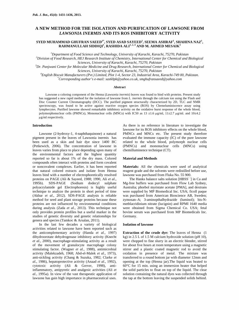
The major red fraction was evaluated for its dye binding capacity described in the earlier studies (Abdullah et al., 2008). The protein binding dye was further purified by thin layer chromatography, using separately the silica and cellulose as the solid supports using solvent A as the mobile phase. The purity of the compound was further confirmed by HPTLC in solvent systems A and B. Identification of isolated dye by UV spectrophotometer: The isolated compound in methanol was analyzed with the help of automatic programming system used in UV spectroscopy. The results are given in Figs. 1 and 2.
Fig. 1. UV Spectral analysis of lawsone purchased from FLUKA.
Fig. 2. UV Spectral analysis of isolated lawsone by novel method.
PURIFICATION OF LAWSONE FROM LAWSONIA INERMIS AND ITS ROS INHIBITORY ACTIVITY 1433
Determination of mass-spectra: The mass spectra of the dye was determined using a 'Finnigan' MAT 312 double focusing mass spectrometer connected to MAT 188 data system with PDP 11/34 DEC computer system. Peak matching, linked to scan field desorption (SFD) , field ionization (FI) and fast atom bombardment (FAB) measurement were also performed on the MAT 312 mass spectrometer.
FABMS (fast atom bombardment mass spectra) was measured in glycerol: water in the ratio of 1:1 in the presence of potassium iodide. An accurate mass measurement was made with the FAB source using an internal standard. HRMS (high resolution mass spectra) were recorded on JEOL MS-HX 110 mass spectrometer connected to DEC PDP 11/73 computer system. Identification by nuclear magnetic resonance spectroscopy (NMR): The NMR spectrum was determined in deuterated methanol solvent as an internal standard at 300 MHz (1H-NMR) on Avance AV-300. The results are shown in Fig. 3. Lawsone binding with food proteins: The lawsone-protein binding assay was performed according to our previously reported PAGE methods (Badaruddin et al., 2007; Saeed et al., 2010). Briefly describing the lawsone (0.2g) was dissolved in 7.5 mL of glacial acetic acid and 5 mL of methanol. The volume was made up to 100mL with DDD (double distilled deionized) water. The same recipe was followed for preparing the standard dye Commassie Blue R250 for comparison. The destining solution was prepared by mixing 10mL of glacial acetic acid and 30mL of methanol together. The volume was made up to 100mL with DDD water.
The gel was left overnight in the staining solution and was washed twice with the destaining solution with a gap of 15 minutes to produce the clear bands on the colorless gel while the other half portion of the gel was stained overnight with Commassie Blue R250 and destained by washing several times with the destining solution as in Figs. 4 and 5. Isolation of Polymorphoneutrophils (PMNs) and Mononuclear (MNCs): The human blood (10mL) was collected from young healthy volunteers (24-35 years) by vein-puncture in heparin containing tubes and was used immediately. The whole blood was diluted with HBSS -- (pH 7.4), and then mixed with one third of its volume of 6% dextran solution for differential sedimentation and for removal of erythrocytes, after gentle mixing. The mixture was kept at room temperature for 20 minutes undisturbed. The upper layer containing leucocytes was collected and was gently layered over equal volume of Ficoll paque it was then subjected to centrifugation at 400g for 25 minutes at room temperature. The upper layer was removed; the mononuclear cells in the junction portion were collected gently which consisted of 15-35% monocytes and were washed with HBSS. The neutrophils or MNCs collected were subjected to hypotonic lysis with sterile distilled water for one minute, and then washed twice with HBSS -- (Ca and Mg free). Cells were re-suspended (HBSS++) containing Ca and Mg to a concentration of 1×10⁶. Preparation of opsonized zymosan, luminol and lucignin: The opsonization of zymosan particles was carried out according to the practice of Cohen et al., (1986). Briefly
describing, zymosan (100 mg) was mixed in 5mL of phosphate buffer saline (PBS, pH 7.4) and 5mL fresh pooled serum from healthy volunteers, the mixture was incubated at 37°C in a shaking water bath for 30 minutes and was centrifuged at 300g for 20 minutes followed by washing twice with PBS . The pellets were finally re-suspended in 5mL of PBS. The mixture was kept frozen at -20°C till use and it was brought to room temperature immediately before use.
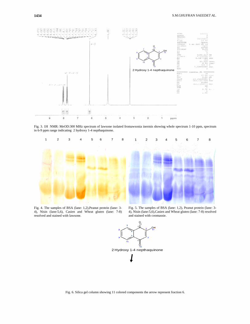
The luminal (1.8 mg) was dissolved in 1 mL borate buffer and stirred in vortexes for 5-10 minutes. The solution then further diluted up to 10ml of HBSS++ to give 180µg luminol/mL. Lucignin (0.5Mm) solution was prepared by dissolving 1.27mg lucignin in 5mL of HBSS++. Chemiluminescence assay: Luminol-enhanced chemiluminescence assay was performed as described by Helfand et al., (1982) and Helkar et al., (2001). Briefly, about 50µL whole blood (1:50 dilution), PMNCs (1×10⁶) or MNCs cells (1×10⁶) were suspended in Hank’s Balance salt solution containing Ca and Mg. The mixture was incubated with 50µL serial dilution of lawsone for 1 hour. To each well, 50µL serum opsonized zymosan was added, followed by 50µL luminol. HBSS++ were added to a 96 well flat bottom plate in a final volume to 0.2mL. HBSS++ alone was taken as a control. Recorded peak and total integral chemiluminescence reading were expressed in the relatively light unit [RLU], the phagocytosis kinetic studies was monitored with luminometer for 50 minutes in repeated scan mode. Results and Discussion Purification and identification of lawsone: We have demonstrated for the first time that color of Henna is a mixture of 11 components by using different solvent systems. Combinations of solvents in different ratio play the key role in extraction of ionized molecules from the natural resources as in case of Henna. Kharbade & Agrawall (1985) has isolated five dyes on the silica TLC plates system using benzene; ethyl-formate and formic acid (74:24:1), Karawya et al., (1969) and Verma & Rai, (1968) had used the TLC method for the isolation of lawsone from the leaves. Lawsone with Rf value as 0.72 was also separated (Masschelein-Kleiner, 1967) on 10% acetylated cellulose powder mixed with 95% ethanol. In the present study, we successfully isolated eleven color bands that were resolved on the silica column using a novel mobile phase "A" [chloroform: petether: acetic acid (4:6:0.5) as shown in Fig. 6.
The fraction 6 was further separated by flash chromatography using silica gel, which was resolved into 2 colored bands, the dim pink color band was first eluted followed by the reddish brown bands as the major components. The fraction 6 was resolved by DCC in BAW, it was resolved in 3 fractions; the first two fractions light pink and mid were followed by a reddish brown band, however when the same fraction 6 was separated by round disc centrifuge chromatography (RDCC) using the same solvent system, only 2 fractions were obtained. Major band showed the Rf value as 0.48. Final purification by HPTLC, using BAW and mobile phase "A” gave two sharp rounded spots having Rf values 0.38 and 0.42 respectively.
S.M.GHUFRAN SAEEDET AL. 1434
2 Hydroxy 1-4 nepthaquinone
Fig. 3. 1H NMR: MeOD:300 MHz spectrum of lawsone isolated fromawsonia inermis showing whole spectrum 1-10 ppm, spectrum in 6-9 ppm range indicating 2 hydroxy 1-4 nepthaquinone.
1 2 3 4 5 6 7 8
Fig. 4. The samples of BSA (lane: 1,2),Peanut protein (lane: 3-4), Nisin (lane:5,6), Casien and Wheat gluten (lane: 7-8) resolved and stained with lawsone.
1 2 3 4 5 6 7 8
Fig. 5. The samples of BSA (lane: 1,2), Peanut protein (lane: 3-4), Nisin (lane:5,6),Casien and Wheat gluten (lane: 7-8) resolved and stained with coomassie.
2 Hydroxy 1-4 nepthaquinone
Fig. 6. Silica gel column showing 11 colored components the arrow represent fraction 6.
PURIFICATION OF LAWSONE FROM LAWSONIA INERMIS AND ITS ROS INHIBITORY ACTIVITY 1435
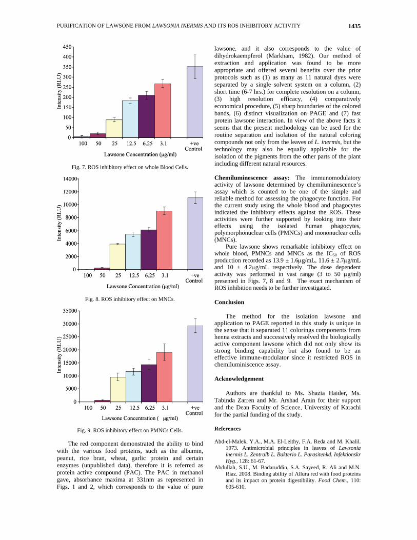
Fig. 7. ROS inhibitory effect on whole Blood Cells.
Fig. 8. ROS inhibitory effect on MNCs.
Fig. 9. ROS inhibitory effect on PMNCs Cells.
The red component demonstrated the ability to bind with the various food proteins, such as the albumin, peanut, rice bran, wheat, garlic protein and certain enzymes (unpublished data), therefore it is referred as protein active compound (PAC). The PAC in methanol gave, absorbance maxima at 331nm as represented in Figs. 1 and 2, which corresponds to the value of pure
lawsone, and it also corresponds to the value of dihydrokaempferol (Markham, 1982). Our method of extraction and application was found to be more appropriate and offered several benefits over the prior protocols such as (1) as many as 11 natural dyes were separated by a single solvent system on a column, (2) short time (6-7 hrs.) for complete resolution on a column, (3) high resolution efficacy, (4) comparatively economical procedure, (5) sharp boundaries of the colored bands, (6) distinct visualization on PAGE and (7) fast protein lawsone interaction. In view of the above facts it seems that the present methodology can be used for the routine separation and isolation of the natural coloring compounds not only from the leaves of L. inermis, but the technology may also be equally applicable for the isolation of the pigments from the other parts of the plant including different natural resources. Chemiluminescence assay: The immunomodulatory activity of lawsone determined by chemiluminescence’s assay which is counted to be one of the simple and reliable method for assessing the phagocyte function. For the current study using the whole blood and phagocytes indicated the inhibitory effects against the ROS. These activities were further supported by looking into their effects using the isolated human phagocytes, polymorphonuclear cells (PMNCs) and mononuclear cells (MNCs).
Pure lawsone shows remarkable inhibitory effect on whole blood, PMNCs and MNCs as the IC50 of ROS production recorded as 13.9 ± 1.6μg/mL, 11.6 ± 2.7μg/mL and 10 ± 4.2μg/mL respectively. The dose dependent activity was performed in vast range (3 to 50 μg/ml) presented in Figs. 7, 8 and 9. The exact mechanism of ROS inhibition needs to be further investigated. Conclusion
The method for the isolation lawsone and application to PAGE reported in this study is unique in the sense that it separated 11 colorings components from henna extracts and successively resolved the biologically active component lawsone which did not only show its strong binding capability but also found to be an effective immune-modulator since it restricted ROS in chemiluminiscence assay. Acknowledgement
Authors are thankful to Ms. Shazia Haider, Ms. Tabinda Zarren and Mr. Arshad Arain for their support and the Dean Faculty of Science, University of Karachi for the partial funding of the study. References Abd-el-Malek, Y.A., M.A. El-Leithy, F.A. Reda and M. Khalil.
1973. Antimicrobial principles in leaves of Lawsonia inermis L. Zentralb L. Bakterio L. Parasitenkd. Infektionskr Hyg., 128: 61-67.
Abdullah, S.U., M. Badaruddin, S.A. Sayeed, R. Ali and M.N. Riaz. 2008. Binding ability of Allura red with food proteins and its impact on protein digestibility. Food Chem., 110: 605-610.
S.M.GHUFRAN SAEEDET AL. 1436
Akbar, F., N. Yousaf, M.A. Rabbani, Z.K. Shinwari and M.S. Masood. 2012. Study of total seed proteins pattern of sesame (Sesamum indicum L.) landraces via sodium dodecyl sulfate polyacrylamide gel electrophoresis (SDSPAGE). Pak. J. Bot., 44: 2009-2014.
Ali, B.H., A.K. Bashir and O.M. Tanira. 1995a. Anti-inflammatory, antipyretic, and analgesic effects of Lawsonia inermis L. (Henna) in rats. Pharmacol., 51: 356-63.
Ali, M. and M.R. Grever. 1998. A cytotoxic naphtho-quinone from Lawsonia inermis. Fitoterapia, LXIX, (2): 1810-1813.
Ali, R. and S.A. Sayeed. 1990, A plant dye from Lawsonia inermis for protein staining after polyacrylamide gel eletrophoresis. Electrophoresis, 11: 343-344.
Ali, R., S.A. Sayeed and A.A. Khan. 1995b. A sensitive novel staining agent for the resolved proteins on PAGE. Int. J. Peptide Protein Res., 45: 97-99.
Ali, R. and S.A. Sayeed. 1988. A novel dye for staining eletrectrophoretically resolved proteins. Proceeding, Elsevier Science Publishers B.V. (Biomedical Division).
Anaad, K.K., B. Singh, D. Chand and B.K. Chandon. 1992. An evaluation of Lawsonia alba extract as hepatoprotective agent. Planta Med., 58: 22-25.
Badaruddin, M., S.U. Abdullah, S.A. Sayeed, R. Ali and M.N. Riaz. 2007. Sunset yellow: A food color for protein staining with SDS-PAGE. Cereal Food World, 52(1): 12-14.
Chang, H. and S.E. Suzuka. 1982. Lawsone derived from the henna. Plant increases the oxygen affinity of sickle cell blood. Biochem. Biophys. Res. Commun., 107: 602-608.
Clarke, D. T., G.R. Jones and M.M. Martin. 1986. The anti-sickling drug lawsone (2-OH-1,4-naphtho-quinone) protects sickled cells against membrane damage. Biochem. Biophys. Res. Commun., 139: 780-786.
Cohen, D., A.A. Aderem, S.D. Wright and Z.A. Cohn. 1986. Bacterial lipopolysaccharides prime macrophages for enhanced release of arachidonic acid metabolites. J. Exp. Med., 164: 165-179.
Handa, G., A. Kapil, S. Sharma and J. Singh. 1997. Lawnermis acid a new anticomplementary triterpenoid from Lawsonia inermis seeds. Indian J. Chem. Sect. B. 36: 252-256.
Helfand, S.L., J.C. Roder and J. Werkmeister. 1982. Chemiluminescence response of human natural killer cells.
1. The relationship between target cell binding, Chemiluminescence and cytolysis. The J. Exp. Med., 298: 569-572.
Helkar, G., E.S. Ozveri, M. Yuksel, A. Aktan and A.S. Yalcin. 2001. Different kinds of reactive oxygen species were detected in colon and breast tumors. Cancer Letters, 165: 219-224.
Karawya, M.S., S.M.A. Wahhab and A.Y. Zaki. 1969. A study of the lawsone content in henna. Lloydia, 32: 76-78.
Kharbade, B.V. and O.P. Agrawal. 1985. Identification of natural red dyes in old Indian textiles: Evaluation of thin-layer chromatographic systems. J. Chromatography, 347: 447-54.
Knecht, W., J. Henseling and M. Loffler. 2000. Kinetics of inhibition of human and rat dihydroorotate dehy drogenase by atovaquone, lawsone derivatives, brequinar sodium and polyporic acid. Chem. Biol. Interact., 124: 61-76.
Malekzadeh, F. 1968. Antimicrobial activity of Law-sonia inermis L. Appl. Microbiol., 16: 663-664.
Markham, K.R. 1982. Techniques of Flavonoid Identification", Academic Press, London.
Masschelein-Kleiner, L. 1967. Microanalysis of hydroquinones in red lakes. Mikrochim. Acta., 6: 1080.
Petkewich, R. 2006. Dye derived from green henna leaves is used to decorate the body with intricate designs. Chem. Eng., News, 84: 28.
Saeed, S.M.G., S.U. Abdullah, S.A. Sayeed and R. Ali. 2010. Food protein: Food color interactions and its application in rapid protein assay. Czech J. Food Sci., 28(6): 506-513.
Tamkoc, A. and E. Arsalan. 2011. Inter and intra-specific variation in SDS-PAGE of seed proteins of three Poa L. (Poaceae) species. Pak. J. Bot., 43(2): 1105-1110.
Verma, M. R. and J. Rai. 1968. Spectrophotometric and chromatographic studies of henna leaf and powder extracts, Indian Standards Institute Bulletin, 20: 495-497.
Wagner, H., B. Kreher and K. Jurcic. 1988. In vitro sti- mulation of human granulocytes and lymphocytes by pico- and femtogram quantities of cytostatic agents. Arzneim. Forsch., 38: 273-275.
Zada, M., Z.K. Shinwari, N. Zakir and M.A. Rabbani. 2013. Study of total seed storage proteins in ethiopian mustard (Brassica carinata A. Braun) germplasm. Pak. J. Bot., 45: 443-448.
(Received for publication 24 January 2012)