Embed Size (px)
Citation preview
Accepted by S. Carranza: 3 May 2011; published: 15 Jun. 2011
ZOOTAXAISSN 1175-5326 (print edition)
ISSN 1175-5334 (online edition)Copyright © 2011 · Magnolia Press
Zootaxa 2918: 47–67 (2011) www.mapress.com/zootaxa/ Article
47
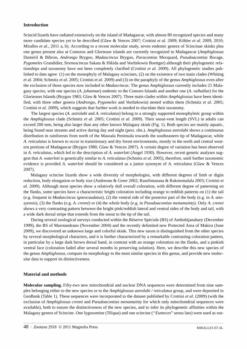
A new large and colorful skink of the genus Amphiglossus from Madagascar revealed by morphology and multilocus molecular study
AURÉLIEN MIRALLES1,5, ACHILLE P. RASELIMANANA2, DOMOINA RAKOTOMALALA3, MIGUEL VENCES1 & DAVID R. VIEITES4
1Technical University of Braunschweig, Zoological Institute, Mendelssohnstr. 4, 38106 Braunschweig, Germany.2Département de Biologie Animale, Université d’Antananarivo, B.P 906, Antananarivo (101), Madagascar, and Association Vahatra,BP 3972, Antananarivo 101, Madagascar3WWF Madagascar and West Indian Ocean Programme Office, BP. 738, Antananarivo (101), Madagascar4Department of Biodiversity and Evolutionary Biology, Museo Nacional de Ciencias Naturales–CSIC, C/ José Gutiérrez Abascal 2,28006 Madrid, Spain5Corresponding author. E-mail: [email protected]
Abstract
We describe a new species of Amphiglossus skink from the western edge of the Central Highlands of Madagascar in theReserve of Makira, and also found in the Réserve Spéciale of Ambohijanahary and in the Réserve Spéciale of Marotandra-no. Amphiglossus meva n. sp. is characterized and differentiated from other species of the genus by a combination of mor-phological, chromatic and molecular characters: 1) a relatively large size (SVL of adults from 126 to 150 mm); 2) acharacteristic pattern of coloration, Amphiglossus meva being the only skink in Madagascar together with Amphiglossuscrenni with dark grey dorsum contrasting with orange flanks and ventrum; 3) the absence of a postnasal scale; 4) the pre-subocular frequently absent, 5) the presence of single elongated tertiary temporal bordering lower secondary temporal and6) pentadactyl limbs. In addition to the morphological approach, a multi-locus genetic analysis based on eight mitochon-drial and nuclear genes clearly supports the distinctiveness of A. meva. This new species was found in areas of rainforest,sometimes containing transitional deciduous forest elements. It was typically observed under large rotten logs associatedwith dense layers of decomposed wood retaining certain humidity and providing habitat for invertebrate larvae and ter-mites.
Key words: Amphiglossus, conservation, Madagascar, molecular phylogeny, rainforest, Squamata: Scincomorpha: Scin-cidae
Résumé
Une nouvelle espèce de scinque de genre Amphiglossus est décrite de la bordure ouest des hauts plateaux de Madagascar,dans la Réserve de Makira, et dans les Réserves Spéciales de Marotandrano et d’Ambohijanahary. Amphiglossus meva sp.nov. se distingue des autres espèces du genre par la combinaison des caractères suivants: 1) une taille relativement impor-tante (distance tête-cloaque comprise entre 126 et 150 mm chez les adultes); 2) un modèle de coloration caractéristique,puisqu’il s’agit avec Amphiglossus crenni des deux seuls scinques malgaches dotés d’une face dorsal gris foncée con-trastant avec des flancs et une face ventrale orange; 3) l’absence d’écaille postnasale; 4) l’écaille presuboculaire qui estfréquemment absente, 5) la présence d’une unique temporale tertiaire, allongée et bordant la temporale secondaire in-férieure, et 6) des membres pentadactyles. En plus de l’approche morphologique, une analyse génétique basé sur septgènes mitochondriaux et nucléaires soutient également la validité taxinomique de A. meva. Cette espèce n’est actuellementconnue que par quelques spécimens récoltés dans des secteurs de forêt pluviale contenant parfois des éléments de forêtdécidue transitionnel. Son biotope préférentiel semble être constitué par les larges souches et troncs d’arbres en décom-position retenant l’humidité et hébergant des larves d’invertébrés et des termites.
MIRALLES ET AL.48 · Zootaxa 2918 © 2011 Magnolia Press
Introduction
Scincid lizards have radiated extensively on the island of Madagascar, with almost 80 recognized species and manymore candidate species yet to be described (Glaw & Vences 2007; Crottini et al. 2009; Köhler et al. 2009, 2010;Miralles et al., 2011 a, b). According to a recent molecular study, seven endemic genera of Scincinae skinks plusone genus present also at Comoros and Glorieuse islands are currently recognized in Madagascar (AmphiglossusDuméril & Bibron, Androngo Brygoo, Madascincus Brygoo, Paracontias Mocquard, Pseudoacontias Bocage,Pygomeles Grandidier, Sirenoscincus Sakata & Hikida and Voeltzkowia Boettger) although their phylogenetic rela-tionships and taxonomy have not been completely clarified (Crottini et al. 2009). All phylogenetic studies pub-lished to date agree (1) on the monophyly of Malagasy scincines, (2) on the existence of two main clades (Whitinget al. 2004; Schmitz et al. 2005; Crottini et al. 2009) and (3) on the paraphyly of the genus Amphiglossus even afterthe exclusion of those species now included in Madascincus. The genus Amphiglossus currently includes 21 Mala-gasy species, with one species (A. johannae) endemic to the Comoro Islands and another one (A. valhallae) for theGlorieuses Islands (Brygoo 1983; Glaw & Vences 2007). Three main clades within Amphiglossus have been identi-fied, with three other genera (Androngo, Pygomeles and Voeltzkowia) nested within them (Schmitz et al. 2005;Crottini et al. 2009), which suggests that further work is needed to elucidate their taxonomy.
The largest species (A. astrolabi and A. reticulatus) belong to a strongly supported monophyletic group withinthe Amphiglossus clade (Schmitz et al. 2005; Crottini et al. 2009). Their snout-vent length (SVL) in adults canexceed 200 mm, being also larger than any other known Malagasy skink (Fig. 1). Both species are mainly aquatic,being found near streams and active during day and night (pers. obs.). Amphiglossus astrolabi shows a continuousdistribution in rainforests from north of the Masoala Peninsula towards the southeastern tip of Madagascar, whileA. reticulatus is known to occur in transitionary and dry forest environments, mostly in the north and central west-ern portions of Madagascar (Brygoo 1980, Glaw & Vences 2007). A certain degree of variation has been observedin A. reticulatus, which led to the description of A. waterloti (Angel 1930). However, recent genetic analyses sug-gest that A. waterloti is genetically similar to A. reticulatus (Schmitz et al. 2005), therefore, until further taxonomicevidence is provided A. waterloti should be considered as a junior synonym of A. reticulatus (Glaw & Vences2007).
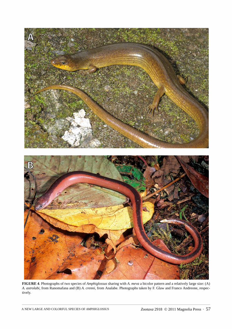
Malagasy scincine lizards show a wide diversity of morphologies, with different degrees of limb or digitsreduction, body elongation or body size (Andreone & Greer 2002; Raselimanana & Rakotomalala 2003; Crottini etal. 2009). Although most species show a relatively dull overall coloration, with different degree of patterning onthe flanks, some species have a characteristic bright coloration including orange to reddish patterns on (1) the tail(e.g. frequent in Madascincus igneocaudatus), (2) the ventral side of the posterior part of the body (e.g. in A. ano-syensis), (3) the flanks (e.g. A. crenni) or (4) the whole body (e.g. in Pseudoacontias menamainty). Only A. crennishows a very contrasting pattern between the bright pink/reddish lateral and ventral sides of the body and tail, witha wide dark dorsal stripe that extends from the snout to the tip of the tail.
During several zoological surveys conducted within the Réserve Spéciale (RS) of Ambohijanahary (December1999), the RS of Marotandrano (November 2004) and the recently delimited new Protected Area of Makira (June2009), we discovered an unknown large and colorful skink. This new taxon is distinguished from the other speciesby several morphological characters, and it is further characterized by a remarkable contrasting coloration pattern,in particular by a large dark brown dorsal band, in contrast with an orange coloration on the flanks, and a pinkishventral face (coloration faded after several months in preserving solution). Here, we describe this new species ofthe genus Amphiglossus, compare its morphology to the most similar species in this genus, and provide new molec-ular data to support its distinctiveness.
Material and methods
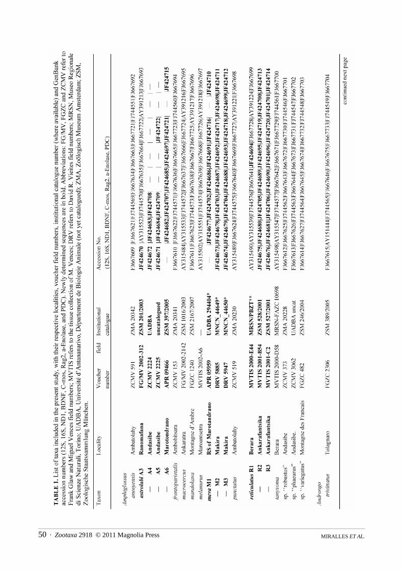
Molecular sampling. Fifty-two new mitochondrial and nuclear DNA sequences were determined from nine sam-ples belonging either to the new species or to the Amphiglossus astrolabi / reticulatus group, and were deposited inGenBank (Table 1). These sequences were incorporated to the dataset published by Crottini et al. (2009) (with theexclusion of Amphiglossus crenni and Pseudoacontias menamainty for which only mitochondrial sequences wereavailable), both to ensure the distinctiveness of the new species, and to infer its phylogenetic affinities within theMalagasy genera of Scincine. One lygosomine (Tiliqua) and one scincine (‘‘Eumeces” sensu lato) were used as out-
Zootaxa 2918 © 2011 Magnolia Press · 49A NEW LARGE AND COLORFUL SPECIES OF AMPHIGLOSSUS
groups. Among the non-Malagasy skinks, previous more inclusive studies (Whiting et al. 2004; Schmitz et al.2005) suggested that species of the genus Eumeces sensu lato are relatively close to the Malagasy radiation. Forthese two outgroup taxa, concatenated "chimera" sequences of different species were compiled from GenBank (seeCrottini et al. 2009).
Laboratory techniques. Total genomic DNA was extracted using proteinase K (10 mg/ml) digestion followedby a standard salt-extraction protocol (Bruford et al. 1992). From the mitochondrial DNA (mtDNA), we amplifiedthree fragments of the 12S rRNA, 16S rRNA and ND1 genes. Additionally, fragments of five nuclear DNA genes(nuDNA) were amplified: brain-derived neurotrophic factor (BDNF); recombination activating gene 2 (Rag2); α-enolase (enol); oocyte maturation factor (C-mos) and phosducin (PDC). Standard polymerase chain reactions wereperformed in a final volume of 12.5 μl containing 0.3 μl each of 10 pmol primer, 0.25 μl of total dNTP 10 mM(Promega), 0.1 μl of 5 U/ml GoTaq, and 2.5 μl of GoTaq Reaction Buffer (Promega). See Crottini et al. (2009) forprimers and PCR conditions used. The successfully amplified products were purified using ExoSAP-IT purifica-tion kit according to the manufacturer’s instruction. Purified PCR templates were sequenced using dye-labeleddideoxy terminator cycle sequencing on an ABI 3130 automated DNA sequencer.
Analysis of molecular data. All obtained DNA sequences were edited and checked for errors using Codon-Code Aligner (v. 2.0.6, Codon Code Corporation). No stop codons were found in protein coding genes. The datamatrix included 34 samples representing 32 taxa with an aligned sequence length of 3936 base pairs (Table 1). Fouradditional specimens, for which not all the genes could be successfully sequenced, were included in a separateanalysis based on a reduced number of markers. Maximum parsimony (MP) and partitioned Bayesian inferencesearches based on the full concatenated dataset, were performed to infer trees. We used PAUP* 4.0b10 (Swofford2002) to perform MP analyses with 100 random addition sequence replicates, equal character weighting, tree bisec-tion and reconnection (TBR) branch swapping, and gaps coded as missing data. Nodal support was obtained usingbootstrap analyses, with 10000 replicates, 10 random addition sequences replicates and TBR branch swapping.Partitioned Bayesian analyses were performed using the 21 partitions and the same parameters as previously usedby Crottini et al. (2009) with MrBayes 3.1.2 (Ronquist & Huelsenbeck 2003). We performed one run of 20 milliongenerations (started on random trees) and four incrementally heated Markov chains (using default heating values)each, sampling the Markov chains at intervals of 1000 generations. The first 10 million generations were conserva-tively discarded and 10000 trees were retained post burn-in and summed to generate a majority rule consensus tree.Genetic divergences were estimated with MEGA 4.1 (Tamura et al. 2007) by calculating uncorrected p-distancesfrom the 16S and ND1 genes.
Morphological characters and coloration. Specimens were collected using opportunistic searches and pitfalltraps (see Raxworthy & Nussbaum, 1994). The specimens captured were euthanized with a 4% chloro-butanolsolution, tissue samples were collected and conserved in pure ethanol for molecular studies, and specimens weresubsequently preserved in 70% ethanol (with exception of the UADBA specimens that have been fixed in a 12%formalin solution before the final conservation in alcohol). Specimens examined for the present study are depositedin the Göteborg Natural History Museum, Göteborg, Sweden (GNM); Museo Nacional de Ciencias Naturales,Madrid, Spain (MNCN); Muséum National d’Histoire Naturelle, Paris, France (MNHN); Département de BiologieAnimale, Université d'Antananarivo, Madagascar (UADBA); Zoologisches Forschungsmuseum AlexanderKoenig, Bonn, Germany (ZFMK) and Zoologische Staatssammlung München, Germany (ZSM). All additionalspecimens used for comparisons with the new species are listed in the Appendix 1.
Measurements of specimens were recorded to the nearest 0.1 mm using a dial caliper (except the tail lengthwhich was measured with a string). Meristic, mensural and qualitative characters examined here are routinely usedin the taxonomy of Scincidae, such as scale counts, presence or absence of homologous scale fusions or the vari-ability in color patterns. Scale nomenclature, scale counts, and measurements used in the morphological analysesessentially follow Andreone & Greer (2002). Nuchal scales are defined as enlarged scales of the nape, occupyingtransversally the place of two or more rows of dorsal cycloid scale (see Miralles 2006). The ventral scales arecounted in a single row from the postmentals to the preanal scales (both included in the count), with mental scaleexcluded. The frontal scale is considered hourglass-shaped when constricted by first supraocular, bell-shapedwhen this is not the case (Greer & Shea 2000).
For several voucher specimens, color pictures were taken to record alive natural coloration. Drawings weremade using Adobe Illustrator CS2 and a WACOM graphic tablet CTE-640, after photographs were taken through aZEISS stereomicroscope SteREO Discovery V12.
MIRALLES ET AL.50 · Zootaxa 2918 © 2011 Magnolia Press
Zootaxa 2918 © 2011 Magnolia Press · 51A NEW LARGE AND COLORFUL SPECIES OF AMPHIGLOSSUS
MIRALLES ET AL.52 · Zootaxa 2918 © 2011 Magnolia Press
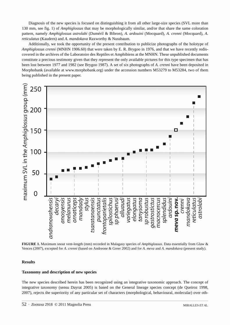
Diagnosis of the new species is focused on distinguishing it from all other large-size species (SVL more than130 mm, see fig. 1) of Amphiglossus that may be morphologically similar, and/or that share the same colorationpattern, namely Amphiglossus astrolabi (Duméril & Bibron), A. ardouini (Mocquard), A. crenni (Mocquard), A.reticulatus (Kaudern) and A. mandokava Raxworthy & Nussbaum.
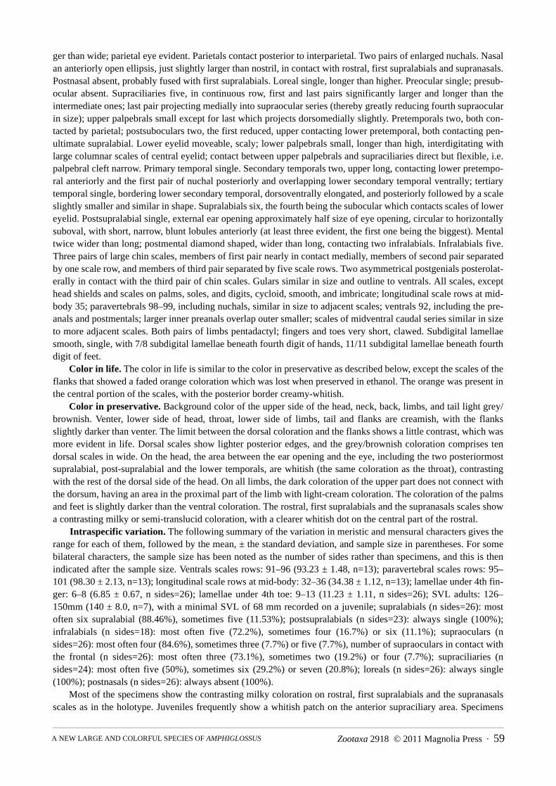
Additionally, we took the opportunity of the present contribution to publicize photographs of the holotype ofAmphiglossus crenni (MNHN 1906.60) that were taken by E. R. Brygoo in 1976, and that we have recently redis-covered in the archives of the Laboratoire des Reptiles et Amphibiens at the MNHN. These unpublished documentsconstitute a precious testimony given that they represent the only available pictures for this type specimen that hasbeen lost between 1977 and 1982 (see Brygoo 1987). A set of six photographs of A. crenni have been deposited inMorphobank (available at www.morphobank.org) under the accession numbers M53279 to M53284, two of thembeing published in the present paper.
FIGURE 1. Maximum snout vent-length (mm) recorded in Malagasy species of Amphiglossus. Data essentially from Glaw &Vences (2007), excepted for A. crenni (based on Andreone & Greer 2002) and for A. meva and A. mandokava (present study).
Results
Taxonomy and description of new species
The new species described herein has been recognized using an integrative taxonomic approach. The concept ofintegrative taxonomy (sensu Dayrat 2005) is based on the General lineage species concept (de Queiroz 1998,2007), rejects the superiority of any particular set of characters (morphological, behavioural, molecular) over oth-
Zootaxa 2918 © 2011 Magnolia Press · 53A NEW LARGE AND COLORFUL SPECIES OF AMPHIGLOSSUS
ers, and advocates the combined and integrated use of various such methods (Padial et al. 2010). In the presentcase, both morphological data (qualitative and quantitative scalation characteristics, coloration pattern) and molec-ular data (phylogenetic position, genetic distances) congruently support the distinctiveness of this new species:
Amphiglossus meva sp. nov. (figs. 2, 3)
Holotype. MNCN 44648 (field no. ZCMV 11324), collected in the western portion of the Makira plateau, close toa campsite locally named Angozongahy, at 15°26'13.3''S 49°07'07.0''E, 1009 m above sea level, district of Mandrit-sara, region of Sofia, province of Mahajanga, northeastern Madagascar, by D.R. Vieites, M. Vences, F. Ratsoavinaand R.-D. Randrianiaina on 28 June 2009. The holotype is in a good state of preservation; it was fixed and pre-served in alcohol. At the time of preservation the specimen was shedding, which explains the somewhat faded col-oration.
Paratypes (n=10). One adult specimen, MNCN 44650 (field no. DRV 5947), a subadult, MNCN 44649 (fieldno. DRV 5885), and a juvenile, ZSM 0487/2009 (field no. ZCMV 11323), collected by the same collectors and atthe same locality as the holotype; a juvenile, UADBA 29402 (field no. APR 05957), collected on 24 November2004 in the transitional rainforest at Riamalandy, 16°17.1’S 48°48.9’E, 850 m elevation within the RS ofMarotandrano, Region of Sofia, Madagascar, by A. P. Raselimanana; a juvenile, UADBA 29403 (field no. APR05958) and an adult male, UADBA 29404 (field no. APR 05959), collected at the same date and in the same areaas above, but both were found together in a different rotten log, by A. P. Raselimanana; an adult male, UADBA
29405 (field no. APR 06021) captured on 26th November 2004 in the transitional rainforest at Riamalandy, sameconditions as above, by A. P. Raselimanana; an adult female, UADBA 29406 (field no. APR 06039) and two juve-niles, UADBA 29407 and UADBA 29408 (field no. APR 06040 and APR 06041), collected in the same rotten logson 27th November 2004 in transitional forest at Riamalandy, 16°16.9’S 48°49.1’E, 800 m elevation within the RSof Marotandrano, Region of Sofia, Madagascar, by A. P. Raselimanana.
Additional specimens (n=2). Two additional specimens were collected in the RS of Ambohijanahary. Werefrain to include these specimens in the type series given that (1) this locality is far away from both the Makirareserve and the RS of Marotandrano, (2) these specimens have a narrower brown dorsal stripe than those fromMakira and from the RS of Marotandrano (6 scales rows vs. 10) and (3) no tissue sample from this locality wasavailable for molecular analysis. These specimens are: an adult male, UADBA 12209 (field no. RD 1225) and anadult female, UADBA 12210 (field no. RD 1269) in excellent condition of preservation, collected on 18 and 19December 1999, in the “forêt d’ Ankazotsihitafototra”, 18°15.7’S 45°25.2’E, 1150 m elevation within the RS ofAmbohijanahary, Region of Bongolava, Madagascar, by D. Rakotomalala and S. M. Goodman.
Diagnosis. A member of the phenetic Amphiglossus/Madascincus group which differs (1) from the Malagasygenera in the subfamily Lygosominae (Cryptoblepharus and Trachylepis) by the presence of entirely movable andscaly eyelids (versus fused immovable eyelids forming spectacles over the eyes in Cryptoblepharus; or movableeyelids with a translucent disk or window in the lower eyelid in Trachylepis), absence of prefrontals (present inboth Cryptoblepharus and Trachylepis), and lack of frontoparietal scales (present in Trachylepis); (2) from all theother Malagasy scincine genera by the presence of four legs.
Within the Amphiglossus/Madascincus group, it is placed in the lineage called Amphiglossus (sensu Crottini etal. 2009) by molecular data. Within Amphiglossus, it is distinguished from all the other species by a combination of(1) a relatively large size (SVL of adults from 126 to 150 mm); (2) a characteristic pattern of coloration with dark/grey dorsum contrasting with bright orange to yellowish flanks and ventrum, including the ventral side of the tail;(3) absence of a postnasal scale; (4) presubocular frequently absent, (5) presence of a single elongated tertiary tem-poral bordering lower secondary temporal.
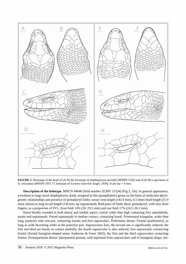
Among large-sized Amphiglossus (A. astrolabi, A. reticulatus, A. ardouini, A. mandokava, A. crenni), the newspecies can be distinguished from the superficially similar Amphiglossus astrolabi (see fig. 4A, 5A–D) by showingsignificantly shorter fingers and toes with lower numbers of lamellae under fourth finger (6–8 versus 10–13) andfourth toe (11–13 versus 15–21); a smaller size (SVL max = 150 mm versus 226 mm); more compact head; a lowernumber of ventrals (91–96 versus 99–113); absence of postnasal; presubocular frequently absent (versus alwayspresent, most often two on each sides). From A. reticulatus (see fig. 5E–H) it can be distinguished by showing sig-
MIRALLES ET AL.54 · Zootaxa 2918 © 2011 Magnolia Press
nificantly shorter limbs in proportion to body size; smaller size (SVL max = 150 mm versus 212 mm); a less prom-inent parietal area and more compact head; a lower number of ventral scales (91–96 versus 95–108) and of scalerows around midbody (32–36 versus 39–41); absence of postnasal; by the uniform dark dorsal and light ventral col-oration (versus complex patterns). From A. ardouini it differs by the absence of postnasals (versus presence), ahigher number of scale rows around midbody (32–36 versus 31–33), shorter fingers with a lower number of lamel-lae under fourth finger (6–8 versus 7–10) and toe (9–13 versus 17–21), a uniform dark dorsal and light ventral col-oration (versus complex patterns, including dark transversal dark stripes in the anterior part of body). From A.mandokava it differs by the absence of postnasals (versus present), a lower number of ventrals (91–96 versus 103–120) and paravertebrals (95–101 versus 129–141), by the uniform dark dorsal and light ventral coloration (versuscomplex patterns, including dark transversal dark stripes in the anterior part of body). From A. crenni (see fig. 4B,6), it differs by the absence of postnasals (versus presence); a more compact body with 32–36 scales around mid-body (versus a slender elongated body with 26–28 scales around mid-body), and pentadactyl limbs (versusextremely reduced limbs, usually with two toes and two fingers, but sometimes with up to four). See also table 2for a summary of morphological characteristics of the new species. Furthermore, the new species differs from allAmphiglossus and Madascincus species for which DNA sequences were available, by high sequence divergencesin mitochondrial and nuclear genes (see below).
TABLE 2. Comparison of some characteristics distinguishing the new species from other “large-sized” (SVL 130 mm) and/orsuperficially similar species of Amphiglossus. For each character, range, mean ± standard deviation (SD) and sample size (n;inside parentheses) are given. For some bilateral characters, the sample size has been noted as the number of sides rather thanspecimens.
1Partly based on Angel (1942) and Brygoo (1983). 2 Based on Andreone & Greer (2002) and on the photographs of the lostholotype taken by Brygoo in 1976 (see fig. 6). 3Partly based on Raxworthy & Nussbaum (1993). 4Two types of color patternsare presently distinguished: (1) the “bicolor pattern” with a dorsal side uniformly dark contrasting with a ventral side uniformlylight, and (2) the “variegated patterns” that may be composed by dark transversal or longitudinal stripes, dash lines, or reticula-tions on a lighter background.
A. ardouini1 A. astrolabi A. crenni2 A. mandokava3 A. reticulatus A. meva
SVL max (mm) 137 226 164 171 212 150
Color pattern4 variegated bicolor bicolor variegated bicolor or variegated
bicolor
Postnasals PresentAbsentN sides
100%–(8)
100%–(24)
X–(1)
100%–(10)
100%–(12)
–100%(26)
Presubocular N=0 N=1 N=2n sides:
–100 %–(4)
–25%75%(24)
–X–(1)
–100 %–(10)
–100 %–(12)
42,3%57,7%–(26)
N lamellae under 4th finger
min–max:mean±SD:n sides:
7–10–(7)
10–1311.59 ± 0.81(31)
–5–65.38 ± 0.92(8)
7–108.36 ± 0.84(14)
6–86.85 ± 0.67(26)
N lamellae under4th toe
min–max:mean±SD:n sides:
17–21–(8)
15–2118.16 ± 1.27(32)
6–76.7(6)
9–1210.20 ± 0.92(10)
12–1614.19 ± 1.22(16)
9–1311.23 ± 1.11(26)
N ventral scale rows
min–max:mean±SD:n:
94–104–(10)
99–113105.41 ± 4.40(17)
–103–120116.20 ± 7.40(5)
95–108102.13 ± 4.7(8)
91–9693.23 ± 1.48(13)
N paravertebral scale rows
min–max:mean±SD:n:
100–(2)
98–109103.12 ± 2.82(17)
116–132152.66(6)
129–141136.75 ± 5.44(4)
96–108103.13 ± 4.45(8)
95–10198.30 ± 2.13(13)
N longitudinal scalerows at mid–body
min–max:mean±SD:n:
31–33–(10)
34–3835.53 ± 1.06(17)
26–2827.3(6)
36–3836.80 ± 1.10(5)
39–4140.00 ± 0.53(8)
32–3634.38 ± 1.12(13)
Zootaxa 2918 © 2011 Magnolia Press · 55A NEW LARGE AND COLORFUL SPECIES OF AMPHIGLOSSUS
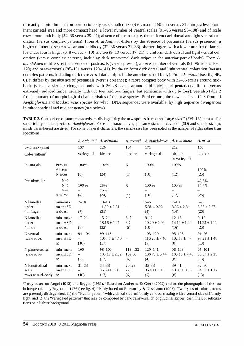
FIGURE 2. Drawings of the head of the holotype of Amphiglossus meva sp. n. (MNCN 44648): (A) dorsal view, (B) ventralview, (C) lateral view, (D) close up of the ocular region. Scale bar = 2 mm.
MIRALLES ET AL.56 · Zootaxa 2918 © 2011 Magnolia Press
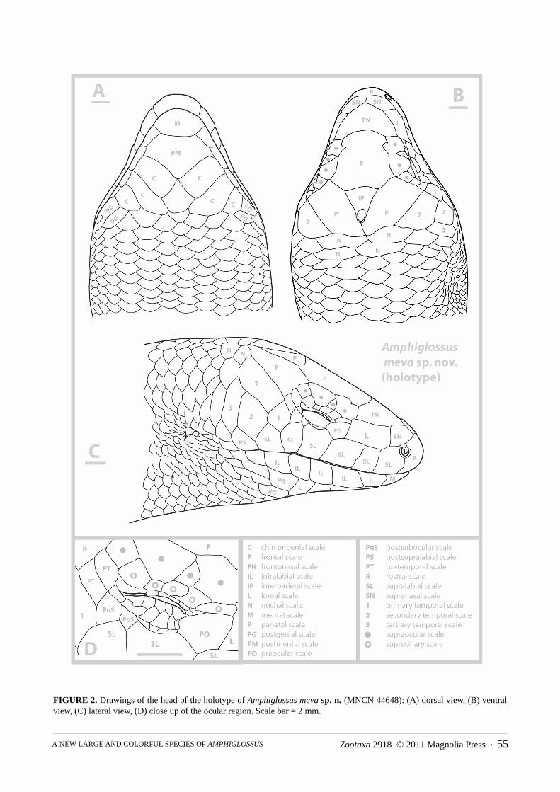
FIGURE 3. Photographs of Amphiglossus meva sp. n.: (A) picture of the holotype specimen in life (MNCN 44648) and (B)four freshly euthanasied specimens including juveniles and an adult, all from the Makira reserve ; (C) living picture of an adultspecimen from Ambohijanahary (UADBA 12209). Photographs by M. Vences and D.R. Vieites (A-B) and Harald Schütz (C),respectively.
Zootaxa 2918 © 2011 Magnolia Press · 57A NEW LARGE AND COLORFUL SPECIES OF AMPHIGLOSSUS
FIGURE 4. Photographs of two species of Amphiglossus sharing with A. meva a bicolor pattern and a relatively large size: (A)A. astrolabi, from Ranomafana and (B) A. crenni, from Analabe. Photographs taken by F. Glaw and Franco Andreone, respec-tively.
MIRALLES ET AL.58 · Zootaxa 2918 © 2011 Magnolia Press
FIGURE 5. Drawings of the head of (A-D) the lectotype of Amphiglossus atrolabi (MNHN 5256) and of (E-H) a specimen ofA. reticulatus (MNHN 1931.77, holotype of Scelotes waterloti Angel, 1930). Scale bar = 4 mm.
Description of the holotype. MNCN 44648 (field number ZCMV 11324) (Fig 2, 3A). In general appearance,a medium to large-sized Amphiglossus skink, assigned to this (paraphyletic) genus on the basis of molecular phylo-genetic relationships and presence of pentadactyl limbs; snout–vent length (143.0 mm), 6.5 times head length (21.9mm), almost as long as tail length (136 mm, tip regenerated). Both pairs of limbs short, pentadactyl, with very shortfingers; as a proportion of SVL, front limb 14% (20–19.5 mm) and rear limb 17% (24.5–26.2 mm).
Snout bluntly rounded in both lateral and medial aspect; rostral wider than high contacting first supralabials,nasals and supranasals. Paired supranasals in median contact, contacting loreal. Frontonasal triangular, wider thanlong, posterior side concave, contacting loreals and first suproculars. Prefrontals absent. Frontal quadrilateral, aslong as wide becoming wider at the posterior part. Supraoculars four, the second one is significantly reduced, thefirst and third are barely in contact medially, the fourth supraocular is also reduced; first supraocular constrictingfrontal (frontal hourglass-shaped sensu Andreone & Greer 2002), the first and the third supraoculars contactingfrontal. Frontoparietals absent. Interparietal present, well separated from supraoculars and of triangular shape, lon-
Zootaxa 2918 © 2011 Magnolia Press · 59A NEW LARGE AND COLORFUL SPECIES OF AMPHIGLOSSUS
ger than wide; parietal eye evident. Parietals contact posterior to interparietal. Two pairs of enlarged nuchals. Nasalan anteriorly open ellipsis, just slightly larger than nostril, in contact with rostral, first supralabials and supranasals.Postnasal absent, probably fused with first supralabials. Loreal single, longer than higher. Preocular single; presub-ocular absent. Supraciliaries five, in continuous row, first and last pairs significantly larger and longer than theintermediate ones; last pair projecting medially into supraocular series (thereby greatly reducing fourth supraocularin size); upper palpebrals small except for last which projects dorsomedially slightly. Pretemporals two, both con-tacted by parietal; postsuboculars two, the first reduced, upper contacting lower pretemporal, both contacting pen-ultimate supralabial. Lower eyelid moveable, scaly; lower palpebrals small, longer than high, interdigitating withlarge columnar scales of central eyelid; contact between upper palpebrals and supraciliaries direct but flexible, i.e.palpebral cleft narrow. Primary temporal single. Secondary temporals two, upper long, contacting lower pretempo-ral anteriorly and the first pair of nuchal posteriorly and overlapping lower secondary temporal ventrally; tertiarytemporal single, bordering lower secondary temporal, dorsoventrally elongated, and posteriorly followed by a scaleslightly smaller and similar in shape. Supralabials six, the fourth being the subocular which contacts scales of lowereyelid. Postsupralabial single, external ear opening approximately half size of eye opening, circular to horizontallysuboval, with short, narrow, blunt lobules anteriorly (at least three evident, the first one being the biggest). Mentaltwice wider than long; postmental diamond shaped, wider than long, contacting two infralabials. Infralabials five.Three pairs of large chin scales, members of first pair nearly in contact medially, members of second pair separatedby one scale row, and members of third pair separated by five scale rows. Two asymmetrical postgenials posterolat-erally in contact with the third pair of chin scales. Gulars similar in size and outline to ventrals. All scales, excepthead shields and scales on palms, soles, and digits, cycloid, smooth, and imbricate; longitudinal scale rows at mid-body 35; paravertebrals 98–99, including nuchals, similar in size to adjacent scales; ventrals 92, including the pre-anals and postmentals; larger inner preanals overlap outer smaller; scales of midventral caudal series similar in sizeto more adjacent scales. Both pairs of limbs pentadactyl; fingers and toes very short, clawed. Subdigital lamellaesmooth, single, with 7/8 subdigital lamellae beneath fourth digit of hands, 11/11 subdigital lamellae beneath fourthdigit of feet.
Color in life. The color in life is similar to the color in preservative as described below, except the scales of theflanks that showed a faded orange coloration which was lost when preserved in ethanol. The orange was present inthe central portion of the scales, with the posterior border creamy-whitish.
Color in preservative. Background color of the upper side of the head, neck, back, limbs, and tail light grey/brownish. Venter, lower side of head, throat, lower side of limbs, tail and flanks are creamish, with the flanksslightly darker than venter. The limit between the dorsal coloration and the flanks shows a little contrast, which wasmore evident in life. Dorsal scales show lighter posterior edges, and the grey/brownish coloration comprises tendorsal scales in wide. On the head, the area between the ear opening and the eye, including the two posteriormostsupralabial, post-supralabial and the lower temporals, are whitish (the same coloration as the throat), contrastingwith the rest of the dorsal side of the head. On all limbs, the dark coloration of the upper part does not connect withthe dorsum, having an area in the proximal part of the limb with light-cream coloration. The coloration of the palmsand feet is slightly darker than the ventral coloration. The rostral, first supralabials and the supranasals scales showa contrasting milky or semi-translucid coloration, with a clearer whitish dot on the central part of the rostral.
Intraspecific variation. The following summary of the variation in meristic and mensural characters gives therange for each of them, followed by the mean, ± the standard deviation, and sample size in parentheses. For somebilateral characters, the sample size has been noted as the number of sides rather than specimens, and this is thenindicated after the sample size. Ventrals scales rows: 91–96 (93.23 ± 1.48, n=13); paravertebral scales rows: 95–101 (98.30 ± 2.13, n=13); longitudinal scale rows at mid-body: 32–36 (34.38 ± 1.12, n=13); lamellae under 4th fin-ger: 6–8 (6.85 ± 0.67, n sides=26); lamellae under 4th toe: 9–13 (11.23 ± 1.11, n sides=26); SVL adults: 126–150mm (140 ± 8.0, n=7), with a minimal SVL of 68 mm recorded on a juvenile; supralabials (n sides=26): mostoften six supralabial (88.46%), sometimes five (11.53%); postsupralabials (n sides=23): always single (100%);infralabials (n sides=18): most often five (72.2%), sometimes four (16.7%) or six (11.1%); supraoculars (nsides=26): most often four (84.6%), sometimes three (7.7%) or five (7.7%), number of supraoculars in contact withthe frontal (n sides=26): most often three (73.1%), sometimes two (19.2%) or four (7.7%); supraciliaries (nsides=24): most often five (50%), sometimes six (29.2%) or seven (20.8%); loreals (n sides=26): always single(100%); postnasals (n sides=26): always absent (100%).
Most of the specimens show the contrasting milky coloration on rostral, first supralabials and the supranasalsscales as in the holotype. Juveniles frequently show a whitish patch on the anterior supraciliary area. Specimens
MIRALLES ET AL.60 · Zootaxa 2918 © 2011 Magnolia Press
from the western population (Ambohijanahary) have a narrower dark dorsal stripe (always six scale rows on theneck, n=2) than those from Makira and Marotandrano (always ten scale rows on the neck, n=11). The life color-ation of juveniles is similar to the adults, with a bright contrasting orange / pink on the flanks, venter, lower side ofhead, throat, lower side of limbs, and tail. Nevertheless, in Makira, the pattern shown by the available series ofspecimens suggests that the orange coloration fades in parallel with the age of the specimen, what does not seem tobe the case in the populations of Marotandrano and Ambohijanahary. In all specimens, the orange coloration disap-peared after fixation, becoming cream or peach colored. See also table 2.
FIGURE 6. Photographs (A, B) of the lost holotype of Amphiglossus crenni (MNHN 1906.60; unpublished pictures taken byE. R. Brygoo in 1976, archives of the Laboratoire des Reptiles et Amphibiens at the MNHN), and (C) a drawing made after thephotograph A. Scale borders hardly distinguishable and doubtful have been represented by dashlines. Scale bar = 2 mm.
Etymology. Meva, pronounced “mæva or mœva”, is a Malagasy word used to express beauty and refers to thesplendid bicoloration of this skink. It is used as a noun in apposition.
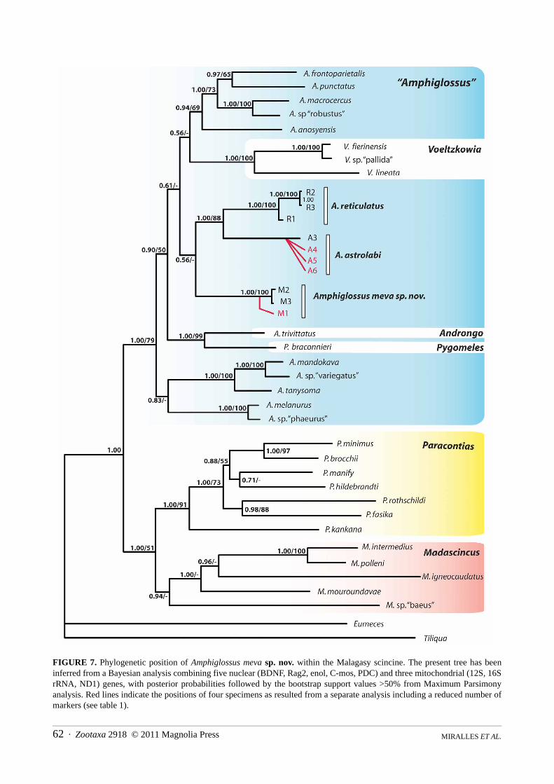
Phylogenetic position and genetic differentiation. The results of the phylogenetic analyses are summarizedin Figure 7. Unsurprisingly, the phylogenetic tree obtained is highly congruent with the one published by Crottini et
Zootaxa 2918 © 2011 Magnolia Press · 61A NEW LARGE AND COLORFUL SPECIES OF AMPHIGLOSSUS
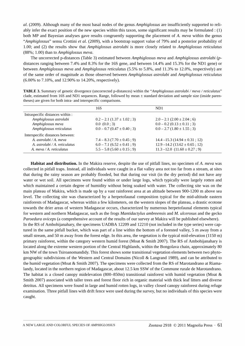
al. (2009). Although many of the most basal nodes of the genus Amphiglossus are insufficiently supported to reli-ably infer the exact position of the new species within this taxon, some significant results may be formulated : (1)both MP and Bayesian analyses gave results congruently supporting the placement of A. meva within the genus“Amphiglossus” sensu Crottini et al. (2009), with a bootstrap support value of 79% and a posterior probability of1.00; and (2) the results show that Amphiglossus astrolabi is more closely related to Amphiglossus reticulatus(88%; 1.00) than to Amphiglossus meva.
The uncorrected p-distances (Table 3) estimated between Amphiglossus meva and Amphiglossus astrolabi (p-distances ranging between 7.4% and 8.3% for the 16S gene, and between 14.4% and 15.3% for the ND1 gene) orbetween Amphiglossus meva and Amphiglossus reticulatus (5.5% to 5.8%, and 11.3% to 12.0%, respectively) areof the same order of magnitude as those observed between Amphiglossus astrolabi and Amphiglossus reticulatus(6.00% to 7.10%, and 12.90% to 14.20%, respectively).
TABLE 3. Summary of genetic divergence (uncorrected p-distances) within the “Amphiglossus astrolabi / meva / reticulatus”clade, estimated from 16S and ND1 sequences. Range, followed by mean ± standard deviation and sample size (inside paren-theses) are given for both intra- and interspecific comparisons.
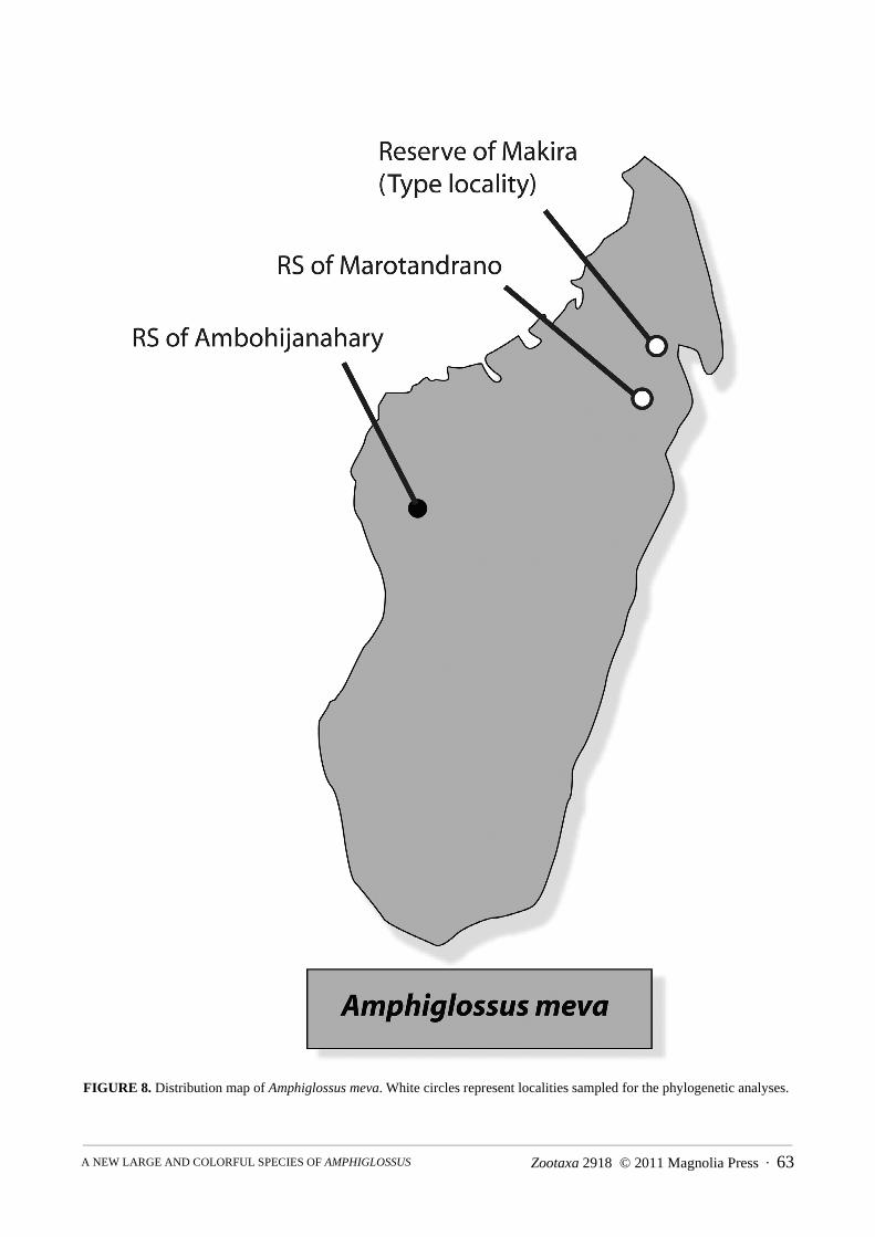
Habitat and distribution. In the Makira reserve, despite the use of pitfall lines, no specimen of A. meva wascollected in pitfall traps. Instead, all individuals were caught in a flat valley area not too far from a stream, at sitesthat during the rainy season are probably flooded, but that during our visit (in the dry period) did not have anywater or wet soil. All specimens were found within or under large logs, which typically were largely rotten andwhich maintained a certain degree of humidity without being soaked with water. The collecting site was on themain plateau of Makira, which is made up by a vast rainforest area at an altitude between 900-1200 m above sealevel. The collecting site was characterized by a herpetofaunal composition typical for the mid-altitude easternrainforests of Madagascar, whereas within a few kilometers, on the western slopes of the plateau, a drastic ecotonetowards the drier areas of western Madagascar occurs, characterized by numerous herpetofaunal elements typicalfor western and northern Madagascar, such as the frogs Mantidactylus ambreensis and M. ulcerosus and the geckoParoedura oviceps (a comprehensive account of the results of our survey at Makira will be published elsewhere). In the RS of Ambohijanahary, the specimens UADBA 12209 and 12210 (not included in the type series) were cap-tured in the same pitfall bucket, which was part of a line within the bottom of a forested valley, 5 m away from asmall stream, and 50 m away from the forest edge. In this area, the vegetation is the typical mid-elevation (1150 m)primary rainforest, within the category western humid forest (Moat & Smith 2007). The RS of Ambohijanahary islocated along the extreme western portion of the Central Highlands, within the Bongolava chain, approximately 80km NW of the town Tsiroanomandidy. This forest shows some transitional vegetation elements between two phyto-geographic subdivisions of the Western and Central Domains (Nicoll & Langrand 1989), and can be attributed tothe humid vegetation (Moat & Smith 2007). The specimens were collected from the RS of Marotandrano at Riama-landy, located in the northern region of Madagascar, about 12.5 km SSW of the Commune rurale de Marotandrano.The habitat is a closed canopy midelevation (800–850m) transitional rainforest with humid vegetation (Moat &Smith 2007) associated with taller trees and forest floor rich in organic material with thick leaf litters and diversedetritus. All specimens were found in large and humid rotten logs, in valley closed canopy rainforest during refugeexamination. Three pitfall lines with drift fence were used during the survey, but no individuals of this species werecaught.
16S ND1
Intraspecific distances within : Amphiglossus astrolabi Amphiglossus meva Amphiglossus reticulatus
0.2 – 2.1 (1.37 ± 1.02 ; 3)0.0 (0.0 ; 3)0.0 – 0.7 (0.47 ± 0.40 ; 3)
2.0 – 2.1 (2.00 ± 2.04 ; 6)0.0 – 0.2 (0.13 ± 0.11 ; 3)0.0 – 2.7 (1.80 ± 1.55 ; 3)
Interspecific distances between: A. astrolabi / A. meva A. astrolabi / A. reticulatus A. meva / A. reticulatus
7.4 – 8.3 (7.70 ± 0.45 ; 9)6.0 – 7.1 (6.52 ± 0.41 ; 9)5.5 – 5.8 (5.60 ± 0.15 ; 9)
14.4 –15.3 (14.94 ± 0.31 ; 12)12.9 –14.2 (13.62 ± 0.65 ; 12)11.3 –12.0 (11.60 ± 0.27 ; 9)
MIRALLES ET AL.62 · Zootaxa 2918 © 2011 Magnolia Press
FIGURE 7. Phylogenetic position of Amphiglossus meva sp. nov. within the Malagasy scincine. The present tree has beeninferred from a Bayesian analysis combining five nuclear (BDNF, Rag2, enol, C-mos, PDC) and three mitochondrial (12S, 16SrRNA, ND1) genes, with posterior probabilities followed by the bootstrap support values >50% from Maximum Parsimonyanalysis. Red lines indicate the positions of four specimens as resulted from a separate analysis including a reduced number ofmarkers (see table 1).
Zootaxa 2918 © 2011 Magnolia Press · 63A NEW LARGE AND COLORFUL SPECIES OF AMPHIGLOSSUS
FIGURE 8. Distribution map of Amphiglossus meva. White circles represent localities sampled for the phylogenetic analyses.
MIRALLES ET AL.64 · Zootaxa 2918 © 2011 Magnolia Press
Previous herpetological surveys using pitfall traps in the same reserve (Raxworthy, unpublished; Biodev,unpublished), the Central Highland sites of the RS d'Ambohitantely (C.J. Raxworthy and collaborators and S.M.Goodman and collaborators, unpublished), Ankazomivady forest (Goodman et al., 1998), Parc National d’Andrin-gitra (Raxworthy et al., 1996), and Andranomay forest (Raselimanana 1998), the west part of Parc National duTsingy de Bemaraha (Bora et al. 2010), Kirindy forest (Raselimanana 2008, Raxworthy et al, unpublished), thesouthwestern PK 32 forest (Raxworthy et al., umpublished,), Parc National de Zombitse (Raxworthy et al. 1994),and Vohibasia (Goodman et al. 1997), and the northwestern RS d'Ankarafantsika (Ramanamanjato & Rabibisoa,2002; Raselimanana, 2008) did not provide any evidence for the occurrence of A. meva n. sp. although numerousother burrowing species (skinks and frogs) were collected in these surveys. The species appears to show a prefer-ence for large rotten logs retaining a certain degree of humidity but in general in parts of the rainforest with rela-tively dry soils. Based on our finding in Marotandrano and Makira, this new species is not rare but probably has aquite strict ecological specificity with respect to its microhabitat (Fig. 8).
Ecological notes. In Ambohijanahary, two specimens were captured on two consecutive days, both in the samepitfall trap, 5 m away from a 2 m wide stream. They may have been a breeding pair. No other individuals werefound in the RS d’Ambohijanahary despite an additional 165 trap days with pitfall devices. The specimens fromMarotandrano were all captured during refuge examination including rotten logs excavation and removal of barksand leaf litter accumulated under taller and big dead trees. In Makira, a group of three individuals was foundtogether under a big log. Several larvae of coleopterans, other insects and termites were found in the same micro-habitat, suggesting that this new skink may feed on these preys. In contrast to the other large species within thesame genus, A. meva was never found in water.
Threats and Conservation status. Habitat loss due to slash and burn agriculture, bush fires, and wood extrac-tion are the main pressures on the Ambohijanahary and Marotandrano reserve. Forests at these reserves, especiallyAmbohijanahary, are extensively fragmented. Although this forest is classified as a Réserve Spéciale, no manage-ment plan has been proposed. The reserve and surrounding areas are known to be the domain of zebu cattle thieves(dahalo). The forested areas within the reserve are often used by local people as a site to shelter stolen zebu. Theimportance of this form of refuge has provided some protection to the remaining forest. Moreover, MNP has anoffice and agents in Marotandrano Village and a coordination office is operational in Mandritsara. On the contrary,Makira reserve currently appears to be relatively well preserved at its western edge, where the type locality of thenew species is located. Despite the common use of the forest for cattle grazing, in 2009 we could not detect majorforest destruction nor human settlements directly in the forested area. Efforts need to be undertaken to maintain thisapparently stable situation and to reduce forest destruction at the eastern lowland borders of Makira, which appearsto be relatively intense in some areas.
Discussion
Madagascar is one of the richest and more biologically diverse places on Earth, and its diversity is still largelyunknown with many new species being described every year and many more yet to be discovered (Köhler et al.2005; Vieites et al. 2009). There are several reptile species in Madagascar with bright coloration; some of themhave been considered aposematic. For example, several snakes such as Stenophis citrinus Domergue and Liophid-ium pattoni Vieites, Fanomezana, Ratsoavina, Randrianiaina, Nagy & Vences present unique color patterns consist-ing of alternation of yellow and black cross bands or red, yellow and blue in contrast with black respectively. Noneof these species are poisonous, and the factors triggering the evolution of their aposematic coloration is unclear. Inskinks there are several examples of bright coloration that may be related to escaping or warning predators. Skinkssuch as Madascincus igneocaudatus have a contrasting bright red tail, which can be autotomized if a predator bitesit, while other species show an overall bright coloration like A. crenni or Pseudoacontias menamainty that could beconsidered as aposematic. However, these species are highly fossorial and as far as it is known they are not poison-ous. It is unclear if the bright coloration evolved as a warning signal to predators, but also other burrowing taxa areknown to have aposematic coloration (Wollenberg & Measey 2009).
Both morphological and coloration characters distinguish A. meva sp. nov. from any other skink species fromMadagascar. It is remarkable that such a large and conspicuous species of vertebrate has not been detected untilnow, despite this new species being relatively widespread on the island. This suggests that many more species of
Zootaxa 2918 © 2011 Magnolia Press · 65A NEW LARGE AND COLORFUL SPECIES OF AMPHIGLOSSUS
vertebrates can still be expected from Madagascar and encourages more field and taxonomic work on its fauna.Despite the species is only known from three sites, we have found many specimens in a short period. Until moredata is gathered, we suggest to consider this species as Data Deficient for conservation purposes following IUCNcriteria.
Acknowledgements
We are grateful to Theo Rajoafiarison, Jim and Carol Patton, Emile Rajeriarison, Fanomezana Mihaja. Ratsoavinaand Roger-Daniel Randrianiaina who helped collecting specimens at Makira, to Florent and François Randriana-solo from the WCS for their help and companionship in the field at Makira, and to our drivers Claude and Samy forsafely carrying us to the basis of the Makira slopes. Steven M. Goodman helped in the capture of several specimensand made valuable comments on earlier versions of this manuscript. Harald Schütz took the photograph of the liv-ing specimen from Ambohijanahary. Franco Andreone, Edouard R. Brygoo and Frank Glaw contributed photo-graphs of Amphiglossus crenni and A. astrolabi. We also thank the Département de Biologie Animale, Universitéd’Antananarivo, Madagascar National Park team in Mandritsara, the brigade of Gendarmerie in Marotandrano thatensuring our security during the field survey, the Ministères des Eaux et Forêts, Antananarivo, and the TechnicalUniversity of Braunschweig. Many thanks to Annemarie Ohler and Ivan Ineich (MNHN), Frank Glaw (ZSM) andGöran Nilson (GNM) for providing us access to collection specimens. We are grateful to the Malagasy authoritiesfor research and export permits, and to the Wildlife Conservation Society for supporting our research activities atMakira. The field inventory in Ambohijanahary was funded by a grant from the National Geographic Society toS.M. Goodman and D. Rakotondravony, and the mission to Marotandrano was generously supported by the Mac-Arthur Foundation in the context of the RAP–Gasy project. We are indebted to the Ecology Training Program andthe World Wide Fund for Nature, Antananarivo, for logistical support. Funding was provided by a postdoctoralresearch fellowship of the Alexander von Humboldt Foundation and by a SYNTHESYS grant (FR–TAF–842) toAM, by the Volkswagen Foundation to MV, and by a Spanish Ministry of Science and Innovation grant(CGL2009–10198) to DRV.
References
Andreone, F. & Greer, A.E. (2002) Malagasy scincid lizards: descriptions of nine new species, with notes on the morphology,reproduction and taxonomy of some previously described species (Reptilia, Squamata: Scincidae). Journal of Zoology(London), 258, 139–181.
Angel, M.F. (1930) Diagnoses d’espèces nouvelles de lézards, de Madagascar, appartenant au genre Scelotes. Bulletin duMuséum National d’Histoire Naturelle, Paris, 2, 506–509.
Bora, P., Randrianantoandro, J.C., Randrianavelona, R., Hantalalaina, E.F., Andriantsimanarilafy, R.R., Rakotondravony, D.,Ramilijaona, O.R., Vences, M., Jenkins, R.K.B., Glaw, F. & Köhler, J. (2010) Amphibians and reptiles of the Tsingy deBemaraha Plateau, western Madagascar: Checklist, biogeography and conservation Herpetological Conservation andBiology, 5(1), 111–125.
Brygoo, E.R. (1980) Systématique des lézards scincidés de la région malgache. II. Amphiglossus astrolabi Duméril et Bibron,1839; Gongylus polleni Grandidier, 1869; Gongylus stumpffi Boettger, 1882, et Scelotes waterloti, Angel, 1930. Bulletindu Muséum national d´Histoire naturelle. Section A, 2(2), 525–539.
Brygoo, E.R. (1983) Systématique des lézards scincidés de la région malgache. X. Rapports de Gongylus johannae Günther,1880, des Comores, et de Sepsina valhallae Boulenger, 1909, des Glorieuses, avec les espèces malgaches. Bulletin duMuséum National d’Histoire Naturelle, Paris, 4ème série, 5, A, n°2, 651–660.
Brygoo, E.R. (1987) Les types de Scincidés (Reptilia, Sauriens) du Muséum national d’Histoire naturelle. Catalogue critique.Bulletin du Muséum national d’Histoire naturelle, Paris, Section A, (3), 1–126.
Crottini, A., Dordel, J., Köhler, J., Glaw, F., Schmitz, A. & Vences, M. (2009) A multilocus phylogeny of Malagasy scincid liz-ards elucidates the relationships of the fossorial genera Androngo and Cryptoscincus. Molecular Phylogenetics and Evolu-tion, 53, 345–350.
Dayrat, B. (2005) Toward integrative taxonomy. Biological Journal of the Linnean Society, 85, 407–415.de Queiroz, K. (2007) Species concepts and species delimitation. Systematic Biology, 56, 879–886.Glaw, F. & Vences, M. (2007) A field guide to the amphibians and reptiles of Madagascar. Third edition. Cologne, Vences &
Glaw Verlag, 496 pp. Goodman, S.M., Ramanamanjato, J.-B. & Raselimanana, A.P. (1997) Les amphibiens et les reptiles. In: Langrand, O and S. M.
MIRALLES ET AL.66 · Zootaxa 2918 © 2011 Magnolia Press
Goodman (Eds.). Inventaire biologique Forêts de Vohibasia et Isoky-Vohimena. Recherches pour le Développement, sérieSciences Biologiques, n°12 , pp.110–129.
Goodman, S.M., Duplantier, J.-M., Rakotomalaza, P.J., Raselimanana, A.P., Rasoloarison, R., Ravokatra, M., Soarimalala, V. &Wilme, L. (1998) Inventaire biologique de la forêt d’Ankazomivady, Ambositra. Akon’ny Ala, 24, 19–32.
Greer, A.E. & Shea, G. (2000) A major new head scale character in non-lygosomine scincid lizards. Journal of Herpetology, 34,631–636.
Köhler, J., Vieites, D.R., Bonett, R. M., García, F.H., Glaw, F., Steinke, D. & Vences, M. (2005) New amphibians and globalconservation: A boost in species discoveries in a highly endangered vertebrate group. BioScience, 55(8), 693–696.
Köhler, J., Vences, M., Erbacher, M. & Glaw, F. (2010) Systematics of limbless scincid lizards from northern Madagascar: mor-phology, phylogenetic relationships and implications for classification (Squamata: Scincidae). Organisms Diversity andEvolution, 10, 147–159.
Köhler, J., Vieites, D.R, Glaw, F., Kaffenberger, N. & Vences, M. (2009) A further new species of limbless skink, genus Para-contias, from eastern Madagascar. African Journal of Herpetology, 58, 98–105.
Miralles, A. (2006) A New Species of Mabuya (Reptilia, Squamata, Scincidae) from the Isolated Caribbean Island of SanAndrés, with a new interpretation of nuchal scales, character of systematic importance. The Herpetological Journal, 16, 1–7.
Miralles, A., Köhler, J., Glaw, F. & Vences, M. (2011a) A molecular phylogeny of the “Madascincus polleni species complex”,with description of a new species of scincid lizard from the coastal dune area of northern Madagascar. Zootaxa, 2876, 1–16.
Miralles, A., Köhler, J., Vieites, D.R., Glaw, F. & Vences, M. (2011b) Hypotheses on rostral shield evolution in fossorial lizardsderived from the phylogenetic position of a new species of Paracontias (Squamata, Scincidae). Organisms Diversity andEvolution, 11, 135–150.
Moat, J. & Smith, P. (2007) Atlas de la végétation de Madagascar. Kew: Royal Botanic Gardens, Kew, 124pp.Nicoll, M.E. & Langrand, O. (1989) Madagascar: Revue de la conservation et des aires protégées. World Wildlife Fund,
Gland, 374pp.Padial, J.M., Miralles, A., De la Riva, I. & Vences, M. (2010) The integrative future of taxonomy. Frontiers in Zoology, 7, 16.
doi:10.1186/1742-9994-7-16.Ramanamanjato, J.-B. & Rabibisoa, N. (2002) Evaluation rapide de la diversité biologique des reptiles et amphibiens de la
Réserve Naturelle Intégrale d’Ankarafantsika, In: Alonso, L. E., Schulenberg, T. S., Radilofe, S. & Missa O. (Eds): Uneevaluation biologique de la Réserve Naturelle Intégrale d’Ankarafantsika, Madagascar. – Bulletin RAP d’évaluationrapide 23, Conservational International, Washington, DC, pp 98–103 + 135–138.
Raselimanana, A.P. (1998) La diversité de la faune de reptiles et d’amphibiens. In: Rakotondravony D., Goodman S. M. (Eds.).Inventaire biologique de la forêt d’Andranomay, Anjozorobe. Recherches pour le Développement, série SciencesBiologiques, 13, pp 43–59.
Raselimanana, A.P. (2008) Herpétofaune des forêts sèches malgaches. Dans Les forêts sèches de Madagascar. In: Goodman S.M. & Wilmé L. (eds.). Malagasy Nature, 1, 46–75.
Raselimanana, A.P. & Rakotomalala, D. (2003) Scinicidae, skinks. In: Goodman, S. M., Benstead, J. P. (Eds.) The natural his-tory of Madagascar. The University of Chicago Press, Chicago, pp. 986–993.
Raxworthy, C.J., Ramanamanjato, J.-B. & Raselimanana, A.P. (1994) Les reptiles et les amphibiens. In: Goodman S. M., Lan-grand O. (Eds.) Inventaire Biologique de Forêt de Zombitse. Recherches pour le Développement, série SciencesBiologiques, numéro spécial, pp. 41–57.
Raxworthy, C.J. & Nussbaum, R.A. (1993) Four new species of Amphiglossus from Madagascar (Squamata: Scincidae). Her-petologica, 49, 326–341.
Raxworthy, C.J. & Nussbaum, R.A. (1994) A rainforest survey of amphibians, reptiles and small mammals at Montagned’Ambre, Madagascar. Biological Conservation, 69, 65–74.
Raxworthy, C.J. & Nussbaum, R.A. (1996) Amphibians and reptiles of the réserve naturelle intégrale d’Andringitra, Madagas-car: a study of elevational distribution and local endemicity. In: Goodman, S. M. (ed.): A floral and faunal inventory of theeastern slopes of the réserve naturelle intégrale d’Andringitra, Madagascar: with reference to elevational variation. Fiel-diana Zoology (new series), 85, 158–170.
Ronquist, F. & Huelsenbeck, J.P. (2003) MRBAYES 3: Bayesian phylogenetic inference under mixed models. Bioinformatics,19, 1572–1574.
Schmitz, A., Brandley, M.C., Mausfeld, P., Vences, M., Glaw, F., Nussbaum, R.A. & Reeder, T.W. (2005) Opening the blackbox: phylogenetics and morphological evolution of the Malagasy fossorial lizards of the subfamily ‘‘Scincinae’’. Molecu-lar Phylogenetics and Evolution, 34, 118–133.
Swofford, D.L. (2002) PAUP*. Phylogenetic Analysis Using Parsimony (*and other Methods). Version 4.0.b10. Sunderland,MA: Sinauer Associates.
Tamura, K., Dudley, J., Nei, M. & Kumar, S. (2007) MEGA4: Molecular Evolutionary Genetics Analysis (MEGA) softwareversion 4.0. Molecular Biology and Evolution. doi: 10.1093/molbev/msm092
Vieites, D.R., Wollenberg, K.C., Andreone, F., Köhler, J., Glaw, F. & Vences, M. (2009) Vast underestimation of Madagascar’sbiodiversity evidenced by an integrative amphibian inventory. Proceedings of the National Academy of Sciences of theUnited States of America, 106, 8267–8272.
Zootaxa 2918 © 2011 Magnolia Press · 67A NEW LARGE AND COLORFUL SPECIES OF AMPHIGLOSSUS
Whiting, A.S., Sites J.W. Jr. & Bauer, A.M. (2004) Molecular phylogenetics of Malagasy skinks (Squamata: Scincidae). Afri-can Journal of Herpetology, 53(2), 135–146.
Wollenberg, K.C. & Measey, G.J. (2009) Why colour in subterranean vertebrates? Exploring the evolution of colour patterns incaecilian amphibians. Journal of Evolutionary Biology, 22, 1046–1056.
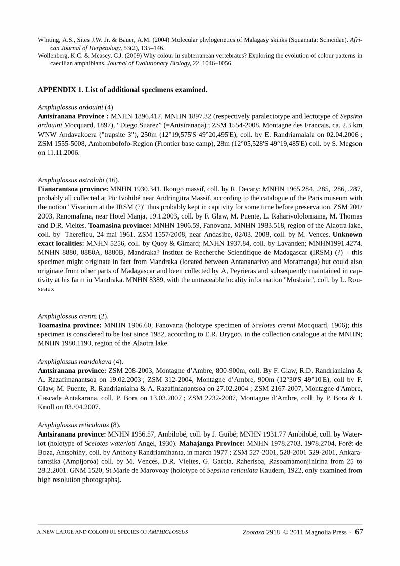
APPENDIX 1. List of additional specimens examined.
Amphiglossus ardouini (4)Antsiranana Province : MNHN 1896.417, MNHN 1897.32 (respectively paralectotype and lectotype of Sepsinaardouini Mocquard, 1897), “Diego Suarez” (=Antsiranana) ; ZSM 1554-2008, Montagne des Francais, ca. 2.3 kmWNW Andavakoera ("trapsite 3"), 250m (12°19,575'S 49°20,495'E), coll. by E. Randriamalala on 02.04.2006 ;ZSM 1555-5008, Ambombofofo-Region (Frontier base camp), 28m (12°05,528'S 49°19,485'E) coll. by S. Megsonon 11.11.2006.
Amphiglossus astrolabi (16).Fianarantsoa province: MNHN 1930.341, Ikongo massif, coll. by R. Decary; MNHN 1965.284, .285, .286, .287,probably all collected at Pic Ivohibé near Andringitra Massif, according to the catalogue of the Paris museum withthe notion "Vivarium at the IRSM (?)" thus probably kept in captivity for some time before preservation. ZSM 201/2003, Ranomafana, near Hotel Manja, 19.1.2003, coll. by F. Glaw, M. Puente, L. Raharivololoniaina, M. Thomasand D.R. Vieites. Toamasina province: MNHN 1906.59, Fanovana. MNHN 1983.518, region of the Alaotra lake,coll. by Therefieu, 24 mai 1961. ZSM 1557/2008, near Andasibe, 02/03. 2008, coll. by M. Vences. Unknownexact localities: MNHN 5256, coll. by Quoy & Gimard; MNHN 1937.84, coll. by Lavanden; MNHN1991.4274.MNHN 8880, 8880A, 8880B, Mandraka? Institut de Recherche Scientifique de Madagascar (IRSM) (?) – thisspecimen might originate in fact from Mandraka (located between Antananarivo and Moramanga) but could alsooriginate from other parts of Madagascar and been collected by A, Peyrieras and subsequently maintained in cap-tivity at his farm in Mandraka. MNHN 8389, with the untraceable locality information "Mosbaie", coll. by L. Rou-seaux
Amphiglossus crenni (2).Toamasina province: MNHN 1906.60, Fanovana (holotype specimen of Scelotes crenni Mocquard, 1906); thisspecimen is considered to be lost since 1982, according to E.R. Brygoo, in the collection catalogue at the MNHN;MNHN 1980.1190, region of the Alaotra lake.
Amphiglossus mandokava (4).Antsiranana province: ZSM 208-2003, Montagne d’Ambre, 800-900m, coll. By F. Glaw, R.D. Randrianiaina &A. Razafimanantsoa on 19.02.2003 ; ZSM 312-2004, Montagne d’Ambre, 900m (12°30'S 49°10'E), coll by F.Glaw, M. Puente, R. Randrianiaina & A. Razafimanantsoa on 27.02.2004 ; ZSM 2167-2007, Montagne d'Ambre,Cascade Antakarana, coll. P. Bora on 13.03.2007 ; ZSM 2232-2007, Montagne d’Ambre, coll. by P. Bora & I.Knoll on 03./04.2007.
Amphiglossus reticulatus (8).Antsiranana province: MNHN 1956.57, Ambilobé, coll. by J. Guibé; MNHN 1931.77 Ambilobé, coll. by Water-lot (holotype of Scelotes waterloti Angel, 1930). Mahajanga Province: MNHN 1978.2703, 1978.2704, Forêt deBoza, Antsohihy, coll. by Anthony Randriamihanta, in march 1977 ; ZSM 527-2001, 528-2001 529-2001, Ankara-fantsika (Ampijoroa) coll. by M. Vences, D.R. Vieites, G. Garcia, Raherisoa, Rasoamamonjinirina from 25 to28.2.2001. GNM 1520, St Marie de Marovoay (holotype of Sepsina reticulata Kaudern, 1922, only examined fromhigh resolution photographs).