Embed Size (px)
Citation preview
B R A I N R E S E A R C H R E V I E W S 5 2 ( 2 0 0 6 ) 2 7 5 – 2 9 2
ava i l ab l e a t www.sc i enced i rec t . com
www.e l sev i e r. com/ loca te /b ra in res rev
Review
A new insight on Al-maltolate-treated aged rabbit asAlzheimer's animal model
Bharathia, N.M. Shamasundarb, T.S. Sathyanarayana Raoc, M. Dhanunjaya Naidud,R. Ravide, K.S.J Raoa,⁎aDepartment of Biochemistry and Nutrition, Central Food Technological Research Institute, Mysore-570013, IndiabDepartment of Anatomy, J.S.S Medical College, Mysore, IndiacDepartment of Psychiatry, J.S.S. Medical College, Mysore, IndiadDepartment of Biotechnology, S.V. University, Tirupati, IndiaeNetherlands Brain Bank, Amsterdam, The Netherlands
A R T I C L E I N F O
⁎ Corresponding author. Fax: +91 821 2517233E-mail address: [email protected] (K.S.J. R
0165-0173/$ – see front matter © 2006 Elsevidoi:10.1016/j.brainresrev.2006.04.003
A B S T R A C T
Article history:Accepted 4 April 2006Available online 16 June 2006
Lack of anadequate animalmodel forAlzheimer's disease (AD) has limited anunderstandingof the pathogenesis of the disease and the development of therapeutic agents targeting keypathophysiological processes. There are undoubtedly few satisfactory animal models forexploring therapies targeting at amyloid beta (Aβ) secretion, deposition, aggregation, andprobably the inflammatory response. However, an understanding of the complex events –tau, Aβ, oxidative stress, redox active iron, etc. – involved in the neuronal cell loss is stillunclear due to the lack of a suitable animal model system. The use of neurotoxic agentsparticularly aluminum–organic complexes, especially Al-maltolate, expands the scope of ADresearch by providing new animal models exhibiting neurodegenerative processes relevantto AD neuropathology. Examination of different species of aged animals including therapidly advancing transgenic mouse models revealed very limited AD-like pathology. Mostother animal models have single event expression such as extracellular Aβ deposition,intraneuronal neurofilamentous aggregation of proteins akin to neurofibrillary tangles,oxidative stress or apoptosis. To date, there are no paradigms of any animal in which all thefeatures of AD were evident. However, the intravenous injection of Al-maltolate into agedNew zealand white rabbits results in conditions which mimics a number ofneuropathological, biochemical and behavioral changes observed in AD. Suchneurodegenerative effects include the formation of intraneuronal neurofilamentousaggregates that are tau positive, immunopositivity of Aβ, presence of redox active iron,oxidative stress and apoptosis, adds credence to the value of this animal model system. Theuse of this animalmodel should not be confusedwith the ongoing controversy regarding thepossible role of Al in the neuropathogenesis, a debate which by no means has beenconcluded. Above all this animal model involving neuropathology induced by Al-maltolateprovides a new information in understanding the mechanism of neurodegeneration.
© 2006 Elsevier B.V. All rights reserved.
Keywords:Alzheimer's diseaseAl-maltolateAged rabbitAβ depositionNeurofibrillary tangleOxidative stressApoptosis
.ao).
er B.V. All rights reserved.
276 B R A I N R E S E A R C H R E V I E W S 5 2 ( 2 0 0 6 ) 2 7 5 – 2 9 2
Contents
1. Introduction . . . . . . . . . . . . . . . . . . . . . . . . . . . . . . . . . . . . . . . . . . . . . . . . . . . . . . . . . . 2762. Al-maltolate as novel compound for inducing AD like pathology in aged rabbits . . . . . . . . . . . . . . . . 2773. Why choose aged v/s young rabbits . . . . . . . . . . . . . . . . . . . . . . . . . . . . . . . . . . . . . . . . . . . . . 278
3.1. Susceptibility of aged rabbits in inducing AD neuropathology compared to young ones. . . . . . . . . . . . . 2783.2. Assessment of neurofibrillary degeneration based on neuroanatomical susceptibility of Al-induced
neurodegeneration . . . . . . . . . . . . . . . . . . . . . . . . . . . . . . . . . . . . . . . . . . . . . . . . . . 2784. Strong evidences supporting Al-maltolate/rabbit model for AD . . . . . . . . . . . . . . . . . . . . . . . . . . . . . . 2785. Contradiction and paradox in Al-maltolate-induced neuropathology in comparison with Alzheimer's disease . . . . 281
5.1. Behavioral features . . . . . . . . . . . . . . . . . . . . . . . . . . . . . . . . . . . . . . . . . . . . . . . . . . 2815.2. Immunohistochemical features . . . . . . . . . . . . . . . . . . . . . . . . . . . . . . . . . . . . . . . . . . . 281
6. Characteristics of tangles associated with Al-maltolate-treated aged rabbits in comparison with AD . . . . . . . . . 2827. How do the abeta fibrillar deposition and NFTs evolve? . . . . . . . . . . . . . . . . . . . . . . . . . . . . . . . . . . 2828. Similarities and differences in degenerative aspects in Al-maltolate-treated rabbits . . . . . . . . . . . . . . . . . . 282
8.1. Oxidative stress . . . . . . . . . . . . . . . . . . . . . . . . . . . . . . . . . . . . . . . . . . . . . . . . . . . . 2838.2. Apoptosis. . . . . . . . . . . . . . . . . . . . . . . . . . . . . . . . . . . . . . . . . . . . . . . . . . . . . . . . 283
8.2.1. Effect of Al-maltolate on the mitochondrial-mediated apoptosis pathway. . . . . . . . . . . . . . . . 2838.2.2. Effect of Al-maltolate on apoptosis-regulatory proteins that mediate endoplasmic reticulum . . . . . 285
8.3. NFT formation . . . . . . . . . . . . . . . . . . . . . . . . . . . . . . . . . . . . . . . . . . . . . . . . . . . . . 2859. Neurochemical features observed in Al-maltolate-treated aged rabbits with that of AD. . . . . . . . . . . . . . . . . 285
9.1. Alterations in the levels of NPY . . . . . . . . . . . . . . . . . . . . . . . . . . . . . . . . . . . . . . . . . . . 2869.2. Imbalances in the levels of N-acetyl-aspartyl-glutamate (NAAG) and its precursor N-acetyl-L-aspartate (NAL) 2869.3. D-Aspartate levels in AD/DNA alterations in AD . . . . . . . . . . . . . . . . . . . . . . . . . . . . . . . . . . 286
10. Why other animal models including transgenic fails to reproduce total AD neuropathology? . . . . . . . . . . . . . 28611. Conclusion and future perspectives . . . . . . . . . . . . . . . . . . . . . . . . . . . . . . . . . . . . . . . . . . . . . 287Acknowledgments . . . . . . . . . . . . . . . . . . . . . . . . . . . . . . . . . . . . . . . . . . . . . . . . . . . . . . . . . . 287References . . . . . . . . . . . . . . . . . . . . . . . . . . . . . . . . . . . . . . . . . . . . . . . . . . . . . . . . . . . . . . 287
1. Introduction
AD is a complex neurodegenerative disorder comprisingcomplex neurobiochemical and neuropathological events,characterized by three typical pathological features, namelythe extracellular deposition of Aβ (Selkoe, 1989, 1991; Hardyand Selkoe, 2002; Hardy and Higgins, 1992), the formation ofintraneuronal neurofibrillary tangles (NFTs) (Doll, 1993; Perryand Perry, 1985; Perl and Brondy, 1980; Lovell et al., 1993;Wisniewski and Sofer, 1979), and selective neuronal loss.However, it is still unclear which of these pathologicalfeatures is the primary event in the initiation and progres-sion of this disease. The etiological factors of AD includegenetics, head trauma, oxidative stress, infectious agents,and environmental factors including aluminum (Al) toxicity.The pioneering studies on neurotoxicity of Al in experimen-tal animals were first reported in 1897 by Dollken (1897).Many scientific studies have brought to light the potentialtoxicity of Al in experimental animal models and inhumans under different clinical conditions (Spafforth,1921; McLaughlin et al., 1962). But the usage of Al inexperimental animal came to light following the extraordi-nary discovery of Klatzo et al. (1965) who showed thatinjections of Al-salts into rabbit brain led to the formationof NFTs which appeared similar to the NFTs of AD (Klatzoet al., 1965; Terry and Peña, 1986). Later, these results werereplicated in cats by Crapper et al. (1973). The complexchemistry of Al and the fact that there was no readily
available radioisotope for experimental purposes thushindered the clarification of this element's involvement inthe etiology of AD. However, studies by Priest (2004) onhumans and animal using the 26Al radioisotope (Yumoto etal., 2001) have demonstrated that Al can indeed enter thecentral nervous system following systemic administration(Walton et al., 1995). In addition, there is documentedevidence that Al is neurotoxic, both in human disease, aswell as in experimental animals (Wills and Savory, 1983).Studies by Wen and Wisniewski (1985) histochemicallylocalized Al in rabbit CNS further supported by Uemura(1984) illustrated intranuclear Al accumulation in chronicanimals in turn led to neurofibrillary changes. Thereby Alsalts administered intracerebrally or peripherally in rabbit(Klatzo et al., 1965), cat (Crapper et al., 1973), monkey(Games et al., 1995), rat (Brining et al., 1996), and dog (Unoet al., 1999) induce the formation of neurofibrillary aggre-gates (NFAs) which has contributed to the argument that Alis one of the contributing factor to several neurodegener-ative disorders, mainly AD. However, this hypothesisremains controversial.
Although understanding of the complex events involved inneuropathogenesis and neurobiochemical events in ADrequires the availability of suitable animal model systems.Understanding the neurodegeneration pathways in relation-ship to Aβ deposition, NFT and neuritic plaque formationusing human tissue is limited since only a single time point,an intrinsic limitation resulting from the use of human
277B R A I N R E S E A R C H R E V I E W S 5 2 ( 2 0 0 6 ) 2 7 5 – 2 9 2
autopsy tissue. To date, examination of different species ofaged animals including transgenic mice have revealed verylimited AD-like neuropathology (Sugaya et al., 1997). Recently,Bishop and Robinson (2000) stated that, “Mice are notHumans,” and they could have a much different response tothe presence of a neurotoxin. Thereby rabbits may beparticularly relevant to the investigation of human diseasesince they belong to the mammalian order Lagomorpha (Grauret al., 1996), a group reported to closely resemble primatesthan rodents (Graur et al., 1996) and provide a unique animalsystem for the consistent production of neurofibrillary pa-thology (Klatzo et al., 1965; Yokel and O'Callaghan, 1998).Expansion of the use of new animal models is obviouslyneeded; hence, rabbits have been considered the most widelyused experimental animal for such studies because of itsvulnerability to Al and its availability (Klatzo et al., 1965; Yokeland O'Callaghan, 1998). Moreover rabbits, along with cats,develop intraneuronal NFAs in response to the intracerebraladministration of Al salts, whereas rodents do not developthese lesions (Yokel and O'Callaghan, 1998). Chronic intracis-ternal or intracerebral injection of minute quantities of Al intoexperimental animals, especially rabbits, induces progressive-ly severe neurologic signs associated neuropathologic featuresof neurodegeneration (Hof et al., 1992), particularly theproduction of intraneuronal argyrophilic protein aggregates(Klatzo et al., 1965) that bear biochemical similarities to theNFTs observed in AD. Studies by Savory et al. (1994, 1995,1996b, 1999, 2001, 2003) and Rao et al. (2000) employed Newzealand aged white rabbits as the experimental animal (4 yearsold). Besides intraneuronal neurofilamentous changes in thehippocampus, cerebral cortex, brainstem, and spinal cord,which demonstrate many biochemical features, are in com-mon with as seen in AD (Hof et al., 1992), intracisternaladministration of Al-maltolate to rabbits also leads tobiochemical changes suggestive of apoptosis similar toAD (Savory et al., 1999). Here, in this review, we havetried to convey that this Al/rabbit model system helps tounravel the events associated against the fatal Alneurotoxicity in relevance to AD (Ghribi et al., 2001d).In precise Al-maltolate-treated aged rabbits could bereliable and a sensitive animal model for understandingAD neuropathology.
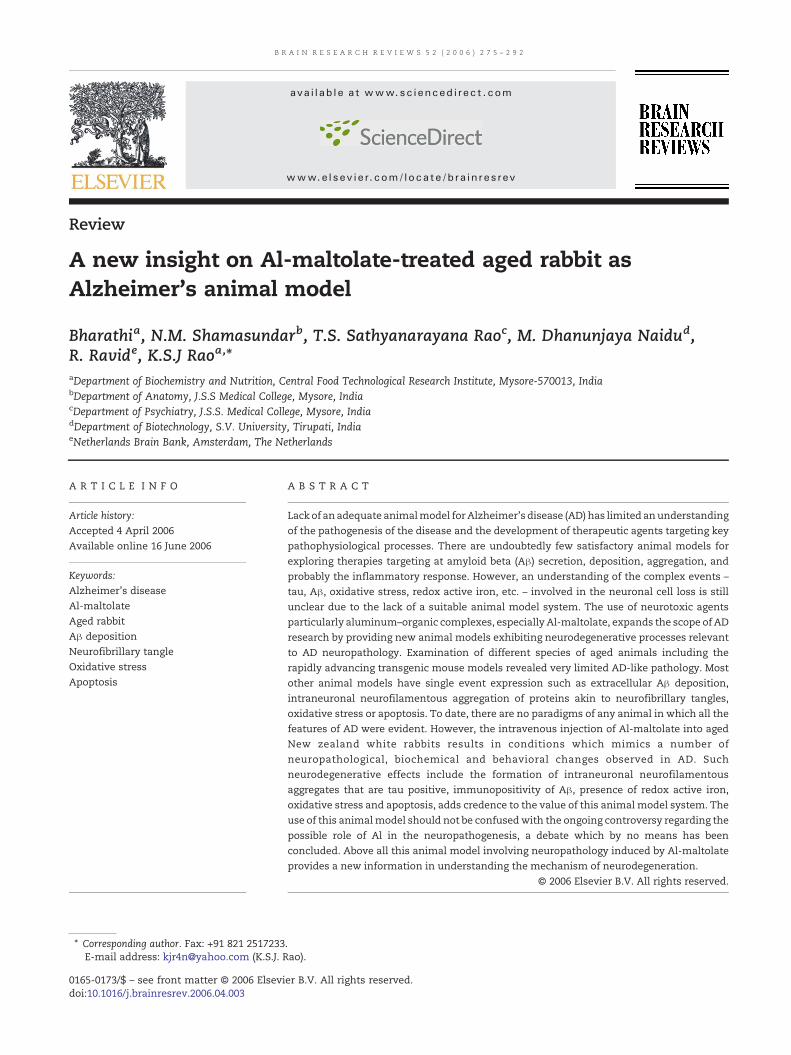
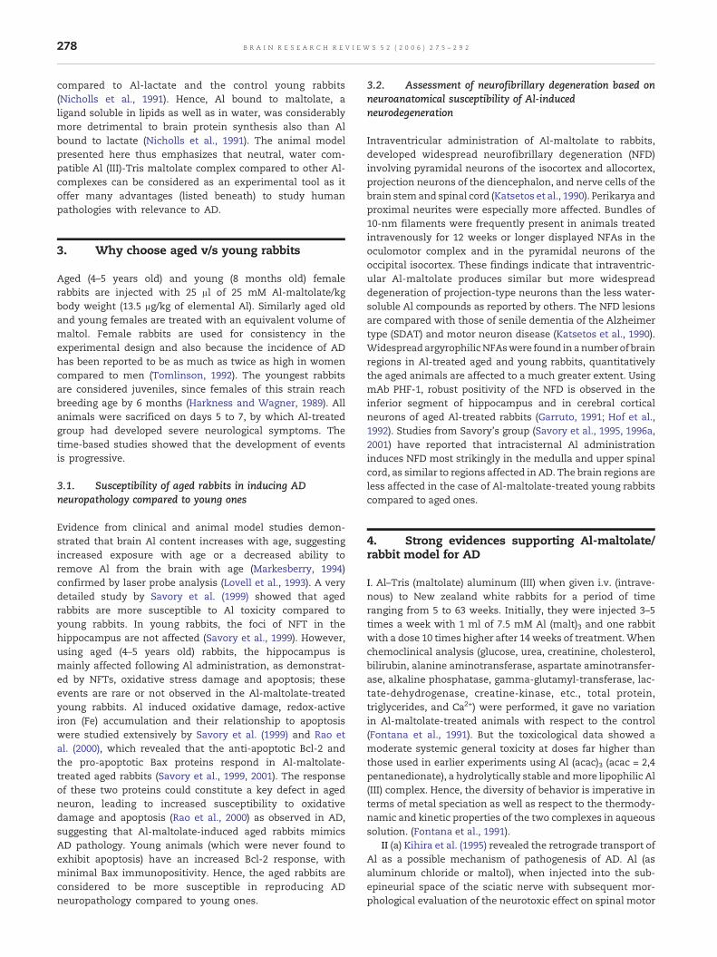
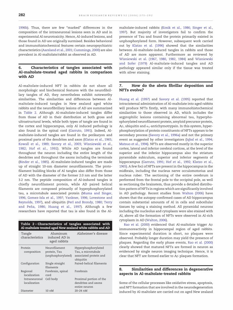
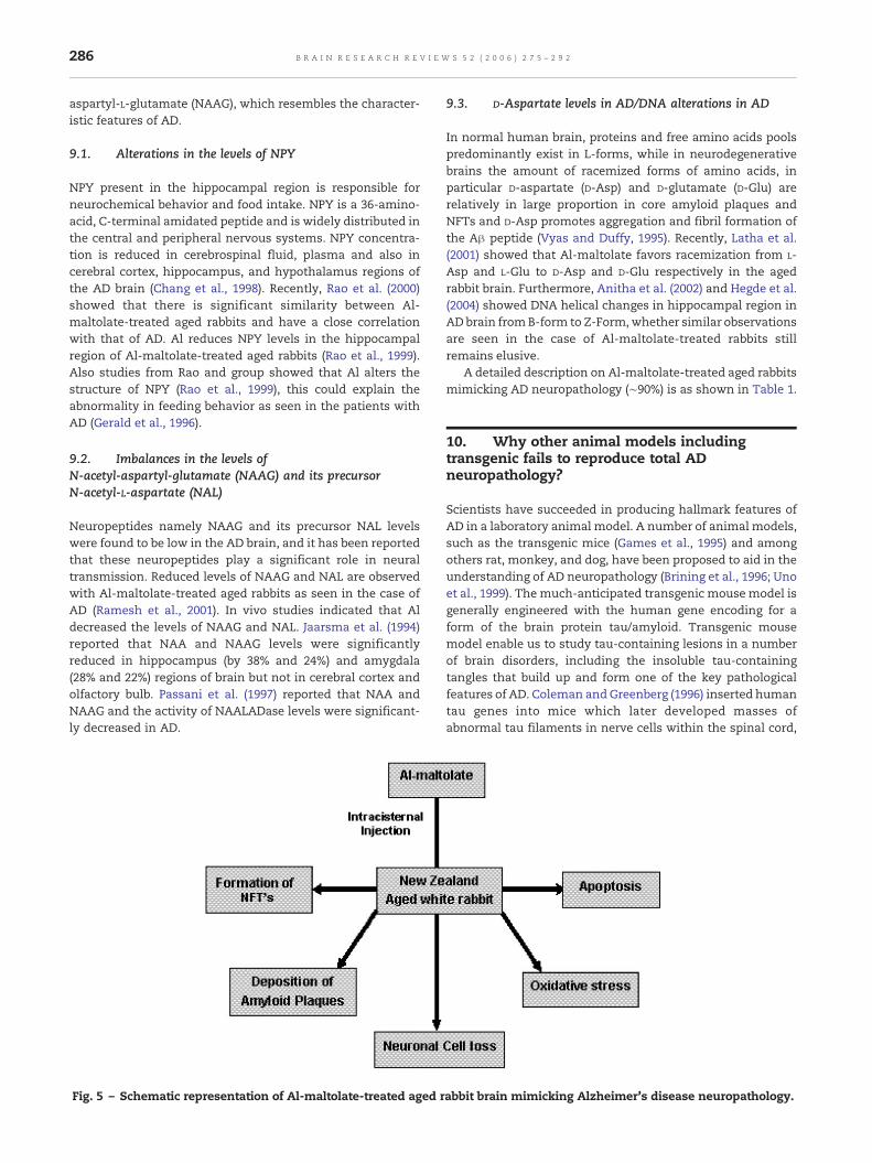
Fig. 1 – Represents the localization of Aβ (Vascular region)in the Al-maltolate-treated aged rabbits which is alsoobserved in the vascular region of demented people.
2. Al-maltolate as novel compound forinducing AD like pathology in aged rabbits
Although Al, still remains as a mystery, even after manydecades of research because of its intrinsic difficulties inunderstanding the role of chemical speciation in biologicalsystems. Hence, to understand the mechanism of Alinduced neuropathology, the selection of an appropriate Alcompound is important. Scientists have employed theelectroneutral Al-maltolate (Al(mal)3) complex (Bertholf etal., 1987) on experimental animals since this compound candeliver a significant amount of free aqueous Al at physio-logical pH (Martin, 1986). In contrast, most other Al salts,such as AlCl3, produce insoluble complexes at neutral pH(Martin, 1986). The uniqueness of Al-maltolate compound isthat this Al-complex increases the soluble Al concentration
from 4–6 mM compared to other organic Al salts like Al-lactate or Al-aspartate (soluble Al concentration is ∼55–330 μM). Al-maltolate is soluble from pH 3.0 to 10.0,possesses hydrolytic stability at pH 7.0, and does not havespeciation chemistry problems (Martin, 1986). Al-maltolate ispreferred over other Al compounds because of its followingproperties: (a) very high metal solubility at pH 7.0, (b)prominent kinetic restrictions to ligand exchange reactionsin neutral solution (Corain et al., 1994; Finneagan et al.,1986), hence suitable for toxicological studies and also tounderstand the neuropathology.
Administration of different Al compounds to a certainextent induces AD neuropathology, but compared to othercompounds, Al-maltolate seems to be more effective. Toresolve some of the questions related to the designing of theanimalmodel, investigators have studied a variety of Al salts –Al lactate, AlCl3 (He and Strong, 2000), AlF, and AlSiO4 (Garrutoet al., 1984) – on aged rabbits. Certain Al-organic and Al-inorganiccomplexesadministration todifferentanimalgroupslike cats, ferrets, and dogs also did not mimic the ADneuropathology, though the NFAs were prominent in the CA1of the hippocampus, the subiculum cortex and in theposterior cingulated gyrus (Strong et al., 1991a; Strong andGarruto, 1991b; Wakayama et al., 1993). Whereas in case ofAl-maltolate-treated aged rabbits NFAs formation or pairedhelical filaments (PHF) were observed in the axons imagedin hippocampal neurons (Fig. 1) (Maccioni and Cambiazo,1995; Geula et al., 1998; Rao et al., 2000). Studies fromNicholls et al. (1991) also support this concept that Al-maltolate is comparatively more efficient than the other Al-complexes. They reported that when young rabbits (infants)were given s.c. (subcutaneous) injections 3 times weekly oflow doses of Al-maltolate (0.5–1.5 mg Al/kg body wt) or Al-lactate (8 mg Al/kg body weight) from 5 or 10 days of age to14 or 22 days of age. The cell-free protein synthesizingsystem in the brain exhibited increased activity in Alexposed infants. The mRNA fraction obtained from thebrain polysomal RNA were more active in Al exposed
278 B R A I N R E S E A R C H R E V I E W S 5 2 ( 2 0 0 6 ) 2 7 5 – 2 9 2
compared to Al-lactate and the control young rabbits(Nicholls et al., 1991). Hence, Al bound to maltolate, aligand soluble in lipids as well as in water, was considerablymore detrimental to brain protein synthesis also than Albound to lactate (Nicholls et al., 1991). The animal modelpresented here thus emphasizes that neutral, water com-patible Al (III)-Tris maltolate complex compared to other Al-complexes can be considered as an experimental tool as itoffer many advantages (listed beneath) to study humanpathologies with relevance to AD.
3. Why choose aged v/s young rabbits
Aged (4–5 years old) and young (8 months old) femalerabbits are injected with 25 μl of 25 mM Al-maltolate/kgbody weight (13.5 μg/kg of elemental Al). Similarly aged oldand young females are treated with an equivalent volume ofmaltol. Female rabbits are used for consistency in theexperimental design and also because the incidence of ADhas been reported to be as much as twice as high in womencompared to men (Tomlinson, 1992). The youngest rabbitsare considered juveniles, since females of this strain reachbreeding age by 6 months (Harkness and Wagner, 1989). Allanimals were sacrificed on days 5 to 7, by which Al-treatedgroup had developed severe neurological symptoms. Thetime-based studies showed that the development of eventsis progressive.
3.1. Susceptibility of aged rabbits in inducing ADneuropathology compared to young ones
Evidence from clinical and animal model studies demon-strated that brain Al content increases with age, suggestingincreased exposure with age or a decreased ability toremove Al from the brain with age (Markesberry, 1994)confirmed by laser probe analysis (Lovell et al., 1993). A verydetailed study by Savory et al. (1999) showed that agedrabbits are more susceptible to Al toxicity compared toyoung rabbits. In young rabbits, the foci of NFT in thehippocampus are not affected (Savory et al., 1999). However,using aged (4–5 years old) rabbits, the hippocampus ismainly affected following Al administration, as demonstrat-ed by NFTs, oxidative stress damage and apoptosis; theseevents are rare or not observed in the Al-maltolate-treatedyoung rabbits. Al induced oxidative damage, redox-activeiron (Fe) accumulation and their relationship to apoptosiswere studied extensively by Savory et al. (1999) and Rao etal. (2000), which revealed that the anti-apoptotic Bcl-2 andthe pro-apoptotic Bax proteins respond in Al-maltolate-treated aged rabbits (Savory et al., 1999, 2001). The responseof these two proteins could constitute a key defect in agedneuron, leading to increased susceptibility to oxidativedamage and apoptosis (Rao et al., 2000) as observed in AD,suggesting that Al-maltolate-induced aged rabbits mimicsAD pathology. Young animals (which were never found toexhibit apoptosis) have an increased Bcl-2 response, withminimal Bax immunopositivity. Hence, the aged rabbits areconsidered to be more susceptible in reproducing ADneuropathology compared to young ones.
3.2. Assessment of neurofibrillary degeneration based onneuroanatomical susceptibility of Al-inducedneurodegeneration
Intraventricular administration of Al-maltolate to rabbits,developed widespread neurofibrillary degeneration (NFD)involving pyramidal neurons of the isocortex and allocortex,projection neurons of the diencephalon, and nerve cells of thebrain stemand spinal cord (Katsetos et al., 1990). Perikarya andproximal neurites were especially more affected. Bundles of10-nm filaments were frequently present in animals treatedintravenously for 12 weeks or longer displayed NFAs in theoculomotor complex and in the pyramidal neurons of theoccipital isocortex. These findings indicate that intraventric-ular Al-maltolate produces similar but more widespreaddegeneration of projection-type neurons than the less water-soluble Al compounds as reported by others. The NFD lesionsare compared with those of senile dementia of the Alzheimertype (SDAT) and motor neuron disease (Katsetos et al., 1990).Widespread argyrophilicNFAswere found in anumberof brainregions in Al-treated aged and young rabbits, quantitativelythe aged animals are affected to a much greater extent. UsingmAb PHF-1, robust positivity of the NFD is observed in theinferior segment of hippocampus and in cerebral corticalneurons of aged Al-treated rabbits (Garruto, 1991; Hof et al.,1992). Studies from Savory's group (Savory et al., 1995, 1996a,2001) have reported that intracisternal Al administrationinduces NFD most strikingly in the medulla and upper spinalcord, as similar to regions affected in AD. The brain regions areless affected in the case of Al-maltolate-treated young rabbitscompared to aged ones.
4. Strong evidences supporting Al-maltolate/rabbit model for AD
I. Al–Tris (maltolate) aluminum (III) when given i.v. (intrave-nous) to New zealand white rabbits for a period of timeranging from 5 to 63 weeks. Initially, they were injected 3–5times a week with 1 ml of 7.5 mM Al (malt)3 and one rabbitwith a dose 10 times higher after 14 weeks of treatment.Whenchemoclinical analysis (glucose, urea, creatinine, cholesterol,bilirubin, alanine aminotransferase, aspartate aminotransfer-ase, alkaline phosphatase, gamma-glutamyl-transferase, lac-tate-dehydrogenase, creatine-kinase, etc., total protein,triglycerides, and Ca2+) were performed, it gave no variationin Al-maltolate-treated animals with respect to the control(Fontana et al., 1991). But the toxicological data showed amoderate systemic general toxicity at doses far higher thanthose used in earlier experiments using Al (acac)3 (acac = 2,4pentanedionate), a hydrolytically stable andmore lipophilic Al(III) complex. Hence, the diversity of behavior is imperative interms of metal speciation as well as respect to the thermody-namic and kinetic properties of the two complexes in aqueoussolution. (Fontana et al., 1991).
II (a) Kihira et al. (1995) revealed the retrograde transport ofAl as a possible mechanism of pathogenesis of AD. Al (asaluminum chloride or maltol), when injected into the sub-epineurial space of the sciatic nerve with subsequent mor-phological evaluation of the neurotoxic effect on spinal motor
279B R A I N R E S E A R C H R E V I E W S 5 2 ( 2 0 0 6 ) 2 7 5 – 2 9 2
neurons in rabbits—spheroid/globules, and peripheral chro-matolysis, and neuronal degeneration were observed in thespinal anterior horn of Al-maltolate-treated aged rabbits. Thesoma and dendrites of neurons in the anterior horn of Al-treated rabbit showed marked edematous change, fragmen-tation of granular endoplasmic reticulum, increased accumu-lation of neurofilament, and accumulation of free ribosomesand lipid-droplet-like structures.
(b) Theabove findings indicate that the retrograde transportof Al into spinal motor neurons via the peripheral nervoussystem may exacerbate neuronal degeneration in ALS immu-nohistochemically in adult New zealand white rabbits afterintraventricular (subacute) and intravenous (chronic) admin-istration of a water-soluble aluminum compound, Al-malto-late (Katsetos et al., 1990; Kihira et al., 1995; Liwincz et al., 1974).
III. Al-maltolate induces cytochrome c translocation intothe cytosol as early as 3 h in aged but not in young rabbithippocampus. Pretreatment with cyclosporin A, an inhibitorof the mitochondria permeability transition pore (MTP),blocks cytochrome c release. Therefore, it appears thataluminum maltolate-induced cytochrome c release resultsfrom opening of the MTP. This effect implicates aging as aprerequisite factor, since the MTP does not open in younganimals. Mitochondrial injury thus may represent a primaryinitiator of neurodegeneration (Ghribi et al., 2001c). Recentreports by Ghribi et al. (2002b) showed that pretreatment ofAl-maltolate for 14 days with 7 mm of lithium carbonate indrinking water prevents aluminum-induced translocation ofcytochrome c and upregulates Bcl-2 and Bcl-X(L), down-regulates Bax, abolishes caspase-3 activity, and reduces DNAdamage. The regulatory effect of lithium on the apoptosis-controlling proteins occurs in both the mitochondria andendoplasmic reticulum (ER) and inhibits the Aβ inducedstress in ER of rabbit hippocampus (Ghribi et al., 2003). Wepropose that the neuroprotective effect of lithium involvesthe modulation of apoptosis-regulatory proteins present inthe subcellular organelles of rabbit brain (Ghribi et al., 2002b,2003).
IV. Ghribi et al. (2002a) put forth that rabbits treatedintracisternally with Al-maltolate had higher levels of pro-caspase in the cytosolic fractions, whereas p17, the activecaspase-3 localized in the ER. This distribution was supportedimmunohistochemically for the colocalization of p17 withcalnexin, a specific marker of the ER, these observation are inaccordance with the biochemical changes seen in AD patients.
V. Garruto et al. (1988) carried out imaging of Al in NFT-bearing neurons within sommer's sector of the hippocampusin Guamanian patients, using a method of computer-con-trolled electron beam X-ray micro-analysis and wavelengthdispersive spectrometry. Al was distributed in cell bodies andaxonal processes of NFT-bearing neurons. The elementalimages showed that Al deposits occur within the same NFT-bearing hippocampal neuron, suggesting this element in-volvement in NFT formation. No prominent concentrations ofAl were imaged in non-NFT-containing regions within thepyramidal cell layer compared to control cases.
VI. The extraordinary work carried out by Savory et al.(1993) on the quantitation of Al in the brain and spinal cordand its effects on neurofilament protein expression andphosphorylation gave a new proof for the involvement of Al
in AD. When Al-maltolate was treated to aged rabbits,decreasing concentration was observed (∼10 μg/g dry tissue)in the brain and spinal cord, whereas in lumbar cord (∼2.1 μg/gdry tissue), argyrophilic tangles were observed in perikaryaand proximal neurites of neurons as far distal as the lumbarand sacral cord areas (Savory et al., 1993). Immunoblot studiesfailed to detect changes in three neurofilament proteinisoforms, and also no significant alterations in the totalphosphate content of these proteins were observed, thegenes encoding for the 200-kDa and 68-kDa neurofilamentprotein also were unaffected on Al-maltolate treatment(Savory et al., 1993).
VII. Studies were carried out on neuronal culture systemby Hewitt et al. (1991) to evaluate the neurotoxic effects ofAl-maltolate on rabbit fetal midbrain sections containing theoculomotor nucleus. Cultures were treated with 5, 7, 9, 11,13, and 15 μmol/l Al-maltolate, or 39 and 45 μmol/l maltol(molal equivalents to 13 and 15 μmol/l Al-maltolate), at thesame control cultures were maintained. The number oftangles produced in Al-maltolate-treated cultures wascounted and compared to untreated controls, a total of 7%of neurons following treatment with 11, 13, 15 μmol/l Al-maltolate respectively and none in the controls. Immuno-histochemical studies show that NFTs were immunoreactivewith MAbs to phosphorylated (SMI-31), nonphosphorylated,phosphorylation dependent (SMI-32) and phosphorylationindependent (SMI-33) epitopes of the high (–H) and middle(–M) molecular weight neurofilament subunits (NF-H/M). Bycontrast these lesions were nonreactive with MAbs recog-nizing tau, MAP2 or different beta-tubulin isotypes (Mumaand Singer, 1996; Hewitt et al., 1991). The above neurocy-toskeletal changes observed as seen similar to AD may aid inthe assessment of the possible role of Al in the etiology ofAD.
VIII. AD is associated with changes affecting numerousneurotransmitter systems (Nordberg, 1992). Of the systemsaffected in AD, the cholinergic system shows the greatestchanges demonstrating a decrease in high affinity cholineuptake (HACU); a deficit in activity of acetylcholinesterase(AChE) (Dai et al., 2002); choline acetyl transferase (ChAT)(Gibson and Peterson, 1981); and a decrease in acetylcholine(Ach) concentrations (Slotkin et al., 1990); monoamines, andtheir precursors. These cholinergic changes are observed infrontal and temporal cortex of postmortem brains of AD(Langlais et al., 1993). It has been hypothesized that to acertain extent cholinergic abnormalities might contribute tocognitive decline in AD (Terry and Buccafusco, 2003; Bartus,2000).
Studies of the cholinergic system of animals showed anAl-induced decrease in HACU by rat synaptosomes (Lai et al.,1980). It has been shown that AChE decline in mouse brain(Zatta et al., 2002), increase in monoamine oxidase activityin rat brain (Zatta et al., 1999), and decrease in ChAT activityin rabbit brain (Hofstetter et al., 1987). Neurotransmittersystem changes in the Al-intoxicated rabbit model mimicthose seen in AD (Beal et al., 1989). Lavond et al. (1993)reported that there is an interlink between the Ach overflowand CRs. Al-treated rabbits showed a delay in conditionedeyeblink acquisition and greatly attenuated Ach overflow(Yokel et al., 1994). Thus Al-induced attenuation of Ach
280 B R A I N R E S E A R C H R E V I E W S 5 2 ( 2 0 0 6 ) 2 7 5 – 2 9 2
overflow might in turn contribute to the Al-induced learningdeficit which mimics the similar observations made in AD(Gron et al., 2005; Yokel et al., 1994). Furthermore, theneurotransmitter alterations which in turn accompany Alneurofibrillary degeneration play a significant role in medi-ating long term potentiation, a synaptic model of learning(Yokel et al., 1994). There was a significant reduction inacetylcholinesterase activity in entorhinal cortex and hippo-campus as well as significant reductions in cortical concen-trations of serotonin and norepinephrine in the Al-treatedrabbits (Nordberg, 1992). Significant reductions in glutamate,aspartate, and taurine were found in frontoparietal andposterior parietal cortex. However, the concentrations ofgamma-amino butyric acid were unchanged in cerebralcortex. Both substance P and cholecystokinin immunoreac-tivity were significantly reduced in entorhinal cortex, butthere were no significant changes in somatostatin andvasoactive intestinal polypeptide in Al-treated rabbit (Nord-berg, 1992; Yokel et al., 1994). These findings show aparallence analogous between Al-treated rabbits to AD interms of neurotransmitter changes.
IX. Inflammation is found to be a key player in the onset ofneurodegeneration in AD. In AD patients, there is anupregulation of pro-inflammatory genes (McGeer andMcGeer, 1999; Colangelo et al., 2002), and levels of cytokineslike interleukin-1 (IL-1), IL-1β, IL-6, tumor necrosis factor andneurotrophins were elevated in the microglia (Zhao et al.,2003) as well as in cerebrospinal fluid and plasma (Sun et al.,2003). There are few significant reports on the role of Al inneuroinflammation. Campbell et al. (2002) have reported thatAl increases cell proliferation, cytokine secretion, and nuclearfactor-κB (NF-κB) activation in human glioblastoma cells(Campbell et al., 2002), interleukin-1β precursor, cytosolicphospholipase A2 (Lukiw et al., 2005). Further, inflammationis always correlated with the duration of exposure and theamount of Al accumulated (Dale et al., 1991). There is limiteddirect evidence of Al induced inflammatory events in theCNS. Glial fibrillary acidic protein (GFAP) has been shown tobe associated with gliosis, a generic response of the CNS toneural injury. Further Tsunoda and Sharma (1999) reportedthat the level of tumor necrosis factor-α, another cytokineimplicated in neuronal damage, was significantly increasedin the cerebrum of mice exposed to Al compared withcontrols. (Demircan et al., 1998). Yokel and O'Callaghan(1998) reported that Al increases the frontal cortical GFAPincreased (approximately twofold above control) in Al-treatedrabbits; whereas hippocampal and cerebellar GFAP concen-trations were not affected. Thus, Al-treated rabbits might actas a model system in reproducing AD related inflammatorychanges.
X. AD is characterized by impairment in working memory(Baddeley et al., 1991; Germano and Kinsella, 2005), visuo-perception, attention, semantic memory, and episodic mem-ory (Scahill et al., 2005; Hodges et al., 1990). To evaluate this,Starr et al. (2005) carried out a study on 9 AD patients (meanage 73.6) and 10 healthy control (mean age 71.8) subjects,who underwent an fMRI memory paradigm. Healthy controlsubjects activated the right parahippocampal gyrus, whereassubjects with AD activated the right superior frontal gyrusand left uncus. Further, Nordahl et al. (2005) reported that
mild cognitive impairment in AD defined as episodicmemory impairment is associated with hippocampal atro-phy. Hence, cognitive deficits associated with AD still needsto be operationalized both in human subjects and inanimals.
In case of Al-treated rabbits, there is a progressive declinein specific memory functions such as deficits in short-termmemory and response acquisition (Crapper and Dalton, 1973;Petit et al., 1980; Rabe et al., 1982). The neurobehavioraltoxicity studies of Al-treated rabbits were based on theprocedure developed by Gormezano (1996) to measurelearning and memory. Yokel et al. (1994) has made in-depth study on the Al-induced behavioral toxicity. Theystudied the rate of acquisition and the retention of theclassically conditioned (Pavlovian conditioning) eyeblinkreflex, measured as the extension of the third eyelid(nictitating membrane) was determined in a differentialand delayed conditioning procedure. A retention and extinc-tion session were studied for 10 days after the lastconditioning session by presentation of 100 trials of eachtone alone. Eyeblinks initiated within 550 ms after toneonset were considered conditioned responses (CRs). Thecomparison of the learning curves of rabbits exposed to Alearly in development reveals some initial Al-associatedimprovement in CR acquisition versus Al-induced inhibitionof CR acquisition in the adult and aged rabbits (Yokel et al.,1994). The Al-induced inhibition of CR acquisition seen inadult and aged, but not in younger rabbits, suggests that themature mammalian brain is more susceptible to Al-inducedneurobehavioral toxicity. Further, Clark and Squire (1998)suggested that the study of memory processing in thehippocampus might lead to insights about consciousness.They also suggested that hippocampal lesions do notprevent rabbits from learning to blink in response to atone when they were trained on a conventional protocol inwhich the tone begins before and then slightly overlaps withan air puff to the eye (Eichenbaum, 1999). But rabbits withhippocampal damage fail to learn on a variant of thisprotocol in which the tone and air puff are separated by ahalf-second ‘trace’ interval (Eichenbaum, 1999). (This gap isprobably too brief for rabbits to forget the tone, judging bythe short-term/working memory capacity retained byhumans with similar damage). The Al-treated rabbit can beacquainted as an animal model for studying behavioralfeatures particularly learning and memory in relevance toneurodegeneration.
XI. The first contribution on therapeutic potential ofchelation on Al was reported by Mclachlan et al. (1991).Further Janson (2001) reported that low dose of the injectabledesferrioxamine (DFO), to remove Al from the brain ofelderly patients were benefited to 50%. Savory et al. (1994)demonstrated the partial reversal of Al-induced neurofibril-lary degeneration by DFO in rabbits. Recently, Gong et al.(2005) showed the protective effects of Ginkgo biloba extracton Al-induced brain dysfunction. These findings indicate apartial reversal of Al-induced neurodegeneration by DFO andGinkgo. Further iron-specific chelator like deferiprone is alsofound to be effective in the chelation of Al (Janson, 2001).The chelation therapy of Al potentially indicates that therabbit model can be used for research on chelators.
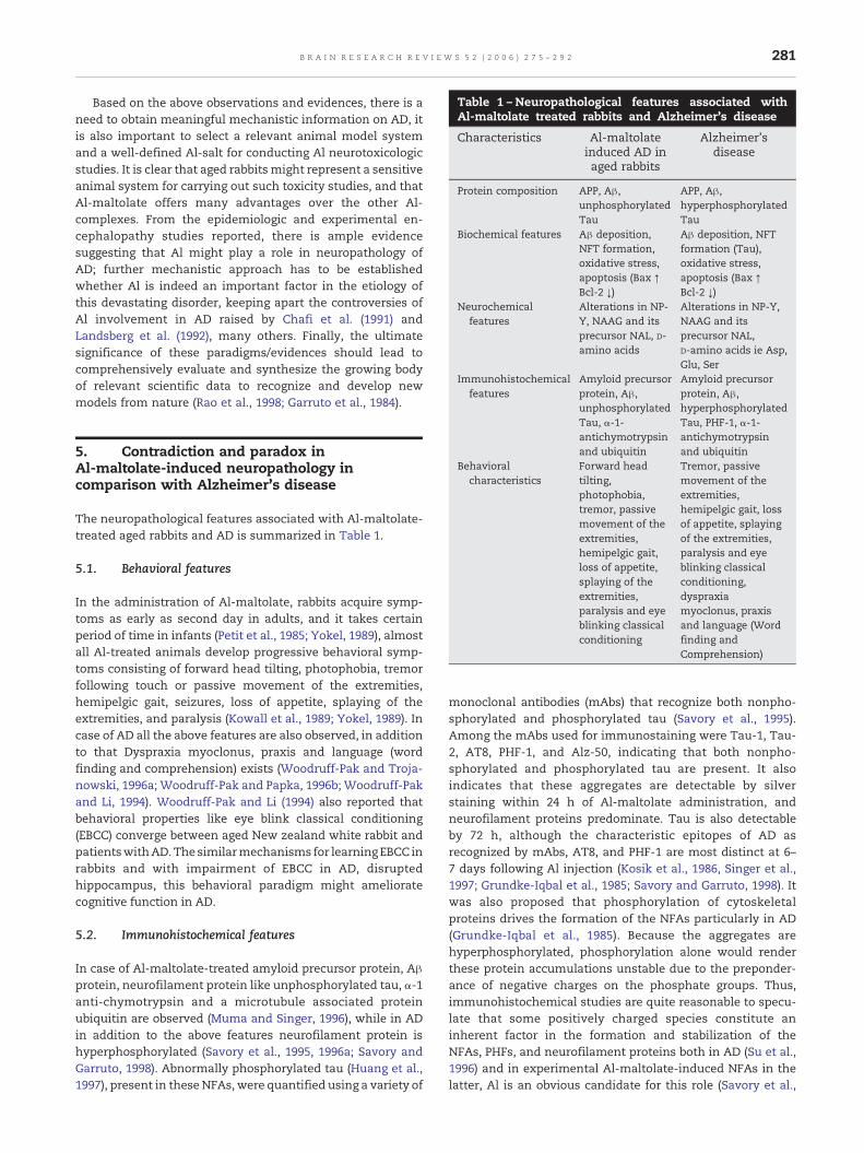
Table 1 – Neuropathological features associated withAl-maltolate treated rabbits and Alzheimer's disease
Characteristics Al-maltolateinduced AD inaged rabbits
Alzheimer'sdisease
Protein composition APP, Aβ,unphosphorylatedTau
APP, Aβ,hyperphosphorylatedTau
Biochemical features Aβ deposition,NFT formation,oxidative stress,apoptosis (Bax ↑Bcl-2 ↓)
Aβ deposition, NFTformation (Tau),oxidative stress,apoptosis (Bax ↑Bcl-2 ↓)
Neurochemicalfeatures
Alterations in NP-Y, NAAG and itsprecursor NAL, D-amino acids
Alterations in NP-Y,NAAG and itsprecursor NAL,D-amino acids ie Asp,Glu, Ser
Immunohistochemicalfeatures
Amyloid precursorprotein, Aβ,unphosphorylated
Amyloid precursorprotein, Aβ,hyperphosphorylated
281B R A I N R E S E A R C H R E V I E W S 5 2 ( 2 0 0 6 ) 2 7 5 – 2 9 2
Based on the above observations and evidences, there is aneed to obtain meaningful mechanistic information on AD, itis also important to select a relevant animal model systemand a well-defined Al-salt for conducting Al neurotoxicologicstudies. It is clear that aged rabbitsmight represent a sensitiveanimal system for carrying out such toxicity studies, and thatAl-maltolate offers many advantages over the other Al-complexes. From the epidemiologic and experimental en-cephalopathy studies reported, there is ample evidencesuggesting that Al might play a role in neuropathology ofAD; further mechanistic approach has to be establishedwhether Al is indeed an important factor in the etiology ofthis devastating disorder, keeping apart the controversies ofAl involvement in AD raised by Chafi et al. (1991) andLandsberg et al. (1992), many others. Finally, the ultimatesignificance of these paradigms/evidences should lead tocomprehensively evaluate and synthesize the growing bodyof relevant scientific data to recognize and develop newmodels from nature (Rao et al., 1998; Garruto et al., 1984).
Tau, α-1-antichymotrypsinand ubiquitin
Tau, PHF-1, α-1-antichymotrypsinand ubiquitin
Behavioralcharacteristics
Forward headtilting,photophobia,tremor, passivemovement of theextremities,hemipelgic gait,loss of appetite,splaying of theextremities,paralysis and eyeblinking classicalconditioning
Tremor, passivemovement of theextremities,hemipelgic gait, lossof appetite, splayingof the extremities,paralysis and eyeblinking classicalconditioning,dyspraxiamyoclonus, praxisand language (Wordfinding andComprehension)
5. Contradiction and paradox inAl-maltolate-induced neuropathology incomparison with Alzheimer's disease
The neuropathological features associated with Al-maltolate-treated aged rabbits and AD is summarized in Table 1.
5.1. Behavioral features
In the administration of Al-maltolate, rabbits acquire symp-toms as early as second day in adults, and it takes certainperiod of time in infants (Petit et al., 1985; Yokel, 1989), almostall Al-treated animals develop progressive behavioral symp-toms consisting of forward head tilting, photophobia, tremorfollowing touch or passive movement of the extremities,hemipelgic gait, seizures, loss of appetite, splaying of theextremities, and paralysis (Kowall et al., 1989; Yokel, 1989). Incase of AD all the above features are also observed, in additionto that Dyspraxia myoclonus, praxis and language (wordfinding and comprehension) exists (Woodruff-Pak and Troja-nowski, 1996a; Woodruff-Pak and Papka, 1996b; Woodruff-Pakand Li, 1994). Woodruff-Pak and Li (1994) also reported thatbehavioral properties like eye blink classical conditioning(EBCC) converge between aged New zealand white rabbit andpatientswithAD.The similarmechanisms for learningEBCC inrabbits and with impairment of EBCC in AD, disruptedhippocampus, this behavioral paradigm might amelioratecognitive function in AD.
5.2. Immunohistochemical features
In case of Al-maltolate-treated amyloid precursor protein, Aβprotein, neurofilament protein like unphosphorylated tau, α-1anti-chymotrypsin and a microtubule associated proteinubiquitin are observed (Muma and Singer, 1996), while in ADin addition to the above features neurofilament protein ishyperphosphorylated (Savory et al., 1995, 1996a; Savory andGarruto, 1998). Abnormally phosphorylated tau (Huang et al.,1997), present in theseNFAs, were quantified using a variety of
monoclonal antibodies (mAbs) that recognize both nonpho-sphorylated and phosphorylated tau (Savory et al., 1995).Among the mAbs used for immunostaining were Tau-1, Tau-2, AT8, PHF-1, and Alz-50, indicating that both nonpho-sphorylated and phosphorylated tau are present. It alsoindicates that these aggregates are detectable by silverstaining within 24 h of Al-maltolate administration, andneurofilament proteins predominate. Tau is also detectableby 72 h, although the characteristic epitopes of AD asrecognized by mAbs, AT8, and PHF-1 are most distinct at 6–7 days following Al injection (Kosik et al., 1986, Singer et al.,1997; Grundke-Iqbal et al., 1985; Savory and Garruto, 1998). Itwas also proposed that phosphorylation of cytoskeletalproteins drives the formation of the NFAs particularly in AD(Grundke-Iqbal et al., 1985). Because the aggregates arehyperphosphorylated, phosphorylation alone would renderthese protein accumulations unstable due to the preponder-ance of negative charges on the phosphate groups. Thus,immunohistochemical studies are quite reasonable to specu-late that some positively charged species constitute aninherent factor in the formation and stabilization of theNFAs, PHFs, and neurofilament proteins both in AD (Su et al.,1996) and in experimental Al-maltolate-induced NFAs in thelatter, Al is an obvious candidate for this role (Savory et al.,
282 B R A I N R E S E A R C H R E V I E W S 5 2 ( 2 0 0 6 ) 2 7 5 – 2 9 2
1996b). Thus, there are few “marked” differences in thecomposition of the intraneuronal lesions seen in AD and inexperimental Al neurotoxicity. Hence, Al-induced lesions, andthose found in AD are originally surmised. Besides behavioraland immunohistochemical features certain neuropsychiatriccharacteristics (Aarsland et al., 2001; Cummings, 2000) are alsoprevalent in Al-maltolate/rabbit as observed in AD.
6. Characteristics of tangles associated withAl-maltolate-treated aged rabbits in comparisonwith AD
Al-maltolate-induced NFT in rabbits do not share allmorphologic and biochemical features with the neurofibril-lary tangles of AD, they nevertheless exhibit noteworthysimilarities. The similarities and differences between Al-maltolate-induced tangles in New zealand aged whiterabbits and the neurofibrillary lesions of AD are summarizedin Table 2. Although Al-maltolate-induced tangles differfrom those of AD in their distribution at both gross andultrastructural levels, while both types of tangle are found inthe cortex and hippocampus, only Al induced pathology isalso found in the spinal cord (Garruto, 1991). Indeed, Al-maltolate-induced tangles are found in the perikaryon andproximal parts of the dendrites and axon (Klatzo et al., 1965;Kowall et al., 1989; Savory et al., 2003; Wisniewski et al.,1982; Hof et al., 1992). While AD tangles are foundthroughout the neuron including the entire length of thedendrites and throughout the axons including the terminals(Binder et al., 1985). Al-maltolate-induced tangles are madeup of straight 10-nm diameter neurofilaments. The proto-filament building blocks of Al tangles also differ from thoseof AD with the diameter of the former 2.0 nm and the latter3.2 nm. The peptide composition of Al-induced tangles ischiefly neurofilament protein, while AD paired helicalfilaments are composed primarily of hyperphosphorylatedtau, a microtubule associated protein (Muma and Singer,1996; Gomez-Isla et al., 1997; Yankner, 1996; Lovestone andReynolds, 1997), and ubiquitin (Perl and Brondy, 1980; Terryand Peña, 1986; Huang et al., 1997). Although a fewresearchers have reported that tau is also found in the Al-
Table 2 – Characteristics of tangles associated withAl-maltolate treated agedNewzealandwhite rabbits andAD
Tanglecharacteristics
Aluminuminduced AD inaged rabbits
Alzheimer's disease
Proteincomposition
Neurofilamentprotein, Tau(unphosphorylated)
HyperphosphorylatedTau, a microtubuleassociated protein andubiquitin
Configuration Single straightfilaments
Paired-helical filaments
Regionallocalization
Forebrain, spinalcord
Forebrain
Intraneuronallocalization
Cell body Proximal portion of thedendrites and axonsentire neuron
Diameter 10 nM 20–24 nM
maltolate-induced rabbits (Kosik et al., 1986; Singer et al.,1997). But majority of investigators fail to confirm thepresence of Tau and found the protein primarily existed inunphosphorylated form. However, subsequent work carriedout by Klatzo et al. (1996) showed that the similaritiesbetween Al-maltolate-induced tangles in rabbits and thoseof AD are more apparent. Furthermore as reviewed byWisniewski et al. (1967, 1980, 1982, 1984) and Wisniewskiand Sofer (1979) Al-maltolate-induced tangles and ADpathology appeared similar only if the tissue was treatedwith silver staining.
7. How do the abeta fibrillar deposition andNFTs evolve?
Huang et al. (1997) and Savory et al. (1995) reported thatintracisternal administration of Al-maltolate into aged rabbitswill produce NFTs firstly, with many immunohistochemicalsimilarities to those observed in AD, which includes theargyrophilic lesions containing abnormal tau, hyperpho-sphorylatedneurofilament protein, amyloid precursor protein,Aβ, ubiquitin and α1-antichymotrypsin. In this system, hyper-phosphorylation of protein constituents of NFTs appears to besecondary process (Savory et al., 1996a) and not the primaryevent as suggested by other investigators (Iqbal et al., 1994;Matsuo et al., 1994). NFTs are observed mostly in the superiorcortex, lateral and inferior cerebral cortices, at the level of thesuperior and the inferior hippocampus also the striatumpyramidale subiculum, superior and inferior segments ofhippocampus (Garruto, 1991; Hof et al., 1992; Klatzo et al.,1965). A few foci of NFTs are present in the hippocampus in themidbrain, including the nucleus nerve occulomotorius andnucleus ruber. The sectioning of the entire cerebrum isperformed from the frontal pole to the occipital pole, as wellas sectioning the brainstem, thus provide a detailed distribu-tionpattern ofNFTs in regionswhich are significantly involvedin AD pathology. Recent studies from Walton (2006) haveshown that the autopsy-confirmed cases of AD hippocampuscontain substantial amounts of Al in cells and subcellulartissues by using a staining method. All pyramidal neuronsincluding the nucleolus and cytoplasmwere also stained withAl, above all the formation of NFTs were observed in Al-richcytoplasm in AD (Walton, 2006).
Rao et al. (2000) evidenced that Al-maltolate trigger Aβimmunoreactivity in hippocampal region of aged rabbits.Since experimental duration is short, no plaques wereobserved. Probably longer duration may yield the presence ofplaques. Regarding the early phase events, Rao et al. (2000)clearly showed that matured NFTs are formed in neuron asevidenced by single neuron imaging technique. Hence, it isclear that NFT are formed earlier to Aβ plaques formation.
8. Similarities and differences in degenerativeaspects in Al-maltolate-treated rabbits
Some of the cellular processes like oxidative stress, apoptosis,andNFT formation that are involved in the neurodegenerationinduced by Al-maltolate are carried out on aged New zealand
283B R A I N R E S E A R C H R E V I E W S 5 2 ( 2 0 0 6 ) 2 7 5 – 2 9 2
white rabbits through intravenous administration. Based onthe recent literature, data available on the Al-maltolate-induced neuropathology in relevance to AD have focused onthe neuronal injury resulting in the understanding of neuro-pathogenesis in relevance to AD.
8.1. Oxidative stress
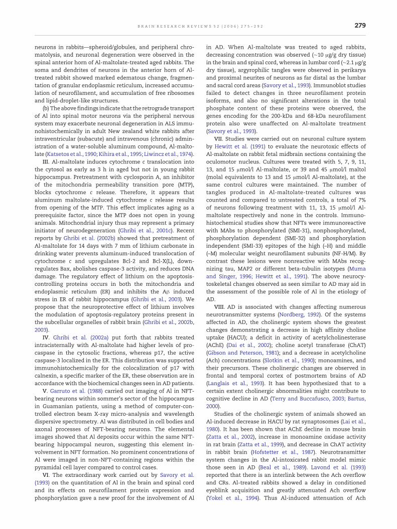
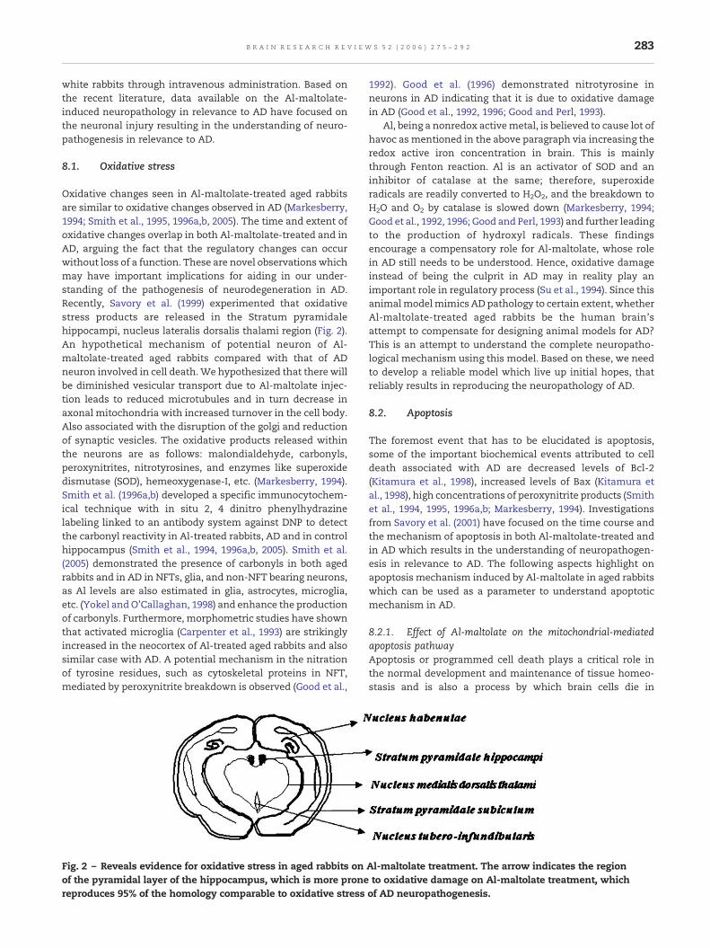
Oxidative changes seen in Al-maltolate-treated aged rabbitsare similar to oxidative changes observed in AD (Markesberry,1994; Smith et al., 1995, 1996a,b, 2005). The time and extent ofoxidative changes overlap in both Al-maltolate-treated and inAD, arguing the fact that the regulatory changes can occurwithout loss of a function. These are novel observations whichmay have important implications for aiding in our under-standing of the pathogenesis of neurodegeneration in AD.Recently, Savory et al. (1999) experimented that oxidativestress products are released in the Stratum pyramidalehippocampi, nucleus lateralis dorsalis thalami region (Fig. 2).An hypothetical mechanism of potential neuron of Al-maltolate-treated aged rabbits compared with that of ADneuron involved in cell death.We hypothesized that there willbe diminished vesicular transport due to Al-maltolate injec-tion leads to reduced microtubules and in turn decrease inaxonal mitochondria with increased turnover in the cell body.Also associated with the disruption of the golgi and reductionof synaptic vesicles. The oxidative products released withinthe neurons are as follows: malondialdehyde, carbonyls,peroxynitrites, nitrotyrosines, and enzymes like superoxidedismutase (SOD), hemeoxygenase-I, etc. (Markesberry, 1994).Smith et al. (1996a,b) developed a specific immunocytochem-ical technique with in situ 2, 4 dinitro phenylhydrazinelabeling linked to an antibody system against DNP to detectthe carbonyl reactivity in Al-treated rabbits, AD and in controlhippocampus (Smith et al., 1994, 1996a,b, 2005). Smith et al.(2005) demonstrated the presence of carbonyls in both agedrabbits and in AD in NFTs, glia, and non-NFT bearing neurons,as Al levels are also estimated in glia, astrocytes, microglia,etc. (Yokel andO'Callaghan, 1998) and enhance the productionof carbonyls. Furthermore, morphometric studies have shownthat activated microglia (Carpenter et al., 1993) are strikinglyincreased in the neocortex of Al-treated aged rabbits and alsosimilar case with AD. A potential mechanism in the nitrationof tyrosine residues, such as cytoskeletal proteins in NFT,mediated by peroxynitrite breakdown is observed (Good et al.,
Fig. 2 – Reveals evidence for oxidative stress in aged rabbits onof the pyramidal layer of the hippocampus, which is more pronereproduces 95% of the homology comparable to oxidative stress
1992). Good et al. (1996) demonstrated nitrotyrosine inneurons in AD indicating that it is due to oxidative damagein AD (Good et al., 1992, 1996; Good and Perl, 1993).
Al, being a nonredox activemetal, is believed to cause lot ofhavoc as mentioned in the above paragraph via increasing theredox active iron concentration in brain. This is mainlythrough Fenton reaction. Al is an activator of SOD and aninhibitor of catalase at the same; therefore, superoxideradicals are readily converted to H2O2, and the breakdown toH2O and O2 by catalase is slowed down (Markesberry, 1994;Good et al., 1992, 1996; Good and Perl, 1993) and further leadingto the production of hydroxyl radicals. These findingsencourage a compensatory role for Al-maltolate, whose rolein AD still needs to be understood. Hence, oxidative damageinstead of being the culprit in AD may in reality play animportant role in regulatory process (Su et al., 1994). Since thisanimalmodelmimics AD pathology to certain extent, whetherAl-maltolate-treated aged rabbits be the human brain'sattempt to compensate for designing animal models for AD?This is an attempt to understand the complete neuropatho-logical mechanism using this model. Based on these, we needto develop a reliable model which live up initial hopes, thatreliably results in reproducing the neuropathology of AD.
8.2. Apoptosis
The foremost event that has to be elucidated is apoptosis,some of the important biochemical events attributed to celldeath associated with AD are decreased levels of Bcl-2(Kitamura et al., 1998), increased levels of Bax (Kitamura etal., 1998), high concentrations of peroxynitrite products (Smithet al., 1994, 1995, 1996a,b; Markesberry, 1994). Investigationsfrom Savory et al. (2001) have focused on the time course andthe mechanism of apoptosis in both Al-maltolate-treated andin AD which results in the understanding of neuropathogen-esis in relevance to AD. The following aspects highlight onapoptosis mechanism induced by Al-maltolate in aged rabbitswhich can be used as a parameter to understand apoptoticmechanism in AD.
8.2.1. Effect of Al-maltolate on the mitochondrial-mediatedapoptosis pathwayApoptosis or programmed cell death plays a critical role inthe normal development and maintenance of tissue homeo-stasis and is also a process by which brain cells die in
Al-maltolate treatment. The arrow indicates the regionto oxidative damage on Al-maltolate treatment, whichof AD neuropathogenesis.
284 B R A I N R E S E A R C H R E V I E W S 5 2 ( 2 0 0 6 ) 2 7 5 – 2 9 2
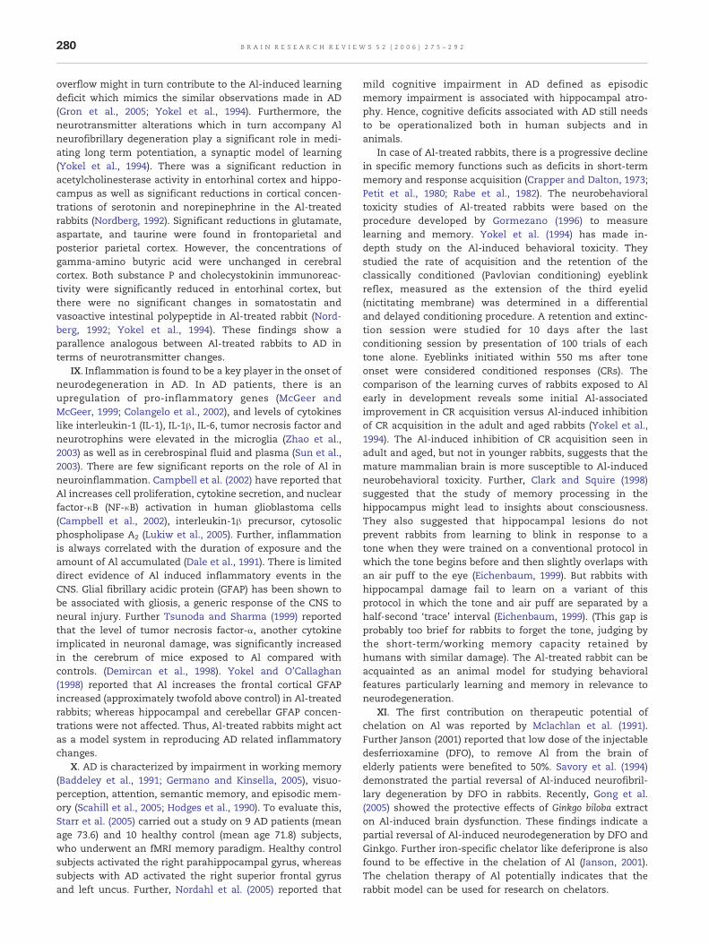
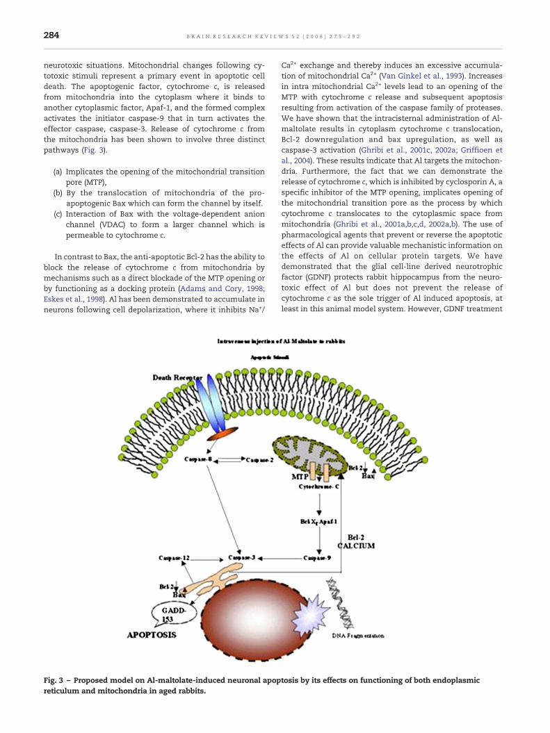
neurotoxic situations. Mitochondrial changes following cy-totoxic stimuli represent a primary event in apoptotic celldeath. The apoptogenic factor, cytochrome c, is releasedfrom mitochondria into the cytoplasm where it binds toanother cytoplasmic factor, Apaf-1, and the formed complexactivates the initiator caspase-9 that in turn activates theeffector caspase, caspase-3. Release of cytochrome c fromthe mitochondria has been shown to involve three distinctpathways (Fig. 3).
(a) Implicates the opening of the mitochondrial transitionpore (MTP),
(b) By the translocation of mitochondria of the pro-apoptogenic Bax which can form the channel by itself.
(c) Interaction of Bax with the voltage-dependent anionchannel (VDAC) to form a larger channel which ispermeable to cytochrome c.
In contrast to Bax, the anti-apoptotic Bcl-2 has the ability toblock the release of cytochrome c from mitochondria bymechanisms such as a direct blockade of the MTP opening orby functioning as a docking protein (Adams and Cory, 1998;Eskes et al., 1998). Al has been demonstrated to accumulate inneurons following cell depolarization, where it inhibits Na+/
Fig. 3 – Proposed model on Al-maltolate-induced neuronal apopreticulum and mitochondria in aged rabbits.
Ca2+ exchange and thereby induces an excessive accumula-tion of mitochondrial Ca2+ (Van Ginkel et al., 1993). Increasesin intra mitochondrial Ca2+ levels lead to an opening of theMTP with cytochrome c release and subsequent apoptosisresulting from activation of the caspase family of proteases.We have shown that the intracisternal administration of Al-maltolate results in cytoplasm cytochrome c translocation,Bcl-2 downregulation and bax upregulation, as well ascaspase-3 activation (Ghribi et al., 2001c, 2002a; Griffioen etal., 2004). These results indicate that Al targets the mitochon-dria. Furthermore, the fact that we can demonstrate therelease of cytochrome c, which is inhibited by cyclosporin A, aspecific inhibitor of the MTP opening, implicates opening ofthe mitochondrial transition pore as the process by whichcytochrome c translocates to the cytoplasmic space frommitochondria (Ghribi et al., 2001a,b,c,d, 2002a,b). The use ofpharmacological agents that prevent or reverse the apoptoticeffects of Al can provide valuable mechanistic information onthe effects of Al on cellular protein targets. We havedemonstrated that the glial cell-line derived neurotrophicfactor (GDNF) protects rabbit hippocampus from the neuro-toxic effect of Al but does not prevent the release ofcytochrome c as the sole trigger of Al induced apoptosis, atleast in this animal model system. However, GDNF treatment
tosis by its effects on functioning of both endoplasmic
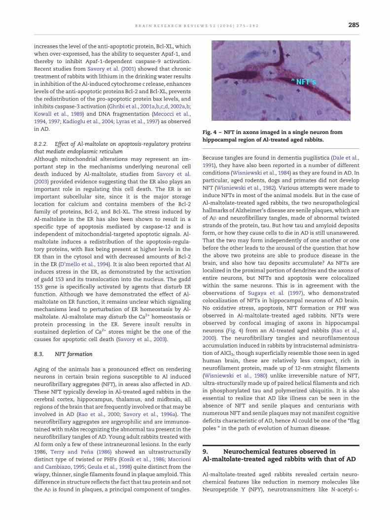
Fig. 4 – NFT in axons imaged in a single neuron fromhippocampal region of Al-treated aged rabbits.
285B R A I N R E S E A R C H R E V I E W S 5 2 ( 2 0 0 6 ) 2 7 5 – 2 9 2
increases the level of the anti-apoptotic protein, Bcl-XL, whichwhen over-expressed, has the ability to sequester Apaf-1, andthereby to inhibit Apaf-1-dependent caspase-9 activation.Recent studies from Savory et al. (2001) showed that chronictreatment of rabbits with lithium in the drinking water resultsin inhibition of the Al-induced cytochrome c release, enhanceslevels of the anti-apoptotic proteins Bcl-2 and Bcl-XL, preventsthe redistribution of the pro-apoptotic protein bax levels, andinhibits caspase-3 activation (Ghribi et al., 2001a,b,c,d, 2002a,b;Kowall et al., 1989) and DNA fragmentation (Mecocci et al.,1994, 1997; Kadioglu et al., 2004; Lyras et al., 1997) as observedin AD.
8.2.2. Effect of Al-maltolate on apoptosis-regulatory proteinsthat mediate endoplasmic reticulumAlthough mitochondrial alterations may represent an im-portant step in the mechanisms underlying neuronal celldeath induced by Al-maltolate, studies from Savory et al.(2003) provided evidence suggesting that the ER also plays animportant role in regulating this cell death. The ER is animportant subcellular site, since it is the major storagelocation for calcium and contains members of the Bcl-2family of proteins, Bcl-2, and Bcl-XL. The stress induced byAl-maltolate in the ER has also been shown to result in aspecific type of apoptosis mediated by caspase-12 and isindependent of mitochondrial-targeted apoptotic signals. Al-maltolate induces a redistribution of the apoptosis-regula-tory proteins, with Bax being present at higher levels in theER than in the cytosol and with decreased amounts of Bcl-2in the ER (D'mello et al., 1994). It is also been reported that Alinduces stress in the ER, as demonstrated by the activationof gadd 153 and its translocation into the nucleus. The gadd153 gene is specifically activated by agents that disturb ERfunction. Although we have demonstrated the effect of Al-maltolate on ER function, it remains unclear which signalingmechanisms lead to perturbation of ER homeostasis by Al-maltolate. Al-maltolate may disturb the Ca2+ homeostasis orprotein processing in the ER. Severe insult results insustained depletion of Ca2+ stores might be the one of thecauses for apoptotic cell death (Savory et al., 2003).
8.3. NFT formation
Aging of the animals has a pronounced effect on renderingneurons in certain brain regions susceptible to Al inducedneurofibrillary aggregates (NFT), in areas also affected in AD.These NFT typically develop in Al-treated aged rabbits in thecerebral cortex, hippocampus, thalamus, and midbrain, allregions of the brain that are frequently involved or thatmay beinvolved in AD (Rao et al., 2000; Savory et al., 1996a). Theneurofibrillary aggregates are argyrophilic and are immunos-tainedwithmAbs recognizing the abnormal tau present in theneurofibrillary tangles of AD. Young adult rabbits treated withAl form only a few of these intraneuronal lesions. In the early1986, Terry and Peña (1986) showed an ultrastructurallydistinct type of twisted or PHFs (Kosik et al., 1986; Maccioniand Cambiazo, 1995; Geula et al., 1998) quite distinct from thewispy, thinner, single filaments found in plaque amyloid. Thisdifference in structure reflects the fact that tau protein and notthe Aβ is found in plaques, a principal component of tangles.
Because tangles are found in dementia pugilistica (Dale et al.,1991), they have also been reported in a number of differentconditions (Wisniewski et al., 1984) as they are found in AD. Inparticular, aged rodents, dogs and primates did not developNFT (Wisniewski et al., 1982). Various attempts were made toinduce NFTs in most of the animal models. But in the case ofAl-maltolate-treated aged rabbits, the two neuropathologicalhallmarks of Alzheimer's disease are senile plaques, which areof Aβ and neurofibrillary tangles, made of abnormal twistedstrands of the protein, tau. But how tau and amyloid depositsform, or how they cause cells to die in AD is still unanswered.That the two may form independently of one another or onebefore the other leads to the arousal of the question that howthe above two proteins are able to produce disease in thebrain, and also how tau deposits accumulate? As NFTs arelocalized in the proximal portion of dendrites and the axons ofentire neurons, but NFTs and apoptosis were colocalizedwithin the same neurons. This is in agreement with theobservations of Sugaya et al. (1997), who demonstratedcolocalization of NFTs in hippocampal neurons of AD brain.No oxidative stress, apoptosis, NFT formation or PHF wasobserved in Al-maltolate-treated aged rabbits. NFTs wereobserved by confocal imaging of axons in hippocampalneurons (Fig. 4) from an Al-treated aged rabbits (Rao et al.,2000). The neurofibrillary tangles and neurofilamentousaccumulation induced in rabbits by intracisternal administra-tion of AlCl3, though superficially resemble those seen in agedhuman brain, these are relatively less compact, rich inneurofilament protein, made up of 12-nm straight filaments(Wisniewski et al., 1980) unlike irreversible nature of NFT,ultra-structurally made up of paired helical filaments and richin phosphorylated tau and polymerized ubiquitin. It is alsoessential to realize that AD like illness can be seen in theabsence of NFT and senile plaques and centurians withnumerous NFT and senile plaquesmay not manifest cognitivedeficits characteristic of AD, hence Al could be one of the “flagpoles “ in the path of evolution of human disease.
9. Neurochemical features observed inAl-maltolate-treated aged rabbits with that of AD
Al-maltolate-treated aged rabbits revealed certain neuro-chemical features like reduction in memory molecules likeNeuropeptide Y (NPY), neurotransmitters like N-acetyl-L-
286 B R A I N R E S E A R C H R E V I E W S 5 2 ( 2 0 0 6 ) 2 7 5 – 2 9 2
aspartyl-L-glutamate (NAAG), which resembles the character-istic features of AD.
9.1. Alterations in the levels of NPY
NPY present in the hippocampal region is responsible forneurochemical behavior and food intake. NPY is a 36-amino-acid, C-terminal amidated peptide and is widely distributed inthe central and peripheral nervous systems. NPY concentra-tion is reduced in cerebrospinal fluid, plasma and also incerebral cortex, hippocampus, and hypothalamus regions ofthe AD brain (Chang et al., 1998). Recently, Rao et al. (2000)showed that there is significant similarity between Al-maltolate-treated aged rabbits and have a close correlationwith that of AD. Al reduces NPY levels in the hippocampalregion of Al-maltolate-treated aged rabbits (Rao et al., 1999).Also studies from Rao and group showed that Al alters thestructure of NPY (Rao et al., 1999), this could explain theabnormality in feeding behavior as seen in the patients withAD (Gerald et al., 1996).
9.2. Imbalances in the levels ofN-acetyl-aspartyl-glutamate (NAAG) and its precursorN-acetyl-L-aspartate (NAL)
Neuropeptides namely NAAG and its precursor NAL levelswere found to be low in the AD brain, and it has been reportedthat these neuropeptides play a significant role in neuraltransmission. Reduced levels of NAAG and NAL are observedwith Al-maltolate-treated aged rabbits as seen in the case ofAD (Ramesh et al., 2001). In vivo studies indicated that Aldecreased the levels of NAAG and NAL. Jaarsma et al. (1994)reported that NAA and NAAG levels were significantlyreduced in hippocampus (by 38% and 24%) and amygdala(28% and 22%) regions of brain but not in cerebral cortex andolfactory bulb. Passani et al. (1997) reported that NAA andNAAG and the activity of NAALADase levels were significant-ly decreased in AD.
Fig. 5 – Schematic representation of Al-maltolate-treated aged r
9.3. D-Aspartate levels in AD/DNA alterations in AD
In normal human brain, proteins and free amino acids poolspredominantly exist in L-forms, while in neurodegenerativebrains the amount of racemized forms of amino acids, inparticular D-aspartate (D-Asp) and D-glutamate (D-Glu) arerelatively in large proportion in core amyloid plaques andNFTs and D-Asp promotes aggregation and fibril formation ofthe Aβ peptide (Vyas and Duffy, 1995). Recently, Latha et al.(2001) showed that Al-maltolate favors racemization from L-Asp and L-Glu to D-Asp and D-Glu respectively in the agedrabbit brain. Furthermore, Anitha et al. (2002) and Hegde et al.(2004) showed DNA helical changes in hippocampal region inAD brain fromB-form to Z-Form, whether similar observationsare seen in the case of Al-maltolate-treated rabbits stillremains elusive.
A detailed description on Al-maltolate-treated aged rabbitsmimicking AD neuropathology (∼90%) is as shown in Table 1.
10. Why other animal models includingtransgenic fails to reproduce total ADneuropathology?
Scientists have succeeded in producing hallmark features ofAD in a laboratory animal model. A number of animal models,such as the transgenic mice (Games et al., 1995) and amongothers rat, monkey, and dog, have been proposed to aid in theunderstanding of AD neuropathology (Brining et al., 1996; Unoet al., 1999). The much-anticipated transgenic mouse model isgenerally engineered with the human gene encoding for aform of the brain protein tau/amyloid. Transgenic mousemodel enable us to study tau-containing lesions in a numberof brain disorders, including the insoluble tau-containingtangles that build up and form one of the key pathologicalfeatures of AD. Coleman and Greenberg (1996) inserted humantau genes into mice which later developed masses ofabnormal tau filaments in nerve cells within the spinal cord,
abbit brain mimicking Alzheimer's disease neuropathology.
287B R A I N R E S E A R C H R E V I E W S 5 2 ( 2 0 0 6 ) 2 7 5 – 2 9 2
cortex, and brainstem, three critical regions of the centralnervous system. As the mice aged, insoluble masses of taufilaments grew in number. The transgenic animals alsoshowed evidence of nerve cell degeneration and impairedmovement, unlike their littermates lacking the inserted taugene. Transgenicmice have also been usedmainly to examinethe process of Aβ deposition (Games et al., 1995), whileindividual events such as apoptosis and NFT formation havebeen explored in other animals (Brining et al., 1996; Uno et al.,1999). Coleman and Greenberg (1996) have suggested that thetransgenic animal systemmay aid in an understanding of AD,only with reference to Aβ deposition, single event expression.Hence, researchers need to develop an animal model whichwould demonstrate all the following events namely, Aβdeposition, PHF formation, neuronal death, cholinergic defi-cits, inflammatory processes, and cognitive deficits in order tounderstand the spectrum of AD neuropathology which aids indeveloping new drug therapies for AD (Savory et al., 1999;Smith and Perry, 1997; Wisniewski and Sofer, 1979; Wis-niewski et al., 1980, 1982, 1984). The transgenic mice do notcompletely mimic AD pathological features, but they closelyresemble the other human brain disease affected by neuro-degeneration. Thus, the Al-maltolate-treated aged rabbitenables the demonstration of Aβ deposition, and the coloca-lization of NFT, PHF1, oxidative stress with apoptosis inhippocampal neurons, this brain region is usually andselectively involved in AD pathology (Grundke-Iqbal et al.,1985; Markesberry, 1994; Smith et al., 1996a,b; Su et al., 1994,1996).
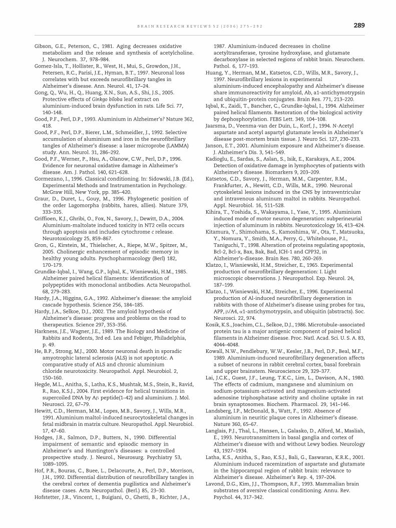
The understanding of AD neurochemistry and neuropa-thology is a big challenge due to unavailability of a suitableanimal model, which mimics AD pathology. Recently, Savoryet al. (2003) indicated that aged rabbits after Al-maltolatetreatment mimic AD-like neuropathology in terms of neuro-fibrillary tangles, β-amyloid deposition, oxidative stress andapoptosis in forebrain, hippocampus and midbrain regions(Fig. 5). Besides Savory's contribution in developing animalmodel for AD, Prof. Rao (2000) has made significant piece ofwork in providing circumstantial evidences on the neuro-pathological features in Al-maltolate-treated rabbits as sim-ilar in the case of AD. Hence, this animal model might be apromising model for pursuing further studies related to ADwithout involving the ongoing controversies of Al.
11. Conclusion and future perspectives
It is clear that the existing animal models tested so far fallshort in providing reliable and valid information on theneuropathology of AD, irrespective of whether their purposeis for analysis of the disease or developing more effectivetherapies than those that are presently available. Severalmodels have intrinsic limitations, and on the whole, they donot reproduce the pathogenetic process and are unlikely tohelp in the development of effective neuroprotective thera-pies. The genetic, particularly transgenic, technologies thatappeared to offer greater construct validity have so far failedto live up initial hopes and do not reliably result in reprodu-cing the neuropathology of AD. It is not yet been resolvedwhether the molecular basis in developing animal models are
incorrect or fundamental speciation differences in Al leadingto cellular processing of normal and abnormal proteins. Theextent and causes of the neurodegeneration and behavioraldeficits seen in these animal models require additional studyandmay involve higher inputs in this regard. Transgenic micewith the focus on APO-E4, presenilins and tau genes arecontinuing to be developed. However, Al-maltolate-treatedaged rabbits ameliorate AD pathology in behavioral, neuro-chemical, and immunohistochemical features, but furtherrefinements are required to develop an effective animalmodel. Al-maltolate-treated aged rabbits might act as areliable and efficient system in understanding the neuro-pathogenesis among the currently available ones. This opensup new avenues in developing therapeutic strategies fortreating this tragic, devastating disease.
Acknowledgments
The authors profoundly thank Dr. V. Prakash, Director,Central Food Technological Research Institute, Mysore forall his support and encouragement. This work was sup-ported by the grant from Department of Biotechnology, Indiafor National Facility for food safety, DBT Overseas Asso-ciateship awarded to KSJ Rao, and CSIR-CNR Joint Project ontoxicity of metals in human brain. Bharathi is thankful toCouncil for Scientific and Industrial Research for awardingSenior research fellowship.
This review is dedicated to Prof. John Savory, Universityof Virginia, Dr. Mary Herman, NIMH/NIH, with due respectfor their pioneering contribution on Al neurotoxicity.
R E F E R E N C E S
Aarsland, D., Cummings, J.L., Larsen, J.P., 2001.Neuropsychiatric differences between Parkinson's diseasewith dementia and Alzheimer's disease. Int. J. Geriatr.Psychiatry 16, 184–191.
Adams, J.M., Cory, S., 1998. The Bcl-2 protein family arbiters of cellsurvival. Science 281, 1322–1326.
Anitha, S., Rao, K.S.J., Latha, K.S., Viswamitra, M.A., 2002. Firstevidence to show the topological change of DNA fromB-DNA toZ-DNA conformation in the hippocampus of Alzheimer's brain.Neuromol. Med. 2, 289–297.
Baddeley, A.D., Bressi, S., Sala, S.D., Logie, R., Spinnler, H., 1991.The decline of working memory in Alzheimer's disease. Alongitudinal study. Brain 6, 2521–2542.
Bartus, R.T., 2000. On neurodegenerative diseases, models andtreatment strategies: lessons learned and lessons forgotten ageneration following the cholinergic hypothesis. Exp. Neurol.163, 495–529.
Beal, M.F., Mazurek, M.F., Ellison, D.W., Kowall, N.W., Solomon,P.R., Pendlebury, W.W., 1989. Neurochemical characteristicsof aluminum-induced neurofibrillary degeneration in rabbits.Neuroscience 29, 339–346.
Bertholf, R.L., Nicholson, J.R.P., Wills, M.R., Savory, J., 1987.Measurement of lipid peroxidation products in rabbit brain andorgans (response to aluminium exposure). Ann. Clin. Lab. Sci.17, 418–423.
Binder, L.I., Frankfurter, A., Rebhun, L.I., 1985. The distribution oftau in the mammalian central nervous system. J. Cell Biol. 101,1371–1378.
288 B R A I N R E S E A R C H R E V I E W S 5 2 ( 2 0 0 6 ) 2 7 5 – 2 9 2
Bishop, G.M., Robinson, S.R., 2000. β-Amyloid helps to protectneurons against oxidative stress. Neurobiol. Aging Suppl. 21,S226.
Brining, S.K., Jones, C.R., Chang, M.C., 1996. Effects of chronicbeta-amyloid treatment on fatty acid incorporation into ratbrain. Neurobiol. Aging 17, 301–310.
Campbell, A., Yang, E.Y., Tsai-Turton, M., Bondy, S.C., 2002.Pro-inflammatory effects of aluminum in human glioblastomacells. Brain Res. 933, 60–65.
Carpenter, A.F., Carpenter, P.W., Markesberry, W.R., 1993.Morphometric analyses of microglia in Alzheimer's disease.J. Neuropathol. Exp. Neurol. 52, 601–608.
Chafi, A.H., Hauw, J-J., Rancurel, G., Berry, J.P., Galle, C., 1991.Absence of aluminium in Alzheimer's disease brain tissue:electron microprobe and ion microprobe studies. Neurosci.Lett. 123, 61–64.
Chang, R.S.L., Lotti, V.C., Chen, T-B., 1998. Specific [1H]Propionyl-Neuropeptide Y (NPY) binding in rabbit aorticmembranes: comparisons with binding in rat brain andbiological responses in rat vas deferens. Biochem. Biophys. Res.Commun. 151, 1213–1219.
Clark, R.E., Squire, L.R., 1998. Classical conditioning and brainsystems: the role of awareness. Science 280, 77–81.
Colangelo, V., Schurr, J., Ball, M.J., Pelaez, R.P., Bazan, N.G.,Lukiw, W.J., 2002. Gene expression profiling of 12633 genesin Alzheimer hippocampal CA1: transcription andneurotrophic factor down-regulation and upregulation ofapoptotic and pro-inflammatory signaling. J. Neurosci. Res.70, 462–473.
Coleman, Greenberg, 1996. Animal model for Alzheimer's disease.Neurobiol. Aging 17, 1–2.
Corain, B., Abdiqqfrar Osman, A., Bertani, R., Tapparo, A., Zatta,P.F., Bombi, G.G., 1994. The aqueous solution state ofα-hydroxocarboxylate complexes of aluminium (III): an IRand NMR approach. Life Sci. Report. 11, 103–109.
Crapper, D.R., Dalton, A.J., 1973. Alterations in short-termretention, conditioned avoidance response acquisition andmotivation following aluminium induced neurofibrillarydegeneration. Physiol. Behav. 10, 925–933.
Crapper, D.R., Krishnan, S.S., Dalton, A.J., 1973. Brain aluminiumdistribution in Alzheimer's disease and experimentalneurofibrillary degeneration. Science 180, 511–513.
Cummings, J.L., 2000. Cognitive and behavioral heterogeneity inAlzheimer's disease, seeking the neurobiological basis.Neurobiol. Aging 21, 345–361.
Dai, J., Buijs, R.M., Kamphorst, W., Swaab, D.F., 2002. Impairedaxonal transport of cortical neurons in Alzheimer's disease isassociated with neuropathological changes. Brain Res. 948,138–144.
Dale, G.E., Leigh, P.N., Luthert, P., Anderton, B.H., Roberts, G.W.,1991. Neurofibrillary tangles in dementia pugilistica areubiquitinated. J. Neurol., Neurosurg. Psychiatry 54,116–118.
Demircan, M., Ergun, O., Coker, C., Avanoglu, S., Ozok, G., 1998.Aluminium in total parenteral nutrition solutions producesportal inflammation in rats. J. Pediatr. Gastroenterol. Nutr. 26,274–278.
D'mello, S.R., Anelli, R., Calissano, P., 1994. Induction of apoptosisin immature granule cells but promotes survival of matureneurons. Exp. Cell Res. 211, 232–238.
Doll, R., 1993. Alzheimer's disease and environmental aluminium.Age Ageing 22, 138–153.
Dollken, V., 1897. Uber die Wirkung des Aluminium mitbesbonderer Berucksichtigung der durch das Aluminiumverusachten lasionen im Zentralnervensystem. Arch. Exp.Pathol. 98–120.
Eichenbaum, H., 1999. Conscious awareness, memory and thehippocampus. Nat. Neurosci. 2, 775–776.
Eskes, R., Antonsson, B., Osen-Sand, A., Montessuit, S., Richter, C.,
Sadoul, R., Mazzei, G., Nichols, A., Martinou, J.C., 1998.Bax-induced cytochrome c release from mitochondria isindependent of the permeability transition pore but highlydependent on Mg2+ ions. J. Cell Biol. 143, 217–224.
Finneagan, M.M., Rettig, S., Orvig, C.A., 1986. A neutral watersoluble aluminium complex of neurological interest. J. Am.Chem. Soc. 108, 5033–5035.
Fontana, L., Perazzolo, M., Stella, M.P., Tapparo, A., Corain, B.,Favarato, M., Zatta, P., 1991. A long term toxicologicalinvestigation on the effect of tris(maltolate) aluminium (III) inrabbits. Biol. Trace Elem. Res. 2, 183–191.
Games, D., Adams, R., Alesandrini, R., Barbour, R., Berthelette, P.,Blackwell, C., Carr, T., Clemens, J., Donaldson, T., Gillespie, F.,1995. Alzheimer's-type neuropathology in transgenic miceoverexpressing V71F beta amyloid precursor protein. Nature373, 523–527.
Garruto, R.M., 1991. Pacific paradigms of environmentally-inducedneurological disorders: clinical, epidemiological and molecularperspectives. Neurotoxicology 12, 347–377.
Garruto, R.M., Fukatsu, R., Yanagihara, R., Gajdusek, D.C., Hook,G., Fiori, C.E., 1984. Imaging of calcium and aluminium inneurofibrillary tangle-bearing neurons in parkinsonismdementia of Guam. Proc. Natl. Acad. Sci. U. S. A. 81,1875–1879.
Garruto, R.M., Yanagihara, R., Shankar, S.K., Wolff, A., Salazar,A.M., Amyx, H.L., 1988. Experimental models ofmetal-induced neurofibrillary degeneration. In: Tsubaki, T.,Yase, Y. (Eds.), Amyotrophic Lateral Sclerosis. Elsevier,Amsterdam, pp. 41–50.
Gerald, C., Walker, M.W., Cyisolone, L., 1996. A receptor subtypeinvolved in neuropeptide Y-induced food Intake. Nature 382,162–171.
Germano, C., Kinsella, G.J., 2005. Working memory and learning inearly Alzheimer's disease. Neuropsychol. Rev. 15, 1–10.
Geula, C., Wu, C.K., Saroff, D., Lorenzo, A., Yuan, M., Yankner, B.A.,1998. Aging renders the brain vulnerable to amyloid β-proteinneurotoxicity. Nat. Med. 4, 827–831.
Ghribi, O., Herman, M.M., Forbes, M.S., DeWitt, D.A., Savory,J., 2001a. GDNF protects against aluminum-inducedapoptosis in rabbits by upregulating Bcl-2 and Bcl-XL andinhibiting mitochondrial Bax translocation. Neurobiol. Dis.5, 764–773.
Ghribi, O., Dewitt, D.A., Forbes, M.S., Herman, M.M., Savory, J.,2001b. Co-involvement of mitochondria and endoplasmicreticulum in regulation of apoptosis: changes in cytochrome-c,Bcl-2 and Bax in the hippocampus of aluminium treatedrabbits. Brain Res. 8, 66–73.
Ghribi, O., Dewitt, D.A., Forbes, M.S., Arad, A., Herman, M.M.,Savory, J., 2001c. Cyclosporin A inhibits Al-induced cytochromec release from mitochondria in aged rabbits. J. Alzheimer's Dis.3, 387–391.
Ghribi, O., Herman, M.M., Dewitt, D.A., Forbes, M.S., Savory, J.,2001d. Abeta (1–42) and aluminium induce stress in theendoplasmic reticulum in rabbit hippocampus, involvingnuclear translocation of gadd 153 and NF-kappa B. Brain Res.Mol. Brain Res. 96, 30–38.
Ghribi, O., Herman, M.M., Savory, J., 2002a. The endoplasmicreticulum is the main site for caspase activation followingaluminium-induced neurotoxicity in rabbit hippocampus.Neurosci. Lett. 324, 217–221.
Ghribi, O., Herman, M.M., Spaulding, N.K., Savory, J., 2002b.Lithium inhibits aluminium induced apoptosis in rabbithippocampus, by preventing cytochrome c translocation, Bcl-2decrease, Bax elevation and caspase-3 activation. J.Neurochem. 82, 137–145.
Ghribi, O., Herman, M.M., Savory, J., 2003. Lithium inhibitsabeta-induced stress in endoplasmic reticulum of rabbithippocampus but does not prevent oxidative damage and tauphosphorylation. J. Neurosci. Res. 71, 853–862.
289B R A I N R E S E A R C H R E V I E W S 5 2 ( 2 0 0 6 ) 2 7 5 – 2 9 2
Gibson, G.E., Peterson, C., 1981. Aging decreases oxidativemetabolism and the release and synthesis of acetylcholine.J. Neurochem. 37, 978–984.
Gomez-Isla, T., Hollister, R., West, H., Mui, S., Growdon, J.H.,Petersen, R.C., Parisi, J.E., Hyman, B.T., 1997. Neuronal losscorrelates with but exceeds neurofibrillary tangles inAlzheimer's disease. Ann. Neurol. 41, 17–24.
Gong, Q., Wu, H., Q., Huang, X.N., Sun, A.S., Shi, J.S., 2005.Protective effects of Ginkgo biloba leaf extract onaluminium-induced brain dysfunction in rats. Life Sci. 77,140–148.
Good, P.F., Perl, D.P., 1993. Aluminium in Alzheimer's? Nature 362,418.
Good, P.F., Perl, D.P., Bierer, L.M., Schmeidler, J., 1992. Selectiveaccumulation of aluminium and iron in the neurofibrillarytangles of Alzheimer's disease: a laser microprobe (LAMMA)study. Ann. Neurol. 31, 286–292.
Good, P.F., Werner, P., Hsu, A., Olanow, C.W., Perl, D.P., 1996.Evidence for neuronal oxidative damage in Alzheimer'sdisease. Am. J. Pathol. 140, 621–628.
Gormezano, I., 1996. Classical conditioning. In: Sidowski, J.B. (Ed.),Experimental Methods and Instrumentation in Psychology.McGraw Hill, New York, pp. 385–420.
Graur, D., Duret, L., Gouy, M., 1996. Phylogenetic position ofthe order Lagomorpha (rabbits, hares, allies). Nature 379,333–335.
Griffioen, K.J., Ghribi, O., Fox, N., Savory, J., Dewitt, D.A., 2004.Aluminium-maltolate induced toxicity in NT2 cells occursthrough apoptosis and includes cytochrome c release.Neurotoxicology 25, 859–867.
Gron, G., Kirstein, M., Thielscher, A., Riepe, M.W., Spitzer, M.,2005. Cholinergic enhancement of episodic memory inhealthy young adults. Pyschopharmacology (Berl) 182,170–179.
Grundke-Iqbal, I., Wang, G.P., Iqbal, K., Wisniewski, H.M., 1985.Alzheimer paired helical filaments: identification ofpolypeptides with monoclonal antibodies. Acta Neuropathol.68, 279–283.
Hardy, J.A., Higgins, G.A., 1992. Alzheimer's disease: the amyloidcascade hypothesis. Science 256, 184–185.
Hardy, J.A., Selkoe, D.J., 2002. The amyloid hypothesis ofAlzheimer's disease: progress and problems on the road totherapeutics. Science 297, 353–356.
Harkness, J.E., Wagner, J.E., 1989. The Biology and Medicine ofRabbits and Rodents, 3rd ed. Lea and Febiger, Philadelphia,p. 49.
He, B.P., Strong, M.J., 2000. Motor neuronal death in sporadicamyotrophic lateral sclerosis (ALS) is not apoptotic. Acomparative study of ALS and chronic aluminiumchloride neurotoxicity. Neuropathol. Appl. Neurobiol. 2,150–160.
Hegde, M.L., Anitha, S., Latha, K.S., Mushtak, M.S., Stein, R., Ravid,R., Rao, K.S.J., 2004. First evidence for helical transitions insupercoiled DNA by Aβ peptide(1–42) and aluminium. J. Mol.Neurosci. 22, 67–79.
Hewitt, C.D., Herman, M.M., Lopes, M.B., Savory, J., Wills, M.R.,1991. Aluminiummaltol-induced neurocytoskeletal changes infetal midbrain in matrix culture. Neuropathol. Appl. Neurobiol.17, 47–60.
Hodges, J.R., Salmon, D.P., Butters, N., 1990. Differentialimpairment of semantic and episodic memory inAlzheimer's and Huntington's diseases: a controlledprospective study. J. Neurol., Neurosurg. Psychiatry 53,1089–1095.
Hof, P.R., Bouras, C., Buee, L., Delacourte, A., Perl, D.P., Morrison,J.H., 1992. Differential distribution of neurofibrillary tangles inthe cerebral cortex of dementia pugilistica and Alzheimer'sdisease cases. Acta Neuropathol. (Berl.) 85, 23–30.
Hofstetter, J.R., Vincent, I., Buigiani, O., Ghetti, B., Richter, J.A.,
1987. Aluminium-induced decreases in cholineacetyltransferase, tyrosine hydroxylase, and glutamatedecarboxylase in selected regions of rabbit brain. Neurochem.Pathol. 6, 177–193.
Huang, Y., Herman, M.M., Katsetos, C.D., Wills, M.R., Savory, J.,1997. Neurofibrillary lesions in experimentalaluminium-induced encephalopathy and Alzheimer's diseaseshare immunoreactivity for amyloid, Ab, a1-antichymotrypsinand ubiquitin-protein conjugates. Brain Res. 771, 213–220.
Iqbal, K., Zaidi, T., Bancher, C., Grundke-Iqbal, I., 1994. Alzheimerpaired helical filaments. Restoration of the biological activityby dephosphorylation. FEBS Lett. 349, 104–108.
Jaarsma, D., Veenma-van der Duin, L., Korf, J., 1994. N-Acetylaspartate and acetyl aspartyl glutamate levels in Alzheimer'sdisease post-mortem brain tissue. J. Neuro Sci. 127, 230–233.
Janson, E.T., 2001. Aluminium exposure and Alzheimer's disease.J. Alzheimer's Dis. 3, 541–549.
Kadioglu, E., Sardas, S., Aslan, S., Isik, E., Karakaya, A.E., 2004.Detection of oxidative damage in lymphocytes of patients withAlzheimer's disease. Biomarkers 9, 203–209.
Katsetos, C.D., Savory, J., Herman, M.M., Carpenter, R.M.,Frankfurter, A., Hewitt, C.D., Wills, M.R., 1990. Neuronalcytoskeletal lesions induced in the CNS by intraventricularand intravenous aluminum maltol in rabbits. Neuropathol.Appl. Neurobiol. 16, 511–528.
Kihira, T., Yoshida, S., Wakayama, I., Yase, Y., 1995. Aluminiuminduced mode of motor neuron degeneration: subperineurialinjection of aluminum in rabbits. Neurotoxicology 16, 413–424.
Kitamura, Y., Shimohama, S., Kamoshima, W., Ota, T., Matsuoka,Y., Nomura, Y., Smith, M.A., Perry, G., Whitehouse, P.I.,Taniguchi, T., 1998. Alteration of proteins regulating apoptosis,Bcl-2, Bcl-x, Bax, Bak, Bad, ICH-1 and CPP32, inAlzheimer's-disease. Brain Res. 780, 260–269.
Klatzo, I., Wisniewski, H.M., Streicher, E., 1965. Experimentalproduction of neurofibrillary degeneration: I. Lightmicroscopic observations. J. Neuropathol. Exp. Neurol. 24,187–199.
Klatzo, I., Wisniewski, H.M., Streicher, E., 1996. Experimentalproduction of Al-induced neurofibrillary degeneration inrabbits with those of Alzheimer's disease using probes for tau,APP, β/A4, α1-antichymotrypsin, and ubiquitin (abstracts). Soc.Neurosci. 22, 974.
Kosik, K.S., Joachim, C.L., Selkoe, D.J., 1986. Microtubule-associatedprotein tau is a major antigenic component of paired helicalfilaments in Alzheimer disease. Proc. Natl. Acad. Sci. U. S. A. 83,4044–4048.
Kowall, N.W., Pendlebury, W.W., Kesler, J.B., Perl, D.P., Beal, M.F.,1989. Aluminium-induced neurofibrillary degeneration affectsa subset of neurons in rabbit cerebral cortex, basal forebrainand upper brainstem. Neuroscience 29, 329–377.
Lai, J.C.K., Guest, J.F., Leung, T.K.C., Lim, L., Davison, A.N., 1980.The effects of cadmium, manganese and aluminium onsodium-potassium-activated and magnesium-activatedadenosine triphosphatase activity and choline uptake in ratbrain synaptosomes. Biochem. Pharmacol. 29, 141–146.
Landsberg, I.P., McDonald, B., Watt, F., 1992. Absence ofaluminium in neuritic plaque cores in Alzheimer's disease.Nature 360, 65–67.
Langlais, P.J., Thal, L., Hansen, L., Galasko, D., Alford, M., Masliah,E., 1993. Neurotransmitters in basal ganglia and cortex ofAlzheimer's disease with and without Lewy bodies. Neurology43, 1927–1934.
Latha, K.S., Anitha, S., Rao, K.S.J., Bali, G., Easwaran, K.R.K., 2001.Aluminium induced racemization of aspartate and glutamatein the hippocampal region of rabbit brain: relevance toAlzheimer's disease. Alzheimer's Rep. 4, 197–204.
Lavond, D.G., Kim, J.J., Thompson, R.F., 1993. Mammalian brainsubstrates of aversive classical conditioning. Annu. Rev.Psychol. 44, 317–342.

290 B R A I N R E S E A R C H R E V I E W S 5 2 ( 2 0 0 6 ) 2 7 5 – 2 9 2
Liwincz, B.H., Kristensson, K., Wisniewski, H.M., Shelanski, M.L,Terry, R.D., 1974. Observations on axoplasmic transport inrabbits with aluminium-induced neurofibrillary tangles. BrainRes. 80, 413–420.
Lovell, M.A., Ehmann, W.D., Markesberry, W.R., 1993. Lasermicroprobe analysis of brain aluminium in Alzheimer'sdisease. Ann. Neurol. 33, 36–42.
Lovestone, S., Reynolds, C.H., 1997. The phosphorylation of tau: acritical stage in neurodevelopmental and neurodegenerativeprocess. Neuroscience 78, 309–324.
Lukiw, W.J., Percy, M.E., Kruck, T.P., 2005. Nanomolar aluminiuminduces pro-inflammatory and pro-apoptotic gene expressionin human brain cells in primary culture. J. Inorg. Biochem. 99,1895–1898.
Lyras, L., Cairns, N.J., Jenner, A., Jenner, P., Halliwell, B., 1997. Anassessment of oxidative damage to proteins, lipids, and DNA inbrain from patients with Alzheimer's disease. J. Neurochem.68, 2061–2069.
Maccioni, R.B., Cambiazo, V., 1995. Role of microtubule-associatedproteins in the control of microtubule assembly. Physiol. Rev.75, 835–864.
Markesberry, W.R., 1994. Oxidative stress hypothesis inAlzheimer's disease. Free Radical Biol. Med. 23, 134–147.
Martin, R.B., 1986. Aluminium in chemistry, biology andmedicine.Clin. Chem. 32, 1797–1806.
Matsuo, E.S., Shin, R.W., Billingsley, M.L., Van de Voorde, A.,O' Connor, M., Trojanowski, J.Q., Lee, V.M., 1994. Biopsy derivedadult human brain tau is phosphorylated at many of the samesites as Alzheimer's disease paired helical filament tau.Neuron 13, 989–1002.
McGeer, P.L., McGeer, E.G., 1999. Inflammation of the brain inAlzheimer's disease: implications for therapy. J. Leukocyte Biol.65, 409–415.
Mclachlan, D.R.C., Dalton, A.J., Kruck, T.P., Bell, M.Y., Smith, W.L.,Kalow, W., Andrews, D.F., 1991. Intramusculardesferrioxamine in patients with Alzheimer's disease. Lancet337, 1304–1308.
McLaughlin, A.I.G., Kazantis, G., King, E., Teare, D., Porter, R.J.,Owen, R., 1962. Pulmonary fibrosis and encephalopathyassociated with the inhalation of aluminium dust. Br. J. Ind.Med. 19, 253–263.
Mecocci, P., MacGarvey, U., Beal, M.F., 1994. Oxidative damage tomitochondrial DNA is increased in Alzheimer's disease. Ann.Neurol. 36, 747–751.
Mecocci, P., Beal, M.F., Cecchetti, R., Polidori, M.C., Cherubini, A.,Chionne, F., Avellini, L., Ronano, G., Senin, U., 1997.Mitochondrial membrane fluidity and oxidative damage tomitochondrial DNA in aged and AD human brain. Mol. Chem.Neuropathol. 31, 53–56.
Muma, N.A., Singer, S.M., 1996. Aluminium-inducedneuropathology: transient changes in micro-tubule associatedproteins. Neurotoxicol. Teratol. 18, 679–690.
Nicholls, D.M., Speares, G.M., Miller, A.C., Math, J., DelBianco, G., 1991. Brain protein synthesis in rabbitsfollowing low level aluminium exposure. Int. J. Biochem. 8,737–741.
Nordahl, C.W., Ranganath, C., Yonelinas, A.P., DeCarli, C., Reed,B.R., Jagust, W.J., 2005. Different mechanisms of episodicmemory failure in mild cognitive impairment.Neuropsychologia 43, 1688–1697.
Nordberg, A., 1992. Biological markers and the cholinergichypothesis in Alzheimer's disease. Acta Neurol. Scand., Suppl.139, 54–58.
Passani, L.A., Vonsattel, J.P., Coyle, J.T., 1997. Distribution ofN-acetylaspartyl glutamate immunoreactivity in human brainand its alterations in neurodegenerative disease. Brain Res.772, 9–22.
Perl, D.P., Brondy, A.R., 1980. Alzheimer's disease: X-rayspectrometric evidence of aluminium accumulation in
neurofibrillary tangle-bearing neurons. Science 208,297–299.
Perry, E.K., Perry, R.H., 1985. New insights into the nature of senile(Alzheimer type) plaques. Trends Neurosci. 8, 301–303.
Petit, T.L., Biederman, G.B., McMullen, P.A., 1980. Neurofibrillarydegeneration, dendritic dying back and learning memorydeficits after aluminium administration: implications for brainaging. Exp. Neurol. 67, 152–162.
Petit, T.L., Biederman, G.B., Jonas, P., Leboutiller, J.C., 1985.Neurobehavioral development of following aluminiumadministration in infant rabbits. Exp. Neurol. 640–651.
Priest, N.D., 2004. The biological behaviour and bioavailability ofaluminium in man, with special reference to studiesemploying aluminium-26 as a tracer: review and study update.J. Environ. Monit. 5, 375–403.
Rabe, A., Moon, H.L., Shek, J., Wisniewski, H.M., 1982. Learningdeficit in immature rabbits with aluminium inducedneurofibrillary changes. Exp. Neurol. 76, 441–446.
Ramesh, J., Rao, K.S.J., Easwaran, K.R.K., 2001. The effect of anacidic neurotransmitter, N-acetyl-L-aspartyl-L-glutamate andits precursor N-acetyl-L-aspartate. Alzheimer's Rep. 4,87–92.
Rao, K.S.J., Katsetos, C.D., Herman, M.M., Savory, J., 1998.Experimental aluminium encephalopathy. Clin. Lab. Med. 18,687–697.
Rao, K.S.J., Ramesh, J., Easwaran, K.R.K., 1999. Neuropeptide Ystructure is modulated by aluminium in the hypothalamus.Alzheimer's Rep. 2, 99–103.
Rao, K.S.J., Anitha, S., Latha, K.S., 2000. Aluminium inducedneurodegeneration in hippocampus of aged rabbits mimicsAlzheimer's disease. Alzheimer's Rep. 3, 83–88.
Savory, J., Garruto, R.M., 1998. Aluminium, tau protein andAlzheimer's disease: an important link? Nutrition 3,313–314.
Savory, J., Herman, M.M., Hundley, J.C., Seward, R.L., Griggs, C.M.,Katsetos, C.D., Wills, M.R., 1993. Quantitative studies onaluminium deposition and its effects on neurofilament proteinexpression and phosphorylation, following theintraventricular administration of aluminium maltolate toadult rabbits. Neurotoxicology 14, 9–12.
Savory, J., Herman, M.M., Erasmus, R.T., Boyd, J.C., Wills, M.R.,1994. Partial reversal of aluminium-induced neurofibrillarydegeneration by desferrioxamine in adult male rabbits.Neuropathol. Appl. Neurobiol. 20, 31–37.
Savory, J., Huang, Y., Herman, M.M., Reyes, M.R., Wills, M.R., 1995.Tau immuno reactivity associated with aluminiummaltolate-induced neurofibrillary degeneration in rabbits.Brain Res. 669, 325–329.
Savory, J., Huang, Y., Herman, M.M., Wills, M.R., 1996a.Quantitative image analysis of temporal changes in tau andneurofilament proteins during the course of acuteexperimental neurofibrillary degeneration;nonphosphorylated epitopes precede phosphorylation. BrainRes. 707, 272–281.
Savory, J., Exley, C., Forbes, W.F., Huang, Y., Joshi, J.G., Kruck, T.,Mclachlan, D.R., Wakayama, I., 1996b. Can the controversy ofthe role of aluminium in Alzheimer's disease be resolved?What are the suggested approaches to this controversy andmethodological issues to be considered? J. Toxicol. Environ.Health 48, 615–635.
Savory, J., Rao, K.S.J., Huang, Y., 1999. Age related hippocampalchanges in Bcl-2: Bax ratio, oxidative stress, redox-active ironand apoptosis associated with aluminium-inducedneurodegeneration: increased susceptibility with aging.Neurotoxicology 20, 805–815.
Savory, J., Ghribi, O., Forbes, M.S., Herman, M.M., 2001. Aluminiumand neuronal cell injury: inter-relationships betweenneurofilamentous arrays and apoptosis. J. Inorg. Biochem. 87,15–19.
291B R A I N R E S E A R C H R E V I E W S 5 2 ( 2 0 0 6 ) 2 7 5 – 2 9 2
Savory, J., Herman, M.M., Ghribi, O., 2003. Intracellularmechanisms underlying aluminium-induced apoptosis inrabbit brian. J. Inorg. Biochem. 97, 154–157.
Scahill, V.L., Hodges, J.R., Graham, K.S., 2005. Can episodicmemorytasks differentiate semantic dementia from Alzheimer'sdisease. Neurocase 11, 441–451.
Selkoe, D.J., 1989. Biochemistry of altered brain proteins inAlzheimer's disease. Annu. Rev. Neurosci. 12, 463–490.
Selkoe, D.J., 1991. Themolecular pathology of Alzheimer's disease.Neuron 6, 487–498.
Singer, S.M., Chambers, C.B., Newfry, G.A., Norhund, M.A., Muma,N.A., 1997. Tau in aluminium-induced neurofibrillary tangles.Neurotoxicology 18, 63–76.
Slotkin, T.A., Seidler, F.J., Crain, B.J., Bell, J.M., Bissette, G.,Nemeroff, C.B., 1990. Regulatory changes in pre-synapticcholinergic function assessed in rapid autopsy material frompatients with Alzheimer's disease: implications for etiologyand therapy. Proc. Natl. Acad. Sci. U. S. A. 87, 2452–2455.
Smith, M.A., Perry, G., 1997. The pathogenesis of Alzheimerdisease: an alternative to the amyloid hypothesis.J. Neuropathol. Exp. Neurol. 56, 217–222.
Smith, M.A., Kutty, R.K., Richey, P.L., Yan, S.D., Stern, D., 1994.Hemeoxygenase-I is associated with the neurofibrillarypathology of Alzheimer's disease. Am. J. Pathol. 145,42–47.
Smith, M.A., Sayre, L.M., Monnier, V.M., Perry, G., 1995. Radicalageing in Alzheimer's disease. Trends Neuosci. 18,172–176.
Smith, M.A., Perry, G., Richey, P.L., 1996a. Oxidative damage inAlzheimer's. Nature 382, 120–121.
Smith, M.A., Siedlak, S.L., Richey, P.L., Nagaraj, R.H., Elhammer, A.,Perry, G., 1996b. Quantitative solubilization and analysis ofinsoluble paired helical filaments from Alzheimer's disease.Brain Res. 717, 99–108.
Smith, M.A., Nunomura, A., Lee, H.G., Zhu, X., Moreira, P.I., Avila, J.,Perry, G., 2005. Chronological primary of oxidative stress inAlzheimer's disease. Neurobiol. Aging 5, 579–580.
Spafforth, J., 1921. Case of aluminium poisoning. Lancet 1301.Starr, J.M., Loeffler, B., Abousleiman, Y., Simonotto, E., Marshall, I.,
Goddard, N., Wardlaw, J.M., 2005. Episodic and semanticmemory tasks activate different brain regions in Alzheimer'sdisease. Neurology 65, 266–269.
Strong, M.J., Garruto, R.M., 1991b. Chronic aluminium inducedmotor neuron degeneration: clinical, neuropathological andmolecular biological aspects. Can. J. Neurol. Sci. 18,428–431.
Strong, M.J., Wolff, A.V., Wakayama, I., Garruto, R.M., 1991a.Aluminum induced chronic myelopathy in rabbits.Neurotoxicology 12, 9–21.
Su, J.H., Anderson, A.J., Cummings, B.J., 1994.Immunohistochemical evidence for apoptosis inAlzheimer's disease. NeuroReport 5, 2529–2533.
Su, J.H., Cummings, B.J., Cotman, C.W., 1996. Plaque biogenesis inbrain aging and Alzheimer's disease: I. Progressive changes inphosphorylation states of paired helical filaments andneurofilaments. Brain Res. 739, 79–87.
Sugaya, K., Reeves, M., McKinney, M., 1997. Topographicassociation between DNA fragmentation and Alzheimer'sdisease neuropathology in the hippocampus. Neurochem. Int.31, 275–281.
Sun, Y.X., Minthon, L., Wallmark, A., Warkentin, S., Blennow,K., Janciauskiene, S., 2003. Inflammatory markers inmatched plasma and cerebrospinal fluid from patients withAlzheimer's disease. Dementia Geriatr. Cognit. Disord. 16,136–144.
Terry, A.V., Buccafusco, J.J., 2003. The cholinergic hypothesis of ageand Alzheimer's disease related cognitive deficits: recentchallenges and their implications for novel drug development.J. Pharmacol. Exp. Ther. 3, 821–827.
Terry, R.D., Peña, C., 1986. Experimental production ofneurofibrillary degeneration. 2. Electron microscopy,phosphatase histochemistry and electron probe analysis.J. Neuropathol. Exp. Neurol. 24, 200–210.
Tomlinson, B.E., 1992. Aging and the dementias, In: Adams, J.H.,Duchen, L.W. (Eds.), Greenfield's Neuropathology, 5th ed.Edward Arnold, London, pp. 1284–1411.
Tsunoda, M., Sharma, R.P., 1999. Modulation of tumornecrosis factor alpha expression in mouse brain afterexposure to aluminium in drinking water. Arch. Toxicol.73, 419–426.
Uemura, E., 1984. Intranuclear aluminium accumulation inchronic animals with experimental neurofibrillary changes.Exp. Neurol. 85, 10–18.
Uno, H., Aslum, P.B., Dong, S., 1999. Cerebral amyloid angiopathyand plaques, and visceral amyloidosis in aged macaques.Neurobiol. Aging 17, 275–282.
Van Ginkel, M.F., Heijink, E., Dekker, P.B., Miralem, T., VanderVoet, G.B., de Wolff, F.A., 1993. Effect of aluminiumchloride, -citrate and -maltol on the calcium mediateddegradation of neurofilament proteins. Neurotoxicology 14,13–18.
Vyas, S.B., Duffy, L.K., 1995. Stabilization of secondary structure ofAlzheimer's beta protein by Al (III) ions and D-Asp sustitution.Biochem. Biophys. Res. Commun. 206, 718–723.
Wakayama, I., Nerunkar, V.R., Garruto, R.M., 1993.Immunohistochemical and ultrastructural evidence ofdendritic degeneration in motor neurons of Al-intoxicatedrabbits. Acta Neuropathol. 85, 122–128.
Walton, J.R., 2006. Aluminium in hippocampal neurons fromhumans with Alzheimer's disease. Neurotoxicology 3, 385–394.
Walton, J., Tuniz, C., Fink, D., Jacobsen, G., Wilcox, D., 1995. Uptakeof trace amounts of aluminium into the brain from drinkingwater. Neurotoxicology 16, 187.
Wen, G.Y., Wisniewski, H.M., 1985. Histochemical localization ofaluminium in the rabbit CNS. Acta Neuropathol. (Berl.) 3,175–184.
Wills, M.R., Savory, J., 1983. Aluminium poisoning: dialysisencephalopathy, osteomalacia and anaemia. Lancet 11, 29.
Wisniewski, H.M., Sofer, D., 1979. Neuro-fibrillary pathology:current status and research perspectives. Mech. Aging Dev. 1,119–142.
Wisniewski, H.M., Narkiewicz, O., Wisniewski, K., 1967.Topography and dynamics of neurofibrillary degeneration inaluminium encephalopathy. Acta Neuropathol. (Berl.) 9,127–133.
Wisniewski, H.M., Sturman, J.A., Shek, J.W., 1980. Aluminiumchloride induced neurofibrillary changes developing rabbits bymetallic aluminium. Ann. Neurol. 8, 479–490.
Wisniewski, H.M., Shek, J.W., Gruca, S., Sturman, J.A., 1982.Chronic model of neurofibrillary changes induced by metallicaluminium. Neurobiol. Aging 3, 11–22.
Wisniewski, H.M., Shek, J.W., Gruca, S., Sturman, J.A., 1984.Aluminium induced neurofibrillary changes in axons anddendrites. Acta Neuropthol. (Berlin) 3, 190–197.
Woodruff-Pak, D.S., Li, Y.-T., 1994. Nefiracetam. Effect on eye blinkclassical conditioning in rabbits. Psychopharmacology (Berlin)114, 200–208.
Woodruff-Pak, D.S., Papka, M., 1996b. Alzheimer's disease and eyeblink conditioning: 750 ms trace vs 400 ms delay paradigm.Neurobiol. Aging 3, 397–404.
Woodruff-Pak, D.S., Trojanowski, J.Q., 1996a. The older rabbit as ananimalmodel: implications for Alzheimer's disease. Neurobiol.Aging 17, 283–290.
Yankner, B.A., 1996. New clues to Alzheimer's disease: unravelingthe roles of amyloid and tau. Nat. Med. 2, 850–852.
Yokel, R.A., 1989. Aluminium produces age related behavioraltoxicity in the rabbit. Neurotoxicol. Teratol. 11, 237–242.
Yokel, R.A., O'Callaghan, J.P., 1998. An aluminum-induced

292 B R A I N R E S E A R C H R E V I E W S 5 2 ( 2 0 0 6 ) 2 7 5 – 2 9 2
increase in GFAP is attenuated by some chelators.Neurotoxicol. Teratol. 20, 55–60.
Yokel, R.A., Allen, D.D., Meyer, J.J., 1994. Studies of aluminumneurobehavioral toxicity in the intact mammal. Cell. Mol.Neurobiol. 14, 791–808.
Yumoto, S., Nagai, H., Matsuzaki, H., Matsumura, H., Tada, W.,Nagatsuma, E., Kobayashi, K., 2001. Aluminium-26incorporation into the brain of rat fetuses and sucklings. BrainRes. Bull. 55, 229–234.
Zatta, P., Zambenedetti, P., Milanese, M., 1999. Activation of
monoamine oxidase type-B by aluminum in rat brainhomogenate. NeuroReport 10, 3645–3648.
Zatta, P., Ibn-Lkhayat-Idrissi, M., Zambenedetti, P., Kilyen, M., Kiss,T., 2002. In vivo and in vitro effects of aluminium on theactivity of mouse brain acetylcholinesterase. Brain Res. Bull.59, 41–45.
Zhao, M., Cribbs, D.H., Anderson, A.J., Cummings, B.J., Su, J.H.,Wasserman, A.J., Cotman, C.W., 2003. The induction of the TNFalpha death domain signaling pathway in Alzheimer's diseasebrain. Neurochem. Res. 28, 307–318.