Embed Size (px)
Citation preview

A new era in premium ultrasoundPhilips EPIQ 7 system specifi cations
452299101151.indd 1 25/04/14 15:44

2
Contents
1 Introduction 41.1 Applications 4
2 System overview 52.1 System architecture 52.2 Imaging formats 62.3 Imaging modes 6 M-mode 7 Spectral Doppler 7 Auto color and auto Doppler 8 Steerable continuous wave (CW) Doppler 8 Tissue Doppler Imaging (TDI/TDI PW) 8 iRotate echo (X5-1 and X7-2t) 8 Live xPlane imaging 8 Live 3D echo 8 Live 3D/4D and MPR/iSlice imaging 9 3D/4D and MPR imaging 9
(all electronic array transducer) 3D/4D and MPR imaging (hybrid transducers) 9 Freehand 3D volume and MPR imaging 9 Spatio-Temporal Image Correlation (STIC) imaging 9 iSTIC imaging 9 Panoramic imaging 10 Contrast imaging – cardiovascular 10 Contrast imaging – general imaging 10 Interventional imaging 10 2D imaging 11 Tissue Harmonic Imaging (THI) 11 Color Doppler 11 Color Power Angio imaging (CPA) 12 Strain-based elastography 12 Shear wave elastography 12 3D panoramic imaging 12
3 System controls 133.1 Optimization controls 13 2D grayscale imaging 13 Next generation SonoCT real-time compound imaging 13 Elevation compound imaging 14 XRES adaptive image processing 14 Live volume imaging/Live 3D Echo (CV) 14
Volume imaging (GI/WHC) 15 Tissue aberration correction (TAC) 15 Coded beamforming 15 iSCAN intelligent optimization 15 AutoSCAN intelligent optimization 16 iOPTIMIZE intelligent optimization 163.2 Control panel 163.3 Touch screen 16
4 Workflow 174.1 Ergonomics 174.2 Display annotation 174.3 SmartExam protocols 174.4 Stress echo 184.5 Volume imaging solutions for connected
radiology departments 184.6 QuickSAVE feature 194.7 Image presentation 194.8 Cineloop review 194.9 Exam management features 194.10 Rapid Procedure Setup 194.11 Connectivity 19 NetLink connectivity option
(standard on premium and high end) 20
5 Transducers 215.1 Transducer selection 21 Compact transducers 21 PureWave crystal technology 21
xMATRIX technology 21 Curved array 21 3D9-3v broadband curved array 21
C10-3v broadband curved array with PureWave crystal technology 21 C10-4ec broadband curved array 21 C9-2 broadband curved array with PureWave crystal technology 21 C8-5 broadband curved array 22 C5-1 broadband curved array with PureWave crystal technology 22
452299101151.indd 2 25/04/14 15:44

3
Volume array 22 V6-2 broadband curved array 22 Linear array 22 VL13-5 broadband linear array 22 L18-5 broadband linear array 22
L15-7io broadband compact linear array 22 L12-5 50 broadband linear array 23 L12-3 broadband linear array 23 Sector array 23 S5-1 broadband sector array with PureWave crystal technology 23 S8-3 sector array 23 S12-4 sector array 23
S7-3t sector array TEE 23 xMATRIX array 24 X5-1 xMATRIX array
with PureWave technology 24 X6-1 xMATRIX array with PureWave crystal technology 24 X7-2 xMATRIX array with PureWave
crystal technology 24 X7-2t xMATRIX array TEE with PureWave technology 24 Non-imaging 24 D5cwc CW transducer (Pedoff) 24 D2cwc CW transducer (Pedoff) 24 D2tcd PW transducer (Pedoff) 245.2 Transducer application guide 25 6 PercuNav image fusion and
interventional navigation 29 6.1 Overview 29 Image fusion 29 Interventional navigation and planning software 29 Interventional navigation tracking instrumentation 30 Anatomical measurements 30 Connectivity 30
PercuNav accessories 31
7 Measurementsandanalysis 32 Measurement tools and general description 327.1 Measurement tools and quantification 32 QLAB quantification software 32 Cardiac 3D Quantification (3DQ) 32 Cardiac 3D Quantification Advanced (3DQ Advanced) 32 General Imaging 3D Quantification (GI 3DQ) 33 Mitral Valve NavigatorA.I. (MVNA.I.) 33 Fetal Heart Navigator (FHN) 34 Automated Cardiac 2D QuantificationA.I. (a2DQA.I..) 34 Automated Cardiac Motion 2D QuantificationA.I.
(aCMQA.I.) 34 Cardiac Motion/Mechanics 2D Quantification
for Stress (CMQ Stress) 35 Elastography Quantification (EQ) 35 Intima Media Thickness (IMT) measurements 36 MicroVascular Imaging (MVI) 36 Region of Interest (ROI) Quantification 36 Strain Quantification (SQ) 367.2 High Q automatic Doppler analysis 377.3 Clinical option analysis packages 37
8 Physicalspecifications 38 System cart 38 Monitor 38
Control panel 38 Physio 39 Peripherals 39 Input/output ports 39 Power requirements and video parameters 39 Electrical safety standards 39
9 Maintenanceandservices 40 Maintenance 40 Services 40 Clinical education 40 Philips Remote Connectivity 41 Warranty 41
452299101151.indd 3 25/04/14 15:44

4
Together we’re changing the course of medicine through the unexpected intelligence of pure sound and its ability to help you save and improve lives. We believe in premium ultrasound for a new era, one that meets the expanding use of ultrasound worldwide with answers that excel in the face of greater case volume, diagnostic complexity, and demand for certainty that ultrasound must deliver.
1. Introduction
1.1 ApplicationsAbdominal Obstetrical Fetal echo Cerebrovascular Vascular (peripheral, cerebrovascular, temporal TCD, and abdominal)Abdominal vascular Gynecological and fertility Small parts and superficial Musculoskeletal Pediatric general imaging Prostate Echocardiography (adult, pediatric, fetal) Stress echocardiography Transesophageal echocardiography (adult and pediatric)Surgical imaging Interventional imaging Contrast imaging Bowel imagingStrain elastography, elastography,shear wave elastography (ElastPQ)PerioperativeEpicardial echocardiography
452299101151.indd 4 25/04/14 15:44

5
• Live 3D color imaging• iRotate imaging
– Electronic rotation available with the X5-1, X7-2, and X7-2t transducers
– Standard 2D views from the same apical or parasternal window without moving scanning hand
– Part of a stress echo protocol for fast acquisitions and more consistent views between resting phase and stress phase
– iRotate achieves frame rates up to 290 Hz• Philips next generation SonoCT real-time compound imaging
– High precision beam-steered image compounding that acquires more tissue information and reduces angle-generated artifacts
– Up to nine lines of sight, obtained by steering the ultrasound beam, available on linear, curved and tightly curved arrays, and mechanical volume arrays
– WideSCAN capability to expand field of view during SonoCT imaging
– SonoCT capability available during contrast imaging modes – Elevation compound imaging on X5-1, X6-1, X7-2, and X7-2t transducer which compounds two or more lines of sight in the elevation dimensio
New for EPIQ – variable XRES is an extension of Philips exclusive XRES speckle noise reduction feature that allows the user to select progressive amounts of noise reduction, edge enhancement and textural smoothing. Available with specific transducers under certain tissue specific presets, users have the option to select the imaging characteristics of their choice from crisp to smooth tissue textures providing enhanced visualization of target anatomic structures.
2.1 System architecture• Powerful Philips nSIGHT imaging combines a proprietary
massive parallel processing architecture and new precision beamforming for real-time coherent beam reconstruction. Capable of processing multiple data streams for structural, functional and Live 3D/4D imaging.
– Built for 2D, Live xPlane, Live 3D, Live 3D zoom, Live full volume, up to 102° x 101°, high volume rate (HVR) imaging, Live 3D color, MPR (multiplanar reconstruction), electronic rotational echocardiography (iRotate) and panoramic imaging capability; true real-time volume image-forming capability with multiple rendering engines
– Supports both strain and shear wave elastography – Next generation Live 3D, PureWave xMATRIX transducers, with microbeamforming and single ASIC beamforming architecture
– Live 3D/4D, high volume rate (HVR) imaging with over 9,200 imaging elements, allowing outstanding 2D and 3D imaging from the single ergonomic transducer, eliminating the need to change between 2D and specialty 3D transducers
– Offers up to 7,071,744 total digital channels (xMATRIX configuration)
– Offers up to 4,718,592 total digital channels (non-xMATRIX configuration)
• Next generation ultra-low noise, wide dynamic range digital broadband acoustic beamforming with proprietary architecture
– 3D volume scan conversion that processes 460 megavoxels per second and renders 2300 mega-ray cast samples per second
– Live full volumes with one-beat, two-beat, four-beat and six-beat options
• 320 dB maximum dynamic range • Powerful distributed multi-core processing architecture capable
of achieving 450 x 109 40-bit Multiply-Accumulates/second. Includes three hard drives, 1 TB plus 120 MB SSD, and 4 GB graphics display card for fast frame rate large live volume 3D rendering
– Advanced pulse shaping, pulse coding and multivariant harmonics technologies incorporated
– Support for transducer frequencies up to 20 MHz• Optimized for high definition 21.5 inch LCD display• Designed to support virtually any array configuration: sector,
linear, curved, tightly curved, TEE, and xMATRIX electronic volume arrays
• Contrast echo with low MI, mid MI, and LVO modes – Pulse inversion and Power modulation technology for low MI Imaging
– Pulse inversion technology
2. System overview
452299101151.indd 5 25/04/14 15:44

6
• Live 3D/4D volume • Live 3D/4D zoom • 3D full volume • 2D, MPR, and volume • Dual Volume for full volume, 3D Zoom and iCrop
2.3 Imaging modes• 2D grayscale imaging with advanced pulse coding, pulse shaping,
and frequency compounding technologies • New xMATRIX-based 2D elevation compounding• M-mode• M-mode color Doppler• M-mode tissue Doppler• Anatomical M-mode• Live 3D Echo (instantaneous volume rendering of cardiac anatomy) • 3D imaging• 3D imaging with color Doppler• 4D imaging• Live xPlane imaging (simultaneous display of two live
imaging planes)• Tissue Harmonic Imaging (THI) with pulse inversion technology• Coded beamforming • Multivariate Tissue Harmonic Imaging including pulse inversion
technology and coded harmonics• Left ventricular opacification (LVO) with pulse inversion and
power modulation technologies• Contrast detection technology using pulse inversion and
power modulation imaging techniques 3D Contrast imaging on the X5-1 and X6-1
• SonoCT beam-steered real-time compound imaging• Harmonic SonoCT imaging• Up to three levels of XRES adaptive image processing technology• iSCAN intelligent scanning for one-button TGC and
gain optimization• iSCAN with adaptive gain compensation (AGC) for real-time
user-initiated frame-by-frame TGC optimization• AutoSCAN with adaptive gain compensation (AGC) for real-time
frame-by-frame TGC optimization• Simultaneous 2D M-mode• Color Doppler• Color Power Angio imaging (CPA) and directional CPA• Strain-based elastography• Shear wave elastography point quantification imaging (ElastPQ)• High-PRF pulsed wave (PW) Doppler• Duplex and simultaneous 2D/PW Doppler• Duplex continuous wave (CW) Doppler
• Philips next generation XRES adaptive image processing for noise and artifact reduction that improves tissue and border definition.
– Performs 350 million calculations per frame of image data over 2800 frames per second
– Operates in 2D and 2D/CFI/Doppler/TDI mixed modes over 2800 frames per second
– Offers XRES capability in contrast imaging modes – Provides user selectable levels of XRES adaptive processing for C5-1, C9-2, X5-1, and X7-2
• Philips adaptive Broadband Flow imaging – Doppler bandwidth that automatically adjusts for optimal flow sensitivity and resolution
– Advanced dynamic motion suppression algorithms that reduce flash artifacts
• Fully independent triplex multiple mode operation for extraordinary ease of use during Doppler procedures
• Auto Doppler flow optimization for carotid and arterial applications using linear array transducers
– Automatically adjusts color box position and angle – Automatically adjusts PW sample volume placement and angle – Includes Auto Flow Tracking for automatic angle correction with sample volume movements
• Advanced stress echo applications – Stress protocols with up to 10 stages – Forty views per stage by five modes
• Multi-application SmartExam workflow protocols – Stress echo, echo, abdominal, small parts, OB/GYN, and vascular applications
– Step-by-step on-screen guidance during exam – Full user customization – Record function for creation of custom protocols – Automatic mode switching including 3D
• Fast system boot up: from OFF, approximately 110 seconds• Transport mode: from sleep mode to on, approximately
20 seconds – Transport mode lasts 45 minutes before recharge is needed
2.2 Imaging formats • 2D linear: WideSCAN with SonoCT • 2D curved: WideSCAN with SonoCT • 2D sector • 2D virtual apex sector imaging with wide field of view• 2D trapezoid• Dual 2D • Panoramic • 3D Panoramic
452299101151.indd 6 25/04/14 15:44

7
M-mode• Available on all imaging transducers• Selectable sweeping rates• Time markers: 0.1 and 0.2 seconds• Acquisition zoom capability• Selectable display format prospective or retrospective
(1/3-2/3, 1/2-1/2, 2/3-1/3, side by side, full screen)• Chroma colorization with multiple color maps• Cineloop review for retrospective analysis of M-mode data
256 (8 bits) discrete gray levels
Spectral Doppler• Display annotations including Doppler mode, scale (cm/sec)
Nyquist limit, wall filter setting, gain, acoustic output status, sample volume size, normal/inverted, angle correction, grayscale curve
• Ultra-high resolution millisecond spectral FFT rate• Angle correction with automatic velocity scale adjustment• Adjustable velocity display ranges• Nine position shifts (including 0)• Normal/invert display around horizontal zero line• Selectable sweep speeds• Selectable low-frequency signal filtering with adjustable
wall filter settings• Selectable grayscale curve for optimal display• Selectable Chroma colorization maps• Selectable display format prospective or retrospective −
1/3-2/3, 1/2-1/2, 2/3-1/3, side by side, full screen• Doppler review for retrospective analysis of Doppler data• 256 (8 bits) discrete gray levels• Post-processing in PW frozen mode includes map, baseline,
invert, and Chroma• Available on all imaging transducers• Adjustable sample volume size: 1.0-20 mm
(transducer dependent)• Simultaneous or duplex mode of operation• Simultaneous 2D, color Doppler, pulsed Doppler• High-PRF capability in all modes including duplex,
simultaneous duplex and triplex• iSCAN optimization that automatically adjusts scale and baseline
• Duplex color flow and CW Doppler• Duplex 2D, color flow, PW Doppler• Duplex 2D, CPA, PW Doppler• Auto Doppler optimization: Auto PW Doppler, color Doppler,
flow optimization for one-button angle correction and steering• Tissue Doppler Imaging (TDI)• Adaptive Doppler• Adaptive Broadband Color Flow• Color Compare mode• Independent triplex mode for simultaneous 2D, color flow,
PW Doppler• Independent triplex mode for simultaneous 2D, CPA, PW Doppler• Dual imaging with:
– Two work flow choices; single buffer or dual buffer – Mixed mode display with one image live while other is frozen, for example, 2D/2D, 2D/color, color/color, color/CPA
• High definition zoom (write zoom)• Reconstructed zoom with pan (read zoom)• Panoramic imaging• SonoCT panoramic imaging with XRES and harmonic modes• 3D Panoramic imaging for full organ visualization• Chroma imaging in 2D, 3D, QLAB MPR and iSlice, Panoramic,
M-mode and Doppler modes• Dynamic colorization in Live 3D mode on X5-1, X7-2, and X7-2t,
freehand 3D on C10-3v, and 3D/4D on V6-2, X6-1, and X7-2• Live MVI• Spatio-Temporal Image Correlation (STIC)• iSTIC on X6-1 transducer
452299101151.indd 7 25/04/14 15:44

8
Live 3D echo• Available on X5-1, X7-2, and X7-2t xMATRIX transducers• Live full volume imaging• High volume rate imaging (HVR)• ECG display• Live one-beat, two-beat, four-beat and six-beat
3D volume imaging• Long live volume loop acquire• Beat-by-beat retrospective 3D loop selection• Live 3D color flow imaging• High volume rate (HVR) echo and color• xMATRIX with LVO, hi MI and low MI, xMATRIX pulse inversion
and power modulation• Contrast and interventional modes• Live 3D zoom and Live 3D zoom preview• One-beat focused volume• Half clam shell• Left and right clam shell switching• Two volume viewing display• Crop adjust with cropping• 3D color flow• 3D Zoom: 2D and Color• 3D Zoom: 2D and Color Preview• Enhanced Live 3D dynamic colorization for enhanced 3D effect• Full volume sweep• Adjustable live volume angle control• Volume rotation using 3D Rotate and Rotate-Z• Dynamic colorization• Adjustable vision preset control• Adjustable center, back, front, volume imaging control• Maximum 102° by 101° live volume imaging (mode dependent)• Support of volume rates up to 90 vps
Auto color and auto Doppler• In live imaging provides the following capabilities:
– Automatically adjusts color box position and angle – Automatically adjusts PW sample volume placement and angle – Includes Auto Flow Tracking for automatic angle correction with sample volume movements
– Automatically adjusts PW scale and baseline• When image is frozen and Doppler is active, automatically
adjusts PW scale and baseline• Auto color and Auto Doppler is available on the linear
transducers, L12-3, L12-5 50, L18-5, VL13-5, and L15-7io in carotid and arterial vascular applications
• Auto Doppler is available on the curvilinear transducers, C5-1, C8-5, C9-2, C10-3v, C10-4ec, V6-2
Steerable continuous wave (CW) Doppler• Available on all cardiac applications using sector transducers• Steerable through 90° sector• Maximum velocity range: 19 m/sec (transducer dependent)
Tissue Doppler Imaging (TDI/TDI PW)• Available on all cardiac imaging transducers (except S7-3t) • Frame rate control: High frame rate acquisition of tissue motion
(up to 240 fps) • TDI gain, TGC and LGC compatible• TDI Opt: Optimized transmit and receive frequencies• Eight maps
iRotateecho(X5-1,X7-2,andX7-2t)• Ability to image in 2D and rotate the image without
moving the transducer• Home rotational key• High frame rate rotational imaging• iRotate with stress echo acquire• iRotate for contrast echo• iRotate with color flow and CMQ speckle technology
Live xPlane imaging• Available on X5-1, X6-1, X7-2, and X7-2t xMATRIX transducers• Simultaneous display of two live imaging planes• Color and grayscale modes• Lateral, rotational, and elevation steering• Contrast and interventional modes
452299101151.indd 8 25/04/14 15:44

9
Freehand3DvolumeandMPRimaging• Qualitative grayscale volume acquisition supported
on all imaging transducers• Volume display with surface rendering (transparency, brightness,
and lighting controls)• Multiplanar view display• Specialized algorithms and maps increase 3D display• Trim tools on both volume and multiplanar reconstructed
(MPR) views• Supported by SonoCT and XRES modes to reduce
noise artifacts• Resize control that adjusts for different sweep speeds• On-screen orientation markers
Spatio-TemporalImageCorrelation(STIC)imaging• Available on V6-2 transducer• Automated volume acquisition of fetal cardiac cycle allowed• Grayscale and 3D Color• CPA and Directional CPA (DCPA)• Default 25° elevation angle• User-configurable acquisition time• Ability to stop acquisition and return to standby• Ability to accept or reject detected heart rate• Compatible with QLAB quantification software
iSTIC imaging• Available on X6-1 transducer• Automated volume acquisition of fetal cardiac cycle allowed• Grayscale and color modes• Automated detection of fetal heart rate • Acquisition of multiple subvolumes of the fetal heart • Multiple full volumes in one fetal heart cardiac cycle
Live3D/4DandMPR/iSliceimaging• Supported on X5-1, X6-1, X7-2, and X7-2t xMATRIX transducers• Volume display with surface rendering (transparency, brightness,
and lighting controls)• Multiplanar reconstruction (MPR) and iSlice view display with
QLAB software, including nine simultaneous views from 3D• Specialized algorithms and maps that increase 3D display• Cropping tools on volume views with image reference red,
green, and blue crop planes, arbitrary plane cropping and ROI-directed cropping with iCrop
• Two and three 2D reference planes optionally available Live 3D, full volume and 3D zoom imaging, live and review
• Supported XRES modes to reduce noise artifacts
3D/4DandMPRimaging (all electronic array transducer)• Available on X6-1 xMATRIX transducer• Volume display with surface rendering (transparency, brightness,
and lighting controls)• Multiplanar reconstruction (MPR) view display• Cropping tools on both volume and multiplanar reconstruction
(MPR) views• Slice control on MPR and volume displays• Supported by elevation compound imaging and XRES modes
to reduce noise artifacts• Full volume sweep• Adjustable X, Y, Z rotation• Dynamic colorization• Adjustable vision preset control• Contrast mode• Support of volume rates of at least 156 vps• Zoom• 3D color flow
3D/4DandMPRimaging(hybridtransducers)• Volume display with surface rendering (transparency, brightness,
and lighting controls)• Multiplanar reconstruction (MPR) view display• Specialized algorithms and maps maximize
three-dimensional display• Cropping tools on both volume and multiplanar reconstruction
(MPR) views• Slice control on MPR and volume displays• Supported by SonoCT and XRES modes to reduce noise artifacts
452299101151.indd 9 25/04/14 15:44

10
Contrast imaging – general imaging• System optimized for detecting contrast agent signatures
as they are approved for use• Contrast modes available on C5-1, C9-2, C10-4ec, C10-3v,
L12-3, L12-5 50, and X6-1 transducers• Live MicroVascular Imaging (MVI)• Mid-MI contrast modes available on C5-1, C9-2, and X6-1
transducers• Pulse inversion contrast imaging available with SonoCT
and XRES technologies• Power modulation (PM), pulse inversion (PI), and flash contrast
imaging modes• Touch screen display timer• Advanced non-linear pulsing schemes with SonoCT and XRES
for increased contrast sensitivity• Low MI color flow contrast• High frequency contrast capability• Flash imaging• Dual imaging mode for simultaneous fundamental
and contrast displays• ECG/timed triggering• Long loop capture mode during contrast procedures
(3-10 minutes)• QLAB ROI and MVI display
Interventional imaging• TSI available on selected transducers for optimal performance
during interventional and biopsy procedures• Enhanced needle visualization displays• Biopsy guide selection menus• Contrast and interventional modes• Support of multiple biopsy angles on S5-1, X6-1, C5-1, C9-2,
C10-4ec, V6-2, and L12-3
Panoramic imaging
• Real-time extended field-of-view composite imaging, acquired in fundamental or SonoCT mode
• Ability to acquire composite image in XRES mode• Ability to back up and realign the image during acquisition• Full zoom, pan, Cineloop review, and image rotation capabilities• Auto fit of composite image• Distance, curved-linear distance and area in review mode can
be measured with distance marker displayed via skin-line ruler• Ability to display or remove skin-line ruler• Cineloop review that allows measurement on individual frames• Scaling information included for connectivity prints allowing
for measurements on a workstation• Available on linear and curved array transducers
(not available on endovaginal transducers)
Contrast imaging – cardiovascular• System optimized for left ventricular opacification and low
MI imaging• One-touch solution (one-button access in LVO preset)
with settings for bolus and infusion• 2D, Live xPlane, Live 3D Echo, and full volume 3D• X5-1 and S5-1 broad bandwidth pulse inversion and power
modulation technologies for high sensitivity and high resolution visualization of contrast agent at low MI
• LVO and low MI contrast on and off, and contrast optimization choices and transmit power settings that can be saved with Gain Save feature for stress echo studies, eliminating setup time for image acquisition at peak stress
• Low MI with flash• Low MI with triggered replenishment imaging (TRI) that provides
excellent 2D image quality on the S5-1 transducer• X5-1 with iRotational contrast imaging and iRotational stress
contrast imaging• X5-1 with Live xPlane for contrast imaging• X5-1 with xMATRIX elevation compounding for contrast imaging• X5-1 with triggered replenishment imaging• X5-1 with 3D contrast echo• Supported on the S5-1 and, X5-1 transducers
452299101151.indd 10 25/04/14 15:44

11
Color Doppler• Available on all imaging transducers• Color gain• Region of Interest (ROI)• Freq Opt: Fixed transmit/receive frequencies including
adaptive flow• Seventeen selectable baseline positions for CV, nine selectable
baseline positions for GI, WHC• Baseline invert• B/W suppress• Color blending• Color compare dual display (B/W on left, color on right)• Color map• Color persistence• Flow optimization: GI, WHC• Output power• Magnify (range from 0.8X to 8X)• Scale sector width and position on curved and phased
array transducers• Simultaneous mode during PW mode• Smoothing• Ability to steer between ±3 degree steer angle on linear
array transducers• Variance• Wall filter• Write priority• Zoom• Cineloop review with full playback control
– Advanced motion suppression with intelligent algorithms; adapts to various application types to selectively eliminate virtually all color motion artifact
• 256 color bins• Parallelogram steering (three angles) on linear array
transducers; three angles on L12-5 50 and L18-5, twenty-one angles on L12-3 and L15-7io
• Trackball-controlled color region of interest: size and position• Maps, filters, color sensitivity, line density, smoothing, echo
write priority, color persistence, gain, and baseline optimized automatically by exam type or is user-selectable
• Velocity and variance displays• Color invert in live and frozen imaging• Frequency optimization control for spatial resolution and
penetration optimization• Color and 2D line density control• Automatically adapts transmit and receive bandwidth processing
based on the color box position providing optimal sensitivity and color resolution
2D imaging• Available with all imaging transducers• Adjustable sector width and position during live imaging• Ability to invert image left and right, top and bottom• Receive gain• LGC (lateral gain compensation) on cardiac sector transducers• Selection between one and eight focal zones• Dynamic range or echo compression, transducer and tissue
specific preset (TSP) dependent• Gray map• Chroma imaging providing colorized luminance maps• Acquisition zoom (HD zoom): ability to position the zoom
ROI anywhere within the image, and change the height and width of the zoom ROI
• Display zoom and magnify on live or frozen images up to 16 times• Three levels of frame rate• Support of frame rates of over 2800 frames per second• Tissue optimization• Contrast resolution enhancement• Tissue Harmonic Imaging• SonoCT imaging• Live Compare imaging; side-by-side comparison of 2D images
where the current live image is compared to a stored image from the same study or retrieved multi-modality image
• WideSCAN imaging• Next generation XRES technology, including up to three
selection levels on some transducers• Persistence (frame averaging)• Grayscale standard display• AutoSCAN with adaptive gain compensation (AGC) for real-time
line-by-line TGC optimization
Tissue Harmonic Imaging (THI)• Provides second harmonic processing to reduce artifacts
and improve image clarity• Multivariate pulsing including patented pulse inversion phase
cancellation technology for increased detail resolution during harmonic imaging
• Available on all imaging transducers with the exception of the C8-5
• Extends high performance imaging capabilities to all patient body types
• Support of SonoCT (Harmonic SonoCT) and XRES modes• Coded harmonics available with C5-1 in selected modes
452299101151.indd 11 25/04/14 15:44

12
Strain-basedelastography• Strain-based elastography for breast and gynecological imaging• Available for breast imaging on the L18-5 and L12-5 50
transducers, and on the C10-3v for gynecological and urology imaging
• One-touch entry into elastography mode• Elastogram applied as a Region of Interest box with user control
of size and location through entire field of view• Indicator for compression level• Display options• Single-screen 2D with elastogram • Side-by-side display of 2D image and 2D with elastogram• Shadow duplication (size compare) and measurement capability
in side-by-side display• Distance and area tools• Duplication from either side of the display• Eight selectable elastogram display maps• Ability to hide or show the elastogram display• Blend capability to increase 2D visibility through
elastogram display• Four smoothing selections• Five persistence selections• Two dynamic resolution system (DRS) selections to alternate
between elastogram resolution and penetration• Four dynamic range selections for elastogram display • Two elastogram optimization settings for different
tissue compositions• AI – anechoic imaging for enhancing areas without ultrasound
signals such as cystic and complex cystic structures
Shear wave elastography• Tissue deformation from special ultrasound push pulses• Detection pulses used to calculate shear wave velocity• Available on C5-1 for liver imaging
3D Panoramic imaging• Real-time extended field-of-view volume imaging, acquired
in echo xPlane imaging mode on the X6-1• Calibrated grayscale volumes for measurements• Volume display with surface rendering (transparency, brightness,
and lighting controls)• Multiplanar view display• Specialized algorithms and maps increase 3D display• Trim tools on both volume and multiplanar reconstructed
(MPR) views
Color Power Angio imaging (CPA)• Automatically adapts transmit and receive bandwidth processing
based on the color box position providing optimal sensitivity and color resolution
• Highly sensitive mode for small vessel visualization• Available on all imaging transducers for general imaging
and women’s healthcare• Cineloop review• Multiple color maps• Individual controls for gain, filters, sensitivity, echo write priority,
and color invert• Adjustable CPA Region of Interest: size and position• User-selectable persistence• User-selectable blending on/off• Cineloop review with full playback control• Advanced motion suppression with intelligent algorithms;
adapts to various application types to selectively eliminate virtually all color motion artifact
• 256 color bins• Parallelogram steering (three angles) on linear array transducers;
three angles on L12-5 50 and L18-5, twenty-one angles on L12-3 and L15-7io
• Trackball-controlled color region of interest: size and position• Maps, filters, color sensitivity, line density, smoothing, echo
write priority, color persistence, gain, and baseline optimized automatically by exam type or is user-selectable
• Velocity and variance displays• Color invert in live and frozen imaging• Frequency optimization control for spatial resolution and
penetration optimization• Color and 2D line density control• Automatically adapts transmit and receive bandwidth processing
based on the color box position providing optimal sensitivity and color resolution
452299101151.indd 12 25/04/14 15:44

13
3.1 Optimization controls2D grayscale imaging• Smart TGC: pre-defined TGC curves optimized for consistently
excellent imaging with minimal TGC adjustment• Lateral gain compensation (LGC) and Smart LGC for cardiac
sector transducers• Adjustable temporal resolution and spatial resolution with
DRS control• Depth: adjustment from 1.0 to 30 cm depending on transducer
and exam• Selection between one and eight transmit focal zones• 16-level digital reconstructed zoom with pan capability• High definition zoom that concentrates all image processing
power into a user-defined area of interest; possible to combine high definition zoom with pan zoom
• Cineloop image review• Selectable 2D compression settings• Tissue aberration correction• Sector size and steering control for sector and curved
array image formats• Selectable 2D line density with DRS control• Dual imaging with either independent Cineloop buffers
or split screen imaging• Dual imaging with color compare• Dual imaging with fundamental and contrast optimization• Chroma imaging with multiple color maps• 256 (8 bits) discrete gray levels• 2D acquisition frame rate over 2800 frames/sec
(dependent on field of view, depth and angle)• Live MVI
Next generation SonoCT real-time compound imaging• Available on all transducers except sector and xMATRIX arrays• Virtually all clutter and artifacts eliminated• Automatic selection of the number of steering angles based
on the user-selected resolution/frame rate (Res/Speed) condition• Up to nine lines of sight automatically adjusted via DRS control• Operates in conjunction with Tissue Harmonic Imaging,
volume modes, panoramic imaging, and duplex Doppler• Operates in conjunction with XRES• Available in contrast modes• Available with WideSCAN format during 2D imaging for extended
field-of-view operation
3. System controls
Philips common user experience provides readily accessible and logically grouped primary controls along with an easy-to-learn graphical user interface.
452299101151.indd 13 25/04/14 15:44

14
• Post processing• Left/right Invert• Res/speed control• Capture• Compress• Gain• ECG• ECG trigger• Cineloop/Live volume imaging/Live 3D Echo• Review/full volume• Save volume in native or native loop• Distance and area measurements on rendered volumes• 2D optimization settings• Tissue Harmonic Imaging• Density• 3D optimization settings• Sector width• Angle• Elevation width control• Lateral width control• Lateral position control• Elevation position control• 3D volume: front, center, back• 3D view control: up, down, left, right, front, back• 3D home• 3D swivel• Magnify• 3D LVO setting (available on X5-1 transducer)• Rotation in either absolute or relative trackball motion• Live 3D zoom mode with zoom preview• 3D color optimization• 3D size and position• Independent trackball control of lateral and elevation sizing and
positioning in live full volume and Live 3D color modes• iSlice – automated cropping of 3D volume into four MPR views
using standard or user-defined slicing protocols• iCrop: two orthogonal MPR views with volume mode
– Enables cropping during the exam or from review – Separates elevation and lateral rotation of cropping tools – Provides variable view directions and view direction color indicator
– Allows prospective or retrospective long volume loop acquire• Beat-by-beat 3D loop selection• Review/full volume• Calibrated 3D grid• Generic distance and area measurements available on volume• Distance and area measurements on MPRs
Elevation compound imaging• Available on X5-1, X6-1, X7-2, and X7-2t transducers• Reduces speckle and improves contrast resolution• Operates with at least two lines of sight• Operates in 2D in conjunction with fundamental imaging,
Tissue Harmonic Imaging, and duplex Doppler• Operates in conjunction with XRES imaging • Has no adverse impact on frame rates
XRES adaptive image processingNew for EPIQ - Variable XRES is an extension of Philips exclusive XRES speckle noise reduction feature that allows the user to select progressive amounts of noise reduction, edge enhancement and textural smoothing. Available with specific transducers under certain tissue specific presets, users have the option to select the imaging characteristics of their choice from crisp to smooth tissue textures providing enhanced visualization of target anatomic structures. • Available on all imaging transducers• Eliminates virtually all speckle noise and enhances border definition• Available in all imaging modes including color flow and Doppler• Available in contrast modes• Operates in conjunction with SonoCT imaging• Provides user three levels of user selectable choices on C5-1,
C9-2, X5-1, and X7-2• Provides high resolution algorithms for advanced speckle noise
reduction, refined tissue pattern displays, and fine border definition• Provides high speed processing that allows over 2800
frames-per-second displays
Live volume imaging/Live 3D Echo (CV)• Grayscale imaging controls• 3D Vision control• Dynamic volume colorization• Chroma colorization• Reset orientation• Up/down invert• XRES technology, including variable selection on X5-1 and X7-2• Zoom• Show/hide color• Reset controls• Rotate X, Y, Z• Auto crop• Plane/Manual crop• Brightness• Smoothing• Reference images
452299101151.indd 14 25/04/14 15:44

15
• Smoothing• Lighting• Transparency• xHair display• Save volume in native or native loop• Acquisition sweep save• MPR sweep save• Generic distance and area measurements available
on rendered volumes• Distance and area measurements on MPRs• QLAB plugins, including GI 3DQ and FHN
Tissue aberration correction (TAC)• Automatically enabled when ABD maximum penetration
TSI is selected on C5-1 transducer – Corrects for speed of sound disturbances due to excessive adipose layer on obese patients
• User selections with the L18-5, L12-5 50 for advanced breast, superficial, thyroid, and testicle TSIs
– Corrects for speed of sound disturbances in fatty tissue
Coded beamforming• Automatically enabled when ABD, OB or GYN maximum
penetration TSI is selected on C5-1 transducer• Coded excitation using new chirp transmit technology that
improves penetration and recovers more tissue information for detailed resolution at extended depths
• Coded harmonics mode that reduces image degrading artifacts while maintaining penetration qualities
iSCAN intelligent optimization• One-touch image optimization
– In 2D mode, one-button automatic adjustment of system gain and TGC to achieve balanced brightness of tissues
• Available in contrast imaging for selected transducers/applications – Independent settings based upon whether the contrast timer is active
• In Doppler mode, one-button automatic adjustment of: – Doppler PRF based on detected velocity – Doppler baseline based on detected flow direction
• Available on all imaging transducers• Operates in conjunction with SonoCT and XRES imaging• AutoSCAN continuous automatic optimization• Adaptive gain compensation (AGC) dynamically adjusts
(every pixel on every scan line) low level 2D echoes to reduce gain artifacts (shadows/through transmission) and improve image uniformity with 2D and 3D imaging
• Multiple 3D display layouts (volume, volume + 2 MPR, volume + 3 MPR)
• Dual volume display• 3D color zoom• 3D rotate and rotate-Z• X-hair manipulation controls for MPR alignment• Volume acquisition method (1, 2, 4, and 6 Beat, HVR)
Live volume imaging/Live 3D echo (GI/WHC)• Single sweep, 4D, STIC, iSTIC, and panoramic 3D• 3D preview ROI size and position• 3D preview ROI curve adjust• Sector width• Angle• Res/speed control• Grayscale imaging controls• 2D optimization settings• 2D color optimization settings• 2D power optimization settings• Tissue Harmonic Imaging• Rotate X, Y, Z• Slice• ROI size and position• ROI curve adjust• Pointer trim adjust• Pointer xHair move• Pointer cine• Edit/accept• Hide volume• Up/down invert• QuickFlip• 3D rotate: 0, 180, 90, 270• 3D view control: up, down, left, right, front, back• Reset orientation• Magnify• 3D vision control• Dynamic volume colorization• Chroma colorization• Layout• Reference• XRES technology• Zoom• Show/hide echo or color• Reset controls• Pan• Sculpt• Threshold• Brightness
452299101151.indd 15 25/04/14 15:44

16
AutoSCAN intelligent optimization• Continuous, real-time adjustment of system gain and TGC
to achieve balanced brightness of tissues – When activated, applies gain balancing to all grayscale image data including 2D, X-plane, 3D, 4D, and m-mode grayscale data
– Every image frame has individually adjusted image brightness – Available from 2D touch screen controls
iOPTIMIZE intelligent optimization Multiple technologies for one-button approach to automatically and instantly adjust system performance for different patient sizes, flow states, and clinical requirements• TissueSpecificImaging – adjusts over 7,500 parameters during
transducer/application selection• Patient optimization – adjusts 2D performance to instantly
adapt to different patient sizes• Flow optimization – adjusts broadband flow performance
to instantly adapt to different flow states• Dynamic resolution system (DRS) – one control adjusts
nearly 40 parameters simultaneously for user preference of spatial resolution or temporal resolution during clinical procedures
• One control optimizes functions such as: – Line density – Persistence – Pulse inversion harmonics – Synthetic aperture – Number of lines of sight (SonoCT) – RF interpolation – Parallel beamforming
3.2 Control panel • Easy-to-learn graphical user interface with reduced number
of hard controls• Primary controls concentrated in cluster around trackball• Tri-state control panel lighting (active, available, and unavailable)• Ambient lighting control for optimal image viewing in both light
and dark environments• Full color 12 inch capacitive touch screen, complete with
swipe technology, enables easy navigation of controls and system interaction
• Dual function mode switch and independent gain controls for 2D, CPA, M-mode, Color, PW, CW Doppler, TDI, and 3D
• Eight-slide pot control adjustment of TGC curve• iSCAN control for 2D/Doppler automatic optimization• High definition/pan zoom control• Dual mode control• Freeze control• Two programmable acquire controls• Pull-out alphanumeric backlit keyboard for text entry
3.3 Touch screen • Widescreen touch screen for dynamic presentation
of controls• Workflow related controls (Patient, Review, Report,
End Exam, Help) always present on touch screen• Direct selection of any attached transducer• Automatic or manual selection of tissue-specific
imaging parameters• Tabbed layout and swipe capability for quick access
to hidden controls• Touch screen control adjustment of LGC curve• Touch screen alphanumeric keyboard for text entry
452299101151.indd 16 25/04/14 15:44

17
• TGC curve (On/Auto/Off display)• TGC values (On/Off display)• Tool Tips provides a brief description of the abbreviated on-screen
image parameters• Trackball icon displaying functions assigned to trackball buttons• Informative trackball arbitration prompts• Thumbnail display of images printed/stored• On screen selection and display of calculations• On screen selection and editing of protocols• Calculations results and analysis labels• Graphical tabs that allow navigation to other analysis features• Network and connectivity icons to allow instant feedback about
network and printer conditions• Icons to display status of and/or allow access to the following
functions: Print Job status, media read/write status, battery level, wireless connectivity, remote service, microphone, HIPAA status, iScan status, acquisition status, physio status
• Cineloop frame number display• Cineloop bar with trim markers• Prompt region for display of informational text and icons• Trackball icon displaying functions assigned to trackball buttons• Contrast specification• Protocol procedure list with status
4.3 SmartExam protocols• On screen selection and editing of protocols• Exam guide with on-screen display• Required views based on exam type• SmartExam customization
– Creates a protocol as the user performs an exam – Saves all annotation, body markers, and labeled measurements defined in each view
– Records modes used to capture each view – Captures the acquisition method (print, capture, 3D dataset) in each individual view
– Provides user ability to pause and resume recording process if needed
– Allows user to edit views before finalizing the new protocol• Fully customizable protocol capability for any clinical application
supported on the system with flexibility to conduct the examination protocol in any sequence
4.1 Ergonomics• Advanced control panel design with fewer, clustered controls
and easily accessed mode keys to reduce reach• Tri-state lighting that provides immediate feedback of active,
available, and unavailable controls• Widescreen touch screen allows more controls to be available
at a time• Touch screen controls are grouped for quick recognition• Many touch screen controls can also be accessed from the
main display allowing user to maintain consistent visual focus• Independent adjustment of height, rotation and lateral movement
of monitor and control panel allowing improved user posture, increasing comfort during exams (meets industry standards recommendation for the prevention of WRMSD)
• Highly mobile cart with single pedal brake operation facilitating portable exams and positioning in confined space environments
4.2 Display annotation• On-screen annotation of all pertinent imaging parameters for
complete documentation, including transducer type and frequency, active clinical options and optimized presets, display depth, TGC curve, grayscale, color map, frame rate, compression map value, color gain, color image mode, hospital name, and patient demographic data
• User-selectable display of patient birth date, patient gender, institution name, system name and user
• Fixed position title area for consistent annotation• Patient name, ID, birth date gender and system date that can
be turned off (hidden) for generating still images for publication• Additional patient information can be displayed on demand• Sector steering icon for endocavitary transducers• Scan plane orientation marker• User selectable depth scale display• Real-time display of mechanical index (MI)• Real-time display of thermal index (TIb, TIc, TIs)• Multiple trackball-driven annotation arrows• Pre-defined annotations and body markers (application specific
and user selectable), with two body markers supported in dual imaging format
• Doppler baseline invert in live and frozen imaging• Compression changes available live or scrolling loop
4. Workflow
The EPIQ 7 combines a complete range of premium imaging technologies with superior sophisticated ergonomics, dynamic adaptive software and ease-of-use to help keep busy departments on schedule.
452299101151.indd 17 25/04/14 15:44

18
– Support between 1 and 40 views per stage – Support user-defined view names – Prompt for a particular stage and view – Assign stage and view names – Set clip length for each image or group of images – Set the number of cycles/beats for each image – Define prospective, retrospective, or multi-cycle/full disclosure acquisition
– Define the capture format of each image or group of images – Define the default replay mode for each protocol – Set mode acquisition for each view – Support for up to five modes – Save user-defined protocols within a preset – Save user-defined protocols to removable media for import onto separate systems at the same software level
– Modify protocols during use – Add stages at any point after the current stage
• CMQ stress – Pre and post data curves – Pre and post bull’s-eye maps – Pre and post strain comparisons
4.5 Volume imaging solutions for connected radiology departments• Customizable to your workflow• Fast, one-button press volume acquisition and on-cart review• Advanced volume and MPR visualization with QLAB GI 3DQ
– iSlice and thick slice on cart• Capability to export freehand, electronic, and hybrid acquired
3D grayscale data for visualization on most PACS in a stacked “fly-through” manner (like CT/MR)
• Off-cart evaluation of volume data on a multimodality clinical workstation
• Powerful 3D manipulation tools including volume rendering, MPR, MIP, slab viewing (thick slice), 3D orientation graphics
• Advanced 3D visualization with QLAB GI 3DQ including ability to handle 3D color flow and xMATRIX data
• Orientation labels feature for spatial orientation of 3D data sets – Adult orientation labels for non-fetal applications – Fetal orientation labels for fetal applications
• MPR Export capability – Ability to export A, B, and C planes as a multiframe loop for review on a DICOM device
– Available on all transducers, but not supported for any STIC or iSTIC files
• Preset protocols including but not limited to abdominal, vascular, cardiac, and OB/GYN exams based on industry and accreditation guidelines
• Automatic launching of annotation and body marker icon on required views
• Ability to automatically launch modes (2D, 3D, color modes, Doppler, dual, color compare) defined in a SmartExam
• Ability to pause and resume SmartExam function at any time• System analysis capabilities supported in all defined protocols
4.4 Stress echo• Acquisition of echocardiography single frame or loops of
the left ventricle in any imaging mode including 2D, color, and spectral Doppler.
• Gain Save that adjusts automatically to different views and automatically saves your preferred control settings, such as gain, depth, ROI, position, and many other parameters:
– For each view while acquiring resting images – At immediate post-exercise, automatic retrieval of saved settings for each view
– Different gain profiles for parasternal LAX and SAX views, AP4 and AP2 views allowed
• Length of acquired images that is user-adjustable between 1 and 180 seconds
• Ability to acquire routine cardiac images in timed and R-R interval clip (varies with selected compression ratio and available system memory)
• For timed acquisition, the ability to start acquisition on the R-wave if the ECG is active and an R-wave is present
• Your preferred control settings automatically saved – such as MI (mechanical index), gain and depth for each view while acquiring resting images
• Live Compare• Ability to defer selection by stage• Default stress protocols
– Factory-provided non-editable default protocols include: – Two-stage exercise stress – Four-stage pharmacological stress – Three-stage exercise stress (bicycle) – Four-stage quantitative: wall motion and contrast
• Default protocols that may be used as the basis for user-defined versions
– Support between 1 and 10 stages – Support user-defined stage names
452299101151.indd 18 25/04/14 15:44

19
4.9 Exam management features• Internal storage • Data export • Temporary ID feature
– One-click start of exam from patient data entry screen with system-provided information
– Storage of images that were created without a patient name with a temporary identification
4.10RapidProcedureSetup• With a single selection, choose transducer, preset, study type,
study description, and optionally gender• Procedure definitions are built-in for built-in study types• Additional procedure definitions may be added by the user• Procedure may be automatically selected based on modality
Worklist scheduled procedure information
4.11 Connectivity Standard connectivity features• Digital image acquisition and on-board patient exam storage
– Direct digital storage of B/W and color loops to internal hard disk drives
– Combined 1 TB storage capacity – Storage capacity of approximately 350 patient exams (assuming 40 images, 6 seconds of clips and reports per exam)
– Fully-integrated user interface – User configurable “auto delete” capability – On-screen recall, measurement and text editing – Exam directory – Append exam – To existing study – To new study using existing patient information
• Data types – 2D, M-mode, Doppler spectral frame acquisition – 2D clip acquisition up to 2,200 frames per clip – Scrolling M-mode, Doppler acquisition – Cartisian volume acquisition: 3D, 4D, STIC, 3D panoramic – Cardiac temporal volume acquisition: Live 3D, full volume 3D – 3D clips: volume render views and MPR views – Q-Apps frames and clips – PercuNav frames and clips
• Printing – Local print to on-board or off-board video printers – Page report print – DICOM grayscale or color print
4.6 QuickSAVE feature• The system provides the ability to quickly save preferred system
settings as individual exam types• Over 40 QuickSAVE exams can be created per transducer• Saved parameters include virtually all imaging parameters
as well as color box size• QuickSAVE exams can be copied to USB/DVD and transferred
to other systems of like configuration
4.7Imagepresentation• Up/down• Left/right• Multiple duplex image formats (1/3-2/3, 1/2-1/2, 2/3-1/3,
50/50 and full screen)• Depth from 1 cm to 30 cm (transducer dependent)
4.8 Cineloop review• Acquisition, storage in local memory, and display in real-time and
duplex modes of up to 2,200 frames of 2D and color images or up to 48 seconds of Doppler data and M-mode for retrospective review and image selection
• Prospective or retrospective loop acquire “accept” prior to store or clip store
• Trackball control of image selection• Variable playback speed• 3D iCROP from Cineloop review• 3D iSlice from Cineloop review• Trim capability of 2D data• Capture of over 20 seconds of Live 3D imaging per loop• Available in all imaging modes plus:
– Panoramic imaging – 3D panoramic imaging – 3D imaging – Independent control of 2D image or spectral data in duplex mode
– Simultaneous control of 2D and spectral data in simultaneous mode
• On-screen display of current 2D frame number• Many controls available in Cineloop review for post-processing
such as 2D gain, dynamic range/compress, XRES, magnify zoom
452299101151.indd 19 25/04/14 15:44

20
• DICOM compression options – Uncompressed (Explicit VR Little Endian, Implicit VR Little Endian)
– JPEG lossy compression (loops) with configurable quality factor 60-100
– RLE lossless compression – JPEG lossless compression (frames)
• Other DICOM export options – Monochrome or true color – Configurable image size/loop export 640 x 480 or 800 x 600 or 1,024 x 768
– Grayscale mapping choices - DICOM Grayscale Standard Display Function (GSDF) - 25 additional grayscale curves, user selectable
– Export optimization tool to aid user in evaluating PACS display monitor calibration and in selecting which grayscale curve to use for exported images
– Native data attached to DICOM ultrasound images (lossless compressed) - 2D native data types: tissue, flow, tissue-Doppler, spectral Doppler, M-mode, and elastography
- 3D volume data including crop, resize, gain, compression, automated border tracking, color baseline, 3D vision control, colorize, color suppress, B/W suppress, XRES and 3D quantification
– Ultrasound region calibration (standard for ultrasound images) – Pixel spacing attribute for measurement calibration (optional) – DICOM query/retrieve of other modality images (CT/MRI/X-ray/mammography/PET)
– De-identification feature – Send images to PACS and media without burned-in identifying information burned in to the image
– Images exported to media may optionally have patient information removed from DICOM attributes or PC format names
– All pages sent to DICOM printer have patient identification overlay – not configurable
• All pages sent to local printers are configurable to include or exclude patient identification overlay
• Media storage and retrieval – Export DICOM Image and structured report export to removable media
– Export PC Format image export to removable media – Supported media - Read and write (single session) to CD (CD+R) - DVD read-only (DVD+R) - DVD read + write (single session) (DVD+RW, CD-R and DVD-R)
- USB storage (flash memory or hard drives) – DICOM image import - Ultrasound images - Multi-modality images (CT/MRI/X-Ray/Mammography/PET)
– OB trending data - Export OB trending information via USB storage device - Import OB trending information via USB storage device - Export and import of trending data is compatible with iU22
• RS-232 serial storage – Export of report data to off-line analysis computer programs
• Basic networking connectivity – Wired gigabit Ethernet – Wireless networking 802.11n - WPA/WPA2 Personal security - WPA/WPA2 Enterprise security
– Network addressing - IPV4 addressing: static or DHCP for system address, static or hostnames (DNS lookup) for server addresses
- IPV6 addressing: link local, router discovery, or DHCP for system address, hostnames for server addresses
NetLink connectivity option (standard on premium and high end)• Supported DICOM services
– Image storage – Structured Report (SR) storage includes Ob/Gyn, vascular, adult echo, pediatric echo, fetal echo, and congenital cardiology
– Modality Worklist with automatic patient demographic entry – Modality performed procedure step (MPPS) – Storage commitment push model – Query/retrieve of ultrasound images (study-root)
• Image and structured report export to network storage servers – Send images after each Print/Acquire – Send images at End of Exam (batch send) – Send images and report on-demand during exam – Send images or exams manually – Send to up to 5 storage SCP's concurrently (at End Exam or after each Print/Acquire)
– Independently configurable destinations for each acquisition control (e.g., Acquire1, Acquire2, Save 3D, etc.)
452299101151.indd 20 25/04/14 15:44

21
5. Transducers
5.1 Transducer selection• Electronic switching of transducers using four universal connectors• Dedicated (Pedoff) continuous wave Doppler connector is available • Automatic parameter optimization of each transducer for exam
type through Tissue Specific Imaging (TSI) software • If two transducers are connected that both support the same TSI
the system supports instantaneous switching between transducers while maintaining current depth parameter if possible
• User customizable imaging presets for each transducer• Automatic dynamic receive focal optimization• Transmission of focal characteristics automatically controlled
through TSI, iFOCUS, and DRS functions
Compact transducers• Ergonomic designs with lightweight super flexible cables• Virtually pinless micro connectors• Advanced low-loss lens technology for better penetration
with less artifacts• Breakthrough broadband frequency response• Support for very high frequencies up to 20 MHz• Advanced micro-electronics in linear, curved, tightly-curved,
sector, hybrid volume array, and xMATRIX configurations• High precision automated volume transducers
PureWave crystal technology• Available on the X5-1, X6-1, X7-2, X7-2t, S5-1, C5-1, C9-2,
and C10-3v transducers• Breakthrough crystal technology that allows greater acoustic
efficiency and bandwidth
xMATRIX technology• Available on the X5-1, X6-1, X7-2 and X7-2t transducers• Unique array configuration of fully-sampled elements
that allows 2D, Live xPlane, and volume imaging
Curved array3D9-3vbroadbandcurvedarray• 9 to 3 MHz extended operating frequency range• 164° field of view (wide scan enabled)• Support of high resolution 2D imaging• Support of high resolution, quantitative, single sweep 3D volume
acquisitions (hybrid and freehand)• Support of 4D imaging up to 22 volumes per second• Steerable pulsed wave and color Doppler, Color Power Angio,
SonoCT, XRES, and harmonic imaging• Endovaginal applications• Interventional applications• Supports biopsy guide capabilitiesC10-3vbroadbandcurvedarray with PureWave crystal technology• 10 to 3 MHz extended operating frequency range• End-fire sector, 11.5 mm radius of curvature, 163° field of view
(wide scan enabled)• Steerable pulsed wave and color Doppler, Color Power Angio
(CPA), directional CPA, SonoCT, XRES, and harmonic imaging• Endovaginal applications• Elastography – strain-based• Contrast applications• Supports biopsy guide capabilitiesC10-4ecbroadbandcurvedarray• 10 to 4 MHz extended operating frequency range• End-fire sector, 8 mm radius of curvature, 147° field of view
(wide scan enabled)• Steerable pulsed wave and color Doppler, Color Power Angio
(CPA), directional CPA, SonoCT, XRES, and harmonic imaging• Endocavitary applications, including vaginal, and rectal• Contrast application• Supports biopsy guide capabilitiesC9-2broadbandcurvedarray with PureWave crystal technology• 9 to 2 MHz extended operating frequency range• End-fire sector, 45 mm radius of curvature, 102° field of view
(wide scan enabled)• Steerable pulsed wave and color Doppler, Color Power Angio
(CPA), directional CPA, SonoCT, XRES, and harmonic imaging• General purpose obstetrical and gynecological, small adult and
pediatric abdominal applications• Supports biopsy guide capabilities (4 angle)• Contrast application
452299101151.indd 21 25/04/14 15:44

22
Linear arrayVL13-5broadbandlineararray• 13 to 5 MHz extended operating frequency range• Fine pitch, 192 element, high resolution linear array• Support of high resolution 2D imaging• Support of high resolution, quantitative, single sweep 3D
volume acquisition• Support of 4D imaging• Steerable pulsed wave and color Doppler, Color Power Angio,
SonoCT, XRES, and harmonic imaging• High resolution superficial applications including small parts,
breast, and vascular imaging• Tissue aberration correction selection for advanced breast
imaging TSI• Supports biopsy guide capabilitiesL18-5broadbandlineararray• 18 to 5 MHz extended operating frequency range• Ultra-fine pitch, 288 element, high resolution linear array• Steerable pulsed wave and color Doppler, Color Power Angio
(CPA), SonoCT, panoramic, XRES, and harmonic imaging• High resolution superficial applications including small parts,
breast, vascular, and musculoskeletal imaging• Tissue aberration correction selection for MSK and breast
imaging TSI• Auto Doppler flow optimization• Elastography – strain-based• Supports biopsy guide capabilitiesL15-7iobroadbandcompactlineararray• 15 to 7 MHz extended operating frequency range• Unique lens design allowing high resolution imaging at transducer
surface• Steerable pulsed wave and color Doppler, Color Power Angio
(CPA), panoramic, and XRES imaging• High resolution intraoperative vascular and superficial
(MSK and small parts) applications• Auto Doppler flow optimization• Fine angle steering of color and pulsed wave Doppler
C8-5broadbandcurvedarray
• 8 to 5 MHz extended operating frequency range• End-fire sector, 14 mm radius of curvature, 122° field of view
(wide scan enabled)• Steerable pulsed wave and color Doppler, Color Power Angio
(CPA), directional CPA, SonoCT, and XRES imaging• Pediatric abdominal and neonatal cephalic imaging• Supports biopsy guide capabilitiesC5-1broadbandcurvedarray with PureWave crystal technology
• 5 to 1 MHz extended operating frequency range• End-fire sector, 45 mm radius of curvature, 111° field of view
(wide scan enabled)• High density curved array with 160 elements• Steerable pulsed, High-PRF and color Doppler; and Color
Power Angio (CPA), directional CPA, SonoCT, and mulitvariate harmonic imaging
• General purpose abdominal, obstetrical, gynecological, and interventional applications
• Discreet TSI for deep abdominal, obstetrical, and gynecology penetration enables:
– Tissue aberration correction – Coded beamforming with chirp transmit and coded harmonics
• Intervention application• Elastography – shear wave • Contrast application • Supports biopsy guide capabilities
Volume arrayV6-2broadbandcurvedarray• 6 to 2 MHz extended operating frequency range• Steerable pulsed wave, High-PRF, and color Doppler;
Color Power Angio (CPA), directional CPA, SonoCT, XRES, harmonic imaging, and STIC
• End-fire sector, 55 mm radius of curvature, 100° field of view (wide scan enabled)
• Support of high resolution 2D imaging• Support of high resolution, quantitative, single sweep 3D
volume acquisition• Support of 4D imaging up to 36 volumes per second• General purpose obstetrical volume applications• Supports biopsy guide capabilities
452299101151.indd 22 25/04/14 15:44

23
Sector arrayS5-1broadbandsectorarray with PureWave crystal technology
• 5 to 1 MHz extended operating frequency range• Phased array, 80 elements• 2D; CW, steerable pulsed wave, High PRF and color Doppler;
tissue Doppler, XRES, AutoSCAN/iSCAN, and harmonic imaging• Adult echo, pediatric echo and TCD applications• Contrast applicationS8-3sectorarray• 8 to 3 MHz extended operating frequency range• Phased array, 96 elements• 2D, steerable PW Doppler, CW Doppler, High PRF
Doppler, color Doppler, tissue Doppler, advanced XRES, and harmonic imaging
• Adult and pediatric cardiac applications S12-4sectorarray• 12-4 MHz extended operating frequency range• Phased array, 96 elements• 2D, steerable PW Doppler, CW Doppler, High PRF
Doppler, color Doppler, tissue Doppler, advanced XRES, and harmonic imaging
• Pediatric and adult cardiac applications, neonatal head applicationS7-3tsectorarrayTEE• 7 to 3 MHz extended operating frequency range• Transesophageal sector array with 48 elements• Physical dimensions:
– Tip: 10.7 x 8 x 27 mm (0.42 x 0.31 x 1.1 in) – Shaft: 7.4 mm (0.29 in) diameter, 70 cm (27.6 in) L
• Manually rotatable array from 0 to 180 degrees• 2D, steerable PW Doppler, CW Doppler, color Doppler, XRES,
and harmonic imaging• Pediatric and adult TEE applications: patients > 3.5 kg (7.7 lb)
L12-550broadbandlineararray• 12 to 5 MHz extended operating frequency range• Fine pitch, 256 element, high resolution linear array• Steerable pulsed wave and color Doppler, Color Power Angio
(CPA), SonoCT, XRES, and harmonic imaging• High resolution superficial applications including small parts,
breast, vascular, and musculoskeletal imaging• Tissue aberration correction selection for advanced MSK
and breast imaging TSI• Auto Doppler flow optimization• Contrast application• Elastography – strain-based• Panoramic imaging• Pediatric application• High frame rates available• Supports biopsy guide capabilitiesL12-3broadbandlineararray• 12 to 3 MHz extended operating frequency range• Fine angle steering of color and pulsed wave Doppler• Steerable pulsed wave and color Doppler, Color Power
Angio (CPA), SonoCT, XRES, and harmonic imaging• Vascular (carotid, surgical, arterial and venous) and
superficial imaging applications• Cerebrovascular (carotids, vertebrals), peripheral
vascular (venous, arterial), internal mammary vessels and musculoskeletal imaging
• Surgical application• Contrast application• Auto Doppler flow optimization• Supports biopsy guide capabilities
452299101151.indd 23 25/04/14 15:44

24
X7-2xMATRIXarraywithPureWavecrystaltechnology• 7 to 2 MHz extended operating frequency range• Fully-sampled matrix phased array with 2,500 elements• Triple-high line density in live volume and full volume modes• 2D, biplane (Live xPlane), triggered full volume, Live 3D Echo,
elevation compounding imaging, 2D, biplane and 3D imaging color Doppler, pulsed Doppler, XRES, and harmonic imaging
• Pediatric applications: abdomen, neonatal head, and fetal echoX7-2txMATRIXarrayTEEwithPureWavetechnology• 7 to 2 MHz extended operating frequency range• Live 3D Echo transesophageal xMATRIX array transducer
with 2,500 elements• Physical dimensions:
– Tip: 1.7 x 3.8 cm (0.7 x 1.5 in) WxL – Shaft: 1 cm (0.4 in) diameter, 1 m (39.4 in) L
• Electronically rotatable array from 0 to 180 degrees• Electrocautery suppression• 2D, advanced XRES, harmonic imaging, M-mode, color M-mode,
color flow, PW Doppler, CW Doppler, Live xPlane imaging, Live 3D Echo, Live 3D zoom, 3D zoom color, 3D zoom color preview, two-volume view, triggered full volume and triggered 3D color volume
• Adult TEE applications: patients >30 kg (66 lb)
Non-imagingD5cwc CW transducer (Pedoff)• Dedicated 5 MHz continuous wave Doppler• Deep venous and arterial applicationsD2cwc CW transducer (Pedoff)• Dedicated 2 MHz continuous wave Doppler• Adult cardiology applicationsD2tcd PW transducer (Pedoff)• Dedicated 2 MHz pulsed wave Doppler• Transcranial Doppler applications
xMATRIXarrayX5-1xMATRIXarraywithPureWavetechnology• 5 to 1 MHz extended operating frequency range• Ergonomic xMATRIX handle with lightweight cable• Shorter length for easy apical fit• 2D and Live 3D Echo solution with all modes• 3,040 elements with microbeamforming• 2D, Live 3D volume, high volume rate (HVR), one-beat,
two-beat, four-beat, six-beat, Live volume, color flow, Live 3D color, PW, CW, M-mode, color M-mode, contrast hi MI, contrast low MI, pulse inversion, flash imaging, high 2D frame rate mode, TDI, TDI PW, CMQ quantification, Live xPlane imaging
• 3D color zoom, 3D color zoom preview, high volume rate (HVR) color, and two-volume view display
• iRotate – rotatable scan angle from 0 to 360 degrees• Extended cable length• Single ASIC architecture• Adult, contrast LVO, pediatric CHD, contrast low MI,
cardiology coronary, adult general• Physical dimensions:
– Dimensions: 9.2 x 3.9 x 2.9 cm (3.6 x1.5 x 1.1 in) LWD with a shallow waist and push ridges for optimal scanning comfort. The shortened 3D length helps to fit into the bed for apical views
– Lens: 1.7 x 2.3 cm (0.67 x 0.9 in)• Green label approved transducer
(environmental improvement measure)X6-1xMATRIXarraywithPureWavecrystaltechnology• 6 to 1 MHz extended operating frequency range• Fully-sampled matrix phased array with 9,212 elements• Dynamic focusing over range in both lateral and elevation
dimensions• Three line densities in 3D and 4D modes• Supports biopsy guide capabilities• General purpose abdominal, obstetrical, fetal echo, gynecological,
and interventional applications• 2D modes including 2D, M-mode, color Doppler, Color Power
Angio including directional CPA, pulsed Doppler, elevation compounding, biplane (Live xPlane) with color, 3D with color and CPA, 4D, triggered full volume with color, advanced XRES, and harmonic imaging
• Auto-heart rate detection for triggered full volume fetal echo in grayscale and color
• Contrast application
452299101151.indd 24 25/04/14 15:44

25
Transducer – Curved 3D9-3v C10-3v C10-4ec C9-2 C8-5 C5-1 V6-2Type of array Tightly curved Tightly curved Tightly curved Curved Tightly curved Curved CurvedNumber of elements 128 128 128 192 128 160 192Scanplane aperture 26.1 mm 24.3 mm 53.76 mm 22.4 mm 55.5 mm 63.36 mmField of view 164º 163º 147º 102º 122º 111º 100ºVolume field of view 156º x 85º 100 ºx85 ºBroadband frequency range 9-3 MHz 10-3 MHz 10-4 MHz 9-2 MHz 8-5 MHz 5/1 MHz 6-2 MHzPureWave technology
Application Exam typeAbdominal General
Renal
Bowel
Vascular
Penetration
Intervention
Obstetrics Early OB
General OB
Fetal echo
NT
Penetration
Fetal Fetal echo
Gynecology Pelvis
Fertility
Penetration
Cardiology AdultPediatricAdult congenital
Vascular Carotid
Arterial
Venous
Abdominal
TCDCerebrovascularIntraoperativeEpiAorticSuperficial
Pediatric Abdomen
HipNeonatal cephalic
RenalSmall parts Superficial
ThyroidTesticleBreast
Musculoskeletal SuperficialGeneral
Urology Prostate
Bladder
RenalContrast
Surgical VascularPerioperative Adult TEE
Pediatric TEEEpicardialIntraoperativeIntervention
Biopsy guide 4 angle 4 angle 2 angle
5.2 Transducer application guide
452299101151.indd 25 25/04/14 15:44

26
Transducer – Linear VL13-5 L18-5 L15-7io L12-550 L12-3Type of array Linear Linear Linear Linear LinearNumber of elements 192 288 128 256 160Scanplane aperture 38.9 mm 23 mm 50 mm 38 mmField of view 38 mmVolume field of view 38 mm x 30ºBroadband frequency range 13-5 MHz 18-5 MHz 15-7 MHz 12-5 MHz 12-3 MHzPureWave technologyApplication Exam typeAbdominal General
RenalBowel
VascularPenetrationIntervention
Obstetrics Early OBGeneral OB
Fetal echoNTPenetration
Fetal Fetal echoGynecology Pelvis
FertilityPenetration
Cardiology AdultPediatricAdult congenital
Vascular Carotid
Arterial
Venous
AbdominalTCDCerebrovascularIntraoperative
EpiAortic
Superficial
Pediatric Abdomen
Hip
Neonatal cephalicRenal
Small parts Superficial
Thyroid
Testicle
Breast
Musculoskeletal Superficial
General
Urology ProstateBladderRenalContrast
Surgical Vascular
Perioperative Adult TEEPediatric TEEEpicardialIntraoperativeIntervention
Biopsy guide 3 angle
5.2 Transducer application guide
452299101151.indd 26 25/04/14 15:44

27
Transducer – Sector S12-4 S7-3t S8-3 S5-1Type of array Sector Sector Sector SectorNumber of elements 96 48 96 80Scanplane aperture 9.78 mm 7.25 mm 15.4 mm 20.3 mmField of view 90˚ 90˚ 90˚ 90˚Volume field of viewBroadband frequency range 12-4 MHz 7-3 MHz 8-3 MHz 5-1 MHzPureWave technology
Application Exam typeAbdominal General
RenalBowelVascularPenetrationIntervention
Obstetrics Early OBGeneral OBFetal echoNTPenetration
Fetal Fetal echoGynecology Pelvis
FertilityPenetration
Cardiology Adult
Pediatric
Adult congenitalVascular Carotid
ArterialVenousAbdominalTCD
CerebrovascularIntraoperativeEpiAorticSuperficial
Pediatric AbdomenHipNeonatal cephalic
RenalSmall parts Superficial
ThyroidTesticleBreast
Musculoskeletal SuperficialGeneral
Urology ProstateBladderRenalContrast
Surgical VascularPerioperative Adult TEE
Pediatric TEE
EpicardialIntraoperativeIntervention
Biopsy guide 3 angle
5.2 Transducer application guide
452299101151.indd 27 25/04/14 15:44

28
Transducer–xMATRIX X7-2 X7-2t X6-1 X5-1Type of array xMATRIX xMATRIX xMATRIX xMATRIXNumber of elements 2500 2500 9212 3040Scanplane aperture Proprietary Proprietary ProprietaryField of view 90º 90˚ 100˚ 90˚Volume field of view 86º x 86º 98˚ x 98˚ 90˚ x 90˚ 98˚ x 98˚Broadband frequency range 7-2 MHz 7-2 MHz 6-1 MHz 5-1 MHzPureWave technology
Application Exam typeAbdominal General
Renal
Bowel
Vascular
Penetration
Intervention
Obstetrics Early OB
General OB
Fetal echo
NT
Penetration
Fetal Fetal echo
Gynecology Pelvis
FertilityPenetration
Cardiology Adult
Pediatric
Adult congenitalVascular Carotid
ArterialVenousAbdominal
TCD
CerebrovascularIntraoperativeEpiAorticSuperficial
Pediatric AbdomenHipNeonatal cephalicRenal
Small parts SuperficialThyroidTesticleBreast
Musculoskeletal SuperficialGeneral
Urology ProstateBladderRenalContrast
Surgical VascularPerioperative Adult TEE
Pediatric TEE
EpicardialIntraoperativeIntervention
Biopsy guide 3 angle
5.2 Transducer application guide
452299101151.indd 28 25/04/14 15:44

29
Interventional navigation and planning software• Use the trackball and set key to scroll through images
and annotate and update targets and points of needle entry to pre-plan procedures and guide to a pre-defined trajectory
• Navigate instruments for diagnostic and therapeutic procedures such as biopsy, ablation, drainages, and injections
• Perform out-of-plane navigation using the PercuNav Ultrasound Tracker and PercuNav navigation tracked instrumentation
• Display a tracked instrument position graphic on CT, MRI, or PET/CT images without the use of ultrasound
• Display a tracked instrument position graphic on ultrasound with and without fusion to CT, MRI, or PET/CT.
• Display an ultrasound guidance bar representing the distance between the ultrasound scan plane and the operator-selected target
• Perform live multi-planar (Coronal/Sagittal/Axial) reconstructions on CT, MRI, or PET/CT images
• Live reformatting of CT, MRI, or PET/CT images around the tracked needle to visualize structures
• Use color Doppler or contrast enhanced ultrasound functional imaging to identify areas of interest and guide interventions
• Monitor the status of, and configure, connected PercuNav instruments directly from the primary monitor
• Manual ablation parameter entry for treatment planning, and intra-procedural ablation monitoring and navigation
• On-screen help instructions for guidance through various fusion or navigation workflows
• Select layouts or save a customized layout with configurable views from the touchscreen
6.1 OverviewThe PercuNav image fusion and interventional navigation system provides the operator with the following capabilities:
Image fusion• Track a variety of ultrasound transducers to fuse ultrasound (US)
with CT, MRI, or PET/CT to find regions of interest that are easier to see when combining imaging modalities
• Fuse up to two advanced modality images (CT, MRI, or PET) to one another to find regions of interest that are easier to see when combining imaging modalities
• Fuse previously acquired (historic) advanced images (CT, MRI, or PET/CT) with live ultrasound
• Perform fast, automated, fusion setup (image registration) using reusable patient trackers attached to the patient
– PercuNav patient trackers ensure that fusion accuracy is maintained during unexpected patient motion without the need to perform fusion setup again during a procedure
– PercuNav patient trackers are used to monitor respiratory motion to ensure consistent interventional access
• Perform fusion and navigation procedures using integrated color Doppler controls
• Perform fusion setup (image registration) using internal landmarks with fast internal plane matching or by selecting corresponding internal landmarks in each imaging modality
• Perform fusion setup (image registration) using external landmarks by manually selecting corresponding external landmarks using a PercuNav tracked instrument
• Acquire a 3D ultrasound sweep and fuse to live 2D ultrasound for pre-/intra-/post-procedural imaging comparison (US-to-US fusion)
• Use the transducer or the control panel trackball for fast fusion adjustment in 3D and 2D, respectively
• Adjust image registration landmarks to achieve optimum fusion accuracy using the control panel trackball and set keys
• Manually adjust the fusion overlay opacity (blend) using the control panel scroll knob or have PercuNav automatically scroll through the opacity to verify fusion accuracy
6. PercuNav image fusion and interventional navigation
452299101151.indd 29 25/04/14 15:44

30
Interventional navigation tracking instrumentation• Track instruments within a large dome-shaped tracking volume
generated by the PercuNav field generator (FG)• Connect up to six instruments to the tool connection unit (TCU)
for attachment of multiple instruments including PercuNav patient trackers, PercuNav ultrasound trackers, PercuNav coaxial needle trackers and the PercuNav adaptive needle tracker
• Carry and attach the portable TCU with its handle and flexible clip, respectively
• Easily store TCU on the back of the ultrasound system cart• Two options for tracking interventional devices: the fast PercuNav
adaptive needle tracker or the highly accurate PercuNav coaxial needle tracker
• Turn any interventional device (10G-18G) into a tracked instrument by attaching the reusable PercuNav adaptive needle tracker
• Use the PercuNav adaptive needle tracker and accompanying stylus to locate and match external landmarks during fusion setup (image registration), and to plan interventional trajectories
• Track the exact tip location and orientation using miniature electro-magnetic sensors implanted at the tip of the PercuNav coaxial needle tracker
• Use the PercuNav coaxial needle tracker to guide a wide range of ablation, biopsy or other instruments with gauge compatibility from 13G-20G and length compatibility from 9 cm to more than 20 cm
Anatomical measurements• 2D and 3D distance and angle measurements for measurement
of skin-to-target, target size, interventional window, distance between multiple trajectories, and other distances
Connectivity• Support for network DICOM image transfer from advanced
imaging scanners or PACS on a hospital network• Support for DICOM image import from USB or CD/DVD media• Support for DICOM image export to external media
(USB, CD/DVD) or PACS
452299101151.indd 30 25/04/14 15:44
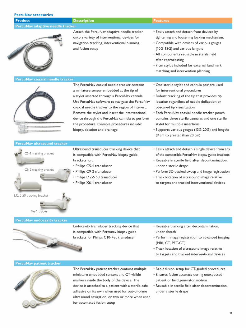
31
C5-1 tracking bracket
L12-5 50 tracking bracket
C9-2 tracking bracket
PercuNav accessoriesProduct Description FeaturesPercuNav adaptive needle tracker
Attach the PercuNav adaptive needle tracker onto a variety of interventional devices for navigation tracking, interventional planning, and fusion setup
• Easily attach and detach from devices by tightening and loosening locking mechanism.
• Compatible with devices of various gauges (10G-18G) and various lengths
• All components reusable in sterile field after reprocessing
• 7 cm stylus included for external landmark matching and intervention planning
PercuNav coaxial needle trackerThe PercuNav coaxial needle tracker contains a miniature sensor embedded at the tip of a stylet inserted through a PercuNav cannula. Use PercuNav software to navigate the PercuNav coaxial needle tracker to the region of interest. Remove the stylet and insert the interventional device through the PercuNav cannula to perform the procedure. Example procedures include: biopsy, ablation and drainage
• One sterile stylet and cannula pair are used for interventional procedures
• Robust tracking of the tip that provides tip location regardless of needle deflection or obscured tip visualization
• Each PercuNav coaxial needle tracker pouch contains three sterile cannulas and one sterile stylet for multiple insertions
• Supports various gauges (13G-20G) and lengths (9 cm to greater than 20 cm)
PercuNav ultrasound trackerUltrasound transducer tracking device that is compatible with PercuNav biopsy guide brackets for:• Philips C5-1 transducer• Philips C9-2 transducer• Philips L12-5 50 transducer• Philips X6-1 transducer
• Easily attach and detach a single device from any of the compatible PercuNav biopsy guide brackets
• Reusable in sterile field after decontamination, under a sterile drape
• Perform 3D tracked sweep and image registration• Track location of ultrasound image relative
to targets and tracked interventional devices
PercuNav endocavity trackerEndocavity transducer tracking device that is compatible with Percunav biopsy guide brackets for Philips C10-4ec transducer
• Reusable tracking after decontamination, under sheath
• Perform image registration to advanced imaging (MRI, CT, PET-CT)
• Track location of ultrasound image relative to targets and tracked interventional devices
PercuNav patient trackerThe PercuNav patient tracker contains multiple miniature embedded sensors and CT-visible markers inside the body of the device. The device is attached to a patient with a sterile-safe adhesive on its own when used for out-of-plane ultrasound navigation, or two or more when used for automated fusion setup
• Rapid fusion setup for CT-guided procedures• Ensures fusion accuracy during unexpected
patient or field generator motion• Reusable in sterile field after decontamination,
under a sterile drape
X6-1 tracker
452299101151.indd 31 25/04/14 15:44

32
• Cardiac 3D measurements, 3D quantification from MPR views includes the following measurements
– Distance – Area – Biplane LV volume (Simpson’s) – Biplane LV ejection fraction – Biplane LV mass
Cardiac 3D Quantification Advanced (3DQ Advanced) • Left ventricle global and regional volume and timing analyses with
no geometric assumption • Comprehensive report page with AHA/ASE 17-segment bull’s-eye
plots and numeric values • Image quality index using dedicated color scale for 3D volume
quality control • Display and manipulation of dynamic 3D rendering and left
ventricular (LV) true volumes of Live 3D data sets• Displays of 3D or dynamic 3D renderings in grayscale, single
colorization, or dynamic colorization • Multiplanar reconstruction (MPR) views • Option to flip LV apical two-chamber display and corresponding
septal, anterior, lateral, inferior (SALI) sequence • iSlice display-compatible • Measurements of LV endocardial true 3D volumes, LV ejection
fraction, and stroke volume using semi-automated 3D border detection
• Computation of regional volumes based on AHA/ASE 17-segment LV model
• Edit mode that adds flexibility for optimal 3D border tracking in four dimensions
• Display of global LV volume waveforms, all 17 regional volume waveforms, or a subset of user-selected regional volume waveforms
• Displays of dyskinetic segments and corresponding volume waveforms in specific color and format
• Display of regional end-diastolic normalized regional volume waveforms
• User-selectable waveforms: single, by wall, by level (ring) modes • A bull’s-eye visualization of all 17 regional segments or the
user-defined and user-selected regional segments • Global and regional reports that provide 3D LV global values and
regional timing indices from all or a subset of 17 regional segments and bull’s-eye-based parametric imaging display
– 3D true volume-based EDV, ESV, stroke volume, and EF – Standard deviation and maximal difference of time to minimum systolic volume (Tmsv) based on all or a subset of 17 regional segments
Measurementtoolsandgeneraldescription• 2D distance• 2D circumference/area by ellipse, continuous trace, trace by points• Auto conversion of distance to ellipse• 2D curved-linear distance• 2D angle: intersection of two lines• 3D: ellipse and distance on 2 MPR views• 3D: stacked contours on one MPR• M-mode distance (depth, time, slope)• Manual Doppler distance• Manual Doppler trace• Time/slope measurements in Doppler and M-mode• High Q automatic Doppler analysis (general imaging only)
– Doppler values including PI, RI, S/D indices• Volume flow• 2D volume (2 volume methods)• Heart rate• Trackball-controlled electronic measurement calipers: 8 sets• User-defined protocols, measurements and equations• On-the-fly measurement labels• Fully editable results data sheet• Integrated patient exam report• Delete last measurement• Embed image and graphs in report
7.1MeasurementtoolsandquantificationQLAB quantification softwareCardiac 3D Quantification (3DQ) • On-cart and off-cart access• Customize capabilities via optional plug-ins• Capable of performing 2D measurements from 3D volume
and 3D color volume multiplanar reconstruction (MPR) views • Review and quantification of Live 3D, 3D zoom, 3D full volume
and color full volume files• 3D image controls: 3D vision map, 3D single or dynamic
colorization, 3D color render, 3D color suppress control • Multiplanar reconstruction (MPR) views
– 3D slice plane – Parallel plane – Unlimited MPR manipulation – Plane rotation, tilt, movement controls to reduce left ventricle foreshortening
• 3D annotation • 3D spatial reference icon
7. Measurements and analysis
452299101151.indd 32 25/04/14 15:44

33
Mitral Valve NavigatorA.I. (MVNA.I.) • 3D assessment of mitral valve anatomy and associated structures • Review and quantification of Live 3D and full volume data sets from
X7-2t and xMATRIX transducer (Live 3D TEE) • Task-driven workflow provides user direction and guidance
illustrations • Automated ES selection • Automated 3D annulus segmentation and leaflet surface • Associated 2D, 3D, and projected measurements and calculations
sorted by group – Annulus – Leaflet – Aortic-Mitral – Coaptation – Papillary
• 3D image controls – 3D vision map – 3D single or dynamic colorization – Auto-view – Absolute and relative rotation – Three 3D render modes: volume, slices, model
• Multiplanar reconstruction (MPR) views – 3D slice plane – Unlimited MPR manipulation – Slice thickness – MPR smooth
• 3D mitral labels • 3D mitral model
– Model displays: tenting surface, leaflet surface, minimum surface – Enhanced coaptation line tracing – Leaflet discontinuity traces – Leaflet segmentation – Up to 53 measurements overlay – Exposed and coapted leaflet length and surfaces – Continuous display during loop playback
• Mitral valve 3D measurements and 2D/3D quantification from model view include the following measurements
– Distances – Curve distances – Areas – Projected areas – Volumes – Angles – Ratio
– Tmsv values displayed in time (msec) or normalized to the R-R interval (%)
– Bull’s-eye showing the user-selected segments for time to minimum systolic volume (Tmsv) calculation
– LV timing and radial excursion parametric images in bull’s-eye format using effective color-coded scales
– Parametric imaging supporting AHA/ASE 17-segment overlay on the bull’s-eye for direct and rapid visualization
– Parametric imaging that provides a radial excursion threshold slider for selective visualization of LV segments in the timing parametric display
– Measurement data exported in Excel or DICOM SR formats
General Imaging 3D Quantification (GI 3DQ) • Access to 3D viewing controls • Simple annotation • Linear distance measurements
– Polygon, free-form polygon, spline, free-form spline, rectangle measurements
• Curved distance measurements • Ellipse measurements • Area measurements • Auto area measurements semi-automated tool to simplify
measurement of hypoechoic structures • Angle measurements • 3D measurement tools
– Stacked contour measurements – Auto-stacked contour measurements semi-automated tool to simplify volume measurements of hypoechoic structures
– Auto-volume tool – 3D distance/curved iSlice – Stacked ellipsoid measurements – Ellipsoidal measurements
• Calculation of the following color indices using the 2D and/or 3D measurement tools
– Vascularization index (VI) – Flow index (FI) – Vascularization flow index (VFI) – Pixel intensity index (PII) and echo histogram display
• Support of the display of 3D orientation labels (when enabled) – Trending capability (available only on stand-alone PC)
• Display of timer (when enabled )
452299101151.indd 33 25/04/14 15:44

34
• Automated Tissue Motion Annular Displacement (aTMAD) – Mitral valve and other valve annular motion tracking over time – Computation of valve annular displacement curves over time – Color Kinesis overlay to visualize valve annular plane motion parametrically
– Measurement data exported in Excel or DICOM SR formats – Simplified workflow with SmartExam
Automated Cardiac Motion 2D QuantificationA.I. (aCMQA.I.) • Automated Region of Interest for selected anatomical views • Objective assessment of left ventricle global function and regional
wall motion, deformation, and timing using the next generation of 2D speckle tracking technology
• File compatibility – Display and quantification of 2D native ultrasound DICOM images
• Available methods with dedicated preferences settings – Global workflow
• Latest generation of 2D speckle tracking technology • Automated border detection for cardiac chambers and
vessel cavities • Auto aortic valve closure time detection • Smooth color transited bull’s eye presentation
– Multiple cardiac view/images capable – AHA/ASE 17 left ventricle segmentation templates (three apical views and three short-axis view templates)
– Easy-to-edit template position and shape – Intuitive step-by-step user interface – Tracking quality tool editable threshold help to display various quality tracking
– User-editable post LV segments display consistent display with corresponding waveform and reported values beat-to-beat selection
– Tracking that can be initiated from any frame • Display
– Border (hide or show) – Vector velocity field (hide or show) – Image ROI Overlay (hide or show) – Cardiac phases (overlay of AVO, AVC, MVO, and MVC mechanical events auto-imported from ultrasound cart analysis via DICOM SR or manual entry)
• 2D speckle parameters – Volume/EF and area/FAC – Longitudinal strain and strain rate – Circumferential strain and strain rate – Radial and transversal displacement – Radial fractional shortening
• Measurement and calculation definition and overlay on 3D model • Comprehensive reporting • Data exported in Excel or DICOM SR formats • Measurement data exported in Excel or DICOM SR formats
Fetal Heart Navigator (FHN) • Guides the user in obtaining the standard diagnostic views of the
fetal heart • Improves reproducibility and consistency between users • Protocol-driven workflow
– Automates the initial ductal arch view – Guides user in obtaining view recommended in ISUOG Fetal Cardiac Screening Guidelines
– Obtains the fetal heart view: 4-chamber, LVOT, and RVOT – Supports X6-1 iSTIC and V6-2 STIC data sets containing eight or more frames
• Presets supported: OB Difficult, OB Early, OB Fetal Echo, OB Fetal Echo CV, OB General, OB Max Pen, GYN Pelvic
• Visualization controls allow users to change display settings at any stage in the protocol
– Chroma map – Gray map – Slice thickness – Brightness
Automated Cardiac 2D QuantificationA.I. (a2DQA.I..) • Left ventricle global volume analysis from 2D and biplane images • Display of 2D ultrasound native DICOM images• Automated border detection for cardiac chambers and
vessel cavities • Computation of area, LV volumes, and advanced parameters for
LV systolic and diastolic function including fractional area change (FAC), ejection fraction (EF), peak ejection rate (PER), peak rapid filling rate (PRFR), and atrial filling fraction (AFF)
• Single-plane volume measurements based on 5/6 Area-Length Method and Simpson’s Single Plane Method of Disks (MOD)
• Biplane volume measurements based on Simpson’s Biplane Method of Disks (MOD)
• Color Kinesis (CK) tool that provides color-coded visualization of global and regional wall motion displaying automated borders over time parametrically
• Transparency control to visualize echo grayscale under a semi-transparent CK overlay
• CK display for arbitrary frame rates (high frame rate CK display) • Manual user-editable timing overrides for the onset and duration
of the CK parametric display
452299101151.indd 34 25/04/14 15:44

35
– Radial velocity – Speed (absolute angle independent velocity) – Regional rotation and rotation velocity – Global rotation (SAX) – Endo-Epi mural torsion and local rotation
• Measurement and calculations – Waveform auto peak or systolic peak detection to report – Time to peak and peak values – Timing caliper – Global result display one view and global result display – Results display in AHA/ASE 17 LV segment bull’s-eye plot format and numerical table
– Longitudinal strain per view and global longitudinal strain – Circumferential strain per view and global circumferential strain – User-defined workflow for specific local strain analysis
• Up to 17 dedicated colors to help differentiate each cord and corresponding waveform
• Up to three waveform auto peak detections to report time to peak and peak values
• Color Kinesis (CK) overlay for color-coded visualization • Simplified workflow with SmartExam • Measurement data exported in Excel or DICOM SR formats
Cardiac Motion/Mechanics 2D Quantification for Stress (CMQ Stress) Designed to help objectify stress echo exam interpretation, CMQ Stress employs a user interface specifically designed for stress echo exams and around stress echo practitioners. • Automated Region of Interest for selected anatomical views • Objective assessment of left ventricle global function and regional
wall motion, deformation, and timing using the next generation of 2D speckle tracking technology
• File compatibility – Display and quantification of 2D native ultrasound DICOM Stress Echo images
• Global workflow – Latest generation of 2D speckle tracking technology – Semi-automated border detection for cardiac chambers and vessel cavities
– Auto aortic valve closure time detection – Smooth color transited bull’s-eye presentation – Multiple cardiac view/images capable – AHA/ASE 17 left ventricle segmentation templates (three apical views and three short-axis view templates)
– Easy-to-edit template position and shape – Intuitive step-by-step user interface – Tracking quality tool editable threshold helps to display various quality tracking
– User-editable post LV segments display consistent display with corresponding waveform and reported values beat-to-beat selection
– Tracking that can be initiated from any frame • Display
– Border (hide or show) – Vector velocity field (hide or show) – Image ROI Overlay (hide or show) – Cardiac phases (overlay of AVO, AVC, MVO, MVC mechanical events auto-imported from ultrasound cart analysis via DICOM SR or manual entry)
• 2D speckle parameters – Volume/EF and area/FAC – Longitudinal strain and strain rate – Circumferential strain and strain rate – Radial and transversal displacement – Radial fractional shortening – Radial velocity – Speed (absolute angle independent velocity) – Regional rotation and rotation velocity – Global rotation (SAX) – Endo-Epi mural torsion and local rotation
• Measurement and calculations – Waveform auto peak or systolic peak detection to report time to peak and peak values
– Timing calipers – Global result display one view and global result display – Results display in AHA/ASE 17 LV segment bull’s-eye plot format and numerical table
– Longitudinal strain per view and global longitudinal strain – Circumferential strain per view and global circumferential strain – User-defined workflow for specific local strain analysis
• Up to 17 dedicated colors to help differentiate each cord and corresponding waveform
• Up to three waveform auto peak detections to report time to peak and peak values
• Color Kinesis (CK) overlay for color-coded visualization – Measurement data exported in Excel formats
Elastography Quantification (EQ) Provides strain elastography quantification of relative tissue deformation based on an elastogram. • Ability to generate up to ten user-defined Regions of Interest
(ROIs) • Thumbnail display of frames • Measurement results • Calculation and display of strain rate and total strain • Size comparison between two ROIs
452299101151.indd 35 25/04/14 15:44

36
• Strain ratio • Calculation of ratio between two user-defined ROIs • Graphical display strain ratio parametric image • Color-coded display of strain ratios with parametric imaging • Compatible with EPIQ systems using Elastography mode with
L12-5, C10-3V and L17-5 transducers
Intima Media Thickness (IMT) measurements • Automated measurements of intima media thickness in carotids
and other superficial vessels • Automated measurement technique on user-selected frames • Selector chart to record location and side of vessel from where
IMT is measured • Report of the IMT values in mean millimeters and standard
deviation • Quick optimization for thin or thick intima media complexes • User-adjustable Region of Interest • User-defined measurement capability • Persistent storage of up to ten measurements with image files
for future reference• Measurement data exported in Excel or DICOM SR formats
MicroVascular Imaging (MVI)• Review loops including side-by-side display files• Motion compensation algorithm selectable in Preferences menu• Export of single frames as BMP, JPG, or TIF file format• Export of movie files as AVI file format
Region of Interest (ROI) Quantification • Vascular, general radiology, and cardiac images • Trending capability (available only on stand-alone PC) • Up to ten user-defined regions • Thumbnail display of frames for easy trimming • Pixel intensity index (PII) and echo histogram display, supported
data types: echo, velocity, or power (angio) • Custom tools to auto trim relative to ECG trigger on cardiac
and other triggered files for quantification of specific parts of cardiac cycle
• Region shaping tools – Polygon – Free-form polygon – Spline – Free-form spline – Rectangle – Square 5 mm
• Single frame tools – Angle – Annotation – Curved distance – Distance – Ellipse – Live xPlane ROI combination
• Auto area for semi-automated quantification of hypoechoic structures
• Adjustable ROI dynamic tool • Motion compensation algorithm selectable in Preferences menu • Data display selection
– Log – Linear
• Data display smoothing option • Mean, median, and standard deviation calculation • Time-intensity curves • Curve fitting tools
– Gamma-variate (wash-in and wash-out) – One minus exponential – Linear – Log normal WI – Log normal WIWO – LDRW WIWO
• Graphic results with dB, intensity or velocity/ frequency, time to peak, “A” value, area under curve, and peak intensity (when a gamma variate curve has been applied) for each frame
• Calculation of the following color indices using the 2D measurement tools
– Vascularization index (VI) – Flow index (FI) – Vascularization flow index (VFI)
Strain Quantification (SQ) • Used in the evaluation of regional myocardial function • Measures the myocardial velocity from color TDI data set and
derives the displacement, strain, and strain rate along user-defined M-lines
• Includes ability to overlay opening and closing of aortic and mitral valves on SQ curves to display left ventricle mechanical events
• User-selectable waveform display that makes SQ curves easier to read
• Capable of drawing up to four M-lines at a time • Point of Interest (POI) tool that obtains values from any point
on the M-mode display • M-mode (hide or display) control
452299101151.indd 36 25/04/14 15:44

37
• User-defined and automatic (using speckle tracking algorithms) M-line motion compensation to follow myocardial motion
• Able to present TDI results in two display formats – Anatomical M-mode display – Graph display
• User-selectable waveforms for optimal sub-region visualization • Curve processing modes • Cardiac mechanical timing reference overlay • TDI velocity, displacement, strain and strain rate timing
measurements with dedicated time calipers and labels • Automatic subdivision of M-line into a customizable number
of sub-regions • Averages up to 20 cardiac beat cycles in both M-mode and
graph displays • Measurement data exported in Excel format
7.2HighQautomaticDoppleranalysis• Automatic real-time and retrospective tracing of:
– Instantaneous peak velocity – Instantaneous intensity weighted mean velocity
• Automatic real-time display of (user selectable up to six): – Volume flow – Time-averaged peak velocity – Time-averaged mean velocity – Resistive index – Pulsatility index – Systolic/diastolic ratio – Acceleration/deceleration times – Illustrated High Q
7.3Clinicaloptionanalysispackages• Cardiac analysis• Volume by area/length method• M-mode ejection fraction (via Teichholz or cubed method)• Novel 3-point adjustable Simpson’s template• Simpson’s biplane and single plane volume and ejection fraction• Area, length, volume and ejection fraction• LV mass• 2D all points• M-mode all points• Peak velocity• Maximum and mean pressure gradients• Pressure half time• E/A ratio• D/E slope• Continuity equation• Diastolic function
• Cardiac output• Acceleration time• Heart rate• Vascular analysis
– Right and left carotid artery protocols – ICA/CCA ratio – Bilateral lower extremity arterial and venous labels – Bilateral upper extremity arterial and venous labels – Percent diameter and area reduction – Vascular graft measurement package – User comments – High Q automatic Doppler analysis
• OB analysis – Fetal echo application – Fetal biometry (up to quintuplets) – Biophysical profile – Amniotic fluid index – Early gestation – Fetal long bones – Fetal cranium – Other OB measurements - 2D echo - Fetal heart M-mode - Fetal Doppler - Fetal echo
• Gynecology/fertility – Uterine volume – Right and left ovary volumes – Right and left follicles (10) – Endometrial thickness – Cervical length
• Abdominal vascular – Labels for all major abdominal arteries and veins – Left and right segmentation for kidneys
• General imaging – General – User-defined labels
• Prostate – Prostate gland
• Pediatric – General – d:D radio
• Small parts – General – Breast with right and left protocols for up to five lesions per breast
452299101151.indd 37 25/04/14 15:44

38
8. Physical specifications
System cart• State-of-the-art ergonomic design for comfort and convenience• Easy maneuverability and mobility
– Wheel-lock and monitor adjustments that facilitate bedside exams• Independent height adjustment of control panel and display monitor• Easily accessed transducer connector ports, USB, and DVD
media drive, if equipped• Transducer and gel bottle holders• Mobility through high quality, shock-absorbing casters with foot
pedal controls for: – 4-wheel swivel – 2-wheel swivel lock – 2-wheel brakes
• Integrated footrests • Transducer connector and OEM bay illumination for easy visibility
in scanning rooms• Digitally-enhanced 2-speaker high fidelity stereo output with
rear mounted subwoofer• On-board storage tray behind control panel touch screen
and in rear bay left and right storage drawers• Universal peripheral bay that provides easy access for up to two
on-board hardcopy or documentation devices• Built-in A/C line conditioner that provides isolation from voltage
fluctuations and electrical noise interference• Three high-capacity fans with automatic speed adjustment
to optimize cooling efficiency with minimal audible noise
Monitor• Flat panel LCD display monitor
– 21.5 in/54.6 cm wide format high definition flat panel TFT/IPS display
– High contrast ratio >1000:1 – Extended viewing angle >178º (horizontal and vertical) – Response time: <14 ms – Virtually flicker-free technology for reduced eyestrain – Mounted on fully articulating extension arm – Four-way articulation with 34.5 in/87.6 cm lateral and 7 in/17.8 cm vertical adjustment range - Nearly infinite positioning adjustments: height, swivel, and tilt
Control panel• Articulation facilitates nearly infinite positioning adjustments
for optimum scanning ergonomics: height, swivel, and tilt – Up and down 10 in/25.4 cm – Rotates 180º from center – Complete freedom for side-to-side slide movement, infinite positions
– Operates on battery for adjustment during mobile exams – Retractable, backlit alphanumeric keyboard – Palm rest
Physical dimensionsWidth 23.9 in/60.6 cm
Height 57.5-67.5 in/146-171.5 cm
Depth 43 in/109.2 cm
Weight 230 lbs/104.3 kg without peripheral devices
452299101151.indd 38 25/04/14 15:44

39
Physio• One 3-lead ECG input
– Gain, sweep rate, and display position controls – Automatic heart rate calculation and display – Fault condition display – Cineloop locator displayed on one ECG input from an ECG source like stress ECG or ECG monitor
Peripherals • The system supports up to two on-board peripheral devices
(excluding report printers) – Video-recording peripherals, operated via system user interface – DVD recorder (cart-dependent) – Small format digital color printer (USB) – Small format digital B/W printer (USB) – PercuNav
• Support for large format external color printer• Support for various Hewlett-Packard and Epson brand color
and monochrome report printers (USB, externally mounted)
Input/output ports• Export of measurement and analysis data to off-line reporting
software packages (USB)• Display port video export available for either full screen resolution
of 1920x1080 (1080p) or display area 1024x768
Powerrequirementsandvideoparameters• 100V-240 V, 50 Hz/60 Hz – PAL/NTSC• Integrated A/C line conditioning and battery back-up system• Power consumption: <600 VA depending on system configuration
Electrical safety standards • Electromechanical Safety Standards met
– CAN/CSA 22.2 No. 60601-1, Medical Electrical Equipment: General requirements for basic safety and essential performance
– IEC 60601-1, Medical Electrical Equipment: General requirements for basic safety and essential performance
– IEC 60601-1-2, Collateral Standard, Electromagnetic compatibility – requirements and tests
– IEC 60601-2-37, Particular Requirements for the basic safety and essential performance of ultrasonic medical diagnostic and monitoring equipment
– ANSI/AAMI ES60601-1, Medical Electrical Equipment: General requirements for basic safety and essential performance
• Electromechanical Safety Standards met (EU Only) – EN60601-2-37, Particular requirements for the basic safety and essential performance of ultrasonic medical diagnostic and monitoring equipment
• Agency approvals – Canadian Standards Association (CSA) – CE Mark in accordance with the European Medical Device Directive issued by British Standards Institute (BSI)
452299101151.indd 39 25/04/14 15:44
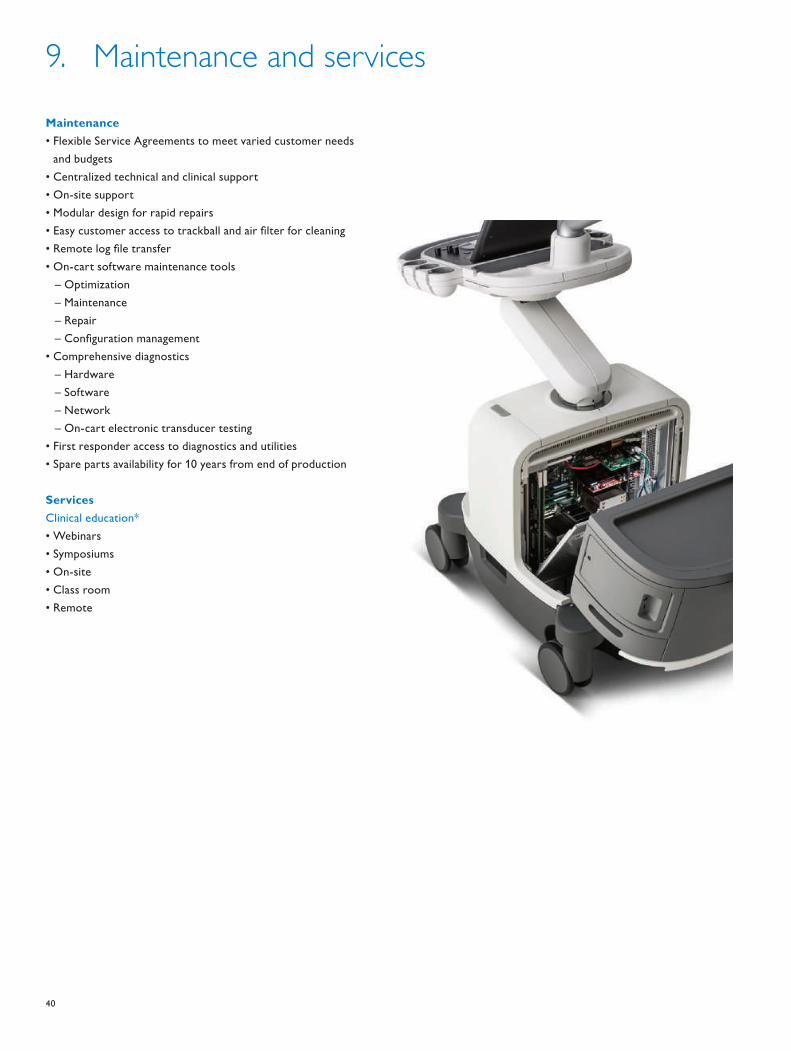
40
9. Maintenance and services
Maintenance• Flexible Service Agreements to meet varied customer needs
and budgets • Centralized technical and clinical support• On-site support• Modular design for rapid repairs• Easy customer access to trackball and air filter for cleaning• Remote log file transfer• On-cart software maintenance tools
– Optimization – Maintenance – Repair – Configuration management
• Comprehensive diagnostics – Hardware – Software – Network – On-cart electronic transducer testing
• First responder access to diagnostics and utilities• Spare parts availability for 10 years from end of production
ServicesClinical education*• Webinars• Symposiums• On-site• Class room• Remote
452299101151.indd 40 25/04/14 15:44

41
Philips Remote Connectivity*• iSSL and encryption• Patient De-identify• Security• Remote Desktop
– Remote technical support – Remote clinical support – Remote clinical education
• On-cart remote support request• Proactive monitoring with alerting capability
– Monitors key system parameters - Voltage - Temp - Fan speeds - Error conditions - Local alert handling and response
Warranty• Philips standard product warranty
*Service agreement required for access to Philips Remote Services. Access to the internet required. Not all remote features available in all countries; contact your Philips representative for details.
452299101151.indd 41 25/04/14 15:44

© 2014 Koninklijke Philips N.V.All rights are reserved.
Philips Healthcare reserves the right to make changes in specifications and/or to discontinue any product at any time without notice or obligation and will not be liable for any consequences resulting from the use of this publication.
Printed in The Netherlands.4522 991 01151 * APR 2014
Please visit www.philips.com/EPIQ
Philips Healthcare is part of Royal Philips
How to reach uswww.philips.com/[email protected]
Asia+49 7031 463 2254
Europe, Middle East, Africa+49 7031 463 2254
Latin America+55 11 2125 0744
North America+1 425 487 7000800 285 5585 (toll free, US only)
452299101151.indd 42 25/04/14 15:44