Embed Size (px)
Citation preview
NanoscaleAdvances
PAPER
Ope
n A
cces
s A
rtic
le. P
ublis
hed
on 1
1 m
árci
us 2
020.
Dow
nloa
ded
on 2
022.
04.
04.
14:
50:1
2.
Thi
s ar
ticle
is li
cens
ed u
nder
a C
reat
ive
Com
mon
s A
ttrib
utio
n-N
onC
omm
erci
al 3
.0 U
npor
ted
Lic
ence
.
View Article OnlineView Journal | View Issue
A nano-integrate
aState Key Laboratory of Molecular Vaccinol
Public Health, Xiamen University, Xiame
[email protected] Zhongshan Hospital of Dalian U
China. E-mail: [email protected]
† Electronic supplementary informa10.1039/c9na00786e
Cite this: Nanoscale Adv., 2020, 2,2192
Received 16th December 2019Accepted 7th March 2020
DOI: 10.1039/c9na00786e
rsc.li/nanoscale-advances
2192 | Nanoscale Adv., 2020, 2, 2192
d diagnostic and therapeuticplatform with oxidation–reduction reactions intumor microenvironments†
Lei Zhao,ab Guihua Qiu,a Kai Wang,a Hu Chen,a Fengkai Ruan,a Nan Liu,a Zifeng Deng,a
Youliang Yao,a Dongbei Guo,a Dai Wang,a Li Sha,b Xiangyu Kong,b Wenzhi Liu*b
and Yongxing Zhang *a
In the present study, we developed a nano-integrated diagnostic and therapeutic platform with oxidation–
reduction reactions in tumormicroenvironments (TMEs). The proposed platform resolved the contradiction
of particle size between the enhanced permeability and retention (EPR) effect and tumor interstitial
penetration, as well as poor circulation and low drug-loading efficiency. Flower-like MnO2 NPs were
used as the core and modified with hyaluronate (HA) and H2PtCl6 to obtain MnO2–HA@H2PtCl6 (MHP).
The maximum drug-loading efficiency rate of H2PtCl6 reached 35% due to its chelation with HA. MHP
showed satisfactory integrity and stability during circulation and can also be used as a magnetic
resonance imaging (MRI) contrast agent. In addition, MHP as a radiosensitizer achieved an excellent
tumor inhibition effect in combination with radiotherapy. Importantly, MHP released ultra-small
nanoparticles, USNPs, (�20 nm) through the supramolecular self-assembly abilities of Mn2+, HA, and
H2PtCl6 in TMEs, leading to the increase of penetration into multicellular spheres and solid tumors
(Scheme), as well as prolonging its retention in tumors.
1 Introduction
The transportation of nanoparticles (NPs) in solid tumors ismainly associated with their diameter and surface potential.1–4
A dense extracellular matrix, high interstitial uid pressure(IFP), and uneven distribution of tumor vessel diameter are themain reasons for poor clinical treatment.5,6 NPs with diametersin the range of 60–100 nm have shown a satisfactory enhancedpermeability and retention (EPR) effect and high tendencytowards extravasation across the tumor vessels, while theycannot uniformly and effectively penetrate into the tumorstroma.3,7 The NPs with diameters in the range of 10–20 nm cansignicantly penetrate into the tumor stroma and spreadevenly, while the removal rate is faster, and the retention time oftumor is shortened.5
Radiation therapy is a therapeutic method, utilizing ionizingradiation to induce DNA damage to kill cancer cells.8 Besides, itsdisadvantages mainly include lack of selectivity of radiation loca-tion and poor efficacy due to the hypoxic regions in solid tumors.9
ogy and Molecular Diagnostics, School of
n, Fujian, 361102, PR China. E-mail:
niversity, Dalian, Liaoning, 116001, PR
tion (ESI) available. See DOI:
–2202
In order to solve the above mentioned problems, radiosensitizers(e.g., gold nanoparticles10 and titanium oxide nanoparticles11) havebeen developed. However, a number of these nanoparticles are notbiodegradable and cannot be therefore used in clinical practicedue to their long-term retention in vivo. It has been reported thatthe nanomaterial peruorocarbon is helpful to transport oxygen,but there are a number of shortcomings, such as short retentiontime of oxygen in nanomaterials, low efficiency of oxygen trans-port, and the need for a particular range of temperature to controland release.12 It is well-known that high concentration reactiveoxygen species (ROS) (e.g., H2O2, �100 mM) in tumor microenvi-ronments (TMEs) can limit the efficacy of radio-chemotherapy intumors.13Moreover,M2 phenotype tumor-associatedmacrophages(TAMs) promote tumor metastasis and drug resistance in hypoxicregions and tumor hypoxic environments.14
In the present study, we synthesized a biodegradable nano-integrated diagnosis and treatment platform, namely MHP, inorder to overcome the above-mentioned problems. For thispurpose, hyaluronan (HA), as a hydrophilic polysaccharide, wasused to modify the surface of ower-like MnO2 cores andincrease the loading rate of H2PtCl6. MHP is sensitive tooxidation–reduction reactions and has acceptable biocompati-bility and can be utilized as an ultra-small nanoparticle (USNP)triggered by TMEs. With consuming excessive endogenousH2O2 in TMEs, it degrades from the original parental NPs (100nm) to USNPs (�20 nm), leading to deeper penetration andlonger retention in the tumor stroma. Due to its initial
This journal is © The Royal Society of Chemistry 2020

Paper Nanoscale Advances
Ope
n A
cces
s A
rtic
le. P
ublis
hed
on 1
1 m
árci
us 2
020.
Dow
nloa
ded
on 2
022.
04.
04.
14:
50:1
2.
Thi
s ar
ticle
is li
cens
ed u
nder
a C
reat
ive
Com
mon
s A
ttrib
utio
n-N
onC
omm
erci
al 3
.0 U
npor
ted
Lic
ence
.View Article Online
appropriate hydrodynamic diameter (100 nm), MHP hasa promising EPR effect. MHP as a radiosensitizer can achieve aneffective tumor inhibition in combination with radiotherapy. Inaddition, by means of multi-elemental and spectral detection,we preliminarily conrmed the basic components of the USNPsby combining different centrifugation and dialysis methods.Our research was conducted on the basis of the formation ofUSNPs by supermolecular recombination aer the metabolismof nanomaterials induced by TMEs, aiming to provide a newidea for penetration of radio-chemotherapy into clinicaltumors.
2 Results and discussion2.1 Synthesis and characterization
In the current research, we synthesized MnO2–HA@H2PtCl6nanoparticles by the layer-by-layer (LBL) method as a nanoscalecoating and surface functionalization technique. Next, oleic
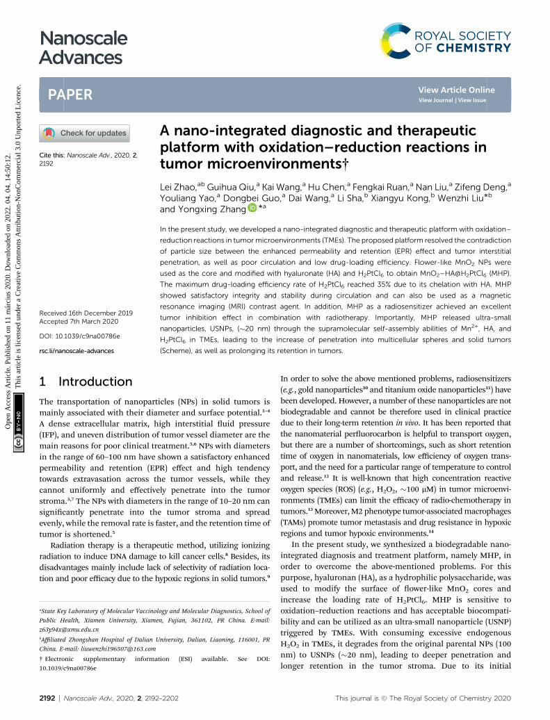
Fig. 1 (a) TEM image of MnO2 NPs, scale bar: 100 nm; (b) TEM image oscale bar: 100 nm; (d) schematic illustration of synthesis of MHP; (e) HCTTEM images of HA-H2PtCl6 at different ratios of HA/H2PtCl6. The scalesH2PtCl6, MH, and MHP; (j) loading efficiency of H2PtCl6 at different ratio
This journal is © The Royal Society of Chemistry 2020
acid was utilized to reduce potassium permanganate to obtainower-like MnO2 NPs with a size of 70–110 nm (average size: 90nm) (Fig. 2b), and then, they were characterized by transmissionelectron microscopy (TEM; Fig. 1a) and (EDS; Fig. S1†).
Aerwards, the HA was adsorbed on the surface of MnO2 toform MnO2–HA. A number of scholars demonstrated that HApossesses a series of appropriate properties, such as biocom-patibility, biodegradability, and non-immunogenicity.15,16 TEMrevealed that core-MnO2 was surrounded by light-coloredsubstances (Fig. 1b), suggesting that HA has been successfullyloaded around the core. Zeta potential almost showed the samesurface charge as HA (Fig. 1i), suggesting that the combinationof HA and core-MnO2 is electrostatic adsorption.
Before nal loading, we tested the inhibitory effect ofH2PtCl6 on cancer cells. It was disclosed that treatment with 10mg mL�1 H2PtCl6 could signicantly inhibit the proliferation ofHCT-116, and treatment with 20 mg mL�1 H2PtCl6 for 48 h couldinhibit cell proliferation by within 45% (Fig. 1e). Additionally,
f MnO2–HA, scale bar: 100 nm; (c) TEM image of MnO2–HA@H2PtCl6,-116 cell viabilities after incubation with H2PtCl6 for 24 and 48 h; (f–h)are 50, 100, and 200 nm, respectively; (i) zeta potential of MnO2, HA,s; (k) UV-Vis-NIR of MnO2, HA, H2PtCl6, MH, and MHP. *P < 0.05.
Nanoscale Adv., 2020, 2, 2192–2202 | 2193
Nanoscale Advances Paper
Ope
n A
cces
s A
rtic
le. P
ublis
hed
on 1
1 m
árci
us 2
020.
Dow
nloa
ded
on 2
022.
04.
04.
14:
50:1
2.
Thi
s ar
ticle
is li
cens
ed u
nder
a C
reat
ive
Com
mon
s A
ttrib
utio
n-N
onC
omm
erci
al 3
.0 U
npor
ted
Lic
ence
.View Article Online
treatment with 20 mg mL�1 H2PtCl6 for 8 h could cause cellapoptosis by around 81% (Fig. S2†), in addition to remarkabledouble-strand breaks in DNA (Fig. S3†). In vitro, different ratiosof HA/H2PtCl6 might form nanoparticles with different sizes.When the mass ratio turns out to be 8 : 1, 5 : 1, and 4 : 1, theparticle size is less than 50 nm (Fig. 1b–d). Moreover, when theproportion of H2PtCl6 is excessive (Fig. S4†), a ratio of 1 : 5seems to be appropriate to form regular nanoparticles; whenthe ratio is 1 : 4, regular nanoparticles cannot be formed;however, when the ratio is 1 : 8, the proportion of H2PtCl6 maybe excessive. Hence, we can speculate that the binding of HA toH2PtCl6 may be based on the chelation of the Pt-centered atomand HA-ligand.
Eventually, H2PtCl6 was added to the surface of MH to formMnO2–HA@H2PtCl6 (MHP). With the help of inductivelycoupled plasma mass spectrometry (ICP-MS), it was revealedthat the drug-loading efficiency of H2PtCl6 was increased withthe increase of H2PtCl6, and this may be due to high specicsurface area of MnO2. Furthermore, when HA was used as themedium, the drug-loading efficiency of H2PtCl6 was
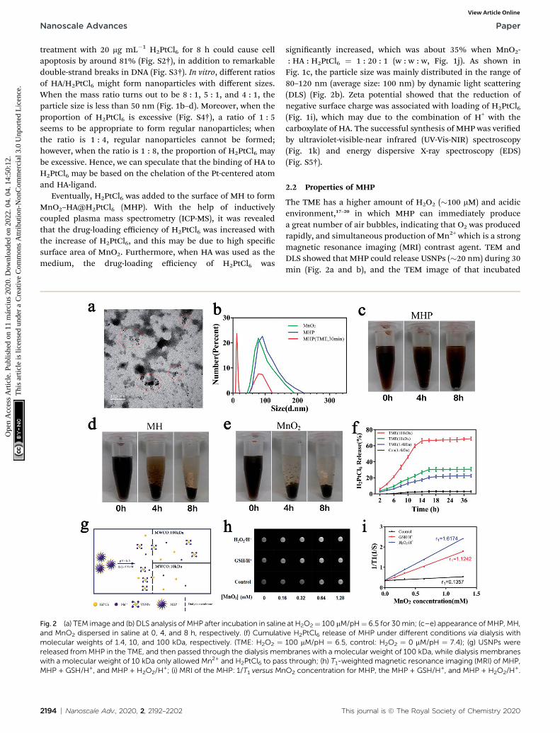
Fig. 2 (a) TEM image and (b) DLS analysis of MHP after incubation in salineand MnO2 dispersed in saline at 0, 4, and 8 h, respectively. (f) Cumulatimolecular weights of 1.4, 10, and 100 kDa, respectively. (TME: H2O2 ¼released from MHP in the TME, and then passed through the dialysis memwith a molecular weight of 10 kDa only allowed Mn2+ and H2PtCl6 to pasMHP + GSH/H+, and MHP + H2O2/H
+; (i) MRI of the MHP: 1/T1 versus Mn
2194 | Nanoscale Adv., 2020, 2, 2192–2202
signicantly increased, which was about 35% when MnO2-: HA : H2PtCl6 ¼ 1 : 20 : 1 (w : w : w, Fig. 1j). As shown inFig. 1c, the particle size was mainly distributed in the range of80–120 nm (average size: 100 nm) by dynamic light scattering(DLS) (Fig. 2b). Zeta potential showed that the reduction ofnegative surface charge was associated with loading of H2PtCl6(Fig. 1i), which may due to the combination of H+ with thecarboxylate of HA. The successful synthesis of MHP was veriedby ultraviolet-visible-near infrared (UV-Vis-NIR) spectroscopy(Fig. 1k) and energy dispersive X-ray spectroscopy (EDS)(Fig. S5†).
2.2 Properties of MHP
The TME has a higher amount of H2O2 (�100 mM) and acidicenvironment,17–20 in which MHP can immediately producea great number of air bubbles, indicating that O2 was producedrapidly, and simultaneous production of Mn2+ which is a strongmagnetic resonance imaging (MRI) contrast agent. TEM andDLS showed that MHP could release USNPs (�20 nm) during 30min (Fig. 2a and b), and the TEM image of that incubated
at H2O2¼ 100 mM/pH¼ 6.5 for 30min; (c–e) appearance of MHP, MH,ve H2PtCl6 release of MHP under different conditions via dialysis with100 mM/pH ¼ 6.5, control: H2O2 ¼ 0 mM/pH ¼ 7.4); (g) USNPs werebranes with a molecular weight of 100 kDa, while dialysis membraness through; (h) T1-weighted magnetic resonance imaging (MRI) of MHP,O2 concentration for MHP, the MHP + GSH/H+, and MHP + H2O2/H
+.
This journal is © The Royal Society of Chemistry 2020
Paper Nanoscale Advances
Ope
n A
cces
s A
rtic
le. P
ublis
hed
on 1
1 m
árci
us 2
020.
Dow
nloa
ded
on 2
022.
04.
04.
14:
50:1
2.
Thi
s ar
ticle
is li
cens
ed u
nder
a C
reat
ive
Com
mon
s A
ttrib
utio
n-N
onC
omm
erci
al 3
.0 U
npor
ted
Lic
ence
.View Article Online
overnight is shown in Fig. S6.† Centrifugation at 16 000 rpm for10 min and dialysis (14 000 MW) were carried out, and ICP-MSdemonstrated [Mn] : [Pt] in the supernatant before dialysis, inthe supernatant aer dialysis, and precipitation aer dialysis,which were (1.27097–1.28031), (1.15368–1.16724), and(1.16223–1.16856), respectively. However, [Mn] : [Pt] wasapproximately equal to 3.772 in MHP, which showed the releaseof superuousMn2+ during transformation. The UV-Vis-NIR didnot show the absorption peak of HA in the supernatant aerdialysis, suggesting that HA was approximately 100% trans-formed. Therefore, we can preliminarily speculate that thechemical formula of the USNPs is as follows: (Mn)4n (H2PtCl6)n(C14H20NO11Na)x, which may be formed by the self-assembly ofMn2+, HA, and H2PtCl6.
Next, the stability of the NPs was tested with saline. It wasobserved that MnO2 was almost fully aggregated and precipi-tated during 4 h (Fig. 2e), reecting a poor hydrodynamicstability, which is consistent with ndings of a previous study.21
Importantly, MH showed a large amount of precipitation, whileMHP revealed nearly no precipitation during 4 h (Fig. 2c and d);aer 8 h, MH was almost fully precipitated, whereas MHP onlyrevealed a small amount of precipitation, demonstrating thatMHP had superior stability to MnO2 and MH in saline, and thismay be related to the surface charge and dispersion of NPs.Although it was disclosed that the hydrodynamic stability of
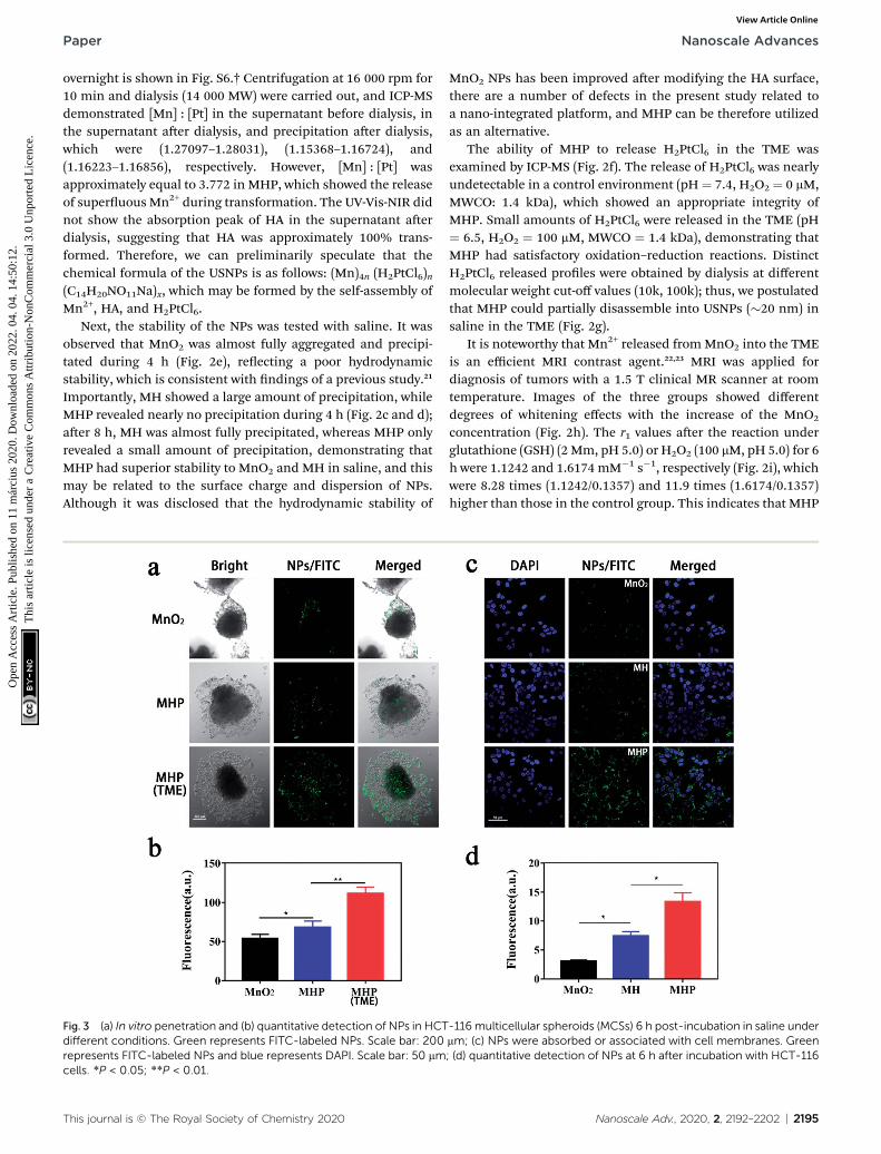
Fig. 3 (a) In vitro penetration and (b) quantitative detection of NPs in HCTdifferent conditions. Green represents FITC-labeled NPs. Scale bar: 200represents FITC-labeled NPs and blue represents DAPI. Scale bar: 50 mmcells. *P < 0.05; **P < 0.01.
This journal is © The Royal Society of Chemistry 2020
MnO2 NPs has been improved aer modifying the HA surface,there are a number of defects in the present study related toa nano-integrated platform, and MHP can be therefore utilizedas an alternative.
The ability of MHP to release H2PtCl6 in the TME wasexamined by ICP-MS (Fig. 2f). The release of H2PtCl6 was nearlyundetectable in a control environment (pH ¼ 7.4, H2O2 ¼ 0 mM,MWCO: 1.4 kDa), which showed an appropriate integrity ofMHP. Small amounts of H2PtCl6 were released in the TME (pH¼ 6.5, H2O2 ¼ 100 mM, MWCO ¼ 1.4 kDa), demonstrating thatMHP had satisfactory oxidation–reduction reactions. DistinctH2PtCl6 released proles were obtained by dialysis at differentmolecular weight cut-off values (10k, 100k); thus, we postulatedthat MHP could partially disassemble into USNPs (�20 nm) insaline in the TME (Fig. 2g).
It is noteworthy that Mn2+ released fromMnO2 into the TMEis an efficient MRI contrast agent.22,23 MRI was applied fordiagnosis of tumors with a 1.5 T clinical MR scanner at roomtemperature. Images of the three groups showed differentdegrees of whitening effects with the increase of the MnO2
concentration (Fig. 2h). The r1 values aer the reaction underglutathione (GSH) (2 Mm, pH 5.0) or H2O2 (100 mM, pH 5.0) for 6h were 1.1242 and 1.6174 mM�1 s�1, respectively (Fig. 2i), whichwere 8.28 times (1.1242/0.1357) and 11.9 times (1.6174/0.1357)higher than those in the control group. This indicates that MHP
-116multicellular spheroids (MCSs) 6 h post-incubation in saline undermm; (c) NPs were absorbed or associated with cell membranes. Green; (d) quantitative detection of NPs at 6 h after incubation with HCT-116
Nanoscale Adv., 2020, 2, 2192–2202 | 2195

Nanoscale Advances Paper
Ope
n A
cces
s A
rtic
le. P
ublis
hed
on 1
1 m
árci
us 2
020.
Dow
nloa
ded
on 2
022.
04.
04.
14:
50:1
2.
Thi
s ar
ticle
is li
cens
ed u
nder
a C
reat
ive
Com
mon
s A
ttrib
utio
n-N
onC
omm
erci
al 3
.0 U
npor
ted
Lic
ence
.View Article Online
has a strong T1 imaging function in the TME, and that it isfeasible as a positive MRI contrast agent.
2.3 In vitro penetration ability and cellular uptake test
In this stage, a multicellular sphere (MCS) model was used toassess the in vitro penetration ability of MHP. We incubatedequivalent uorescein-5-isothiocyanate (FITC)-labeled NPs withMCS under different conditions for 6 h, and their penetrationdepth was measured by confocal laser scanning microscopy(CLSM) (Fig. 3a). Compared with the MnO2 group, whose greenuorescence only appeared in small amounts of cells outsidethe sphere, uorescence was slightly observed both inside andaround the cell sphere in the MHP group. The cellular uptaketest showed that NPs absorbed by HCT-116 cells or associated
Fig. 4 (a) Cytotoxicity of HCT-116 cells treatedwithMH, H2PtCl6, andMH4, and 8 Gy; (b) flow cytometry analysis; (c) quantitative analysis of apcombined with or without radiation (5 Gy); (d) immunofluorescence stain(MnO2/H2PtCl6 ¼ 50/17.5 mg mL�1), with or without radiation (5 Gy). BlueScale bar: 50 mm; (e) quantitative analysis of g-H2AX foci (g-H2AX foci/10
2196 | Nanoscale Adv., 2020, 2, 2192–2202
with cell membranes were MHP/FITC, MH/FITC, and MnO2/FITC, which ranked from maximum to minimum (Fig. 3c andd). This may be due to the hierarchical dispersion of NPs andCD44 receptors' targeting effect of HA.24,25 Importantly, uo-rescence in the MCS was the strongest in all groups whenincubated in the TME (H2O2 ¼ 100 mM/pH ¼ 6.5) (Fig. 3b),which further indicated that MHP released USNPs under thecircumstance of the TME.
2.4 Cytotoxicity assay
We evaluated the in vitro cytotoxicity of MH, H2PtCl6, and MHPcombined with radiation (Fig. 4a). Before any treatment, a smallamount of sterile paraffin was covered on the surface of the cellculture medium to simulate the hypoxic environment of
P (MnO2/H2PtCl6¼ 50/17.5 mgmL�1) at different radiation doses at 0, 2,optosis induced by MH or MHP (MnO2/H2PtCl6 ¼ 50/17.5 mg mL�1)ing of g-H2AX foci in HCT-116 cells treated with saline, MH, and MHPrepresents DAPI and red represents Cy3 double-strand breaks in DNA.0 mm2) for n > 100 cells in each treatment group. *P < 0.05; **P < 0.01.
This journal is © The Royal Society of Chemistry 2020

Paper Nanoscale Advances
Ope
n A
cces
s A
rtic
le. P
ublis
hed
on 1
1 m
árci
us 2
020.
Dow
nloa
ded
on 2
022.
04.
04.
14:
50:1
2.
Thi
s ar
ticle
is li
cens
ed u
nder
a C
reat
ive
Com
mon
s A
ttrib
utio
n-N
onC
omm
erci
al 3
.0 U
npor
ted
Lic
ence
.View Article Online
tumors. The results showed that the cell proliferation rategradually decreased in all the groups with the increase of irra-diation dose. Additionally, MHP with the same radiation dosehad the strongest inhibition rate in all the groups, whichdemonstrated the necessity of MH for loading H2PtCl6, layinga foundation for clinical application and transformation. It maybe related to the following factors: (1) MnO2 had the effect ofradio-sensitization. (2) HA had the function of transforming M2phenotype TAMs into M1 phenotype TAMs, thereby reducingthe resistance of tumors to chemotherapy.14 (3) MHP was moresusceptible to be absorbed or associated with cell membranes.
We then assessed whether MH-loaded H2PtCl6 increased theapoptosis-inducing effect on HCT-116 cells (Fig. 4b and c). Thendings manifested that MH can slightly induce cell apoptosis.Whether early or late apoptosis, the proportion of cells inducedby MHP was similar to that of radiation (5 Gy), while beinggreater than that of MH. Moreover, MHP combined with radi-ation induced the largest proportion of apoptotic cells in all thegroups. Therefore, we speculated that the apoptotic inductionof MHP may be one of the reasons for its cytotoxicity.
Fig. 5 (a) T1-weighted MR images; (b) MR signal intensities of tumors (bla¼ 3; (c) fluorescence (FL) imaging; (d) average radiant efficiency in tumoMHP/Cy5.5 or MnO2/Cy5.5 in HCT-116 tumor-bearingmice, n¼ 3; (e) FLat 48 h following MHP/Cy5.5 or MnO2/Cy5.5 treatment (He: Heart, Li: Liv0.01.
This journal is © The Royal Society of Chemistry 2020
Several studies have shown that MnO2 can consumeendogenous H2O2 in the TME26–29 and consistently produceoxygen to overcome hypoxic environments, and also attenuatethe resistance of radio-chemotherapy.30–32 In the present study,Cy3-labeled g-H2AX antibody was used to reect double-strandbreaks in DNA (Fig. 4d). Different groups, including control,MH, RT alone, and MHP, showed no obvious DNA breaks.Compared with the MH or MHP group, the MH + RT group andMHP + RT group had a signicantly higher percentage of cellswith DNA strand breaking (Fig. 4e), indicating that both MHand MHP had satisfactory radio-sensitization.
2.5 In vivo MRI and uorescence (FL) imaging
Given their biodegradability and T1-weighted MRI capabilities,MnO2 NPs were chosen as the carriers of a nano-integrateddiagnostic and therapeutic platform. HCT-116-tumor-bearingmice were injected with MHP nanoparticles (5 mg kg�1)through their tail vein. Then, T1-weighted MRI images wereacquired at different time points (Fig. 5a). The signal intensityof both groups similarly showed upward, downward, and slow
ck circles) in mice injected with 5 mg kg�1 MHP or MnO2 via tail veins, nrs (black circles) calculated at different time points after i.v. injection ofimaging; (f) average radiant efficiency in vital organs ofmice and tumorser, Sp: Spleen, Lu: Lung, Ki: Kidney, Tu: Tumor), n ¼ 3. *P < 0.05; **P <
Nanoscale Adv., 2020, 2, 2192–2202 | 2197

Nanoscale Advances Paper
Ope
n A
cces
s A
rtic
le. P
ublis
hed
on 1
1 m
árci
us 2
020.
Dow
nloa
ded
on 2
022.
04.
04.
14:
50:1
2.
Thi
s ar
ticle
is li
cens
ed u
nder
a C
reat
ive
Com
mon
s A
ttrib
utio
n-N
onC
omm
erci
al 3
.0 U
npor
ted
Lic
ence
.View Article Online
decline trends, while the peak value and the steady-state valueaer 48 h in the MHP group were higher than those in theMnO2
group. Aerwards, MHP andMnO2 were labeled with equivalentCy5.5, and the metabolism and distribution of MHP and MnO2
in mice were determined by FL imaging (Fig. 5c). Within 24 h,the average radiant efficiency in the MHP group was notablyhigher than that in the MnO2 group (Fig. 5d). The mice weresacriced aer 48 h and the tumors and organs were removed(Fig. 5e), in which the retention of MHP in tumors was higherthan that of MnO2, and there was no signicant differencebetween them in themain organs (Fig. 5f). On the one hand, theEPR effect of MHP was found to be stronger than that of MnO2
NPs, which might be related to the superior hydrodynamicstability of MHP, and HA also played a substantial role in tumorbinding in vivo. On the other hand, due to the metabolicproperties of MHP, the accumulation in the main organs waslimited.
2.6 In vivo penetration and combination therapy
In addition to using the MCS to study the penetration of NPs, wealso assessed in vivo penetration by co-localization percentageof a vascular marker, namely CD31 (red) and FITC-labeled NPs(green). Moreover, MnO2 NPs/FITC and MHP/FITC (5 mg kg�1)
Fig. 6 (a) Confocal images of tumor slices dissected frommice at 24 h poanti-CD31-stained blood vessels, respectively. Scale bar: 50 mm; (b) the P(a); (c) tumor growth, n ¼ 5; (d) representative tumor images on dayimmunohistochemical staining on the 3rd day post therapy. Scale bar: 2
2198 | Nanoscale Adv., 2020, 2, 2192–2202
were injected into tumor-bearing mice via the tail vein, andtumor tissues were collected aer 24 h, and then frozen tissuesections were examined by CLSM (Fig. 6a). In the MHP group,there was less co-localization between NPs and blood vessels,while more yellow appeared in the MnO2 group. On the onehand, it was revealed that MnO2 NPs had limited penetrationability because of their large particle size and physical proper-ties and could easily aggregate and precipitate, and the majorityof them remained on the vascular wall. On the other hand, dueto oxidation–reduction reactions of MHP in the TME, once thetumors were entered, MHP released USNPs (�20 nm), whichresulted in an excellent interstitial penetration.
As MRI and FL imaging in vivo showed that MHP hada promising EPR inuence, we further studied the anti-cancereffect of MHP combined with radiotherapy in vivo. MH andMHP (5 mg kg�1) were injected into HCT-116-tumor-bearingmice via the tail vein. Aer 8 h, 5 Gy radiation was carried out onthe tumor site. Aer this, the volume of tumors was monitoredevery other day for 14 days (Fig. 6c). The mice in the controlgroup grew faster, and the volume of tumors in the mice on the14th day was about 9 times higher than that at the beginning.The MHP + RT group (about 1.07 times) showed the best tumor
st i.v. injection of MnO2 or MHP. Blue and red signals represent NPs andearson correlation coefficient (Rr) calculated in the image, as shown in14th; (e) images of hematoxylin and eosin (H & E) staining and Ki6750 mm (H & E); 25 mm (Ki67). *P < 0.05; **P < 0.01.
This journal is © The Royal Society of Chemistry 2020

Paper Nanoscale Advances
Ope
n A
cces
s A
rtic
le. P
ublis
hed
on 1
1 m
árci
us 2
020.
Dow
nloa
ded
on 2
022.
04.
04.
14:
50:1
2.
Thi
s ar
ticle
is li
cens
ed u
nder
a C
reat
ive
Com
mon
s A
ttrib
utio
n-N
onC
omm
erci
al 3
.0 U
npor
ted
Lic
ence
.View Article Online
suppression effect. Fig. 6d illustrates tumor entities in eachtreatment group obtained on the 14th day.
To further perceive the inhibitory inuence of MHPcombined with radiotherapy on tumor growth, hematoxylin andeosin (H & E) staining and Ki67 immunohistochemistry (Fig. 6e)were carried out in tumor tissues on the 2nd day aer treat-ment, and it was revealed that distinct tissue necrosis and Ki67expression decreased in both the MHP group and MHP + RTgroup.
2.7 Blood testing and histological examinations
There was no aberration in mice during the entire post-treat-ment period. Although a small amount of MHP was accumu-lated in the liver and kidneys aer injection via the tail vein, thetargets of the blood routine and blood biochemical values onthe 1st and 7th days aer treatment are shown in Fig. 7a.Besides, the white blood cell (WBC), red blood cell (RBC),hemoglobin (HGB), platelet count (PLT), alanine aminotrans-ferase (ALT), aspartate aminotransferase (AST), blood ureanitrogen (BUN), creatinine (CREA), mean corpuscular hemo-globin concentration (MCHC), hematocrit (HCT), meancorpuscular volume (MCV), mean corpuscular hemoglobin(MCH), mean platelet volume (MPV), and platelet distributionwidth (PDW) showed no signicant difference compared withthe control group. Additionally, 48 h post-treatment, H & Estaining of the main organs of the mice showed no signicant
Fig. 7 (a) Blood routine and biochemical tests on the 1st and 7th days aftestaining images of major organs (heart, liver, spleen, lungs, and kidneys) at250 mm; (c) body weight monitoring of HCT-116-tumor-bearing mice e
This journal is © The Royal Society of Chemistry 2020
damage compared with the control group (Fig. 7b). Mice treatedwith radiotherapy slightly lost weight at the beginning, butrecovered rapidly later (Fig. 7c). These results demonstratedthat MHP may be a relatively safe nano-drug in the short term,and further in-depth toxicity assessments should be carried outin the future.
3 Experimental testing3.1 Materials
HA (10 K) was purchased from Lifecore Biomedical LLC(Chaska, MN, USA); H2PtCl6$6H2O was provided by Macklin(Shanghai, China); Cy5.5 was purchased from Meilunbio(Dalian, China). Moreover, a cell counting kit-8 (CCK-8) waspurchased from Dojindo Molecular Technologies, Inc. (Kuma-moto, Japan); g-H2AX human anti-rabbit antibody was providedby Arigo Biolaboratories Inc. (Hsinchu, Taiwan); an Annexin V-FITC/PI apoptosis assay kit was purchased from TransDetect(Beijing, China); and Ki67 human anti-rabbit antibody wasprovided by Affinity Inc. (New York, NY, USA).
3.2 Synthesis and characterization of MnO2 NPs
The synthesis of MnO2 NPs was conducted as previouslydescribed, which was slightly modied accordingly.33 Firstly, 0.1g KMnO4 was added to 50 mL ultra-pure water and stirred for 10min to be fully dissolved. Furthermore, 35 mL oleic acid was
r i.v. injection of MHP (5 mg kg�1) in female nude mice, n¼ 5; (b) H & E48 h after i.v. injection of MHP or MHP+ RT (5mg kg�1, 5 Gy). Scale bar:very other day post-treatment, n ¼ 5.
Nanoscale Adv., 2020, 2, 2192–2202 | 2199

Nanoscale Advances Paper
Ope
n A
cces
s A
rtic
le. P
ublis
hed
on 1
1 m
árci
us 2
020.
Dow
nloa
ded
on 2
022.
04.
04.
14:
50:1
2.
Thi
s ar
ticle
is li
cens
ed u
nder
a C
reat
ive
Com
mon
s A
ttrib
utio
n-N
onC
omm
erci
al 3
.0 U
npor
ted
Lic
ence
.View Article Online
added to the solution and stirred for 20 h at room temperature.The color of the solution changed from dark purple to brownblack, suggesting that Mn7+ was reduced to Mn4+. The colloidalsolution was centrifuged for 8 min at room temperature at 8000rpm; the supernatant was taken and centrifuged for 8 min at15 000 rpm, and the precipitate was taken and washed vetimes with 75% ethanol using ultrasonication. Finally, ata constant temperature of 60 �C, TEM, EDS, UV-Vis-NIR, andDLS (Malvern) were utilized for detection.
3.3 Synthesis and characterization of MnO2–HA (MH) andMnO2–HA@H2PtCl6 (MHP)
Synthesis of MH andMHP was performed based on the layer-by-layer (LBL) method. For this purpose, rstly, the pre-synthesizedMnO2 NPs were added to a certain amount of ultra-pure waterand treated using an ultrasonic cell breaker (amplitude: 35%)for 30 s � 5 times. Secondly, HA and chloroplatinic acid witha certain mass ratio were added; the colloidal solution wassubjected to ultrasonication 30 s � 3 times under the sameconditions, and stirred for 8 h. Aerwards, it was centrifuged at15 000 rpm for 8 min to obtain a precipitate. Finally, this waswashed with ultra-pure water and dried at a constant tempera-ture of 60 �C to achieve MH and MHP. TEM, DLS, ICP-MS andUV-Vis-NIR were used for detection as well.
3.4 Cellular experiments
To explore the ability of MHP to penetrate into tumors, weproduced HCT-116multicellular spheres.34,35 Briey, the bottomof a 6-well plate was covered with 0.5 mL agarose liquid (1.5 w/v%), aer being cooled at room temperature, and a layer ofagaropectin was formed on its surface. Then, 3 � 105 HCT-116cells and medium were co-incubated in each well for about 12days, in which the suspension was mixed with 1 mL pipettesevery 3 days. The same amount of FITC-labeled NPs (20 mg mL�1
of MnO2) was added to the approximate equal amount ofmulticellular spheres for 6 h. Additionally, HCl and H2O2 wereused to adjust the experimental conditions. The CLSM was usedto observe the results. Cellular uptake was herein conducted asfollows: 3 � 105 HCT-116 cells were seeded in a glass-bottomculture dish. MnO2 NPs/FITC, MH/FITC, and MHP/FITC (20 mgmL�1 of MnO2) were incubated for 6 h. Green represents FITC-labeled NPs and blue represents the nucleus. Then, the ndingswere observed with a laser confocal microscope. Cytotoxicityassay: 3� 103 HCT-116 cells were seeded into 96-well plates andincubated for 24 h. Micro-amounts of sterile paraffin were usedto cover the surface of the culture medium for 12 h in order toprovide an appropriate hypoxic environment. Aerwards, MH,H2PtCl6, and MHP (MnO2/H2PtCl6 ¼ 50/17.5 mg mL�1) wereadded to the culture medium in the presence of paraffin,respectively. Aer 12 h, 0/2/4/8 Gy of radiation were given, theold medium was removed from the 96-well plate, and freshmedium was added for another 72 h. Relative cell viabilitieswere analyzed by the CCK-8 assay. Detection of double-strandbreaks in DNA was performed as well: MH, H2PtCl6, and MHP(MnO2/H2PtCl6 ¼ 50/17.5 mg mL�1) were added into a glass-bottom culture dish containing pre-seeded HCT-116 cells.
2200 | Nanoscale Adv., 2020, 2, 2192–2202
Radiation (5 Gy) was given aer incubation for 8 h. Then,incubation was followed for another 30 min. Red represents Cy3labeled double-strand breaks in DNA, and blue represents DAPI.A laser confocal microscope was utilized for visualization.
3.5 In vivo MR and FL imaging
All animals were purchased from the Animal ExperimentalCenter of Xiamen University (Xiamen, China) and were used inaccordance with the Institutional Animal Care and UseCommittee (IACUC) and approved by the Ethics Committee ofthe Xiamen University. Moreover, 5 � 106 HCT-116 cells mixedwith Matrigel were subcutaneously injected into female BALB/cnude mice. For FL imaging, MnO2/Cy5.5 and MHP/Cy5.5 (5 mgkg�1) were injected into HCT-116-tumor-bearing mice (300–350mm3) via the tail vein. These mice were scanned with an opticalimaging system (IVIS Lumina II) at intervals and were executedat 48 h, aer which their tumors and main organs were used forFL imaging. For in vitro MRI, MHP solutions with differentMnO2 concentrations treated with or without H2O2/GSH underacidic conditions were scanned using a 1.5 T clinical MRscanner at room temperature. The relaxation rate r1(1/T1) wasaccordingly calculated. For in vivo MRI, MnO2 NPs and MHP (5mg kg�1) were injected into HCT-116-tumor-bearing mice (300–350 mm3) via the tail vein. A MRI instrument (9.4 T; BrukerCorp., Billerica, MA, USA) was used for monitoring as well.
3.6 In vivo penetration
HCT-116 tumor-bearing mice (300–350 mm3) were injected withMnO2 NPs/FITC and MHP/FITC (5 mg kg�1) through the tailvein, respectively. These mice were executed aer 24 h. Tumortissues were frozen and sliced. Red represents Alexa Fluor 488-labeled CD31 antibody and green represents FITC-labeled NPs.A laser confocal microscope was then utilized for visualization.
3.7 In vivo combined therapy
HCT-116 tumor-bearing mice (100 mm3) were randomly dividedinto 6 groups (n ¼ 5). Besides, MH and MHP (5 mg kg�1) wereinjected into the tail vein, respectively. Aer 8 h, covering thearea with a lead plate except the tumor, mice underwent radi-ation at a dose of 5 Gy. The volume of tumor (V ¼ length �width2/2) and body weight of these mice were recorded by everyother day post-treatment for 2 weeks. Then, those mice wereexecuted on the 14th day, and the tumors of themice were takenout and photographed. Another array of tumor-bearing micewere executed 48 h aer treatment. Furthermore, H & E stainingand Ki67 immunohistochemical staining were performed.
3.8 Blood testing and histological examinations
Female-nude mice received the same amount of saline andMHP (5 mg kg�1) through the tail vein. The values of routineblood and blood biochemical tests were detected aer 24 h andon the 7th day, respectively (WBC, RBC, HGB, PLT, ALT, AST,BUN, CREA-S, MCHC, HCT, MCV, MCH, MPV, and PDW).Another array of female tumor-bearing mice received saline andMHP (5 mg kg�1) through the tail vein. Combined with
This journal is © The Royal Society of Chemistry 2020

Paper Nanoscale Advances
Ope
n A
cces
s A
rtic
le. P
ublis
hed
on 1
1 m
árci
us 2
020.
Dow
nloa
ded
on 2
022.
04.
04.
14:
50:1
2.
Thi
s ar
ticle
is li
cens
ed u
nder
a C
reat
ive
Com
mon
s A
ttrib
utio
n-N
onC
omm
erci
al 3
.0 U
npor
ted
Lic
ence
.View Article Online
radiation (5 Gy), these mice were executed at 48 h post-treat-ment, and major organs (e.g., heart, liver, spleen, lung, kidney)were taken out to carry out H & E staining.
4 Conclusions
In the present study, we synthesized short-term biosafety MHPas an ultra-small NP (USNP) generator by electrostatic adsorp-tion between MnO2 NPs and HA, and the chelation between HAand H2PtCl6. The maximum loading efficiency of H2PtCl6reached 35%. Saline stable MHP with an appropriate initial size(average: 100 nm) was highly aggregated at the tumor sitethrough the EPR effect and HA targeting, stayed longer, inresponse to acidic and relatively high H2O2 TME conditions,and was rapidly decomposed into numerous USNPs (�20 nm)through supramolecular self-assembly of Mn2+, HA, andH2PtCl6. Then we preliminarily speculated the chemicalformula of USNP: (Mn)4n (H2PtCl6)n (C14H20NO11Na)x. So, MHPcould uniformly penetrate into MCSs and solid tumors, and wasmore conducive to radio-chemotherapy. It also has biodegrad-ability and bioimaging functions that allow use of MRI tomonitor drug delivery and scientic radiotherapy programadjustment.
Conflicts of interest
There are no conicts to declare.
References
1 P. Wardman, Chemical radiosensitizers for use inradiotherapy, Clin. Oncol., 2007, 19(6), 397–417.
2 L. Tang, X. Yang, Q. Yin, K. Cai, H. Wang, I. Chaudhury,C. Yao, Q. Zhou, M. Kwon, J. A. Hartman, I. T. Dobrucki,L. W. Dobrucki, L. B. Borst, S. Lezmi, W. G. Helferich,A. L. Ferguson, T. M. Fan and J. Cheng, Investigating theoptimal size of anticancer nanomedicine, Proc. Natl. Acad.Sci. U. S. A., 2014, 111(43), 15344–15349.
3 J. Wang, W. Mao, L. L. Lock, J. Tang, M. Sui, W. Sun, H. Cui,D. Xu and Y. Shen, The Role of Micelle Size in TumorAccumulation, Penetration, and Treatment, ACS Nano,2015, 9(7), 7195–7206.
4 H. R. Jia, Y. X. Zhu, X. Y. Liu, G. Y. Pan, G. Gao, W. Sun,X. D. Zhang, Y. W. Jiang and F. G. Wu, Construction ofDually Responsive Nanotransformers with Nanosphere-Nanober-Nanosphere Transition for Overcoming the SizeParadox of Anticancer Nanodrugs, ACS Nano, 2019, 13(10),11781–11792.
5 R. K. Jain and T. Stylianopoulos, Delivering nanomedicine tosolid tumors, Nat. Rev. Clin. Oncol., 2010, 7(11), 653–664.
6 C. L. Waite and C. M. Roth, Nanoscale drug delivery systemsfor enhanced drug penetration into solid tumors: currentprogress and opportunities, Crit. Rev. Biomed. Eng., 2012,40(1), 21–41.
7 S. D. Perrault, C. Walkey, T. Jennings, H. C. Fischer andW. C. Chan, Mediating tumor targeting efficiency of
This journal is © The Royal Society of Chemistry 2020
nanoparticles through design, Nano Lett., 2009, 9(5), 1909–1915.
8 P. Wardman, L. K. Folkes, S. M. Bentzen, M. R. Stratford,P. J. Hoskin, H. Phillips and S. Jackson, Inuence ofplasma glutathione levels on radiation mucositis, Int. J.Radiat. Oncol., Biol., Phys., 2001, 51(2), 460–464.
9 Y. Zhou, S. Hua, J. H. Yu, P. Dong, F. J. Liu and D. B. Hua, Astrategy for effective radioprotection by chitosan-based long-circulating nanocarriers, J. Mater. Chem. B, 2015, 3(15), 2931–2934.
10 X. Yang, M. Yang, B. Pang, M. Vara and Y. Xia, GoldNanomaterials at Work in Biomedicine, Chem. Rev., 2015,115(19), 10410–10488.
11 R. Brown, M. Tehei, S. Oktaria, A. Briggs, C. Stewart,K. Konstantinov, A. Rosenfeld, S. Corde and M. Lerch,High-Z Nanostructured Ceramics in Radiotherapy: FirstEvidence of Ta2O5-Induced Dose Enhancement onRadioresistant Cancer Cells in an MV Photon Field, Part.Part. Syst. Charact., 2014, 31(4), 500–505.
12 G. Song, C. Liang, X. Yi, Q. Zhao, L. Cheng, K. Yang andZ. Liu, Peruorocarbon-Loaded Hollow Bi2Se3Nanoparticles for Timely Supply of Oxygen under Near-Infrared Light to Enhance the Radiotherapy of Cancer, Adv.Mater., 2016, 28(14), 2716–2723.
13 F. Klemm and J. A. Joyce, Microenvironmental regulation oftherapeutic response in cancer, Trends Cell Biol., 2015, 25(4),198–213.
14 M. Song, T. Liu, C. Shi, X. Zhang and X. Chen, BioconjugatedManganese Dioxide Nanoparticles Enhance ChemotherapyResponse by Priming Tumor-Associated Macrophagestoward M1-like Phenotype and Attenuating TumorHypoxia, ACS Nano, 2016, 10(1), 633–647.
15 Y. Liu, J. Sun, W. Cao, J. Yang, H. Lian, X. Li, Y. Sun, Y. Wang,S. Wang and Z. He, Dual targeting folate-conjugatedhyaluronic acid polymeric micelles for paclitaxel delivery,Int. J. Pharm., 2011, 421(1), 160–169.
16 K. Y. Choi, H. Chung, K. H. Min, H. Y. Yoon, K. Kim,J. H. Park, I. C. Kwon and S. Y. Jeong, Self-assembledhyaluronic acid nanoparticles for active tumor targeting,Biomaterials, 2010, 31(1), 106–114.
17 H. E. Barker, J. T. Paget, A. A. Khan and K. J. Harrington, Thetumour microenvironment aer radiotherapy: mechanismsof resistance and recurrence, Nat. Rev. Cancer, 2015, 15(7),409–425.
18 Y. Lou, P. C. McDonald, A. Oloumi, S. Chia, C. Ostlund,A. Ahmadi, A. Kyle, U. Auf dem Keller, S. Leung,D. Huntsman, B. Clarke, B. W. Sutherland, D. Waterhouse,M. Bally, C. Roskelley, C. M. Overall, A. Minchinton,F. Pacchiano, F. Carta, A. Scozzafava, N. Touisni,J. Y. Winum, C. T. Supuran and S. Dedhar, Targetingtumor hypoxia: suppression of breast tumor growth andmetastasis by novel carbonic anhydrase IX inhibitors,Cancer Res., 2011, 71(9), 3364–3376.
19 S. E. Rademakers, P. N. Span, J. H. Kaanders, F. C. Sweep,A. J. van der Kogel and J. Bussink, Molecular aspects oftumour hypoxia, Mol. Oncol., 2008, 2(1), 41–53.
Nanoscale Adv., 2020, 2, 2192–2202 | 2201
Nanoscale Advances Paper
Ope
n A
cces
s A
rtic
le. P
ublis
hed
on 1
1 m
árci
us 2
020.
Dow
nloa
ded
on 2
022.
04.
04.
14:
50:1
2.
Thi
s ar
ticle
is li
cens
ed u
nder
a C
reat
ive
Com
mon
s A
ttrib
utio
n-N
onC
omm
erci
al 3
.0 U
npor
ted
Lic
ence
.View Article Online
20 I. Lohse, C. Lourenco, E. Ibrahimov, M. Pintilie, M. S. Tsaoand D. W. Hedley, Assessment of hypoxia in the stroma ofpatient-derived pancreatic tumor xenogras, Cancers, 2014,6(1), 459–471.
21 Y. Hao, L. Wang, B. Zhang, D. Li, D. Meng, J. Shi, H. Zhang,Z. Zhang and Y. Zhang, Manganese dioxide nanosheets-based redox/pH-responsive drug delivery system for cancertheranostic application, Int. J. Nanomed., 2016, 11, 1759–1778.
22 L. Meng, Y. Cheng, S. Gan, Z. Zhang, X. Tong, L. Xu, X. Jiang,Y. Zhu, J. Wu and A. Yuan, Facile Deposition of ManganeseDioxide to Albumin-Bound Paclitaxel Nanoparticles forModulation of Hypoxic Tumor Microenvironment ToImprove Chemoradiation Therapy, Mol. Pharmaceutics,2018, 15(2), 447–457.
23 Y. Feng, D. Ding, W. Sun, Y. Qiu, L. Luo, T. Shi, S. Meng,X. Chen and H. Chen, Magnetic Manganese OxideSweetgum-Ball Nanospheres with Large MesoporesRegulate Tumor Microenvironments for Enhanced TumorNanotheranostics, ACS Appl. Mater. Interfaces, 2019, 11(14),37461–37470.
24 J. Shi, R. Ma, L. Wang, J. Zhang, R. Liu, L. Li, Y. Liu, L. Hou,X. Yu, J. Gao and Z. Zhang, The application of hyaluronicacid-derivatized carbon nanotubes in hematoporphyrinmonomethyl ether-based photodynamic therapy for in vivoand in vitro cancer treatment, Int. J. Nanomed., 2013, 8,2361–2373.
25 T. H. Tran, J. Y. Choi, T. Ramasamy, D. H. Truong,C. N. Nguyen, H. G. Choi, C. S. Yong and J. O. Kim,Hyaluronic acid-coated solid lipid nanoparticles fortargeted delivery of vorinostat to CD44 overexpressingcancer cells, Carbohydr. Polym., 2014, 114, 407–415.
26 C. R. Gordijo, A. Z. Abbasi, M. A. Amini, H. Y. Lip, A. Maeda,P. Cai, P. J. O'Brien, R. S. DaCosta, A. M. Rauth and X. Y. Wu,Design of Hybrid MnO2-Polymer-Lipid Nanoparticles withTunable Oxygen Generation Rates and TumorAccumulation for Cancer Treatment, Adv. Funct. Mater.,2015, 25(12), 1858–1872.
27 W. Fan, W. Bu, B. Shen, Q. He, Z. Cui, Y. Liu, X. Zheng,K. Zhao and J. Shi, Intelligent MnO2 Nanosheets Anchored
2202 | Nanoscale Adv., 2020, 2, 2192–2202
with Upconversion Nanoprobes for Concurrent pH-/H2O2-Responsive UCL Imaging and Oxygen-Elevated SynergeticTherapy, Adv. Mater., 2015, 27(28), 4155–4161.
28 H. Fan, Z. Zhao, G. Yan, X. Zhang, C. Yang, H. Meng,Z. Chen, H. Liu and W. Tan, A smart DNAzyme-MnO2
nanosystem for efficient gene silencing, Angew. Chem., Int.Ed. Engl., 2015, 54(16), 4801–4805.
29 C. He, D. Liu and W. Lin, Self-assembled core-shellnanoparticles for combined chemotherapy andphotodynamic therapy of resistant head and neck cancers,ACS Nano, 2015, 9(1), 991–1003.
30 K. F. Xu, H. R. Jia, Y. X. Zhu, X. Y. Liu, G. Gao, Y. H. Li andF. G. Wu, Cholesterol-Modied Dendrimers forConstructing a Tumor Microenvironment-Responsive DrugDelivery System, ACS Biomater. Sci. Eng., 2019, 5(11), 6072–6081.
31 T. D. Eubank, R. D. Roberts, M. Khan, J. M. Curry,G. J. Nuovo, P. Kuppusamy and C. B. Marsh, Granulocytemacrophage colony-stimulating factor inhibits breastcancer growth and metastasis by invoking an anti-angiogenic program in tumor-educated macrophages,Cancer Res., 2009, 69(5), 2133–2140.
32 C. C. Huang, W. T. Chia, M. F. Chung, K. J. Lin, C. W. Hsiao,C. Jin, W. H. Lim, C. C. Chen and H. W. Sung, AnImplantable Depot That Can Generate Oxygen in Situ forOvercoming Hypoxia-Induced Resistance to AnticancerDrugs in Chemotherapy, J. Am. Chem. Soc., 2016, 138(16),5222–5225.
33 H. M. Chen, J. H. He, C. B. Zhang and H. He, Self-assembly ofnovel mesoporous manganese oxide nanostructures andtheir application in oxidative decomposition offormaldehyde, J. Phys. Chem. C, 2007, 111(49), 18033–18038.
34 M. A. Oberli, Lipid Nanoparticle Assisted mRNA Delivery forPotent Cancer Immunotherapy, Nano Lett., 2017, 17(3),1326–1335.
35 P. Zhang, J. Wang, H. Chen, L. Zhao, B. Chen, C. Chu, H. Liu,Z. Qin, J. Liu, Y. Tan, X. Chen and G. Liu, TumorMicroenvironment-Responsive Ultrasmall NanodrugGenerators with Enhanced Tumor Delivery andPenetration, J. Am. Chem. Soc., 2018, 140(44), 14980–14989.
This journal is © The Royal Society of Chemistry 2020