Embed Size (px)
Citation preview
226
Scientific
Aust Vet J Vol 81, No 4, April 2003
A multifocal symmetrical necrotising encephalomyelopathyin Angus calves
AW PHILBEYab and KS MARTELc
Objective To define a neurological disorder in Angus calves.
Procedure Clinical and pathological examinations wereperformed on affected Angus calves from a herd experiencing1% annual mortality from neurological disease.
Clinical signs Angus calves developed ataxia, nystagmus,strabismus, muscular tremors, opisthotonus, bruxism, hyper-aesthesia, tetanic spasms and episodic convulsions at 2 to 6weeks of age. Death occurred 4 to 7 days after the onset ofclinical signs.
Gross pathology Bilaterally symmetrical, yellow-grey fociwere present in the medulla oblongata.
Histopathology Symmetrical degenerative lesionsaffected the dorsal vagal motor, lateral cuneate and olivarynuclei in the medulla oblongata and sometimes the spinalcord, substantia nigra and cerebellar peduncles. Malacia wascharacterised by spongiosis of the neuropile, vascular hyper-plasia, infiltration of gitter cells, spheroid formation anddelayed degeneration of neurones.
Conclusion Angus calves may develop a multifocalsymmetrical necrotising encephalomyelopathy.Aust Vet J 2003;81:226-229
AST Aspartate transaminaseCNS Central nervous systemCPK Creatine phosphokinaseEDTA Ethylenediaminetetraacetic acidGDPT Gel diffusion precipitin testLDH Lactate dehydrogenaseMSNE Multifocal symmetrical necrotising
encephalomyelopathy
Subacute necrotising encephalomyelopathy (Leigh’s disease)is an inherited neurological disorder of humans charac-terised clinically by ataxia, limb paresis, disorders of move-
ment, intellectual deterioration, nystagmus and episodicseizures.1,2 The age of onset, clinical presentation and distribu-tion of lesions in Leigh’s disease are heterogeneous. Symmetricalgrey-brown malacic lesions typically occur in the brain stem anddiencephalon and may also be found in the globus pallidus,spinal cord, optic nerves and cerebellum.2 The malacia is char-acterised histologically by spongiosis or dissolution of theneuropile, degeneration and necrosis of neurones, segmentalspheroid formation, vascular hyperplasia, infiltration of gittercells and gliosis.2 Leigh’s disease can be caused by mutations in
genes in the mitochondria and nucleus encoding a number ofrespiratory chain complex enzymes, resulting in impairedenergy production in cells.3-6 Symmetrical malacic lesions withsome similarities to those found in Leigh’s disease occur inWernicke-Korsakoff syndrome due to a deficiency of thiamine,which also results in impairment of cellular respiration.7
Inherited disorders similar to Leigh’s disease have beendescribed in Australian cattle dogs and Alaskan huskies butspecific metabolic and genetic defects in these disorders havenot been identified.8,9 MSNEs with histological lesions resem-bling those of Leigh’s disease have also been reported in pureand crossbred Limousin calves in Australia and the UK, as wellas pure and crossbred Simmental cattle in Australia, NewZealand and North America.10-16 Features of the histologicallesions in MSNEs of cattle that are shared with Leigh’s diseaseare early destruction of the neuropile in the CNS with delayeddegeneration of neurones, resulting in relative sparing of cytonsand axons.2 MSNE has been identified in Angus cattle inAustralia and the USA16 but there are no published descriptionsof the disease in this breed. We now record the clinical signs andpathology of MSNE in Angus calves.
HistoryIn a purebred Angus cattle herd at Cootamundra, on the
south western slopes of New South Wales, replacement bullshad for many years been bred from within the herd or obtainedfrom a single local source. A progressive neurological disorderdeveloped in approximately 1% of 500 to 600 calves born ineach of five successive years during the period 1987 to 1991.Male and female calves were affected, with no apparent sex bias.
Materials and methodsClinical examinations and pathology
Neurological examinations were performed on four clinicallyaffected Angus calves in June 1990 and May 1991 (Table 1).Calves were killed by intravenous injection of barbiturate andnecropsied. Another calf that died naturally was necropsiedwithin 12 h of death. Brains, spinal cords, eyes and other tissueswere fixed in 10% buffered formol-saline, processed by standardmethods and 5 µm thick paraffin wax sections were stained withhaematoxylin and eosin.
Haematology and biochemistrySamples of blood from two calves were collected into EDTA
and haematological examinations performed using standardmethods. Serum samples from these calves were assayedbiochemically using an automated analyser (Ektachem, Kodak).
VirologySerum samples from two calves were tested in the GDPT for
antibodies against pestiviruses and inoculated onto confluentmonolayers of primary bovine testis cells. After two passages at
aNew South Wales Agriculture, Regional Veterinary Laboratory, WaggaWagga, New South Wales 2650bCurrent address: Department of Veterinary Pathology, University of Glasgow,Bearsden Road, Glasgow G61 1QH, Scotland.E-mail: [email protected] c40 Cooper Street, Cootamundra, New South Wales 2590
Aust Vet J Vol 81, No 4, April 2003 227
Scientific
weekly intervals the cell cultures were screened for pestivirusesby indirect immunofluorescence.
ResultsClinical signs
Commencing at 2 to 6 weeks of age, affected calves hadataxia, a wide-based stance, nystagmus and strabismus,progressing to lateral recumbency, muscular tremors, opistho-tonus and bruxism. Proprioceptive deficits were demonstratedin one ambulatory animal. Calves were hyperaesthetic and wentinto tetanic spasms, extensor rigidity and convulsions whenstimulated. Death occurred 4 to 7 days after the onset of clinicalsigns.
Gross pathologyThe only gross abnormalities observed were bilaterally
symmetrical, yellow-grey foci, 1 to 2 mm in diameter, in themedulla oblongata of all five calves.
HistopathologyHistologically, all calves had symmetrical degenerative lesions
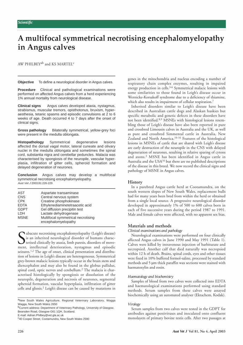
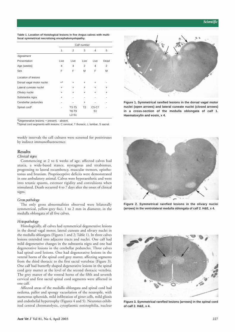
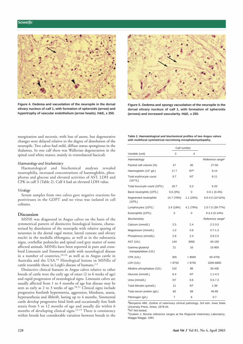
in the dorsal vagal motor, lateral cuneate and olivary nuclei inthe medulla oblongata (Figures 1 and 2; Table 1). In three calveslesions extended into adjacent tracts and nuclei. One calf hadmild degenerative changes in the substantia nigra and one haddegenerative lesions in the cerebellar peduncles. Three calveshad spinal cord lesions. One had degenerative lesions in theventral horns of the spinal cord grey matter, affecting segmentsfrom the third thoracic to the first sacral vertebrae (Figure 3).One calf had butterfly-shaped degenerative lesions in the spinalcord grey matter at the level of the second thoracic vertebra.The grey matter of the ventral horns of the fifth and seventhcervical and first sacral spinal cord segments were affected inone calf.
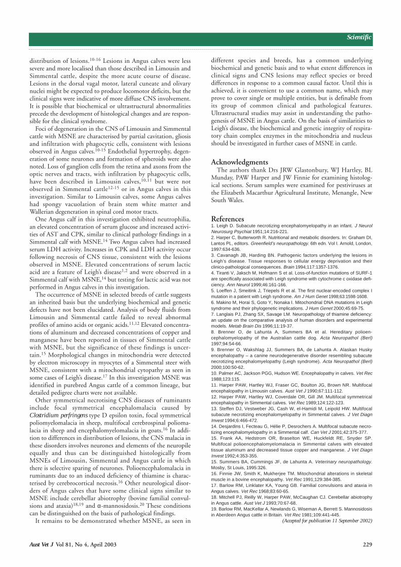
Affected areas of the medulla oblongata and spinal cord hadoedema, pallor and spongy vacuolation of the neuropile, withnumerous spheroids, mild infiltration of gitter cells, mild gliosisand endothelial hypertrophy (Figures 4 and 5). Neurones exhib-ited central chromatolysis, cytoplasmic eosinophilia, nuclear
Table 1. Location of histological lesions in five Angus calves with multi-focal symmetrical necrotising encephalomyelopathy.
Calf number
1 2 3 4 5
Signalment
Presentation Live Live Live Live Dead
Age (weeks) 4 4 2 4 2
Sex F F M F M
Location of lesions
Dorsal vagal motor nuclei +a + + + -
Lateral cuneate nuclei + + + + +
Olivary nuclei + + + + +
Substantia nigra + - - - -
Cerebellar peduncles - - + - -
Spinal cordb - T3-T5 T2 C5-C7 -T8-T9 S1 L2-S1
aDegenerative lesions: + present, - absent.bSpinal cord segments with lesions: C cervical, T thoracic, L lumbar, S sacral.
Figure 1. Symmetrical rarefied lesions in the dorsal vagal motornuclei (open arrows) and lateral cuneate nuclei (closed arrows)in a cross-section of the medulla oblongata of calf 1.Haematoxylin and eosin, x 4.
Figure 2. Symmetrical rarefied lesions in the olivary nuclei(arrows) in the ventrolateral medulla oblongata of calf 2. H&E, x 4.
Figure 3. Symmetrical rarefied lesions (arrows) in the spinal cordof calf 2. H&E, x 4.
228 Aust Vet J Vol 81, No 4, April 2003
Scientific
margination and necrosis, with loss of axons, but degenerativechanges were delayed relative to the degree of dissolution of theneuropile. Two calves had mild, diffuse status spongiosus in thethalamus. In one calf there was Wallerian degeneration in thespinal cord white matter, mainly in ventrolateral fasciculi.
Haematology and biochemistryHaematological and biochemical analyses revealed
neutrophilia, increased concentrations of haemoglobin, phos-phorus and glucose and elevated activities of AST, LDH andCPK in calf 3 (Table 2). Calf 4 had an elevated LDH value.
VirologySerum samples from two calves gave negative reactions for
pestiviruses in the GDPT and no virus was isolated in cellcultures.
DiscussionMSNE was diagnosed in Angus calves on the basis of the
symmetrical pattern of distinctive histological lesions, charac-terised by dissolution of the neuropile with relative sparing ofneurones in the dorsal vagal motor, lateral cuneate and olivarynuclei in the medulla oblongata, as well as in the substantianigra, cerebellar peduncles and spinal cord grey matter of someaffected animals. MSNEs have been reported in pure and cross-bred Limousin and Simmental cattle with neurological diseasein a number of countries,10-16 as well as in Angus cattle inAustralia and the USA.16 Histological lesions in MSNEs ofcattle resemble those in Leigh’s disease of humans.1,2
Distinctive clinical features in Angus calves relative to otherbreeds of cattle were the early age of onset (2 to 6 weeks of age)and rapid progression of neurological signs. Limousin calves areusually affected from 1 to 4 months of age but disease may beseen as early as 2 to 3 weeks of age.10,11 Clinical signs includeprogressive forelimb hypermetria, aggression, blindness, ataxia,hyperaesthesia and illthrift, lasting up to 4 months. Simmentalcattle develop progressive hind limb and occasionally fore limbataxia from 5 to 12 months of age and usually die within 6months of developing clinical signs.12-15 There is consistencywithin breeds but considerable variation between breeds in the
Figure 4. Oedema and vacuolation of the neuropile in the dorsalolivary nucleus of calf 1, with formation of spheroids (arrow) andhypertrophy of vascular endothelium (arrow heads). H&E, x 250.
Figure 5. Oedema and spongy vacuolation of the neuropile in thedorsal olivary nucleus of calf 1, with formation of spheroids(arrows) and increased vascularity. H&E, x 250.
Table 2. Haematological and biochemical profiles of two Angus calveswith multifocal symmetrical necrotising encephalomyelopathy.
Calf number
Variable (unit) 3 4
Haematology Reference rangea
Packed cell volume (%) 47 36 27-50
Haemoglobin (101 g/L) 17.7 NTb 9-14
Total erythrocyte count 9.7 NT 6-11(1012/L)
Total leucocyte count (109/L) 18.7 5.3 5-20
Band neutrophils (109/L) 0.6 (3%) 0 0-0.1 (0-4%)
Segmented neutrophils 14.7 (79%) 1.1 (20%) 0.6-4.0 (10-52%)(109/L)
Lymphocytes (109/L) 3.4 (18%) 4.1 (79%) 2.5-7.5 (39-77%)
Eosinophils (109/L) 0 0 0-2.4 (0-14%)
Biochemistry Reference rangec
Calcium (mmol/L) 2.5 2.4 2.2-3.0
Magnesium (mmol/L) 1.0 0.8 0.7-1.3
Phosphorus (mmol/L) 2.8 2.4 0.8-2.5
AST (U/L) 144 3060 40-150
Gamma glutamyl 21 16 15-900 transpeptidase (U/L)
CPK (U/L) 355 > 8000 50-4700
LDH (U/L) > 8750 > 8750 3200-6800
Alkaline phosphatase (U/L) 102 98 30-430
Glucose (mmol/L) 6.4 NT 1.1-4.3
Urea (mmol/L) NT 6.6 0.5-7.3
Total bilirubin (µmol/L) 11 NT 1-39
Total serum protein (g/L) 60 58 45-85
Fibrinogen (g/L) 7 6 3-7
aBenjamin MM. Outline of veterinary clinical pathology, 3rd edn. Iowa StateUniversity Press, Ames, 1978:44.bNT Not tested.cGodwin J. Bovine reference ranges at the Regional Veterinary Laboratory,Wagga Wagga. 1991
Aust Vet J Vol 81, No 4, April 2003 229
Scientific
distribution of lesions.10-16 Lesions in Angus calves were lesssevere and more localised than those described in Limousin andSimmental cattle, despite the more acute course of disease.Lesions in the dorsal vagal motor, lateral cuneate and olivarynuclei might be expected to produce locomotor deficits, but theclinical signs were indicative of more diffuse CNS involvement.It is possible that biochemical or ultrastructural abnormalitiesprecede the development of histological changes and are respon-sible for the clinical syndrome.
Foci of degeneration in the CNS of Limousin and Simmentalcattle with MSNE are characterised by partial cavitation, gliosisand infiltration with phagocytic cells, consistent with lesionsobserved in Angus calves.10-15 Endothelial hypertrophy, degen-eration of some neurones and formation of spheroids were alsonoted. Loss of ganglion cells from the retina and axons from theoptic nerves and tracts, with infiltration by phagocytic cells,have been described in Limousin calves,10,11 but were notobserved in Simmental cattle12-15 or in Angus calves in thisinvestigation. Similar to Limousin calves, some Angus calveshad spongy vacuolation of brain stem white matter andWallerian degeneration in spinal cord motor tracts.
One Angus calf in this investigation exhibited neutrophilia,an elevated concentration of serum glucose and increased activi-ties of AST and CPK, similar to clinical pathology findings in aSimmental calf with MSNE.14 Two Angus calves had increasedserum LDH activity. Increases in CPK and LDH activity occurfollowing necrosis of CNS tissue, consistent with the lesionsobserved in MSNE. Elevated concentrations of serum lacticacid are a feature of Leigh’s disease1,2 and were observed in aSimmental calf with MSNE,14 but testing for lactic acid was notperformed in Angus calves in this investigation.
The occurrence of MSNE in selected breeds of cattle suggestsan inherited basis but the underlying biochemical and geneticdefects have not been elucidated. Analysis of body fluids fromLimousin and Simmental cattle failed to reveal abnormalprofiles of amino acids or organic acids.11,12 Elevated concentra-tions of aluminum and decreased concentrations of copper andmanganese have been reported in tissues of Simmental cattlewith MSNE, but the significance of these findings is uncer-tain.15 Morphological changes in mitochondria were detectedby electron microscopy in myocytes of a Simmental steer withMSNE, consistent with a mitochondrial cytopathy as seen insome cases of Leigh’s disease.17 In this investigation MSNE wasidentified in purebred Angus cattle of a common lineage, butdetailed pedigree charts were not available.
Other symmetrical necrotising CNS diseases of ruminantsinclude focal symmetrical encephalomalacia caused byClostridium perfringens type D epsilon toxin, focal symmetricalpoliomyelomalacia in sheep, multifocal cerebrospinal polioma-lacia in sheep and encephalomyelomalacia in goats.16 In addi-tion to differences in distribution of lesions, the CNS malacia inthese disorders involves neurones and elements of the neuropileequally and thus can be distinguished histologically fromMSNEs of Limousin, Simmental and Angus cattle in whichthere is selective sparing of neurones. Polioencephalomalacia inruminants due to an induced deficiency of thiamine is charac-terised by cerebrocortical necrosis.16 Other neurological disor-ders of Angus calves that have some clinical signs similar toMSNE include cerebellar abiotrophy (bovine familial convul-sions and ataxia)18,19 and α-mannosidosis.20 These conditionscan be distinguished on the basis of pathological findings.
It remains to be demonstrated whether MSNE, as seen in
different species and breeds, has a common underlyingbiochemical and genetic basis and to what extent differences inclinical signs and CNS lesions may reflect species or breeddifferences in response to a common causal factor. Until this isachieved, it is convenient to use a common name, which mayprove to cover single or multiple entities, but is definable fromits group of common clinical and pathological features.Ultrastructural studies may assist in understanding the patho-genesis of MSNE in Angus cattle. On the basis of similarities toLeigh’s disease, the biochemical and genetic integrity of respira-tory chain complex enzymes in the mitochondria and nucleusshould be investigated in further cases of MSNE in cattle.
AcknowledgmentsThe authors thank Drs JRW Glastonbury, WJ Hartley, BL
Munday, PAW Harper and JW Finnie for examining histolog-ical sections. Serum samples were examined for pestiviruses atthe Elizabeth Macarthur Agricultural Institute, Menangle, NewSouth Wales.
References1. Leigh D. Subacute necrotizing encephalomyelopathy in an infant. J NeurolNeurosurg Psychiat 1951;14:216-221.2. Harper C, Butterworth R. Nutritional and metabolic disorders. In: Graham DI,Lantos PL, editors. Greenfield’s neuropathology. 6th edn. Vol I. Arnold, London,1997:634-636.3. Cavanagh JB, Harding BN. Pathogenic factors underlying the lesions inLeigh’s disease. Tissue responses to cellular energy deprivation and theirclinico-pathological consequences. Brain 1994;117:1357-1376.4. Tiranti V, Jaksch M, Hofmann S et al. Loss-of-function mutations of SURF-1are specifically associated with Leigh syndrome with cytochrome c oxidase defi-ciency. Ann Neurol 1999;46:161-166.5. Loeffen J, Smeitink J, Triepels R et al. The first nuclear-encoded complex Imutation in a patient with Leigh syndrome. Am J Hum Genet 1998;63:1598-1608.6. Makino M, Horai S, Goto Y, Nonaka I. Mitochondrial DNA mutations in Leighsyndrome and their phylogenetic implications. J Hum Genet 2000;45:69-75.7. Langlais PJ, Zhang SX, Savage LM. Neuropathology of thiamine deficiency:an update on the comparative analysis of human disorders and experimentalmodels. Metab Brain Dis 1996;11:19-37.8. Brenner O, de Lahunta A, Summers BA et al. Hereditary polioen-cephalomyelopathy of the Australian cattle dog. Acta Neuropathol (Berl)1997;94:54-66.9. Brenner O, Wakshlag JJ, Summers BA, de Lahunta A. Alaskan Huskyencephalopathy – a canine neurodegenerative disorder resembling subacutenecrotizing encephalomyelopathy (Leigh syndrome). Acta Neuropathol (Berl)2000;100:50-62.10. Palmer AC, Jackson PGG, Hudson WE. Encephalopathy in calves. Vet Rec1988;123:115.11. Harper PAW, Hartley WJ, Fraser GC, Boulton JG, Brown NR. Multifocalencephalopathy in Limousin calves. Aust Vet J 1990;67:111-112.12. Harper PAW, Hartley WJ, Coverdale OR, Gill JM. Multifocal symmetricalencephalopathy in Simmental calves. Vet Rec 1989;124:122-123.13. Steffen DJ, Vestweber JG, Cash W, el-Hamidi M, Leipold HW. Multifocalsubacute necrotizing encephalomyelopathy in Simmental calves. J Vet DiagnInvest 1994;6:466-472.14. Desjardins I, Fecteau G, Hélie P, Desrochers A. Multifocal subacute necro-tizing encephalomyelopathy in a Simmental calf. Can Vet J 2001;42:375-377.15. Frank AA, Hedstrom OR, Braselton WE, Huckfeldt RE, Snyder SP.Multifocal polioencephalomyelomalacia in Simmental calves with elevatedtissue aluminum and decreased tissue copper and manganese. J Vet DiagnInvest 1992;4:353-355.15. Summers BA, Cummings JF, de Lahunta A. Veterinary neuropathology.Mosby, St Louis, 1995:326.16. Finnie JW, Smith K, Mukherjee TM. Mitochondrial alterations in skeletalmuscle in a bovine encephalopathy. Vet Rec 1991;129:384-385.17. Barlow RM, Linklater KA, Young GB. Familial convulsions and ataxia inAngus calves. Vet Rec 1968;83:60-65.18. Mitchell PJ, Reilly W, Harper PAW, McCaughan CJ. Cerebellar abiotrophyin Angus cattle. Aust Vet J 1993;70:67-68.19. Barlow RM, MacKellar A, Newlands G, Wiseman A, Berrett S. Mannosidosisin Aberdeen Angus cattle in Britain. Vet Rec 1981;109:441-445.
(Accepted for publication 11 September 2002)