Embed Size (px)
Citation preview
A Multicenter, International CollaborativeStudy for American Joint Committee onCancer Staging of Retinoblastoma
Part II: Treatment Success and Globe Salvage
Ankit Singh Tomar, MD,1 Paul T. Finger, MD,1 Brenda Gallie, MD,2 Ashwin Mallipatna, MBBS, MS,11
Tero T. Kivelä, MD,3 Chengyue Zhang, MD,4 Junyang Zhao, MD,4 Matthew W. Wilson, MD,5,6
Rachel C. Brenna, MD,5,6 Michala Burges, BS,5,6 Jonathan Kim, MD,7 Vikas Khetan, MBBS, MS,8
Suganeswari Ganesan, MBBS, MS,8 Andrey Yarovoy, MD,9 Vera Yarovaya, MD,9 Elena Kotova, MD,9
Yacoub A. Yousef, MD,10 Kalle Nummi, MD,3 Tatiana L. Ushakova, MD,12,13 Olga V. Yugay, MD,12
Vladimir G. Polyakov, MD,12,13 Marco A. Ramirez-Ortiz, MD, MPH,14 Elizabeth Esparza-Aguiar, MD,14
Guillermo Chantada, MD,15 Paula Schaiquevich, MD,15 Adriana Fandino, MD,16 Jason C. Yam, MD,17
Winnie W. Lau, MD,17 Carol P. Lam, MD,17 Phillipa Sharwood, MBBS,18 Sonia Moorthy, MD,19
Quah Boon Long, MD,19 Vera Adobea Essuman, MD,20 Lorna A. Renner, MD,21 Ekaterina Semenova, MD,1
Jaume Català, MD,22 Genoveva Correa-Llano, MD,23 Elisa Carreras, MD,23 for the American Joint Committee onCancer Ophthalmic Oncology Task Force*
Purpose: To evaluate the ability of the American Joint Committee on Cancer (AJCC) 8th edition to predictlocal tumor control and globe salvage for children with retinoblastoma (RB).
Design: International, multicenter, registry-based retrospective case series.Participants: A total of 2854 eyes of 2097 patients from 18 ophthalmic oncology centers from 13 countries
over 6 continents.Methods: International, multicenter, registry-based data were pooled from patients enrolled between
January 2001 and December 2013. All RB eyes with adequate records to allow tumor staging by the AJCC 8thedition criteria and follow-up to ascertain treatment outcomes were included.
Main Outcome Measures: Globe-salvage rates were estimated by AJCC clinical (cTNMH) categories andtumor laterality. Local treatment failure was defined as use of enucleation or external beam radiation therapy(EBRT), with or without plaque brachytherapy or intra-arterial chemotherapy (IAC).
Results: Unilateral RB occurred in 1340 eyes (47%). Among the 2854 eyes, tumor categories were cT1 tocT4 in 696 eyes (24%), 1334 eyes (47%), 802 eyes (28%), and 22 eyes (1%), respectively. Of these, 1275 eyes(45%) were salvaged, and 1179 eyes (41%) and 400 eyes (14%) underwent primary and secondary enucleation,respectively. The 2- and 5-year KaplaneMeier cumulative globe-salvage rates without the use of EBRT bycTNMH categories were 97% and 96% for category cT1a tumors, 94% and 88% for cT1b tumors, 68% and 60%for cT2a tumors, 66% and 57% for cT2b tumors, and 32% and 25% for cT3 tumors, respectively. Risk of localtreatment failure increased with increasing cT category (P < 0.001). Cox proportional hazards regression analysisconfirmed a higher risk of local treatment failure in categories cT1b (hazard ratio [HR], 3.5; P ¼ 0.004), cT2a (HR,15.1; P < 0.001), cT2b (HR, 16.4; P < 0.001), and cT3 (HR, 45.0; P < 0.001) compared with category cT1a. Use ofplaque brachytherapy and IAC improved local tumor control in categories cT1a (P ¼ 0.031) and cT1b (P < 0.001).
Conclusions: Multicenter, international, internet-based data sharing validated the 8th edition AJCC RBstaging to predict globe-salvage in a large, heterogeneous, real-world patient population withRB. Ophthalmology 2020;127:1733-1746 ª 2020 by the American Academy of Ophthalmology. This is an openaccess article under the CC BY-NC-ND license (http://creativecommons.org/licenses/by-nc-nd/4.0/).
Supplemental material available at www.aaojournal.org.
Cancer classification systems provide a universal languagefor determining disease extent, planning treatment strate-gies, and estimating prognosis.1 A standardized system of
ª 2020 by the American Academy of OphthalmologyThis is an open access article under the CC BY-NC-ND license(http://creativecommons.org/licenses/by-nc-nd/4.0/). Published by Elsevier Inc.
cancer staging enhances communication among eye cancerspecialists, pediatric oncologists, radiologists, radiationtherapists, ophthalmic pathologists, geneticists, and
1733https://doi.org/10.1016/j.ophtha.2020.05.051ISSN 0161-6420/20
Ophthalmology Volume 127, Number 12, December 2020
researchers who treat patients with retinoblastoma (RB).Classifications for RB have evolved over the last fewdecades as a result of the development of new treatmentmodalities used to improve local tumor control and globesalvage.2-5 These rates have improved considerably from26% to 43%6-9 in the early chemotherapy era to 67% to80%9-11 with primary and rescue intra-arterial chemotherapy(IAC), intravitreal chemotherapy, use of plaque brachy-therapy, and other focal treatments.12,13 Treatment successrates differ in various parts of the world, especially inthe lower-income countries where patients present withadvanced disease.14
Reese and Ellsworth3 developed a classification systemto predict globe salvage after external beam radiotherapy(EBRT) as primary treatment.3 Then, as systemicchemotherapy replaced EBRT, the InternationalIntraocular Retinoblastoma Classification (IIRC), alsotermed as “Murphree’s Children’s Hospital of LosAngeles” (CHLA) classification,4 and the InternationalClassification for Retinoblastoma (ICRB), also termed as“Shield’s Wills Eye Hospital” (WEH) classification,5
emerged to predict globe salvage after chemotherapy.These classification systems were formulated by single-center experience or small-group consensus and still lackpublished multicenter validation.15 More importantly, bothCHLA and WEH grouping systems use the same groupnames such as group A to E, but each group definitioncontains key differences that hinder the ability to collateevidence that predict treatment success and to compareclinical outcomes in centers across the world.16-18 Thelack of a universal RB cancer classification has harmed bothresearch and clinical care.17
An evidence-based accurate staging of the eye and childwith RB would serve to predict which eyes are safe to besalvaged and provide a quantitative risk for salvage failure,the need for secondary enucleation, and the risk for extra-ocular relapse. The 8th edition American Joint Committeeon Cancer (AJCC) RB staging system was created by 18 RBspecialist centers from 13 countries on 6 continents. Globalconsensus was derived from the Ophthalmic Oncology TaskForce (OOTF).2 More comprehensive than previouseditions, the resultant TNMH staging system definedanatomic stages of growth of the primary tumor (T), withregional lymph node (N), and systemic metastasis (M)framework, and uniquely includes heritability (H) as anindependent category. It was adopted by the Union forInternational Cancer Control (UICC) and has beenaccepted by multiple ophthalmology, medical oncology,radiation oncology, and medical journals around theworld.19-21
Multicenter international data collection to enroll a globalspectrum of cases was used to evaluate the ability of theAJCC 8th edition classification to predict outcomes withrespect to eye salvage and long-term tumor control.
Methods
All participating centers obtained internal Institutional ReviewBoard approval to perform retrospective medical record reviews
1734
and contribute de-identified data to the AJCC OphthalmicOncology Task Force (OOTF) Retinoblastoma Registry at PrincessMargaret Cancer Center (Toronto, Ontario, Canada). All centersagreed that individual patient consent was not required becausethere were no patient identifiers collected. Each site was anophthalmic oncology subspecialty center. Patients with RB werediagnosed and treated according to the best practices defined byeach institute. Patient records were excluded from analysis if keyvariables, such as demographic data, clinical variables essential forRB classification (tumor location, size, and extent), treatment data(date and type of treatment), and outcome (globe salvage, primary,or secondary enucleation) were missing or inconsistent. This studyadhered to the tenets of the Declaration of Helsinki and the HealthInsurance Portability and Accountability Act of 1996.
The Registry
Internet-based, retrospective registry was created to evaluate thestaging system for RB in the AJCC Cancer Staging Manual.2
Through a consensus process, OOTF committee members(primarily ophthalmic oncologists and pathologists) developedthe epidemiologic, clinical, and pathological data fields.
Internet Database and Security
International standards for patient privacy protection and statisticalanalysis were used. Security measures included the lack of personalpatient identifiers, Secure Sockets Layer encryption, protectionagainst Structured Query Language injection, variable and sessionmanagement, record locking, and trail auditing (e.g., failed loginattempts and web page accessing). Access to the online surveyrequired user accounts issued by the coordinating center. Eachcenter could only access their patient records. When documentationof the local ethics approval was received by the coordinatingcenter, unique login passwords were provided to initiate patiententry. Each center created a random alphanumeric identifier foreach patient.
Definitions
Primary enucleation: removal of treatment naive RB eyes.Secondary enucleation: removal of an eye after an attempt at
eye salvage, irrespective of the reason for enucleation (significantresidual disease, recurrent tumor).
CHLA/IIRC classification: termed CHLA for the purpose ofthis study to avoid any confusion regarding the 2 similar soundingbut distinct classification systems IIRC and ICRB.
WEH/ICRB classification: termed WEH for the purpose of thisstudy.
Each center used its own best diagnostic and therapeuticmethods. Data collected included date of diagnosis, age at diag-nosis (months), hereditary pattern (familial, sporadic), laterality(unilateral, bilateral), and the eye involved (right, left). The clinicalinformation included size and location of intraocular tumor, pres-ence of glaucoma, presence and type of vitreous seeds, subretinalseeds, and macroscopic anterior chamber seeds, and neo-vascularization of the iris. Reese-Ellsworth, CHLA, WEH, andcTNMH staging of RB were noted for each eye. Treatment detailswere noted. The eyes with substantial residual or recurrent diseaseafter chemotherapy and focal consolidation were treated withfurther focal laser, cryotherapy, plaque brachytherapy, IAC, EBRT,or enucleation. Local treatment failure after conservative treatmentwas defined as the need for EBRT or secondary enucleation. Pla-que brachytherapy and IAC are standard in multimodal treatmentfor globe salvage, but they are not universally available. To assessthe difference among the success rates of different main treatment

Tomar et al � AJCC Staging of Retinoblastoma: Globe Salvage
modalities, the second criterion for treatment failure was defined asthe need for plaque brachytherapy, IAC, EBRT, or secondaryenucleation. The latter criterion was formulated purely as astatistical tool to study the impact on globe salvage if plaquebrachytherapy and IAC were not available. Their use should not beconsidered as treatment failure, quite the opposite. Based on these2 definitions, failure-free globe salvage refers to eye conservationwithout need of above listed modalities.
TNMH Retinoblastoma Staging
In contrast to CHLA and WEH, the 8th edition AJCC RBclassification included more complex information focusing notonly on the patient’s primary tumor but also on regional lymphnode spread, metastatic disease, and heritable trait (Table 1). In thatthe registry data fields and collection predated 2013, we used theraw clinical data to classify all the tumors accurately by AJCC8th edition. Data were available for all necessary fields exceptfor the involvement of pars plana and ciliary body (cT3b) by thetumor.
Table 1. American Joint Committee on Cancer 8th Edition TNM CTumor Stagi
cTX Unknown evidence of intraocular tumorcT0 No evidence of intraocular tumorcT1 Intraocular tumor(s) with subretinal fluid �5 mm from
cT1a Tumors �3 mm and >1.5 mm from the disc and foveacT1b Tumors >3 mm or closer than 1.5 mm to the disc and
cT2 Intraocular tumor(s) with retinal detachment, vitreous scT2a Subretinal fluid >5 mm from the base of any tumorcT2b Tumors with vitreous seeding or subretinal seeding
cT3 Advanced intraocular tumor(s)cT3a Phthisis or pre-phthisis bulbicT3b Tumor invasion of the pars plana, ciliary body, lens, zoncT3c Increased intraocular pressure with neovascularization orcT3d Hyphema or massive vitreous hemorrhagecT3e Aseptic orbital cellulitis
cT4 Extraocular tumor(s) involving the orbit, including thecT4a Radiologic evidence of retrobulbar optic nerve involvemcT4b Extraocular tumor clinically evident with proptosis and
Definitions for Regional Lym
cNX RegiocN0 No recN1 Evide
Definitions for Distant M
cM0 No signs or symptomscM1 Distant metastasis wit
cM1a Tumor(s) involving ancM1b Tumor involving the
pM1 Distant metastasis witpM1a Histopathologic confirpM1b Histopathologic confir
Definitions for Heritabl
HX Unknown or insufficient evidence of a conH0 Normal RB1 alleles in blood tested with dH1 Bilateral RB, RB with an intracranial CNS
of RB, or molecular definition of constit
CNS ¼ central nervous system; RB ¼ retinoblastoma.
Statistical Analysis
Continuous variables were described using medians, ranges, andinterquartile ranges (IQRs), and categoric variables were describedusing frequencies and proportions. KaplaneMeier plots with log-rank test for trend were implemented to test whether tumor cate-gory is related to treatment success. All the eyes with intraocularRB that were not primarily enucleated were analyzed for localfailure-free globe salvage analysis. Eyes were censored at the timeof the last follow-up. Cumulative proportion of local failure-freeglobe salvage estimates at 1, 2, 5, and 10 years were tabulated.SPSS (version 23.0, IBM, Armonk, New York, NY) was used togenerate KaplaneMeier plots and to perform all other statisticalanalyses. Statistical significance was set at P <0.05, and no ad-justments were made for multiple tests.
Results
Eighteen eye cancer specialty centers from 13 countries over 6continents successfully entered data online into an internet-based
lassification for Retinoblastoma2 Definitions for AJCC Primaryng (cT)
the base of any tumor
foveaeeding, or subretinal seeding
ules, iris, or anterior chamberbuphthalmos
optic nerveent or thickening of the optic nerve or involvement of the orbital tissuesorbital mass
ph Node Staging (cN)
nal lymph nodes cannot be assessedgional lymph node involvementnce of preauricular, submandibular, and cervical lymph node involvement
etastasis Staging (M)
of intracranial or distant metastasishout microscopic confirmationy distant site (e.g., bone marrow, liver) on clinical or radiologic testscentral nervous system on radiologic imaging (not including trilateral RB)h microscopic confirmationmation of tumor at any distant site (e.g., bone marrow, liver, or other)mation of tumor in the cerebrospinal fluid or CNS parenchyma
e Trait Staging (H)
stitutional RB1 gene mutationemonstrated high-sensitivity assaysmidline embryonic tumor (i.e., trilateral RB), patient with family history
utional RB1 gene mutation
1735

Ophthalmology Volume 127, Number 12, December 2020
registry to evaluate the predictive value of the 8th edition AJCCTNMH staging system for globe salvage after RB treatment. Be-tween January 2001 and December 2013, 2905 eyes of 2097 RBpatients were enrolled. Because of incomplete data, 51 eyes wereexcluded, leaving 2854 (98.2%) complete records for analysis.
Patient Features
The median age at diagnosis was 17.0 months (mean, 21.6; SD,20.9; IQR, 8e29; range, 1e365 months). Of the 2854 eyes, RBwas unilateral in 1340 eyes (47%) and bilateral in 1514 eyes(53%). Among patients with unilateral RB, the right eye wasinvolved in 688 (51.3%).
Classifications
Comparative staging was performed using the Reese-Ellsworth,CHLA, and WEH systems. The Reese-Ellsworth classification3
was reported in 1250 eyes; 168 eyes were in group I (13.3%);119 eyes were in group II (9.4%); 126 eyes were in group III(10.0%); 91 eyes were in group IV (7.3%); and 746 were ingroup V (59.9%).
The CHLA4 was reported in 2835 eyes as follows: 176 eyeswere in group A (6.2%); 476 eyes were in group B (16.8%); 208
Table 2. Classification of Retinoblastoma
Group
No. of Eyes byCHLA
Classification (%) Treatment Outcome N
A N ¼ 176 (6.2%) Globe salvage 174 (Primary enucleation 1 (Secondary enucleation 1 (
B N ¼ 476 (16.8%) Globe salvage 425 (Primary enucleation 22 (Secondary enucleation 29 (
C N ¼ 208 (7.3%) Globe salvage 155 (Primary enucleation 30 (Secondary enucleation 23 (
D N ¼ 1101 (38.9%) Globe salvage 388 (Primary enucleation 498 (Secondary enucleation 215 (
E N ¼ 874 (30.8%) Globe salvage 116 (Primary enucleation 626 (Secondary enucleation 132 (
Data Not Available N ¼ 19
Tumor Category No. of Eyes
cT1 cT1a N ¼ 230 (8.1%)
cT1b N ¼ 466 (16.5%)
cT2 cT2a N ¼ 280 (9.9%)
cT2b N ¼ 1054 (37.2%)
cT3 N ¼ 802 (28.3%)
AJCC ¼ American Joint Committee on Cancer; CHLA ¼ Children’s Hospita
1736
eyes were in group C (7.3%); 1101 eyes were in group D(38.9%); and 874 eyes were in group E (30.8%) (Table 2).
The WEH5 was reported in 2835 eyes as follows: 188 eyes werein group A (6.6%); 600 eyes were in group B (21.2%); 40 eyeswere in group C (1.4%); 277 eyes were in group D (9.8%); and1730 eyes were in group E (61.0%) (Table 2). The largestdisparity between CHLA and WEH can be seen in group Eclassified eyes.
AJCC Clinical Classification
In 2854 eyes, the AJCC clinical T category that indicates theanatomic extent of the primary tumor (cT) was in 696 eyeswith cT1 (24.4%), in 1334 eyes with cT2 (46.7%), in 802eyes with cT3 (28.1%), and in 22 eyes with cT4 (0.8%)(Table 2). Regional lymph node involvement (cN) wasdemonstrated in 12 patients (0.6%) as cN1. The involvedlymph nodes were preauricular, cervical, or submandibular.Twenty-five (1.2%) of 2085 patients presented with distantmetastasis (cM), including 13 with cM1a (0.6%, including 3with distant lymph nodes) and 12 with central nervous system(CNS) metastasis, cM1b (0.6%). Trilateral RB in the AJCCsystem is classified as a brain tumor and thus does not in-fluence the cM category.
Eyes with Local Treatment Outcomes
(%)
No. of Eyes byWEH
Classification (%) Treatment Outcome N (%)
98.8%) N ¼ 188 (6.6%) Globe salvage 184 (97.9%)0.6%) Primary enucleation 2 (1.1%)0.6%) Secondary enucleation 2 (1.1%)89.3%) N ¼ 600 (21.2%) Globe salvage 534 (89%)4.6%) Primary enucleation 30 (5.0%)6.1%) Secondary enucleation 36 (6.0%)74.5%) N ¼ 40 (1.4%) Globe salvage 25 (62.5%)14.4%) Primary enucleation 7 (17.5%)11.1%) Secondary enucleation 8 (20.0%)35.2%) N ¼ 277 (9.8%) Globe salvage 122 (44.0%)45.2%) Primary enucleation 102 (36.8%)19.5%) Secondary enucleation 53 (19.1%)13.2%) N ¼ 1730 (61.0%) Globe salvage 400 (23.1%)71.6%) Primary enucleation 1030 (59.5%)15.1%) Secondary enucleation 300 (17.3%)
N ¼ 19
Treatment Outcome N (%)
Globe salvage 201 (87.4%)Primary enucleation 23 (10.0%)Secondary enucleation 6 (2.6%)Globe salvage 423 (90.8%)Primary enucleation 14 (3.0%)Secondary enucleation 29 (6.2%)Globe salvage 116 (41.3%)Primary enucleation 119 (42.5%)Secondary enucleation 45 (16.0%)Globe salvage 428 (40.6%)Primary enucleation 430 (40.8%)Secondary enucleation 196 (18.6%)Globe salvage 100 (12.5%)Primary enucleation 587 (73.2%)Secondary enucleation 115 (14.3%)
l of Los Angeles; WEH ¼ Wills Eye Hospital.

Table 3. KaplaneMeier Cumulative Proportion of Avoiding Local Treatment Failure Based on Different Retinoblastoma Classifications
Local Treatment Failure Defined as Need for EBRT or Enucleation
Classification Variable
KaplaneMeier Point Estimates (95% CI) %
1 Yr 2 Yrs 5 Yrs 10 Yrs
All patients (n ¼ 1574) 78 (77e79) 75 (74e76) 68 (67e69) 42 (38e46)CHLA Classification (n ¼1556) A (n ¼ 168) 100 100 98 (96e100) 98 (96e100)
B (n ¼ 438) 96 (95e97) 94 (93e95) 89 (87e91) 59 (50e68)C (n ¼ 167) 89 (86e92) 87 (85e89) 86 (83e89) 69 (62e76)D (n ¼ 563) 66 (64e68) 61 (59e63) 49 (46e52) 16 (11e21)E (n ¼ 220) 39 (35e43) 29 (25e33) 23 (19e27) 15 (10e20)
WEH Classification (n ¼1563) A (n ¼ 179) 99 (98e100) 99 (98e100) 98 (96e100) 98 (96e100)B (n ¼ 555) 96 (95e97) 94 (93e95) 89 (87e91) 63 (56e70)C (n ¼ 32) 73 (65e81) 73 (65e81) 73 (65e81) 65 (54e76)D (n ¼ 164) 71 (67e75) 68 (64e72) 56 (51e61) 15 (8e22)E (n ¼ 633) 56 (54e58) 49 (47e51) 39 (36e42) 15 (10e20)
AJCC cT size category (n ¼1574) cT1a (n ¼ 200) 97 (96e98) 97 (96e98) 96 (94e98) 96 (94e98)cT1b (n ¼ 436) 96 (95e97) 94 (93e95) 88 (86e90) 59 (50e68)cT2a (n ¼ 144) 74 (70e78) 68 (64e72) 60 (55e65) 38 (29e47)cT2b (n ¼ 592) 70 (68e72) 66 (64e68) 57 (54e60) 25 (20e30)cT3 (n ¼ 202) 39 (35e43) 32 (28e36) 25 (20e30) 17 (12e22)
For CHLA Classification
Overall Comparison: Log-Rank test, P < 0.001
Pairwise Comparison:
A B C D
AB <0.001C <0.001 0.005D <0.001 <0.001 <0.001E <0.001 <0.001 <0.001 <0.001
For WEH Classification
Overall Comparison: Log-Rank test, P < 0.001
Pairwise Comparison:
A B C D
AB 0.001C <0.001 <0.001D <0.001 <0.001 0.429E <0.001 <0.001 0.009 <0.001
For cT Category
Overall Comparison: Log-Rank Test, P < 0.001
Pairwise Comparison:
cT1a cT1b cT2a cT2b
cT1acT1b 0.035cT2a <0.001 <0.001cT2b <0.001 <0.001 ¼0.584cT3 <0.001 <0.001 <0.001 <0.001
Local Treatment Failure Defined as Need for Plaque Brachytherapy, IAC, EBRT, or Enucleation
Classification Variable
KaplaneMeier Point Estimates (95% CI), %
1 Yr 2 Yrs 5 Yrs 10 Yrs
All patients(n ¼ 1574)
76 (75e77) 72 (71e73) 61 (59e63) 25 (22e28)
(Continued)
Tomar et al � AJCC Staging of Retinoblastoma: Globe Salvage
1737

Table 3. (Continued.)
Local Treatment Failure Defined as Need for Plaque Brachytherapy, IAC, EBRT, or Enucleation
Classification Variable
KaplaneMeier Point Estimates (95% CI), %
1 Yr 2 Yrs 5 Yrs 10 Yrs
CHLA/IIRC Classification (n ¼ 1556) A (n ¼ 168) 98 (97e99) 96 (94e98) 88 (84e92) 62 (50e84)B (n ¼ 438) 93 (92e94) 89 (87e91) 78 (76e80) 30 (23e37)C (n ¼ 167) 88 (85e91) 85 (82e88) 78 (74e82) 33 (24e42)D (n ¼ 563) 64 (62e66) 59 (57e61) 42 (39e45) 12 (8e16)E (n ¼ 220) 39 (35e43) 29 (25e33) 23 (19e27) 15 (10e20)
WEH/ICRB Classification (n ¼ 1563) A (n ¼ 179) 98 (97e99) 95 (93e97) 88 (82e92) 61 (50e72)B (n ¼ 555) 93 (92e94) 90 (89e91) 78 (76e80) 32 (26e38)C (n ¼ 32) 73 (65e81) 69 (60e78) 63 (53e73) 27 (11e43)D (n ¼ 164) 68 (64e72) 65 (61e69) 48 (43e53) 12 (7e17)E (n ¼ 633) 55 (53e57) 48 (46e50) 36 (33e39) 12 (8e16)
AJCC cT size category (n ¼ 1574) cT1a (n ¼ 200) 95 (93e97) 93 (91e95) 87 (84e90) 62 (51e73)cT1b (n ¼ 436) 93 (92e94) 89 (87e91) 78 (76e80) 29 (22e36)cT2a (n ¼ 144) 72 (68e76) 64 (60e68) 47 (42e52) 21 (14e28)cT2b (n ¼ 592) 69 (67e71) 65 (63e67) 51 (48e54) 16 (12e20)cT3 (n ¼ 202) 39 (35e43) 32 (28e36) 25 (20e30) 17 (12e22)
For CHLA Classification
Overall Comparison: Log-Rank Test, P < 0.001
Pairwise Comparison:
A B C D
AB ¼0.003C <0.001 0.100D <0.001 <0.001 <0.001E <0.001 <0.001 <0.001 <0.001
For WEH Classification
Overall Comparison: Log-Rank test, P < 0.001
Pairwise Comparison:
A B C D
AB 0.002C <0.001 ¼0.001D <0.001 <0.001 0.459E <0.001 <0.001 0.012 <0.001
For cT Category
Overall Comparison: Log-Rank Test, P < 0.001
Pairwise Comparison:
cT1a cT1b cT2a cT2b
cT1acT1b 0.031cT2a <0.001 <0.001cT2b <0.001 <0.001 ¼0.983cT3 <0.001 <0.001 <0.001 <0.001
Comparison between the KaplaneMeier Curves Stratified by cT Categories Using 2 Different Definitions of Local Treatment Failure
Pairwise Comparison:
P Value
cT1a 0.031cT1b <0.001
Ophthalmology Volume 127, Number 12, December 2020
1738
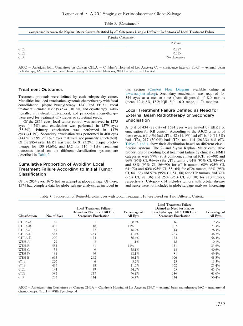
Table 3. (Continued.)
Comparison between the KaplaneMeier Curves Stratified by cT Categories Using 2 Different Definitions of Local Treatment Failure
Pairwise Comparison:
P Value
cT2a 0.387cT2b 0.535cT3 No difference
AJCC ¼ American Joint Committee on Cancer; CHLA ¼ Children’s Hospital of Los Angeles; CI ¼ confidence interval; EBRT ¼ external beamradiotherapy; IAC ¼ intra-arterial chemotherapy; RB ¼ retinoblastoma; WEH ¼ Wills Eye Hospital.
Tomar et al � AJCC Staging of Retinoblastoma: Globe Salvage
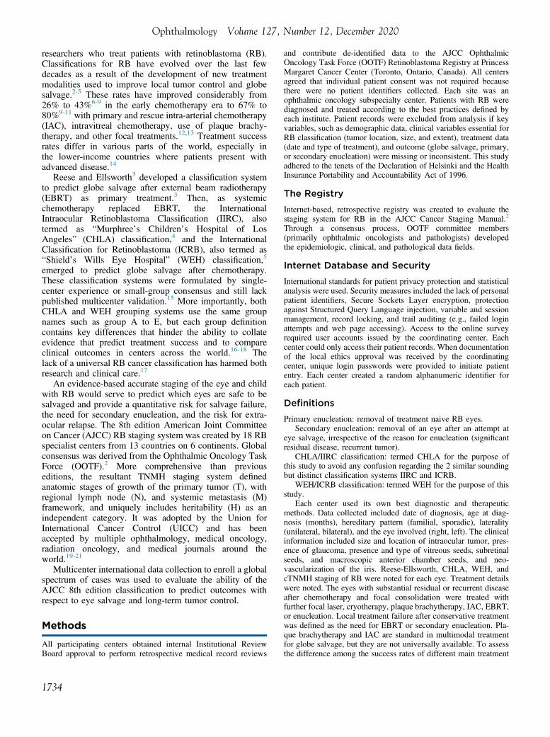
Treatment Outcomes
Treatment protocols were defined by each subspecialty center.Modalities included enucleation, systemic chemotherapy with focalconsolidation, plaque brachytherapy, IAC, and EBRT. Focaltreatment included laser (532 or 810 nm) and cryotherapy. Addi-tionally, intravitreal, intracameral, and periocular chemotherapywere used for treatment of vitreous or subretinal seeds.
Of the 2854 eyes, local tumor control was achieved in 1275eyes (44.7%) and enucleation was performed in 1579 eyes(55.3%). Primary enucleation was performed in 1179eyes (41.3%). Secondary enucleation was performed in 400 eyes(14.0%, 23.9% of 1675 eyes that were not primarily enucleated).Of the 2854 eyes, EBRT was used for 91 (3.2%), plaque brachy-therapy for 130 (4.6%), and IAC for 116 (4.1%). Treatmentoutcomes based on the different classification systems aredescribed in Table 2.
Cumulative Proportion of Avoiding LocalTreatment Failure According to Initial TumorClassification
Of the 2854 eyes, 1675 had an attempt at globe salvage. Of these,1574 had complete data for globe salvage analysis, as included in
Table 4. Proportion of Retinoblastoma Eyes with Local
Classification No. of Eyes
Local Treatment FailureDefined as Need for EBRT or
Secondary EnucleationP
CHLA-A 168 1CHLA-B 438 48CHLA-C 167 27CHLA-D 563 233CHLA-E 220 124WEH-A 179 2WEH-B 555 61WEH-C 32 9WEH-D 164 69WEH-E 633 292cT1a 200 6cT1b 436 48cT2a 144 49cT2b 592 217cT3 202 114
AJCC ¼ American Joint Committee on Cancer; CHLA ¼ Children’s Hospitalchemotherapy; WEH ¼ Wills Eye Hospital.
this section (Consort Flow Diagram available online atwww.aaojournal.org). Secondary enucleation was required for344 eyes at a median time (from diagnosis) of 8.0 months(mean, 12.4; SD, 12.2; IQR, 5.0e16.0, range, 1e74 months).
Local Treatment Failure Defined as Need forExternal Beam Radiotherapy or SecondaryEnucleation
A total of 434 (27.6%) of 1574 eyes were treated by EBRT orenucleation for RB control. According to the AJCC criteria, ofthese eyes, 6 (1.4%) had cT1a, 48 (11.1%) had cT1b, 49 (11.3%)had cT2a, 217 (50.0%) had cT2b, and 114 (26.3%) had cT3.Tables 3 and 4 show their distribution based on different classi-fication systems. The 2- and 5-year KaplaneMeier cumulativeproportions of avoiding local treatment failure by clinical cTNMHcategories were 97% (95% confidence interval [CI], 96e98) and96% (95% CI, 94e98) for cT1a tumors, 94% (95% CI, 93e95)and 88% (95% CI, 86e90) for cT1b tumors, 68% (95% CI,64e72) and 60% (95% CI, 55e65) for cT2a tumors, 66% (95%CI, 64e68) and 57% (95% CI, 54e60) for cT2b tumors, and 32%(95% CI, 28e36) and 25% (95% CI, 20e30) for cT3 tumors,respectively. Category cT4 includes tumors with orbital diseaseand hence were not included in globe salvage analyses. Increasing
Treatment Failure Based on Two Different Criteria
ercentage ofAll Eyes
Local Treatment FailureDefined as Need for Plaque
Brachytherapy, IAC, EBRT, orSecondary Enucleation
Percentage ofAll Eyes
0.6% 16 9.5%11% 102 23.3%16.2% 44 26.3%41.4% 263 46.7%56.4% 124 56.4%1.1% 18 10.1%
11% 131 23.6%28.1% 13 40.6%42.1% 81 49.4%46.1% 306 48.3%3.0% 23 11.5%11.0% 102 23.4%34.0% 65 45.1%36.7% 246 41.6%56.4% 114 56.4%
of Los Angeles; EBRT ¼ external beam radiotherapy; IAC ¼ intra-arterial
1739
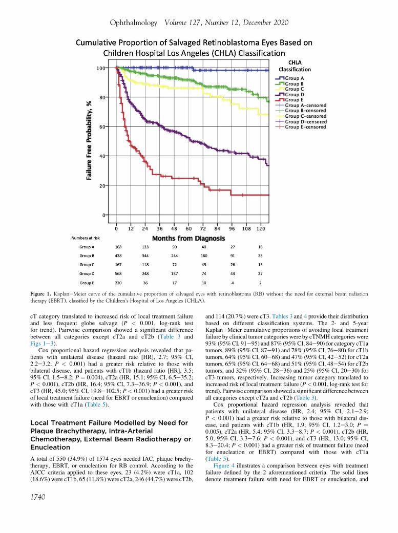
Figure 1. KaplaneMeier curve of the cumulative proportion of salvaged eyes with retinoblastoma (RB) without the need for external beam radiationtherapy (EBRT), classified by the Children’s Hospital of Los Angeles (CHLA).
Ophthalmology Volume 127, Number 12, December 2020
cT category translated to increased risk of local treatment failureand less frequent globe salvage (P < 0.001, log-rank testfor trend). Pairwise comparison showed a significant differencebetween all categories except cT2a and cT2b (Table 3 andFigs 1e3).
Cox proportional hazard regression analysis revealed that pa-tients with unilateral disease (hazard rate [HR], 2.7; 95% CI,2.2e3.2; P < 0.001) had a greater risk relative to those withbilateral disease, and patients with cT1b (hazard ratio [HR], 3.5;95% CI, 1.5e8.2; P ¼ 0.004), cT2a (HR, 15.1; 95% CI, 6.5e35.2;P < 0.001), cT2b (HR, 16.4; 95% CI, 7.3e36.9; P < 0.001), andcT3 (HR, 45.0; 95% CI, 19.8e102.5; P < 0.001) had a greater riskof local treatment failure (need for EBRT or enucleation) comparedwith those with cT1a (Table 5).
Local Treatment Failure Modelled by Need forPlaque Brachytherapy, Intra-ArterialChemotherapy, External Beam Radiotherapy orEnucleation
A total of 550 (34.9%) of 1574 eyes needed IAC, plaque brachy-therapy, EBRT, or enucleation for RB control. According to theAJCC criteria applied to these eyes, 23 (4.2%) were cT1a, 102(18.6%) were cT1b, 65 (11.8%) were cT2a, 246 (44.7%) were cT2b,
1740
and 114 (20.7%) were cT3. Tables 3 and 4 provide their distributionbased on different classification systems. The 2- and 5-yearKaplaneMeier cumulative proportions of avoiding local treatmentfailure by clinical tumor categories were by cTNMH categories were93% (95%CI, 91e95) and 87% (95%CI, 84e90) for category cT1atumors, 89% (95% CI, 87e91) and 78% (95% CI, 76e80) for cT1btumors, 64% (95% CI, 60e68) and 47% (95% CI, 42e52) for cT2atumors, 65% (95% CI, 64e68) and 51% (95% CI, 48e54) for cT2btumors, and 32% (95% CI, 28e36) and 25% (95% CI, 20e30) forcT3 tumors, respectively. Increasing tumor category translated toincreased risk of local treatment failure (P< 0.001, log-rank test fortrend). Pairwise comparison showed a significant difference betweenall categories except cT2a and cT2b (Table 3).
Cox proportional hazard regression analysis revealed thatpatients with unilateral disease (HR, 2.4; 95% CI, 2.1e2.9;P < 0.001) had a greater risk relative to those with bilateral dis-ease, and patients with cT1b (HR, 1.9; 95% CI, 1.2e3.0; P ¼0.005), cT2a (HR, 5.4; 95% CI, 3.3e8.7; P < 0.001), cT2b (HR,5.0; 95% CI, 3.3e7.6; P < 0.001), and cT3 (HR, 13.0; 95% CI,8.3e20.4; P < 0.001) had a greater risk of treatment failure (needfor enucleation or EBRT) compared with those with cT1a(Table 5).
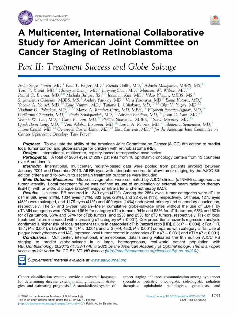
Figure 4 illustrates a comparison between eyes with treatmentfailure defined by the 2 aforementioned criteria. The solid linesdenote treatment failure with need for EBRT or enucleation, and
Figure 2. KaplaneMeier curve of cumulative proportion of salvaged retinoblastoma (RB) eyes without the need for external beam radiation therapy(EBRT), classified by Wills Eye Hospital (WEH) classification.
Tomar et al � AJCC Staging of Retinoblastoma: Globe Salvage
the dotted lines represent treatment failure with need for IAC,plaque brachytherapy, EBRT, or enucleation. KaplaneMeiersurvival curves showed a significant pairwise difference for cT1a(P ¼ 0.031) and cT1b (P < 0.001) but not for cT2a (P ¼ 0.390)and cT2b (P ¼ 0.530). For example, cT1b, the divergent greenlines (1 solid, 1 broken), represents the difference in globe salvageresults, which is statistically significant. If IAC and plaquebrachytherapy are not available, the globe salvage curve wouldshift from a solid to a dotted line.
Discussion
We present a multicenter, international, internet-basedregistry to study the ability of the 8th edition of AJCC RBStaging System to predict globe salvage without the needfor EBRT. We found that increasing AJCC cT category wassignificantly related to increasing risk of local treatmentfailure as defined by need for EBRT or enucleation. Spe-cifically, as the T-categories increased from cT1a to cT3, thehazard of treatment failure increased. We found a 3.5-foldrisk for cT1b, 15.1-fold risk for cT2a, 16.4-fold risk forcT2b, and 45.0-fold risk for cT3 compared with cT1a.
This study included patients from 18 international RBsubspecialty centers from 13 countries in 6 continents. Theirparticipation allowed for the inclusion of an unusuallydiverse real-world sample sampling of patients from aroundthe globe. The numbers of enrolled patients with RB werelarge enough to obtain statistically significant results.
The existence of multiple RB classification systems hasled to confusion and miscommunication.16-18 The CHLAand WEH classification systems were designed to predicttreatment success using a combination of systemic chemo-therapy and focal consolidation. Both had the same “A” to“E” categories with subtle but significant criteria differ-ences, leading to non-comparable results.16 The mostclinically relevant discrepancy is the size criteria foradvanced tumors, which essentially classifies large CHLAgroup D tumors to group E tumors in the WEH system.17
This disparity was evident in our study, in which the samecohort had 30.8% eyes classified in group E as per CHLAand 61.0% per WEH classification (Table 2). Comparedwith the 8th edition AJCC cTNMH classification, cT3most closely resembles CHLA group E and includes28.3% of all eyes. With most literature using either of the
1741
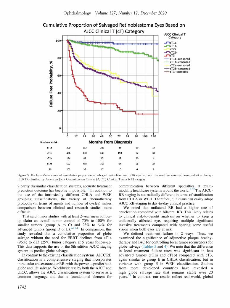
Figure 3. KaplaneMeier curve of cumulative proportion of salvaged retinoblastoma (RB) eyes without the need for external beam radiation therapy(EBRT), classified by American Joint Committee on Cancer (AJCC) Clinical Tumor (cT) category.
Ophthalmology Volume 127, Number 12, December 2020
2 partly dissimilar classification systems, accurate treatmentprediction outcome has become impossible.18 In addition tothe use of the intrinsically different CHLA and WEHgrouping classifications, the variety of chemotherapyprotocols (in terms of agents and number of cycles) makescomparison between clinical and research studies moredifficult.
That said, major studies with at least 2-year mean follow-up claim an overall tumor control of 70% to 100% forsmaller tumors (group A to C) and 23% to 64% foradvanced tumors (group D or E).9,11,17 In comparison, thisstudy revealed that a cumulative proportion of globesalvage without the need for EBRT declines from cT1a(96%) to cT3 (25%) tumor category at 5 years follow-up.This data supports the use of the 8th edition AJCC stagingsystem to predict globe salvage.
In contrast to the existing classification systems, AJCCRBclassification is a comprehensive staging that incorporatesintraocular and extraocular RB,with the power to predict bothglobe and life salvage. Worldwide use by both the AJCC andUICC, allows the AJCC classification system to serve as acommon language and thus a foundational element for
1742
communication between different specialties at multi-modality healthcare systems around theworld.1,22 TheAJCC-RB staging is not radically different in terms of stratificationfrom CHLA or WEH. Therefore, clinicians can easily adaptAJCC RB-staging to day-to-day clinical practice.
We noted that unilateral RB had a higher rate ofenucleation compared with bilateral RB. This likely relatesto clinical risk-to-benefit analysis on whether to keep aunilaterally affected eye, requiring multiple significantinvasive treatments compared with sparing some usefulvision when both eyes are at risk.
We defined treatment failure in 2 ways. Thus, weexamined the significance of adjunctive plaque brachy-therapy and IAC for controlling local tumor recurrences forglobe salvage (Tables 3 and 4). We note that the differencein local treatment failure rates was significant in lessadvanced tumors (cT1a and cT1b) compared with cT3,again similar to group E in CHLA classification, but invariance with group E in WEH classification. Studiesfrom more developed countries have revealed ahigh globe salvage rate that remains stable over 20years.11 In contrast, our results reflect real-world, global
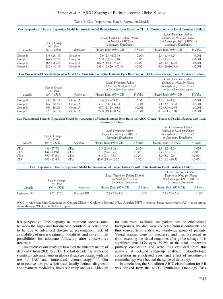
Table 5. Cox Proportional Hazard Regression Models
Cox Proportional Hazards Regression Model for Association of Retinoblastoma Eyes Based on CHLA Classification with Local Treatment Failure
Variable
Eyes in Group,No. (%)
(N ¼ 1556) Reference
Local Treatment Failure Definedas Need for EBRT orSecondary Enucleation
Local Treatment FailureDefined as Need for Plaque
Brachytherapy, IAC, EBRT, orSecondary Enucleation
Hazard Ratio (95% CI) P Value Hazard Ratio (95% CI) P Value
Group B 438 (28.1%) Group A 17.9 (2.5e129.5) 0.004 2.4 (1.4e4.0) 0.001Group C 167 (10.7%) Group A 29.0 (3.9e213.8) 0.001 2.9 (1.7e5.2) <0.001Group D 563 (36.2%) Group A 102.9 (14.4e733.8) <0.001 7.6 (4.6e12.6) <0.001Group E 220 (14.1%) Group A 252.4 (35.2e1807.5) <0.001 17.6 (10.4e29.6) <0.001
Cox Proportional Hazards Regression Model for Association of Retinoblastoma Eyes Based on WEH Classification with Local Treatment Failure
Variable
Eyes in Group,No. (%)
(N ¼ 1563) Reference
Local Treatment FailureDefined as Need for EBRTor Secondary Enucleation
Local Treatment FailureDefined as Need for PlaqueBrachytherapy, IAC, EBRT,or Secondary Enucleation
Hazard Ratio (95% CI) P Value Hazard Ratio (95% CI) P Value
Group B 438 (28.1%) Group A 9.6 (2.4e39.1) 0.002 2.3 (1.4e3.7) 0.001Group C 167 (10.7%) Group A 30.7 (6.6e142.3) 0.001 5.1 (2.5e10.3) <0.001Group D 563 (36.2%) Group A 45.7 (11.2e186.4) <0.001 6.0 (3.6e10.0) <0.001Group E 220 (14.1%) Group A 77.8 (19.4e312.7) <0.001 9.8 (6.1e15.8) <0.001
Cox Proportional Hazards Regression Model for Association of Retinoblastoma Eyes Based on AJCC Clinical Tumor (cT) Classification with LocalTreatment Failure
Variable
Eyes in Group,No. (%)
(N ¼ 1574) Reference
Local Treatment FailureDefined as Need for EBRT or
Secondary Enucleation
Local Treatment FailureDefined as Need for PlaqueBrachytherapy, IAC, EBRTor Secondary Enucleation
Hazard Ratio (95% CI) P Value Hazard Ratio (95% CI) P Value
cT1b 436 (27.7%) cT1a 3.5 (1.5e8.2) 0.004 1.9 (1.2e3.0) 0.005cT2a 144 (9.1%) cT1a 15.1 (6.4e35.2) <0.001 5.4 (3.3e8.7) <0.001cT2b 592 (37.6%) cT1a 16.4 (7.3e36.9) <0.001 5.0 (3.2e7.6) <0.001cT3 202 (12.8%) cT1a 45.0 (19.8e102.5) <0.001 13.0 (8.3e20.3) <0.001
Cox Proportional Hazards Regression Model for Association of Tumor Laterality with Retinoblastoma Local Treatment Failure
Variable
Eyes in Group,No. (%)
(N ¼ 1574) Reference
Local Treatment Failure Definedas Need for EBRT orSecondary Enucleation
Local Treatment FailureDefined as Need for Plaque
Brachytherapy, IAC, EBRT orSecondary Enucleation
Hazard Ratio (95% CI) P Value Hazard Ratio (95% CI) P Value
Unilateral RB 425 (27%) Bilateral RB 2.7 (2.2e3.2) <0.001 2.4 (2.0e2.9) <0.001
AJCC ¼ American Joint Committee on Cancer; CHLA ¼ Children’s Hospital of Los Angeles; EBRT ¼ external beam radiotherapy; IAC ¼ intra-arterialchemotherapy; WEH ¼ Wills Eye Hospital.
Tomar et al � AJCC Staging of Retinoblastoma: Globe Salvage
RB perspective. The disparity in treatment success ratesbetween the high- and low-income countries is consideredto be due to advanced disease at presentation, lack ofavailability of newer treatment modalities, and more limitedpossibilities for adequate follow-up after conservativetreatment.14
Limitations of our study are based on the inherent nature ofdata entry from 2001 to 2013. The last decade has witnessedsignificant advancements in globe salvage associated with theuse of IAC and intravitreal chemotherapy.12,13 Ourretrospective design, which used locally defined diagnosticand treatment modalities, limits subgroup analysis. Although
no data were available on patient sex or ethnic/racialbackgrounds, the data were collected from 6 continents andthus sourced from a diverse, worldwide group of patients.Visual acuities were not measured and thus prevented usfrom assessing the visual outcomes after globe salvage. It issignificant that 1179 eyes, 39.2% of the total, underwentprimary enucleation and were thus excluded from thisanalysis. A detailed subgroup analysis, histopathologiccorrelation in enucleated eyes, and effect of neoadjuvantchemotherapy were beyond the scope of this study.
In conclusion, the 8th edition AJCC classification for RBwas derived from the AJCC Ophthalmic Oncology Task
1743
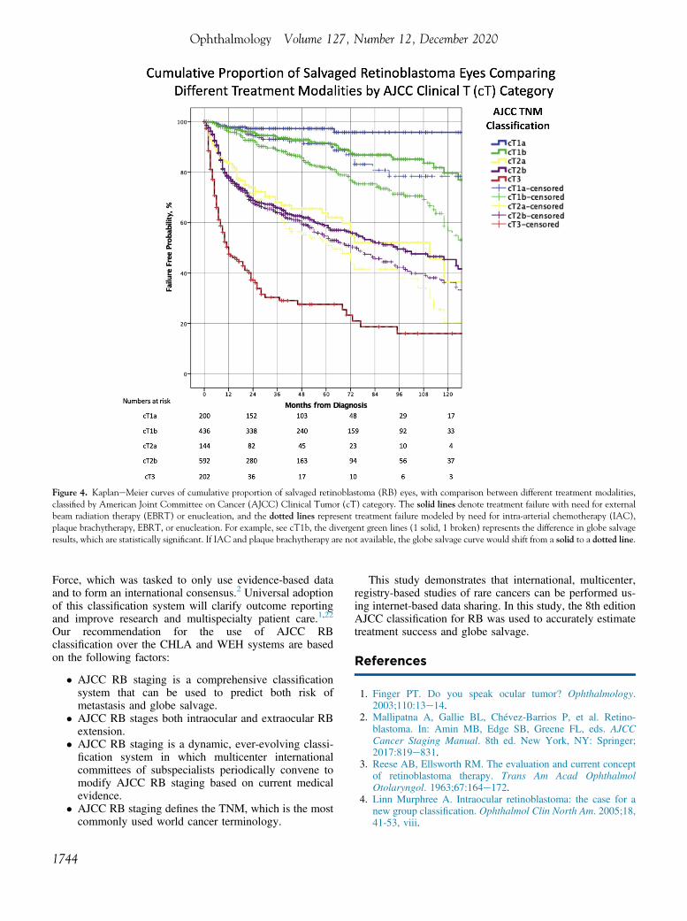
Figure 4. KaplaneMeier curves of cumulative proportion of salvaged retinoblastoma (RB) eyes, with comparison between different treatment modalities,classified by American Joint Committee on Cancer (AJCC) Clinical Tumor (cT) category. The solid lines denote treatment failure with need for externalbeam radiation therapy (EBRT) or enucleation, and the dotted lines represent treatment failure modeled by need for intra-arterial chemotherapy (IAC),plaque brachytherapy, EBRT, or enucleation. For example, see cT1b, the divergent green lines (1 solid, 1 broken) represents the difference in globe salvageresults, which are statistically significant. If IAC and plaque brachytherapy are not available, the globe salvage curve would shift from a solid to a dotted line.
Ophthalmology Volume 127, Number 12, December 2020
Force, which was tasked to only use evidence-based dataand to form an international consensus.2 Universal adoptionof this classification system will clarify outcome reportingand improve research and multispecialty patient care.1,22
Our recommendation for the use of AJCC RBclassification over the CHLA and WEH systems are basedon the following factors:
� AJCC RB staging is a comprehensive classificationsystem that can be used to predict both risk ofmetastasis and globe salvage.
� AJCC RB stages both intraocular and extraocular RBextension.
� AJCC RB staging is a dynamic, ever-evolving classi-fication system in which multicenter internationalcommittees of subspecialists periodically convene tomodify AJCC RB staging based on current medicalevidence.
� AJCC RB staging defines the TNM, which is the mostcommonly used world cancer terminology.
1744
This study demonstrates that international, multicenter,registry-based studies of rare cancers can be performed us-ing internet-based data sharing. In this study, the 8th editionAJCC classification for RB was used to accurately estimatetreatment success and globe salvage.
References
1. Finger PT. Do you speak ocular tumor? Ophthalmology.2003;110:13e14.
2. Mallipatna A, Gallie BL, Chévez-Barrios P, et al. Retino-blastoma. In: Amin MB, Edge SB, Greene FL, eds. AJCCCancer Staging Manual. 8th ed. New York, NY: Springer;2017:819e831.
3. Reese AB, Ellsworth RM. The evaluation and current conceptof retinoblastoma therapy. Trans Am Acad OphthalmolOtolaryngol. 1963;67:164e172.
4. Linn Murphree A. Intraocular retinoblastoma: the case for anew group classification. Ophthalmol Clin North Am. 2005;18,41-53, viii.
Tomar et al � AJCC Staging of Retinoblastoma: Globe Salvage
5. Shields CL, Mashayekhi A, Au AK, et al. The InternationalClassification of Retinoblastoma predicts chemoreductionsuccess. Ophthalmology. 2006;113:2276e2280.
6. Gallie BL, Budning A, DeBoer G, et al. Chemotherapy withfocal therapy can cure intraocular retinoblastoma withoutradiotherapy. Arch Ophthalmol. 1996;114:1321e1328.
7. Wilson MW, Haik BG, Liu T, et al. Effect on ocular survivalof adding early intensive focal treatments to a two-drugchemotherapy regimen in patients with retinoblastoma. Am JOphthalmol. 2005;140:397e406.
8. Chantada GL, Fandiño AC, Raslawski EC, et al. Experiencewith chemoreduction and focal therapy for intraocular retino-blastoma in a developing country. Pediatr Blood Cancer.2005;44:455e460.
9. Munier FL, Beck-Popovic M, Chantada GL, et al. Conserva-tive management of retinoblastoma: challenging orthodoxywithout compromising the state of metastatic grace. “Alive,with good vision and no comorbidity.” Prog Retin Eye Res.2019;73:100764.
10. Shields CL, Say EAT, Pointdujour-Lim R, et al. Rescue intra-arterial chemotherapy following retinoblastoma recurrenceafter initial intra-arterial chemotherapy. J Fr Ophtalmol.2015;38:542e549.
11. Shields CL, Bas Z, Tadepalli S, et al. Long-term (20-year)real-world outcomes of intravenous chemotherapy (chemo-reduction) for retinoblastoma in 964 eyes of 554 patients at asingle centre. Br J Ophthalmol; 2020 Feb 12. bjophthalmol-2019-315572 https://doi.org/10.1136/bjophthalmol-2019-315572. Online ahead of print.
12. Yousef YA, Soliman SE, Astudillo PPP, et al. Intra-arterialchemotherapy for retinoblastoma: a systematic review. JAMAOphthalmol. 2016;134:584e591.
13. American Brachytherapy Society - Ophthalmic Oncology TaskForce. Electronic address: [email protected],ABSeOOTF Committee. The American BrachytherapySociety consensus guidelines for plaque brachytherapy
of uveal melanoma and retinoblastoma Brachytherapy.2014;13:1e14.
14. Global Retinoblastoma Study Group, Fabian ID, Abdallah E,et al. Global retinoblastoma presentation and analysis bynational income level. JAMA Oncol. 2020;6:1e12.
15. Chantada GL, Sampor C, Bosaleh A, et al. Comparison ofstaging systems for extraocular retinoblastoma: analysisof 533 patients. JAMA Ophthalmol. 2013;131:1127e1134.
16. Novetsky DE, Abramson DH, Kim JW, Dunkel IJ. Publishedinternational classification of retinoblastoma (ICRB) defini-tions contain inconsistencies–an analysis of impact.Ophthalmic Genet. 2009;30:40e44.
17. Kim JW, Shah SN, Green S, et al. Tumour size criteria forGroup D and E eyes in the International Classification Systemfor Retinoblastoma: effects on rates of globe salvage and high-risk histopathologic features. Acta Ophthalmol (Copenh).2020;98:e121ee125.
18. Scelfo C, Francis JH, Khetan V, et al. An international surveyof classification and treatment choices for group D retino-blastoma. Int J Ophthalmol. 2017;10:961e967.
19. AJCC Ophthalmic Oncology Task Force. International vali-dation of the American Joint Committee on Cancer’s 7thEdition Classification of Uveal Melanoma. JAMA Ophthalmol.2015;133:376e383.
20. Finger PT; 7th Edition, AJCC-UICC Ophthalmic OncologyTask Force. The 7th edition AJCC staging system for eyecancer: an international language for ophthalmic oncology.Arch Pathol Lab Med. 2009;133:1197e1198.
21. Jain P, Finger PT, Damato B, et al. Multicenter,International Assessment of the Eighth Edition of theAmerican Joint Committee on Cancer Staging Manual forConjunctival Melanoma. JAMA Ophthalmol. 2019;137:905e911.
22. Finger PT. Foundational elements for collaboration inophthalmic oncology. Ophthalmol Retina. 2017;1:263e265.
Footnotes and Financial Disclosures
Originally received: April 20, 2020.Final revision: May 28, 2020.Accepted: May 29, 2020.Available online: June 8, 2020. Manuscript no. D-20-00973.1 Department of Ocular Tumor and Orbital Disease, The New York EyeCancer Center, New York, New York.2 The Eye Cancer Clinic, Princess Margaret Cancer Centre, Toronto,Ontario, Canada.3 Ocular Oncology Service, Department of Ophthalmology, University ofHelsinki and Helsinki University Hospital, Helsinki, Finland.4 Pediatric Oncology Center, Beijing Children’s Hospital, Beijing, China.5 Department of Ophthalmology, Hamilton Eye Institute, University ofTennessee Health Science Center, College of Medicine, Memphis,Tennessee.6 Department of Surgery, St. Jude Children’s Research Hospital, Memphis,Tennessee.7 USC Roski Eye Institute, Keck Medical School of the University ofSouthern California, Los Angeles, California The Vision Center at Chil-dren’s Hospital Los Angeles, Los Angeles, California.8 Department of Vitreoretina Services, Sankara Nethralaya, Chennai, TamilNadu, India.9 Ocular Oncology Department, The S.N. Fyodorov Eye MicrosurgeryFederal State Institution, Moscow, Russian Federation.
10 Department of Surgery (Ophthalmology), King Hussein Cancer Center,Amman, Jordan.11 Department of Ophthalmology and Vision Sciences, Hospital for SickChildren, Toronto, Canada.12 SRI of Pediatric Oncology and Hematology of N.N. Blokhin NationalMedical Research Center Oncology of Russian Federation, Moscow,Russian Federation.13 Medical Academy of Postgraduate Education, Moscow, RussianFederation.14 Department of Ophthalmology, Hospital Infantil de México FedericoGómez, Mexico City, Mexico.15 Precision Medicine Coordination Hospital JP Garrahan, Buenos Aires,Argentina and CONICET, National Scientific and Technical ResearchCouncil, Argentina.16 Ophthalmology Service Hospital JP Garrahan, Buenos Aires, Argentina.17 Department of Ophthalmology and Visual Sciences, The Chinese Uni-versity of Hong Kong, Kowloon, Hong Kong.18 Save Sight Institute, Discipline of Ophthalmology, Sydney MedicalSchool, University of Sydney, Sydney, Australia.19 KK Women’s and Children’s Hospital, Singapore.20 Ophthalmology Unit, Department of Surgery, School of Medicine andDentistry, College of Health Sciences, University of Ghana, Accra, Ghana.21 Department of Child Health, University of Ghana Medical School,Accra, Ghana.
1745
Ophthalmology Volume 127, Number 12, December 2020
22 Retinoblastoma Unit, Department of Ophthalmology, Hospital Sant Joande Déu, Esplugues de Llobregat, Barcelona, Spain.23 Retinoblastoma Unit, Department of Oncology, Hospital Sant Joan deDéu, Esplugues de Llobregat, Barcelona, Spain.
*The American Joint Committee on Cancer Ophthalmic Oncology TaskForce members appear in the Supplemental Appendix (available at www.aaojournal.org).
Financial Disclosure(s):The author(s) have no proprietary or commercial interest in any materialsdiscussed in this article.
Writing Committee: Ankit S. Tomar, MD, Paul T. Finger, MD, BrendaGallie, MD, Aswhin Mallipatna, MD, and Tero Kivelä, MD. All authorsparticipated in critical review of the manuscript before publication. YuliyaGavrylyuk, MD, MHA, Princess Margaret Cancer Centre, Toronto, Ontario,Canada, assisted with multicenter institutional review board, ethics com-mittee, privacy, and other contractual relationships.
The Myrna and John Daniels Charitable Trust, the Paul Finger Fund, andThe Eye Cancer Foundation provided monetary support to the PrincessMargaret Cancer Centre’s Internet Technology Program, which has (in turn)participated in the design, construction, and maintenance of this retino-blastoma registry. Dr. Tomar received an ophthalmic oncology fellowshipgrant to study with Dr. Finger (from The Eye Cancer Foundation). Dr.Kivelä reported receiving a governmental grant from the Helsinki Univer-sity Hospital Research Fund. The funding sources had no role in the designand conduct of the study; collection, management, analysis, and interpre-tation of the data; preparation, review, or approval of the manuscript; anddecision to submit the manuscript for publication.
HUMAN SUBJECTS: Human subjects were included in this study. Allparticipating centers obtained internal institutional review board approval toperform retrospective medical record reviews and contribute de-identifieddata. All research adhered to the tenets of the Declaration of Helsinki.All participants provided informed consent.
No animal subjects were used in this study.
1746
Author Contributions:
Conception and design: Finger, Gallie
Data collection: Tomar, Finger, Gallie, Mallipatna, Kivelä, Zhang, Zhao,Wilson, Brenna, Burges, Kim, Khetan, Ganesan, Yarovoy, Yarovaya,Yousef, Nummi, Ushakova, Yugay, Polyakov, Ramirez-Ortiz, Esparza-Aguiar, Chantada, Schaiquevich, Fandino, Yam, Lau, Lam, Sharwood,Moorthy, Long, Essuman, Renner, Semenova, Jaume Català, Correa-Llano,Carreras-Bertran
Analysis and interpretation: Tomar, Finger, Gallie, Mallipatna, Kivelä,Zhang, Zhao, Wilson, Brenna, Burges, Kim, Khetan, Ganesan, Yarovoy,Yarovaya, Yousef, Nummi, Ushakova, Yugay, Polyakov, Ramirez-Ortiz,Esparza-Aguiar, Chantada, Schaiquevich, Fandino, Yam, Lau, Lam, Shar-wood, Moorthy, Long, Essuman, Renner, Semenova, Jaume Català, Correa-Llano, Carreras-Bertran
Obtained funding: N/A; Each author’s time contributions for data entry andmanuscript review were performed as part of their regular employment ortaken from their free time. No additional funding was provided for thesetasks.
Overall responsibility: Tomar, Finger, Gallie, Mallipatna, Kivelä, Zhang,Zhao, Wilson, Brenna, Burges, Kim, Khetan, Ganesan, Yarovoy, Yarovaya,Yousef, Nummi, Ushakova, Yugay, Polyakov, Ramirez-Ortiz, Esparza-Aguiar, Chantada, Schaiquevich, Fandino, Yam, Lau, Lam, Sharwood,Moorthy, Long, Essuman, Renner, Semenova, Jaume Català, Correa-Llano,Carreras-Bertran
Abbreviations and Acronyms:AJCC ¼ American Joint Committee on Cancer; CHLA ¼ Children’sHospital of Los Angeles; CI ¼ confidence interval; EBRT ¼ external beamradiotherapy; HR ¼ hazard ratio; IAC ¼ intra-arterial chemotherapy;ICRB ¼ International Classification for Retinoblastoma;IIRC ¼ International Intraocular Retinoblastoma Classification;IQR ¼ interquartile range; OOTF ¼ Ophthalmic Oncology Task Force;RB ¼ retinoblastoma; WEH ¼ Wills Eye Hospital.
Correspondence:Paul T. Finger, MD, FACS, The New York Eye Cancer Center, 115 East61st St., 5th Floor, New York, NY 10065. E-mail: [email protected].