Embed Size (px)
Citation preview
109
THEMATIC REVIEW
The skeleton: a multi-functional complex organ. The growth platechondrocyte and endochondral ossification
E J Mackie, L Tatarczuch and M Mirams
School of Veterinary Science, University of Melbourne, Parkville, Victoria 3010, Australia
(Correspondence should be addressed to E J Mackie; Email: [email protected])
Abstract
Endochondral ossification is the process that results in both
the replacement of the embryonic cartilaginous skeleton
during organogenesis and the growth of long bones until
adult height is achieved. Chondrocytes play a central role in
this process, contributing to longitudinal growth through a
combination of proliferation, extracellular matrix (ECM)
secretion and hypertrophy. Terminally differentiated hyper-
trophic chondrocytes then die, allowing the invasion of a
mixture of cells that collectively replace the cartilage tissue
with bone tissue. The behaviour of growth plate chondro-
cytes is tightly regulated at all stages of endochondral
ossification by a complex network of interactions between
This paper is one of three papers that form part of a thematic review
section on the skeleton: a multi-functional complex organ. The Guest
Editor for this section was Colin Farquharson, Roslin Institute,
University of Edinburgh, UK.
Journal of Endocrinology (2011) 211, 109–1210022–0795/11/0211–109 q 2011 Society for Endocrinology Printed in Great
circulating hormones (including GH and thyroid hormone),
locally produced growth factors (including Indian hedgehog,
WNTs, bone morphogenetic proteins and fibroblast growth
factors) and the components of the ECM secreted by the
chondrocytes (including collagens, proteoglycans, thrombos-
pondins and matrilins). In turn, chondrocytes secrete factors
that regulate the behaviour of the invading bone cells,
including vascular endothelial growth factor and receptor
activator of NFkB ligand. This review discusses how the
growth plate chondrocyte contributes to endochondral
ossification, with some emphasis on recent advances.
Journal of Endocrinology (2011) 211, 109–121
Introduction
Bones in different parts of the skeleton develop through two
distinct processes, intramembranous ossification and endo-
chondral ossification. Intramembranous ossification, which
occurs in the flat bones of the skull, involves direct
differentiation of embryonic mesenchymal cells into the
bone-forming osteoblasts. In contrast, endochondral ossifica-
tion, which occurs in the remainder of the skeleton, involves
the replacement of a cartilage model by bone tissue.
The cartilage model of a prospective bone is formed as
embryonic mesenchymal cells condense and differentiate into
chondrocytes, which secrete the various components of
cartilage extracellular matrix (ECM), including collagen type
II and the proteoglycan aggrecan. The model expands
through chondrocyte proliferation. Ossification of the
cartilage model is preceded by hypertrophy of the chon-
drocytes in the prospective mid-shaft of the bone, and
deposition of a periosteal bone collar by recently differ-
entiated osteoblasts surrounding the mid-shaft (Fig. 1). Blood
vessels, osteoclasts (cartilage- and bone-resorbing cells), as
well as bone marrow and osteoblast precursors then invade the
model from the bone collar and proceed to form the primary
centre of ossification. The primary centre expands towards
the ends of the cartilage model, as the osteoclasts remove
cartilage ECM and osteoblasts deposit bone on cartilage
remnants. In long bones, a secondary ossification centre
subsequently forms at each end of the cartilage model, leaving
a cartilaginous growth plate between the primary and
secondary ossification centres, as well as the prospective
permanent articular cartilages at each end of the bone. The
growth plate is responsible for longitudinal growth of bones.
Skeletal maturity occurs when the expanding primary centre
of ossification meets the secondary centre of ossification, thus
obliterating the growth plate (Fig. 1).
Endochondral ossification is initiated during foetal life, and
continues until growth ceases in early adulthood. Although
endochondral ossification is dependent on the concerted
actions of a number of cell types, it is the chondrocyte that
drives the process. This review will focus on the role of
the chondrocyte in endochondral ossification, with some
emphasis on areas in which recent advances have been made.
DOI: 10.1530/JOE-11-0048Britain Online version via http://www.endocrinology-journals.org
A B
Cartilage
Bone
Bone marrow (including blood vessels)
Primarycentre ofossification
Growthplate
Epiphysis
Metaphysis
Diaphysis
Articularcartilage
Secondarycentre ofossification
C D E
Periostealbonecollar
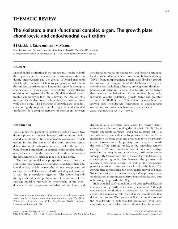
Figure 1 Overview of development of a bone by endochondralossification. (A) The cartilage model of the future bone.(B) A periosteal bone collar has formed and formation of the primarycentre of ossification has been initiated. (C) The primary centre ofossification is starting to expand towards the ends of the cartilagemodel. (D) The secondary centres of ossification have formed ateach end of the bone, leaving a cartilaginous growth plate betweenprimary and secondary centres of ossification. (E) Skeletal maturityhas been achieved, with complete replacement of the growth platecartilage by bone. The only cartilage remaining is the articularcartilage at the ends of the bone.
Figure 2 Growth plate morphology. Section through the proximalgrowth plate of the tibia from a 3-week-old mouse; the section isoriented longitudinally with respect to the bone; barZ50 mm.
E J MACKIE and others . Growth plate chondrocytes110
The growth plate chondrocyte models its ownenvironment
At all stages of endochondral ossification, from the initiation
of formation of the primary centre to the final stages of
adolescent growth, the chondrocytes contributing to this
process participate in an orderly sequence of events that are
reflected in their morphology; the different stages of the
chondrocyte lifespan are visible in distinct zones in sections
through the growth plate (Fig. 2). First, chondrocytes
undergo proliferation, which is observed as the presence of
pairs of chondrocytes in a single lacuna within the cartilage
ECM, before their separation from each other by secretion of
ECM (Figs 2 and 3). Following proliferation, the chon-
drocytes undergo a period of high secretory activity, as they
deposit the typical cartilage ECM components around
themselves, while remaining in multicellular clusters, often
arranged in columns parallel to the long axis of the bone.
These cells gradually undergo hypertrophy, modelling their
surrounding ECM as they expand, and then mineralising it.
Following hypertrophy, chondrocytes undergo physiological
death, and the transverse septa of the cartilage ECM
surrounding them are removed, allowing entry of the mixture
of cells responsible for the expansion of the ossification centre.
Thus, the growth plate chondrocyte plays multiple important
roles during its lifespan. It constructs the transient growth
plate tissue, which has the necessary capacity to move in space
through continued self-renewal and localised degradation, but
simultaneously maintains the mechanical stability of the
growing bone. Accumulating evidence indicates that the
Journal of Endocrinology (2011) 211, 109–121
growth plate chondrocyte orchestrates the invasion of its own
domain by the ossification front not only through preparation
of the cartilage tissue, but also by secreting soluble molecules
that regulate the behaviour of the invading cells.
Contribution to bone elongation
The growth plate chondrocyte contributes to bone elongation
through a combination of proliferation, ECM secretion and
hypertrophy. The relative contributions of these parameters
vary with growth rate, which varies with anatomical location,
age and species; the higher the growth rate the greater the
contribution from cellular hypertrophy and the smaller the
contribution from matrix synthesis (Wilsman et al. 1996).
These functions of the chondrocyte are tightly controlled by
circulating molecules such as hormones, as well as by
substances produced locally by the chondrocytes themselves
(summarised in Fig. 4). Mutations in the genes encoding these
regulatory molecules result in abnormal chondrocyte
behaviour and skeletal dysplasias, most of which are associated
with dwarfism, i.e. inadequate growth of the bones that form
by endochondral ossification (Krakow & Rimoin 2010).
Proliferation
An important stimulator of chondrocyte proliferation in the
growth plate is GH, which is produced in the pituitary gland.
GH exerts its effects on the growth plate predominantly
www.endocrinology-journals.org
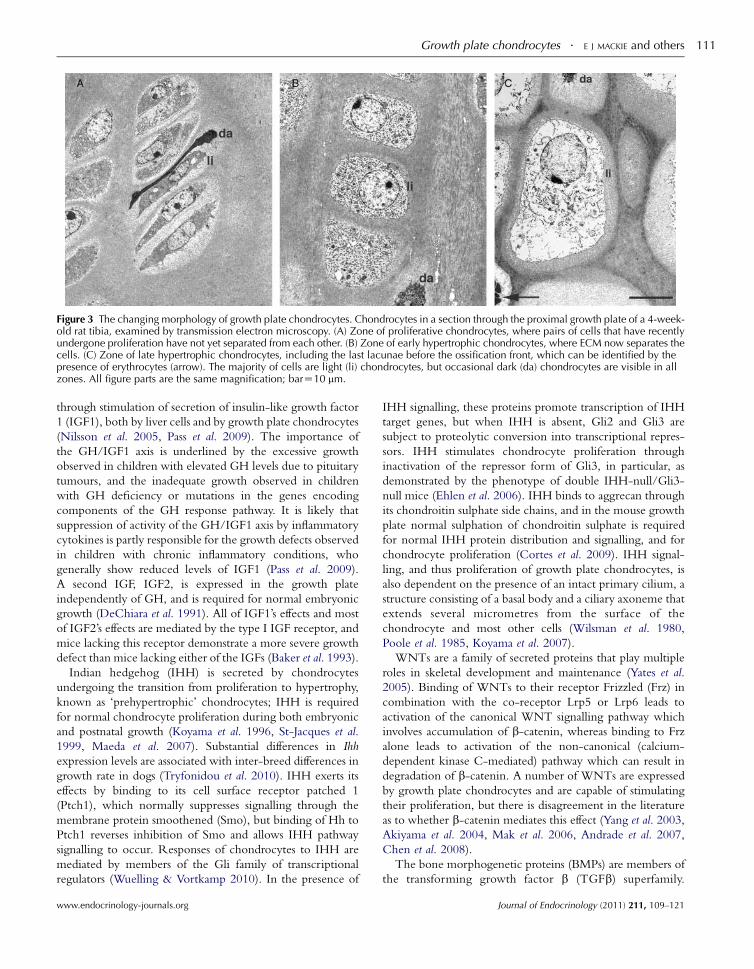
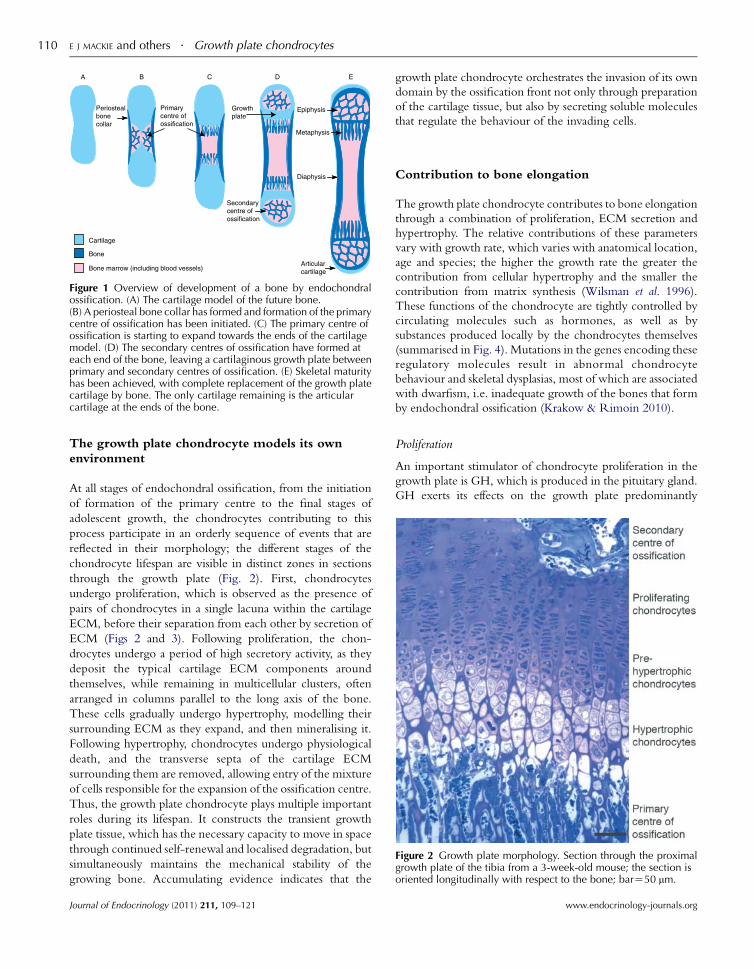
Figure 3 The changing morphology of growth plate chondrocytes. Chondrocytes in a section through the proximal growth plate of a 4-week-old rat tibia, examined by transmission electron microscopy. (A) Zone of proliferative chondrocytes, where pairs of cells that have recentlyundergone proliferation have not yet separated from each other. (B) Zone of early hypertrophic chondrocytes, where ECM now separates thecells. (C) Zone of late hypertrophic chondrocytes, including the last lacunae before the ossification front, which can be identified by thepresence of erythrocytes (arrow). The majority of cells are light (li) chondrocytes, but occasional dark (da) chondrocytes are visible in allzones. All figure parts are the same magnification; barZ10 mm.
Growth plate chondrocytes . E J MACKIE and others 111
through stimulation of secretion of insulin-like growth factor
1 (IGF1), both by liver cells and by growth plate chondrocytes
(Nilsson et al. 2005, Pass et al. 2009). The importance of
the GH/IGF1 axis is underlined by the excessive growth
observed in children with elevated GH levels due to pituitary
tumours, and the inadequate growth observed in children
with GH deficiency or mutations in the genes encoding
components of the GH response pathway. It is likely that
suppression of activity of the GH/IGF1 axis by inflammatory
cytokines is partly responsible for the growth defects observed
in children with chronic inflammatory conditions, who
generally show reduced levels of IGF1 (Pass et al. 2009).
A second IGF, IGF2, is expressed in the growth plate
independently of GH, and is required for normal embryonic
growth (DeChiara et al. 1991). All of IGF1’s effects and most
of IGF2’s effects are mediated by the type I IGF receptor, and
mice lacking this receptor demonstrate a more severe growth
defect than mice lacking either of the IGFs (Baker et al. 1993).
Indian hedgehog (IHH) is secreted by chondrocytes
undergoing the transition from proliferation to hypertrophy,
known as ‘prehypertrophic’ chondrocytes; IHH is required
for normal chondrocyte proliferation during both embryonic
and postnatal growth (Koyama et al. 1996, St-Jacques et al.
1999, Maeda et al. 2007). Substantial differences in Ihh
expression levels are associated with inter-breed differences in
growth rate in dogs (Tryfonidou et al. 2010). IHH exerts its
effects by binding to its cell surface receptor patched 1
(Ptch1), which normally suppresses signalling through the
membrane protein smoothened (Smo), but binding of Hh to
Ptch1 reverses inhibition of Smo and allows IHH pathway
signalling to occur. Responses of chondrocytes to IHH are
mediated by members of the Gli family of transcriptional
regulators (Wuelling & Vortkamp 2010). In the presence of
www.endocrinology-journals.org
IHH signalling, these proteins promote transcription of IHH
target genes, but when IHH is absent, Gli2 and Gli3 are
subject to proteolytic conversion into transcriptional repres-
sors. IHH stimulates chondrocyte proliferation through
inactivation of the repressor form of Gli3, in particular, as
demonstrated by the phenotype of double IHH-null/Gli3-
null mice (Ehlen et al. 2006). IHH binds to aggrecan through
its chondroitin sulphate side chains, and in the mouse growth
plate normal sulphation of chondroitin sulphate is required
for normal IHH protein distribution and signalling, and for
chondrocyte proliferation (Cortes et al. 2009). IHH signal-
ling, and thus proliferation of growth plate chondrocytes, is
also dependent on the presence of an intact primary cilium, a
structure consisting of a basal body and a ciliary axoneme that
extends several micrometres from the surface of the
chondrocyte and most other cells (Wilsman et al. 1980,
Poole et al. 1985, Koyama et al. 2007).
WNTs are a family of secreted proteins that play multiple
roles in skeletal development and maintenance (Yates et al.
2005). Binding of WNTs to their receptor Frizzled (Frz) in
combination with the co-receptor Lrp5 or Lrp6 leads to
activation of the canonical WNT signalling pathway which
involves accumulation of b-catenin, whereas binding to Frz
alone leads to activation of the non-canonical (calcium-
dependent kinase C-mediated) pathway which can result in
degradation of b-catenin. A number of WNTs are expressed
by growth plate chondrocytes and are capable of stimulating
their proliferation, but there is disagreement in the literature
as to whether b-catenin mediates this effect (Yang et al. 2003,
Akiyama et al. 2004, Mak et al. 2006, Andrade et al. 2007,
Chen et al. 2008).
The bone morphogenetic proteins (BMPs) are members of
the transforming growth factor b (TGFb) superfamily.
Journal of Endocrinology (2011) 211, 109–121
HypertrophyThyroidhormones
WNTsIGF1FGFsIHH
PTHrP
Proliferation
GH IGF1
IHHBMPs
FGFs
WNTs
Factors secretedby chondrocytes
Hormones
Blood vessel
Osteoclast
Osteoblast
VEGF
HMGB1
RANKL
WNTsBMPs
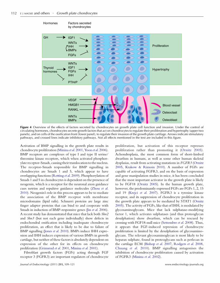
Figure 4 Overview of the effects of factors secreted by chondrocytes on growth plate cell function and invasion. Under the control ofcirculating hormones, chondrocytes secrete growth factors that act on chondrocytes to regulate their proliferation and hypertrophy (upper twopanels), and on cells of the ossification front (lower panel), to regulate their invasion of the growth plate cartilage. Arrows indicate stimulatorypathways, and crossed lines indicate inhibitory pathways. Not all effects mentioned in the text are included in this figure.
E J MACKIE and others . Growth plate chondrocytes112
Activation of BMP signalling in the growth plate results in
chondrocyte proliferation (Minina et al. 2001, Yoon et al. 2006).
BMP receptors are complexes of type I and type II serine/
threonine kinase receptors, which when activated phosphor-
ylate receptor-Smads, causing their translocation to thenucleus.
The receptor-Smads responsible for BMP signalling in
chondrocytes are Smads 1 and 5, which appear to have
overlapping functions (Retting et al. 2009). Phosphorylation of
Smads 1 and 5 in chondrocytes is dependent on the presence of
neogenin, which is a receptor for the neuronal axon guidance
cues netrins and repulsive guidance molecules (Zhou et al.
2010). Neogenin’s role in this process appears to be to mediate
the association of the BMP receptor with membrane
microdomains (lipid rafts). Schnurri proteins are large zinc
finger adapter proteins that can bind to and cooperate with
Smads in induction of BMP-responsive genes (Jin et al. 2006).
A recent study has demonstrated that mice that lack both Shn2
and Shn3 (but not each gene individually) show defects in
endochondral ossification including inadequate chondrocyte
proliferation, an effect that is likely to be due to failure of
BMP signalling (Jones et al. 2010). BMPs induce IHH expre-
ssion and IHH induces expression of various BMPs in growth
cartilage, but neither of these factors is absolutely dependent on
expression of the other for its effects on chondrocyte
proliferation (Grimsrud et al. 2001, Minina et al. 2001).
Fibroblast growth factors (FGFs) acting through FGF
receptor 3 (FGFR3) are important regulators of chondrocyte
Journal of Endocrinology (2011) 211, 109–121
proliferation, but activation of this receptor represses
proliferation rather than promoting it (Ornitz 2005).
Achondroplasia, the most common form of short-limbed
dwarfism in humans, as well as some other human skeletal
dysplasias, result from activating mutations in FGFR3 (Ornitz
2005, Krakow & Rimoin 2010). A number of FGFs are
capable of activating FGFR3, and on the basis of expression
and gene manipulation studies in mice, it has been concluded
that the most important activator in the growth plate is likely
to be FGF18 (Ornitz 2005). In the human growth plate,
however, the predominantly expressed FGFs are FGFs 1, 2, 15
and 19 (Krejci et al. 2007). FGFR3 is a tyrosine kinase
receptor, and its suppression of chondrocyte proliferation in
the growth plate appears to be mediated by STAT1 (Ornitz
2005). The activity of FGFs, like that of IHH, is modulated by
glycosaminoglycans. Mice that lack sulphatase-modifying
factor 1, which activates sulphatases (and thus proteoglycan
desulphation) show dwarfism, which can be rescued by
crossing with FGF18-null mice (Settembre et al. 2008). Thus,
it appears that FGF-induced repression of chondrocyte
proliferation is limited by the desulphation of glycosamino-
glycans. The relevant glycosaminoglycan is most likely to be
heparan sulphate, found in proteoglycans such as perlecan in
the cartilage ECM (Bishop et al. 2007, Rodgers et al. 2008,
Chuang et al. 2010). BMP signalling antagonises the
inhibition of chondrocyte proliferation caused by activation
of FGFR3 (Minina et al. 2002).
www.endocrinology-journals.org
Growth plate chondrocytes . E J MACKIE and others 113
Chondrocyte proliferation in response to growth factors
including IHH is mediated by cyclins which form complexes
with cyclin-dependent kinases, ultimately leading to acti-
vation of E2F transcription factors and cell cycle progression
(Wuelling & Vortkamp 2010). Cyclin D1, in particular, is
required for normal proliferation of growth plate chondro-
cytes, as demonstrated by studies in cyclin D1-null mice
(Beier et al. 2001). The ability of chondrocytes to express
normal levels of cyclin D1, and thus undergo normal
proliferation, is dependent on the presence of the transcrip-
tional repressor TRPS1. This protein derives its name from
tricho–rhino–phalangeal syndrome, a condition in humans
involving skeletal malformations, which results from
mutations in the TRPS1 gene. TRPS1-null mice demon-
strate abnormally low levels of proliferation in growth plate
chondrocytes, which can be attributed to a role for TRPS1 in
repression of STAT3 expression, which in turn allows for
elevated expression of cyclin D1 (Suemoto et al. 2007). It is
interesting to note that TRPS1 binds the transcriptional
activator form of Gli3, but the implications of this binding for
chondrocyte proliferation in the growth plate are as yet
unclear (Wuelling et al. 2009, Wuelling & Vortkamp 2010).
Conversely, inhibition of chondrocyte proliferation in
response to activation of FGFR3 is accompanied by induction
of inhibitors of cyclin-dependent kinase including p21,
presumably thereby causing cell cycle arrest (Dailey et al.
2003). Further information about the mechanism by which
FGFR3 causes early cell cycle exit is provided by the results of
expression array studies comparing chondrocytes isolated
from human foetuses affected by thanatophoric dysplasia
caused by activating FGFR3 mutations with chondrocytes
isolated from normal foetuses; among other observations
concerning cell cycle-related genes, the FGFR3 mutations
are associated with lower levels of cell cycle-related E2F target
genes (Schibler et al. 2009).
Extracellular matrix
Secretion of ECM by growth plate chondrocytes makes an
important contribution to growth. Cartilage ECM consists
primarily of large aggregates of aggrecan and the glycosami-
noglycan hyaluronan, packed in amongst fibrils of collagen
type II (Gentili & Cancedda 2009, Heinegard 2009). These
three components of cartilage ECM confer on the growth
plate the mechanical stability required by this integral
component of the growing skeleton. The collagen fibrils
provide the framework for the tissue and the strongly
hydrophilic hyaluronan–aggrecan aggregates allow the tissue
to withstand compression. Both collagen type II and aggrecan
are almost exclusively expressed in cartilage.
Cartilage ECM also contains a number of less abundant
collagens, proteoglycans and other non-collagenous proteins,
which, together with the three major cartilage constituents,
form a complex network of interacting molecules (Gentili &
Cancedda 2009, Heinegard 2009). The relative abundance of
many of the cartilage ECM constituents varies between zones
www.endocrinology-journals.org
of the growth plate. Minor cartilage collagens include
collagen types VI, IX, X, XI, XII and XIV. Cartilage
proteoglycans, in addition to aggrecan, include the small
leucine-rich proteoglycans decorin, biglycan and fibro-
modulin, as well as the large proteoglycan perlecan. Other
non-collagenous proteins found in cartilage ECM include
the matrilins and thrombospondin family members, such as
thrombospondin-5, also known as cartilage oligomeric
matrix protein (COMP). Many of the less abundant
molecules make important contributions to cartilage ECM
assembly and, together with the major cartilage constituents,
influence the behaviour of chondrocytes (Heinegard 2009,
Klatt et al. 2011).
The importance of the complex interactions between
cartilage ECM components for growth is illustrated by the
effects of mutations in humans or mice in the genes encoding
a number of the proteins. For some of these molecules, the
complete absence of the protein has no detrimental effect, but
mutations causing a failure of secretion from the endoplasmic
reticulum (ER) result in retention of the mutant protein and
its binding partners in the ER as well as growth defects
(Zaucke & Grassel 2009, Klatt et al. 2011). Pseudoachon-
droplasia in humans is caused by mutations in COMP, and the
milder multiple epiphyseal dysplasia can be caused by
mutations in the genes encoding COMP, collagen type IX
or matrilin-3 (Briggs & Chapman 2002). Many of the COMP
mutations result in misfolding of COMP, and the chon-
drocytes from these patients retain not only COMP but also
collagen type IX and matrilin-3 in their ER, and the ECM is
depleted of all these proteins (Hecht et al. 2005). Secretion of
aggrecan and collagen type II is not affected by the mutations,
but the collagen does not form organised fibril bundles,
indicating that the COMP/collagen IX/matrilin-3
complexes are required for normal cartilage matrix organis-
ation and growth plate structure. Mice lacking COMP or
matrilin-3 show no abnormality in skeletal development or
growth, and mice lacking collagen type IX exhibit a mild
growth defect, suggesting that these proteins are at least partly
able to substitute for each other (Fassler et al. 1994, Hagg et al.
1997, Svensson et al. 2002, Ko et al. 2004). Generation of
mice lacking both COMP and collagen type IX did not show
any greater disturbance of the growth plate than did mice
lacking collagen type IX alone, but the ECM of these growth
plates still contains some matrilin-3, so perhaps the additional
deletion of matrilin-3 and/or possibly other proteins is
required for the demonstration of functional redundancy
(Blumbach et al. 2008, Posey et al. 2008). It is interesting to
note that in these mice the additional deletion of the COMP-
related protein thrombospondin-3, which alone causes no
obvious growth plate defect, caused a significantly greater
disruption of growth plate organisation and limb length
reduction (Posey et al. 2008).
In addition to the protein components of cartilage ECM,
hyaluronan plays an important role in the contribution of ECM
secretion to growth. Mice in which the gene for hyaluronan
synthase 2 (Has2) is inactivated in tissues derived from limb bud
Journal of Endocrinology (2011) 211, 109–121
E J MACKIE and others . Growth plate chondrocytes114
mesoderm possess abnormally short limbs (Matsumoto et al.
2009). The growth plates from these mice contain abnormally
low levels of hyaluronan; they show a decrease in the deposition
of aggrecan and a decrease in the amount of matrix separating
the chondrocytes, manifest as an increase in cell density in the
absence of any effect on proliferation.
Expression and secretion of components of cartilage ECM,
including collagen type II and aggrecan are stimulated by a
variety of soluble factors present in the growth plate, including
IGF1, BMPs and other members of the TGFb superfamily, and
are absolutely dependent on the transcription factor SOX9 (Bi
et al. 1999, Lefebvre & Smits 2005, Tew et al. 2008). SOX9-
activated transcription appears to be modulated by epigenetic
mechanisms, since it occurs predominantly in hyperacetylated
chromatin; the histone acetyltransferase p300 associates with
SOX9 and enhances SOX9-dependent transcription. More-
over, inhibition of histone deacetylases (HDACs) stimulates
expression of SOX9-activated cartilage ECM genes and
induces histone acetylation in the region of the Col2a1
enhancer in primary chondrocyte cultures (Furumatsu et al.
2005). Overexpression of HDAC1 or 2 in chondrocytes results
in down-regulation of expression of Aggrecan and Col2a1,
providing further evidence for epigenetic control of this aspect
of chondrocyte function (Hong et al. 2009).
Hypertrophy
As post-proliferative chondrocytes undergo hypertrophy, they
experience changes in gene expression that allow them to
modify the structure and composition of the surrounding
ECM. The synthesis of collagen type II is down regulated and
the synthesis of the non-fibrillar collagen type X, expression
of which is specific to hypertrophic chondrocytes, is initiated
(van der Eerden et al. 2003). Hypertrophic chondrocytes
also selectively express matrix metalloproteinase 13
(MMP13), a collagenase capable of degrading fibrils of
collagen type II (Johansson et al. 1997, Cawston & Young
2010). Post-proliferative growth plate chondrocytes exist as
two populations, which are described as light and dark cells
on the basis of their appearance when viewed by transmission
electron microscopy (Fig. 3; Anderson 1964, Hwang 1978,
Wilsman et al. 1981, Roach & Clarke 2000, Ahmed et al.
2007). The fact that these two populations could only be
identified by electron microscopy has made it difficult to study
their functional differences, but the recent identification of
periostin as a dark chondrocyte-associated protein will assist
in research in this area (Chen et al. 2010).
It may seem logical to assume that chondrocytes would
need to degrade the ECM immediately surrounding
themselves to undergo the enormous increase in volume
described as hypertrophy (Fig. 3), however, none of the
enzymes known to both degrade the components of cartilage
ECM and be expressed by hypertrophic chondrocytes are
required for the process of hypertrophy. The chondrocytes of
MMP13-null mice undergo apparently normal hypertrophy,
indicating that collagen degradation is not necessary (Inada
Journal of Endocrinology (2011) 211, 109–121
et al. 2004, Stickens et al. 2004). Similarly, aggrecan
degradation does not appear to be required. There are no
morphological defects in the growth cartilage of mice in
which aggrecan is rendered resistant to MMP cleavage (Little
et al. 2005a). A disintegrin and metalloproteinase with
thrombospondin motifs (ADAMTS) family of enzymes
includes ADAMTS-1, -4 and -5, which are capable of
cleaving aggrecan; mice lacking each of these individually
(Little et al. 2005b, Stanton et al. 2005) or ADAMTS-4 and -5
in combination (Rogerson et al. 2008) show normal growth
plate morphology. The results of the latter study indicate that
an additional, as yet unidentified, aggrecan-degrading activity
is present in mouse cartilage, thus a role for aggrecan cleavage
in chondrocyte hypertrophy cannot yet be ruled out. It seems
likely, however, that ECM degradation is not in fact required
for chondrocytes to expand in volume. Observations on the
role of hyaluronan in the growth plate may provide some
insight into this problem of how the chondrocyte is able to
expand so dramatically. Results of studies in which growth
plate organ cultures were treated with hyaluronidase have
provided evidence for a role for hyaluronan in the
enlargement of the lacunae of hypertrophic chondrocytes
(Pavasant et al. 1996). Moreover, in the Has2-null mice
mentioned-above, as well as having reduced matrix volume,
the growth plates show an almost complete failure of
chondrocyte hypertrophy, based on both morphology and
expression of hypertrophy-associated genes (Matsumoto et al.
2009). As noted by these authors, the role of hyaluronan may
be to initiate hypertrophy-inducing intracellular signalling in
chondrocytes emerging from the proliferative phase. Given
the importance of hyaluronan–aggrecan complexes for
swelling of the growth plate ECM, however, it is likely that
this swelling per se is required for the concomitant expansion
of the chondrocyte. Recently proliferated chondrocytes are
flattened in the longitudinal axis of the growing bone, and the
subsequent increase in cell volume is manifest as a
disproportionate increase in height, relative to width of the
cell, that is, in the direction of bone growth (Fig. 3; Breur
et al. 1994). Perhaps the role of hyaluronan is to provide
sufficient space between collagen fibrils to ensure that the
chondrocyte can displace the surrounding matrix as it expands.
Just as proliferation of the growth plate chondrocyte is
regulated, both positively and negatively, by systemic and
locally produced extracellular molecules, so is the transition
from the proliferative to the hypertrophic state. Thyroid
hormones are important systemic regulators of chondrocyte
hypertrophy (Shao et al. 2006). In vitro, the thyroid hormone
triiodothyronine (T3) stimulates morphological hypertrophy
as well as molecular markers of hypertrophy, without
stimulating proliferation (Burch & Lebovitz 1982, Bohme
et al. 1992, Ballock & Reddi 1994, Wang et al. 2007).
Hypothyroidism in humans results in slowing of longitudinal
bone growth, with abnormally thin growth plates and
impaired chondrocyte hypertrophy. Studies in genetically
manipulated mice have demonstrated that the receptor
www.endocrinology-journals.org
Growth plate chondrocytes . E J MACKIE and others 115
responsible for these effects is thyroid hormone receptor a(and not thyroid hormone receptor b; Kaneshige et al. 2001).
The mediators of thyroid hormone-induced chondrocyte
hypertrophy are starting to be identified, but the mechanism
of this response has not yet been fully elucidated. One such
pathway appears to be WNT/b-catenin signalling. WNTs
not only stimulate chondrocyte proliferation as noted above,
but also promote chondrocyte hypertrophy (Enomoto-
Iwamoto et al. 2002, Dong et al. 2006). A number of studies
in genetically manipulated mice with either constitutive
activation or depletion of components of the b-catenin
signalling pathway have demonstrated that activation of this
pathway promotes chondrocyte hypertrophy; thus, it appears
most likely that the effect of WNTs on chondrocyte
hypertrophy is mediated by the canonical WNT signalling
pathway (Tamamura et al. 2005, Chen et al. 2008, Kawasaki
et al. 2008). In chondrocytes in vitro, T3 up-regulates Wnt4
mRNA and protein expression as well as cellular accumu-
lation of b-catenin, and inhibition of WNT signalling by
secreted WNT antagonists inhibits T3-induced hypertrophy,
providing strong evidence that WNT signalling contributes
to thyroid hormone-induced hypertrophy (Wang et al. 2007).
In addition, these effects of T3 are partially inhibited by
inhibitors of IGF1 signalling, indicating that IGF1 also makes
some contribution to thyroid hormone-induced hypertrophy
(Wang et al. 2010). T3 also induces expression of FGFR3 in
chondrocyte-like cells and the growth plates of thyroid
hormone receptor a-null mice express substantially lower
levels of FGFR3 than do those of wild-type animals (Barnard
et al. 2005). Since activation of FGFR3 results in acceleration
of hypertrophy, this receptor appears to be another mediator
of thyroid hormone-induced chondrocyte hypertrophy
(Minina et al. 2002, Ornitz 2005).
The IHH secreted by prehypertrophic chondrocytes is
another factor that not only influences proliferation, but also
hypertrophy, which it is generally considered to inhibit
(Vortkamp et al. 1996, Maeda et al. 2007). This effect of IHH
is mediated by Gli2, and Gli2-dependent stimulation of
secretion of parathyroid hormone-related peptide (PTHrP;
Vortkamp et al. 1996, Kronenberg 2006, Joeng & Long 2009).
PTHrP suppresses hypertrophy, thus keeping the cells in a
proliferative state (Lee et al. 1996). PTHrP exerts this effect at
least in part through inhibition of expression of the
transcription factor RUNX2; RUNX2 induces chondrocytic
transcription of hypertrophy-associated genes including
col10a1 and promotes hypertrophy (Lefebvre & Smits 2005,
Guo et al. 2006). The suppression of hypertrophy by PTHrP is
probably also mediated by SOX9, which is activated by
PTHrP through protein kinase A-dependent phosphorylation
and indirectly inhibits RUNX2 expression (Huang et al. 2000,
2001, Akiyama et al. 2002, Yamashita et al. 2009). PTHrP
stimulates cyclin D1 expression in chondrocytes, and is unable
to down-regulate RUNX2 expression in chondrocytes from
cyclin D1-null mice, apparently because cyclin D1 contrib-
utes to proteasomal degradation of RUNX2 (Beier et al. 2001,
Zhang et al. 2009). The transcriptional co-regulator Zfp521,
www.endocrinology-journals.org
which is induced by PTHrP, has also recently been identified
as an effector of PTHrP’s actions in the growth plate; the
growth cartilage of mice with chondrocyte-specific deletion
of Zfp521 resembles that of PTHrP-null mice, and PTHrP is
unable to stimulate cyclin D1 expression or inhibit RUNX2
expression in the absence of Zfp521 (Correa et al. 2010).
Expression of PTHrP by growth plate chondrocytes is
inhibited by TRPS1, as demonstrated by studies in TRPS1-
null mice and accompanying in vitro studies (Nishioka et al.
2008). Mice with a heterozygous in-frame deletion of the
DNA-binding domain of TRPS1 show elevated expression of
RUNX2 in the growth plate; moreover, TRPS1 directly
interacts with RUNX2 to inhibit its function (Napierala et al.
2008). These observations suggest that TRPS1 acts to fine-
tune cell cycle exit and progression to the hypertrophic state,
by limiting both the antihypertrophic activity of PTH and the
pro-hypertrophic activity of RUNX2, while at the same time
supporting proliferation in a cyclin D1-dependent manner
(mentioned above in the section on proliferation; Suemoto
et al. 2007).
In vivo studies in rats indicate that thyroid hormones
suppress expression of both PTHrP and its receptor, thus
providing another potential mechanism by which thyroid
hormones induce chondrocyte hypertrophy (Stevens et al.
2000). It has recently been demonstrated that in the
absence of PTHrP, IHH promotes chondrocyte hypertrophy,
an effect that is likely to be mediated by WNT and
BMP signalling pathways (Mak et al. 2008). Furthermore,
complex interactions between the locally secreted factors
that regulate chondrocyte behaviour in the growth plate
include the fact that FGFs antagonise BMP-induced
IHH expression, an effect that is relevant to the FGFs’ effect
on hypertrophy, but not proliferation (Minina et al. 2002,
Retting et al. 2009).
Degradation and invasion of growth plate cartilage
Mineralisation
Hydroxyapatite crystals (composed primarily of calcium and
phosphate) are deposited in the ECM surrounding late
hypertrophic chondrocytes. The matrix vesicles released by
these cells contain a combination of proteins including
phosphate transporters, phosphatases and annexins and
provide the nucleation site for mineralisation (Anderson
1969, Kirsch et al. 1997, Kirsch 2006). In vitro studies
identified annexins 5 and 6 as calcium channels with a
potential role in calcium deposition in matrix vesicles,
however, the results of a recent study in mice lacking both
of these proteins have ruled out an essential role for them in
mineralisation of growth plate cartilage (Kirsch et al. 2000,
Genge et al. 2007a,b, Belluoccio et al. 2010). It may be
that other members of the annexin family expressed
by hypertrophic chondrocytes compensate for the loss of
these two proteins. The phosphatases PHOSPHO1 and tissue
Journal of Endocrinology (2011) 211, 109–121
E J MACKIE and others . Growth plate chondrocytes116
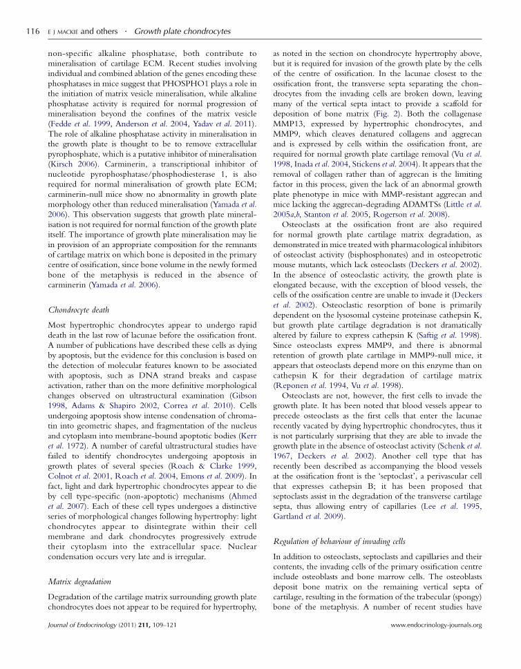
non-specific alkaline phosphatase, both contribute to
mineralisation of cartilage ECM. Recent studies involving
individual and combined ablation of the genes encoding these
phosphatases in mice suggest that PHOSPHO1 plays a role in
the initiation of matrix vesicle mineralisation, while alkaline
phosphatase activity is required for normal progression of
mineralisation beyond the confines of the matrix vesicle
(Fedde et al. 1999, Anderson et al. 2004, Yadav et al. 2011).
The role of alkaline phosphatase activity in mineralisation in
the growth plate is thought to be to remove extracellular
pyrophosphate, which is a putative inhibitor of mineralisation
(Kirsch 2006). Carminerin, a transcriptional inhibitor of
nucleotide pyrophosphatase/phosphodiesterase 1, is also
required for normal mineralisation of growth plate ECM;
carminerin-null mice show no abnormality in growth plate
morphology other than reduced mineralisation (Yamada et al.
2006). This observation suggests that growth plate mineral-
isation is not required for normal function of the growth plate
itself. The importance of growth plate mineralisation may lie
in provision of an appropriate composition for the remnants
of cartilage matrix on which bone is deposited in the primary
centre of ossification, since bone volume in the newly formed
bone of the metaphysis is reduced in the absence of
carminerin (Yamada et al. 2006).
Chondrocyte death
Most hypertrophic chondrocytes appear to undergo rapid
death in the last row of lacunae before the ossification front.
A number of publications have described these cells as dying
by apoptosis, but the evidence for this conclusion is based on
the detection of molecular features known to be associated
with apoptosis, such as DNA strand breaks and caspase
activation, rather than on the more definitive morphological
changes observed on ultrastructural examination (Gibson
1998, Adams & Shapiro 2002, Correa et al. 2010). Cells
undergoing apoptosis show intense condensation of chroma-
tin into geometric shapes, and fragmentation of the nucleus
and cytoplasm into membrane-bound apoptotic bodies (Kerr
et al. 1972). A number of careful ultrastructural studies have
failed to identify chondrocytes undergoing apoptosis in
growth plates of several species (Roach & Clarke 1999,
Colnot et al. 2001, Roach et al. 2004, Emons et al. 2009). In
fact, light and dark hypertrophic chondrocytes appear to die
by cell type-specific (non-apoptotic) mechanisms (Ahmed
et al. 2007). Each of these cell types undergoes a distinctive
series of morphological changes following hypertrophy: light
chondrocytes appear to disintegrate within their cell
membrane and dark chondrocytes progressively extrude
their cytoplasm into the extracellular space. Nuclear
condensation occurs very late and is irregular.
Matrix degradation
Degradation of the cartilage matrix surrounding growth plate
chondrocytes does not appear to be required for hypertrophy,
Journal of Endocrinology (2011) 211, 109–121
as noted in the section on chondrocyte hypertrophy above,
but it is required for invasion of the growth plate by the cells
of the centre of ossification. In the lacunae closest to the
ossification front, the transverse septa separating the chon-
drocytes from the invading cells are broken down, leaving
many of the vertical septa intact to provide a scaffold for
deposition of bone matrix (Fig. 2). Both the collagenase
MMP13, expressed by hypertrophic chondrocytes, and
MMP9, which cleaves denatured collagens and aggrecan
and is expressed by cells within the ossification front, are
required for normal growth plate cartilage removal (Vu et al.
1998, Inada et al. 2004, Stickens et al. 2004). It appears that the
removal of collagen rather than of aggrecan is the limiting
factor in this process, given the lack of an abnormal growth
plate phenotype in mice with MMP-resistant aggrecan and
mice lacking the aggrecan-degrading ADAMTSs (Little et al.
2005a,b, Stanton et al. 2005, Rogerson et al. 2008).
Osteoclasts at the ossification front are also required
for normal growth plate cartilage matrix degradation, as
demonstrated in mice treated with pharmacological inhibitors
of osteoclast activity (bisphosphonates) and in osteopetrotic
mouse mutants, which lack osteoclasts (Deckers et al. 2002).
In the absence of osteoclastic activity, the growth plate is
elongated because, with the exception of blood vessels, the
cells of the ossification centre are unable to invade it (Deckers
et al. 2002). Osteoclastic resorption of bone is primarily
dependent on the lysosomal cysteine proteinase cathepsin K,
but growth plate cartilage degradation is not dramatically
altered by failure to express cathepsin K (Saftig et al. 1998).
Since osteoclasts express MMP9, and there is abnormal
retention of growth plate cartilage in MMP9-null mice, it
appears that osteoclasts depend more on this enzyme than on
cathepsin K for their degradation of cartilage matrix
(Reponen et al. 1994, Vu et al. 1998).
Osteoclasts are not, however, the first cells to invade the
growth plate. It has been noted that blood vessels appear to
precede osteoclasts as the first cells that enter the lacunae
recently vacated by dying hypertrophic chondrocytes, thus it
is not particularly surprising that they are able to invade the
growth plate in the absence of osteoclast activity (Schenk et al.
1967, Deckers et al. 2002). Another cell type that has
recently been described as accompanying the blood vessels
at the ossification front is the ‘septoclast’, a perivascular cell
that expresses cathepsin B; it has been proposed that
septoclasts assist in the degradation of the transverse cartilage
septa, thus allowing entry of capillaries (Lee et al. 1995,
Gartland et al. 2009).
Regulation of behaviour of invading cells
In addition to osteoclasts, septoclasts and capillaries and their
contents, the invading cells of the primary ossification centre
include osteoblasts and bone marrow cells. The osteoblasts
deposit bone matrix on the remaining vertical septa of
cartilage, resulting in the formation of the trabecular (spongy)
bone of the metaphysis. A number of recent studies have
www.endocrinology-journals.org
Growth plate chondrocytes . E J MACKIE and others 117
provided evidence that growth plate chondrocytes produce
factors that regulate the behaviour of these invading cells
(summarised in Fig. 4), thus presumably ensuring that the rate
of replacement of cartilage by bone is matched to the
preparation of the growth plate for invasion.
As chondrocytes undergo hypertrophy, under the control
of RUNX2 they increase their expression of vascular
endothelial growth factor, which promotes the vascular
invasion of the growth plate (Zelzer et al. 2001, 2004).
High-mobility group box 1 protein (HMGB1) is secreted by
hypertrophic chondrocytes, and acts as a chemoattractant for
endothelial cells, osteoclasts and osteoblasts; HMGB1-null
mice display delayed invasion of the growth plate by cells of
the ossification centre (Taniguchi et al. 2007). Another factor
expressed by hypertrophic chondrocytes that promotes
invasion by osteoclasts is receptor activator of NFkB ligand
(RANKL), which is essential for osteoclast differentiation
(Kishimoto et al. 2006). Hypertrophic chondrocytes stimulate
osteoclast differentiation in vitro in a RANKL-dependent
manner, and RANKL expression by cultured chondrocytes is
stimulated by BMP2 and mediated by RUNX2 (Usui et al.
2008). A possible role for WNTs released by chondrocytes in
increasing osteoblast numbers has been suggested by the
finding that levels of b-catenin protein and markers of
osteoblast differentiation are abnormally low in the primary
spongiosa of mice with a postnatally induced chondrocyte-
specific knockout of IHH (Maeda et al. 2007). Furthermore,
evidence for regulation of osteoblast behaviour by growth
plate chondrocytes has been provided by studies in mice
expressing an activating Fgfr3 mutant under the control of the
Col2a1 (chondrocyte-specific) or Col1a1 (osteoblast-specific)
promoter (Matsushita et al. 2009). Activation of FGFR3
signalling in chondrocytes, but not osteoblasts, caused an
increase in osteoblast numbers at the chondro-osseous
junction, an effect that is likely to be mediated by the
stimulation of BMP ligand expression and inhibition
of BMP antagonist expression observed in these mice
(Matsushita et al. 2009).
Collectively, these observations indicate that chondrocytes of
the growth plate release soluble factors that allow them to delica-
tely control the behaviour of the invading vascular endothelial
cells, osteoclasts and osteoblasts, which all have important roles
in transforming growth plate cartilage into bone tissue.
Growth plate closure
As skeletal maturity approaches, the rate of advancement of the
ossification front is greater than the rate at which growth plate
chondrocytes replace themselves. The growth plate cartilage
disappears as the primary centre of ossification meets the
secondary centre and bony fusion occurs (Fig. 1). In human
males as well as females, oestrogen regulates this process, as
demonstrated by the failure of normal growth plate closure in
patients with oestrogen deficiency or oestrogen resistance
(Smith et al. 1994, Morishima et al. 1995). It is thought that
growth plate closure occurs when the chondrocytes exhaust
www.endocrinology-journals.org
their proliferative potential, and that the role of oestrogen is to
accelerate this process of senescence (Weise et al. 2001).
Oestrogen receptors expressed by growth plate chondrocytes
are likely to mediate these effects (Nilsson et al. 2005).
Conclusion
As demonstrated in this review, the understanding of the role
of the growth plate chondrocyte in endochondral ossification
has increased enormously in recent years, largely as a result of
the identification of gene mutations responsible for human
chondrodysplasias, and discoveries making use of recent
advances in gene manipulation technologies for in vitro and
in vivo studies, supported by careful morphological character-
isation. There is still much to learn about this process. The
molecular bases of some chondrodysplasias remain undiscov-
ered, and for many of those for which the responsible genes
have been identified, the links between mutation and disease
are incompletely elucidated. Although chondrocytes, the
major structural components of the matrix they produce and
components of the major signalling pathways that regulate
their behaviour have been extensively studied, there are still
many gaps in our knowledge. The process by which a single
cell type, the chondrocyte, provides the impetus for the
growth of rigid bones from embryonic life to adulthood is a
fascinating area of study, which will no doubt keep us
occupied for many years to come.
Declaration of interest
The authors declare that there is no conflict of interest that could be perceived
as prejudicing the impartiality of the review reported.
Funding
This review did not receive any specific grant from any funding agency in the
public, commercial or not-for-profit sector.
References
Adams CS & Shapiro IM 2002 The fate of the terminally differentiated
chondrocyte: evidence for microenvironmental regulation of chondrocyte
apoptosis. Critical Reviews in Oral Biology and Medicine 13 465–473. (doi:10.
1177/154411130201300604)
Ahmed YA, Tatarczuch L, Pagel CN, Davies HM, Mirams M & Mackie EJ
2007 Physiological death of hypertrophic chondrocytes. Osteoarthritis and
Cartilage 15 575–586. (doi:10.1016/j.joca.2006.10.016)
Akiyama H, Chaboissier MC, Martin JF, Schedl A & de Crombrugghe B 2002
The transcription factor Sox9 has essential roles in successive steps of the
chondrocyte differentiation pathway and is required for expression of
Sox5 and Sox6. Genes and Development 16 2813–2828. (doi:10.1101/gad.
1017802)
Akiyama H, Lyons JP, Mori-Akiyama Y, Yang X, Zhang R, Zhang Z,
Deng JM, Taketo MM, Nakamura T, Behringer RR et al. 2004 Interactions
between Sox9 and beta-catenin control chondrocyte differentiation. Genes
and Development 18 1072–1087. (doi:10.1101/gad.1171104)
Journal of Endocrinology (2011) 211, 109–121
E J MACKIE and others . Growth plate chondrocytes118
Anderson DR 1964 The ultrastructure of elastic and hyaline cartilage of
the rat. American Journal of Anatomy 114 403–434. (doi:10.1002/aja.
1001140305)
Anderson HC 1969 Vesicles associated with calcification in the matrix of
epiphyseal cartilage. Journal of Cell Biology 4159–72. (doi:10.1083/jcb.41.1.59)
Anderson HC, Sipe JB, Hessle L, Dhanyamraju R, Atti E, Camacho NP &
Millan JL 2004 Impaired calcification around matrix vesicles of growth
plate and bone in alkaline phosphatase-deficient mice. American Journal of
Pathology 164 841–847. (doi:10.1016/S0002-9440(10)63172-0)
Andrade AC, Nilsson O, Barnes KM & Baron J 2007 Wnt gene expression in
the post-natal growth plate: regulation with chondrocyte differentiation.
Bone 40 1361–1369. (doi:10.1016/j.bone.2007.01.005)
Baker J, Liu JP, Robertson EJ & Efstratiadis A 1993 Role of insulin-like
growth factors in embryonic and postnatal growth. Cell 75 73–82.
(doi:10.1016/S0092-8674(05)80085-6)
Ballock RT & Reddi AH 1994 Thyroxine is the serum factor that regulates
morphogenesis of columnar cartilage from isolated chondrocytes in
chemically defined medium. Journal of Cell Biology 126 1311–1318.
(doi:10.1083/jcb.126.5.1311)
Barnard JC, Williams AJ, Rabier B, Chassande O, Samarut J, Cheng SY,
Bassett JH & Williams GR 2005 Thyroid hormones regulate fibroblast
growth factor receptor signaling during chondrogenesis. Endocrinology 146
5568–5580. (doi:10.1210/en.2005-0762)
Beier F, Ali Z, Mok D, Taylor AC, Leask T, Albanese C, Pestell RG & LuValle
P 2001 TGFbeta and PTHrP control chondrocyte proliferation by
activating cyclin D1 expression. Molecular Biology of the Cell 12 3852–3863.
Belluoccio D, Grskovic I, Niehoff A, Schlotzer-Schrehardt U, Rosenbaum S,
Etich J, Frie C, Pausch F, Moss SE, Poschl E et al. 2010 Deficiency of
annexins A5 and A6 induces complex changes in the transcriptome of
growth plate cartilage but does not inhibit the induction of mineralization.
Journal of Bone and Mineral Research 25 141–153. (doi:10.1359/jbmr.090710)
Bi W, Deng JM, Zhang Z, Behringer RR & de Crombrugghe B 1999 Sox9
is required for cartilage formation. Nature Genetics 22 85–89. (doi:10.1038/
8792)
Bishop JR, Schuksz M & Esko JD 2007 Heparan sulphate proteoglycans
fine-tune mammalian physiology. Nature 446 1030–1037. (doi:10.1038/
nature05817)
Blumbach K, Niehoff A, Paulsson M & Zaucke F 2008 Ablation of collagen
IX and COMP disrupts epiphyseal cartilage architecture. Matrix Biology 27
306–318. (doi:10.1016/j.matbio.2007.11.007)
Bohme K, Conscience-Egli M, Tschan T, Winterhalter KH & Bruckner P
1992 Induction of proliferation or hypertrophy of chondrocytes in serum-
free culture: the role of insulin-like growth factor-I, insulin, or thyroxine.
Journal of Cell Biology 116 1035–1042. (doi:10.1083/jcb.116.4.1035)
Breur GJ, Turgai J, Vanenkevort BA, Farnum CE & Wilsman NJ 1994
Stereological and serial section analysis of chondrocytic enlargement in the
proximal tibial growth plate of the rat. Anatomical Record 239 255–268.
(doi:10.1002/ar.1092390304)
Briggs MD & Chapman KL 2002 Pseudoachondroplasia and multiple
epiphyseal dysplasia: mutation review, molecular interactions, and genotype
to phenotype correlations. Human Mutation 19 465–478. (doi:10.1002/
humu.10066)
Burch WM & Lebovitz HE 1982 Triiodothyronine stimulates maturation
of porcine growth-plate cartilage in vitro. Journal of Clinical Investigation 70
496–504. (doi:10.1172/JCI110641)
Cawston TE & Young DA 2010 Proteinases involved in matrix turnover
during cartilage and bone breakdown. Cell Tissue Research 339 221–235.
(doi:10.1007/s00441-009-0887-6)
Chen M, Zhu M, Awad H, Li TF, Sheu TJ, Boyce BF, Chen D & O’Keefe RJ
2008 Inhibition of beta-catenin signaling causes defects in postnatal
cartilage development. Journal of Cell Science 121 1455–1465. (doi:10.1242/
jcs.020362)
Chen KS, Tatarczuch L, Mirams M, Ahmed YA, Pagel CN & Mackie EJ 2010
Periostin expression distinguishes between light and dark hypertrophic
chondrocytes. International Journal of Biochemistry and Cell Biology 42 880–
889. (doi:10.1016/j.biocel.2010.01.018)
Journal of Endocrinology (2011) 211, 109–121
Chuang CY, Lord MS, Melrose J, Rees MD, Knox SM, Freeman C, Iozzo RV
& Whitelock JM 2010 Heparan sulfate-dependent signaling of fibroblast
growth factor 18 by chondrocyte-derived perlecan. Biochemistry 49
5524–5532. (doi:10.1021/bi1005199)
Colnot C, Sidhu SS, Balmain N & Poirier F 2001 Uncoupling of chondrocyte
death and vascular invasion in mouse galectin 3 null mutant bones.
Developmental Biology 229 203–214. (doi:10.1006/dbio.2000.9933)
Correa D, Hesse E, Seriwatanachai D, Kiviranta R, Saito H, Yamana K, Neff L,
Atfi A, Coillard L, Sitara D et al. 2010 Zfp521 is a target gene and key effector
of parathyroid hormone-related peptide signaling in growth plate chon-
drocytes.DevelopmentalCell 19 533–546. (doi:10.1016/j.devcel.2010.09.008)
Cortes M, Baria AT & Schwartz NB 2009 Sulfation of chondroitin sulfate
proteoglycans is necessary for proper Indian hedgehog signaling in the
developing growth plate. Development 136 1697–1706. (doi:10.1242/dev.
030742)
Dailey L, Laplantine E, Priore R & Basilico C 2003 A network of
transcriptional and signaling events is activated by FGF to induce
chondrocyte growth arrest and differentiation. Journal of Cell Biology 161
1053–1066. (doi:10.1083/jcb.200302075)
DeChiara TM, Robertson EJ & Efstratiadis A 1991 Parental imprinting of the
mouse insulin-like growth factor II gene. Cell 64 849–859. (doi:10.1016/
0092-8674(91)90513-X)
Deckers MM, Van Beek ER, Van Der Pluijm G, Wetterwald A, Van Der Wee-
Pals L, Cecchini MG, Papapoulos SE & Lowik CW 2002 Dissociation of
angiogenesis and osteoclastogenesis during endochondral bone formation
in neonatal mice. Journal of Bone and Mineral Research 17 998–1007. (doi:10.
1359/jbmr.2002.17.6.998)
Dong YF, Soung do Y, Schwarz EM, O’Keefe RJ & Drissi H 2006 Wnt
induction of chondrocyte hypertrophy through the Runx2 transcription
factor. Journal of Cell Physiology 208 77–86. (doi:10.1002/jcp.20656)
van der Eerden BC, Karperien M & Wit JM 2003 Systemic and local
regulation of the growth plate. Endocrine Reviews 24 782–801. (doi:10.
1210/er.2002-0033)
Ehlen HW, Buelens LA & Vortkamp A 2006 Hedgehog signaling in skeletal
development. Birth Defects Research. Part C, Embryo Today: Reviews 78
267–279. (doi:10.1002/bdrc.20076)
Emons J, Chagin AS, Hultenby K, Zhivotovsky B, Wit JM, Karperien M &
Savendahl L 2009 Epiphyseal fusion in the human growth plate does not
involve classical apoptosis. Pediatric Research 66 654–659. (doi:10.1203/
PDR.0b013e3181beaa8c)
Enomoto-Iwamoto M, Kitagaki J, Koyama E, Tamamura Y, Wu C, Kanatani
N, Koike T, Okada H, Komori T, Yoneda T et al. 2002 The Wnt antagonist
Frzb-1 regulates chondrocyte maturation and long bone development
during limb skeletogenesis. Developmental Biology 251 142–156. (doi:10.
1006/dbio.2002.0802)
Fassler R, Schnegelsberg PN, Dausman J, Shinya T, Muragaki Y, McCarthy
MT, Olsen BR & Jaenisch R 1994 Mice lacking alpha 1 (IX) collagen
develop noninflammatory degenerative joint disease. PNAS 91 5070–5074.
(doi:10.1073/pnas.91.11.5070)
Fedde KN, Blair L, Silverstein J, Coburn SP, Ryan LM, Weinstein RS,
Waymire K, Narisawa S, Millan JL, MacGregor GR et al. 1999 Alkaline
phosphatase knock-out mice recapitulate the metabolic and skeletal
defects of infantile hypophosphatasia. Journal of Bone and Mineral Research 14
2015–2026. (doi:10.1359/jbmr.1999.14.12.2015)
Furumatsu T, Tsuda M, Yoshida K, Taniguchi N, Ito T, Hashimoto M &
Asahara H 2005 Sox9 and p300 cooperatively regulate chromatin-mediated
transcription. Journal of Biological Chemistry 280 35203–35208. (doi:10.
1074/jbc.M502409200)
Gartland A, Mason-Savas A, Yang M, MacKay CA, Birnbaum MJ & Odgren
PR 2009 Septoclast deficiency accompanies postnatal growth plate
chondrodysplasia in the toothless (tl) osteopetrotic, colony-stimulating
factor-1 (CSF-1)-deficient rat and is partially responsive to CSF-1
injections. American Journal of Pathology 175 2668–2675. (doi:10.2353/
ajpath.2009.090185)
Genge BR, Wu LN & Wuthier RE 2007a In vitro modeling of matrix vesicle
nucleation: synergistic stimulation of mineral formation by annexin A5
and phosphatidylserine. Journal of Biological Chemistry 282 26035–26045.
(doi:10.1074/jbc.M701057200)
www.endocrinology-journals.org
Growth plate chondrocytes . E J MACKIE and others 119
Genge BR, Wu LN & Wuthier RE 2007b Kinetic analysis of mineral
formation during in vitro modeling of matrix vesicle mineralization: effect of
annexin A5, phosphatidylserine, and type II collagen. Analytical Biochemistry
367 159–166. (doi:10.1016/j.ab.2007.04.029)
Gentili C & Cancedda R 2009 Cartilage and bone extracellular matrix.
Current Pharmaceutical Design 15 1334–1348. (doi:10.2174/13816120
9787846739)
Gibson G 1998 Active role of chondrocyte apoptosis in endochondral
ossification. Microscopic Research and Technique 43 191–204. (doi:10.1002/
(SICI)1097-0029(19981015)43:2!191::AID-JEMT10O3.0.CO;2-T)
Grimsrud CD, Romano PR, D’Souza M, Puzas JE, Schwarz EM, Reynolds
PR, Roiser RN & O’Keefe RJ 2001 BMP signaling stimulates chondrocyte
maturation and the expression of Indian hedgehog. Journal of Orthopaedic
Research 19 18–25. (doi:10.1016/S0736-0266(00)00017-6)
Guo J, Chung UI, Yang D, Karsenty G, Bringhurst FR & Kronenberg HM
2006 PTH/PTHrP receptor delays chondrocyte hypertrophy via both
Runx2-dependent and -independent pathways. Developmental Biology 292
116–128. (doi:10.1016/j.ydbio.2005.12.044)
Hagg R, Hedbom E, Mollers U, Aszodi A, Fassler R & Bruckner P 1997
Absence of the alpha1(IX) chain leads to a functional knock-out of the
entire collagen IX protein in mice. Journal of Biological Chemistry 272
20650–20654. (doi:10.1074/jbc.272.33.20650)
Hecht JT, Hayes E, Haynes R & Cole WG 2005 COMP mutations,
chondrocyte function and cartilage matrix. Matrix Biology 23 525–533.
(doi:10.1016/j.matbio.2004.09.006)
Heinegard D 2009 Proteoglycans and more – from molecules to biology.
International Journal of Experimental Pathology 90 575–586. (doi:10.1111/j.
1365-2613.2009.00695.x)
Hong S, Derfoul A, Pereira-Mouries L & Hall DJ 2009 A novel domain in
histone deacetylase 1 and 2 mediates repression of cartilage-specific genes in
human chondrocytes. FASEB Journal 23 3539–3552. (doi:10.1096/fj.09-
133215)
Huang W, Zhou X, Lefebvre V & de Crombrugghe B 2000 Phosphorylation
of SOX9 by cyclic AMP-dependent protein kinase A enhances SOX9’s
ability to transactivate a Col2a1 chondrocyte-specific enhancer. Molecular
and Cellular Biology 20 4149–4158. (doi:10.1128/MCB.20.11.4149-4158.
2000)
Huang W, Chung UI, Kronenberg HM & de Crombrugghe B 2001 The
chondrogenic transcription factor Sox9 is a target of signaling by the
parathyroid hormone-related peptide in the growth plate of endochondral
bones. PNAS 98 160–165. (doi:10.1073/pnas.011393998)
Hwang WS 1978 Ultrastructure of human foetal and neonatal hyaline
cartilage. Journal of Pathology 126 209–214. (doi:10.1002/path.1711260404)
Inada M, Wang Y, Byrne MH, Rahman MU, Miyaura C, Lopez-Otin C &
Krane SM 2004 Critical roles for collagenase-3 (Mmp13) in development
of growth plate cartilage and in endochondral ossification. PNAS 101
17192–17197. (doi:10.1073/pnas.0407788101)
Jin W, Takagi T, Kanesashi SN, Kurahashi T, Nomura T, Harada J & Ishii S
2006 Schnurri-2 controls BMP-dependent adipogenesis via interaction
with Smad proteins. Developmental Cell 10 461–471. (doi:10.1016/j.devcel.
2006.02.016)
Joeng KS & Long F 2009 The Gli2 transcriptional activator is a crucial effector
for Ihh signaling in osteoblast development and cartilage vascularization.
Development 136 4177–4185. (doi:10.1242/dev.041624)
Johansson N, Saarialho-Kere U, Airola K, Herva R, Nissinen L, Westermarck
J, Vuorio E, Heino J & Kahari VM 1997 Collagenase-3 (MMP-13) is
expressed by hypertrophic chondrocytes, periosteal cells, and osteoblasts
during human fetal bone development. Developmental Dynamics 208
387–397. (doi:10.1002/(SICI)1097-0177(199703)208:3!387::AID-
AJA9O3.0.CO;2-E)
Jones DC, Schweitzer MN, Wein M, Sigrist K, Takagi T, Ishii S & Glimcher
LH 2010 Uncoupling of growth plate maturation and bone formation in
mice lacking both Schnurri-2 and Schnurri-3. PNAS 107 8254–8258.
(doi:10.1073/pnas.1003727107)
Kaneshige M, Suzuki H, Kaneshige K, Cheng J, Wimbrow H, Barlow C,
Willingham MC & Cheng S 2001 A targeted dominant negative mutation
www.endocrinology-journals.org
of the thyroid hormone alpha 1 receptor causes increased mortality,
infertility, and dwarfism in mice. PNAS 98 15095–15100. (doi:10.1073/
pnas.261565798)
Kawasaki Y, Kugimiya F, Chikuda H, Kamekura S, Ikeda T, Kawamura N,
Saito T, Shinoda Y, Higashikawa A, Yano F et al. 2008 Phosphorylation of
GSK-3beta by cGMP-dependent protein kinase II promotes hypertrophic
differentiation of murine chondrocytes. Journal of Clinical Investigation 118
2506–2515. (doi:10.1172/JCI35243E1)
Kerr JF, Wyllie AH & Currie AR 1972 Apoptosis: a basic biological
phenomenon with wide-ranging implications in tissue kinetics. British
Journal of Cancer 26 239–257. (doi:10.1038/bjc.1972.33)
Kirsch T 2006 Determinants of pathological mineralization. Current Opinion
in Rheumatology 18 174–180. (doi:10.1097/01.bor.0000209431.59226.46)
Kirsch T, Nah HD, Shapiro IM & Pacifici M 1997 Regulated production of
mineralization-competent matrix vesicles in hypertrophic chondrocytes.
Journal of Cell Biology 137 1149–1160. (doi:10.1083/jcb.137.5.1149)
Kirsch T, Harrison G, Golub EE & Nah HD 2000 The roles of annexins and
types II and X collagen in matrix vesicle-mediated mineralization of growth
plate cartilage. Journal of Biological Chemistry 275 35577–35583. (doi:10.
1074/jbc.M005648200)
Kishimoto K, Kitazawa R, Kurosaka M, Maeda S & Kitazawa S 2006
Expression profile of genes related to osteoclastogenesis in mouse growth
plate and articular cartilage. Histochemistry and Cell Biology 125 593–602.
(doi:10.1007/s00418-005-0103-z)
Klatt AR, Becker AK, Neacsu CD, Paulsson M & Wagener R 2011 The
matrilins: modulators of extracellular matrix assembly. International Journal of
Biochemistry & Cell Biology 43 320–330. (doi:10.1016/j.biocel.2010.12.010)
Ko Y, Kobbe B, Nicolae C, Miosge N, Paulsson M, Wagener R & Aszodi A
2004 Matrilin-3 is dispensable for mouse skeletal growth and development.
Molecular and Cellular Biology 24 1691–1699. (doi:10.1128/MCB.24.4.
1691-1699.2004)
Koyama E, Leatherman JL, Noji S & Pacifici M 1996 Early chick limb
cartilaginous elements possess polarizing activity and express
hedgehog-related morphogenetic factors. Developmental Dynamics 207
344–354. (doi:10.1002/(SICI)1097-0177(199611)207:3!344::AID-
AJA11O3.0.CO;2-4)
Koyama E, Young B, Nagayama M, Shibukawa Y, Enomoto-Iwamoto M,
Iwamoto M, Maeda Y, Lanske B, Song B, Serra R et al. 2007 Conditional
Kif3a ablation causes abnormal hedgehog signaling topography, growth
plate dysfunction, and excessive bone and cartilage formation during mouse
skeletogenesis. Development 134 2159–2169. (doi:10.1242/dev.001586)
Krakow D & Rimoin DL 2010 The skeletal dysplasias. Genetics in Medicine 12
327–341.
Krejci P, Krakow D, Mekikian PB & Wilcox WR 2007 Fibroblast growth
factors 1, 2, 17, and 19 are the predominant FGF ligands expressed in
human fetal growth plate cartilage. Pediatric Research 61 267–272. (doi:10.
1203/pdr.0b013e318030d157)
Kronenberg HM 2006 PTHrP and skeletal development. Annals of the
New York Academy of Sciences 1068 1–13. (doi:10.1196/annals.1346.002)
Lee ER, Lamplugh L, Shepard NL & Mort JS 1995 The septoclast, a cathepsin
B-rich cell involved in the resorption of growth plate cartilage. Journal of
Histochemistry and Cytochemistry 43 525–536. (doi:10.1177/43.5.7730591)
Lee K, Lanske B, Karaplis AC, Deeds JD, Kohno H, Nissenson RA,
Kronenberg HM & Segre GV 1996 Parathyroid hormone-related peptide
delays terminal differentiation of chondrocytes during endochondral bone
development. Endocrinology 137 5109–5118. (doi:10.1210/en.137.11.5109)
Lefebvre V & Smits P 2005 Transcriptional control of chondrocyte fate
and differentiation. Birth Defects Research. Part C, Embryo Today: Reviews 75
200–212. (doi:10.1002/bdrc.20048)
Little CB, Meeker CT, Hembry RM, Sims NA, Lawlor KE, Golub SB, Last K
& Fosang AJ 2005a Matrix metalloproteinases are not essential for aggrecan
turnover during normal skeletal growth and development. Molecular and
Cellular Biology 25 3388–3399. (doi:10.1128/MCB.25.8.3388-3399.2005)
Little CB, Mittaz L, Belluoccio D, Rogerson FM, Campbell IK, Meeker CT,
Bateman JF, Pritchard MA & Fosang AJ 2005b ADAMTS-1-knockout mice
do not exhibit abnormalities in aggrecan turnover in vitro or in vivo. Arthritis
and Rheumatism 52 1461–1472. (doi:10.1002/art.21022)
Journal of Endocrinology (2011) 211, 109–121
E J MACKIE and others . Growth plate chondrocytes120
Maeda Y, Nakamura E, Nguyen MT, Suva LJ, Swain FL, Razzaque MS,
Mackem S & Lanske B 2007 Indian hedgehog produced by postnatal
chondrocytes is essential for maintaining a growth plate and trabecular
bone. PNAS 104 6382–6387. (doi:10.1073/pnas.0608449104)
Mak KK, Chen MH, Day TF, Chuang PT & Yang Y 2006 Wnt/beta–catenin
signaling interacts differentially with Ihh signaling in controlling
endochondral bone and synovial joint formation. Development 133
3695–3707. (doi:10.1242/dev.02546)
Mak KK, Kronenberg HM, Chuang PT, Mackem S & Yang Y 2008 Indian
hedgehog signals independently of PTHrP to promote chondrocyte
hypertrophy. Development 135 1947–1956. (doi:10.1242/dev.018044)
Matsumoto K, Li Y, Jakuba C, Sugiyama Y, Sayo T, Okuno M, Dealy CN,
Toole BP, Takeda J, Yamaguchi Y et al. 2009 Conditional inactivation of
Has2 reveals a crucial role for hyaluronan in skeletal growth, patterning,
chondrocyte maturation and joint formation in the developing limb.
Development 136 2825–2835. (doi:10.1242/dev.038505)
Matsushita T, Wilcox WR, Chan YY, Kawanami A, Bukulmez H, Balmes G,
Krejci P, Mekikian PB, Otani K, Yamaura I et al. 2009 FGFR3 promotes
synchondrosis closure and fusion of ossification centers through the MAPK
pathway. Human Molecular Genetics 18 227–240. (doi:10.1093/hmg/
ddn339)
Minina E, Wenzel HM, Kreschel C, Karp S, Gaffield W, McMahon AP &
Vortkamp A 2001 BMP and Ihh/PTHrP signaling interact to coordinate
chondrocyte proliferation and differentiation. Development 128 4523–4534.
Minina E, Kreschel C, Naski MC, Ornitz DM & Vortkamp A 2002
Interaction of FGF Ihh/Pthlh, and BMP signaling integrates chondrocyte
proliferation and hypertrophic differentiation. Developmental Cell 3
439–449. (doi:10.1016/S1534-5807(02)00261-7)
Morishima A, Grumbach MM, Simpson ER, Fisher C & Qin K 1995
Aromatase deficiency in male and female siblings caused by a novel
mutation and the physiological role of estrogens. Journal of Clinical
Endocrinology and Metabolism 80 3689–3698. (doi:10.1210/jc.80.12.3689)
Napierala D, Sam K, Morello R, Zheng Q, Munivez E, Shivdasani RA &
Lee B 2008 Uncoupling of chondrocyte differentiation and perichondrial
mineralization underlies the skeletal dysplasia in tricho–rhino–phalangeal
syndrome. Human Molecular Genetics 17 2244–2254. (doi:10.1093/hmg/
ddn125)
Nilsson O, Marino R, De Luca F, Phillip M & Baron J 2005 Endocrine
regulation of the growth plate. Hormone Research 64 157–165. (doi:10.
1159/000088791)
Nishioka K, Itoh S, Suemoto H, Kanno S, Gai Z, Kawakatsu M, Tanishima H,
Morimoto Y, Hatamura I, Yoshida M et al. 2008 Trps1 deficiency enlarges
the proliferative zone of growth plate cartilage by upregulation of Pthrp.
Bone 43 64–71. (doi:10.1016/j.bone.2008.03.009)
Ornitz DM 2005 FGF signaling in the developing endochondral skeleton.
Cytokine & Growth Factor Reviews 16 205–213. (doi:10.1016/j.cytogfr.2005.
02.003)
Pass C, MacRae VE, Ahmed SF & Farquharson C 2009 Inflammatory
cytokines and the GH/IGF-I axis: novel actions on bone growth. Cell
Biochemistry and Function 27 119–127. (doi:10.1002/cbf.1551)
Pavasant P, Shizari T & Underhill CB 1996 Hyaluronan contributes to the
enlargement of hypertrophic lacunae in the growth plate. Journal of Cell
Science 109 327–334.
Poole CA, Flint MH & Beaumont BW 1985 Analysis of the morphology and
function of primary cilia in connective tissues: a cellular cybernetic probe?
Cell Motility 5 175–193. (doi:10.1002/cm.970050302)
Posey KL, Hankenson K, Veerisetty AC, Bornstein P, Lawler J & Hecht JT
2008 Skeletal abnormalities in mice lacking extracellular matrix proteins,
thrombospondin-1, thrombospondin-3, thrombospondin-5, and type IX
collagen. American Journal of Pathology 172 1664–1674. (doi:10.2353/ajpath.
2008.071094)
Reponen P, Sahlberg C, Munaut C, Thesleff I & Tryggvason K 1994 High
expression of 92 kD type IV collagenase (gelatinase B) in the osteoclast
lineage during mouse development. Journal of Cell Biology 124 1091–1102.
(doi:10.1083/jcb.124.6.1091)
Retting KN, Song B, Yoon BS & Lyons KM 2009 BMP canonical Smad
signaling through Smad1 and Smad5 is required for endochondral bone
formation. Development 136 1093–1104. (doi:10.1242/dev.029926)
Journal of Endocrinology (2011) 211, 109–121
Roach HI & Clarke NM 1999 "Cell paralysis" as an intermediate stage in the
programmed cell death of epiphyseal chondrocytes during development.
Journal of Bone and Mineral Research 14 1367–1378. (doi:10.1359/jbmr.1999.
14.8.1367)
Roach HI & Clarke NM 2000 Physiological cell death of chondrocytes in vivo
is not confined to apoptosis. New observations on the mammalian growth
plate. Journal of Bone and Joint Surgery. British Volume 82 601–613. (doi:10.
1302/0301-620X.82B4.9846)
Roach HI, Aigner T & Kouri JB 2004 Chondroptosis: a variant of apoptotic
cell death in chondrocytes? Apoptosis 9 265–277. (doi:10.1023/B:APPT.
0000025803.17498.26)
Rodgers KD, San Antonio JD & Jacenko O 2008 Heparan sulfate
proteoglycans: a GAGgle of skeletal–hematopoietic regulators.
Developmental Dynamics 237 2622–2642. (doi:10.1002/dvdy.21593)
Rogerson FM, Stanton H, East CJ, Golub SB, Tutolo L, Farmer PJ & Fosang
AJ 2008 Evidence of a novel aggrecan-degrading activity in cartilage:
studies of mice deficient in both ADAMTS-4 and ADAMTS-5. Arthritis
and Rheumatism 58 1664–1673. (doi:10.1002/art.23458)
Saftig P, Hunziker E, Wehmeyer O, Jones S, Boyde A, Rommerskirch W,
Moritz JD, Schu P & von Figura K 1998 Impaired osteoclastic bone
resorption leads to osteopetrosis in cathepsin-K-deficient mice. PNAS 95
13453–13458. (doi:10.1073/pnas.95.23.13453)
Schenk RK, Spiro D & Wiener J 1967 Cartilage resorption in the tibial
epiphyseal plate of growing rats. Journal of Cell Biology 34 275–291.
(doi:10.1083/jcb.34.1.275)
Schibler L, Gibbs L, Benoist-Lasselin C, Decraene C, Martinovic J, Loget P,
Delezoide AL, Gonzales M, Munnich A, Jais JP et al. 2009 New insight on
FGFR3-related chondrodysplasias molecular physiopathology revealed by
human chondrocyte gene expression profiling. PLoS ONE 4 e7633.
(doi:10.1371/journal.pone.0007633)
Settembre C, Arteaga-Solis E, McKee MD, de Pablo R, Al Awqati Q, Ballabio
A & Karsenty G 2008 Proteoglycan desulfation determines the efficiency of
chondrocyte autophagy and the extent of FGF signaling during endo-
chondral ossification. Genes and Development 22 2645–2650. (doi:10.1101/
gad.1711308)
Shao YY, Wang L & Ballock RT 2006 Thyroid hormone and the growth
plate. Reviews in Endocrine and Metabolic Disorders 7 265–271. (doi:10.1007/
s11154-006-9012-2)
Smith EP, Boyd J, Frank GR, Takahashi H, Cohen RM, Specker B,
Williams TC, Lubahn DB & Korach KS 1994 Estrogen resistance
caused by a mutation in the estrogen-receptor gene in a man.
New England Journal of Medicine 331 1056–1061. (doi:10.1056/
NEJM199410203311604)
Stanton H, Rogerson FM, East CJ, Golub SB, Lawlor KE, Meeker CT, Little
CB, Last K, Farmer PJ, Campbell IK et al. 2005 ADAMTS5 is the major
aggrecanase in mouse cartilage in vivo and in vitro. Nature 434 648–652.
(doi:10.1038/nature03417)
Stevens DA, Hasserjian RP, Robson H, Siebler T, Shalet SM & Williams GR
2000 Thyroid hormones regulate hypertrophic chondrocyte differentiation
and expression of parathyroid hormone-related peptide and its receptor
during endochondral bone formation. Journal of Bone and Mineral Research
15 2431–2442. (doi:10.1359/jbmr.2000.15.12.2431)
Stickens D, Behonick DJ, Ortega N, Heyer B, Hartenstein B, Yu Y, Fosang AJ,
Schorpp-Kistner M, Angel P & Werb Z 2004 Altered endochondral bone
development in matrix metalloproteinase 13-deficient mice. Development
131 5883–5895. (doi:10.1242/dev.01461)
St-Jacques B, Hammerschmidt M & McMahon AP 1999 Indian hedgehog
signaling regulates proliferation and differentiation of chondrocytes and is
essential for bone formation. Genes and Development 13 2072–2086. (doi:10.
1101/gad.13.16.2072)
Suemoto H, Muragaki Y, Nishioka K, Sato M, Ooshima A, Itoh S, Hatamura
I, Ozaki M, Braun A, Gustafsson E et al. 2007 Trps1 regulates proliferation
and apoptosis of chondrocytes through Stat3 signaling. Developmental
Biology 312 572–581. (doi:10.1016/j.ydbio.2007.10.001)
Svensson L, Aszodi A, Heinegard D, Hunziker EB, Reinholt FP, Fassler R &
Oldberg A 2002 Cartilage oligomeric matrix protein-deficient mice have
normal skeletal development. Molecular and Cellular Biology 22 4366–4371.
(doi:10.1128/MCB.22.12.4366-4371.2002)
www.endocrinology-journals.org
Growth plate chondrocytes . E J MACKIE and others 121
Tamamura Y, Otani T, Kanatani N, Koyama E, Kitagaki J, Komori T, Yamada
Y, Costantini F, Wakisaka S, Pacifici M et al. 2005 Developmental regulation
of Wnt/beta–catenin signals is required for growth plate assembly, cartilage
integrity, and endochondral ossification. Journal of Biological Chemistry 280
19185–19195. (doi:10.1074/jbc.M414275200)
Taniguchi N, Yoshida K, Ito T, Tsuda M, Mishima Y, Furumatsu T, Ronfani L,
Abeyama K, Kawahara K, Komiya S et al. 2007 Stage-specific secretion of
HMGB1 in cartilage regulates endochondral ossification. Molecular and
Cellular Biology 27 5650–5663. (doi:10.1128/MCB.00130-07)
Tew SR, Murdoch AD, Rauchenberg RP & Hardingham TE 2008 Cellular
methods in cartilage research: primary human chondrocytes in culture and
chondrogenesis in human bone marrow stem cells. Methods 45 2–9. (doi:10.
1016/j.ymeth.2008.01.006)
Tryfonidou MA, Hazewinkel HA, Riemers FM, Brinkhof B, Penning LC &
Karperien M 2010 Intraspecies disparity in growth rate is associated with
differences in expression of local growth plate regulators. American Journal of
Physiology. Endocrinology and Metabolism 299 E1044–E1052. (doi:10.1152/
ajpendo.00170.2010)
Usui M, Xing L, Drissi H, Zuscik M, O’Keefe R, Chen D & Boyce BF 2008
Murine and chicken chondrocytes regulate osteoclastogenesis by producing
RANKL in response to BMP2. Journal of Bone and Mineral Research 23
314–325. (doi:10.1359/jbmr.071025)
Vortkamp A, Lee K, Lanske B, Segre GV, Kronenberg HM & Tabin CJ 1996
Regulation of rate of cartilage differentiation by Indian hedgehog and
PTH-related protein. Science 273 613–622. (doi:10.1126/science.273.
5275.613)
Vu TH, Shipley JM, Bergers G, Berger JE, Helms JA, Hanahan D, Shapiro SD,
Senior RM & Werb Z 1998 MMP-9/gelatinase B is a key regulator of
growth plate angiogenesis and apoptosis of hypertrophic chondrocytes.
Cell 93 411–422. (doi:10.1016/S0092-8674(00)81169-1)
Wang L, Shao YY & Ballock RT 2007 Thyroid hormone interacts with the
Wnt/beta–catenin signaling pathway in the terminal differentiation of
growth plate chondrocytes. Journal of Bone and Mineral Research 22
1988–1995. (doi:10.1359/jbmr.070806)
Wang L, Shao YY & Ballock RT 2010 Thyroid hormone-mediated growth
and differentiation of growth plate chondrocytes involves IGF-1
modulation of beta-catenin signaling. Journal of Bone and Mineral Research 25
1138–1146. (doi:10.1002/jbmr.5)
Weise M, De-Levi S, Barnes KM, Gafni RI, Abad V & Baron J 2001 Effects of
estrogen on growth plate senescence and epiphyseal fusion. PNAS 98
6871–6876. (doi:10.1073/pnas.121180498)
Wilsman NJ, Farnum CE & Reed-Aksamit DK 1980 Incidence and
morphology of equine and murine chondrocytic cilia. Anatomical Record
197 355–361. (doi:10.1002/ar.1091970309)
Wilsman NJ, Farnum CE, Hilley HD & Carlson CS 1981 Ultrastructural
evidence of a functional heterogeneity among physeal chondrocytes in
growing swine. American Journal of Veterinary Research 42 1547–1553.
Wilsman NJ, Farnum CE, Leiferman EM, Fry M & Barreto C 1996
Differential growth by growth plates as a function of multiple parameters of
chondrocytic kinetics. Journal of Orthopaedic Research 14 927–936. (doi:10.
1002/jor.1100140613)
Wuelling M & Vortkamp A 2010 Transcriptional networks controlling
chondrocyte proliferation and differentiation during endochondral ossifi-
cation. Pediatric Nephrology 25 625–631. (doi:10.1007/s00467-009-1368-6)
www.endocrinology-journals.org
Wuelling M, Kaiser FJ, Buelens LA, Braunholz D, Shivdasani RA, Depping R
& Vortkamp A 2009 Trps1, a regulator of chondrocyte proliferation and
differentiation, interacts with the activator form of Gli3. Developmental
Biology 328 40–53. (doi:10.1016/j.ydbio.2009.01.012)
Yadav MC, Simao AM, Narisawa S, Huesa C, McKee MD, Farquharson C &
Millan JL 2011 Loss of skeletal mineralization by the simultaneous ablation
of PHOSPHO1 and alkaline phosphatase function: a unified model of the
mechanisms of initiation of skeletal calcification. Journal of Bone and Mineral
Research 26 286–297. (doi:10.1002/jbmr.195)
Yamada T, Kawano H, Koshizuka Y, Fukuda T, Yoshimura K, Kamekura S,
Saito T, Ikeda T, Kawasaki Y & Azuma Y 2006 Carminerin contributes to
chondrocyte calcification during endochondral ossification. Nature Medicine
12 665–670. (doi:10.1038/nm1409)
Yamashita S, Andoh M, Ueno-Kudoh H, Sato T, Miyaki S & Asahara H 2009
Sox9 directly promotes Bapx1 gene expression to repress Runx2 in
chondrocytes. Experimental Cell Research 315 2231–2240. (doi:10.1016/j.
yexcr.2009.03.008)
Yang Y, Topol L, Lee H & Wu J 2003 Wnt5a and Wnt5b exhibit distinct
activities in coordinating chondrocyte proliferation and differentiation.
Development 130 1003–1015. (doi:10.1242/dev.00324)
Yates KE, Shortkroff S & Reish RG 2005 Wnt influence on chondrocyte
differentiation and cartilage function. DNA and Cell Biology 24 446–457.
(doi:10.1089/dna.2005.24.446)
Yoon BS, Pogue R, Ovchinnikov DA, Yoshii I, Mishina Y, Behringer RR &
Lyons KM 2006 BMPs regulate multiple aspects of growth-plate chondro-
genesis through opposing actions on FGF pathways. Development 133
4667–4678. (doi:10.1242/dev.02680)
Zaucke F & Grassel S 2009 Genetic mouse models for the functional analysis
of the perifibrillar components collagen IX, COMP and matrilin-3:
implications for growth cartilage differentiation and endochondral
ossification. Histology and Histopathology 24 1067–1079.
Zelzer E, Glotzer DJ, Hartmann C, Thomas D, Fukai N, Soker S & Olsen BR
2001 Tissue specific regulation of VEGF expression during bone
development requires Cbfa1/Runx2. Mechanism of Development 106
97–106. (doi:10.1016/S0925-4773(01)00428-2)
Zelzer E, Mamluk R, Ferrara N, Johnson RS, Schipani E & Olsen BR 2004
VEGFA is necessary for chondrocyte survival during bone development.
Development 131 2161–2171. (doi:10.1242/dev.01053)
Zhang M, Xie R, Hou W, Wang B, Shen R, Wang X, Wang Q, Zhu T,
Jonason JH & Chen D 2009 PTHrP prevents chondrocyte premature
hypertrophy by inducing cyclin-D1-dependent Runx2 and Runx3
phosphorylation, ubiquitylation and proteasomal degradation. Journal of
Cell Science 122 1382–1389. (doi:10.1242/jcs.040709)
Zhou Z, Xie J, Lee D, Liu Y, Jung J, Zhou L, Xiong S, Mei L & Xiong WC
2010 Neogenin regulation of BMP-induced canonical Smad signaling and
endochondral bone formation. Developmental Cell 19 90–102. (doi:10.
1016/j.devcel.2010.06.016)
Received in final form 31 May 2011Accepted 3 June 2011Made available online as an Accepted Preprint3 June 2011
Journal of Endocrinology (2011) 211, 109–121