Embed Size (px)
Citation preview
A MORPHOLOGICAL STUDY OF
HALOBACTERIUM HALOBIUM AND ITS LYSIS IN
MEDIA OF LOW SALT CONCENTRATION
WALTHER STOECKENIUS and ROBERT ROWEN
From The Rockefeller University, New York 10021. Dr. Stoeckenius' present address is the Cardio-vascular Research Institute, the University of California Medical School, San Francisco 94122.Dr. Rowen's present address is the Department of Microbiology and Immunology, the Albert Ein-stein College of Medicine, Yeshiva University, New York 10461.
ABSTRACT
The reported absence of a cell wall in halobacteria cannot be confirmed. Improved fixationtechniques clearly show a cell wall-like structure on the surface of these cells. A stepwisereduction of the salt concentration causes the release of cell wall material before the cellmembrane begins to disintegrate. The cell membrane breaks up into fragments of variablebut rather small size, which are clearly different from a 4S component reported by others tobe the major breakdown product of the cell membrane. It appears more likely that the 4Scomponent arises from the dissolution of the cell wall. A residue cf large membranous sheetsremains even after prolonged exposure of halobacteria envelopes to distilled water. Thelipids in these sheets do not differ significantly from the lipids in the lysed part of the cellmembrane. The sheets, however, contain a purple-colored substance, which is not presentin the lysed part. The easily sedimentable residue that remains after lysis of the cells orenvelopes in distilled water also contains "intracytoplasmic membranes" with unusualstructural characteristics. They can also be identified in sections through intact bacteria orenvelope preparations. Their function is at present unknown but seems to be related to theformation of gas vacuoles in these organisms.
INTRODUCTION
Recently speculations that cellular membranesmay consist of discrete identical subunits haveattracted increasing attention. Attempts havebeen made to isolate and characterize these hypo-thetical subunits. Success has been claimed notonly in their isolation but also in the reconstitutionof membranes from these subunits. It has been
known for some time that by morphologicalcriteria membrane-like structures can be recon-stituted from isolated membrane lipids and protein(31, 32, 33). However, the crucial test for mem-brane reconstitution requires the demonstrationthat typical membrane functions are simultane-
ously restored. One can only hope to achieve this ifmethods are used for isolation that do not irreversi-bly alter the properties of the membrane com-ponents. The membranes of halobacteria, whichdisintegrate readily when the salt concentration ofthe medium is reduced, would appear to be afavorable object for such studies. It has also beenreported that the product of membrane breakdownin this case is a small particle that sediments with asharp boundary in the analytical ultracentrifuge(6). These findings have prompted the studyreported here.
The extremely halophilic bacteria, such as
365
Dow
nloaded from http://rupress.org/jcb/article-pdf/34/1/365/1068324/365.pdf by guest on 30 N
ovember 2021
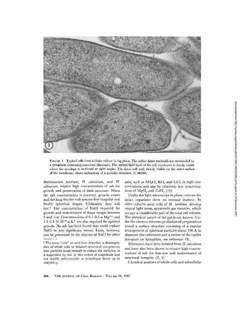
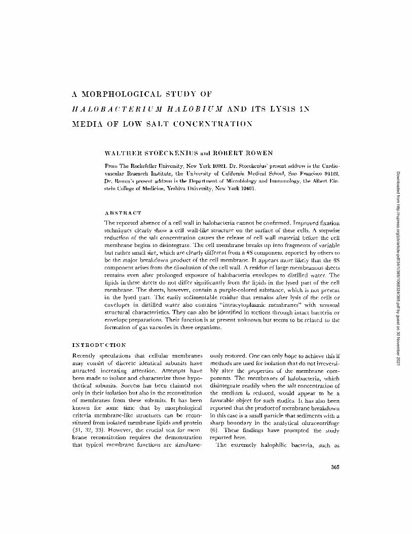
FIGURE 1 Typical cells from a shake culture in log phase. The rather dense nucleoids are surrounded bya cytoplasm containing numerous ribosomes. The central light layer of the cell membrane is clearly visiblewhere the envelope is sectioned at right angles. The dense cell wall, clearly visible on the outer surfaceof the membrane, shows indications of a periodic structure. X 96,000.
Halobacterium halobium, H. cutirubrum, and H.
salinarium, require high concentrations of salt forgrowth and preservation of their structure. When
the salt concentration is lowered, growth ceases
and the long slender rods assume first irregular andfinally spherical shapes. Ultimately they willlyse. The concentration of NaCI required forgrowth and maintenance of shape ranges between
3 and 5 M. Concentrations of 0.1 0.5 M Mg ++ and1.3-2.5 X 10⁻³ M K+ are also required for optimalgrowth. No salt has been found that could replaceNaCl to any significant extent. Lysis, however,can be prevented in the absence of NaCl by other
1 The term lysiss" as used here describes a disintegra-tion of whole cells or isolated structural componentsinto particles small enough to reduce the turbidity ofa suspension by one or two orders of magnitude andnot readily sedimentable at centrifugal forces up to100,000 g.
salts, such as NH4C1, KCI, and LiC1, in high con-centrations, and also by relatively low concentra-
tions of MgCI, and CaCl2 (19).Under the light microscope in phase contrast the
intact organisms show no unusual features. Inolder cultures most cells of H. halobium developcentral light areas, apparently gas vacuoles, which
occupy a considerable part of the total cell volume.The chemical nature of the gas is not known. Un-
der the electron microscope shadowed preparationsreveal a surface structure consisting of a regular
arrangement of spherical particles about 130 A indiameter (for references and a review of the earlier
literature on halophiles, see reference 19).
Ribosomes have been isolated from H. cutirubrum
and have also been shown to require high concen-
trations of salt for function and maintenance of
structural integrity (3, 4).
Chemical analyses of whole cells and subcellular
366 THE JOURN.L OF CELL BIOLOGY VOLUME 34, 1967
Dow
nloaded from http://rupress.org/jcb/article-pdf/34/1/365/1068324/365.pdf by guest on 30 N
ovember 2021
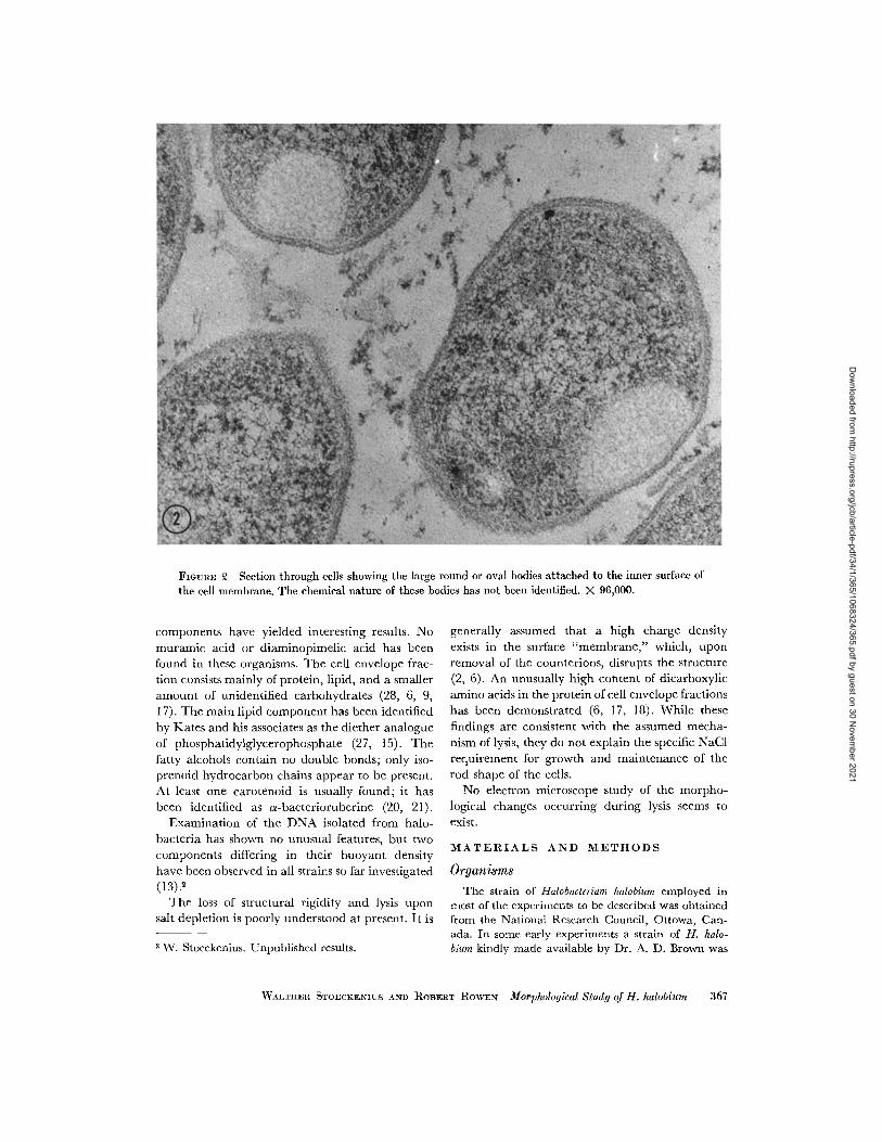
FIGURE 2 Section through cells showing the large round or oval bodies attached to the inner surface ofthe cell membrane. The chemical nature of these bodies has not been identified. X 96,000.
components have yielded interesting results. Nomuramic acid or diaminopimelic acid has beenfound in these organisms. The cell envelope frac-tion consists mainly of protein, lipid, and a smalleramount of unidentified carbohydrates (28, 6, 9,17). The main lipid component has been identifiedby Kates and his associates as the diether analogueof phosphatidylglycerophosphate (27, 15). Thefatty alcohols contain no double bonds; only iso-prenoid hydrocarbon chains appear to be present.At least one carotenoid is usually found; it hasbeen identified as a-bacterioruberine (20, 21).
Examination of the DNA isolated from halo-bacteria has shown no unusual features, but twocomponents differing in their buoyant densityhave been observed in all strains so far investigated(13).2
The loss of structural rigidity and lysis uponsalt depletion is poorly understood at present. It is
2 W. Stoeckenius. Unpublished results.
generally assumed that a high charge densityexists in the surface "membrane," which, uponremoval of the counterions, disrupts the structure(2, 6). An unusually high content of dicarboxylicamino acids in the protein of cell envelope fractionshas been demonstrated (6, 17, 18). While thesefindings are consistent with the assumed mecha-nism of lysis, they do not explain the specific NaCIrequirement for growth and maintenance of therod shape of the cells.
No electron microscope study of the morpho-logical changes occurring during lysis seems toexist.
MATERIALS AND METHODS
Organisms
The strain of Halobacterium halobium employed inmost of the experiments to be described was obtainedfrom the National Research Council, Ottowa, Can-ada. In some early experiments a strain of H. halo-bium kindly made available by Dr. A. D. Brown was
WALTIER STOECKENIUS AND ROBERT ROWEN Morphological Study of H. halobium 367
Dow
nloaded from http://rupress.org/jcb/article-pdf/34/1/365/1068324/365.pdf by guest on 30 N
ovember 2021
used. Both strains yielded identical results but theformer was favored because it appeared to grow morerapidly under the conditions used.
Medium and Growth Conditions
The medium employed was that described byBrown (6), consisting of I 1% Oxoid peptone in thebasal salt solution (BS) used by Sehgal and Gibbons(26).
Organisms were grown at 37 C either as 1 litershake cultures or in 10 liter carboys using forced aera-tion and constant stirring (Microferm Apparatus,New Brunswick Scientific, New Brunswick, N.J.).The carboys were inoculated with 300 ml of a 2-3 dayshake culture grown at 37°C. Foaming was preventedby the addition of 10 ml of Dow-Corning Antifoam ACompound. Unless otherwise indicated, bacteriawere harvested at the end of the log phase of growth(shake cultures, 5-6 days; aerated 10 liter carboy,3 days).
Preparation of Envelopes
Cells were washed once in BS (containing 25j%NaCI) and resuspended in sufficient BS to give a finalvolume which was about J-0 of the original culture.Highly vacuolated cells floated to the surface andwere discarded with the supernatant. Mechanicaldisruption was accomplished by shaking the sus-
FIGoRE Tangentialshowing indications of
X 160,000.
section through the cell walla regular hexagonal structure.
pended organisms with equal volumes of 0.11-0.12mm glass beads in a Bronwill Disintegrator (BronwillScientific, Rochester, N.Y.) for a period of 2 minusing a jet of liquid CO 2 for cooling the disruptionvessel. After removal of the glass beads and furtherdilution with an equal volume of BS to decrease vis-
cosity, the material was spun for 60 min at 20,000 g.
The supernatant was discarded and the pellet wasresuspended in BS and centrifuged for 15 min at 5000
g to remove unbroken cells. The alternate high andlow speed centrifugations were repeated four to five
times until the pellet obtained from the 20,000 g run
appeared free of unbroken cells and the supernatantcontained little or no pigment. The final pellet sus-
pended in a small volume of basal salt solution consti-
tuted our "envelope preparation." This was stored at
4°C and usually used within 2-3 days.
Analytical Procedures
Protein was determined by the method of Lowry
et al. (22) with a freshly prepared solution of
bovine serum albumin as standard. Lipids were ex-
tracted from envelopes and envelope fractions over-night at room temperature with chloroform-metha-
nol, 2:1. Purification of the lipid extract involved
washing with distilled water, drying under nitrogen,and reextraction of the dried material with chloro-
form (29). Total lipids were determined gravimetri-
cally and lipid phosphorus was measured by a modifi-cation of the method of Stewart and Hendry (3()).RNA content was estimated after hot trichloroacetic
acid extraction by an orcinol procedure (23) using
purified yeast RNA as standard.
Thin layer chromatography was carried out on Sil-ica Gel H (Merck, Darmstadt, Germany) using a sol-
vent system of chloroformn-methanol-water, 65:25:4.
Preparation of Samples for
Electron Microscopy
SHADOWING TECHNIQUES: To examine surface
structure of cells and envelope preparations, the mate-rial was diluted to barely visible turbidity in the ap-
propriate salt solution, applied directly to hydro-philic, carbon-coated Formvar grids, and shadowed
with uranium (10 mg at 10 cm and 12°).
For examination of envelope lysate fractions the
material was sprayed onto freshly cleaved mica,
shadowed with platinum-carbon (at an angle at 6),
and coated with a film of evaporated carbon. Thepreparation was reinforced with a thin backing of col-lodion, floated off onto a water surface, and mounted
on grids.NEGATIVE STAIN IN G: Suitable dilutions of the
lysate or lysate fractions were applied to Forinvar-car-
bon coated grids and the grid edge was touched with
a filter paper. Before the remaining film of fluid haddried, one or more drops of the negative stain were
successively applied to the grid and removed in the
368 TE JOURNAL OF CELL BIOLOGY VOLUME 34, 1967
Dow
nloaded from http://rupress.org/jcb/article-pdf/34/1/365/1068324/365.pdf by guest on 30 N
ovember 2021
same manner. The grids were then dried in a desicca-tor and coated with a second thin layer of carbon. 1 0uranyl acetate or uranyl formate or 2 phospho-tungstic acid neutralized with KOH were used asnegative stains.
FIXATION AND EMBEDDING: Pellets of washed
cells, envelope preparations, and envelope frac-tions, approximately 2 mm in diameter, were ob-tained. Many different fixation procedures were triedand the following was found to give the best results.Pellets were covered with a 4% solution of formalde-hyde containing 25% NaCl and 1% CaCI2 at pH 7.0and allowed to remain at 40 C overnight. All subse-quent fixation steps were also carried out in the cold.The pellets were then washed twice for 30 min inter-vals with 25c/% NaCI containing 1% CaCI2 and ex-posed to freshly prepared 2 KMnO 4 solution con-taining 25% NaCI and 1% CaC1 2 (pH 7.0) for aperiod of 30-60 min. In some of the experiments in-volving partial or total lysis of cells or envelopes, thesalt concentration of the fixative and washing solu-tions was reduced to the same amount as in the solu-tion used for lysing the cells or envelope preparations.The specimens were then washed in salt solution andtreated with 2 % aqueous solution of uranyl nitrate for30 min. After several washings with water the fixedmaterial was dehydrated with acetone and portionswere embedded in Epon for sectioning. In most casesthe material was oriented during embedding so that across-section through the center of the pellet could beobtained. Thin sections were cut with a diamondknife and mounted on Formvar films that were lightlycoated with carbon. Sections were double-stainedwith magnesium uranyl acetate and lead citrate (11).
Effects of Salt Concentration
Freshly prepared cells or envelopes were suspendedin BS to give in a Bausch & Lomb Spectronic 20 anOD of 0.2-0.4 at 700 mu when diluted 1:100. Toexamine the changes induced at different salt concen-trations, aliquots were mixed with 100 volumes of BSdiluted to the appropriate concentration with respectto NaCl. The test suspensions were maintained atroom temperature for :30 min. Extent of lysis wasmeasured as decrease in OD at 700 mgu. The materialwas then centrifuged for 15 min at 15,000 g (cells) or30 min at 20,000 g (envelopes). The pellets were re-tained for electron microscopy and the supernatantfluids were examined for released material.
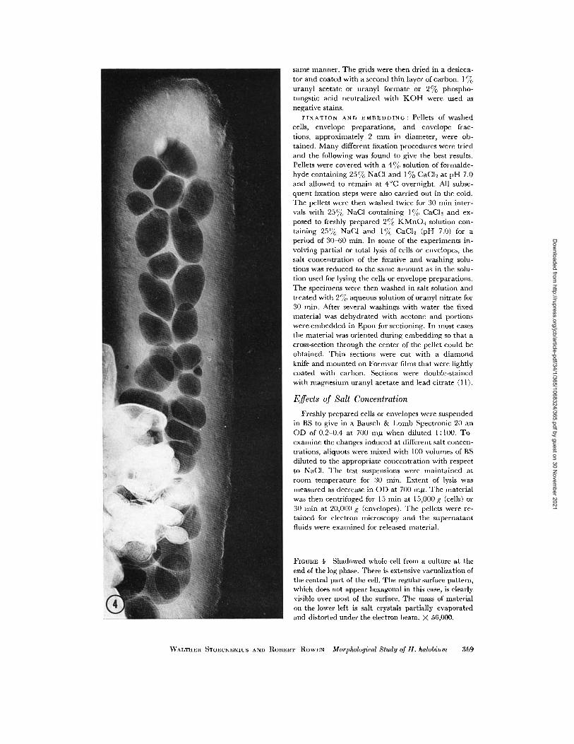
FIGuRE 4 Shadowed whole cell from a culture at theend of the log phase. There is extensive vacuolization ofthe central part of the cell. The regular surface pattern,which does not appear hexagonal in this case, is clearlyvisible over most of the surface. The mass of materialon the lower left is salt crystals partially evaporatedand distorted under the electron beam. X 56,000.
WALTHEI STOECKENICS AND ROBERT ROWEN Morphological Study of H. halobium 369
Dow
nloaded from http://rupress.org/jcb/article-pdf/34/1/365/1068324/365.pdf by guest on 30 N
ovember 2021
RESULTS
Intact Cells
A thin section through cells of H. halobium from a3 day shake culture (early log phase) is shown inFig. 1. The rod-shaped cells have a rather dense
cytoplasm with nucleoids of almost the samedensity. Numerous, ill-defined, denser particlesin the cytoplasm probably represent ribosomes. A
few considerably denser and slightly bigger
particles visible in some of the cells have not beenidentified. Clear round areas of variable size in
the cytoplasm could represent sections through
gas vacuoles.Where the cell envelope is sectioned approxi-
mately radially, several layers can be clearlydiscerned. An innermost clear band about 30 Awide is the most constant feature observed. Thatit actually represents the central light layer of a
typical unit membrane will become obvious from
inspection of sections through isolated envelopesto be described later. On its surface bordering the
cytoplasm a thin dense band can sometimes be seen,but is often not visible at all, presumably becauseits density equals that of the cytoplasm. A denseband on the outer surface of the light band is much
more regularly and clearly observed, but some-times it merges imperceptibly with the 130-150 A
wide layer of material that forms the outer surface
of the cell. In Fig. 1 the outer part of this 130-150 A layer is much denser than the inner, butFig. 2 shows a micrograph where this stratification
is not so obvious, and sometimes, especially in oldercultures, the outer 130-150 A layer appears frayed
and less dense (Fig. 6). This variability in ap-
pearance may be attributable, at least in part, toaccidents of fixation and staining, because the
appearance of the cells sometimes differs in differ-ent regions of the same pellet.
In some places (see Fig. 1) the outermost denselayer of the envelope shows indications of a regular
beaded appearance with a periodicity of approxi-
mately 150 A. Indications of a regular structurecan occasionally also be seen in tangential section(Fig. 3). This appearance is probably related tothe well-known surface structure (12, 16, 24) seen
in shadowed preparations of whole bacteria (Figs.
4 and 5). It has been described as a hexagonalarrangement of spherical particles with a spacingof 120-150 A (16). In our shadowed preparationswe usually did not find a clearly hexagonal lattice,but this may be an artifact due to distortion during
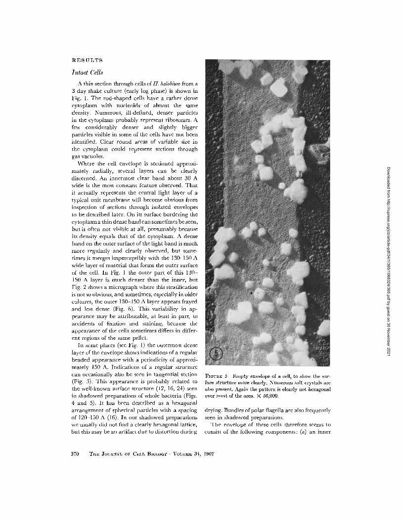
FIGURE 5 Empty envelope of a cell, to show the sur-face structure more clearly. Numerous salt crystals arealso present. Again the pattern is clearly not hexagonalover most of the area. X 56,000.
drying. Bundles of polar flagella are also frequentlyseen in shadowed preparations.
The envelope of these cells therefore seems toconsist of the following components: (a) an inner
370 THE JOURNAL OF CELL BIOLOGY . VOLUME 34, 1967
Dow
nloaded from http://rupress.org/jcb/article-pdf/34/1/365/1068324/365.pdf by guest on 30 N
ovember 2021
unit membrane, the cell membrane proper, whosecentral light layer is always clearly visible; theouter dense layer is usually visible while the innerdense layer is rarely discernible; and (b) a cellwall with a regular surface structure; it may alsoshow a layered appearance in sections, with anouter denser and an inner lighter layer.
It is, of course, possible that the outer denselayer of the cell membrane or at least part of itactually belongs to the cell wall. This problemcannot be resolved before considerably more isknown about the chemical composition of thesestructures, and a separation has been achieved.
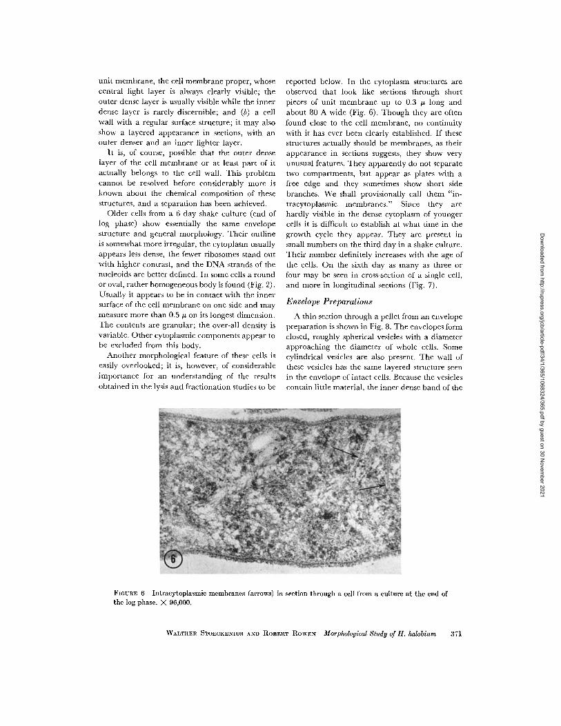
Older cells from a 6 day shake culture (end oflog phase) show essentially the same envelopestructure and general morphology. Their outlineis somewhat more irregular, the cytoplasm usuallyappears less dense, the fewer ribosomes stand outwith higher contrast, and the DNA strands of thenucleoids are better defined. In some cells a roundor oval, rather homogeneous body is found (Fig. 2).Usually it appears to be in contact with the innersurface of the cell membrane on one side and maymeasure more than 0.5 tj on its longest dimension.The contents are granular; the over-all density isvariable. Other cytoplasmic components appear tobe excluded from this body.
Another morphological feature of these cells iseasily overlooked; it is, however, of considerableimportance for an understanding of the resultsobtained in the lysis and fractionation studies to be
reported below. In the cytoplasm structures areobserved that look like sections through shortpieces of unit membrane up to 0.3 long andabout 80 A wide (Fig. 6). Though they are oftenfound close to the cell membrane, no continuitywith it has ever been clearly established. If thesestructures actually should be membranes, as theirappearance in sections suggests, they show veryunusual features. They apparently do not separatetwo compartments, but appear as plates with afree edge and they sometimes show short sidebranches. We shall provisionally call them "in-tracytoplasmic membranes." Since they arehardly visible in the dense cytoplasm of youngercells it is difficult to establish at what time in thegrowth cycle they appear. They are present insmall numbers on the third day in a shake culture.Their number definitely increases with the age ofthe cells. On the sixth day as many as three orfour may be seen in cross-section of a single cell,and more in longitudinal sections (Fig. 7).
Envelope Preparations
A thin section through a pellet from an envelopepreparation is shown in Fig. 8. The envelopes formclosed, roughly spherical vesicles with a diameterapproaching the diameter of whole cells. Somecylindrical vesicles are also present. The wall ofthese vesicles has the same layered structure seenin the envelope of intact cells. Because the vesiclescontain little material, the inner dense band of the
FIGURE 6 Intracytoplasmic membranes (arrows) in section through a cell from a culture at the end ofthe log phase. X 96,000.
WALTHER STOECKENIUS AND ROBERT ROWEN Morphological Study of H. halobium 371
Dow
nloaded from http://rupress.org/jcb/article-pdf/34/1/365/1068324/365.pdf by guest on 30 N
ovember 2021
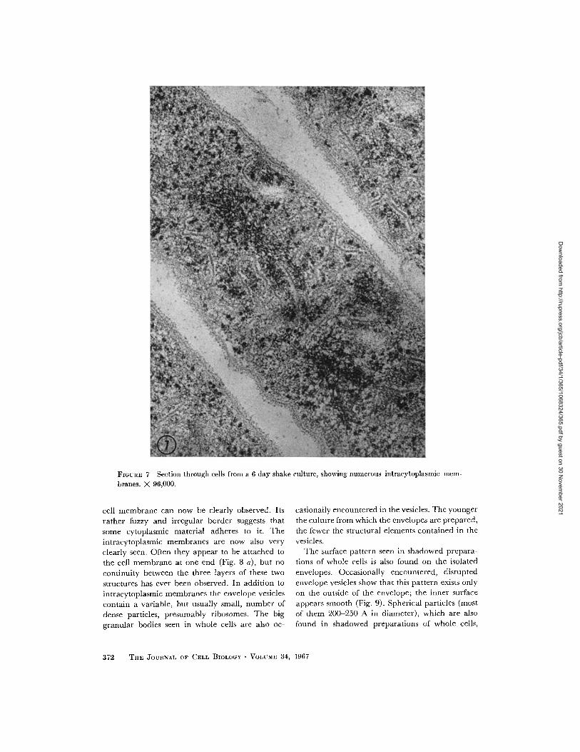
FIGURE 7 Section through cells from a 6 day shake culture, showing numerous intracytoplasmic mem-
branes. X 96,000.
cell membrane can now be clearly observed. Its
rather fuzzy and irregular border suggests that
some cytoplasmic material adheres to it. The
intracytoplasmic membranes are now also very
clearly seen. Often they appear to be attached to
the cell membrane at one end (Fig. 8 a), but no
continuity between the three layers of these two
structures has ever been observed. In addition to
intracytoplasmic membranes the envelope vesicles
contain a variable, but usually small, number of
dense particles, presumably ribosomes. The big
granular bodies seen in whole cells are also oc-
casionally encountered in the vesicles. The younger
the culture from which the envelopes are prepared,
the fewer the structural elements contained in the
vesicles.The surface pattern seen in shadowed prepara-
tions of whole cells is also found on the isolated
envelopes. Occasionally encountered, disrupted
envelope vesicles show that this pattern exists only
on the outside of the envelope; the inner surface
appears smooth (Fig. 9). Spherical particles (most
of them 200-250 A in diameter), which are also
found in shadowed preparations of whole cells,
372 THE JOURNAL OF CELL BIOLOGY VOLUME 34, 1967
Dow
nloaded from http://rupress.org/jcb/article-pdf/34/1/365/1068324/365.pdf by guest on 30 N
ovember 2021
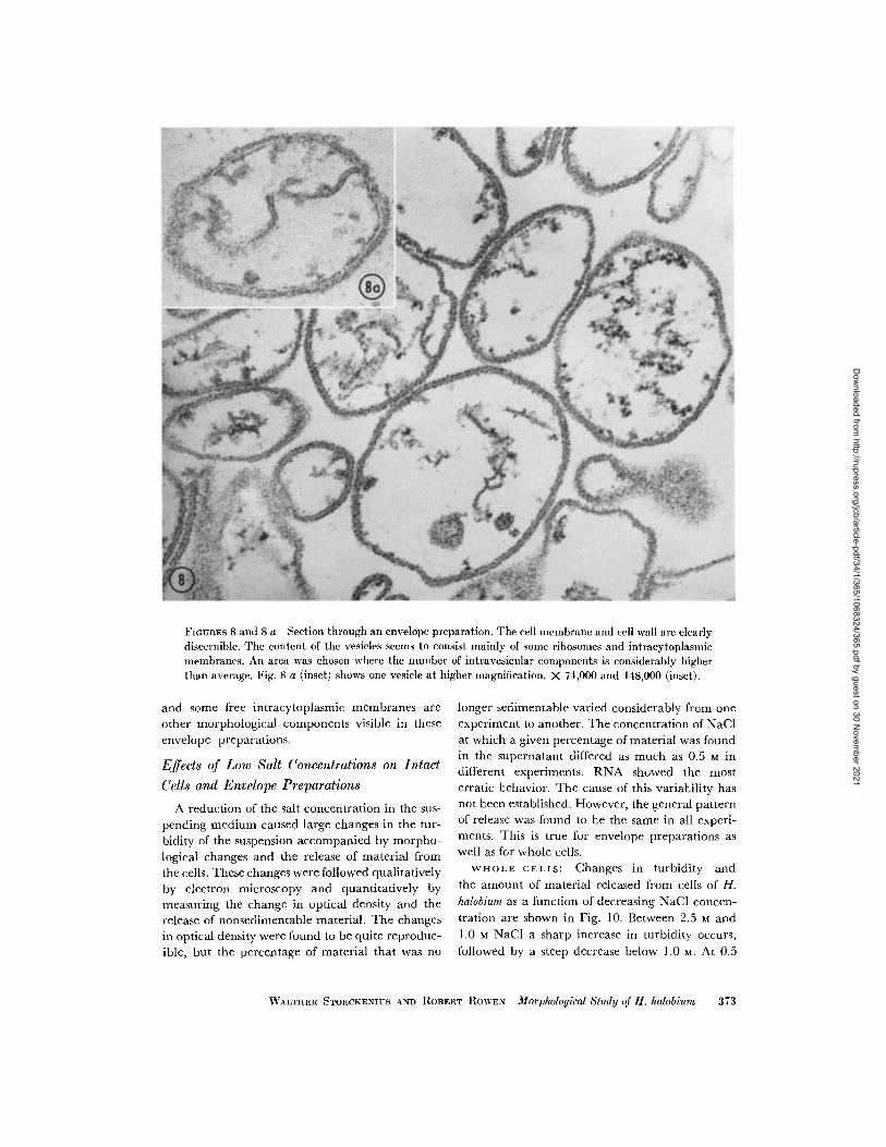
FIGURES 8 and 8 a Section through an envelope preparation. The cell membrane and cell wall are clearlydiscernible. The content of the vesicles seems to consist mainly of some ribosomes and intracytoplasmicmembranes. An area was chosen where the number of intravesicular components is considerably higherthan average. Fig. 8 a (inset) shows one vesicle at higher magnification. X 74,000 and 148,000 (inset).
and some free intracytoplasmic membranes areother morphological components visible in theseenvelope preparations.
Effects of Low Salt Concentrations on Intact
Cells and Envelope Preparations
A reduction of the salt concentration in the sus-pending medium caused large changes in the tur-bidity of the suspension accompanied by morpho-logical changes and the release of material fromthe cells. These changes were followed qualitativelyby electron microscopy and quantitatively bymeasuring the change in optical density and therelease of nonsedimentable material. The changesin optical density were found to be quite reproduc-ible, but the percentage of material that was no
longer sedimentable varied considerably from one
experiment to another. The concentration of NaClat which a given percentage of material was foundin the supernatant differed as much as 0.5 M indifferent experiments. RNA showed the mosterratic behavior. The cause of this variability hasnot been established. However, the general patternof release was found to be the same in all experi-ments. This is true for envelope preparations aswell as for whole cells.
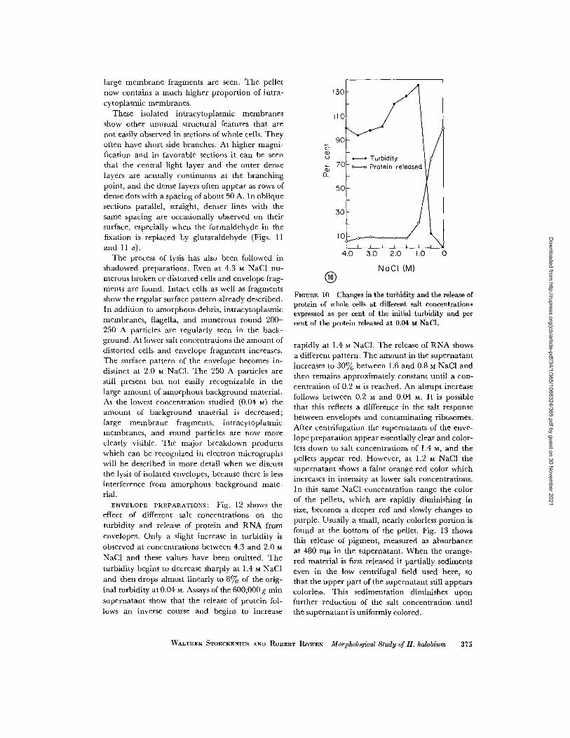
WHOLE CELLS: Changes in turbidity and
the amount of material released from cells of H.
halobium as a function of decreasing NaCl concen-
tration are shown in Fig. 10. Between 2.5 M and
1.0 M NaCl a sharp increase in turbidity occurs,
followed by a steep decrease below 1.0 M. At 0.5
WALTHER STOECKENIUS AND ROBERT ROWEN Morphological Study of H. halobium 373
Dow
nloaded from http://rupress.org/jcb/article-pdf/34/1/365/1068324/365.pdf by guest on 30 N
ovember 2021
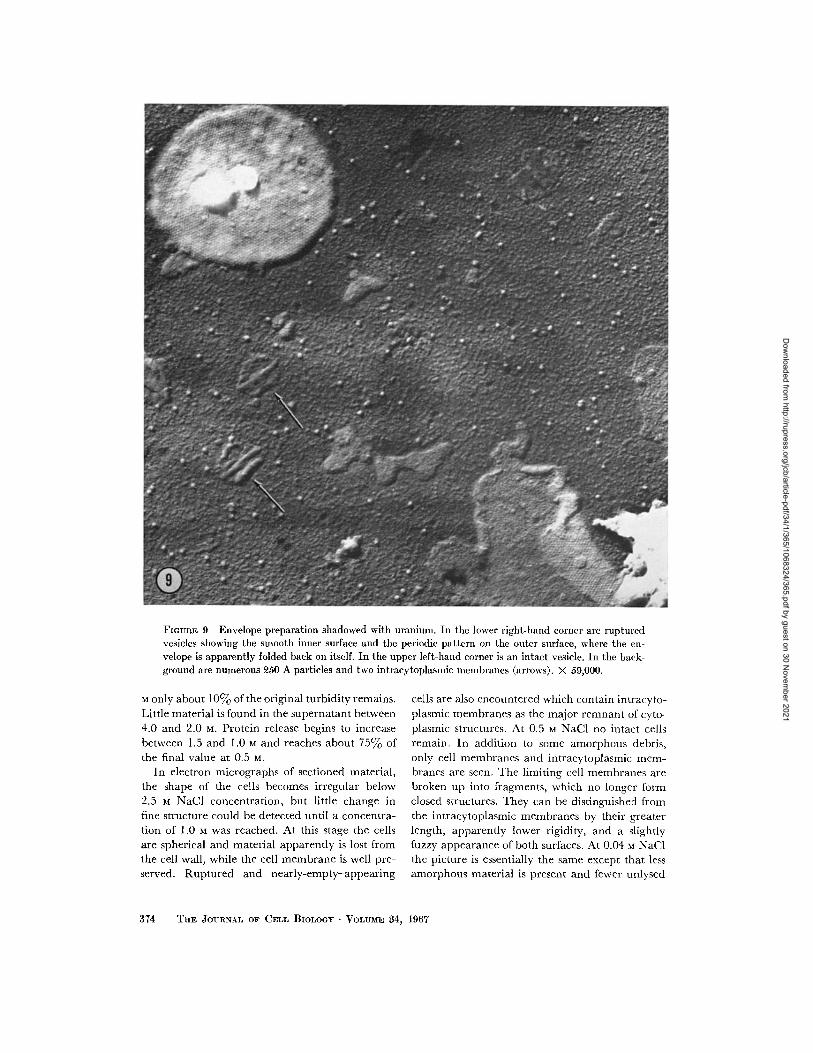
FIGURE 9 Envelope preparation shadowed with uranium. In the lower right-hand corner are rupturedvesicles showing the smooth inner surface and the periodic pattern on the outer surface, where the en-velope is apparently folded back on itself. In the upper left-hand corner is an intact vesicle. In the back-ground are numerous 250 A particles and two intracytoplasmic membranes (arrows). X 59,000.
M only about 10% of the original turbidity remains.Little material is found in the supernatant between4.0 and 2.0 M. Protein release begins to increasebetween 1.5 and 1.0 M and reaches about 75% ofthe final value at 0.5 M.
In electron micrographs of sectioned material,the shape of the cells becomes irregular below2.5 M NaCl concentration, but little change infine structure could be detected until a concentra-tion of 1.0 M was reached. At this stage the cellsare spherical and material apparently is lost fromthe cell wall, while the cell membrane is well pre-served. Ruptured and nearly-empty-appearing
cells are also encountered which contain intracyto-plasmic membranes as the major remnant of cyto-plasmic structures. At 0.5 M NaCl no intact cellsremain. In addition to some amorphous debris,only cell membranes and intracytoplasmic mem-branes are seen. The limiting cell membranes arebroken up into fragments, which no longer formclosed structures. They can be distinguished fromthe intracytoplasmic membranes by their greaterlength, apparently lower rigidity, and a slightlyfuzzy appearance of both surfaces. At 0.04 M NaC1the picture is essentially the same except that lessamorphous material is present and fewer unlysed
374 TaE JOURNAL OF CELL BIOLOGY VOLUME 4, 1967
Dow
nloaded from http://rupress.org/jcb/article-pdf/34/1/365/1068324/365.pdf by guest on 30 N
ovember 2021
large membrane fragments are seen. The pelletnow contains a much higher proportion of intra-cytoplasmic membranes.
These isolated intracytoplasmic membranesshow other unusual structural features that arenot easily observed in sections of whole cells. Theyoften have short side branches. At higher magni-fication and in favorable sections it can be seenthat the central light layer and the outer denselayers are actually continuous at the branchingpoint, and the dense layers often appear as rows ofdense dots with a spacing of about 50 A. In obliquesections parallel, straight, denser lines with thesame spacing are occasionally observed on theirsurface, especially when the formaldehyde in thefixation is replaced by glutaraldehyde (Figs. 11and 11 a).
The process of lysis has also been followed inshadowed preparations. Even at 4.3 M NaCl nu-merous broken or distorted cells and envelope frag-ments are found. Intact cells as well as fragmentsshow the regular surface pattern already described.In addition to amorphous debris, intracytoplasmicmembranes, flagella, and numerous round 200-250 A particles are regularly seen in the back-ground. At lower salt concentrations the amount ofdistorted cells and envelope fragments increases.The surface pattern of the envelope becomes in-distinct at 2.0 M NaCI. The 250 A particles arestill present but not easily recognizable in thelarge amount of amorphous background material.At the lowest concentration studied (0.04 M) theamount of background material is decreased;large membrane fragments, intracytoplasmicmembranes, and round particles are now moreclearly visible. The major breakdown productswhich can be recognized in electron micrographswill be described in more detail when we discussthe lysis of isolated envelopes, because there is lessinterference from amorphous background mate-rial.
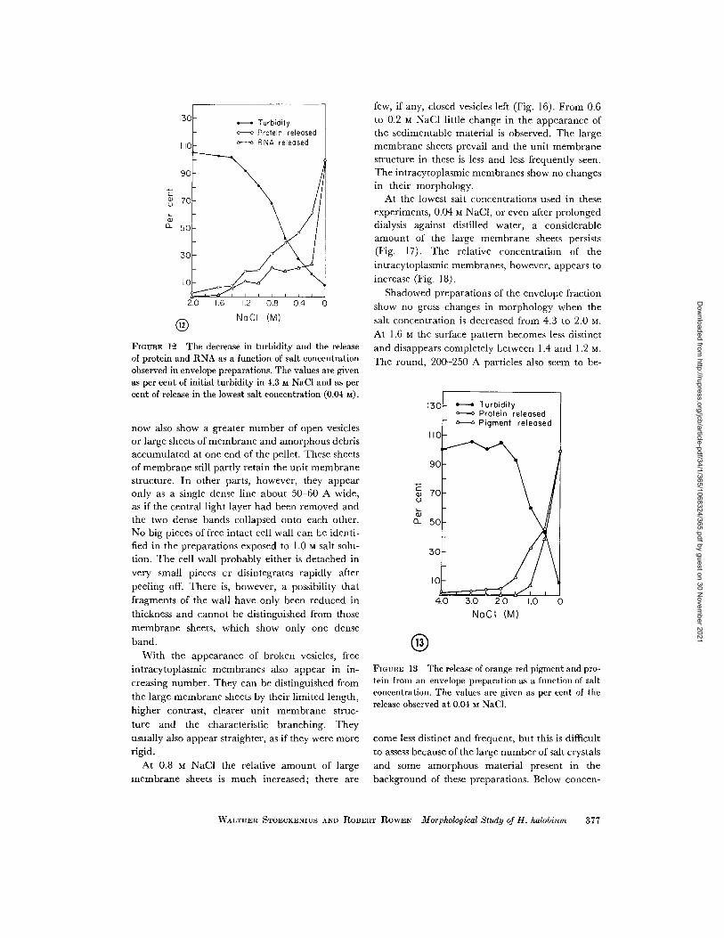
ENVELOPE PREPARATIONS: Fig. 12 shows theeffect of different salt concentrations on theturbidity and release of protein and RNA fromenvelopes. Only a slight increase in turbidity isobserved at concentrations between 4.3 and 2.0 MNaCl and these values have been omitted. Theturbidity begins to decrease sharply at 1.4 M NaCland then drops almost linearly to 8% of the orig-inal turbidity at 0.04 M. Assays of the 600,000 g minsupernatant show that the release of protein fol-lows an inverse course and begins to increase
a
0~L
4.0 3.0 2.0 1.0 0
NaCI (M)
FIGURE 10 Changes in the turbidity and the release ofprotein of whole cells at different salt concentrationsexpressed as per cent of the initial turbidity and percent of the protein released at 0.04 M NaCl.
rapidly at 1.4 M NaC1. The release of RNA showsa different pattern. The amount in the supernatantincreases to 30%0 between 1.6 and 0.8 M NaCl andthen remains approximately constant until a con-centration of 0.2 M is reached. An abrupt increasefollows between 0.2 M and 0.04 M. It is possiblethat this reflects a difference in the salt responsebetween envelopes and contaminating ribosomes.After centrifugation the supernatants of the enve-lope preparation appear essentially clear and color-less down to salt concentrations of 1.4 M, and thepellets appear red. However, at 1.2 M NaCl thesupernatant shows a faint orange-red color whichincreases in intensity at lower salt concentrations.
In this same NaCI concentration range the colorof the pellets, which are rapidly diminishing insize, becomes a deeper red and slowly changes topurple. Usually a small, nearly colorless portion isfound at the bottom of the pellet. Fig. 13 showsthis release of pigment, measured as absorbanceat 480 my in the supernatant. When the orange-red material is first released it partially sedimentseven in the low centrifugal field used here, sothat the upper part of the supernatant still appearscolorless. This sedimentation diminishes uponfurther reduction of the salt concentration untilthe supernatant is uniformly colored.
WVALTHER STOECKENIUS AND ROBERT ROWEN Morphological Study of H. halobium 375
Dow
nloaded from http://rupress.org/jcb/article-pdf/34/1/365/1068324/365.pdf by guest on 30 N
ovember 2021
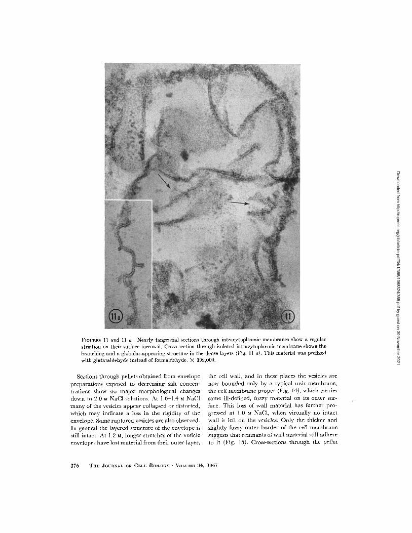
FIGURES 11 and 11 a Nearly tangential sections through intracytoplasmic ilemlbranes show a regular
striation on their surface (arrows). Cross-section through isolated intracytoplasmic membrane shows the
branching and a globular-appearing structure in the dense layers (Fig. 11 a). This material was prefixed
with glutaraldehyde instead of formaldehyde. X 192,000.
Sections through pellets obtained from envelope
preparations exposed to decreasing salt concen-
trations show no major morphological changesdown to 2.0 M NaCl solutions. At 1.6 1.4 M NaCl
many of the vesicles appear collapsed or distorted,
which may indicate a loss in the rigidity of the
envelope. Some ruptured vesicles are also observed.
In general the layered structure of the envelope is
still intact. At 1.2 M, longer stretches of the vesicleenvelopes have lost material from their outer layer,
the cell wall, and in these places the vesicles are
now bounded only by a typical unit membrane,
the cell membrane proper (Fig. 14), which carries
some ill-defined, fuzzy material on its outer sur-
face. This loss of wall material has further pro-gressed at 1.0 M NaCl, when virtually no intact
wall is left on the vesicles. Only the thicker and
slightly fuzzy outer border of the cell membrane
suggests that remnants of wall material still adhere
to it (Fig. 15). Cross-sections through the pellet
376 THE JOURNAL OF CELL BIOLOGY VOLUME 34, 1967
Dow
nloaded from http://rupress.org/jcb/article-pdf/34/1/365/1068324/365.pdf by guest on 30 N
ovember 2021
c
a)
NoCI (M)
FIGURE 12 The decrease in turbidity and the releaseof protein and RNA as a function of salt concentrationobserved in envelope preparations. The values are givenas per cent of initial turbidity in 4.3 M NaCI and as percent of release in the lowest salt concentration (0.04 m).
now also show a greater number of open vesiclesor large sheets of membrane and amorphous debrisaccumulated at one end of the pellet. These sheetsof membrane still partly retain the unit membranestructure. In other parts, however, they appearonly as a single dense line about 50 60 A wide,as if the central light layer had been removed andthe two dense bands collapsed onto each other.No big pieces of free intact cell wall can be identi-fied in the preparations exposed to 1.0 M salt solu-tion. The cell wall probably either is detached in
very small pieces or disintegrates rapidly after
peeling off. There is, however, a possibility that
fragments of the wall have only been reduced in
thickness and cannot be distinguished from those
membrane sheets, which show only one dense
band.
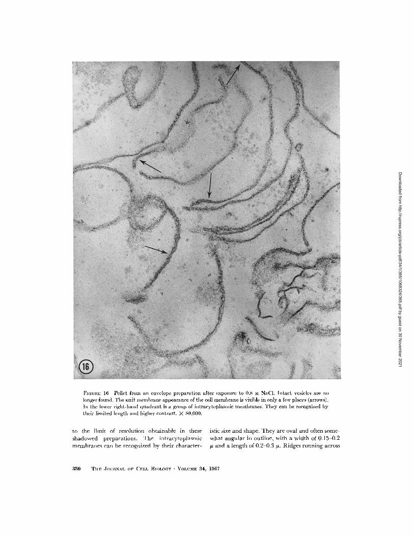
With the appearance of broken vesicles, free
intracytoplasmic membranes also appear in in-
creasing number. They can be distinguished from
the large membrane sheets by their limited length,
higher contrast, clearer unit membrane struc-
ture and the characteristic branching. They
usually also appear straighter, as if they were morerigid.
At 0.8 M NaCl the relative amount of large
membrane sheets is much increased; there are
few, if any, closed vesicles left (Fig. 16). From 0.6to 0.2 M NaCI little change in the appearance ofthe sedimentable material is observed. The largemembrane sheets prevail and the unit membranestructure in these is less and less frequently seen.The intracytoplasmic membranes show no changesin their morphology.
At the lowest salt concentrations used in theseexperiments, 0.04 M NaCI, or even after prolongeddialysis against distilled water, a considerableamount of the large membrane sheets persists(Fig. 17). The relative concentration of theintracytoplasmic membranes, however, appears toincrease (Fig. 18).
Shadowed preparations of the envelope fractionshow no gross changes in morphology when thesalt concentration is decreased from 4.3 to 2.0 M.At 1.6 M the surface pattern becomes less distinctand disappears completely between 1.4 and 1.2 M.The round, 200-250 A particles also seem to be-
a
aa
NoCI (M)
FIGURE 13 The release of orange-red pigment and pro-tein from an envelope preparation as a function of saltconcentration. The values are given as per cent of therelease observed at 0.04 M NaCI.
come less distinct and frequent, but this is difficultto assess because of the large number of salt crystalsand some amorphous material present in thebackground of these preparations. Below concen-
WALTHER STOECKENIUS AND ROBERT ROWEN Morphological Study of H. halobium 377
Dow
nloaded from http://rupress.org/jcb/article-pdf/34/1/365/1068324/365.pdf by guest on 30 N
ovember 2021
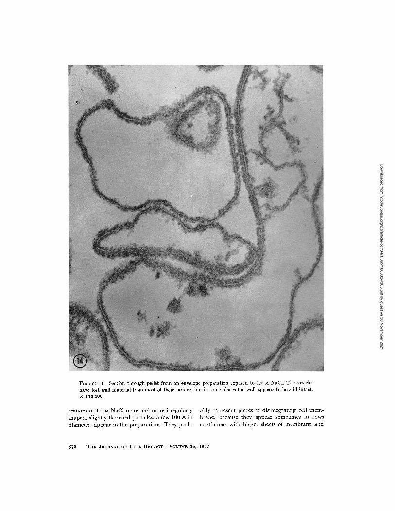
FIGURE 14 Section through pellet from an envelope preparation exposed to 1.2 M NaCl. The vesicleshave lost wall material from most of their surface, but in some places the wall appears to be still intact.
X 176,000.
trations of 1.0 M NaCI more and more irregularly ably represent pieces of disintegrating cell mem-
shaped, slightly flattened particles, a few 100 A in brane, because they appear sometimes in rows
diameter, appear in the preparations. They prob- continuous with bigger sheets of membrane and
378 THE JOURNAL OF CELL BIOLOGY ·VOLUME 34, 1967
Dow
nloaded from http://rupress.org/jcb/article-pdf/34/1/365/1068324/365.pdf by guest on 30 N
ovember 2021
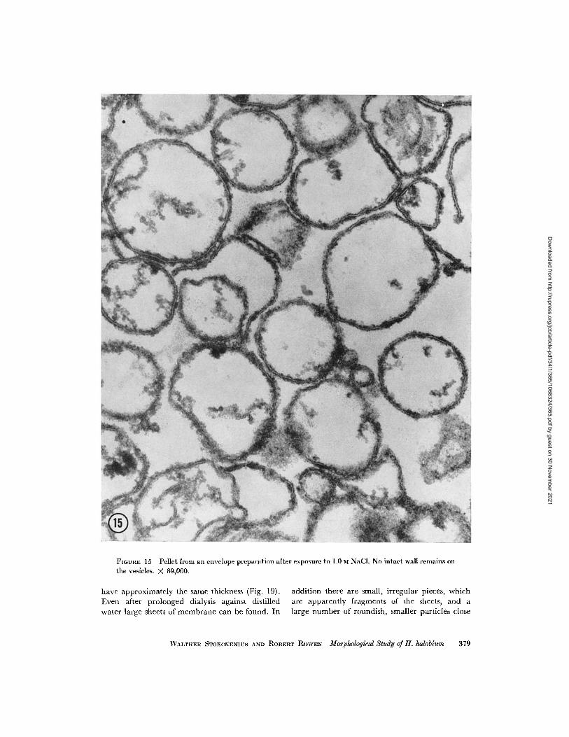
FIGURE 15 Pellet from an envelope preparation after exposure to 1.0 M NaCl. No intact wall remains onthe vesicles. X 89,000.
have approximately the same thickness (Fig. 19).Even after prolonged dialysis against distilled
water large sheets of membrane can be found. In
addition there are small, irregular pieces, whichare apparently fragments of the sheets, and alarge number of roundish, smaller particles close
WALTHER STOECKENIUS AND ROBERT ROWEN Morphological Study of H. halobium 379
Dow
nloaded from http://rupress.org/jcb/article-pdf/34/1/365/1068324/365.pdf by guest on 30 N
ovember 2021
FIGUnE 16 Pellet from an envelope preparation after exposure to 0.8 M NaCI. Intact vesicles are nolonger found. The unit mlembrane appearance of the cell membrane is visible in only a few places (arrows).In the lower right-hand quadrant is a group of intracytoplasmic membranes. They can be recognized bytheir limited length and higher contrast. X 89,000.
to the limit of resolution obtainable in these istic size and shape. They are oval and often some-
shadowed preparations. The intracytoplasmic what angular in outline, with a width of 0.15-0.2membranes can be recognized by their character- u and a length of 0.2 0.3 p. Ridges running across
380 TILE JOURNAL OF CELL BIOLOGY - VOLUME 34, 1967
Dow
nloaded from http://rupress.org/jcb/article-pdf/34/1/365/1068324/365.pdf by guest on 30 N
ovember 2021
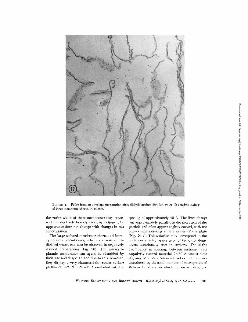
FIGURE 17 Pellet from an envelope preparation after dialysis against distilled water. It consists mainlyof large membrane sheets. X 96,000.
the entire width of these membranes may repre-sent the short side branches seen in sections. Theappearance does not change with changes in saltconcentration.
The large unlysed membrane sheets and intra-cytoplasmic membranes, which are resistant todistilled water, can also be observed in negativelystained preparations (Fig. 20). The intracyto-plasmic membranes can again be identified bytheir size and shape. In addition to this, however,they display a very characteristic regular surfacepattern of parallel lines with a somewhat variable
spacing of approximately 40 A. The lines alwaysrun approximately parallel to the short axis of theparticle and often appear slightly curved, with theconvex side pointing to the center of the plate(Fig. 20 a). This striation may correspond to thedotted or striated appearance of the outer denselayers occasionally seen in sections. The slightdiscrepancy in spacing, between sectioned andnegatively stained material (-50 A versus 40A), may be a preparation artifact or due to errorsintroduced by the small number of micrographs ofsectioned material in which the surface structure
WALTHER STOECKENIUS AND ROBERT ROWEN Morphological Study of H. halobium 381
Dow
nloaded from http://rupress.org/jcb/article-pdf/34/1/365/1068324/365.pdf by guest on 30 N
ovember 2021
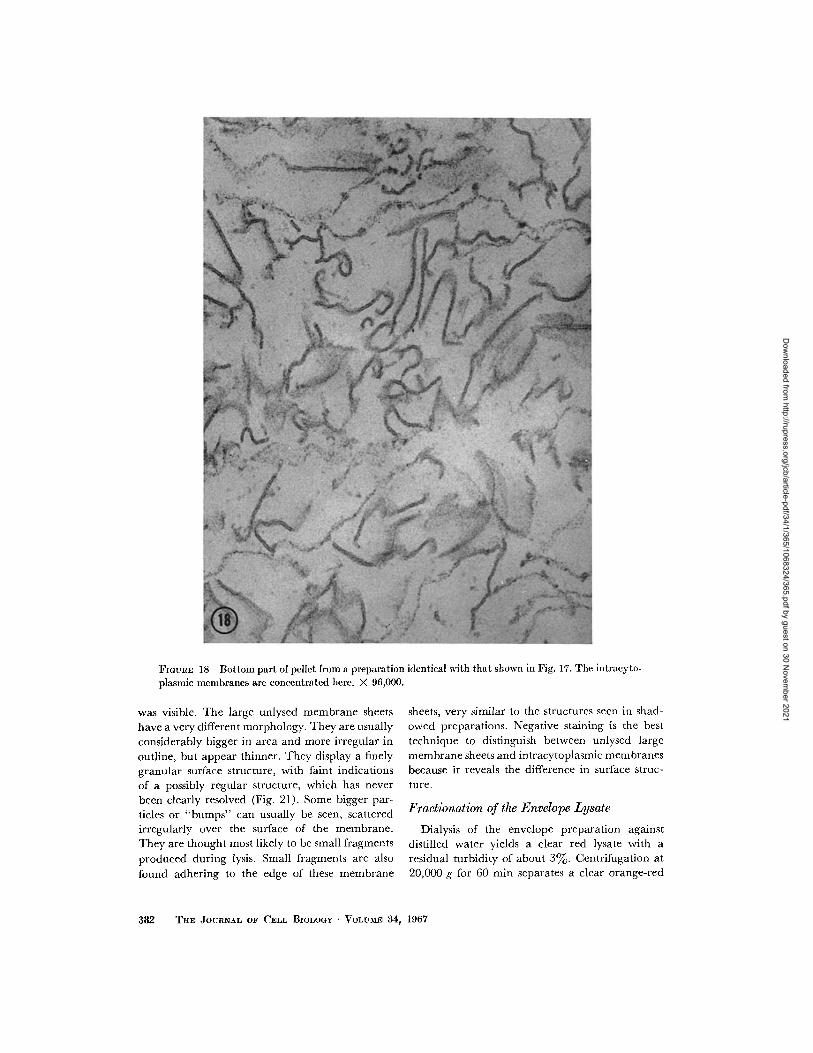
FIGURE 18 Bottom part of pellet from a preparation identical with that shown in Fig. 17. The intracyto-plasmic membranes are concentrated here. X 96,000.
was visible. The large unlysed membrane sheets
have a very different morphology. They are usually
considerably bigger in area and more irregular in
outline, but appear thinner. They display a finelygranular surface structure, with faint indications
of a possibly regular structure, which has never
been clearly resolved (Fig. 21). Some bigger par-
ticles or "bumps" can usually be seen, scattered
irregularly over the surface of the membrane.
They are thought most likely to be small fragments
produced during lysis. Small fragments are also
found adhering to the edge of these membrane
sheets, very similar to the structures seen in shad-owed preparations. Negative staining is the besttechnique to distinguish between unlysed largemembrane sheets and intracytoplasmic membranesbecause it reveals the difference in surface struc-ture.
Fractionation of the Envelope Lysate
Dialysis of the envelope preparation againstdistilled water yields a clear red lysate with aresidual turbidity of about 3%. Centrifugation at20,000 g for 60 min separates a clear orange-red
382 THE JOURNAL OF CELL BIOLOGY VOLUME 34, 1967
Dow
nloaded from http://rupress.org/jcb/article-pdf/34/1/365/1068324/365.pdf by guest on 30 N
ovember 2021

FIGURE 19 Shadowed specimen showing disintegrating membrane sheets from an envelope preparationexposed to 0.5 M NaCI. X 60,000.
NVALTER STOECKENIUS AND ROBERT ROWEN Morphological Study of H. halobium 383
Dow
nloaded from http://rupress.org/jcb/article-pdf/34/1/365/1068324/365.pdf by guest on 30 N
ovember 2021
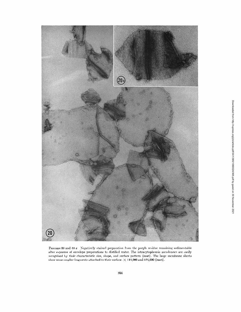
FIGURES 20 and 20 a Negatively stained preparation fom the purple residue remaining sedimentable
after exposure of envelope preparations to distilled water. The intracytoplasmic membranes are easilyrecognized by their characteristic size, shape, and surface pattern (inset). The large membrane sheetsshow some smaller fragments attached to their surface. X 114,000 and 224,000 (inset).
384
Dow
nloaded from http://rupress.org/jcb/article-pdf/34/1/365/1068324/365.pdf by guest on 30 N
ovember 2021
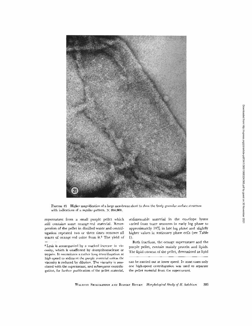
FIGURE 21 Higher magnification of a large membrane sheet to show the finely granular surface structurewith indications of a regular pattern. X 384,000.
supernatant from a small purple pellet which
still contains some orange-red material. Resus-pension of the pellet in distilled water and centrif-
ugation repeated two or three times removes all
traces of orange-red color from it.3 The yield of
3 Lysis is accompanied by a marked increase in vis-cosity, which is unaffected by deoxyribonuclease ortrypsin. It necessitates a rather long centrifugation athigh speed to sediment the purple material unless theviscosity is reduced by dilution. The viscosity is asso-ciated with the supernatant, and subsequent centrifu-gation, for further purification of the pellet material,
sedimentable material in the envelope lysate
varied from trace amounts in early log phase to
approximately 14% in late log phase and slightly
higher values in stationary phase cells (see Table
I).Both fractions, the orange supernatant and the
purple pellet, contain mainly protein and lipids.
The lipid content of the pellet, determined as lipid
can be carried out at lower speed. In most cases onlyone high-speed centrifugation was used to separatethe pellet material from the supernatant.
WALTIER STOECKENIUS A.ND ROBERT ROWEN Morphological Study of H. halobium 385
Dow
nloaded from http://rupress.org/jcb/article-pdf/34/1/365/1068324/365.pdf by guest on 30 N
ovember 2021
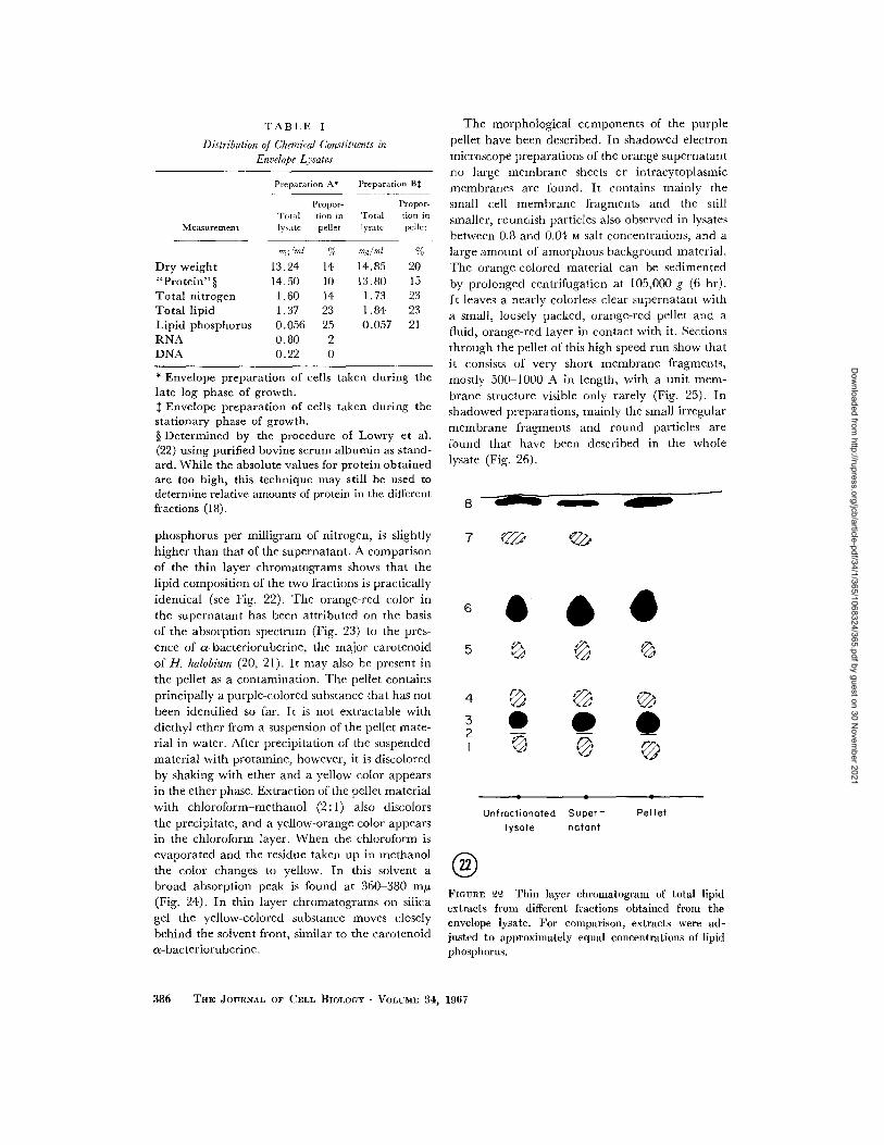
TABLE I
Distribution of Chemical Constituents inEnvelope Lysates
Preparation A- Preparation B
Propor- Propor-Total tion in Total tion in
Measurement lysate pellet lysate pellet
mg ml % mg/ml %
Dry weight 13.24 14 14.85 20"Protein"§ 14.50 10 13.80 15Total nitrogen 1.60 14 1.73 23Total lipid 1.37 23 1.84 23Lipid phosphorus 0.056 25 0.057 21RNA 0.80 2DNA 0.22 0
* Envelope preparation of cells taken during thelate log phase of growth.$ Envelope preparation of cells taken during thestationary phase of growth.§ Determined by the procedure of Lowry et al.(22) using purified bovine serum albumin as stand-ard. While the absolute values for protein obtainedare too high, this technique may still be used todetermine relative amounts of protein in the differentfractions (18).
The morphological components of the purple
pellet have been described. In shadowed electron
microscope preparations of the orange supernatant
no large membrane sheets or intracytoplasmicmembranes are found. It contains mainly the
small cell membrane fragments and the still
smaller, roundish particles also observed in lysates
between 0.8 and 0.04 M salt concentrations, and a
large amount of amorphous background material.
The orange-colored material can be sedimented
by prolonged centrifugation at 105,000 g (6 hr).
It leaves a nearly colorless clear supernatant with
a small, loosely packed, orange-red pellet and afluid, orange-red layer in contact with it. Sections
through the pellet of this high speed run show that
it consists of very short membrane fragments,
mostly 500 1000 A in length, with a unit mem-
brane structure visible only rarely (Fig. 25). Inshadowed preparations, mainly the small irregular
membrane fragments and round particles arefound that have been described in the whole
lysate (Fig. 26).
8 - a
phosphorus per milligram of nitrogen, is slightly
higher than that of the supernatant. A comparisonof the thin layer chromatograms shows that thelipid composition of the two fractions is practicallyidentical (see Fig. 22). The orange-red color inthe supernatant has been attributed on the basisof the absorption spectrum (Fig. 23) to the pres-
ence of a-bacterioruberine, the major carotenoid
of H. halobium (20, 21). It may also be present in
the pellet as a contamination. The pellet contains
principally a purple-colored substance that has not
been identified so far. It is not extractable withdiethyl ether from a suspension of the pellet mate-rial in water. After precipitation of the suspended
material with protamine, however, it is discolored
by shaking with ether and a yellow color appears
in the ether phase. Extraction of the pellet material
with chloroform-methanol (2:1) also discolors
the precipitate, and a yellow-orange color appearsin the chloroform layer. When the chloroform is
evaporated and the residue taken up in methanolthe color changes to yellow. In this solvent abroad absorption peak is found at 360-380 myp(Fig. 24). In thin layer chromatograms on silica
gel the yellow-colored substance moves closelybehind the solvent front, similar to the carotenoida-bacterioruberine.
7 CD C·@
5 0 t
4 C32
1 0 'i
Unfroctionated Super-lysote natant
®Olu
Pellet
FIGunE 22 Thin layer chromatogram of total lipidextracts from different fractions obtained from theenvelope lysate. For comparison, extracts were ad-justed to approximately equal concentrations of lipidphosphorus.
386 THE JOURNAL OF CELL BIOLOGY - VOLUME 34, 1967
Dow
nloaded from http://rupress.org/jcb/article-pdf/34/1/365/1068324/365.pdf by guest on 30 N
ovember 2021
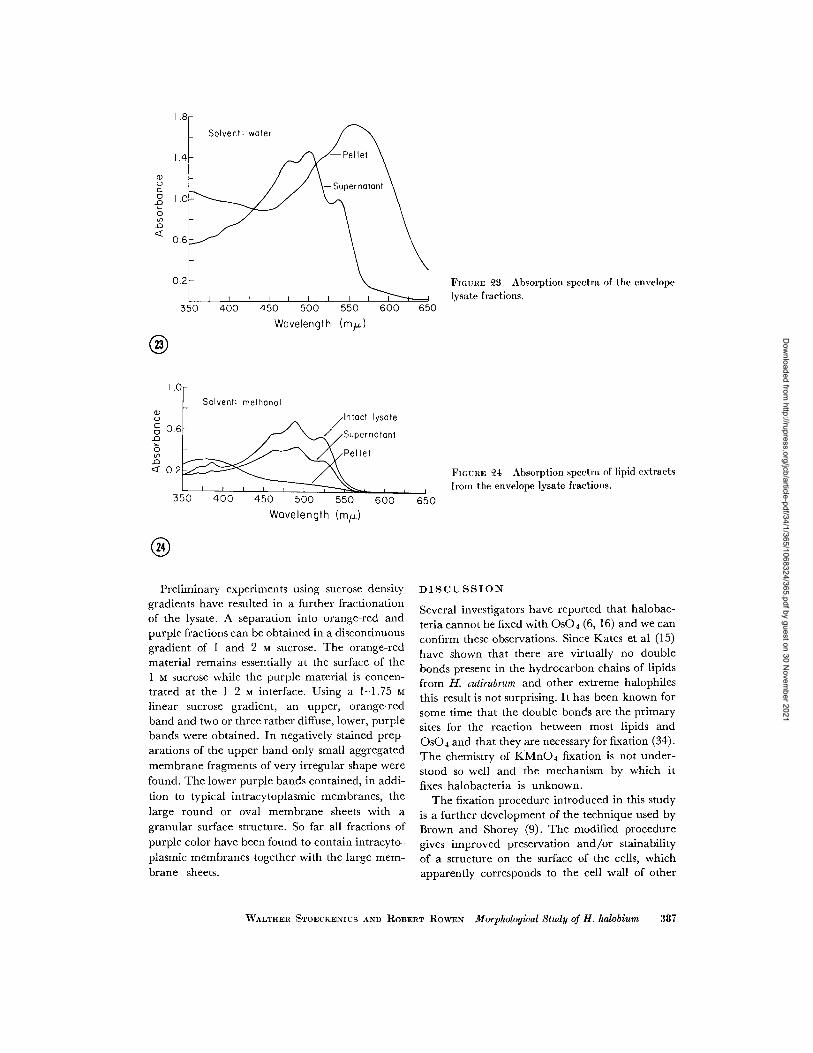
FIGURE 3 Absorption spectra of the envelopelysate fractions.
Wavelength (my.)
0.6
0.2
Solvent: methanol
Intact lysate
/ Supernotont
el let
I I I
350 400 450 500 550 600Wavelength (mt)
Preliminary experiments using sucrose densitygradients have resulted in a further fractionationof the lysate. A separation into orange-red andpurple fractions can be obtained in a discontinuousgradient of I and 2 M sucrose. The orange-redmaterial remains essentially at the surface of theI M sucrose while the purple material is concen-trated at the 1-2 M interface. Using a 1-1.75 Mlinear sucrose gradient, an upper, orange-redband and two or three rather diffuse, lower, purplebands were obtained. In negatively stained prep-arations of the upper band only small aggregatedmembrane fragments of very irregular shape werefound. The lower purple bands contained, in addi-tion to typical intracytoplasmic membranes, thelarge round or oval membrane sheets with agranular surface structure. So far all fractions ofpurple color have been found to contain intracyto-plasmic membranes together with the large mem-brane sheets.
FIGURE 24 Absorption spectra of lipid extractsfrom the envelope lysate fractions.
650
DISCUSSION
Several investigators have reported that halobac-
teria cannot be fixed with OsO4 (6, 16) and we can
confirm these observations. Since Kates et al (15)
have shown that there are virtually no double
bonds present in the hydrocarbon chains of lipids
from H. cutirubrum and other extreme halophiles
this result is not surprising. It has been known for
some time that the double bonds are the primary
sites for the reaction between most lipids and
OsO4 and that they are necessary for fixation (34).
The chemistry of KMnO 4 fixation is not under-
stood so well and the mechanism by which itfixes halobacteria is unknown.
The fixation procedure introduced in this studyis a further development of the technique used byBrown and Shorey (9). The modified proceduregives improved preservation and/or stainabilityof a structure on the surface of the cells, whichapparently corresponds to the cell wall of other
WVALTHER STOECKENIUS AND ROBERT ROWEN Morphological Study of H. halobium 387
a()
C
-oCO.0
CO
o
C)
C
C0
...... i I II I
I nI. r
Dow
nloaded from http://rupress.org/jcb/article-pdf/34/1/365/1068324/365.pdf by guest on 30 N
ovember 2021
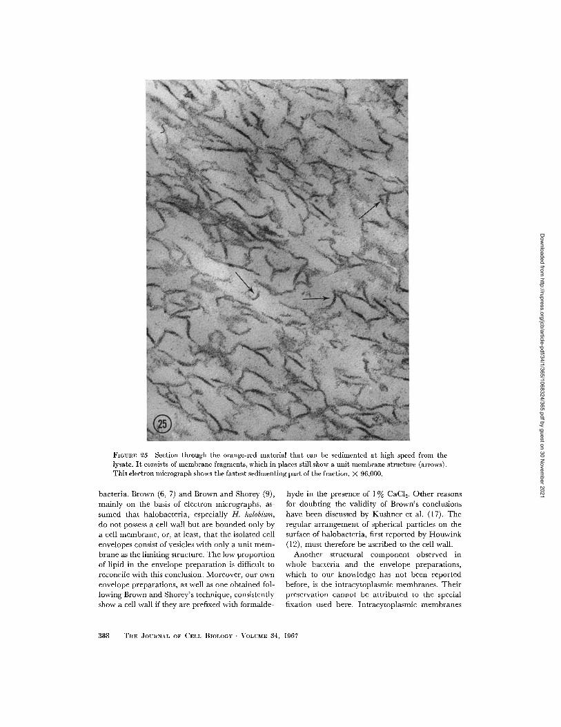
FIGURE 25 Section through the orange-red material that can be sedimented at high speed from thelysate. It consists of membrane fragments, which in places still show a unit membrane structure (arrows).This electron micrograph shows the fastest sedimenting part of the fraction. X 96,000.
bacteria. Brown (6, 7) and Brown and Shorey (9),mainly on the basis of electron micrographs, as-
sumed that halobacteria, especially H. halobium,
do not possess a cell wall but are bounded only bya cell membrane, or, at least, that the isolated cellenvelopes consist of vesicles with only a unit mem-
brane as the limiting structure. The low proportionof lipid in the envelope preparation is difficult toreconcile with this conclusion. Moreover, our ownenvelope preparations, as well as one obtained fol-lowing Brown and Shorey's technique, consistentlyshow a cell wall if they are prefixed with formalde-
hyde in the presence of 1 % CaCI2 . Other reasons
for doubting the validity of Brown's conclusionshave been discussed by Kushner et al. (17). Theregular arrangement of spherical particles on thesurface of halobacteria, first reported by Houwink(12), must therefore be ascribed to the cell wall.
Another structural component observed inwhole bacteria and the envelope preparations,which to our knowledge has not been reportedbefore, is the intracytoplasmic membranes. Theirpreservation cannot be attributed to the specialfixation used here. Intracytoplasmic membranes
388 THE JOURNAL OF CELL BIOLOGY · VOLUME 34, 1967
Dow
nloaded from http://rupress.org/jcb/article-pdf/34/1/365/1068324/365.pdf by guest on 30 N
ovember 2021
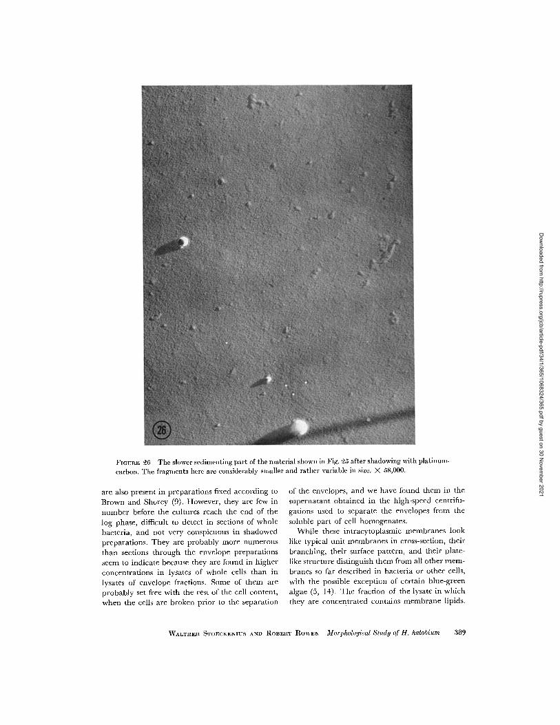
FIGURE 26 The slower sedimcenting part of the material shown in Fig. 25 after shadowing with platinum-
carbon. The fragments here are considerably smaller and rather variable in size. X 58,000.
are also present in preparations fixed according toBrown and Shorey (9). However, they are few innumber before the cultures reach the end of thelog phase, difficult to detect in sections of wholebacteria, and not very conspicuous in shadowedpreparations. They are probably more numerousthan sections through the envelope preparationsseem to indicate because they are found in higherconcentrations in lysates of whole cells than inlysates of envelope fractions. Some of them areprobably set free with the rest of the cell content,when the cells are broken prior to the separation
of the envelopes, and we have found them in thesupernatant obtained in the high-speed centrifu-gations used to separate the envelopes from thesoluble part of cell homogenates.
While these intracytoplasmic membranes looklike typical unit membranes in cross-section, theirbranching, their surface pattern, and their plate-like structure distinguish them from all other mem-branes so far described in bacteria or other cells,with the possible exception of certain blue-greenalgae (5, 14). The fraction of the lysate in whichthey are concentrated contains membrane lipids.
WALTHER STOECKENIUS AND ROBERT RowEN Morphological Study of H. halobium 389
Dow
nloaded from http://rupress.org/jcb/article-pdf/34/1/365/1068324/365.pdf by guest on 30 N
ovember 2021
However, its lipid content cannot be used as anargument for the membrane nature of these struc-tures until fractions of higher purity are obtained.We should emphasize again that the provisionalterm intracytoplasmic membranes is not meantto imply that these structures necessarily have acomposition, fine structure, and function compar-able to those of other cellular membranes. Theirfunction at present is still unknown. They seem tobe related to the formation of gas vacuoles becausethey appear at about the same time and increase innumber with the gas vacuoles. Furthermore, a mu-tant strain of H. halobium isolated by us, which haslost the ability to form gas vacuoles, also lacks theintracytoplasmic membranes. The same holds truefor another strain isolated from Larsen's H. halo-bium strain 5, where the parent strain has gas vac-uoles and intracytoplasmic membranes, and thevariant strain lacks both.4 No reversions to gas vac-uole production have been detected in these strainsso far and therefore the correlation with the pres-ence of intracytoplasmic membranes has not beencarried further. A similar structure has been de-scribed in a short note on the morphology of a blue-green alga (5) which also produces gas vacuoles.The interpretation suggested by the authors, thatthese structures represent collapsed gas vacuoles,cannot be considered definitely proved by theevidence presented, and so far we have no argu-ments to offer for such a role of intracytoplasmicmembranes in halobacteria. In another blue-greenalga, Jost (14) has demonstrated, by means offreeze-etching and negative staining, a structurethat shows the same type of surface pattern as theintracytoplasmic membranes of H. halobium. Al-though the shape of these hohlspindeln is that of acylinder with conical ends, they may very wellhave some relationship to the intracytoplasmicmembranes presently described.
The first visible change in halobacteria, whenthe salt concentration is reduced, is an apparentloss of rigidity and the appearance of irregularlyshaped cells, which on further reduction of the saltconcentration become spherical (1, 2, 24). We have
4 This strain shows a high frequency of loss of gasvacuole formation. Gas-vacuoleless colonies appearclear, whereas the other colonies appear opaque.Many sectored colonies can be observed on plates.We are grateful to Dr. Helge Larsen (Trondheimn,Norway) for drawing our attention to this strain andfor making it available.
not observed any changes in fine structure of theenvelope correlated with these events. The first ob-served change is the loss of the regular surface pat-tern, which occurs at approximately 1.6 M NaClconcentration. The next visible step in the lysis is aloss of cell wall material. It begins at 1.4 M NaCIand appears to be complete at 1.0 M NaCI. At thesame time some of the vesicles open to form sheet-like structures, but the majority are still closed at1.0 M NaCl. Below 1.0 M NaCI more of the vesiclesopen and the cell membranes begin to disintegraterapidly, so that less and less material remains sedi-mentable at 1.2 X 10⁶g min. Qualitatively this se-quence of events is reflected also in the observationson the release of protein and lipid into the super-natant, using the lipid-soluble red carotenoid as anindicator of lipid release (Fig. 13). Quantitatively,one might expect to find a higher proportion of thetotal protein in the supernatant at 1.0 M NaCI thanthe 20-30% actually observed if most of the wallmaterial were released at this salt concentration.However, we do not know how much of the totalprotein of the envelope preparation is soluble pro-tein that is contained inside the vesicles and is onlyreleased when the vesicles break up. Moreover,part of the wall material may still remain attachedto the membrane, or be detached and indistin-guishable from the membrane sheets. This mightexplain the apparent discrepancy.
Perhaps the most interesting result of this studywas the observed heterogeneity of the envelopelysate and the morphology of its components.Brown (6) has reported that he found one majorcomponent in the lysate that sedimented withs20,w _ 4.0 in the analytical ultracentrifuge. He as-sumed this to be a macromolecular constituent ofthe cell membrane. He also obtained a small pelletafter centrifugation of the lysate at 14,500-1 7,000gfor 45 min, and an orange component that sedi-mented ahead of the 4.OS material. Apparentlythese were not further investigated.
Since our studies indicate that Brown's envelopepreparation contained the cell wall in addition tothe cell membrane, a reevaluation of his conclu-sions becomes necessary. Some preliminary dataobtained with the analytical ultracentrifuge t con-firm the presence of the 4.OS component describedby Brown. The orange-red material sediments
I We are grateful to Mr. R. C. Williams, Jr., and Dr.G. M. Edelman for carrying out the ultracentrifugeexperiments.
390 THE JOURNAL OF CELL BIOLOGY VOLUME 34, 1967
Dow
nloaded from http://rupress.org/jcb/article-pdf/34/1/365/1068324/365.pdf by guest on 30 N
ovember 2021
considerably faster, with a very diffuse boundary.Repeated sedimentation of the orange-red materialin the preparative centrifuge before the analyticalrun reduces the 4.OS component to trace amountswithout any apparent loss of orange material. Asdescribed in Results section, the orange-red com-ponent consists of very small, irregular membranefragments. It seems more likely, therefore, thatthe main constituents of the cell membrane arecontained in this fraction. The 4.0S componentmay represent a breakdown product of the cell wall,but further work is required to decide this point.
At least two components have been found in thepurple pellet that can be separated from the lysateat low speed. One, on the basis of its morphology, isreadily identified as the intracytoplasmic mem-branes seen in sections of intact bacteria and un-lysed envelope fractions and has been discussedabove. The origin of the large membrane sheets ismuch less obvious. Electron microscopy of enve-lopes at different salt concentrations seems toindicate that these sheets are unlysed parts of thecell membrane. However, this would imply aheterogeneity of the cell membrane, which we finddifficult to accept. Another possibility is to assumea heterogeneity of the bacterial population, forinstance a structural change in the cell membraneof older cells. This, to us, seems to be a more ac-ceptable explanation, but efforts to fractionate theunlysed cell envelope, in the hope of separating thehypothetical older envelopes from the younger,have so far not yielded any results. The increase inthe mass of nonlysable material with age of theculture which would fit this explanation must beattributed at least partly to the intracytoplasmicmembranes. A third possibility is that the unlysedmembrane sheets constitute a distinct layer of thecell envelope that has not been identified in sec-tions. We feel that so far the electron micrographsof lysing cells do not allow us to exclude this in-terpretation. At present, however, we have noconvincing arguments for or against any of thesepossible explanations. Further work, especiallyseparation and chemical analysis of the two com-ponents found in the pellet, is needed.
The red color of the intact bacteria and of theenvelope preparation is apparently due to at leasttwo different colored substances, which can beseparated with the fractions of the lysate. Theorange-red carotenoid, a-bacterioruberine, is ap-parently bound to the cell membrane, as are the
carotenoids in other bacteria. The purple-coloredsubstance has not been identified but is evidentlybound to the membranous material readily sedi-mentable from the lysate. Of the two identifiedstructural components f this material, the largemembrane sheets are the more likely site, becauselysates of the H. halobium strains which have neithergas vacuoles nor intracytoplasmic membranes(see above) still yield a purple pellet which con-tains membrane sheets.
It has been shown that lysis of halobacteria uponremoval of salt does not depend on enzymic orosmotic processes. It is generally assumed that ahigh charge density on the structural componentsof the organism when not shielded by counterionsleads to a disaggregation (1, 2, 6, 8, 17, 18, 24, 25).Our data are fully compatible with these conclu-sions. They show in addition that a cell wall existsin halobacteria and that cell wall and membranebegin to lyse at different salt concentrations andapparently independently of each other. The proc-ess of lysis is incomplete. Even after prolongeddialysis against distilled water some large mem-brane sheets and the intracytoplasmic membranesare still intact and readily sedimentable. The lysedpart of the cell membrane appears not as a well-defined subunit, but as a mixture of membranefragments of small but variable size. The averagesize of these particles diminishes with diminishingsalt concentrations. However, even in distilledwater membrane structure is still recognizable inthe particles and they are still sedimentable at highspeeds in the preparative ultracentrifuge. They areclearly not subunits and there is no indication thatthey are composed of identical subunits.
This study shows that a thorough characteriza-tion by chemical and physical techniques is neces-sary before the existence and isolation of mem-brane subunits can be claimed. A similar case,
where a supposedly well-defined membrane sub-
unit turned out to be an artifact, has been reported
recently (10, 35).
This work was supported by Public Health ServiceGrant GM 11825 from the National Institute of Gen-eral Medical Sciences.
Receivedfor publication 31 January 1967.
Note Added in Proof: Certain aspects of this work wereinitially presented at the Sixty-Sixth Annual Meetingof The American Society for Microbiology (Rowen,R., and W. Stoeckenius. 1966. Bacteriol. Proc. 107).
WALTHER STOECKENIUS AND IOBERT ROAEX Morphological Study of H. halobiumr 391
Dow
nloaded from http://rupress.org/jcb/article-pdf/34/1/365/1068324/365.pdf by guest on 30 N
ovember 2021
REFERENCES
1. ABRAM, D., and N. E. GIBBONS. 1960. Turbidityof suspensions and morphology of red halo-philic bacteria as influenced by sodium chlo-ride concentration. Can. J. Microbiol. 6:535.
2. ABRAM, D., and N. E. GIBBONS. 1961. The effectof chlorides of monovalent cations, urea, deter-gents, and heat on morphology and the turbid-ity of suspensions of red halophilic bacteria.Can. J. Microbiol. 7:741.
3. BAYLEY, S. T. 1966. Composition of ribosomes ofan extremely halophilic bacterium. J. Mol.Biol. 15:420.
4. BAYLEY, S. T., and D. J. KUSHNER. 1964. Theribosomes of the extremely halophilic bacte-rium, Halobacterium cutirubrum. J. Mol. Biol. 9:654.
5. BOWEN, C. C., and T. E. JENSEN. 1965. Blue-greenalgae: Fine structure of the gas vacuoles. Science.147:1460.
6. BROWN, A. D. 1963. The peripheral structures ofgram-negative bacteria. IV. The cation-sensi-tive dissolution of the cell membrane of thehalophilic bacterium, Halobacterium halobium.Biochim. Biophys. Acta. 75:425.
7. BROWN, A. D. 1964. Aspects of bacterial responseto the ionic environment. Bacteriol. Rev. 28:296.
8. BROWN, A. D. 1965. Hydrogen ion titration of in-tact and dissolved lipoprotein membranes. J.Mol. Biol. 12:491.
9. BROWN, A. D., and C. D. SHOREY. 1963. The cellenvelopes of two extremely halophilic bacteria.J. Cell Biol. 18:681.
10. ENGELMAN, D. M., T. M. TERRY, and H. J.
MOROWITZ. 1967. Characterization of theplasma membrane of Mycoplasma laidlawii. I.Sodium dodecyl-sulfate solubilization. Biochim.Biophys. Acta. In press.
11. FRASCA, J. M., and V. R. PARKS. 1965. A routine
technique for double-staining ultra-thin sec-tions using uranyl and lead salts. J. Cell. Biol.25:157.
12. HOUWINK, A. L. 1956. Flagella, gas vacuoles andcell-wall structure in Halobacterium halobium; anelectron microscope study. J. Gen. Microbiol.15:146.
13. JOSHI, J. G., W. R. GUILD, and P. HANDLER.1963. The presence of two species of DNA inhalobacteria. J. Mol. Biol. 6:34.
14. JOST, M. 1965. Die Ultrastruktur von Oscillatoriarubescens D. C. Arch. Mikrobiol. 50:211.
15. KATES, M., L. S. YENGOYAN, and P. S. SASTRY.
1965. A diether analog of phosphatidyl glycero-phosphate in Halobacterium cutirubrum. Biochim.Biophys. Acta. 98:252.
16. KUSHNER, D. J., and S. T. BAYLEY. 1963. The
effect of pH on surface structure and morphol-ogy of the extreme halophile Halobacteriumcutirubrum. Can. J. Microbiol. 9:53.
17. KUSHNER, D. J., S. T. BAYLEY, J. BORING, M.
KATES, and N. E. GIBBONS. 1964. Morphologi-
cal and chemical properties of cell envelopesof the extreme halophile Halobacterium cutiru-
brum. Can. J. Microbiol. 10:483.18. KUSHNER, D. J., and H. ONISHI. 1966. Contribu-
tion of protein and lipid components to the saltresponse of envelopes of an extremely halo-philic bacterium. J. Bacteriol. 91:653.
19. LARSEN, H. 1962. Halophilism. In The Bacteria.I. C. Gunsalus and R. Y. Stanier, editors. Aca-demic Press, Inc., New York. 4:297.
20. LEDERER, E. 1938. Sur les carotenoides des Cryp-togames. Bull. Soc. Chim. Biol. 20:611.
21. LIAAEN JENSEN, S. 1960. Bacterial carotenoids.
VI. A note on the constitution of bacteriorube-
rine a. Acta Chem. Scand. 14:950.22. LOWRY, O. H., N. J. ROSEBROUGH, A. L. FARR,
and R. J. RANDALL. 1951. Protein measure-ment with the Folin phenol reagent. J. Biol.Chem. 193:265.
23. MEJBAUM, W. 1939. Uber die Bestimmingkleiner Pentosemengen, insbesondere in De-rivaten der Adenylsiure. Z. Physiol. Chem.
258:117.24. MOHR, V., and H. LARSEN. 1963. On the struc-
tural transformations and lysis of Halobacterium
salinarium in hypotonic and isotonic solutions.
J. Gen. Microbiol. 31:267.25. ONISHr, H., and D. J. KUSHNER. 1966. Mecha-
nism of dissolution of envelopes of the extremehalophile Halobacterium cutirubrum. J. Bacteriol.91:646.
26. SEHGAL, S. N., and N. E. GIBBONS. 1960. Effect
of some metal ions on the growth of Halobacte-
rium cutirubrum. Can. J. Microbiol. 6:165.27. SEHGAL, S. N., M. KATES, and N. E. GIBBONS.
1962. Lipids of Halobacterium cutirubrum. Can. J.Biochem. 40:69.
28. SMITHIES, W. R., N. E. GIBBONS, and S. T. BAY-
LEY. 1955. The chemical composition of thecell and cell wall of some halophilic bacteria.
Can. J. Microbiol. 1:605.29. SPERRY, W. M., and F. C. BRAND. 1955. De-
termination of total lipids in blood serum. J.Biol. Chem. 213:69.
30. STEWART, C. P., and E. B. HENDRY. 1935. Thephospholipins of blood. Biochem. J. 29:1683.
31. STOECKENIUS, W. 1959. An electron microscopestudy of myelin figures. J. Biophys. Biochem.Cvtol. 5:491.
32. STOECKENIUS, W. 1962. The molecular structure
392 THE JOURNAL OF CELL BIOLOGY VOLUME 34, 1967
Dow
nloaded from http://rupress.org/jcb/article-pdf/34/1/365/1068324/365.pdf by guest on 30 N
ovember 2021
of lipid-water systems and cell membranemodels studied with the electron microscope.Symp. Intern. Soc. Cell Biol. 1:349.
33. STOECKENIUS, W. 1966. Structural organizationof the mitochondrion. Ciba Found. Symp., Princi-ples Biomol. Organ. 418.
34. STOECKENIUS, W., and S. C. MAHR. 1965. Studies
on the reaction of osmium tetroxide with lipidsand related compounds. Lab. Invest. 14:1196.
35. TERRY, T. M., D. M. ENGELMAN, and H. J.MOROWITZ. 1967. Characterization of theplasma membrane of Mycoplasma laidlawii. II.Modes of aggregation of solubilized membranecomponents. Biochim. Biophys. Acta. In press.
WALTHER STOECKENIUS AND ROBERT ROwEN Morphological Study of H. halobium 393
Dow
nloaded from http://rupress.org/jcb/article-pdf/34/1/365/1068324/365.pdf by guest on 30 N
ovember 2021