Embed Size (px)
Citation preview
A MORPHOLOGICAL AND PHYSIOLOGICAL CLASSIFICATION OF SPIRILLA
by
Jerry Scott Wells, Jr.
Thesis submitted to the Graduate Faculty of the
Virginia Polytechnic Institute
in partial fulfillment of the requirements for the degree of
APPROVED:
Dr. R. E. Benoit
Dr. R. M. Smibert
DOCTOR OF PHILOSOPHY
in
Mfcrobiology
Chai~f~n, Dr.' N. R? Krie&<
Date
April 1970
Dr. R. R. Schmidt
Dr. R. A. Paterson, Head Department of Biology
Blacksburg, Virginia
ACKNOWLEDGEMENTS
The author wishes to express his sincere gratitude
to Dr. N. R. Krieg whose advice, interest, and encour-
agement helped to make this work possible.
The author expresses thanks to Dr. R. A. Paterson,
head of the Department of Biology, for helping to
obtain financial assistance and for the opportunity to
perform this graduate study.
The ~aluable suggestions of Dr. R. M. Smibert and
Dr. R. E. Benoit, are gTatefnlly acknovledged. The
author is indebted to Dr. J. L. Johnson for his advice
on DNA base composition determina tior;.s and especially
for making available his equipment. Thanks are also
due the Department of Veterinary Science for the use
of their facilities.
Appreciation is also extended to Dr. Erwin Lessel,
curator of the American Type C~lture Collection; the
National Collection of Industrial Bacteria; Dr. R. G. E.
Murray; Dr. H. Jannasch; Dr. S. C. Rittenberg; and Dr.
R. M. Smibert for providing me with cultures.
Finally, the help, encouragement, and patience of
my wife are gratefully acknowledged.
ii
TABLE OF CONTfNTS
Page
I. INTRODUCTION
II. REVIEW OF THE LITERATURE
I I I. iLi\ TE RIALS AND METHODS
1
7
26
26 Origins of the Strains
Maintenance of Cultures 28
Media and Chemicals 29
Cc;:1p0siticrn o.f Pcpton.e-Succinate-S a 1 ts ~~~ed. i um . . . .. . . . . . . . . . . . . . . . . 31
Preparation of Inoculum used in Biochemical and Physiological Tests . . . . . . . . . . . . . . . . . . . . . . . . . . . 32
Atmcsp~1eric Conditions 32
EstlmatiJn of Growth Responses 32
Temperature of Incubation 33
Det~rmination of Optimal 33
Biochemical a~d Physiological Methods 33
O.xiclase Test 33
Phosphatase :md SulfBtase Prod1Jction ........................ 34
Deaminase Activity 34
Catalase Test 34
Ure2se Test . . . . . . . " . . . . . . . . . . . . . 36
I-IzS Prodl1ction . . . . . . . . . . . . . . . . . . 36
iii
IV. RESULTS
Casein Hydrolysis
Amylase Activity
Hippurate HyJrolysis
In<lole Production
Aerobic Nitrate and Selenite
Page
36
36
36
37
Reduction . . . . . . . . . . . . . . . . . . . . . . . 37
Gelatin Hydrolysis
Aesculin Hydrolysis
Pigment Production
Nuclease Production
Bile and Glycine Tolerance
Sodium Chloride Tolerance
37
37
37
38
38
38
Growth on EMB and MacConkey Agar 38
Growth on TSI Agar and Seller's Slants . . . . . . . . . . . . . . . . . . . . . . . . . . 39
Reaction in Litmus Milk 39
Acid Reactions from C2rbohydrates 39
Aerobic Growth in the Presence of !~itrate ........................... 39
Methods used for Morphological D~terminations . . . . . . . . . . . . . . . . . . 41
Cultiva~ion of Cells for DNA Isolation . . . . . . . . . . . . . . . . . . . . . . . 41
DNA Isolation 42
Melting Point (Tm) Measurements 43
Cultural and Morphological Tests ' .... 44
44
Relationship to Oxygen 44
iv
v.
Page
Ability to Gro'J Anaerobically in Presence of Nitrate . . . . .. . . . . 44
Te~r.pcrature Range for Growth 45
Colony Characteristics 45
Dimensions of Cells 46
Visibility of Flagellar Tufts by Dark Field . . . . . . . . . . . . . . . . . . . . . . 46
Presence of Coccoid Bodies in Older Cultures ........ ....... ... 49
Physiological Tests 49
Positive Physiological Tests 49
Negative Physiological Tests .5 2
Acid Reactions from Carbohydrates 54
Physiological Tests Giving Varied Responses Among Spi ril 1 a . , • • • • • • 54
Ability of S. lunatum to Grow in PL:)Sh-1"'nT°~' r or:- Sea Water
Deamination and Pigment Production with Aromatic Amino
56
.l\cids . . . . . . . . . . . . . . . . . . . . . . . . . . . 59
Salt Tolerance
tase Composition of DNA
DI.SCUSSION
59
59
63
Limits of Genus a.nd Species Variation 63
The Usefulness of Standardized Conditions in Characterizing Strains of Spirilla . . . . . . . . . . . . . . . . . . . . . . . . . . 65
Discrepancies Arising from a Comparison of the Present Data With Earlier P ... esul~ts ............................... 70
Division of Genus Snirillum on the Basis of Single, Hi~hly-Weighted
Page
Cl1a1~acters . . . . . . . . . . . . . . . . . . . . . . . . . . . 73
Division of Genus Spirillwn on the Basis of Correlated Characters .... ..• 78
Description of Genus Spirillum (Ehrenberg 1830, 38) Based on f.1orphological and Physiological ll es u 1 ts . . . . . . . . . . . . . . . . . . . . . . . . . . . . . . 8 2
Physiological Tests Important for Species Differentiation . . . . . . . . . . . . . . 83
Scheme of Classification of Species · of Genus Spirillum .. ..... ........ ...• 83
§yj_rillum volutm12 (Ehrenberg, 1830) . . . . . . . . . . . . . . . . . . . . . . . . . . . 84
~iri~lum anulus_ (Williams and Rittenberg, 1957) ......... ....... 83
G~_i ri_! lrn~ put ri•li conchyl i um (Tcrasaki, 1961) ... .... .. .. ..... 88
~l'?J-·~ i l_:l_~i~ ;{t_an if e rum ('i\' i 11 i ams and Rittenberg, 1957) ........... 88
Sp~rillu1!.!_ matamorphum (Terasaki, 1961) . . . . . . . . . . . . . . . . . . . . . . . . . . . 89
Spir_illum giosb~_!__g_~ri (Williams and Rittenberg, 1957) ........... 89
~irilh.~m peregrinum (Pretorius, 1963) . . . . . . . . . . . . . . . . . . . . . . . . . . . 90
Spiril~_um i tersonii (Giesberger, 1936) . . . . . . . . . . . . . . . . . . . . . . . . . . . 91
Spirill~im ~er2_en~ Olilller, Winter 1884) .. .. . . .. . . .. . .. . .. • • 92
~~2..t_l l_l}m s inuos um (Wi 11 iams and Rittenberg, 1957) .... ....... 93
§uirillum gracile (Canale-Parola, Rosenthal and Kupfer, 1966) . . . . . 94
vi
VI.
VII.
VIII.
Page
-~Pi..!:} ~:l_um Q9}ZJ.;:_c?_!.flh~g_ (Wi 11 i ams and ,~_1ttenocrg, L951; • • . • • • • • • • • 96
~£ ~ _r ~) 1 UE! ~~ 1 :i-_ _<_:~_~_2Jm (Leif son , 19621 ............................ 96
~pirillun~ lunat_~m (Williams and Rittenberg, 1957"J .. .... .. .. . .. . . 98
:SJ?irillum_ li_null! (Williams and Rittenberg, 1957) . .. . . . .. ..... .. 98
Spirillum beijerinckii (Williams and Rittenberg~ 1957) . . . . . . . . . • . 99
Spirillu~ atlanticu~ (Williams and Rittenberg, 1957) . . . . . . . . . • . 99
§_pir~llum ~onicum (Watanabe, 1959) ........................... 99
Spi_rillum gal.£E.!:tilum (Watanabe, 19 5 9) t t t t t I t t t t t <t t t t t t 6 t t t t • t t t t 10 0
Spirillu:n minutulum (i'fatanabe, 1959) . ',~··-:-:-. ~:............... 100
Un-named Strains of SpLcilla 101
Diagnostic Key to the Species of Genus Spirillum • .. .. .. .. .. . .. .. .. . • .. 103
Key to the Species of Genus Spirillurn . 103
SUMMARY .................................. LI'fERATURE CITED .........................
108
109
113 VITA
IX. ABSTRACT
vii

LIST OF TABLES
Page
TABLE 1. Watanabe's Enrichment and Isolation f.Iedia ............. , ................. 21
TABLE 2. Protocol and Reagents for Microdiffusion Assay of Deaminase Activity . . . . . . . . . . . . . . . . . . . . . . . . . . . 35
TABLE ·.z: ..., . Compounds Employed for Determination of Acid Production from Carbohydrates 40
TABLE 4. Cell Dimensions of Fresh Water Strains of Spirilla .. .............. 47
TABLE s . Cell Dimensions of Marine Strains of Spi1 .. illa ........................... 48
TABLE 6. Positive Physiological Reactions of S1)irilla . . . . . . . . . . . . . . . . . . . . . . . . . . . SO
TABLE 7. Dcaminase Activity of Spirilla as Determined by Microdiffusion A i1 a 1 y s i s . . . . . . . , . . . . . . . . . . . . . . . . . . . 5 1
TABLE 8. Negative Physiological Reactions of .Spirilla . . . . . . . . . . . . . . . . . . . . . . . . . . . 53
TABLE 9. Spirilla Producing Acid from Carbohydrates ...................... 55
TA3LE 10. Physiological Tests Yielding Varied Results Among Fresh Water Strains of (:'. . ·11 ~ d" d uplrJ. a vtU le •••••••••••••••••••
TABLE 11. Physiological Tests Yielding Varied Results Among Marine Straij1s of Spirilla Sttidied ....•.............•
TABLE 12. Formation of Nater Soluble Pigments in Presence of Aromatic Amino Acids
viii
57
58
60

TABLE 13. Spirilla Possessing l!::JA Base Cor:tpos i ti on Va1u"~s from 3 8 to 5 2
Page
~Joles ~ G + C . , . , ............. , . . . . 61
TABLE 14. Spirilla Possessing DNA Base Composition Values from 57 to 65 ~·to 1 es % G + C ..•...•....... , .•. , . , • 6 2
ix
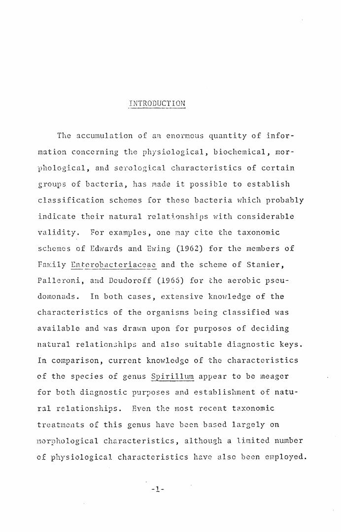
INTRODUCTION
The accumulation of an enormous quantity of infor-
mation concerning t]1e physiological, biochemical, mar-
i_)hological, and se·1'ological characteristics of certain
groups of bacteria, has made it possjble to establish
classification schemes for these bacteria l'lhich probably
indicate their natural relationships with considerable
validity. For examples, one ~ay cite the taxonomic
schemes of Edwards and faring (1962) for the members of
fan:ily En!~ __ ropacteriace§_~ and the scheme of Stanier,
Palleroni, and Dcudorcff (1965) for the aerobic pseu-
domona<ls. In both cases, extensive knowledge of the
characteri.stics of the organisms being classified was
available and was drm,_;n upon for purposes of deciding
natural relationships and also suitable diagnostic keys.
In comparison, current knowledge of the characteristics
of the species of genus ~_pirillum appear to be meager
for both diagnostic purposes and establishment of natu-
ral relationships. Even the most recent taxonomic
treatments of this genus have been ba5ed largely on
1norphological characteristics, although a limited number
of physiological characteristics have also been employed.
-1-
-2-
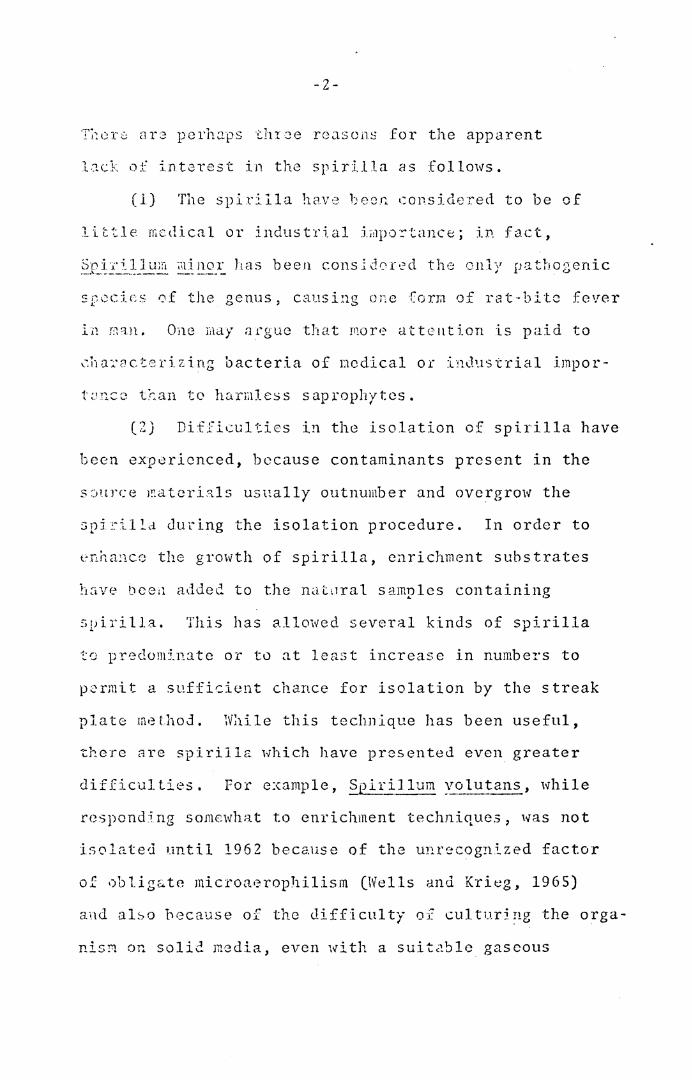
T1~1::r0 <H3 perha.ps thr~e rc;asons for the apparent
l~ck of intaTest in the spirilla as follows.
(l) The spirilla have been considered to be of
little medical or industrial impo~tance; in fact,
Sn:i.·;:·1.11um illinor has been consldor·:d the onlv pathogenic ·-·>-·---------- -- --- - I .
c:-'"l-.:e<.: -i:~\; ... t ....... of the genus, causing one forra of rat-bite fever
One may n1·guc that 1110re attention is paid to
~ha~acterizing bacteria of ncdical or industrial impor-
tQncc t~an to harmless saprophytes. ( .., .,
'-') Difficulties in the isolation of spirilla have
been experienced, because contaminants present in the
s :.:iurce l!ld tcrials usually outnumber and overgrow the
spi~illa during the isolation procedure. In order to
enhance the growth of spirilla, enrichment substrates
h~ive bcei1 added. to the nat.1ral samples containing
svirilla. This has allowed several kinds of spirilla
t0 predominate or to at least increase in numbers to
p~rmit a sufficient chance for isolation by the streak
plate method. Wl1ile this technique has been useful,
there are spirilla which have presented even greater
difficulties. For example, .§_pirillum Y-_olutans, while
responding somewhat to enrichment techniques, was not
isolated until 1962 because of the unrecog21ized factor
of oblig&te microa•3rophilism (Wells and Krieg, 1965)
and al~o because of the difficulty of culturi~g the orga-
nis~ on soli~ media, even with a suitable gaseous
-3-
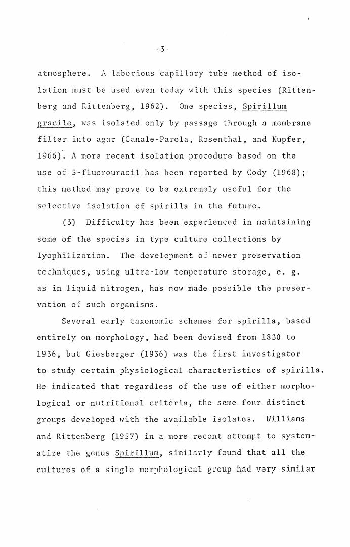
atmosphere. A laborious capillary tube method of iso-
lation ~1st be used even today with this species (Ritten-
berg and Rittcnberg, 1962). One species, Spirillum
gracile, was isolated only by passage through a membrane
filter into agar (Canale-Parola, Rosenthal, and Kupfer,
1966). A more recent isolation procedure based on the
use of 5-fluorouracil has been reported by Cody (1968);
this method may prove to be extremely useful for the
selective isolation of spirilla in the future.
(3) Difficulty has been experienced in maintaining
some of the species in type culture collections by
lyophiliza~ion. The dcvelcpment of newer preservation
techniques, using ultra-low temperature storage, e. g.
as in liquid nitrogen, has now made possible the preser-
vation of such organisms.
Several early taxonomic schemes for spirilla, based
entirely on morphology, had been devised from 1830 to
1936, but Giesberger (1936) was the fiYst investigator
to study c~rtain physiological characteristics of spirilla.
He indicated that regardless of the use of either morpho-
logical or nutritional criteria, the same fonr distinct
groups developed with the available isolates. Williams
and Rittcnberg (1957) in a more recent attempt to system-. ,, r • ']l at1ze cne genus ?p1r1. um, similarly found that all the
cultures of a single morphological grcup had very similar
-4-
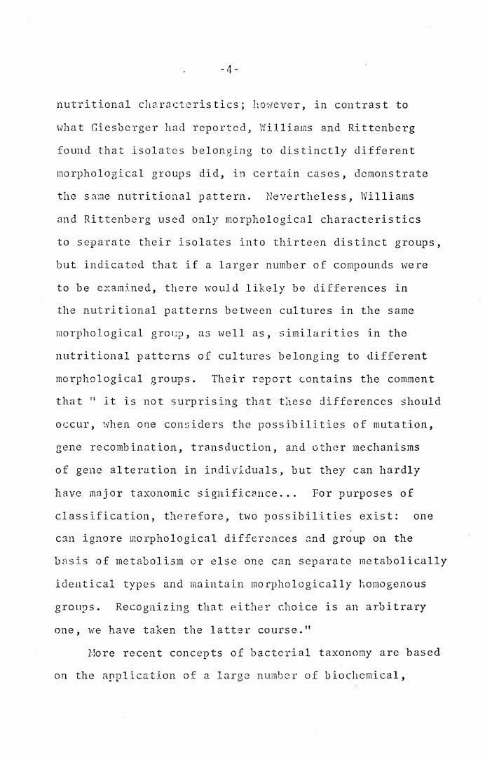
nutritional ch2.rJ.cteristics; however, in contrast to
Hhat Giesberger ha<l reported, Williams and Rittcnberg
found that isolates belonging to distinctly different
morphological groups did, in certain cases, demonstrate
the same nutritional pattern. Nevertheless, Williams
and Rittenberg used only morphological characteristics
to separate their isolates into thirteen distinct groups,
but indicated that if a larger number of compounds were
to be examined, there would likely be differences in
the nutritional patterns between cultures in the same
morphological groujJ, as well as, similarities in the
nutritional patterns of cultures belonging to different
morphological groups. Their repoi~t contains the comment
that 11 lt is not surprising that tl1ese differences should
occur, when one considers the possibilities of mutation,
gene recombination, transduction, and other mechanisms
of gene alteration in individuals, but they can hardly
have major taxonomic signific?nce ... For purposes of
classification, therefore, two possibilities exist: one
can ignore morphological dif fcrcnces a.nd group on the
basis of metabolism or else one can separate metabolically
identical types and maintain morphologically hornogenous
groups. Recognizing that either choice is an arbitrary
one, we have taken the latter course."
More recent concepts of bacterial taxonomy are based
on the application of a large number of biochemical,
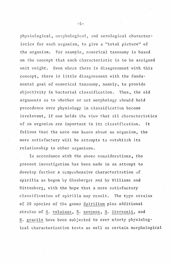
-5-
physiological, raorphological> and serological character-
istics for each 01·ganism, to give a "total picture" of
the organism. For example, numerical taxonomy is based
on the concept that each characteristic is to be assigned
unit weight. Even whe:te there is disagreement with this
concept, there is little disagreement with the funda-
mental goal of numerical taxonomy, namely, to provide
objectivity in bacterial classification. Thus, the old
arguments as to whether or not morphology should hold
precedence over physiology in classification become
irrelevant, if one holds the view that all characteristics
of an organism are important in its classification. It
follows that the more one knows about an organism, the
more satisfactory will be attempts to establish its
relationship to other organisms.
I~ accordance with the above considerations, the
present investigation has been made in an attempt to
develop further a ccmpr·ehensive characterization uf
spirilla as begun by Giesberger and by Williams and
Rittenbcrg, with the hope that a more satisfactory
classification of spirilla may result. The type strains
of 20 species of the genus Spirillum plus additional
strains of S. vol utans, ~. se_?])~~1s, S. i ters_onii, and
S. gracile have been subjected to oveT ninety physiolog-- "--·-- .
ical characterization tests as well as certain morphological
-6-
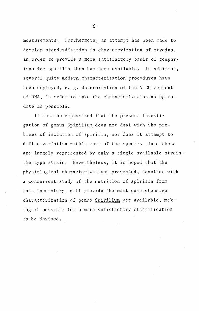
measurements. Furthermore, an attempt has been made to
develop standardization in characterization of strains,
in order to provide a more satisfactory basis of compar-
ison for spirilla than has been available. In addition,
several quite modern characterization procedures have
been employed, e. g. determination of the % GC content
of DNA, in order to make the characterization as up-to·-
date as possible.
It raust be emphasized that the present investi-
gation of genus Spirillum does not deal with the pro-
blems of isolation of spirilla, nor docs it attempt to
define variation within most of the species since these
are largely re?resented by only a single available strain--
the type strain. Nevertheless, it is hoped that the
physiological characteriza~ions presented, together with
a concurr~nt study of the nutrition of spirilla from
this laboratory, will provide the most comprehensive
characterization of genus Spirillum yet available, mak-
ing it possible for a more satisfactory classification
to be devised.
REVIEW OF THE LITERATURE -----·----
The early taxonomic studies of the genus ~irillum
Here entirely morphological and the early taxonomic
history of this genus has been discussed in depth by
Williams and Rittenberg (1957) and Williams (1959).
Although mention will be made of some of the early work,
the present review will be principally concerned with
the work of Vv'ill ianis and Ri t tcnberg (19 5 7) and some of
the more r0ce11t investigations of members of this genus.
The g·~nus Kas originally publishe:C. by Ehrenberg (1830)
1._iith the dl3.gn.osis, "corpora filiforrne rigido spirali",
and contained two species, namely:
(l)
(2)
S-oirillu!T' volut2ns Ehrenberg, originally YJ.~~~l_SJ_ ~J?i:!i~f~~l!_ Milllcr.
Sj)~_ril lum undula (Milller, 1773), originally v i_bno tmd u la.--
Stiles (1905) designated ~· yolutans as the type
species. In his treatment h::; stated: 11 Ju<lged from the
z.oological point of view, YJ.brio ~J?.i!:.L1:..:!:_um is the type
species by absolute tautonomy, but the zoological name
§J~i_ri~l_~un, 1830, was a stillborn homonym, having been
used by Oken, 1815, for a genus of polychaetc worms.
It is not the function of the zoological code to deter-
mine the names in bacteriology, but it is undoubtedly
-7-
-8-
not the most wise or farseeing policy for either zoo-
logists or botanists (including bacteriologists) to
accept unnecessarily a generic name for organisms so
near the border line when that name is a homonym in
either zoology or botany; should further investigations
again bring bacteriology into zoology, the generic
names would come under the zoological code, and in this
case -~pirillum, 1830, would be rejected. If Spirillum
is retained in bacteriology, consistency calls for the
rejection of the name ~:!:_llum volutans Ehrenberg in
favor Of ~irillUffi -~Pi_!:j l_lUTil o,m11er), II
Vuillemin (1913) in a revision of the genera of
bacteria retained the genus Spirillu~ as a genus
conservandum. S. u11dula was considered the type species
by Vuillernin.
The descriptions of the species of Spirillum pub-
lishcd prior to 1936 are, on the whole, incomplete and
inadequate for determiLation purposes. According to
Williams (1959) Giesberger eliminated the majority of
the species as being species dubia. Only those species
were retained whose morphological snd pJ1ysiological
characteTistics were adequately described or whose mor-
phological characteristics were so distinctive that
identification was possible on this basis o.lone, such
as S. volut~ns. Gicsbergcr recognized nine species:
-9 ..
cardionyroaenes. _______ ..L ___ I;? __ _
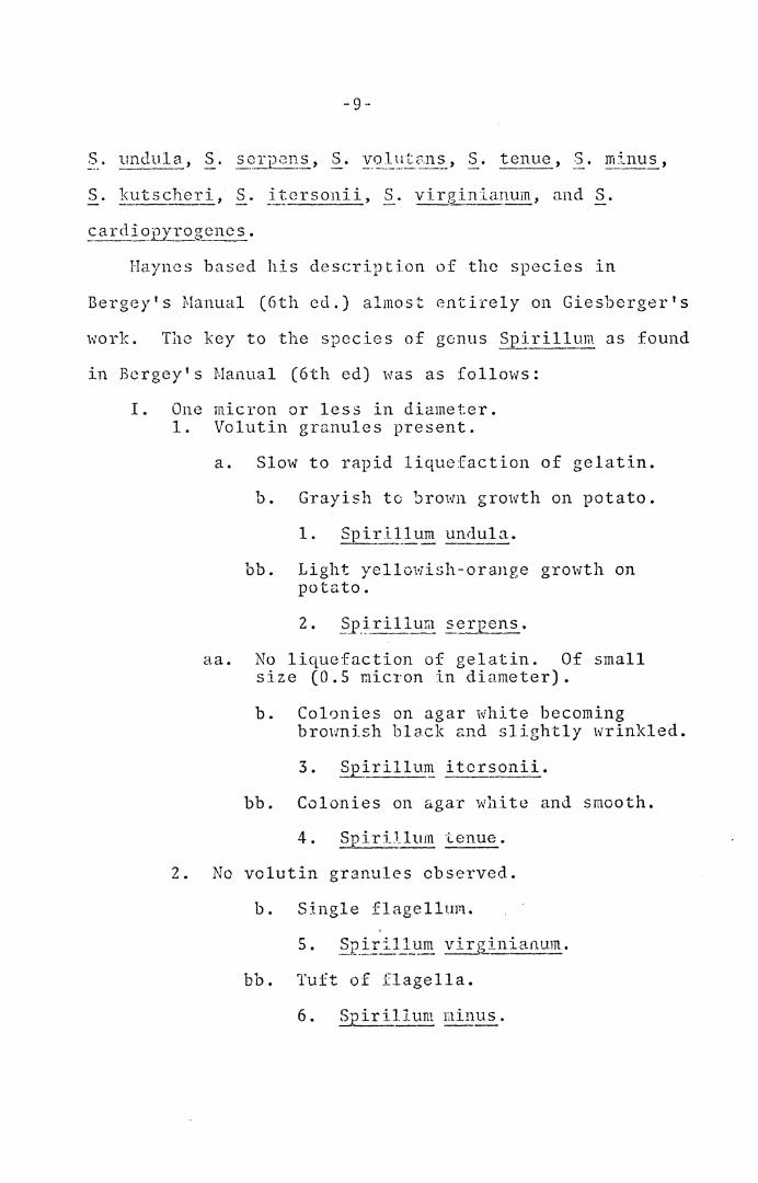
Haynes based his description of tho species in
Bergey's Manual (6th ed.) almost entirely on Giesberger's
work. The key to the species of genus _?pirillum as found
in Bcrgey's Manual (6th ed) was as follows:
I. One micron or less in diameter. 1. Volutin granules present.
a. Slow to rapid liquefaction of gelatin.
b. Grayish to bro1~1 growth on potato.
bb. Light yellowish-orru1ge growth on potato.
aa. No liquefaction of gelatin. Of small size (O.S micron in diameter).
b. Colonies on agar white becoming brownish black and slightly wrinkled.
3. Suirillum itcrsonii.
bb. Colonies on ~gar white and smooth.
4. Spirillum tenue.
2. No vclutin granules observed.
b. Single flagellum.
bb. Tuft of flagella.
-10-
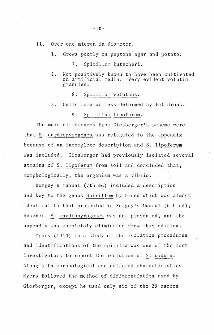
II. Over one micron in diameter.
1. Grows poorly on peptone agar and potato.
7. Spirillum kutscheri.
2. Not positively known to have been cultivated on artificial media. Very evident volutin granules.
8. Spirillum volutans.
3. Cells more or less deformed by fat drops.
9. Spirillum lipoferum.
The main differences from Giesberger's scheme were
that S. cardio.EI_rogenes was relegated to the appendix
because of an incomplete description and S. Ii12oferum
was included. Giesberger had previously isolated several
strains of S. lipoferum from soil and concluded that,
morphologically, the organism was a vibrio.
Bergey's Manual (7th ed) included a description
and key to the genus Spirillum by Breed which was almost
identical to that presented in Bergcy's Manual (6th ed);
however, S. cardiopyrogencs was not presented, and the
appendix uas completely eliminated froii1 this edition.
Myers (1940) in a study of the isolation procedures
and identifications of the spirilla was one of the last
investigators to report the isol&tion of S. undula.
Along with morphological and cultured characteristics
Myers followed the method of differentiation used by
Giesberger, except he used only six of the 29 carbon
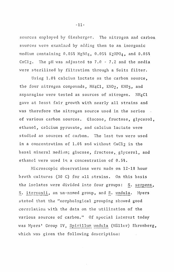
-11-
sources employed by Giesberger. The nitrogen and carbon
sources wer8 examined by adding them to an inorganic
medium containing 0,05% MgS04, 0.05% KzHP04, and 0.05%
CaClz. The pH was adjusted to 7.0 - 7.2 and the media
were sterilized by filtration through a Seitz filter.
Using 1.0% calcium lactate as the carbon source,
the four nitrogen compounds, NH4Cl, KNOz, KN03, and
asparagine were tested as sources of nitrogen. NH4Cl
gave at least fair growth with nearly all strains and
was therefore the nitrogen source used in the series
of various carbon sources, Glucose, fructose, glycerol,
ethanol, calcium pyruvate, and calcium lactate were
studied as sources of carbon. The last two were used
in a concentration of 1,0% and without CaClz in the
basal mineral medium; glucose, fructose,_ glycerol, and
ethanol were used in a concentration of 0.5%.
Microscopic observations were made on 12-18 hour
broth cultures (30 C) for all strains. On this basis
tha isolates were divided into four groups: S. serpens,
-~-· itc~sonii, an un-named group, and§_. undula. Myers
stc:.ted that the "morphological grouping shmved good
correlation ~ith the data on the utilization of the
various sources of carbon." Of special ii1teres t today
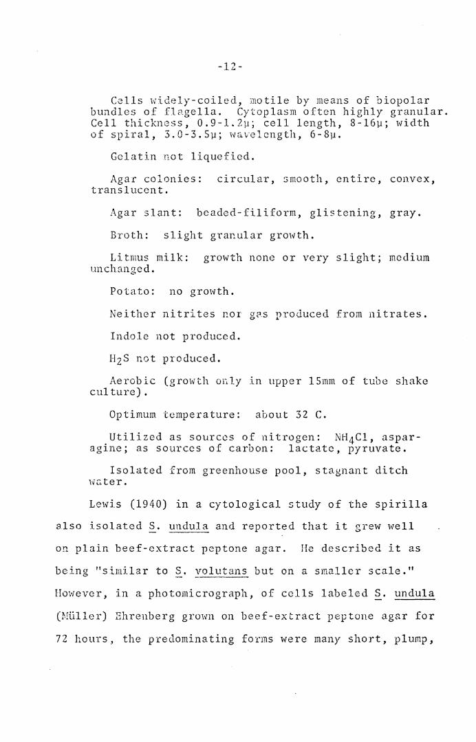
was Myers' Group IV> ~1?ir:i.llum undula (P.H.lller) Ehrenberg,
which ~vas given the following des criptio11:
-12-
Cells widely-coiled, motile by means of biopolar bundles of fl<-1.gella. Cytoplasm often highly granular. Cell thickness, 0.9-1.2µ; cell length, 8-16µ; width o f s p i r a 1 , 3 . 0 - 3 . 5 µ ; iv a. v e 1 c n gt h , 6 - 8 µ •
Gelatin not liquefied.
Agar colonies: circular, smooth, entire, convex, translucent.
Agar slant: beaded-filiform, glistening, gray.
Broth: slight granular growth.
Litmus milk: growth none or very slight; medium unchanged.
Potato: no growth.
Neither nitrites nor g~s produced from nitrates.
Indole not produced.
HzS not produced.
Aerobic (growth or .. ly in upper lSmm of tube shake culture).
Optimum temperature: about 32 C.
Utilized as sources of nitrogen: NH 4Cl, aspar-agine; as sources of carbon: lactate, pyruvate.
Isolated from greenhouse pool, stagnant ditch we, ter.
Lewis (1940) in a cytological study of the spirilla
also isolated ~· undula and reported that it grew well
on plain beef-extract peptone agar. He described it as
being "similar to §_. volutans but on a smaller scale."
However, in a photomicrograph, of cells labeled S. undula
(Milller) Ehrenberg grown on beef-extract peptone agar for
72 hours.• the predominating forms were many short, pl ump,
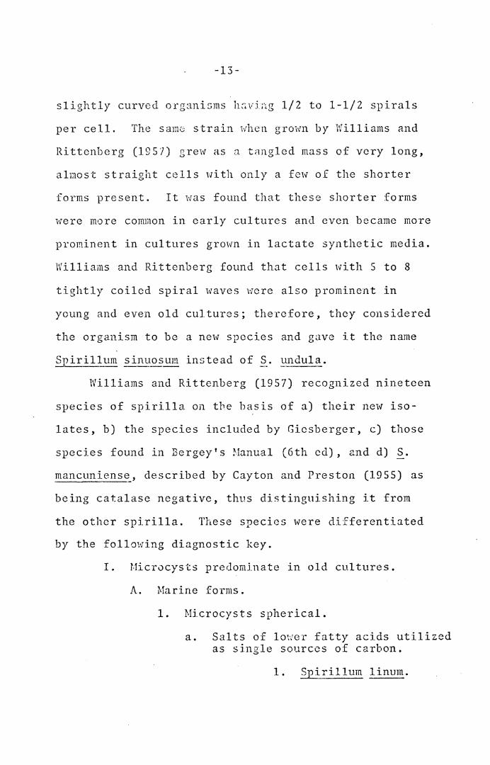
-13-
slightly curved organisms h~vjng 1/2 to 1-1/2 spirals
per cell. The sam0 strain when grown by Williams and
Rittenberg (1957) grew as n tangled mass of very long,
almost straight cells with only a few of the shorter
forms present. It was found that these shorter forms
were more conwon in early cultures and even became more
prominent in cultures grown in lactate synthetic media.
Williams and Rittenberg found that cells with 5 to 8
tightly coiled spiral waves were also prominent in
young and even old cultures; therefore, they considered
the organism to be a new species and gave it the name
Spirillum sinuosum instead of S. undula.
Williams and Rittenberg (1957) recognized nineteen
species of spirilla on the basis of a) their new iso-
lates, b) the species included by Gicsberger, c) those
species found in Bergey's Manual (6th cd), and d) S.
mancuniens~, described by Cayton and Preston (1955) as
being catalase negative, thus distinguishing it from
the other spirilla. These species were differentiated
by the following diagnostic key.
I. Microcysts predominate in old cultures.
A. Marine forms.
1. Microcysts spherical.
a. Salts of lover fatty acids utilized as single sources of carbon.
1. Spirillum linum.
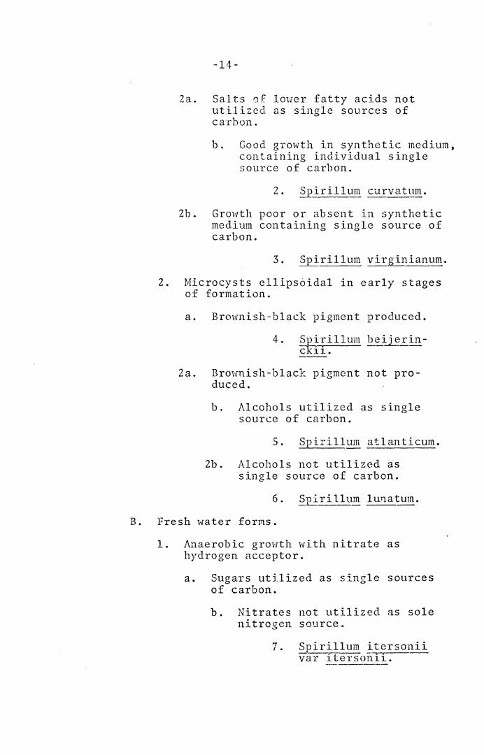
-14-
Za. Salts of lower fatty acids not utilized as single sources of ca-rhon.
b. Good growth in synthetic medium, containing individual single source of carbon.
2. Spirillum curvatnm.
2b. Growth poor or absent in synthetic medium containing single source of carbon.
3. Spirillum virginianum.
2. Microcysts ellipsciidal in early stages of formation.
a. Brownish-black pigment produced.
4. Spirillum beijeE_in-Cl<i1.
2a. Brownish-black pigment not pro-duced.
b. Alcohols utilized as single source of carbon.
5. Spirillum atlanticum.
2b. Alcohols not utilized as single source of carbon.
6. Spirillum lunatum.
B. Fresh water forms.
1. Anaerobic growth with nitrate as hydrogen acceptor.
a. Sugars utilized as single sources of carbon.
b. Nitrates not utilized as sole nitrogen source.
7. ~ir_~llum ttersonii var iterson1i.
-15-
2b. Nitrat0s utilized as sole nitro-gen source.
7a. Suirillum itersonii v~r vulgatum.
2a. Sugars not utilized as single sources of carbon.
8. Spirillum polymorE.!_rnm.
2. No anaerobic growth with nitrate.
9. Spirillum mancuni-cnse.
II. Vegetative cells predominate in old cultures.
A. Good growth on synthetic medium contain-ing single source of carbon.
1. Cell diameter greater than one micron.
a. Single flagellum at each pole.
10. Spi:E_illum anulus.
2a. Tu~ts of flagella at each pole.
11. Spirillum giesbergeri.
2. Cell diameter less than one micron.
a. Gelatin liquefied.
b. Alcohols utilized as single sources of carbon.
12. Spirillum undula.
2b. Alcohols not utilized as single sources of carbon (Nitrates not utilized as sole nitrogen source).
13. Sp_iril~~m serpens var serpens.
2a. Gelatin not liquefied (Nitrates utilized as sole nitrogen source).
-16-
13a. Spirillum serpens var azotum-:--·--
b. Sugars utilized as single carbon sou.cce.
14. Spiril_lum tenue.
2b. Sugars not utilized as single carbon source.
c. Long, multiwaved cells.
2c. Short, S-shaped cells.
16. Spirillurn R.!aniferum.
B. Growth poor or absent in synthetic medium containing single c~rbon source.
1. Isolated in pure culture: growth on enriched pep tone media.
17. ~?irillum kutscheri. ,, L, • Not isolated in pure culture.
a. Large water forms.
18. Spirillum volutans.
2a. Animal pathogen.
19. Spirill~m ~inus.
With regard to a classification scheme Williams
and Rittenbcrg separated their cultures into thirteen
distinct groups based primarily on morphology. For
example, distinction was made between S. _!'Il~!.!us and S.
v.iesben;eri on the basis that the latter, al though ~---.Y.---
similar to ~· anulu_2. metabolically, differed morpho-
logically in that typical cells had two or three wide,
- 1 'J -
regular curves j_n st c~1d of the hal fmoon or S- shaped cells
of S, ~E_nlus; moreover, the d.i.0.1neter was less, the spiral
depth 1\'as much shallower, and the_ granules more prominent.
S, a-ryu!_~ was also distingusihed from-~· _giesbergeri because
it possessed a single polar flagellum. Electron microscopy
has since shown that both species have tufts of polar fla-
gella (Williams, 1960). Another example concerns the dis-
tinction between S. linum and S, lunatum. The latter had
a much larger cell diameter, looser and more irregular
spirals, oval rather than spherical microcysts and did
not grow on hexoses as a sole source of carbon. Here
again, a·distinction based on single vs. multiple polar
flagella was proven to be incorrect (Williams, 1960).
The organisms were further differentiated on their
requirement for sea \·rnter 01· fresh water; the presence or
absence of microcysts in old cultures; whether they grew
on unsupplemented peptone media or required the addition
of yeast extract or casein hydrolysate; physiological tests
such as gelatin liquefaction, nitrate reduction and cata-
lasc production; and the testing of 22 carbon compounds and
4 nitrogen compounds as sole sources of carbon and nitrogen.
As a result of analysis of their isolates, Williams
and Rittenbcrg recognized 10 new species which they
descrlbed and named: s. linum, S. curvatum, s. bei ... - --· --- ~ ----..--.-·- ·- ---
-18-
and S. sinuosum.
In addition to these, they also described two varieties
var _azotuTI_!_, the latter was clistiaguished from S. serpcns
var ~--~..!r~ in its inability to liquefy gelatin. Also
two varieties of S. itersonii were created, ~· itersonii
var itersonii and S. itersonii var vulgatum. Distinction
was made on the basis of the ability of ~· itersoni~ var
vulgatum to utilize nitrates as the sole source of nitro-
gen.
The species S. _!:i.!lu~, S. :~..:'.:l_natum_, ~· curvatum, S.
beijerinckii, and S. atlanticum were the only organisms
isolated from marine sources. These strains grew in
media prepared with either natural or synthetic sea
water however la~ger inocula were required to obtain
growth in the synthetic sea water media as compared to
natural sea: hater. Continued subculture :~n the synthetic
sea water medium eventually resulted in failure of the
organisms to grow on transfer.
Spherical or oval coccoid bcdie3 which predominated
in older cultures of some of the spirj.lla have been
termed "microcysts" by Williams and Rittcnoerg (1956).
They demonstrated that microcyst formation occurred from
helical cells of ~- lunatum by (1) fusion of two entwined
organisms to form one or more microcysts; (2) the pro-
ducticn of a protuberance at some point al.ong the cell
-19-
into which the entire organism was gradually absorbed;
or (3) the gradual shortening <"J.nd rounding of the organ-
ism to form an ov0l to spherical body. h'hen an old
culture of ~· lu~_~turn, completely in the microcyst stage,
was inoculated into fresh media, the microcysts germin-
ated to form a helical cell. Germination occurred by
either unipolar or bipolar emergence of the germ tube.
Williams (1957) demonstrated that fusion of
spirilla also occurred in nonmicrocyst-forming species.
After the fusion process, the fusion cells enlarged
into giant cells, several times the diameter of the
ordinary vegetative cell; a possible sexual mechanism
was suggested.
Although~· serpens was characterized as not produc-
ing microcysts in old cult~res (Williams and Rittenberg,
1957), Bisset and IIale-McCaughey (1967) isolated a strain
of ~· s~_rpens that produced micro cysts. When examined
by electron microscopy, the cyst wall appeared to be
double, and continuous with the double wall of the
spirillum. According to Bisset and Hale-Mccaughey the
development of the cyst represented a true reproductive
process, and resembled the process in Azotobacter.
Mitornycin C, and ultraviolet light were found by
Clark~Walker (1969) to induce the formation of microcysts
in S. iters~tiii. These spherical cells contained phage
-20-
tail parts, rhapidosomes, anJ a granular substance not
seen in normal cells. Spontaneously occurring microcysts
rarely contained phage tails or granular substance; how-
ever, many of these microcysts contained rhapidosomes.
It was suggested that the spherical cells were formed
as the result of the induction of a defective phage,
with the production of phage lysozyme within the cell
leading to the formation of spherical forms as the cells
lose their structural mucopcptide layer.
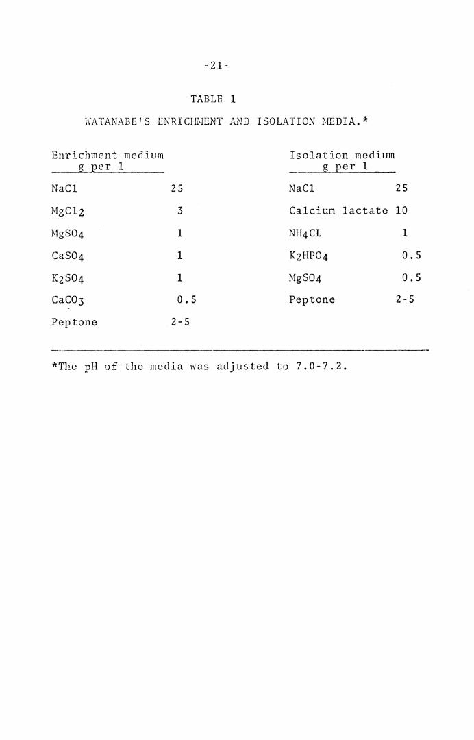
In 1959, Watanabe isolated irom marine shell-fish
the new species ~· j aponic~~!!_, S. halophilum, ~· minut1:1lum,
and S. maritimum. In order to isolate the spirilla
Watanabe devised two types of media (Table 1), one for
enric~nent and one for isolation. Descriptions of the
four species were based on morphological, cultural, and
physiological characteristics. The four species 1vere
reported to be catalase negative. These species resembled
some of the fresh water species from a morphological point
of vieH; for example, ~-· halophilum resembled ~· i t~rs~mii.
However, unlike the fresh 1·rnte-r species all four of these
species required the addition of 2 to 2.5% NaCl to the
medium on isolation and repeated subculture.
Isolates since 1957 included S. uutridfconchylium,
S. metamoruhum, and S. crassum isolated from the putrid ____ __,.___ -
infusions of fresh water snails by Terasaki (1961).
-21-
TABLE 1
WATANABE'S ENRICHMENT AND ISOLATION MEDIA.*
Enrichment medium Isolation medium __ JL__:eer 1 g per 1
NaCl 25 NaCl 25
MgClz 3 Calcium lactate 10
MgS04 1 NII4CL 1
CaS04 1 KzHP04 0.5
KzS04 1 MgS04 o.s CaC03 0.5 Pep tone 2-5
Pep tone 2-5
*The pH of the media was adjusted to 7.0-7.2.
-- 2 2-
Morphological, cultural a11d physiological characteristics
wero presented, which indicated differences from known
species. For example, S. l~_t1~J.:__di~onchyliul:!!_, al though
morphologically similar to many of the species previously
described, differed from them in its ability to liquefy
gelatin, in its catalase reaction, in the type of nitro-
gen and carbon compounds that 1.vere used for growth in
the synthetic media, and in some other minor points.
The three species were reported to be catalase negative.
Lcifson (1962) isolated and named the species S.
-~~li_ca_!un~ from a specimen of distilled water. Leifson
stated that this organism differed so much morphologically
from typical spirilla, (individual cells being vibrio
shaped and averaging 0.4 by 1-2µ), that classification
in the genus ~r>irillum was not entirely satisfactory,
e.g. it was reported to have a single polar flagellum.
While doing a systematic study of the genus ~irillurn
in oxidation ponds, Pretorius (1963) frequently found
the three known species ~· i tersonii, S. _ser:eens, and
S. volutans and one new species which was strictly
aerobic, catalase positive, did not grow under anaerobic
conditions in the presence of nitrate, formed microcysts
(round bodies), and differed in carbon source utilization
from previously described species; it was further charac-
terized and named Spirillum peregrinum.
-23-
Canale-Parola, Rosenthal, and Kupfer (1966) isolated
spirilla measuring 0.25 to 0.30µ in diameter from pond
and stream water. While previous isolations of spirilla
from natural sources were achieved through a number of
procedures, generally after pri1nary enrichments, S.
gracile, because of its small diameter, was isolated
by using cellulose ester filter discs (0.45µ pore size)
which allowed the thin spirilla to penetrate to the
underlying surface of culture plates, but retained
larger unwanted organisms. The organisms were reported
to exhibit a microaerophilic behavior in that all
strains formed colonies beneath the surface of the
agar on initial isolation. After prolonged subcultur-
ing in liquid media a number of the spirilla lost the
ability to diffuse through the agar medium and formed
smaller surface colonies, although a part of each
colony was still below the surface. If one of these
colonies was subcultured in liquid media and then
streaked on agar media a few colonies of the subsurface
type were observed. None of the strains grew anaerobically.
Freshly isolated strains of S. gracile consistently
gave negative catalase tests. However, after prolonged
subculturing, a weak positive reaction was detected in
all strains.
Although there have been reports to the contrary
- 24 ·-
(Belozersky, 1941; Bilnning and Gassel, 1956), it was
not until 1962 that S. volutans was first obtained in
pure culture (Rittenberg and Rittenberg, 1962). In
order to isolate the organism, it was necessary to
exploit one property of S. volutans: its rapid,
unidirectional motion. Observations of the organisms
under the microscope showed that individual spirilla
at the edge of a newly placed drop would swim at a
rapid, steady rate in a straight line to the opposite
edge of the drop before changing their direction of
motion. Using an ingenous capillary tube technique
they were able to observe that S. volutans outdistanced
the contaminants in the mixed culture and they were
able to separate the spirilla from the other organisms
on this principle. Pure cultures were achieved by
growing the separated spirilla inside of dialysis sacs
in contact with mixed cultures on the outside and also
by ~rowth in asparagine-mineral salts medium supplemented
with an extract of Escherichia coli. No chemical sup-
plements added were able to substitute for this depend-
ence on the other bacteria or the extract of E. coli.
Wells an<l Krieg (1965), using the capillary tube
technique of Rittenberg and Rittenberg, isolated S.
yolutan~ and cultured the organism in dialysis sacs
in the presence of other organisms. Wet mounts made
-25-
of the pure culture growing in the dialysis sacs
showed tTte formation of a narroK band of spirilla
some distance in from the cover-slip edge, indicat-
ing a microaerophilic response of~· volutans. Using
nutrient broth, they found that pure cultures of S.
volutans could be propagated indefinitely provided
that the atmospheric oxygen concentration was reduced
to 3%.
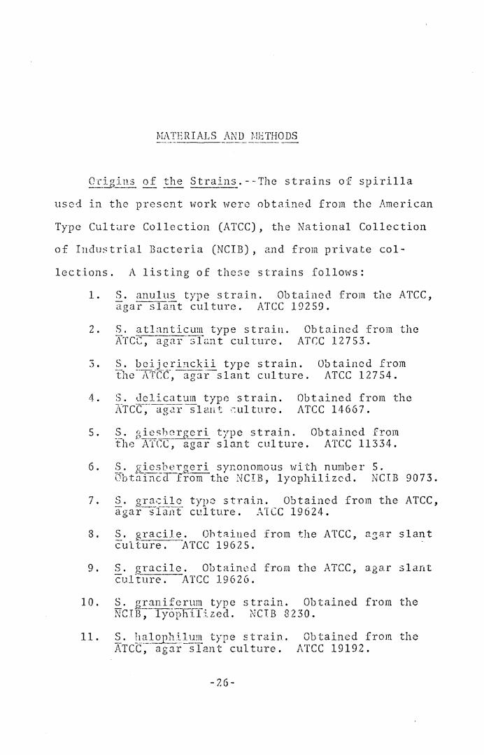
MATERIALS AND J',!ETHODS ----------------
OE_igj~.s o~ ~he Strains. --·The strains of spirilla
used in the present work were obtained from the American
Type Culture Collection (ATCC), the National Collection
of Industrial Bacteria (NCIB), and from private col-
lections. A listing of these strains follows:
1. S. anulus type strain. Obtained from the ATCC, aga-r sla-_nt culture. ATCC 19259.
2.
3.
4.
c .J •
6.
7.
S . at Lm tic um type strain . }\Tcc-;--agar ·-sfu.n t culture.
Obtained from the ATC:C 12753.
S. bcijcrinckii type strain. the-· A'l'C:-C:-,-agar-s lan t cul tu re.
Obtained from ATCC 12754.
S. Jelicatum type strain. XTcc-;·ag~1r sl a11 t ·-:ul turc.
Obtained from the ATCC 14667.
S. gieshcrgeri type strain. Obtained from fhe· A'fCC,agar slant culture. ATCC 11334.
S. giesberaeri synonomous with number 5. Ubtn-incc1T~-om the NCIB, lyophil izcd. NCIB 90 7 3.
S. _g_!_:.:~,:ilc typ2 strain. Obtained from the ATCC, agar slant culture. ATCC 19624.
8 • §_. gr a c i .l e . 0 h t ::i.i u e d from the ATC C , a z Cl r s 1 ant culture. ATCC 19625.
9. S. gracile. Obtained from the ATCC, agar slant cul"iure. ATCC 19626.
10.
11.
S. granifcrum type 'iJrI _________ h_'_l--: d ;,v B ~ lyop 1 ._ze •
strain. Obtained from the NCIB 8230.
~· l1_~~~~.5~2!m type stn1in. ATCC, agar slant culture.
Obtained from the ATCC 19192.
-26-
-27-
12. S. itersonii type strain. Received from Dr. R. I\r:-sinl.bert, Anaerobic Bacteriology Labora-tory, Virginia Polytechnic Institute. ATCC 12639.
13. S. itersonii. Same source as 12. ATCC 11331.
14. S. itersonii. Received from Dr. S. C. Ritten-berg, Department of Bacteriology, University of California.
15. ~· j_~Eonicum type strain. ATCC, agar slant culture.
Obtained from the ATCC 19191.
16. S. linum type strain. Obtained from the ATCC, aga1·-~siant culture. ATCC 11336.
17. S. lunatum type strain. Obtained from the ATCC, agar-s1ant culture. ATCC 11337.
18. S. meta;nor_phum type s tra.in. Obtained from the ATCC~--agar --slint culture. ATCC 15280.
19.
20.
S. minutulum type strain. l\Tcc;-ugar -slant cul turc.
~· percgrinum type strain. ATCC, agar slant culture.
Obtained from the ATCC 19193.
Obtained from the ATCC 15387.
21. S. QOly_E:_orl2.E_~1m type strain. Obtained from the ATCC, agar slant culture. ATCC 11332.
22. ~· ~mo~hum synonomous with number 21. Obtained from the NCIB, lyophilized. NCIB 9072.
2 3. §_. putridiconchyl~~m type strain. from the ATCC, agar slant culture.
Obtained ATCC 15279.
24. ~. ~~~~p~ns type st rain. Same source as 12. ATCC 12638.
25. s. _?erp~p.s. Same source as 12. ATCC 11335.
26. s. ser12e_ns. Same source as 12. ATCC 15278.
27. s. s erpens. Same source as 12. ATCC 11330.
2 8. s. ser~ens unnumbered strain. Same source as 14.
-28-
29. S. :;_crE~-~_s_ Victoria st:!:ain. Sarne source as 14.
30. S. scq~ens. Sarne source as 14. ATCC 12289.
31. S. sery_~:_I~~- strain VI-I. Received from Dr. R. G. E. !<urray, Department of Bacteriology and Immuno-logy, The University of Western Ontario.
32. S. seJ.:_~ns strain VHA. Same source as 31.
33. S. seD!ens strain VHL. Same source as 31.
34. S. ~ erp~n~ st rain VHS. Same source as 31.
35. S. se.!Ee~_?_ St. Rhodes strain. Same source as 31.
36. S. sinuosum type strain. ATc·c·;-a,g·a:1::-s lan t cul tu re.
Obtai.ned from the ATCC 9786.
37. S. volutans type strain. :AT cc T~rss-3-:- Same source as 14.
38. S. volutans Wells strain. Isolated from pond w·ateY-hay infusion. ATCC 19554.
39. Un··named strain 101 of Dr. I-I. iV. Jannasch, Woods Hole Oceanographic Institution.
4 0. Un-named strain 102. Same source as 39.
41. Un-named strain 103. Sarne source as 39.
4 2. Un-named strain 104. Same source as 39.
4 3. Un-:1s.;ned strain 204. Same ::;ource as 39.
44. Un-named strain 206. Same SOl~l'Ce as 39.
~ The Witkin strain of Esr~erichia coli B used as
a standa:rd in determining GC ratios and also the presence
of deaminase, was obtained from Dr. G. R. Carta, Depart-
ment of Biology, Virginia Polytechnic Institute.
Maintenance of Cul tu res. - -Al 1 cultures ~.~·ere main---------- - -- ----tained in semi-solid (0.15% agar) peptone-succinate-salts
-29-
(FSS) medium, with serial transfers being made weekly.
These transfers served as sources of organisms for
preparation of inocula in all experiments except in the
dctcr~ination of the moles % GC of DNA and in the
determination of cleaminase activity.
The cultures were also preserved in a So-Low
Enviro;1men t al Chamber (So- Low Environmental Equipment
Co., Cincinnati, Ohio) set at -80 C in PSS medium+
15% glycerol. On two occasions a nwnber of the cultures
stored in the freezer were ruined due to electric fail-
ure. After each such incident, replacements were
obtained using spirllla m~intaincd by serial transfer.
The frozen cultures '"ierc used as sources for prepar-
ation cf inocula :for the determination of moles % GC
of DNA and deaminase activity; these cultures were
also used for c0nfirmation purposes in several cxperi-
ments which provided Jata contraJictory to that reported
by other i.nvesti~ators, particularly with regard to
nitrate reduction, catalase production and gelatin
h~rd rolys is.
and EMB agars; nutrient, MRVP, and urea broths; litmus
milk, skim milk, peptone, agar, yeast extract, oxgall
biJ.e, deoxyribonucleic acid (DNA), ribonucleic acid (rfrJA),
gelatin, starch (soluble), reagent; melibiose, trehalosc,
~LI1U]J·~ c~ 1 J·~:ll "~~cu]1"n ,·1·1·1~ G1 l'lcitol ~0r~ obtained . . .• H' .> <L.L • -.. .L ' U.v .:> . • ' -... • - - -
-30-
from Difeo Laboratories, Detroit, Michigan.
Ammonium sulfate, reagent; calcium chloride, reagent;
ferrous sulfate, reagent; ferric chloride, reagent; lead
acetate, C.P.; magnesium chloride, reagent; manganese
sulfate, reagent; potassium chloride, reagent; sodium
chloride, reagent; boric acid, reagent; succinic acid,
reagent; and octanol, reagent were products of the J. T.
Baker Chemical Co., Phillipsburg, New Jersey.
Glucose, galactose, lactose, mannose, fructose,
ribose, arabinosc, xylose, rhamnose, raffinose, C. P.;
melezitose, sorbose, cellobiose, glycogen, amygdalin
(Trihydrate), adonitol (adonite), erythritol, mannitol
(mannite), sorbitol (hydrate), dextrin (White, Tech.),
glycerol, C. P.; cysteine, tryosine, phenolphthalein
diphosphate (1Jentasodium), and phenolphthalein disulfate
(Trihydrate) were obtained from Nutritional Biochemical
Corporation, Cleveland, Ohio.
Ethanol, U.S.P. was manufactured by U. S. Industrial
Chemical Co., New York.
Magnesium sulfate, reagent; potassium hydroxide,
reagent; potassium nitrate, reagent; sodium citrate,
reagent; sodium lauryl sulfate, reagent; maltose,
reagent; sucrose, certified A.C.S.; selenous acid,
certified; and ethylene glycol monoethyl ether, puri-
fied were obtained from Fisher Scientific Co., Fairlawn,
-31-
New Jersey.
Oxidase test disks were a product of the Baltimore
Biological Laboratory, Division of B-D Laboratories, Inc.,
Bolt.imore, Md.
Inositol, phenylalanine, M.A., and tryptophan, M.A.
were supplied. by the Mann Research Laboratories, Inc.,
New York.
Bovine pancreatic ribonnclease (RNAase), A-grade
and T1 RNAase (crystalline) were products of Calbiochem,
Los Angeles, California.
Phenol liquefied, A. R. was obtained from the Mall-
inckrodt Chemical Works, St. Louis, Missouri.
Com-oosition of Peptone-Succinate-Salts Jliedium. - -Pep----'------·-- - --- ---------·-- ---- --·---·---tone-succinate-salts (PSS) broth had the following compos-
ition (g per 1): peptone, 10.0; succinic acid, 1.0;
(NII4)zS04, 1.0; MgS04•7HzO, 1.0; FcCl3•6HzO, 0.002;
and MnS04·HzO, 0.002. The pH was adjusted to 6.8 with
2N KOH. For so 1 i<lif icd media, l. 5 % agar T.rns incorpo-
rated into the PSS broth; scmi-~olid meJia were prepared
by adding 0.15% agar to the PSS broth. For the culti-
vation and characterization of marin0 spirilla, artifi-
cial sea water (Society of American Bacteriologists,
1957, p. 113) was employed in the preparation of all
media. S. 1~~~~-a_!un~ grew in the presence or absence of
sea wateT; therefore, this organism ~as examined under
-32-
oot:1 coadi tions with respect to ail tests.
in Biochemical and
those of~· ~~~~1ta.~2, were cultured in screw-capped 13 x
125 mm tubes, containing 10 rnl of PSS broth, for 24 hrs.;
0.0~ ml from the second serial transfer was used as
inoculum for all test media unless otherwise indicated,
A similar inoculum was employed for~· volutans,
with the exception that the organisms were cultivated·
in 75 ml quantities of PSS broth contained in 250 ml
side~arm flasks having a mixture of 30% air and 70%
ni t.:rogcn. Camoun ting to an oxygen conccntra ti on of
approxi~ately 6%).
_y_•~l:~-~!:-:~xis, were groHn and characterized under aerobic
conditions in ali experiments except when determining
ability to grow anaerobically in the presence of
nitrate; ~· volutans, being obligately microaerophilic,
was grown either in liquid media stratified with 0.15%
3go.r or on solid :::i.edia in 2. mixture of 30% air and 70%
nitrogen (amounting to an oxygen concentration of
approxiraately 6%).
Estirnation
for the purpose of determining a growth rasponse was
f;:<ernlined every ch1ring the incubation period; ct~l tures
-33-
were rated by a visual examination with respect to growth
in these'media.
Tef1!I2.~ Ta tu re cf IncE_b a.!:J:.?n • - ··Cul ti va ti on and character-
ization of spirilla were carried out at 30 C, after this
value was determined to be optim2l for all sirains (see
belc·..r).
Qe0rn~in:ition of Qpti~~~~ Ter11per::::!ure_. --The optimal
tamperaturc was determined by incubating the cultures
at temperatures of 10, 15, 20, 25, 30, 37, 42, and 45 C;
for the temperatures 10, 20, and 25 C a Fisher Low Tem-
perature Incubator (Fisher Scientific Co., Fairlawn, N. J.)
was employed, the 15 C temperature was obtained in a
Psycrotherm Incubator Shaker (New Brunswich Scientific
Co., Inc.~ New Brunswick, N. J,), Box Incubators (Pre-
cision ScientifJc Co., Chicago, 111.) were employed for
the t~mperatures 37, 42, and 45 C, and 30 C was obtained
i~ a Fisher Isotemp Incubator (Fisher Scientific Co.,
Fairlawn, H.J.). The medium was cowli~:ioncd to the
various temperature fer 2 hr. prior to inoculation.
After 1 week incubation, a visual estimation of growth
wa~ mr..de.
Bi_~ c.heEJ-~al_ an~ ~?l!i~iolog_!s_<::l ~;e~g.od~.
A number of biochelflical and physiological tests were
e~ployad in the ci1aractertz~tion of the strRins cf spirilla.
· ~2:: id as e_ .!.~ ~!. -. -A po s i ti v e p ·-~ rp 1 e cc lo r reaction was
- 34 ·-
sought with 48 hr. semi-solid (0.15% agar) PSS cultures
by two m~thods: (a) moistened oxidase test disks (BBL)
were smeared Hith a loopful of the culture, and (b) se-
veral drops of paraamino dimcthylaniline solution were
a<ldsd directly into the culture. (
FJ~o~_:p_!rntas_i: and Sulfatasc Production. - -Phenolphthalein-
dipho~phate or disulfate was added to PSS agar at a final
concentration of 0.01%. Inoculation was accomplished by
streaking a loopful of the second of two serial 24 hr. cul-
tures across the agar plates. After incubation for 4 days,
ammonia fumes were used for the detection of free phenol-
phthnlein.
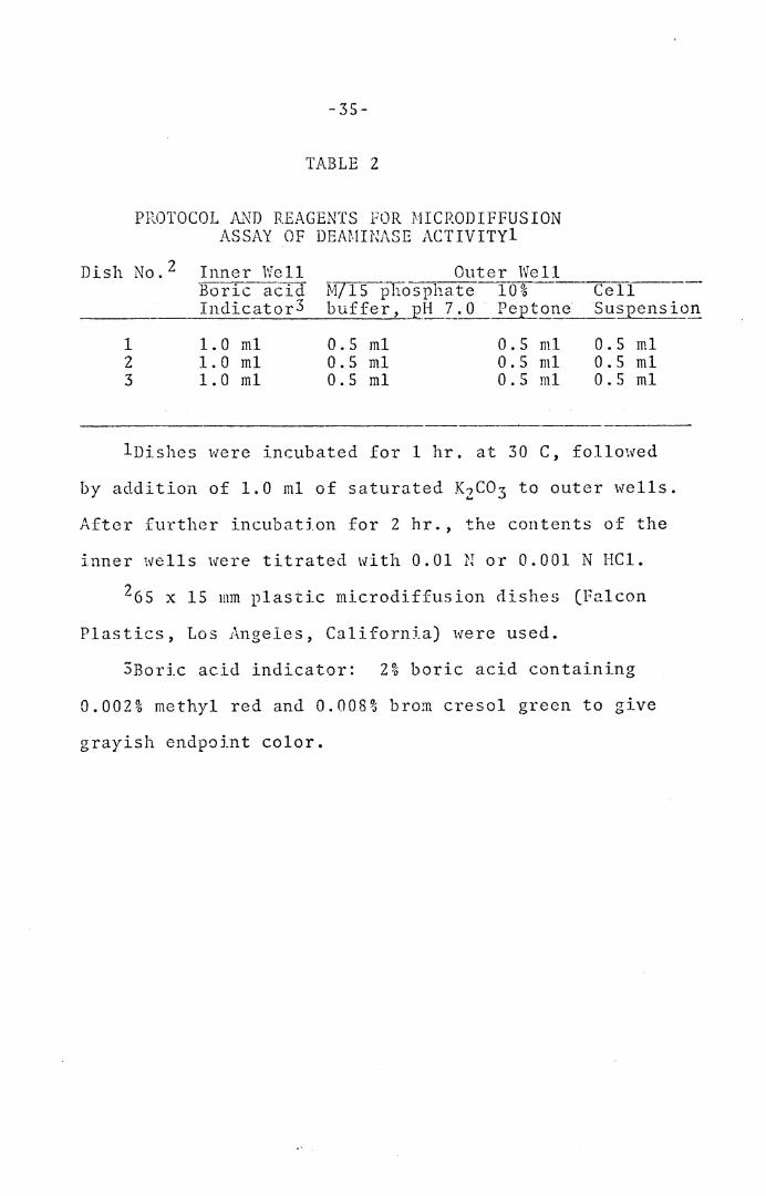
-~an:_~n:i~--~ Act~_vi_ ty. - -The Conway mic rodi f fusion tech~
niquc was employed (Conway, 1950). The spirilla were
cultivated in 300 ml of PSS broth contained in 1 liter
flasks for 24 hrs. The cells were harv~sted, washed
twice in sterile saline, and resuspended in 5 ml of
sterile saline. The turbidity of t~is suspension was
adjusted so that a 1:10 dilution would give a reading
of 200 Klett units. Five-tenths ml of the undiluted
suspension was then used to determine the deaminase
activity, uslng the protocol in Table 2.
-~~~ta~as_~ -~est. --Th<~ Elicroscopic production of a froth
8f bubbles was det8n1ined by the addition of 4-5 drops of
zi IizOz t:o 24 hr. semisolid PSS cultures, with subsGquent
incu~aticn up to 30 minutes.
Dish
1 2 3
-35-
TABLE 2
PROTOCOL At~D REAGENTS FOR MICRODIFFUSION ASSAY OF DEAMINASE ACTIVITYl
No. 2 Inner Well Outer Well Boric acid M/15 phosphate 10% Indicator3 buffer, J2.~Z · o Pep tone
1. 0 ml 0.5 ml 0.5 ml 1. 0 ml 0.5 ml 0.5 ml 1. o ml 0.5 ml 0.5 ml
Cell Suspension
0.5 ml 0.5 ml 0.5 ml
lDishes were incubated for 1 hr. at 30 C, followed
by addition of 1.0 ml of saturated KzC03 to outer wells.
After further incubation for 2 hr., the contents of the
inner wells were titrated with 0.01 N or 0.001 N HCl.
265 x 15 111m plastic microdiffusion dishes (Falcon
Plastics, Los Angeles, California) were used.
3Boric acid indicator: 2% boric acid containing
0.002% methyl red and 0.008% brom cresol green to give
grayish endpoint color.
-36-
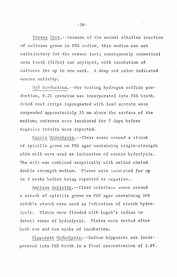
Urease Test.--Because of the normal alkaline reaction
of cul tu res grown in PSS J11•~dium, this medium was not
satisfactory for the urease test; consequently commerical
urea broth (Difeo) was employed, with incubation of
cultures for up to one week. A deep red color indicated
un:1.se activity.
HzS Production.--For testing hydrogen sulfide pro-
duction, 0.2% cysteine was incorporated into PSS broth.
Dried test strips impregnated with lead acetate were
suspended approximately 30 mm above the surface of the
medium; cultures were incubated for 7 days before
n0gativ8 results were reported.
Casein around a streak
of spirilla grown on PSS agar containing single-strength
skim milk were used as indication of casein hydrolysis.
The mil\ was combined aseptically with melted cooled
double strength medium. Plates were incubated for up
to 2 weeks before being repo1·ted as negative. 1
:~Xl~::~ Ac:_ ti v~. - -Clear colorless :lones around
a streak of spirilla grown on PSS agar containing 10%
soluble starch were used as indicatioc of starch hydro-
lysis. Plates were flooded with Lugol's iodine to
detec~ zones of hydYolysis. Plates were tested after
both one anrl two weeks of incubation.
Hi·£p_~i:-_~!:_~ !lY.3:T_o_!z~j.s. - -Sodium hippurate was incor-
porated into PSS broth in a final concentration of 1.0%.
-37-
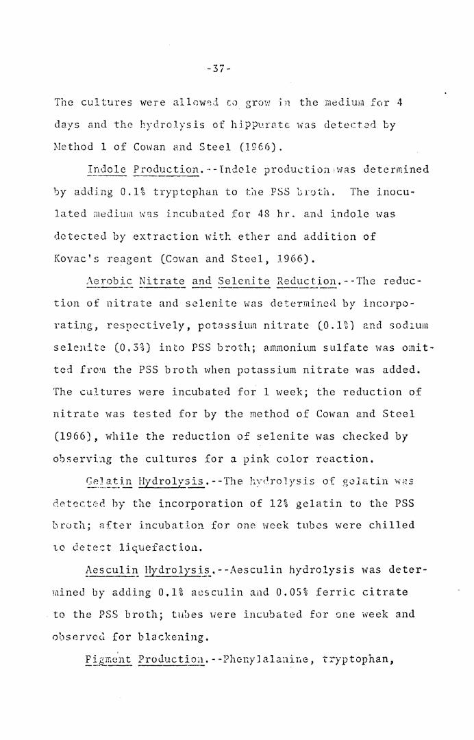
The cultures were allow~d to grow in the medium for 4
days a.nd the hydrolysis of hippu.ratc was detect-3d by
0Iethod 1 of Cmvan and Steel (1966).
In do le Production.·· - lndo le produc tio;1 'was determined
by adding 0.1% tryptophan to t~e PSS broth. The inocu-
lated medirna wg.s incubated for 48 hr. anJ in do le was
detected by extraction with ether and addition of
Kovac's reagent (Cowan and Steel, 1966).
/\erob ic Nitrate and Se lcni te Reduction. - -The reduc-
tion of nitrate and selenite was determined by incorpo-
rating, respectively, pot.'.lssium nitrate (O.l~) and sochum
s e 1 en i t e ( 0 . 3 % ) in to PSS b r o th ; ammonium s u 1 fate was om i t -
ted fro 1n the PSS broth when potassium nitrate was added.
The cultures were incubated for 1 week; the reduction of
nitrate was tested for by the method of Cowan and Steel
(1966), while the reduction of selenite was checked by
observing the cultures for a pink color reaction.
detected hy the incorporation of 12% gelatin to the PSS
broth; after incubation for one week tubes were chilled
to dete=t liquefaction.
~esculi~ !:!Ydrolysi~_.--Aesculin hydrolysis was deter-
mined by adding 0,1% aesculin and 0.05% ferric citrate
to the PSS broth; tubes Here incubated for one week and
observed for blackening.
~~g~r,ent ProJuctiou. - -Phc-nyJ alanir,e, tryptophan,
-38-
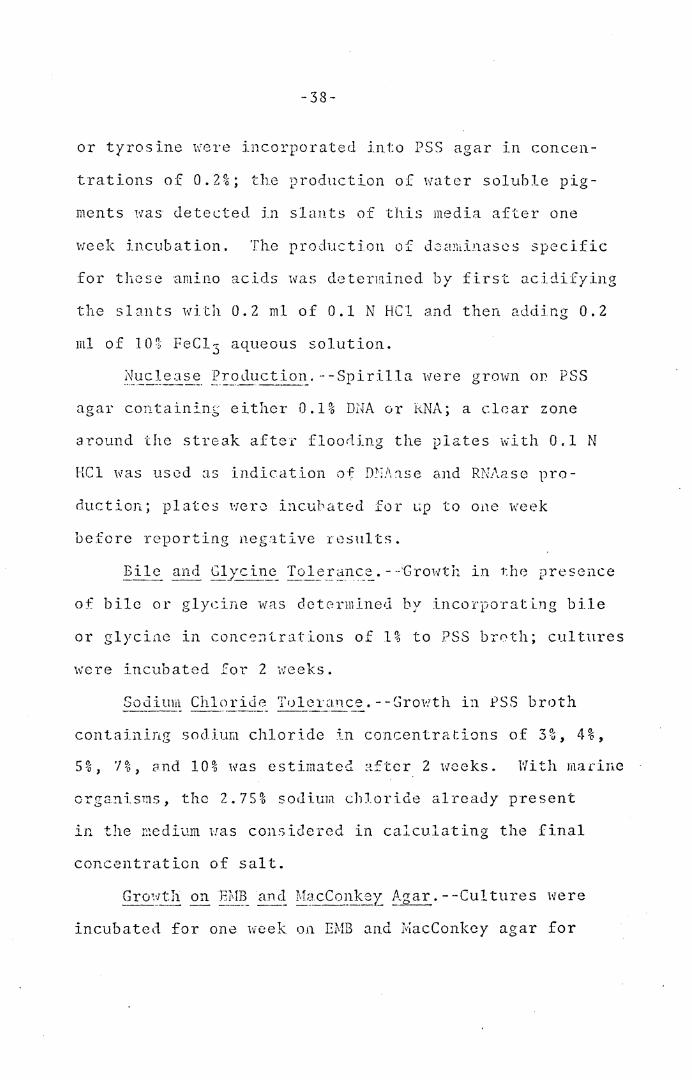
or tyrosine l\'ere incorporated into PSS agar in concen-
trations of 0.2%; the production of iwter soluble pig-
ments was <letected in slants of this media after one
week incubation. The production of d0ct!1iinascs specific
for these amino acids was determined. by first o.cidifying
the slants with 0.2 ml of 0.1 N HC1 and then adding 0.2
ml of 10% FeC1 3 aqueous solution.
agar containin;; either 0.1% D.l~A or h.NA; a clear zone
around the streak after floo~ing the plates with 0.1 N
HCl was used as indication of D?·;J\'.lse and RN1\ase pro-
duction; plates wer0 incubated for LlQ to one week
before reporting negative results.
of bile or glycine was dct0rrnined by incorporating bile
or glyci11c in conc':':-itrations of 1% to PSS brr.th; cultures
were incubated for 2 weeks.
Sodium Chloride Tulcrance.--Growth in PSS broth
containing sodium chloride in concentrations of 3%, 4%,
5%, 1%, and 10% was estimated after 2 weeks. With marine
organisins, the 2. 75~6 sodim;1 chloride already present
in the r::edium 1:as considered in calculating the final
concentration of salt.
incubated for one week on EMB and MacConkcy agar for
-39-
the detection of growth.
_G _ _ro~!th on li'J. Agar a1~! Sel~_er's Slants.--Cultures
were Inoculated on TSI and Seller's slants by stabbing
the butt and streaking the slant with an inoculating
needle. The slants were observed for growth and any
subsequent changes in the media.
Re_9-ction in Litmus Milk. --Spirilla were incubated
in litnrus milk for 72 hrs; the cultures were observed
daily for any reactions producing a color change in
the litmus milk. The marine strains were not exmnined
for their reactions in litmus milk; the artificial sea
water had a precipitating effect on litmus milk even
when added aseptically after autoclaving.
!::~id Reactiop.s _from Ca_Ebohydrates. --For demonstration
of acid reactions from sugars, PSS broth without succin-
ate and with peptone decreased to 0.2% was used; phenol
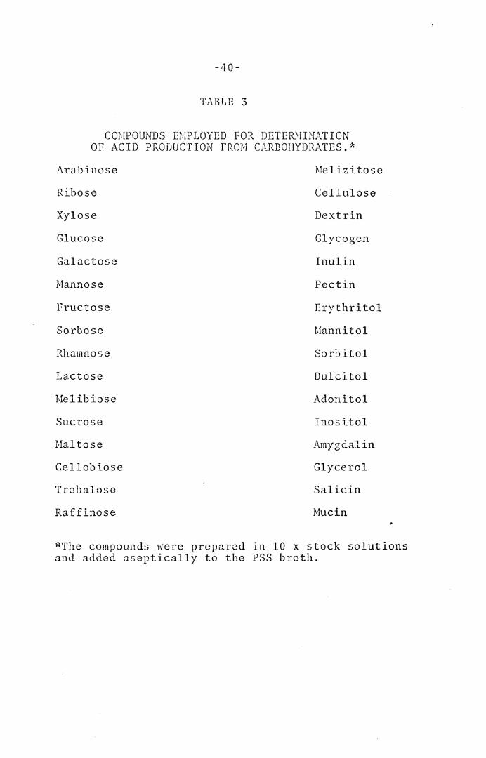
red indicator (18 µg per 1) was added. Thirty-two
compounds (TJ.ble 3) were employed in the determination
of acid production; the compounds w0re added aseptically
to the peptone-salts broth at a final concentration of
1%. The cultures were incubated for up to 3 weeks.
An2erobic Growth in the Presence of Kitrate.--Ability
to grow anaerobically in the presence of nitrate was
detected by incorporating 0.1% potassium nitrate to
freshly-autoclaved PSS semi-solid medium; tubes ·were
-40-
TABLE 3
cm.IPOUNDS Ef.lPLOYED FOR DETERJ1IINAT ION OF ACID PRODUCTION FROM CARBOHYDRATES.*
Arabinose Melizitose
Ribose Cellulose
Xylose Dextrin
Glucose Glycogen
Galactose Inulin
i'v!annose Pectin
Fructose Erythritol
Sorbose Mannitol
Rhamnose Sorbitol
Lactose Dulcitol
Melibiose Adonitol
Sucrose Inositol
Maltose Amygdalin
Cellobiose Glycerol
Trchalose Salicin
Raff inose Mucin
*The compounds were prepared in 10 x stock solutions and added aseptically to the PSS broth.
-41-
inoculated and im.rn.edia te ly sea led with sterile vaspar.
Incubation was for 1 week.
}.iethods used for Mornhological Determinations. - ------ --- -- ___ ..i;;:_. ___ . ____ _
Cell dimensions were obtained from measurement of living
cells from 18 hr. PSS broth cultures, using a Leitz
Ortholux microscope equipped with a Heine phase contrast
condenser and Pv Apo 90x/l.15 objective. In 5 separate
fields, a total of about 50 of the longest and the short-
est cells were chosen for measurement of the range of
cell length; the range of cell diameter, diameter cf
helix, wavelength, and nunilicr of turns were also obtained
from these cells. Flagellar tufts were observed with
dark field microscopy, using a 150 \'latt Xcuo1: lamp for
illumination and a 95x oil Smmersion fluorite objective,
N.A = 1.10. The presence of coccoid bodies was determined
by observing 4 week PSS cultures by phase ~ontrast
microscopy. Measurements of colonies growing on PSS
agar plates were obtained after 72 hr.
Cultivati~.~ ~f Cc:lls ~92. pN~ J_~_ol_~_-t_:_~n. --Spirilla
were cultivated in shaken cotton-stoppered liter flasks
containing 300 ml of medium having the following compos-
ition (g per 1): nutrient broth, 6.0; yeast extract,
2.0; and succinic acid, 1.0. The pH was adjusted to
G.8 with ZN KOH. AeratJon was accomplished by a 30 C
water bath shaker (Rotary Shnkcr, New Brunswick Scientific
-42-
C·J. ' i\Te'.·l n i·t•nc'·•·icli\., N T ) ,. 'i .U .- ""·t :,:) H ..L ' l • l_. • • The cells were centrifuged
at 5000 x g after 18 to 24 hr. E. coli cells were obtained
un<ler similar conditions.
DNA Isolation.--After 1iarvcs ting, the cells 11ere
suspended in 1X salinc--EJJTA (0.1.5 M NaCl, 0.01 M JJDTA,
pH 8.0) ar1d lyscd by the addition of 1% sodium lauryl
sulfate. Ten ml of liquefied pheno 1 \\1e re added, and
•L·ha .. JTIJ0_.~t:1.1··a ',~35 '_~,l.1,~_.k~l,1 ('1~Yl-~t ~rt1"•)·[1 Sl1~~CT 0 UYI"8ll '- r, - '-' • - ~ ' .) .. • ' ,_ - ' • ' ·' ...._ . ' ' J; :,
Corp., Pittsburgh, Pa. at a setting of 2) for 5 to 10
m~1~utes 0r until ho;:1ogen0i:s. The: mixture was cooled
2nd 1:.cntrifuged (5000 x g for 10 ;nin.), and the aqueous
phase was extracted again with liqu~fied phenol. After
precipitation fr.J;n the 8qi.wous ph.::1se by addition of 2
voh;mc~s of ethanol, the DNA was <lis5ol.ved in 0. lX SSC
(lX:.:: 0.15 M N~iCl, ·J.015 M sodium citrate, p!1 7.0).
The SSC concentration ..:as increased to lX (by addition
of sufficient 20X SSC) and the precipitation procedure
repeated. After incrcasiilg the SSC conc0ntration to
1 X, 0.5 ml of a ribonuclease lhixtu:i·e a::. a conccntLition
of 500 µg per ml of bovine pancreatic RNAase and 25
-L1nit:::- f)er· 111.l o.·f '.f1 ~NA;:i_~e lT~ - --dd,..,,' ln 1 ~-1 r arat1· on ., - r~ -· - - 1\ d. ~~ a - "-• (t ( l u '- _1. e p e p . . . -- -
was incubated at 37 C for 2 hr. The DNA was deproteinized
with 5-10 ml of chloroform-octunol
oc t anal) . The i11ixture was sha.ken, cool cd c.md ccn tri-
fuge<l at 5000 x g, and the DNA W3S prec1p1tatcd from
-43-
the actucous phase witlL 2 volumes of ethylene glycol
monoethyl ethe1·. After a f inc.1 ethyl alcohol precipi-
tation, the DNA was dissolved in ca. 5 ml of 0.1 X SSC
and stored at -18 C.
~Iclting £..?J:.!:1t _(Tm)_ rfoasur8mcnts. - -The DNA was
adjusted to a concentration of SO µg per ml with 0.5
x SSC and dialyzed against 0.5 X SSC, prior to Tm
measure~ents. Melting temperature (fm) determinations
were obtained by a variation of the method of Marml1r
3nd Doty (1962); the starting temperature for determin-
ing the Tm profile was 60 C and the temperature was
increased linearly 0.5 degree per minute over the entire
:cange. An auto1:<atic recording Gilford ~1cdel 2400
Spectrophotometer (Gilford Instrmnent Laboratories,
Oberlin, Ohio) was 8mploycd.
RESULTS
g_ul tura"!:_ anc~_Jforphological Tes ts
1he characterization tests included in this category
deal with the relation of spirilla to oxygen, temperature,
colony formation on PSS agar, size of cells growing in
PSS broth, the visibility of flagellar tufts under dark
field, and the presence of coccoid bodies in older cul-
tures.
Relationsh:!:.E_ to Oxygen.--All strains of spirilla
except ~· volutans grew moderately to abundantly well
aerobically in PSS broth in 24 hr. The two strains of
~· vo!utans_, being obligate microaerophilcs, failed to
gro~\f aerobically, but grew well in stratified (0 .15%
agar) PSS broth or in broth lmcler an atmosphere of 30%
air and 70% nitrogen. ~· 6iesbe~gcri c-.ppeared to be a
facultative microaerophile, in that although growth
did occur aerobically, in stratified broth (0.15% agar)
the organism characteristically formed a microacrophilic
band of growth 3-·4 r.rn1 belo1v the surface of the medium.
Anaerobic growth could occur only in the cases of S.
lunatum, and the 3 strains of S. itcrsonii when 0.1% KN03
was added to the PSS medium.
-44-
-45-
ature was 30 C for all strains of spirilla, although
moderate growth did occur at 20, 25, and 37 C. All of
the marine strains had scant to moderate growth at 15 C,
except ~· beijerincki~ which failed to grow; none of
the fresh water strains grew at 15 C. At 42 C, only
co~chylium, and certain strains of S. _serpens: (Victoria,
VH, VHA, VHL, VHS, St. Rhodes, and the unnumbered strain)
were able to grow; growth was scanty. Ne growth occurred
at 10 for any of the strains. 0.:1ly the unnumbered Rit-
ten.berg strain of S. serpens 11as able to grow at 45 C.
Colony Charact~ristics.--Spirilla grown on PSS agar
plates for 72 hr. formed C)lonies ranging from 0. 5 to
1. 5 imn in diameter, except the species S. anull~s, S.
volut~~' S. }2_9-~orphum, ~· _g_"!:_acil_~, and the un-named
strain 101 all of which formed pinpoint colonies. All
colonies were white, circular, and convex with smooth
edges, except for the strains S. volutans and~· g_ra~il~
which produced 1~1ite colonies with fimbriated edges.
The thr0e strains of S. gracile_ formed colonies which
were partially below the surface of the agar, indicating
a possible microaerophilic response. Colonies from S.
vol utans 1·:ere formed best in soft PSS agar (0. 7% agCi.r)
incubated under reduced oxygen.
-46-
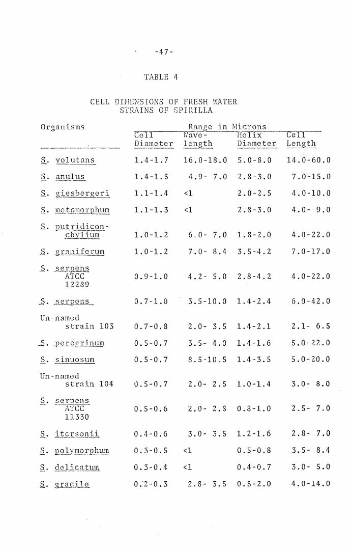
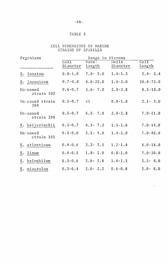
Dimc~_sion_~ Q_f Cell~.--Ranr;es for cell diameter, dia-
meter of helix, wavelength, and length of cells are pre-
sented in Tables 4 and S.
yisi~ility of fla_gell~~ }-~ufts by Dark Fie!d. All
strains of spirilla were actively motile; flagellar
tufts were clearly visible under dark field microscopy
in many of the organisms. The strains of S. _giesbergeri,
S. _!inum, and the un-named strains 101, 102, and 206 all
had helical tufts; similar helical structures were seen
in at least 50% of the cells of S. d~licatum, a species
said to possess predominantly only a single polar fla-
gellum. These flagellar tufts are of interest because
they permit the investigator to view the action of such
structures which, although present on a number of bac-
teria, are invisible by microscopic observations, and
can be seen only after staining; the action of the fla-
gellar tufts can best be described as a series of helical
waves moving down the length of the structures. Straight
tufts of flageJ.la were seen under dark field on the
species§_. gr~nifcrum, S. anulus, S. £UtriC.iconchylium,
S. j_2J~on} .. cu:rr, §_. volut_~ms_ and the S. s<::_-rpe1~~ strain ATCC
12289; _'.?_. vol utan~ flagellci_r tufts were observed by
phase contrast microscopy also. The action of straight
flagcllar tufts were not readily observed on motile
spirilla, except S. ~~lutan~, and were seen only on cells
-47-
TABLE 4
CELL DIME;.JSIONS OF FRESH WATER STRAINS OF SPIRILLA
Organisms Ra_!l_g~ in_lLtcrons Cell Wave- Helix Cell Diameter lenath Diameter Length -----·--· --~~- -----
s. vol utans 1.4-1.7 16.0-18.0 5.0-8.0 14.0-60.0 -----s. anulus 1.4-1.5 4.9- 7.0 2.8-3.0 7.0-15.0 -------_$_. g_i es berg_~ r i 1.l·-1.4 <l 2.0-2.5 4.0-10.0
$_. 1_1}_~_ t.§.]'.l o r 12.b-~lm 1.1-1.3 <l 2.8-3.0 4.0- 9.0
c:: nutridicon-Y-• ~--~~J1y 1{um-- 1.0-1.2 6.0- 7.0 1.8-2.0 4.0-22.0
_s. . g:r_;:tnif erum 1.0-1.2 7.0- 8.4 3.5-4.2 7.0-17.0
_s. _s e r_g_ en__,~ ATCC 0.9-1.0 4. 2- 5.0 2.8-4.2 4.0-22.0 12289
_s. 5_erpons_ 0.7-1.0 3.5-10.0 1.4--2.4 6.0-42.0
Un-named strain 103 0.7-0.8 2.0- 3.5 1.4-2.1 2.1- 6.5
_s. . .1,212..I..Q z r in um 0.5-0.7 3.5- 4.0 1.4-1.6 5.0-22.0
s. sinuosum 0.5-0.7 8.5-10.5 1.4-3.5 5.0-20.0
Un-named str2in 104 0.5-0.7 2.0- 2.5 1.0-1.4 3. 0 :- 8.0
s. _?_e"f_QCHS A'fCC 0.5-0.6 2.0- 2.8 0.3-1.0 2. 5- 7.0 11330
s. itcrsonii 0.4-0.6 3.0- 3.5 1.2-1.6 2. 8- 7.0 --·----·-·------·-·-
ii. J2.. o l:zri:i_QIJ? hum 0.3-0.5 <l O.S-0.8 3.5- 8.4 ro ,'.) . de.Jjc~.i!..!Um 0.3-0.4 <l 0.4-0.7 3.0- 5.0
.s_. . 1 _g_:r_ac:i --~ 0."2··0.3 2.8- 3.5 0.5-2.0 4.0-14.0
-48-
TABLE 5
CELL DIMENSIONS OF MARINE STRAINS OF SPIRILLA
Organisms
S. lunaturn
S. j aponic_vm
Un-named strain 102
Un-na1t1ed strain 204
Un-named strain 206
S. beiierinckii -- __ _.,_ ____ _ Un-named
strain 101
S. atlanticum
S. linum
S. minutulum
Cell--Diameter
0.8-1,0
0.7-0.8
0,6-0.7
0.5-0.7
0.5-0,7
0.5-0.7
0.5-0.6
0.4-0.6
0.4-0.5
0.3-0.6
0.3-0.4
Range in flficrons Wave- Hel1x----c=,e---=-1=-1-Length Diameter Length
7.8- 3.0
8.0-20.0
5.6- 7.0
<l
6.5- 7.0
6.3- 7.2
3.5- 4.0
3.2- 3.5
1.8- 2.0
2.0- 2.8
2.0- ,., 7 i. • ..J
1.0-1.3
1.0-3.0
2.0-2.8
0.8-1.0
2.0-2.8
1.5-1.6
1.4-2.0
1.2-l.4
0.8-1.0
1.4-1.S
0.6-0.8
2.4- 5.4
20.0-75.0
8.5-18.0
2.1- 3.0
7.0-21.0
7.0-14.0
7.0-40.0
4.0-14.0
7.0-30.0
3.5- 8.0
3.0- 8.0
-49-
which had stoppecl their motion; in all such cases the
flagellar tufts extended out from the cell at an angle
of approximately 45 degrees. S. volutans was unique in
its flagellar action because its action during rapid
movement was readily observed (Krieg, Tomelty, and Wells,
1967).
Presence of Coccoid Bodies in Older Cultures.--
The presence of coccoid bodies in 1 to 4 week old cul-
tures was found in all of the marine strains, except
the un-named strain 204; in the fTesh water forms they
S. E_ere_grinu~. The diameters of these coccoid bodies
ranged from 3.0 to 5.0 µ.
Phvsiological Tests _::...!. _______ --··---·-·----
Certain physiological tests were found to be uni-
formly positive or negative for all or nearly all of
the strains studied; other kinds of tests resulted
in a mixture of po~itive and negative results among
the strains.
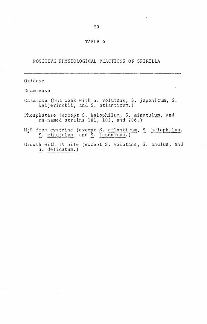
or nearly uniformly positive are summarized in Table 6.
All strains were oxidasc positive, dcaminase positive
(quantitative results are presented in Table 7), and
catalasc positive, although the catalase reaction was
considered very i-1eak with S. volutans, S. iap_smicum, S.
-so-
TABLE 6
POSITIVE PHYSIOLOGICAL RDACTIONS OF SPIRILLA
------ -------Oxidase
Catalase (but weak with~· voJut~~2_, S_. japonJ..~~21!_, ~· !?eij __ erinckii, and§_. atla!1ticum.J
Phosphatase (except ~· ha~~J!._ilum, ~· !!~i_nutulum, and un-named strains 101, 102, and 206.)
HzS from cys teine ( exccp t ~. _a q __ ?-n ti cum, S. L1-':~1=.921!_~-]. u~, ~· rr:inu~~1lum, and ~· ~--~~icum.)
Growth with 1% bile (except S. volt1:!ans, S. _?.~Ul1:1s, and S. ~ __ c].:._ i cat ~m.)
-51-
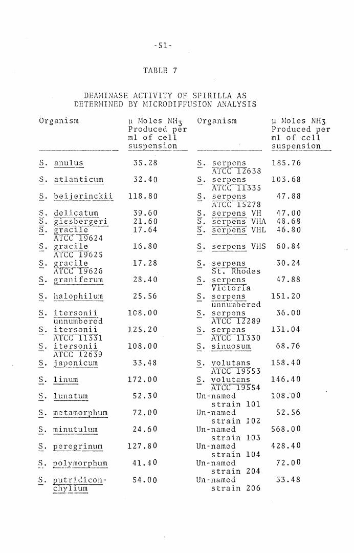
TABLE 7
DEAMINASE ACTIVITY OF SPIRILLA AS DETERMINED BY MICRODIFFUSION ANALYSIS
Organism
S. anulus
S. atlanticum
S. ?e_!:_j erincki~
S. delicatum ~L gre-.Soergeri - ____ ..:._:::._2 __
S. gracile ATCC.19624
S. grac:ile xtcc19625
S. gracile A'FC_c_ 1 ~ 6 2 6
s. zrani:ferum
S. ~a~ophiluni
S. itersonii unnumDere-d
S. itersonii ATCC 11331
S. i te·.csonii ATCC 12639
S. j aP.~nicu~
S. linum
S. lunatum
S. metarnorphum
S. minutulum
S. E_ereg_Iinum
S . pl tr;_ di con -_ _L'_lUID
µ Moles NH 3 Organism Produced. per ml of cell suspension
35.28
32.40
118.80
39.60 21. 60 17.64
16.80
17.28
28.40
25.56
108.00
J.25.20
108.00
33.48
172.00
52.30
72.00
24. 6 0
127.80
41. 40
54.00
S. serpons Arrt-CTI6 3 8
S. scrpens ATC:c 11335
S. ser_:eens ATCC 15278
S. serpens VH "S". serpens VHA EL serpens VHL
S. serpcns VHS
S. ~-E:!_!j)CnS ~-RliOdes
S. serpens VlctorTa
S. ~~~ns unnumbered
S. se~ens ATCC1:2289
S. serpens ATCC tl.330
S. sinuosum
S. volutans ATCC 19553
S. volutans A'(CC 19554
Un-named strain 101
Un-named strain 102
Un-named strain 103
Un-named strain 104
Un-named strain 204
Un-named strain 206
µ Moles NH3 Produced per ml of cell suspension
185.76
103.68
47.88
47.00 48.68 46.80
60.84
30.24
47.88
151. 20
36.00
131. 04
68.76
158.40
146.40
108 .'00
52.56
568.00
428.40
72.00
33.48
-52-
latter 4 species, a froth of bubbles appeared only
after 15 to 30 minutes incubation, with uninoculated
control tubes being negative in all cases. All strains
·were phosphatase po si ti ve except S. halophilum, S. min-
~.!~~lum, and the 3 un-named marine strains 101, 102, and
206. As for the production of hydrogen sulfide from
cysteine all strains were positive except the marine
strains of ~· atlanticu~, S. !1alOj)hilum, S. minutulum,
and _?_. j ap~nicum. All strains were able to grow in the
presence of 1% bile except S. ~-~_lutans_, S. al!-_ulus, and
S. dclicatum.
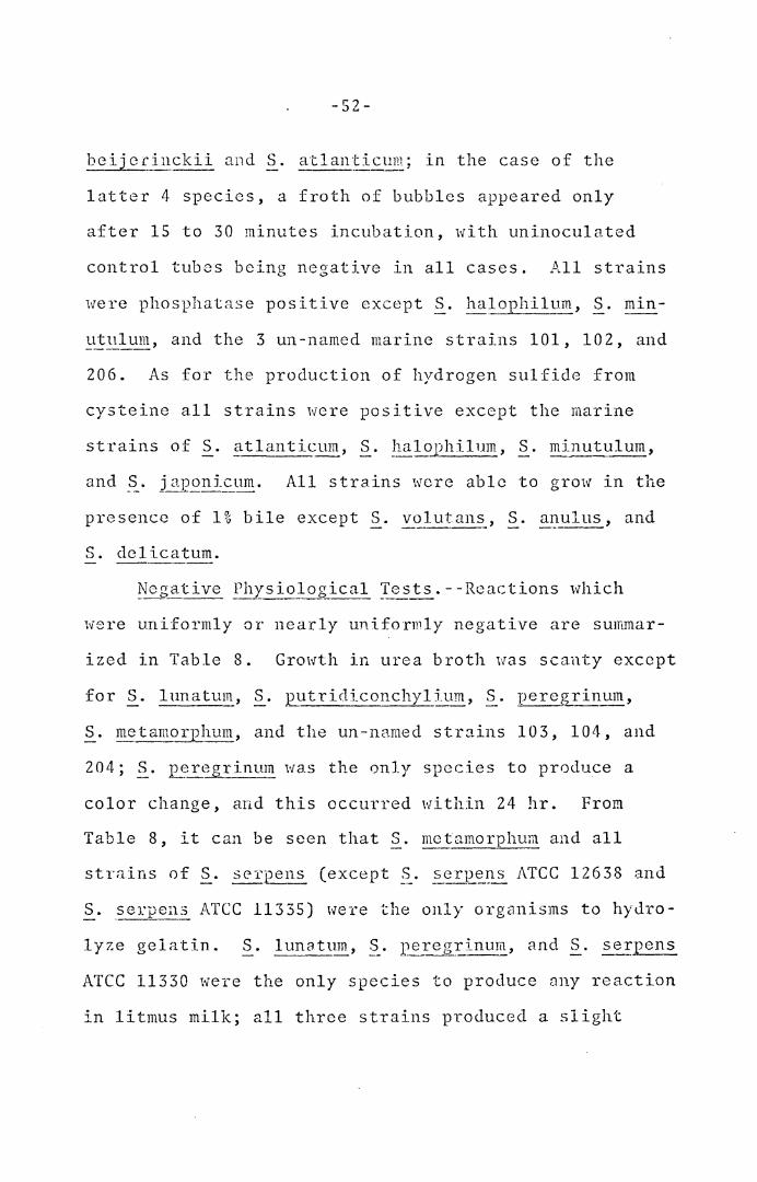
were uniformly or nearly uniformly negative are summar-
ized in Table 8. Growth in urea broth was scanty except
for S. lunatum, §_. putridiconchyl_ium, §_. pereg_~in_ym,
S. metamorphum, and the un-nmned strains 103, 104, and
204; ~. i~regrinum was the only species to produce a
color change, and this occurred within 24 hr. From
Table 8, it can be seen that ~· mctamorphum and all
strains of §_. serpens (except §_. serp_~i:is ATCC 12638 and
S .. ser12ell.3 ATCC 11335) were the only organisms to hydro-
lyze gelatin. .§_. lunatum, §_. peregrinum, and S. se~pens
ATCC 11330 were the only species to produce ::my reaction
in litmus milk; all three strains produced a slight

-53-
TABLE 8
NEGATIVE PHYSIOLOGICAL REACTIONS OF SPIRILLA
Indole production
Hippurate hydrolysis
Amylase
Sulfatase
Casein hydrolysis
Ureasc (except~· £_~regrinum.)
Gelatin hydrolysis (except S. rnctamorphum and 8 out of 10 strains of S. serpens.)
Aesculin hydrolysis (except S. 129J:ymorphum, S. i~crsonii, and S. ~E_~rinu~~·)
Reactions in litmus milk (except S. lw1atum, S. percgrinum, and §_. serpcns ATCC 11330.) - ----- -
Acid reaction from sugars (except S. itersonii, S. gracile, s. 1~E~gxinu~, and the un-namcd strain 204. T
-54-
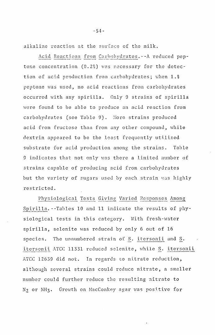
alkaline react ion at the surCLlce of the milk.
tone concentration (0.2%) was necessary for the detec-
tion of acid production from carbohydrates; when 1.%
peptone was used, no acid reactions from carbohydrates
occurred with any spirilla. Only 9 strains of spirilla
were found to be able to produce an acid reaction from
carbohydrates (see Table 9). ~·lore strains produced
acid from fructose than from any other compound, while
dextrin appeared to be the least frequently utilized
substrate for acid production among the strains. Table
9 indicates that not only 1:rns there a limited number of
strains capable of producing acid from carbohydrates
but the variety of sugars used by each strain «1J.s highly
restricted.
Sp~rilla. - -Tables 10 and 11 indicate the results of phy-
siological tests in this category. With fresh-water
spirilla, selenite was reduced by only 6 out of 16
species. The unnumbered strain of S. itersonii and S.
_it_er~_~mi"!:_ ATCC 11331 reduced selenite, while S. itersonii
ATCC 12639 did not. In regards ~o nitrate reduction,
although several strains could reduce nitrate, a smaller
number could further reduce the resulting nitrate to
Nz or NH3. Growth on MacConkey agar was positive for
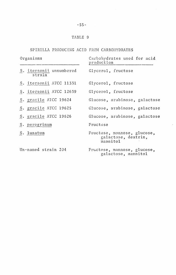
-55-
TABLE 9
SPIRILLA PRODUCING ACID FROM CARBOHYDRATES
Organisms
S. itersonii unnumbered strain
s. itersonii ATCC 11331
s. itersonii ATCC 12639
s. o-racile ~---
ATCC 19624
s. gracil~ ATCC 19625
s. _gracile ATCC 19626
s. percgrinum
s. lunatum
Un-named strain 204
Carbohydrates used for acid production
Glycerol, fructose
Glycerol, fructose
Glycerol, fructose
Glucose, arabinose, galactose
Glucose, arabinose, galactose
Glucose, arabinose, galactose
Fructose
Fructose, mannose, glucose, galactose, dextrin, mannitol
Fructose, mannose, glucose, galactose, mannitol
-56-
the follm·iing strains of §._. ~~~rpens: the unnumbered
strain, Victoria, VI-I, VHA, VHL, VHS, and the St. Rhodes
strain. Growth in MRVP broth was positive for the fol-
lowing strains: Victoria, VH, VHA, VHL, VHS and St.
Rhodes. On Seller's slants, VHL, VHS, and St. Rhodes
strains of§_. _?-~-~ave positive growth reactions.
Growth of spirilla on Seller's slants resulted in all
cases in the production of alkaline slants and greenish
colonies. The 2 un-named strains 103 and 104 produced
alkaline butts in addition to the alkaline slant reaction.
The strains of sp i ri 11 a '·ihich gave a positive growth
response on TSI agar did not produce any gas or color
changes. Ribonuclease, dcoxyribonuclease, or both
were formed in several species. Very few of the fresh-
water species possessed the ability to grow in the
presence of 1% glycine, in contrast to the marine species,
where every strain except ~· beijerincki~ and strain 204
gave a positive response. With regard to marine spirilla,
most tests appeared to be almost uniformly negative for
the various species except for the reduction cf selenite,
growth in MRVP broth and grm·;th on TSI slants: no gas
or color changes occurred in the latter.
{\.bili !l. o~ §_. lunatum to Grow in Fresh-y~ater or
Sea Water.--S. lunatum was the only strain of spirilla
capable of growing in either fresh-water or sea water
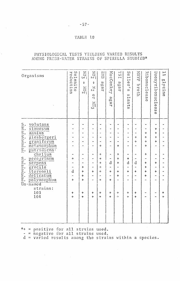
-57-
TABLE 10
PHYSIOLOGICAL TESTS YIELDING VARIED RESULTS M.fONG FRESH-WATER STPJ\I NS OF SP I RI LLA STUDIED*
S. volutans S... s111l"1 .. o sum S-. anul llS--S. gTe s b _e r g e r i ·s. grani fcrum s. ineT~noipll um s. py_~}~f(LJ~con=-
chyl ium S . 2i..t:§g r I!lii~ S. ~-~Epcn~ -;:..- . 1 0 • g_£_~~c L ___ e EL iterson.ii s-. C1cTi cat um ~. ooI~_~1·p-l1um Un-nc;med
strains: 103 104
+
d
+
+ +
+ + + +
+
+ + + +
+ +
+ +
+ +
*+ = =
positive for all strains used. negative for all strains used.
+
+ +
d +
+ +
+
+ +
+
+ ~:-
+
+ +
d d
+ +
+ +
+ +
+ + + +
+
+ + + +
+ + + +
+
+
d -- varied results among the strains within a species.
+ +
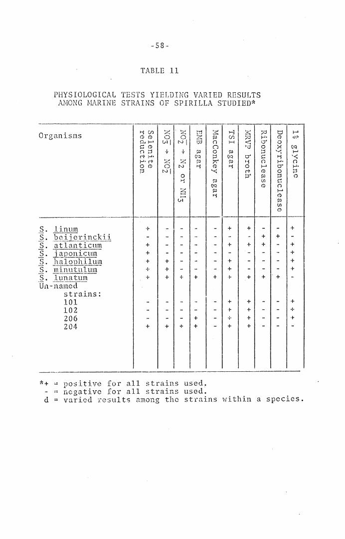
-58-
TABLE 11
PHYSIOLOGICAL TESTS YIELDING VARIED RESULTS AMONG 0L\RINE STRAINS OF SPIRILLA STUDIED*
·-------,-----.,.---:---,---, " "
t-3 I ~I Organisms
S. 1inum S. _be i ·j erinckii S. atlanticnm S. i.~12-Ql_li~um S. ).:1:_al QDh i 1 Ull} S. rninutulum S. lunatum Tfo-named -
strains: 101 102 206 204
1-i en (1) (1) p.. 1-• ~ (1) () :::l er I-'· I-'· rt 0 (1)
~
+ -+ + + + +
---+
z 0
v-1 I + ~~ L. .•
0 NI
----+ + +
---+
~-.<'.'.., 0
NI + z N 0 I-!
~ ........ v1
------+
---+
I I
+
+ +
i
*+ - positive for all strains used. - negative for all strains used~
3:: Pl en n H n 0 ?l ~ CIQ ?;"' Pl (i) >-i '-<
("..) C7Q Pl 1-i
+
+
+ +
+ +
- + + + +
~-· .,.--,. :;;:J f-'· < C) ,..d 0
~ C) ~ >-i n 0 I-' rt (i)
~ ?l Vl (i)
+ +
+ +
+ +
+ + + +
d f-' (i) c:J;.=> 0 >< C7Q ~~ I-' >-i '-< f-'• n C) f-'• 0 ::; ~ (') c n f-' (i) ?l Vl (!I
+ +
+ + + +
·+
+ + +
d = varied TC~'._;ul ts among the strains ,,.,ri thin a species.
-59 -
media; therefore, both types of water were employed in
the characterization tests for this organism, with
identical results in all cases.
De amina ti on _and P i_g_~~-1:!:..~ Pro duet ion wi.!_b-_ Aromatic
Amino Acids.--Deamination of aromatic amino acids was
uniformly negative; however a number of the strains could
not be examined for the specific color reaction because
of the production of distinctive water soluble pigments
which would have obscured the results (Table 12). S.
percgrinum was unusual in that it was the only species
to pro~uce such a pigment in the presence of phenylalanine.
The un-named strain 204 was the only strain to form a jet
black pigment on tyrosine. This pigment could be deco-
lorized by treatment with sodjum hydrosulfite; because
cf this, it is possible that the pigment was melanin.
Salt Tolerance.--All fresh-water strains were inhib-
itcd by the addition of 3% or greater NaCl to the PSS
broth; tolerance to lower concentrations was not determined.
All of the marine strains were capable of growth in 9.75%
NaCl; S. _!unatt1:!!!_, S. halophill:1-m, and the un-named strain
204 were able to grow in a concentration of 12.75% NaCl.
Base Corr .. position of D~'JA. --DNA was isolated f-rom all
42 strains of spirilla and the base compositions, expressed
as moles % G + C (Tm) are presented in Tables 13 and 14
in ascending order.
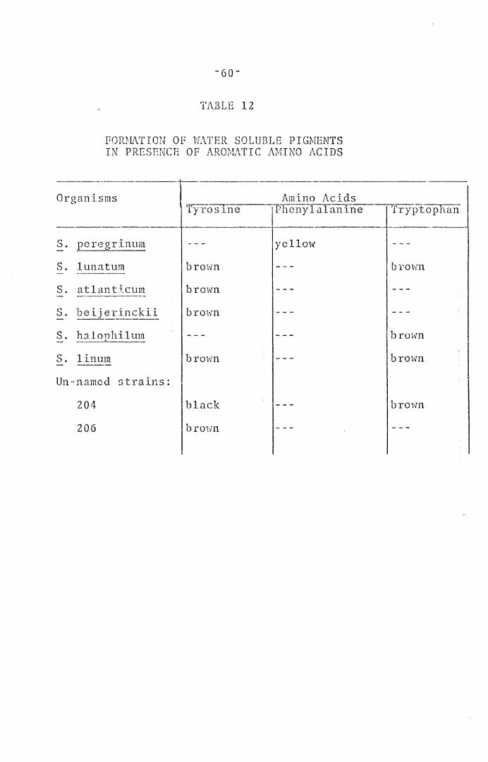
TABLE 12
PORMATION OF WATER SOLUBLE PIGMENTS IN PRESENCE OF AROMATIC AMINO ACIDS
Organisms
S. peregrinum
S. lunatum
S. atlant5_cum
S. halophilum
S. linum
Un-named strains:
204
206
'ryros1ne
brown
brown
brown
brown
black
brm·m
Amino Acids Phenylalanine
yellow
Tryptophan
brown
brown
brown
brmvn

-61-
TABLE 13
SPIRILLA POSSESSING DNA BASE COMPOSITION VALUES FROM 38 TO 52 MOLES % G + C
Species Strain Tm (C) ·----
s. vol utans ATCC 19553 84.8 s. vol utans ATCC 195S4 84.9 s. halop1111um ATCC 19192 86.7 IL minutufl1m- ATCC 19193 86.6 s. j ~onicum ATCC 19191 8 7. 9 Un-11amcc1-- 102 87.9 Un-named 206 8 7. 8 Un-named 101 88.0 s. ?e~j erinck_~~ ATCC 12754 88.6 s. atlanticum ATCC 12753 89.0 s. linum ATCC 11336 88.9 s. serpe~!_lS ATCC 1.2638 89.9 s. scrnens ---L-- ATCC 15?.78 90.0 s. se r:.pens VH 90.0 s. serpens VHA 89 .. 9 s. se_:£pens VHL 90.0 -~;-_ ~er~~s VHS 89.8 s. scrpens Vic Loria 89.9 5-. serr_e1~-~ Un-numbered 89.7 s. scrJ2e~~ St. Rhodes 90.1 s. se~.r~ens ATCC 11335 90.3 s. putridiconchylium ATCC 15279 90.6
·----
9-: 0 GC
33 38 42 42 4S 45 45 46 47 48 48 so 50 50 50
. so so 50 so Sl 51 52
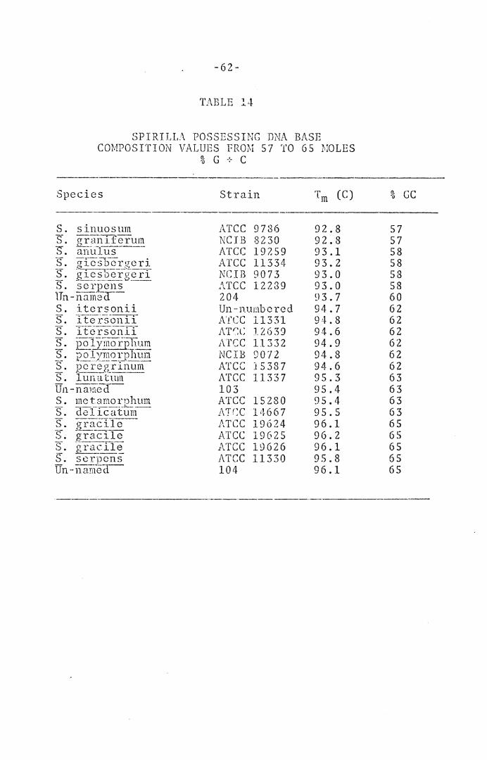
-62-
TABLE 14
SPIRILLA POSSESSING DNA BASE COMPOSITION VALUES fRmr 57 TO 65 MOLES
% G + C -------------------------Species Strain T m (C)
s. sinuosum ATCC 9786 92.8 s-. g~:ah if~rui~ NCIB 8230 92.8 s. anulus ATCC 197.59 93.1 s. gfesFcr!!eri ATCC 11334 93.2 s. ~--r-~----~"L..·--. NCIB 9073 93.0 gicsoenrer1 s. --··-------""--scrpens ATCC 12239 93.0 Un-nam·2-a-- 204 93.7 s. itcrsonii Un-numbered 94.7 s. i tej'.-5-onii Arce 11331 94.8 s. T t e1:.-s-o n if i\Tr:l: 12639 94.6 s. po 1}Ti'no r:pl1um Arce 11332 94.9 s. ----- ·-·- 94.8 polymorphura NCIB 90 '/ 2 s. pc 1:·~8Xli1 u I1!. ATCC 15387 94.6 s. lunutum ATCC 11337 95.3 un-na1rr8lf- 103 95.4 s. mctamorphum ATCC 15280 95.4 S.
a:-_ .. ____ -----elicatum /\.T(:C 1ii6 6 7 95.5
s. gra_ci1c -- ATCC 19624 96.1 ·s. gracile ATCC 19625 96.2 s. -~raciie ATCC 19626 96.1 Q____ __ s. sorpcns ATCC 11330 95.8 Un --11::ini-e<l- 104 96.1
·----------- ---
% GC
57 57 58 58 58 58 60 62 62 62 62 62 62 63 63 63 63 65 65 65 65 65
DISCUSSION
Limits of Genu~ and ~Jecies Variation. Unlike
the work of Williams and Rittenberg (1957), which
·Has based largely on the examination of their own
isolates, and the more recent investigators who
have isolated, described, and named several new
species of the genus Spiri}lurn, the present study
involved a comprehensive examination of all strains
presently available from public collections, plus
some strains from private collections. It is, how-
ever, unfortunate that most of the species are cur-
rently represented by only a single strain, the type,
except in the cases of ~· se_ryen~ (12 strains), S.
gracile (3 strains), ~· itersoni_h (3 strains), and
S. voll~~_ans (2 strains). For some species, such as
S. virginianum and S. curvatum: a type strain could
not be obtained from a collection even though the
collection carried the name of the strain in its
catalog. One possible reason for this is the diffi-
culty of preservation by lyophilization, although
lyophilized cultures of the species S. · graniferum,
S. :e_oly1:10£phum, and ~· giesbe.!::_gc_!_:!:_ were received
-63-

-64-
from the NCI.l3; a.ttempts to obtain technical details
of the method used were unsuccessful. The ATCC has
been unable to maintain strains of spirilla by lyo-
phi li~ation. Examination of several strains belong-
ing to a species is important in determining vari-
ation within a species; for example, it would be
important for the development of a diagnostic key to
be able to know those characteristics that were
positive or negative for 95 - 100% of the strains,
as some of these might represent good diagnostic
characters. Obviously tests yielding mixed react-
ions among the strains of a species would not be of
suitable diagnostic value for the species. With
regard to the two strains of ~· volutans, the type
strain (Pringsheim strain) was first obtained in
mixed culture from the cooling vat of a sugar beet
refinery in England in 1951; this strain was later
obtained in pure cultu~e by Rittenbcrg and Ritten-
berg (1962). The Wells strain of S. volutans was
obtained in mixed culture and isolated in 1964 from
pond-water in Virginia (Wells 2nd Krieg, 1965). It
is notei·.:orthy that al though the t1vo strains originated
from different continents, and although thirteen years
separated their discovery, they proved to be identical
with respect to all the morphological, physiological 1
-65-
and biochemical characteristics used in the present
investigation. In the cases where species were repre-
sented by only one strain, that strain was always the
nomcnclatural type, that is, the strain to which the
nainc of tlle species is pennancntly assigned. It 1vas
felt to be highly desirable to determine the char-
acteristics of such name-bearers because these strains
serve as permc;~ncnt exaTt1ples o:.C 11Jhat is meant by the
various species, although they are not necessarily
typical of the species. An important aspect of
future work, however, would be the isolation and
chara.ctcri zatio:i1 of new strains of spi rill a be long-
ing to species which previously had been character-
ized solely on the basis of the type strains.
The Usefulness of Sts.nclardized Conditions in -- ----·--- -- -- - --·-·
gharacterizing Str.:_::i-_:~~~~ ~i Sj)irilla. The present
investigation of the genus SE_b:illum employed stand-
ardized conditions in the study of all the strains
of spirilla, with the object of providing a valid
comparison of the various strains. Lack of uniformity
in the methodology among previous investigators has
IilJ.de such a comparison very difficult. For example,
previous investigators obtair!ed a number of morpho-
logical measurements for many of the spirilla, but
in a number of cases the morphological climcnsions were
-66-
either obtained £rem stained cc'lls (Williams and Ritten-
berg, 1957; Watanabe, 1959; Terasaki, 1961) or from
living cells (Gicsberger, 1936); in the case of the
latter, the method of observation (brightficld, phase
contrast, or dark field) was not specified. Measure-
ment of stained cells is not as desirable as for liv-
ing cells, because of the distortions produced by dry-
ing and flattening. In the literature, a variety of
media and times of incub2.tion have been used as a
basis for morphological measurements; e.g. Giesberger
(1936) used peptone water m0dium and examined the
cultures after 24 hr. incubation at 30C; Williams and
Rittcnberg (1957) employed a nutrient broth-yeast
au to lys a ti') bro th (time of incubation was not specif icd) ;
1Vatanabe (1959) employed bouillon, peptone-water and
other unspecified media, which contained 2.5% (w/v)
NaCl, besides the two types of artificial sea water
media for purposes of identification, but he did not
specify which was employed for morphological measure-
ments, and.the time of lncubatio11 was not specified;
Canalc-Parola, Rosenthal, and Kupfer (1966) employed
a medium composed of peptone> yeast extract, Tween 80,
KzHP04, and tap water, and the time of incubation was
not specified. In the present investigation, a stand-
ard medium and time of incubation were employed for
-67-
morphological measurements, and all observations were
made of living cells by phase contrast. Growth curves
were not determined for any of the spirilla, and there-
fore one cannot say that morphological dimensions
were obtained from cells growing in the log phase. It
is realized that cells growing in the log phase are
normally more uniform in size and shape than cells from
other stages of growth; however, since conditions
of medium, temperature, age and size of inoculum,
time of incubation, and method of observation were
clearly defined, the stated morphological character-
istics should be reproducible; moreover, since the same
conditions were applied to all strains, a valid com-
parison of the spirilla can be made.
In the literature, a variety of media have been
employed for the determination of physiological character-
istics, such as nitrate reduction, catalase production,
and gelatin hydrolysis. Possibly even more important
was a lack of details concerning the methodology em-
ployed for the performance of the various tests. For
example, in the 7th edition of Bergey's Manual (Breed,
Murray, and Smith, 1957) a number of characteristics
were presented for each species but many details were
no-c explained; e.g. the nature of the media employe<l
for physiological tests; the age of the cells employed;
- 6 8 ·-
the length of time for which certain tests were all0i·1ed
to proceed (especially important in the catalase test) .
Gicsberger (1936) used peptonc water but did not specify
testing techniques; Williams and Rittcnberg (1957) used
nutrient broth-yeast autolysate medium but did not
specify methodology for physiological tests; Canale-Par-
ola, Rosenthal, and Kupfer (1966) did not present details
of their methods for determining catal~;se p.roJuction;
Watanabe (1959) did not present details for determin-
ation of gelatin liquefaction, indole production, reduc-
tion of nit:cate, and cat:ilase production (the latter
being especially important since cultures were reported
as catalase-negativc). Pretorius (1963) did specify
much of his mcthoJolo.~y, in that nitrate broth (Difeo)
was used for <letermirwtion of nitrate re<luction, nut-
rient gelatin (Difeo) fo1 gelatin liquefaction, and
growth on nutrient agar for catalase production; more-
over, times of incubation were given.
In the present co~prchensive study, the types of
me<lia, incubation periods, an-1 methodology for the
dete1mination of react.ions have been clearly specified
for all st rains, so that a val id bas is of co;npar is on
exists. To indicate the import<mce of specifying con-
ditions, a number of examples may be presented. The
time element was critical in determining the production
-69-
of water··soluble pigments from aromatic amino acids,
since the jet black water soluble pigment produced by
the un-name<l strain 204 appeared only after five days
growth. Attempts to reproduce this work would result
in erroneous results if the experiment were to be
terminated cailier. The catalase test, if examined
immediately after the addition of I-I202, gave a neg-
ative reaction in several cases, whereas if the test
were cx~mincd after 30 minutes, a definite froth of
bubbles could be seen, as co1;1pared with uninoculated
controls.
Concc)r<ling acid re<lctions from sugars, the neces-
sity of reducing the pcptone concentration in order to
demonstr0.tc an indicatr)r ch2nge is important due to
the alkaline reactions p rodl1ced by th cs e organisms
growing in the presence of pcptone. Acid reactions
were not detected for any of the spirilla when the
peptone concentration was 1.0~. Because certain com-
pounds may hai.re toxic eff0~cts, the need for specifying
in detail ~oncentration of the mediu1a components cm-
ployed is necessary; for example, pl1osphate is toxic
to S. voluta~s at concentrations gr~ater than 0.0029 M
(Wells, 1966). 1\nother example is the necessity for
explaining whether the nitrate test was carried out
under aerobic or anaerobic conditions, since oxygen
-70-
is known to inhibit nitrate reJuction in some groups of
bacteria (Doelle, 1969).
The possibility exists that sorne of the conditions
employed in the present study are not optimal for detec-
tion of certain characteristics, as perhaps in the employ-
ment of Difeo urease broth for the urease tests or in the
use of only four days of incubation to determine gelatin
liquefaction; nevertheless, because the same conditions
were used for all strains, a valid frame of reference
for comparison docs exist.
Present Data With Earlier Results. In most cases, re-
sults obtain~d in the present study coincided with those
in the literature, but there are a few cases where dis-
crepancies were found. A contTadiction involving the
production of catalase concerned five ~~pecies previously
reprnted as catalase negatjvc: S. ~?-l~p_h:i.lum, S. minu-
~~-1~~' and ~. j~£onicu.m (Wa to.nabe, 19 59) ; .§._. metamorphum
(TerasK:i, 1961; and~· ~~ridiccn~hylium (Terasaki, 1961).
All of these but S. j apo~~~uir: have e:xh i bi ted s t·rong
catalasc reactions; -~· J_a12~-~~~E.:-1~1.'.. exh.ibi ts a weak ro-
action. One possible explanation for these apparent
contradictions in the cntalD.sc re<:J.ctions may be the
same as that suggested by Canale-Parola, Rosenthal,
and Kupfer (1966) for the strains of S. _g_~·a£_~~~-; they
-71-
suggested that s~Jculturing over a period of time may
have selected for cells producing higher levels of
catalase, since all their isolates gave negative
catalasc tests immediately after isolation, while a
definite positive reaction was observed in all strains
after prolonged subculturing.
Such considerations of the possible changes that
may occur upon con t.inw:-d transfer are not only applic-
able to the catalase reactions, but have a more general
implication as well. In the present study, one has no
assurance that a strain used is identical in those
characteristics to the same strain upon its initial
isolation. Fer example, the type strain of S. _!~1n __ atum
at present can grow in PSS broth in tl1c ~Hcscnce or
absence of S8a water; yet Williams and Rittenberg
(1957), who i.3olated this strain, reported that this
strain could not grow unless at least 50% sea water
were present; raorcover, only natural lather than syn-
thetic sea water was effective. It is possible that
a difference in formulation of synthetic sea water is
responsible for the latter effect (no for~ulation for
artificial sea water is given by Williams and Ritten-
bcrg) but it is also possible that the strain has
changed during the thirteen years between its isolation
and the present study. In the cases of discrepancies
-72-
concerning these and other tests, it is equally likely,
however that differences in methodology may be respon-
sible, and no firm decision can be made.
Although the use of nitrate as an electron ac-
ceptor under anaerobic conditions was reported for
S. £Olymorphum by Williams and Rittenbcrg (1957), in
the present study, negative results were obtained
for this organism.
S. delicatum was reported by Leifson (1962) to
possess predominantly a single polar flagellum; our
own darkfield observations of this organism indicate
the presence of visible helical structures, suggest-
ing the pTesence of bundles of flagella. Williams
(1960) was able to detect flagellar bundles in
spirilla previously reported by her to be monotrichous,
and therefore the use of the electron microscope may
clarify the situation with S. delicatum. The problem
is taxonomically important since monotrichous helical
cells would be classified in other genera such as Vibrio
and Cawpylobacter.
Morphological discrepancies arose in regard to
S. E~:!_~!-~dic_<?_l]:_~!piu.m, §_. ~12on~cum, S. haloph~lum,
an<l S. rnLrn tulu111 (Watanabe, 19 59) . From Tables 4
and 5, it can be seen that the cell diameters obtained
in the present study differs from those previously
-73-
reported. The latter were 0,8 - 1.0 µ, 1.2 " 1.5 µ,
0,5 - 0,6 µ, and 0,2 ~ 0,3 ~. respectively. The dif-
ferences could possibly be attributed to the culti-
vation media, leneth of incubation, or methods of
observation.
In regard to hydrogen sulfide production, Terasaki
(1961) reported that S. metamorphu~ did not produce
hydrogen sulfide; however, his methods were not speci-
fied; s incc we obtained positive results when S. ~10ta
morph11~~ was grown in the presence of cysteine, we feel
that differ~nces in methodology may be responsible for
this discrepancy.
A discrepancy concerning the % GC composition of
spirilla was found in the case of S. li_J?._2;lm. In a
review by Hill (1966), an mrnw1bered strain of S. linum
was reported to have a % GC content of 28-30. Our
present investigation of the type strain of S. linum
revealed a % GC content of 48. Ho1.vcvcr, since the
strain reported by Hill was not clearly identified,
the possibility exists th~t this strain may not have
been a strain of S. linum.
p_~v is ion of Genus Sp i ri l lum on the ~as is of Single,
Highly-W"e~ghted Character~. In evaluating a taxonomic
character, the number of genes involved (or at least
some approximate estimate, based on knowledge of the
- 7 4 -
number of enzymes involved in c:x-pression of the char-
acter) whould ideally be determined. The more genes
invclvecl, the greater would be the taxonomic weight.
Some characters nay individually carry great weieht -
such as sporulation, photosynthetic metabolism, me-
thane fermentation, etc., which involve many enzymes.
Do separate characters of sufficient weight exist
;dthin genus Spirillum to justify a division of the
genus into two or more genera?
A possible division of the genus Soirillum into _ _. ___ _ two genera might be s uggcs tc d on the bas is of a
distincti·Jn between obligately microacrophilic vs.
aerobic spiril._ 1_a. Th0 only nwmbers of the obligately
microaerr)philic cc:.tegory wuuld be §_. :1:7:'.)lu(:a~-' and
such a goneT ic group mi~ht IJe further justified by
the low % GC value (4 % l0\1er than any other species
of spirilln;i1). One hesitates to ·weigh obligate
microacrophilism highly, however, because oxygen is
used as electron acceptor _for both aerobic and micro-
<:u~rophilic organisms; thus the difference may be a
5t1.1)erficiaI quantitative di:fferr~nce rather than a
mi.:1 n' fw1da;rrental qualitative difference. To suppoYt
this~ spirilla intermediate between the two categories
appear to exist: the t1·;0 species ~· _giesbcrgeri and
-~· _gra_~_lc appe::::.r to cbr,10nstratc facultativc micro-
<c1eroph.ilic responses in that the former gro1-1s in a
-75-
microaerophilic band a few millimeters below the surface
of PSS medium containing 0.15% agar, while the latter,
growing on PSS agar, forms colonies which ar~ partially
embedded beneath the surface. This partial submergence
of colou:Les suggested a rnicroaerophilic nature to
Canale-Parola, Rosenthal, and Kupfer (1966).
A requirement for sea water might be suggested
as sufficient basis to create separate genera for fresh-
water and marine ~pirilla. In regard to requirement
for sea water, the nun:bcr of genes invol 11cd in distin-
guishing these organi~;ms fro,1: fresh-;va ter forms is not
known; moreover, the di5tinguishing feature separating
marine bacteria frou most nonn'.arin0 species is generally
agreed to reside in the possession of a Tequirement for
Na+ by the marine organisms. A requirement for natural
sea water, a characteristic weighted heavily by Williams
and Rittenbcrg (1957), may carry taxonomic weight
since these a.utho.cs reported that continue cl sub cul tu re
in a synthetic sea water medium did not permit continued
growth of the marine spirilla; however, Watanabe (1959)
was successful in isolating and subculturing four species
of :11:nine spirilla on a synthetic sea >iater; n:orcover,
in the present study we have successfully subcultured
and examined these species, as well as those reported
by Williams and Rittenberg, on an artificial sea witer
-76-
medium. A possible explanation for our success in
obtaining growth on synthetic sea water may be that
continued subculturing over the years has selBcted
for variants 1\Ti th a reduced dependency for natural
sea water; a difference in formulation of artificial
sea water represents another possibility. It was also
observed in the present investigation that S. lunatum
had the ability to grow in media completely devoid of
any type of sea water; this may perhaps be attributed,
as previously mentioned, to continuous subculturing
over the years. In regard to this, MacLeod (1965)
states, "Although much remains to be done to elucidate
the difference3 between marine and terrestrial species
at the molecular level, enough information is avail-
able to suggest that a quite limited number of sue-
ccssive mutations could convert a marine species to
a form ~hich would not be dependent on the sea for its
survival".
The possibility exists of dividing the genus
Spirillym into different genera on a morphological
b2.sis since the spirilla are diverse ~vith respect to
cell di.ameter (see T~i.bles 1J. a.r,_J. 5). However, we
hesitate to divide the genus on this basis because
there appears to be an almost continuous spectrum
of sizes within the genus; and any grouping would be
purely arbitrary.
-77-
Another possibility for division of genus Spirillum
into two or more genera might be based on DNA base com-
position. The % GC composition of only a relatively
few strains of spirilla have been reported in the liter-
ature; these have been reviewed by Hill (1966), where
the range of moles % GC was from 28 to 61.5. Our own
investigation revealed a % GC range of 38 to 65. This
range is a large range for a genus when compared with
certain other genera of bacteria; for example, the GC
values of the genus Pscudomonas range from 57 to 70
moles% (Mandel, 1966); that of the genus Bacillus
from 35 to 51 moles % (Marmur, Falkon, and Mandel, 1963);
and thu.t of the genus Escherichia from SO to 53 moles
% (reviewed by Hill, 1966). The genus Vibrio at one
time possessed a rather large range of % GC, (29.5 to
65 moles %, reviewed by Hill, 1966); however the genus
Vibri~ was separated, on the basis of DNA base composit-
ions and other features, into several genera by Sebald
and V~ron (1963) and such a division represents an
example of what possibly could apply to the genus
Vibrios possessing a GC range of 29.5 to
35,5 moles % were placed in the genus Campylobacter
(also distinguished from Vibrio as being microaero-
philic to anaerobic and being non-fermentative); the
remaining vibrios have been further separated into
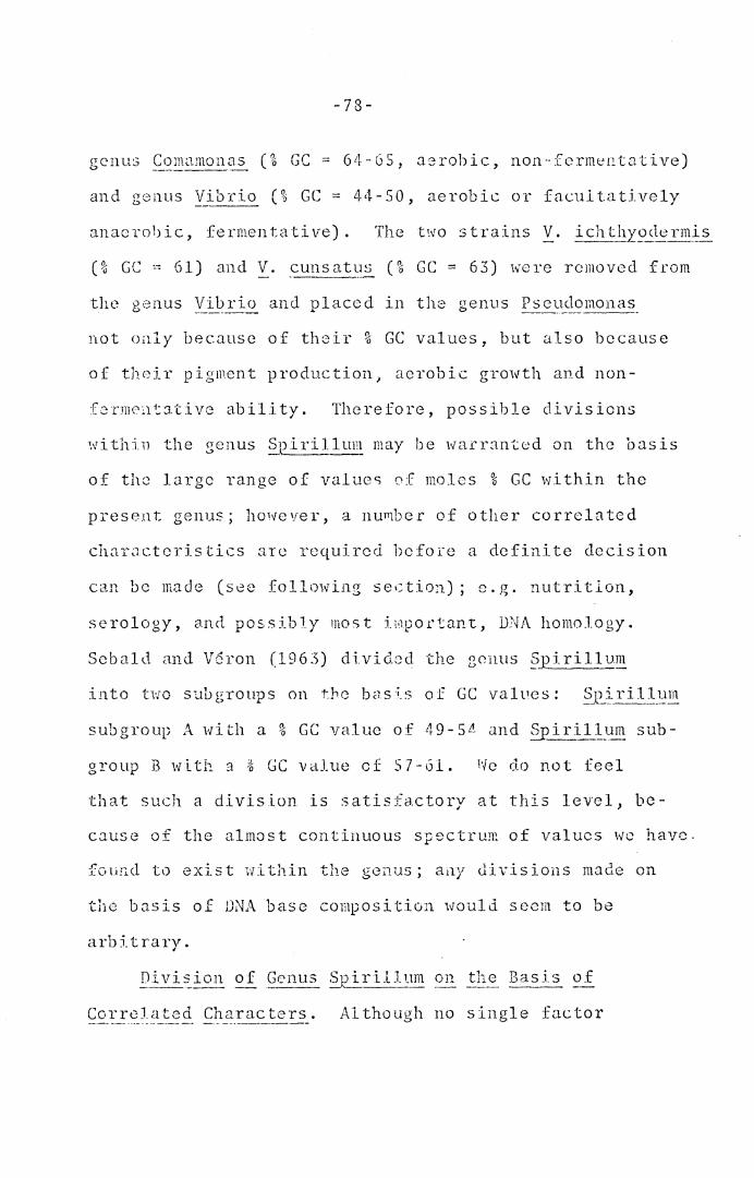
-78-
and genus Vibrio (% GC = 44-50, aerobic or facultatively
anaerobic, fermentative). The t~vo strains '!_. J:cht_1~9~l~_Im~2_
(% GC 0-: 61) and '!_. cunsa\~s (% GC = 63) were removed from
not only because of their % GC values, but also because
of their pigment production, aerobic growth and non-
fcrm0i11.:ativ0 ability. Therefore, possible divisions
within the genus Spirillu~ may be warranted on the basis
of the large range of values o:f moles % GC within the
present genus; however, a nu111bcr of other correlated
charactcris tics are required bcfoi·e a definite decision
can be made (see following section); e.g. nutritlon,
serology, and possj_bJy most irnpoctant, DNA homology.
Sebald and V6ron (1965) di.vicL;d the genus §_pirilJ_~~
into two subgrot~s on the bas~s of GC values: (' . . 1 l ~)niri. .. um _x_:~-----~~--
subgroup A with a % GC value of 49-SL' and §2i~illym sub-
group B with :i % GC value cf 57-61. 1fo do not feel
that such a division is satisfactory at this level, be-
cause of the almost continuous spectrum of values we have.
fc,un.d to exist within the genus; ail)' divisions made on
the basis of DNA base composition Hould seem to be
arbitrarv. ,
Correlated Characters. Although no single factor
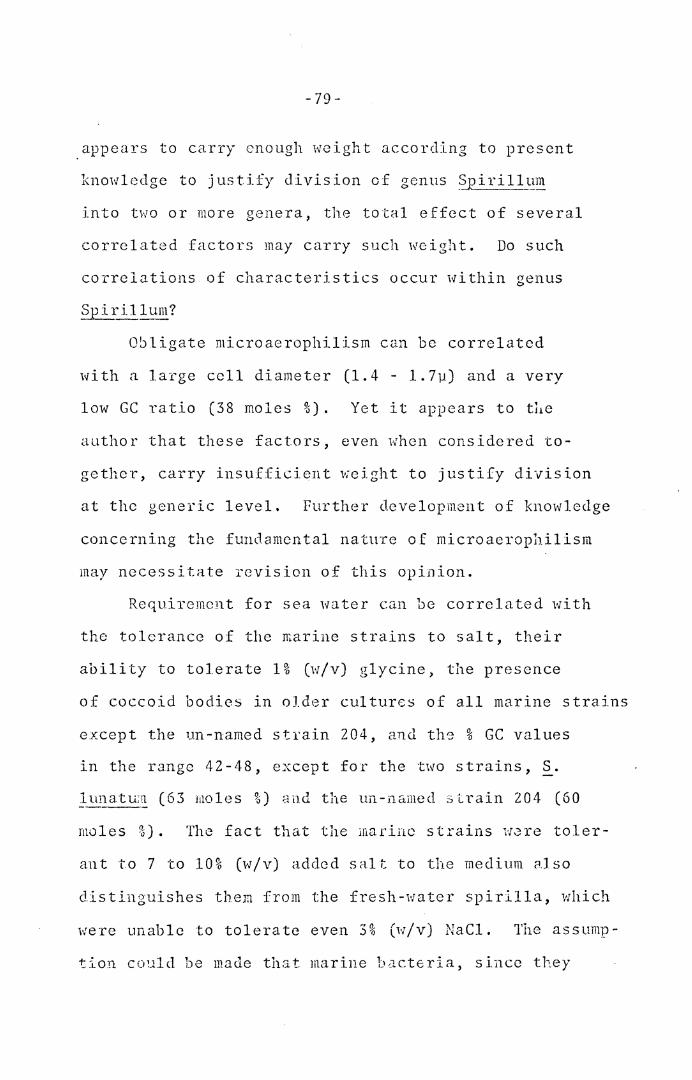
- 79 -
appears to carry enough weight according to present
knowledge to justify division of genus ~irillum
into two or more genera, the total effect of several
correlated factors may carry such weight. Do such
correlations of characteristics occur within genus
Obligate microaerophilism can be correlated
with a large cell diameter (1.4 - 1.7µ) and a very
low GC ratio (38 moles %) • Yet it appears to t11e
author that these factors, even when considered to-
gether, carry insufficient weight to justify division
at the generic level. Further development of knowledge
concerning the fundamental natuTe of microaerophilism
may necessitate revision of this opinion.
Requirement for sea water can be correlated with
the tolerance of the marine strains to salt, their
ability to tolerate 1% (w/v) glycine) the presence
of coccoid bodies in older cultures of all marine strains
except the un-named strain 204, and th9 % GC values
in the range 42-48, except for the two strains, ~-
]-_unatu:~ (63 moles %) and the un-named ;; train 204 (60
moles %) • The fact that the marine sttains 1·1-:;re taler-
ant to 7 to 10% (w/v) added salt to the medium C1Jso
distinguishes them from the fresh-water spirilla, which
were unable to tolerate even 3 % (1·1 /v) NaCl. The ass ump-
tion could be made that marine bacteria, since they
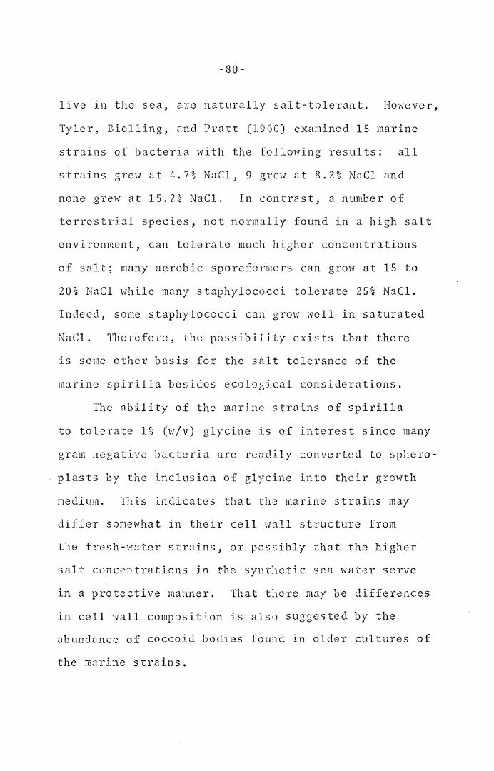
-80-
live in the sea, are naturally salt-tolerant. However,
Tyler, Bielling, and Pratt (1960) examined 15 marine
strains of bacteria with the following results: all
strains grew at 4.7% NaCl, 9 grew at 8.2% NaCl and
none grew at 15.2% NaCl. In contrast, a number of
terrestrial species, not normally found in a high salt
cnvi ronrnent, can to lent te much higher conccn tra tions
of salt; many aerobic sporeformers can grow at 15 to
20% NaCl while many staphylococci tolerate 25% NaCl.
Indeed, some s taphyl oco cci can grow 1vc 11 in saturated
NaCl. Therefore, the possibi.Lity exists that there
is some other basis for the salt tolerance of the
marine spirilla besides ecolog5cal considerations.
The ability of the marine strains of spirilla
to tolerate 1% (w/v) glycine is of interest since many
gram negative bacteria are readily converted to sphero-
plasts by the inclusion of ~lycine into their growth
meJ.ium. This indicates that the marine strains may
differ somewhat in their cell wall structure from
the fresh-water strains, or possibly that the higher
salt concentrations in the synthetic sea water serve
in a protective manner. That there may be differences
in cell wall composit~on is also suggested by the
abundance of coccoi<l bodies found in older cultures of
the marine strains.
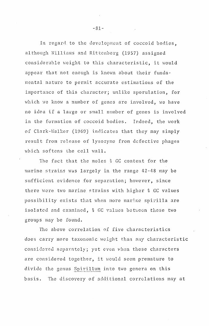
-81-
In regard to the development of coccoid bodies,
although 1Villiarns and Rittcnberg (1957) assigned
considerable weight to this characteristic, it would
appear that not enough is known about their funda-
mental nature to permit accurate estimations of the
importance of this character; unlike sporulation, for
which we know a number of genes are involved, we have
no idea if a large or small number of genes is involved
in the formation of coccoid bodies. Indeed, the work
of Clark-Walker (1969) indicates that they may simply
result from release of lysozyme from defective phages
which softens the cell wall.
The fact that the moles % GC content for the
marine strains was largely in the range 42-48 may be
sufficient evidence for separation; however, since
there were two marine ~trains with higher % GC values
possibility exists that when more mar:bJ.c spL:illa are
isolated and examined, % GC v::i.lues bet~~·ecn these two
groups may be found.
The above correlation of five characteristics
does carry more taxonomic weight than any characteristic
considered separately; yet even when these characters
are considered together, it would seem premature to
divide the genus §.tlEillum into two genera on this
basis. The discovery of additional correlations may at
-82-
some future date provide sufficient support for a
generic separation.
Description of Genus Spirillum (Ehrenberg
183_9_, ~) Based on :Morphological and Physiological
Results. On the basis of the morphological and
physiological results, the following description of
the genus ~irillum can be offered: Rigid, helical
cells, with cell diameters ranging from 0.25 to 1.7 µ,
cell lengths ranging from 3 to 5 µ (as in S. delicatum)
to 14 to 60 µ (as in S. volutans), and number of turns
ranging from less than one to five. Motile with bipolar
polytrichous flagella. Gram Jtegative. Chemoorganotrophic,
having a strictly respiratory metabolism, with oxygen as
the termj.nal electron acceptor. A few species can grow
anaerobically with nitrate as the terminal electron ac-
ceptor. Oxidase, deaminase, and catalase positive. Phos-
phatase usually positive. Hydrogen sulfide usually pro-
duced from cysteine. Indal, sulfatase, and amylase
negative. Casein and hippurate not hydrolyzed. Gelatin
usually not hydrolyzed. Urease usually negative. Acid
reaction usually not produced from carbohydrates; where
this occurs, the kinds of sugars are extremely limited.
Incorporation of natural or artifi~ial sea water into
media is required for marine species. Aerobic,
facultatively microaerophilic, or obligately micro-
aerophilic. Optimum temperature, 30 to 32 C, with
-83-
no growth at 10 or 45 C. Fresh water species do not
grm'l 1-;i th 3% (w/v) NaCl, while marine species grow
in up to 7 to 10% (w/v) NaCl.
Ph_y_:;iologi~al_ Tests Important for Species Dif-
ferentiation. Several physj.ological tests yielded
results so nearly uniform for the genus that the few
exceptions were clearly evident. Such exceptions as
failure to produce hydrogen sulfide from cysteine; urease
production; failure to exhibit phosphatase activity;
gelatin hydrolysis; aesculin hydrolysis; alkaline reac-
tions in litmus milk; ability to grow in presence of
1% glycine and 1% bile; acid reactions from sugars; and
anaerobic growth in the presence of nitrate would appear
to have. taxonomic value in differentiating the species
of spirilla.
In contrast, several physiological tests when
applied to spirilla yielded a greater variety of pos-
itive and negative reactions; these.tests also appear
to be useful in differentiating among species, and are
listed as follows: selenite reduction; DNAase and
RNAase activity; growth on EMB, MacConkey, and Seller's
agar; a:!l.J. ability to grow in MRVP broth.
Sch0me of Classification of Species of Genus
Suirillum. Using the morphological and physiological
characters as determined in the present investigation,
-34-
the following classification has been devised. It
should be emphasized that this scheme is not a diag-
nostic key; such a key follows the present section.
This fresh-water species is distinguished
by a large cell diameter (1.4 - 1.7 microns), by a
great cell length (14 - 60 microns), by the easy
visibility of its flagellar tufts by darkfield or
phase-contrast microscopy, by its obligately micro-
acrophilic nature, and by the low moles % GC content
of its DNA (38%). The organism is only weakly cata-
lase positive.
In regard to the type species of the genus
~ir~_!J:.-~m, we agree with Williams (1959) that -~·
volutans, rati1er than S. ~;11~1::.l'.:1_ 1 should be the type
species of the genus, but for different reasons.
Williams indicated that S. vo_l_utans was the only
species named by Ehrenberg in his paper to the
Berlin Academy (1830), and therefore this was said to
be dcsign&tion of type species by mcnotypy. An exam-
ination of Ehrenberg's paper, however, reveals that
two species were listed: S. volutans and S. undula.
No descriptions were given, but the organisms were
indicated as species previously classified by Mliller
in 1773 as ·1ibrios: V. ~J2irj)lurn (§_. volutan_~_) and
- 85-
V. ~_!1dula (~. ~mdula). In 1905, Stiles designated
S. vo~~;ans as the type species of genus Snirillum,
but pointed out that Ehrenberg should have retained
Milller's specific epithet, and that therefore the
correct name of the organism should be Spirillum
_!?_P.irillum. However, Rule 25 (c) of the International
Code of Nomenclature of Bacteria (International Con-
gress of Microbiology, 1966) states that a specific
epithet is illegitimate when such epithet exactly
repeats the generic name. In view of this, Ehrenberg's
name would appear to stand, and should be written:
.~i-~U!~m volutans Ehrenberg, 1830. In contrast,
Ehrenbcrg's second species should be designated as
~i~i~J:..l!l~ undula (Muller) Ehrenberg, 1830. The desig-
nation of S. volutans as the type species by Stiles
(1905) appears to be the earliest such designation of
the type. Vuillemin (1913) designated S. undula as
the type, and also the 7th edition of Bergey's Manual
(Breed, Murray, and Smith, 1957) designates S. undula
as the type.
We agree with Stiles th.at S. volu~ans is the type
sp<.~cies of the genus, not only on the bas is of priority,
but also because of the following practical reasons:
(a) Strains of S. undula arc not presently
- 86 ·-
available from any collection as far as is known. 1be
last literature references Hhere the use of S. undula
is indicated arc apparently those of Lewis (1940) and
ifyers (1940). It would seem more useful to have a
type species for which strains are available, so that
the strains may be studied by modern methods as they
develop; only in this way can the description of the
type be continually updated and made relevant to
current methodology.
(b) S. volutans can be easily identified on
the basis of its large size by the unmistakable
morphological description given by Tihrenberg (1838):
"Lange de r Sp:i. r.:i.lc l/ J 9 2 bis 1/ 4 8 Linie. Di eke e twa
1/1200 Linie." (A Linie is equivalent to 1/12 of an
inch, therefore the length of §_. ·1olutans 1vas 11 to
43 µand its diameter was about 1.8 µ).
In contrast, Ehrenbcrg's dimensions for S. w1dula
could apply to several current species; such as S.
serpcI_l_?_, §_. E~tr;dlco!~chylium, ~· &.E~nifen1n~, and§_
n1e_!_~1~0 q~~1um. In fact, even the most recent descriptions
of -~--· ~l!~~~t1.}:_?:, namely those of Giesberger (1936), Myers
(1940), Wil.liams and Rittenherg (1957), and Bergey's
i'ianual (Breed, l·!urray, and Smith, 1957), <lo not
provide suf ficicn t information to distinguish §_. ~m<lul a_
from other well-studied species. For exarnple, Giesberger
.. 8 7-
(1936) gives the following <.Jcscription of §_. un~ula:
"In peptone water extremely motile, beautiful, broadly
spiral shaped cells; width of cell 0.9 µ; length of
turn 6 µ; width of turn 3 µ. Motile by means of bipolar
flagellar tufts; volutin occurs as reserve material;
gram negative; catalase positive, non-spore forming;
scanty growth on peptone agar; colonies gray-white,
smooth; liquefies gelatin very slowly; gray-brown
growth on potato .... ". Myers (1940) describes S.
undula as having a cell diameter of 0.9 - 1.2 µ, not
liquefying gelatin, not growing on potato, not protluc-
ing either nitrites or gas from nitrates, not producing
indole or I-I2S u.nd having an optimL1m temperature of
about 32 C. Wjlliams and Rittenb~rg (1957) include
Giesbcrger's description in their treatment of this
species. Bergey's Manual (Breed, Murray, and Smith
1957) use Giesberger's description of S. undula, . -- ·-~-~
although they do incluJe the inJole and nitrate
reactions as observed by Myers.
We therefore believe that with the greater nu1;1ber
of species that arc now available, it would be extremely
difficult to identify any one organism with these descrip-
tions.
(c) Two strains of §_. volutans are presently
available: strain #19553 of the ~nerican Type Culture
-88-
Collection, and the Wells strain, available from the
stock culture collection of the Biology Dept. Virginia
Polyt~chnic Institute, BJacksburg, Virginia 24061. We
propose that the former strain be designated as the
type strain of the species.
2. .?J2l..£il]ym anul_us (Williams and Rittenberg,
1957).
This fresh-water species is also distinguished
by its large cell diameter (1.4 - 1.5 microns) but
it is strictly aerobic and its DNA co~position is 58
moles 95 G ~ C. It possesses l - 3 helical turns, is
strongly catalase positive, and p£oduces both DNAase
and RNAase actj_vity.
3. ~~i~j]}~~!!.l E_~1t£_}:_~iC::_'?E_C::_!~x--~i_u}_~~ (T2rasaki, 1961).
This fresh-water species has a moderately-large
cell diameter (1.0 - 1.2 microns) and exhibits 2 - 4
helical turns. The G + C content of its DNA is 52
moles 0ci. It forms weakly a yellow-green water-soluble
fluorescent pigment (Hylemon, personal communication),
produces hydrogen sulfide from cystein0: grows on TSI
2nd EMB agars, but does not grm·1 in MRVP broth and
cloes not produce DNA2.se activity.
1. ~2-I_~_l1-~1-~~ g__~a~if~_run'._ (Williams and Rit tenberg,
19 5 7) •
This fresh-water species is morphologically similar
-89-
to S. 1211tridiconchyli_um, but has a DNA base composiU.on
of 57 moles % G + C. Moreover, no fluorescent pigment
is produced, hydrogen sulfide is not produced from
cysteinc, growth fails to occur on TSI or HIB agars
but Joes occur in MRVP broth, and DNAasc activity is
produced.
5. Spirillum metam~!phum (Terasaki, 1961).
This fn~sh-wa ter species has a cell diameter
similar to the two preceding species (1.1 - 1.3 microns);
however less than one helical turn is present, and the
G ·~ C content 0£ its DNA is 63%. ~~reuver, an unusual
characteristic, gelatin liquefaction, is present. A
yellow-~rcen watcr·-soluble fluor~scent pigment is
formed (IIylemon, personal communication), growth occurs
on Seller's slants (alkaline reaction in slant only),
and RNAasc Rnd DNAuse activities are not produced.
6. ~pirillum _g_j.csbe_!_g~r~ (Williai11s and Rittenberg,
19 5 7) •
With a cell diameter of 1.1 ·· 1. 4 inicrons, this
fresh-water species is similar to the three preceding
sp~cies; hoi·:evcr unlike ~· 12~:_t1~~idit~.onchxli~~ and S.
gra.niferun1 (but like §_. metamo!:_e_hu~) the cells have
less than one helical turn. The G + C content of the
DNA of 5 8 moles % is .similar to that of S. _g_r_:_anifcrum.
Two most interesting charactcTistics, however, sets
this species apart from the thn~e preceding ones,
-90-
namely: its facultative microaerophilic nature and
its slow rate of growth. When 0.15% agar is incorporated
into PSS broth, a microaerophilic band of growth consis-
tently forms a few mm below the surface of the medium;
as growth increases, this band eventually moves to the
surface. In non-stratified broth, moderate growth occurs
with bottom sediment and clear supenatant. Recent results
of Hylcmon (personal communication, 1970) indicate that
in a defined medium containing succinate as carbon
source, no growth occurs unless 0.15% agar is added;
in ths latter case, a microaerophilic band forms 3 - 6
mm below the surface of the stratified medium, but even
after continucJ growth, this band fails to move to the
surface. The slow rate of growth of S. g_ies~:;r_geri in
PSS broth is unusual, and 3 to 4 days of incubation
are required to achieve a crop of cells comparable
to what most other spirilla produce in 24 hr.
7. ~J2_irill_~u~ percgrinum (Pre tori us, 1963).
This fresh-water species has a cell diameter of
0.5 - 0.7 microns and exhibits 4 - 5 helical turns.
Cocccid bodies predominate in older cultures, and it
is strictly aerobic, with no anaerobic growth in the
presence of nitrate. The G + C content of its DNA
is 62 moles %. It forms weakly a yellow-green water-
soluble :fluorescent pigment (Hylcmon, personal communica-
tion), produces urease, reduces selenite, and produces
a slight alkaline reaction in litmus milk~ but nitrate
is not reduced, no growth occurs on ~iacConkey agar and
RNAase and DNAase activities are not produced. A
yellow water-soluble pigment is produced in the presence
of 0.2% phenylalanine, an<l an acid reaction is produced
from fructose in PSS broth with reduced peptone.
8. §Rir:i.1l:_rn1~ _!tersonii (Gicsbergcr, 1936).
This fresh-water species is similar to S. ~regrinum,
morphologically, with a cell diameter of 0.4 - 0.6 1nicrons;
moreover, coccoid bodiC;s predominate in older cultures,
the G + C content of its DNA is 62 moles %, and it pro-
duces a yellow-green water-soluble fluorescent pigment
(Hylemon, personal communication). However, it exhibits
from less than one to two helical turns, and is strictly
aerobic, but cc.n grow anaerobically in the presence of
nitrate. It reduces nitrata to ammahia or nitrogen,
reduces selenite in only two out of three strains, grows
on MacConkey agar and produces RNAase and DNAase activi-
ties, but it does Eot proU.uce ur13a.se and no reactions
arc produced in litmus milk. A yellow water-soluble
pigment is produced in the presence of 0.2% tryptophan,
a bro1m wc:.tcr-soluble pieTien'..: is produced in the presence
of 0.2% tyrosine, and acid reactions are produced from
glycerol, in addition to fructose, in PSS broth with
reduced peptone.
-92-
Williams and Rittenbcrg (1957) considered two varic-
tics of 2_. Ltcrsof1:i:\._ to exist, based on their nitrogen
meLtbol ism; ~. i tcrsoni i var. i tcrsonii did not reduce
reduced nitrate. The three stfains examined in the
present investigation were all of the latter variety.
9. Sp ~011 um s ~rpe~:2_ (Mill ler, Winter 18 84) .
Strains belonging to this fresh-water species have
cell diameters ranging from 0.6 to 1.0 microns, and
possess from less than one to two helical turns. The
G + C contents of their DNA ranges from SO - 51 moles
9< 0 • Some strains produce a yellow-green water-soluble
fluorescent pigment (IIylemon, p~]rscnal conununication).
Most st rnins produce RNAr .. s e activity, g ro:v on TS I and
EMB agar and tolerate 1% glycine. Some strains grow
on ~.racConkey agar, MRVP broth and on Seller's slants
(when positive, slant only is alkaline). One strain
reJ.uccd nitrate 1veakly. Most strains hydrolyze gelatin.
Williams and Rittenberg (1957) scpardtcd S.
serpe!1s_ into two varieties based on their nitrogen
metabolism c:.nd gelatin hyclrolysis; '.2_. ,:;c __ !:.PGJ!,:>_ var.
azotum reduced nitrate weakly and did not hydrolyze
gelatin, ·while §_. ~e£_2ens var. serpe!lS did not reduce
nitrate but did hydrolyze gelatin. In the present
investigation two out of ten strains failed to hydrolyze
-93-
gelatin, and only one of these very weakly reduced
nitrate; consequcn.tly, it would appear that the dis-
tinction between var. az~:t:_~m and var. serpens is of
questionable value.
Two strains, §_. _se_!:_Pen~ ATCC 12289 and ~· ~_erpens_
ATCC 113?>0, were found to differ from the other strains
to such a degree that they were not considered in the
above classification of this species. ~· serpens ATCC
12289, is morphologically similar to the other strains
of S. ~-~1::_~ns; however, the G + C content of its DNA
is 58 moles % and it ls extremely fastidious in its
nutritional requireme:nts (IIylcmon, 1970). ~· serpel_!_~
ATCC 11330, morphologically, is much smaller than the
other~· se1_:p~ns_ strains; moreover, nitrate is strongly
reduced to ammonia or nitrogen, gelatin is not liquefied,
and the G + C content of the DNA is 65 moles %. There-
fore, we feel that these two strains should not be
placed in S. scrpens, and that their classification
should await the further results of nutritional and
serological studies currently in progress.
10. §_p_ir~ll~~~! si_~u~s~m (Williams and Rittenberg,
1957).
Morphologically, this strain is similar to S.
serp~-~ but it has one to two helical turns and a
DNA base composition of 57 moles %. In regard to its
-94-
DNA base composition, it is similar to~· _0-}H~~~~' ~·
diameter of 0.5 - 0.7 microns, it is smaller than
these species. DNAase activity is produced, but not
RNAase activity (similar to ~· granifcrum); howeveT,
hydrogen sulfide is produced from cystcine and growth
does not occur in i\lRVP broth, unlike ~· _R£aniferum.
Although the production of hydrogen sulfide and
growth in MRVP broth are not major differences when
considered alone, the additional character of morpho-
logical differences tlocs appear to be sufficient for
keeping these species separated.
11. ~p_iJ~_t]L.un _g __ ~~cilc (Can:ile-Parola, Rosenthal,
and Kupfer, 1966).
TI1is fresh-water species is distinguished by a
small c~ll diameter (0.25 to 0.30 microns) and exhibits
3 - 4 helical turns. Coccoid bodies are not formed in
older cultures. The G + C content of its DNA is 65
moles %. It does not produce a yellow-green water-
soluble fluorescent pigment. It produces RNAase activity,
reduces nitrate to nitrite, produces acid reactions from
glucose, galactose, and arabinose, but does not grow on
MacConkey or TSI agar or in M~VP broth; moreover, it . .
does not hydrolyze aesculin or reduce selenite.
It is aerobic and does not form microaerophilic
bands in semi-solid P.SS agar; although Canale-Parola,
-95-
Rosenthal, and Kupfer reported that the spirilla
developed a fe;v :mm belmv the surface of semi··solid
media, forming one to five growth discs. The thick-
ness of the discs varied from a fraction of a ~n to
several mm, and their distances from the surface of
the medium changed during incubation. Sometimes a
growth disc was present immediately under the surface.
This behavior, together with the tendency of the
spirilla to form subsurface colonies, indicates that
low oxygen pressures are optiraal for the growth of the
spirilla. It is likely that available isolates have
undergone some change in their c1xygen requirements
during intervening years, since we did not observe
these discs, and we have not observed subsurface
colonies on agar plates, although colonies which form
partially below the surface arc a common occurrence.
Canale-Parola, Rosenthal, and Kupfer also reported
that on initial isolation, ~· graci~ was catalase
negative by a microscopic test, but that after several
transfers the organisms becam8 catalase positive. In
the present study, all three strains were found to be
- •1 ·t .. 1 ~ ,. . '° . J. • " (..; <• c1 <i .:> C p 0 .> l I .. J. \, e I and many transfers have intervened
since their initial isolation. An initial negative
catalase test would seem to be taxonomically important
for this species, but the possibility exists that if a
-96-
30 minute incubation period for the catalase test, as
used in the present study, had been used after initial
isolation of S. _g_r~1c~~' a weakly positive rcsul t might
have been seen. Therefore it is difficult at this time
to assess properly the catalase test in the classification
of this snecies . ...
12. ~i..!:_:!}~~ polymorphum (Williams and Rittenbcrg,
1957).
This fresh-water species is also distinguished by
its small diameter (0.3-0.5 microns) but it exhibits
less than one helical turn and coccoid bodies prcdomin-
ate in older cultures. The DNA base composition is 62
moles % G + C; similar to S. gracjle, hut it does pro-
duce a yellow-green water-soluble fluorescent pigment
(Hylernon, personal communication), hydrolyzes aesculin,
reduces selenite (weakly) and grows on MacCoTukey agar,
but acid reactions are not proJuced with any sugar in
complex media and it does not grow on TSI agar or in
MRVP broth.
13. ~irillum delicatum (Leifson, 1962).
With a cell diameter of 0.3 - 0.4 nicrcns, this
fresh-water species is very similar to the two preceding
species. It exhibits less than one helical turn,
similar to ~· polymorphum, and with a DNA bJ.se composi-
t ion o f 6 3 mo 1 es % G + C , i t i s s i mi 1 a r to o o th ~-. g_~~ c i 1 e_
-97-
and S. £9 lyi::orphum; hovrever co cco id bodies are not
formed in older cultures and it does not form a yellow-
green water-soluble fluorescent pigment, unlike S.
pol_y_rnorph_un!; moreover, it docs not hydrolyze aesculin,
reduce selenite, nor grow on MacConkcy agar, but it
does grow on TSI agar and in MRVP broth (unlike either
~· g_~1.ctle or S. p_olymorphum). In adcli tion, acid
reactions are not produced with any sugar in complex
. . ( 1·1· s . 1 ) mectia 1.Un i \.e _. graci .. c .
Lcifson (1962) placed this species provisionally
with the spirilla, but because of his observation that
the organisms possessed predominatly single polar fla-
gella rather than flagellar bundles (presumably by
flagella stc~ining), he suggested that the species might
more appropriately be included with the non-fermentative
vibrios. In the present study, darkficld examination
clearly indicated the presence of helical flagella;
indeed, helical waves moving along the filaments could
easily be seen. Such visibility under darkfield sug-
gests that flageJ..lar bundles rather than single fla.gella
:rlay predominate in this species; electron rnicroscopy is
needed to resolve the problem. It is noteworthy that
certain species of spirilla initially reported by
Williams and Rittcnberg (1957) as possessing single
flagella, on the basis of stains, were later reported
-98-
as possessing flagellar bundles on the basis of electron
microscopy (Williams, 1960).
l!J.. ~L~-~·ilJ._~~~ lt~natyr~ (Williams and Rittenberg,
1957).
This marine species has a cell diameter of 0.8 -
1.0 microns and exhibits less than one helical turn.
It possesses a G + C content of 63 moles % and produces
a yellow-green water-s0luble fluorescent pigment (Hyle-
mon, personal communication). It reduces selenite and
nitrate, forms acid reactions from glucose, fructose,
galactose, mannose, mannitol, and dextrin in complex
media (reduced peptone), ?roduces dark brown watcr-
soluble pigment in presence of 0.2% tryptophan and
a light brown water-soluble pigment in presence of
0.2% tyrosine. Anaerobic growth occurs in the preience
of nitrate.
l S. §pi :c i 11 uin J_i_nu11~
1957).
(Williams 2nd Ri.ttenberg,
This marine species possesses a C8ll diameter of
0.4 - 0.5 microns &nd exhibits S-6 h()lical turns. The
base composi.tion of its DNA is 48 moles %. It reduces
selenite weakly but not nitrate, and it does not form
acid reactions with sugars. It produces a deep brown
water-soluble pigcient in the presence of 0.2% tryptophan
or tyrosine. RNAase and DNAase activities are not pro-
duced. Growth occurs on TSI agar and in MRVP broth.
-99-
16. ~r_J:ri13:_1::2i~ )?eij_~ri:]_<::_kii_ (Williams c-J.nd Rittenberg,
1957).
This marine species possesses a cell diameter (0.5-
0. 7 microns) similar to the preceding species, but
exhibits only 1-2 helical turns. The G + C content of
its DNA is 47 moles %, similar to the preceding species;
however, it does not reduce selenite, does not produce
a deep brown water-soluble pigr11ent in the presence of
tryptophan, but it does produce RNAase and DNAase
activities. It docs not grow on TSI agar or in MRVP
broth; hydrogen sulfide is produced from cysteine.
17. ~_pirillum a!J_.antiCUIJ!. (l'filliams and Rittenberg,
1957).
With a cell diameter of 0.4 - 0.6 microns, this
marine species is similar to the two preceding species;
moreover; it possesses a DNA base composition of 48 moles
% G + C. It produces RNAnse activity (unlike S. linum),
but docs not produce DNAase activity (unlike ~· beljcr~
nc_kii). It does not produce water-soluble pigments in
the presence of aromatic amino acids (unlike §_. l inum
and ~· ~_9_ij_~~~ckji), but it does reduce scleni tc and
does grow on TSI agar and in ~-lRVP broth (unlike §_.
beijerinckii_). It does not produce hydrogen sulfide
from cysteine (unlike~· linum and~· beijerinckii).
18. ~Pi!'_illu~ j~p_onicum (Watanabe, 1959).
Nith a cell diameter of 0.7 - 0.8 microns this
-100-
marine species 1s similar to S. lu~_iltl_!:!l~; however it is
distinguished by its great length (20 - 75 microns),
exhlb its less t.hm1 one to one full helical turn, and
has a G + C content of 45 moles %, (similar to~· linw~,
§_. ~~-Jierinck_i:i, and ~-· at:}ar~icum); however it does
not produce hydrogen sulfide from cysteine (unlike S.
!inum and ~· beijerincktJ_, but like ~· atlL1:_!].ticum).
It docs not produce RNAase and DNAase activities and
water-soluble pigments arc not produced from aromatic
amino acids; moreover, growth does not occur on either
TSI agar or in MRVP broth (l~nlike ~· ~!l'!lnti~um).
19. Spirillum_ hal_ophilum (Watanabe, 1959)
This marine species is smaller than the preceding
marine species with a diameter of 0.35 - 0.6 microns;
it exhibits 1-2 helical turns. The base composition of
its DNA is 42 moles % G + C; moreover, it reduces
selenite and nitrate, produces a dark brown water-soluble
pigment in the presence of 0.2% tryptophan, and grows
on TSI agar, but fails to produce phosphatase activity
or hydrogen sulfide from cystclne.
20. ~:e_~r~!l_um minutulurn (Natanabe, 1959).
This marine species is very similar to S. halophilum,
1qith a cell diameter of 0.3 - 0,4 microns and 1-2 helical
turns. The D.NA base cornpositJ.on and all physiological
reactions were identical~ except that this species did
-101-
not produce a pigment in the presence of 0.2% tryptophan.
Therefore, unless future tests indicate that S, halo-
phil!:!E!. and S, ~-inutulum are different, we feel they
should be placed in the same species; since they were
both first named in the same publication, either name
would be suitable for the resulting species. A future
test which would be beneficial in determining their
relationship is a DNA homology determination. In our
diagnostic scheme we will treat S. halophilum and
S. minutulum as synon~ns, with the feeling that pigment
production with tryptophan does not justify separation
of the two species.
Un-named Strains of Spirilla. Un-named strains
101, 102, and 206 appear to be similar to the type
strain of S. linum, ~ut certain differences are evident.
All three strains fail to reduce selenite, and the moles
% G + C contents of their DNA differ slightly from the
value of 48% for the type strain. Both kinds of dif-
ferences appear to be minor and these strains should
be classified as S. linum. The results of future
nutritional and serological studies may necessitate
a revision of this opinion.
Un-named strain 204 is similar to the type strain
of S. lunatum in several respects, particularly in
·102-
regard to its production of an acid reaction from
certain sugars. The base composition of its DNA dif-
fers slightly (60 moles % G + C as compared to 63%
for the type strain), and its cell diameter is some-
what smaller (0.5 - 0.7 µ as compared to 0.8 - 1.0 µ
for the type strain). Unlike the type strain, 204
produces a jet black pigment on tyrosine, and fails
to produce DNAase and RNAase activity. The cumulative
effect of these differences makes it difficult to include
this strain in the species ~· l:_unatum, and it would be
desirable to have additional nutritional and serological
data before making a final decision.
Un-named 3trains 103 and 104 are closely similar
to each other but do not appear to be related to any
of the established species. These fresh-water forms
have a cell diameter of 0.5 - 0.8 µ and tufts of
flagella are not evident under dark field microscopy.
Coccoid bodies are not evident in older cultures.
They are distinguished from all other fresh-water forms
by their ability to grow in the presence of 1% glycine.
Their DNA base compositions are similar (63% for strain
103 and 65% for strain 104). Selenite, nitrate, and
nitrite were reduced; growth occurred in MRVP broth,
MacConkey agar, EMB agar, TSI agar, and Seller's slants
(with alkaline reaction in slant and butt); DNAase and
-103-
RNAase activities were negative. In other respects
the general description for the genus was found to
apply. From the present study, it seems likely that
strains 103 and 104 should be placed in a de nova
species. Ho~ever, a complete description of such a
species must await further data concerning nutrition
and serology, and would be premature at this time.
D~agnostic Key to the Species ~i Genus ~i_Ii_llum.
In order to develop a good diagnostic key, several
strains belonging to a particular species need to be
examirLed, with regard to a large number of characters.
With data of this type, one may select characters
which are good diagnostic characters for that particular
species. Such characteristics would be those which were
positive or negative for 95 to 100% of all the strains
of a species examined. Since strains other than the
type strains were not available for most spacies of
spirilla, it is difficult to present a diagnostic key
that will stand the test of time. However, we consider
the following to be the best diagnostic scheme at
present.
Key to the §_pecies of Genus Spirillum.
I. Growth only in the absence of sea water.
A. Cell diameter, 1.4 to 1.7 microns; cell length,
14 to 60 microns; obligately microaerophilic.
·-104-
1. §.E_irillu~ volut~ns
B'. ·Cell diameter, 1.4 to 1. 5 microns; cell length,
7· to 15 microns; aerobic.
2. Spirillum anulus
C. Average cell diameter between 1.0 and 1.4
microns; aerobic or facultatively microaerophilic.
1. Two to four helical turns.
a. HzS produced from cysteine; no DNAase
activity; no growth in MRVP broth.
3. Spirillum putri-
diconchylium
aa. HzS not produced; DNAase activity; growth
in MRVP broth.
4. Spirillum granife:i:um
2. Less than one helical turn.
a. Gelatin liquefied; strictly aerobic.
5. Spirillum metamorphum
aa. Gelatin not liquefied; facultatively
microaerophilic.
6. Spirillum giesbergeri
D. Average cell diameter between 0.5 and 0.9
microns.
1. Ureasa positive.
7, Spirillum peregrinum.
-105-
2. Urease negative.
a. Anacrob i c growth in the presence of
nitrate.
8. §_piril°lum i tersonii.
aa. No growth anaerobically with nitrate.
b. DNA base composition of 50-51 moles
% G + C.
9. ~pirillum s~ens.
bb. DNA base comuosition of 57 moles % ...
G + C.
10. §.Ei ri 11 um_ s inuosum.
E. Average cell diameter between 0.25 and 0.4
microns.
1. Acid reaction produced in dilute pcptone
media from glucose, galactose, and arabinose.
11. Spirillum gracile.
2. No acid reaction from sugars.
a. Aesculin hydrolyzed; growth on TSI agar
and in MRVP broth.
12. ~irilluE.: ]2_q_!ymor2hum.
aa. Aesculin not hydrolyzed; grcwth occurs
on TSI ~gar and in MRVP broth.
13. §.E_irillum delicatum.
II. Sea water required.
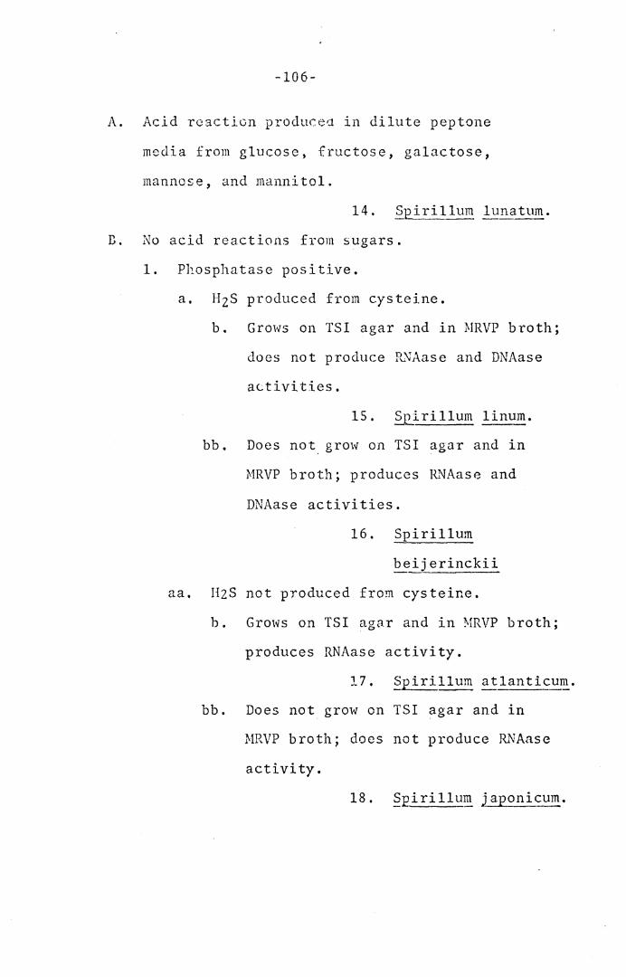
-106-
A. Acid reaction producea in dilute peptone
media from glucose, fructose, galactose,
mannose, and mannitol.
14. Spirillum lunatum.
D. No acid reactions from sugars.
1. Phosphatase positive.
a. HzS produced from cysteine.
b. Grows on TSI agar and in MRVP broth;
does not produce P'"''JAase and DNAase
activities.
15. Spirillum linum.
bb. Does not grow on TSI agar and in
MRVP broth; produces It.'lAase and
DNAase activities.
16. Spirillum
beijerinckii
aa. H2S not produced from cysteine.
b. Grows on TSI agar and in MRVP broth;
produces RNAase activity.
17. ~irillum atlanticum.
bb. Does not grow on TSI agar and in
MRVP broth; does not produce RNAase
activity.
18. Spirillum japonicum.
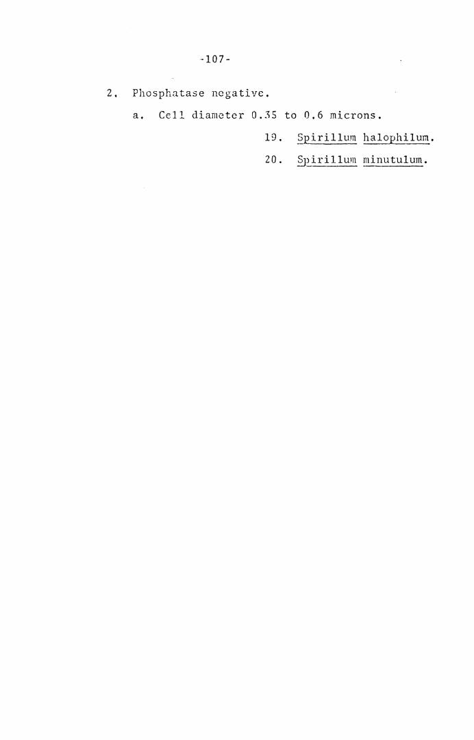
-107-
2. Phosphatase negative.
a. Cell diameter 0.35 to 0.6 microns.
19, Spirillum halophilum.
20. §£irillum minutulum.
SUMMARY
In order to develo~ further a comprehensive
characterization of spirilla and to devise a more
satisfactory classification of the species, the
type strains of 20 marine and fresh-water species
of the genus Spirillum, plus additional strains
of S. gra~ile, ~· itersonii, S. serpens, and S.
volutal)_?_, and 6 un-named strains of spirilla, were
subjected to over 90 physiological tests and also
various morphological tests using standardized
inocula, media, incubation times, and testing
methods. Determination of DNA base compositions
was also performed.
The morphological, physiological, and DNA base
composition data were combined to present a modified
description cf the genus Spirillum (Ehrenberg, 1830). . . .
A scheme for the classification of species of
tho genus ~pirillum was presented, using those characters
~~ich best distinguished each species. The two species
S. ~alC'P!?:.~lum and S. minutulum were combined as a result
of their morphological, physiological, and DNA base com-
position similarities.
-108-
LITERATURE CITED
Belogershy, A. N. 1941. On the composition of the protoplast of _?.2_irillum volutans cells depending on the age of culture. ~!icrobiology 10:185-198.
Bisset, K. A., and C. M. F. Hale--NcCaughcy. 1967. The electron microscopy of sectioned and entire cysts of ~J?irillum ~erpe~. Giorn. Microbiol. 15: 119-121.
Breed~ R. S., E. G. D. i"iurray, and A. P. Hitchens. 1948. Bergey' s 11!anual of Deterrnina ti ve B<1cteriology, 6th ed. The Williams and Wilkins Company, Baltimore, ?-laryland.
Breed~ R. S., E. G. D. Murray, and N. R. Smith. 1957. Bergey's Manual of Determinative Bacteriology, 7th ed. The Williams and Wilkins Company, Baltihlorc, Maryland.
Buning, E., and I. Gassel. 1956. C~loralhydrate auf Spirillen. 21:411-413.
Eine Wirkung von Arch. f.1ikrobiol.
Canale-Parola, E., S. L. Rosenthal, and D. G. Kupfer. 1966. Morphological and physiological character-istics of Spiril_JuH gracile sp. n. Antonie van Lecu~·1enhoeK J. ~Ii crob io 1. Scrol. 32: 113··124.
Cayton, :r. R., and N. W. Preston. 1955. Spirillum ~n~0c'l!._nie~~e n. sp. J. Gen. Microbiol.-1-z:srg-:-szs.
Clark-Walker, G. D. 1969. Association of microcyst fo.:nHtlion in SpLd.llu;n i.t<~rsonii with the s1_:ion-~art•~Ot!S inductioii'oT· c.- dcFec-rive- bacteriophage. J. Bacteriol. 97:885-892.
Cody, R. M. 1968. Selective isolation of Spirillum spec i es • App 1. Micro b i o l. 16 : 19 4 7 - 1948 .
Conway, E. J. 1950. Microdiffusion analysis and volum2tric error. Crosley Lockwood and Son, Ltd,, London.
-109-
-110-
Cowan, S. T., and K. J. Steel. 1966, Manual for the Identification of Medical Bµcteria, Cambridge University Press, New York, N. Y. ·
Doelle, fl, W. 1969. Bacterial Metabolism. Academic Press Inc,, Ne~ York.
:Schrnrds, P. R., nn<l W. H. Ewing. 1962. Identification c1 f [nte:robac tcriaceac, cd. Z, Mihvaukee, Burgess Fublishing Co,
Ehrenberg .• C. G. 1830, Beitragc zur Kenntuis dcr Organisation der Infusorien und ihrer geograph~ ischen Vcrbreitung. A bhandl. I, B8rl. Akad. pp. 1-30.
1838, Die Infusionthicrchen als volkomme -------Organismen. Leipzig, p. 73·86.
Giesbargcr, G. 1936. Beitrage zur Kenntuis der Ga.r."tung ~irjllu1~ Ehbg., Dissertation, Delft.
Hill, L. P. 1966. An index to deoxyribonucleic acid base compositions of bacterial species. J. Gen. Microbial. 44:419-347.
fhc IX International Congress of Microbiology. 1966. International Code of Nomenclature of Bacteria. Intern. J. System. Bacteriol. 16:459-490.
Krieg, N. R., J. P. Tomelty, and J. S. Wells, Jr. 1967. Inhibition of flagellar coordination in Spirillurn volutans. J. Bacterial. 94:1431-1436.
Leifson, E. 1962. The bacterial flora of distilled and stored water. III. New species of the genera ~~27ne~acterium, Flavobacteriuil!., ~iril~u~ and Ps ctlJomonas. In tern. Bul 1. Bae tcd.01. Nomcn. tnxon. 12:161-170.
Lewis, I. M. 1940. Th.e genus Spirillum Ehrenberg; 115. th specia.1 reference to c:·e-iTTi1.cTus icns and the chrornidj.al theory. J. Bacterial. 40:271-283.
MacLeod, R. A. 1965. The Question of the Existence of Specific Marine Bacteria. Bacteriol. Revs. 29;9-23,
Mandel, M. 1966. Deoxyribonucleic acid base composition .in tl1c genus Pseudomo.,~. J. Gen. Microbial. 43:273-292.
-111-
Mar:nur, J., an<l P. :Doty. 1962. Determination of the base composition of deoxyribonucleic acid from its thermal <lenatuxat:ton tempei·ature. J, Mol, Biol. 5;109-118.
Narmur, J,, S. Falkow, and M. Mandel. approaches to bacterial taxonomy. Microbial. 17:329-372.
196.3. New Ann. Rev.
Myers, J. 1940. Studies on the spirilleae: Methods of isolation and identification. J. Bacterial. 40:705-720.
Pre tori us, l\T. A. 1963. A systematic study of the genus .§Jii ril lum which occurs in oxidation ponds, with a description of a new species. J. Gen. ~Iicrobiol. 32:403-408.
Rittenbcrg, B. T., and S. C. Rittenberg. 1962. The g-roHth of §Ri~J_) "!:::tE!. v~!utan_s Ehrenberg in mixed and pure cultures. Arcli. gikrobiol. 42:138-153.
Sebald, M. M., and M. Veron. 1963. Teneur en bases de l'ADN et classification les vibrions. Ann. Inst. Pasteur. 105:897-910.
S'Jciety of American Bacteriologists. 1957. Manual of microbiological methods, p. 113. McGraw-Hill Book Co., Inc., New York.
Stanier, R. Y., N. J. Palleroni, and M. Doudoroff. 1966. The aerobic pseudomonads: a taxonomic st~dy. J. Gen. Microbial. 43:159-271.
Stiles, C. W. 1905. The International Code of Zoological Nomenclature as Applied to Medjcine. Bull Hygienic Laboratory 29. Washington, D. C.
Terasaki, Y. 1961. On ~;r:i}lum. put~id~i.conchylium nov. sp. Botan. Mag. 0Qkyo). 74[87ZJT'T9-85,
, 1961. On tuo new snec:l.es of Sr.irillum. Botan. M~g. (Tokyo), 74(875): 220-22""1. __ _
Tyler, M. E,, M. C. Bielling, and D. B. Pratt. 1960. Mineral requirements and other characters of selected marine bacteria. J. Gen. Micrcbiol. 23: 153-161.
-112-
Vui l lemin, P, 1913.. Gene Ta Schi zomycetum. i\lycologie. Berlin, 11:512-527.
Ann.
Watanabe, N, 1959. On fou·c ne1<l haloDhilic species Botan. Nag. (Tclcyo). 72:77-86. C r• • • 11 c .L' .?.~~'.~.
Wells, J, S., Jr. 1966. The physiology of Spirillum volutans. M. S. Thesis, Newman Library, V1rg1n1a 11-oiytechni.c Institute, Blacksburg, Virginia.
, and N. R. Krieg. 1965. Cultivation of ____ .. __ Sp1 . ."ri llum volutans in a bacteria-free environment.
T.---Bacferiol. 90: 817- 818.
\\'illiarr:s, M. A. 1957. Nuclear phen·Jmena in ~J2.irillum species. Ph.D. thesis, University of Southern California.
. 1959. Some problems in the identification -------- an CI c l as s i f i ca ti on of ,? p i r_~g~-~. I . Ear 1 i er
taxonomy of the genus S_Eirillura. Intern. Bull. Ba ct e r i o i . Nome n , Tax o n,---~f(T) : 3 5 - 5 5 •
. 1959. Some problems in the identification ----·-----~1n<l classification of species ?f ~~irillu_~· II.
Later taxonomy of the genus Sp1rillum. Intern. Bull. Bacterial. Nomen. Taxon. 9l"3) :137-157 •
. 1960. Flagellation in six species of ·-------~j)Trillur.t- -a correction. Intern. Bull. Bacterial.
Nomen. Taxon. 10:193-196.
Williams, M. A., and S. C. Rittenberg. 1956. Micro-cyst formation and germination in Spirillum 1~111c!tt11P. J. Gen. Microbial. 15: 205--:-·z-09-.--
• 1957. A taxonomic study of the genus ---~~l~"I?·i.~}uE! Ehrenberg. Intern. Bull. Bacterial.
; ; o r;ic n • Tax on . 7 : 4 9 - 111.

The vita has been removed from the scanned document
A MORPHOLOGICAL AND PHYSIOLOGICAL
CLASSIFICATION OF SPIRILLA
by
Jerry Scott Wells, Jr.
ABSTRACT
In order to develop further a comprehensive
characterization of spirilla and to devise a more
satisfactory classification of the species, the
type strains of 20 marine and fresh-water species
of the genus Spirillum, plus additional strains of
S. ~cile, ~· itersonii, ~· serpens, and S. volu-
tans, and 6 un-named strains of spirilla, were
subjected to over 90 physiological tests and also
various morphological tests using standardized
inocula, media, incubation times, and testing
methods. Determination of DNA base compositions
was also performed.
All strains of spirill~ grew aerobically except
S, voluta_ns_ which was an obligate microaerophile; S.
giesbergeri and S. gracile appeared to be possible
facultative rnicroacrophiles, S. lunatum and S, iter-
sonii were the only species to. grow anaerobically in
the presence of nitrate, The optimum temperature was
30 C for all strains examined. The cell diameters
for all strains formed an almost continuous spectrum,
ranging from 0.25 to 1.7 microns. All spirilla were
actively motile and a number of the strains possessed
flagellar tufts visible with living cells by dark
field microscopy and in the case of S. volutans also
by phase contrast. Coccoid bodies were present in
4 week cultures in the case of most marine strains;
only 3 fresh-water species exhibited these forms,
Several physiological characteristics were found to
be uniformly or nearly uniformly positive (oxidase, dea-
minase activity with peptone, catalase, phosphatase,
HzS from cysteine, growth in 1% bile) or negative
(indol; amylase; sulfatase; hydrolysis of gelatin,
casein, aesculin, and hippurate; urease; alkaline
reaction in litmus milk; acid reaction from carbo-
hydrates) for all or nearly all of the strains, In
the few cases 1vhere acid was produced from carbohydrates,
a reduced peptane concentration was necessary. Several
physiological characteristics yielded varied reactions
among the spirilla (reduction of selenite and nitrate;
growth on EMB, NacConkey, TSI, and Seller's agars and
in MRVP broth; RNAase and DNAase activity; and
tolerance to 1% glycine}, w~ter~soluble pigments were
produced by a few strains growing on aromatic amino
acids. Three percent NaCl i11hibi ted all fresh-water
strains, while the marine strains could all grow in
the presence of 9. 75% (w/v) NaCl. The DNA base com-
positions of the spirilla, determined from their Tm
values, gave a broad range of values from 38 to 65
moles % G + C.
The limits of genus and species variation, the
usefulness of standardized conditions in characterizing
strains of spirilla, discrepancies arising from a com-
parison of the present data with earlier results, the
division of the genus Spirillum on the basis of single,
highly-weighted characters, and the division of genus
Spirillum on the basis of correlated characters, were
discussed. A modified description of the genus Seiril-
lum (Ehrenberg, 1830) based on the present study was
presented. Various physiological tests important for
species diffeYentiation were indicated. A modified
cl <.ss if ica ti on scheme for the species of genus Sp i ri 1.lum
was devised, and a temporary diagnostic key was also
presented.