Embed Size (px)
Citation preview
A Microbiome-Based Index for Assessing Skin Health andTreatment Effects for Atopic Dermatitis in Children
Zheng Sun,a,i Shi Huang,a,i Pengfei Zhu,a Feng Yue,b Helen Zhao,c Ming Yang,d Yueqing Niu,b Gongchao Jing,a,i
Xiaoquan Su,a,i Huiying Li,e Chris Callewaert,f,g Rob Knight,f Jiquan Liu,c Ed Smith,h Karl Wei,h Jian Xua,i
aSingle-Cell Center and Shandong Key Laboratory of Energy Genetics, Qingdao Institute of BioEnergy and Bioprocess Technology, Chinese Academy of Sciences,Qingdao, Shandong, China
bProcter & Gamble Beijing Innovation Center, Beijing, ChinacProcter & Gamble Singapore Innovation Center, Singapore, SingaporedOffice of General Affairs, Chinese Academy of Sciences, Beijing, ChinaeDepartment of Molecular and Medical Pharmacology, University of California at Los Angeles, Los Angeles, California, USAfCenter for Microbiome Innovation and Departments of Pediatrics, University of California at San Diego, La Jolla, California, USAgCenter for Microbial Ecology and Technology, Ghent University, Ghent, BelgiumhProcter & Gamble Mason Business Center, Mason, Ohio, USAiUniversity of Chinese Academy of Sciences, Beijing, China
ABSTRACT A quantitative and objective indicator for skin health via the micro-biome is of great interest for personalized skin care, but differences among skin sitesand across human populations can make this goal challenging. A three-city (twoChinese and one American) comparison of skin microbiota from atopic dermatitis(AD) and healthy pediatric cohorts revealed that, although city has the greatest ef-fect size (the skin microbiome can predict the originated city with near 100% accu-racy), a microbial index of skin health (MiSH) based on 25 bacterial genera can diag-nose AD with 83 to �95% accuracy within each city and 86.4% accuracy acrosscities (area under the concentration-time curve [AUC], 0.90). Moreover, nonlesionalskin sites across the bodies of AD-active children (which include shank, arm, popli-teal fossa, elbow, antecubital fossa, knee, neck, and axilla) harbor a distinct but le-sional state-like microbiome that features relative enrichment of Staphylococcus au-reus over healthy individuals, confirming the extension of microbiome dysbiosisacross body surface in AD patients. Intriguingly, pretreatment MiSH classifies chil-dren with identical AD clinical symptoms into two host types with distinct microbialdiversity and treatment effects of corticosteroid therapy. These findings suggest thatMiSH has the potential to diagnose AD, assess risk-prone state of skin, and predicttreatment response in children across human populations.
IMPORTANCE MiSH, which is based on the skin microbiome, can quantitatively as-sess pediatric skin health across cohorts from distinct countries over large geo-graphic distances. Moreover, the index can identify a risk-prone skin state and com-pare treatment effect in children, suggesting applications in diagnosis and patientstratification.
KEYWORDS atopic dermatitis, personalized skin care, skin microbiome, spatialvariation, suboptimal health
A central goal of human microbiome projects is to diagnose and predict host statesvia the microbiome (1, 2). The skin, our largest organ and a first line of environ-
mental exposure, hosts a microbiome that is site specific, host specific, and environ-ment specific (3, 4). Particular skin symbionts modulate the host immune response,physiology, and development (5–8). Therefore, the prospect of exploiting the skin
Citation Sun Z, Huang S, Zhu P, Yue F, Zhao H,Yang M, Niu Y, Jing G, Su X, Li H, Callewaert C,Knight R, Liu J, Smith E, Wei K, Xu J. 2019. Amicrobiome-based index for assessing skinhealth and treatment effects for atopicdermatitis in children. mSystems 4:e00293-19.https://doi.org/10.1128/mSystems.00293-19.
Editor Janet K. Jansson, Pacific NorthwestNational Laboratory
Copyright © 2019 Sun et al. This is an open-access article distributed under the terms ofthe Creative Commons Attribution 4.0International license.
Address correspondence to Jian Xu,[email protected].
Z.S. and S.H. contributed equally to this article.
The prospect of exploiting the skinmicrobiota for health protection, diseasetreatment, or personal care has attracted greatinterest. This paper shows that the skinmicrobiota can identify a risk-prone skin stateand assess treatment effect in children.
Received 8 May 2019Accepted 29 July 2019Published
RESEARCH ARTICLETherapeutics and Prevention
July/August 2019 Volume 4 Issue 4 e00293-19 msystems.asm.org 1
20 August 2019
on April 3, 2020 by guest
http://msystem
s.asm.org/
Dow
nloaded from
microbiome for health protection, disease treatment, or personal care has attractedgreat interest (9). The skin microbiome is known to differ between human populations(10, 11); therefore, whether the skin microbiome can serve as an indicator of skin healththat applies across large geographic ranges remains largely unknown (12, 13).
This challenge can be traced to the characteristics of human skin microbiota. Thedominant types of resident skin bacteria appear relatively stable, and less abundanttypes of bacteria account for most of the variability (4). Within an individual, compo-sition of the skin microbiota is determined primarily by body site. Within a skin zone,temporal variability in an individual is small compared to interpersonal variability (3, 4,14, 15). However, how this pattern of spatial variation manifests itself remains poorlyunderstood, particularly in the context of perturbation by disease or medications.Moreover, changes in pathogenic microbiota across individuals, cities, and even at theglobal scale is largely unknown (4, 16, 17). Furthermore, for many microbiome-wideassociation studies, notably those from the gut, applying models of microbial diseasebiomarkers trained in one population to other populations has typically been unsuc-cessful (18), which greatly limits the potential of the microbiome for diagnosis andtreatment-oriented patient stratification.
Atopic dermatitis (AD), a chronic and relapsing inflammatory skin disorder associ-ated with skin barrier impairment, affects 15 to 30% of children (5% of the generalpopulation) worldwide and has been rapidly increasing in prevalence, especially withchildren (19, 20). AD is a heterogeneous disease of different subtypes and with variedand sometimes evasive clinical manifestations (21). Disease severity is typically diag-nosed by physicians via visual observation and diagnosis of disease signs, includingcolor change, pruritus, and swollen and cracked skin, generally done with the well-validated Scoring Atopic Dermatitis Index (SCORAD) (22). Other diagnosis measuresinclude the Eczema Area and the Severity Index (EASI) score (23) or the objectiveSCORAD (24). In addition, several serum biomarker assays, such as thymus andactivation-regulated chemokine (TARC) assay (CC chemokine 17 [CCL17]), pulmonaryand activation-regulated chemokine (PARC) assay (CCL18) (25–28), Staphylococcus au-reus enterotoxin assay (29), etc. are available, yet they are typically invasive. On theother hand, the AD state has been associated with change in the skin microbiome, e.g.,the presence or enrichment of S. aureus (4, 15, 30). These microbiome-based findingsare enabling new opportunities for better assessment or prediction of the disease state,which can potentially overcome the shortcoming of traditional clinical scores orsupplement them, particularly in allowing comparison among patients, examiners, orstudies, and in objective design and administration of skin therapy and care regimens(31).
On the other hand, it remains elusive whether and how skin microbiome plays a rolein AD treatments (30), which aims to reduce symptoms (pruritus and dermatitis),prevent exacerbations, and minimize therapeutic risks. Standard treatment modalitiesfor AD have centered around the use of topical anti-inflammatory preparations andmoisturization of the skin (e.g., corticosteroids, calcineurin inhibitors, and crisaborole),and patients with severe AD may require phototherapy or systemic treatment (forinstance, oral cyclosporine [32, 33]). Topical calcineurin inhibitors are potentially linkedto cancer (34), crisaborole remains uncertain in efficacy (35, 36), and cyclosporine mayinduce side effects, including nephrotoxicity, hypertension, hypertrichosis, etc. (37).Therefore, corticosteroids can serve as a starting point for probing role of the skinmicrobiome in AD treatment, since they are recommended as first-line treatment forAD (38).
In this study, we compared skin microbiota across the body (mainly from theforearm and shank, with the remaining from seven additional skin sites; see Table S1 inthe supplemental material) from healthy and AD active children (3 to 12 years old) fromthree cities (Beijing and Qingdao from China and Denver from United States), andtracked their subsequent response to skin care treatment. We showed that althoughcity has the greatest effect size, a Microbial Index of Skin Health (MiSH) is generallyapplicable to populations across large geographical distances. Moreover, in AD, we
Sun et al.
July/August 2019 Volume 4 Issue 4 e00293-19 msystems.asm.org 2
on April 3, 2020 by guest
http://msystem
s.asm.org/
Dow
nloaded from
confirmed that the microbiome dysbiosis is extended across whole body surface, asnonlesional skin sites of the patient harbor a distinct but lesional state-like microbiome.Intriguingly, pretreatment MiSH classifies children in the Beijing cohort with identicalAD clinical symptoms into two host types with distinct disease severity and sensitivityto corticosteroid therapy (in which corticosteroid-containing ointment was applied onskin surface). Although their reproducibility and generalizability need to be demon-strated in larger cohorts and various populations, our results indicate that, via MiSH, theskin microbiome may potentially serve as a generally applicable, quantitative proxy todiagnose AD, be able to compare the efficacies of AD care products, and be able topredict AD treatment response in children.
RESULTSExperimental design that compared healthy and AD skin microbiota at various
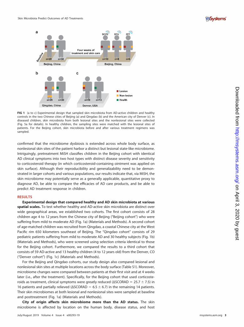
spatial scales. To test whether healthy and AD-active skin microbiota are distinct overwide geographical areas, we established two cohorts. The first cohort consists of 28children age 4 to 12 years from the Chinese city of Beijing (“Beijing cohort”) who weresuffering from mild to moderate AD (Fig. 1a) (Materials and Methods). A second cohortof age-matched children was recruited from Qingdao, a coastal Chinese city at the WestPacific rim 650 kilometers southeast of Beijing. The “Qingdao cohort” consists of 29pediatric patients suffering from mild to moderate AD and 30 healthy subjects (Fig. 1b)(Materials and Methods), who were screened using selection criteria identical to thosefor the Beijing cohort. Furthermore, we compared the results to a third cohort thatconsists of 59 AD-active and 13 healthy children (4 to 12 years old) from the Denver, CO(“Denver cohort”) (Fig. 1c) (Materials and Methods).
For the Beijing and Qingdao cohorts, our study design also compared lesional andnonlesional skin sites at multiple locations across the body surface (Table S1). Moreover,microbiome changes were compared between patients at their first visit and at 4 weekslater (i.e., after the treatment). Specifically, for the Beijing cohort that used corticoste-roids as treatment, clinical symptoms were greatly reduced (ΔSCORAD � 25.7 � 7.5) in16 patients and partially relieved (ΔSCORAD � 6.5 � 6.7) in the remaining 14 patients.Their skin microbiomes at both lesional and nonlesional sites were sampled at baselineand posttreatment (Fig. 1a) (Materials and Methods).
City of origin affects skin microbiome more than the AD status. The skinmicrobiome is affected by location on the human body, disease status, and host
FIG 1 (a to c) Experimental design that sampled skin microbiota from AD-active children and healthycontrols in the two Chinese cities of Beijing (a) and Qingdao (b) and the American city of Denver (c). Indiseased children, skin microbiota from both lesional sites and the nonlesional sites were collected(Fig. 5a for details). In healthy children, the sampling sites were matched with the lesional sites ofpatients. For the Beijing cohort, skin microbiota before and after various treatment regimens wassampled.
Skin Microbiota Predict Outcomes of AD Treatments
July/August 2019 Volume 4 Issue 4 e00293-19 msystems.asm.org 3
on April 3, 2020 by guest
http://msystem
s.asm.org/
Dow
nloaded from
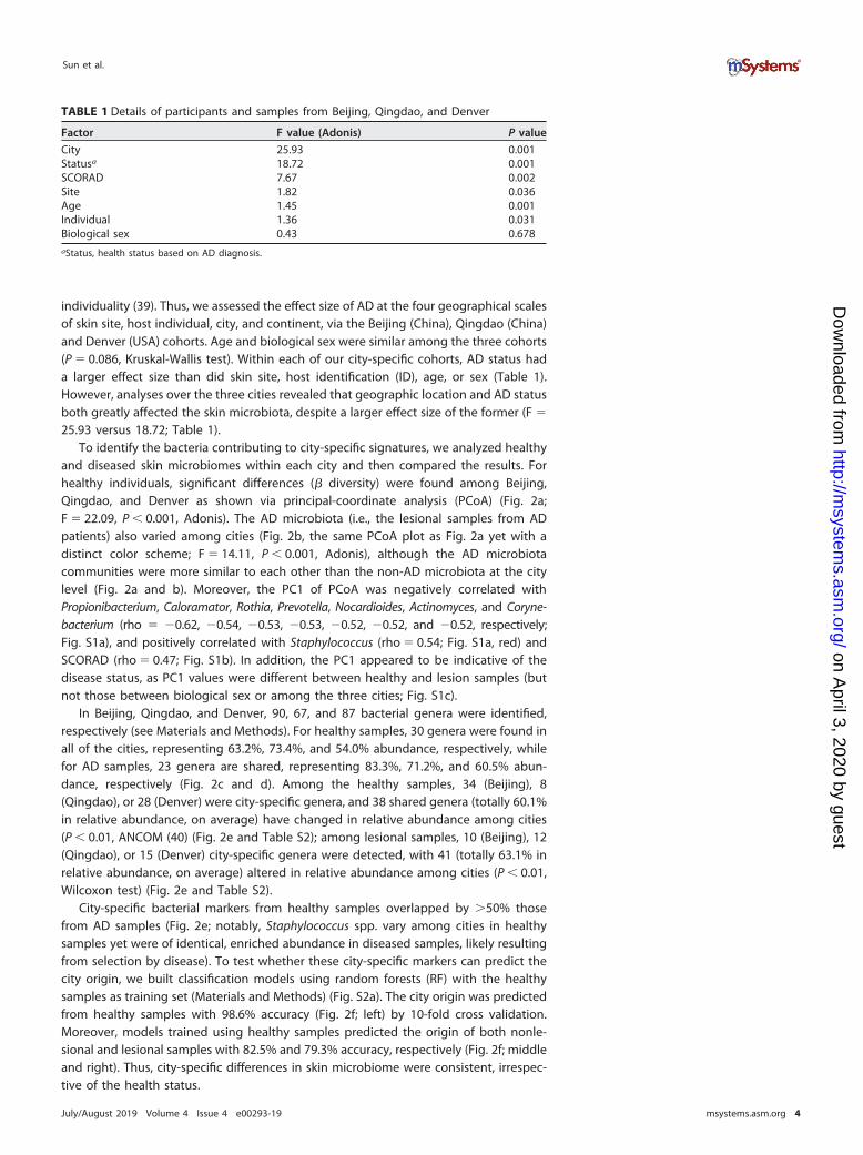
individuality (39). Thus, we assessed the effect size of AD at the four geographical scalesof skin site, host individual, city, and continent, via the Beijing (China), Qingdao (China)and Denver (USA) cohorts. Age and biological sex were similar among the three cohorts(P � 0.086, Kruskal-Wallis test). Within each of our city-specific cohorts, AD status hada larger effect size than did skin site, host identification (ID), age, or sex (Table 1).However, analyses over the three cities revealed that geographic location and AD statusboth greatly affected the skin microbiota, despite a larger effect size of the former (F �
25.93 versus 18.72; Table 1).To identify the bacteria contributing to city-specific signatures, we analyzed healthy
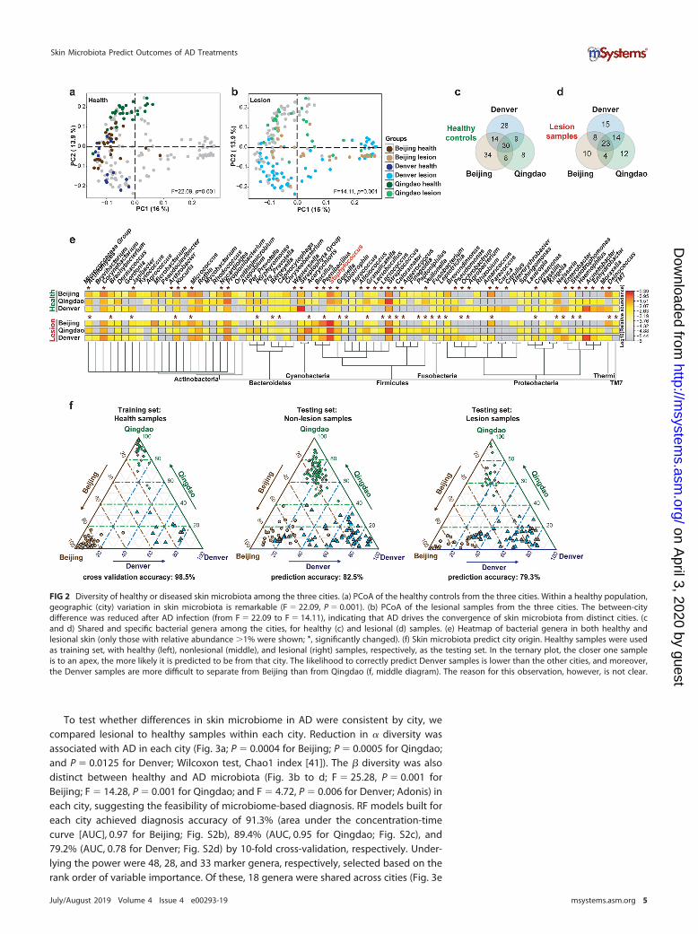
and diseased skin microbiomes within each city and then compared the results. Forhealthy individuals, significant differences (� diversity) were found among Beijing,Qingdao, and Denver as shown via principal-coordinate analysis (PCoA) (Fig. 2a;F � 22.09, P � 0.001, Adonis). The AD microbiota (i.e., the lesional samples from ADpatients) also varied among cities (Fig. 2b, the same PCoA plot as Fig. 2a yet with adistinct color scheme; F � 14.11, P � 0.001, Adonis), although the AD microbiotacommunities were more similar to each other than the non-AD microbiota at the citylevel (Fig. 2a and b). Moreover, the PC1 of PCoA was negatively correlated withPropionibacterium, Caloramator, Rothia, Prevotella, Nocardioides, Actinomyces, and Coryne-bacterium (rho � �0.62, �0.54, �0.53, �0.53, �0.52, �0.52, and �0.52, respectively;Fig. S1a), and positively correlated with Staphylococcus (rho � 0.54; Fig. S1a, red) andSCORAD (rho � 0.47; Fig. S1b). In addition, the PC1 appeared to be indicative of thedisease status, as PC1 values were different between healthy and lesion samples (butnot those between biological sex or among the three cities; Fig. S1c).
In Beijing, Qingdao, and Denver, 90, 67, and 87 bacterial genera were identified,respectively (see Materials and Methods). For healthy samples, 30 genera were found inall of the cities, representing 63.2%, 73.4%, and 54.0% abundance, respectively, whilefor AD samples, 23 genera are shared, representing 83.3%, 71.2%, and 60.5% abun-dance, respectively (Fig. 2c and d). Among the healthy samples, 34 (Beijing), 8(Qingdao), or 28 (Denver) were city-specific genera, and 38 shared genera (totally 60.1%in relative abundance, on average) have changed in relative abundance among cities(P � 0.01, ANCOM (40) (Fig. 2e and Table S2); among lesional samples, 10 (Beijing), 12(Qingdao), or 15 (Denver) city-specific genera were detected, with 41 (totally 63.1% inrelative abundance, on average) altered in relative abundance among cities (P � 0.01,Wilcoxon test) (Fig. 2e and Table S2).
City-specific bacterial markers from healthy samples overlapped by �50% thosefrom AD samples (Fig. 2e; notably, Staphylococcus spp. vary among cities in healthysamples yet were of identical, enriched abundance in diseased samples, likely resultingfrom selection by disease). To test whether these city-specific markers can predict thecity origin, we built classification models using random forests (RF) with the healthysamples as training set (Materials and Methods) (Fig. S2a). The city origin was predictedfrom healthy samples with 98.6% accuracy (Fig. 2f; left) by 10-fold cross validation.Moreover, models trained using healthy samples predicted the origin of both nonle-sional and lesional samples with 82.5% and 79.3% accuracy, respectively (Fig. 2f; middleand right). Thus, city-specific differences in skin microbiome were consistent, irrespec-tive of the health status.
TABLE 1 Details of participants and samples from Beijing, Qingdao, and Denver
Factor F value (Adonis) P value
City 25.93 0.001Statusa 18.72 0.001SCORAD 7.67 0.002Site 1.82 0.036Age 1.45 0.001Individual 1.36 0.031Biological sex 0.43 0.678aStatus, health status based on AD diagnosis.
Sun et al.
July/August 2019 Volume 4 Issue 4 e00293-19 msystems.asm.org 4
on April 3, 2020 by guest
http://msystem
s.asm.org/
Dow
nloaded from
To test whether differences in skin microbiome in AD were consistent by city, wecompared lesional to healthy samples within each city. Reduction in � diversity wasassociated with AD in each city (Fig. 3a; P � 0.0004 for Beijing; P � 0.0005 for Qingdao;and P � 0.0125 for Denver; Wilcoxon test, Chao1 index [41]). The � diversity was alsodistinct between healthy and AD microbiota (Fig. 3b to d; F � 25.28, P � 0.001 forBeijing; F � 14.28, P � 0.001 for Qingdao; and F � 4.72, P � 0.006 for Denver; Adonis) ineach city, suggesting the feasibility of microbiome-based diagnosis. RF models built foreach city achieved diagnosis accuracy of 91.3% (area under the concentration-timecurve [AUC], 0.97 for Beijing; Fig. S2b), 89.4% (AUC, 0.95 for Qingdao; Fig. S2c), and79.2% (AUC, 0.78 for Denver; Fig. S2d) by 10-fold cross-validation, respectively. Under-lying the power were 48, 28, and 33 marker genera, respectively, selected based on therank order of variable importance. Of these, 18 genera were shared across cities (Fig. 3e
FIG 2 Diversity of healthy or diseased skin microbiota among the three cities. (a) PCoA of the healthy controls from the three cities. Within a healthy population,geographic (city) variation in skin microbiota is remarkable (F � 22.09, P � 0.001). (b) PCoA of the lesional samples from the three cities. The between-citydifference was reduced after AD infection (from F � 22.09 to F � 14.11), indicating that AD drives the convergence of skin microbiota from distinct cities. (cand d) Shared and specific bacterial genera among the cities, for healthy (c) and lesional (d) samples. (e) Heatmap of bacterial genera in both healthy andlesional skin (only those with relative abundance �1% were shown; *, significantly changed). (f) Skin microbiota predict city origin. Healthy samples were usedas training set, with healthy (left), nonlesional (middle), and lesional (right) samples, respectively, as the testing set. In the ternary plot, the closer one sampleis to an apex, the more likely it is predicted to be from that city. The likelihood to correctly predict Denver samples is lower than the other cities, and moreover,the Denver samples are more difficult to separate from Beijing than from Qingdao (f, middle diagram). The reason for this observation, however, is not clear.
Skin Microbiota Predict Outcomes of AD Treatments
July/August 2019 Volume 4 Issue 4 e00293-19 msystems.asm.org 5
on April 3, 2020 by guest
http://msystem
s.asm.org/
Dow
nloaded from
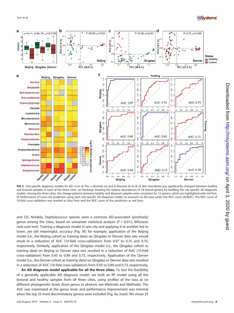
and S3). Notably, Staphylococcus species were a common AD-associated (positively)genus among the cities, based on univariate statistical analysis (P � 0.012, Wilcoxonrank-sum test). Training a diagnosis model in one city and applying it to another led tolower, yet still meaningful, accuracy (Fig. 3f); for example, application of the Beijingmodel (i.e., the Beijing cohort as training data) on Qingdao or Denver data sets wouldresult in a reduction of AUC (10-fold cross-validation) from 0.97 to 0.75 and 0.70,respectively. Similarly, application of the Qingdao model (i.e., the Qingdao cohort astraining data) on Beijing or Denver data sets resulted in a reduction of AUC (10-foldcross-validation) from 0.95 to 0.89 and 0.73, respectively. Application of the Denvermodel (i.e., the Denver cohort as training data) on Qingdao or Denver data sets resultedin a reduction of AUC (10-fold cross-validation) from 0.95 to 0.89 and 0.73, respectively.
An AD diagnosis model applicable for all the three cities. To test the feasibilityof a generally applicable AD diagnosis model, we built an RF model using all thelesional and healthy samples from all three cities, using profiles of the taxa at sixdifferent phylogenetic levels (from genus to phylum; see Materials and Methods). TheAUC was maximized at the genus level, and performance improvement was minimalwhen the top 25 most discriminatory genera were included (Fig. 4a, inset). We chose 25
FIG 3 City-specific diagnosis models for AD. (a to d) The � diversity (a) and � diversity (b to d) of skin microbiota was significantly changed between healthyand lesional samples in each of the three cities. (e) Heatmap showing the relative abundances of 18 shared genera for building the city-specific AD diagnosismodels. Among the three cities, the change patterns between healthy and diseased samples were consistent for 12 genera, which are highlighted with red font.(f) Performance of cross-city prediction using each city-specific AD diagnosis model, as assessed via the area under the ROC curve (AUROC). The ROC curve of10-fold cross-validation was marked as blue lines and the ROC curve of the prediction as red lines.
Sun et al.
July/August 2019 Volume 4 Issue 4 e00293-19 msystems.asm.org 6
on April 3, 2020 by guest
http://msystem
s.asm.org/
Dow
nloaded from
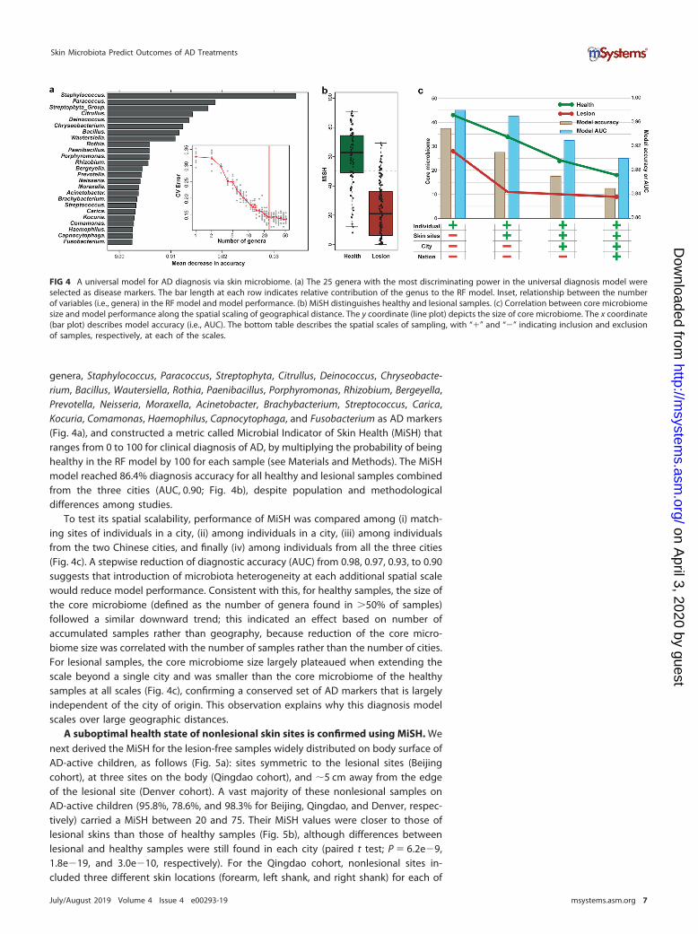
genera, Staphylococcus, Paracoccus, Streptophyta, Citrullus, Deinococcus, Chryseobacte-rium, Bacillus, Wautersiella, Rothia, Paenibacillus, Porphyromonas, Rhizobium, Bergeyella,Prevotella, Neisseria, Moraxella, Acinetobacter, Brachybacterium, Streptococcus, Carica,Kocuria, Comamonas, Haemophilus, Capnocytophaga, and Fusobacterium as AD markers(Fig. 4a), and constructed a metric called Microbial Indicator of Skin Health (MiSH) thatranges from 0 to 100 for clinical diagnosis of AD, by multiplying the probability of beinghealthy in the RF model by 100 for each sample (see Materials and Methods). The MiSHmodel reached 86.4% diagnosis accuracy for all healthy and lesional samples combinedfrom the three cities (AUC, 0.90; Fig. 4b), despite population and methodologicaldifferences among studies.
To test its spatial scalability, performance of MiSH was compared among (i) match-ing sites of individuals in a city, (ii) among individuals in a city, (iii) among individualsfrom the two Chinese cities, and finally (iv) among individuals from all the three cities(Fig. 4c). A stepwise reduction of diagnostic accuracy (AUC) from 0.98, 0.97, 0.93, to 0.90suggests that introduction of microbiota heterogeneity at each additional spatial scalewould reduce model performance. Consistent with this, for healthy samples, the size ofthe core microbiome (defined as the number of genera found in �50% of samples)followed a similar downward trend; this indicated an effect based on number ofaccumulated samples rather than geography, because reduction of the core micro-biome size was correlated with the number of samples rather than the number of cities.For lesional samples, the core microbiome size largely plateaued when extending thescale beyond a single city and was smaller than the core microbiome of the healthysamples at all scales (Fig. 4c), confirming a conserved set of AD markers that is largelyindependent of the city of origin. This observation explains why this diagnosis modelscales over large geographic distances.
A suboptimal health state of nonlesional skin sites is confirmed using MiSH. Wenext derived the MiSH for the lesion-free samples widely distributed on body surface ofAD-active children, as follows (Fig. 5a): sites symmetric to the lesional sites (Beijingcohort), at three sites on the body (Qingdao cohort), and �5 cm away from the edgeof the lesional site (Denver cohort). A vast majority of these nonlesional samples onAD-active children (95.8%, 78.6%, and 98.3% for Beijing, Qingdao, and Denver, respec-tively) carried a MiSH between 20 and 75. Their MiSH values were closer to those oflesional skins than those of healthy samples (Fig. 5b), although differences betweenlesional and healthy samples were still found in each city (paired t test; P � 6.2e�9,1.8e�19, and 3.0e�10, respectively). For the Qingdao cohort, nonlesional sites in-cluded three different skin locations (forearm, left shank, and right shank) for each of
FIG 4 A universal model for AD diagnosis via skin microbiome. (a) The 25 genera with the most discriminating power in the universal diagnosis model wereselected as disease markers. The bar length at each row indicates relative contribution of the genus to the RF model. Inset, relationship between the numberof variables (i.e., genera) in the RF model and model performance. (b) MiSH distinguishes healthy and lesional samples. (c) Correlation between core microbiomesize and model performance along the spatial scaling of geographical distance. The y coordinate (line plot) depicts the size of core microbiome. The x coordinate(bar plot) describes model accuracy (i.e., AUC). The bottom table describes the spatial scales of sampling, with “�” and “�” indicating inclusion and exclusionof samples, respectively, at each of the scales.
Skin Microbiota Predict Outcomes of AD Treatments
July/August 2019 Volume 4 Issue 4 e00293-19 msystems.asm.org 7
on April 3, 2020 by guest
http://msystem
s.asm.org/
Dow
nloaded from
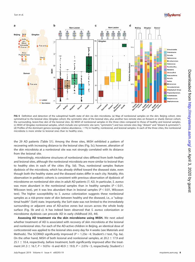
the 29 AD patients (Table S1). Among the three sites, MiSH exhibited a pattern ofrecovering with increasing distance to the lesional sites (Fig. 5c); however, alteration ofthe skin microbiota at a nonlesional site was not strongly correlated with its distancefrom the lesional site.
Interestingly, microbiome structures of nonlesional sites differed from both healthyand lesional sites, although the nonlesional microbiota are more similar to lesional thanto healthy sites in each of the cities (Fig. 5d). Thus, nonlesional samples featuredysbiosis of the microbiota, which has already shifted toward the diseased state, eventhough both the healthy states and the diseased states differ in each city. Notably, thisobservation in pediatric cohorts is consistent with previous observation of dysbiosis ofmicrobiome on nonlesional skin sites in adult AD patients (7, 42). In particular, S. aureuswas more abundant in the nonlesional samples than in healthy samples (P � 0.01,Wilcoxon test), yet it was less abundant than in lesional samples (P � 0.01, Wilcoxontest). The higher susceptibility to S. aureus colonization suggests these nonlesionalsamples as a risk-prone state of skin between healthy and the diseased, i.e., a “subop-timal health” (SoH) state. Importantly, the SoH state was not limited to the immediatelysurrounding or adjacent area of AD-active zones but occurs across the whole bodysurface (Fig. 5b and c). It has indeed been observed that S. aureus colonization ormicrobiome dysbiosis can precede AD in early childhood (43, 44).
Assessing AD treatment via the skin microbiome using MiSH. We next askedwhether treatment of AD is associated with recovery of skin microbiota at the lesionaland nonlesional sites. For each of the AD-active children in Beijing, an identical dose ofcorticosteroid was applied to the lesional sites every day for 4 weeks (see Materials andMethods). The SCORAD significantly improved (P � 1.23e�4; Student’s t test, Fig. 6a).On the other hand, MiSH of both lesional and nonlesional samples, at 20.2 � 17.9 and25.1 � 10.4, respectively, before treatment, both significantly improved after the treat-ment (41.3 � 16.7, P � 9.07e�6 and 40.9 � 19.9, P � 2.07e�5, respectively; Student’s t
FIG 5 Definition and detection of the suboptimal health state of skin via skin microbiota. (a) Map of nonlesional samples on the skin. Beijing cohort, sitessymmetrical to the lesional sites; Qingdao cohort, the symmetric sites of the lesional sites, plus another two remote sites on forearm or shank; Denver cohort,the surrounding, lesion-free skin of the lesional sites. (b) MiSH of nonlesional samples in the three cities compared to those of healthy and lesional samples.(c) MiSH of Qingdao nonlesional samples, which include one symmetric site (arm; “symmetric”) and two remote sites (leg; “distant” and “distant & symmetric”).(d) Profiles of the dominant genera (average relative abundance, �1%) in healthy, nonlesional, and lesional samples. In each of the three cities, the nonlesionalmicrobiota is more similar to lesional ones than to healthy ones.
Sun et al.
July/August 2019 Volume 4 Issue 4 e00293-19 msystems.asm.org 8
on April 3, 2020 by guest
http://msystem
s.asm.org/
Dow
nloaded from
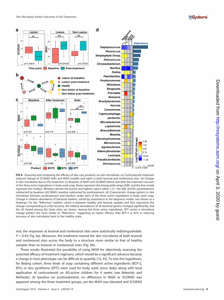
test; the responses at lesional and nonlesional sites were statistically indistinguishable;P � 0.43; Fig. 6a). Moreover, the treatment moved the skin microbiota of both lesionaland nonlesional sites across the body to a structure more similar to that of healthysamples than to lesional or nonlesional ones (Fig. 6b).
These results illustrated the possibility of using MiSH for objectively assessing thepotential efficacy of treatment regimens, which would be a significant advance becausea change in host phenotype can be difficult to quantify (15, 45). To test this hypothesis,for Beijing cohort, three kinds of soap containing different active ingredients (BCP-2,B7U, or zinc pyrithione [ZPT]) were used for body wash (once daily) along with localapplication of corticosteroid on AD-active children for 4 weeks (see Materials andMethods). At baseline (or posttreatment), no difference in MiSH or SCORAD wasapparent among the three treatment groups, yet the MiSH was elevated and SCORAD
FIG 6 Assessing and comparing the efficacy of skin care products via skin microbiota. (a) Corticosteroid treatmentinduced change of SCORAD (left) and MiSH (middle and right) in both lesional and nonlesional sites. (b) Changein skin microbiota due to the treatment. (c) Boxplots of MiSH and SCORAD before and after the treatment via eachof the three active ingredients in body wash soap. Boxes represent the interquartile range (IQR), and the lines insiderepresent the median. Whiskers denote the lowest and highest values within 1.5 the IQR. ΔmiSH, posttreatmentsubtracted by baseline; ΔSCORAD, baseline subtracted by posttreatment. (d) Characteristic change pattern in skinmicrobiota between posttreatment and baseline under each of the three active ingredients in body wash soap.Change in relative abundance of bacterial markers, sorted by importance in the diagnosis model, was shown as aheatmap. For the “Reference” pattern, which is between healthy and lesional samples and thus represents thechange corresponding to a full recovery, the relative abundances of 36 bacterial genera changed significantly, andthe 29 shared among the three cities are shown. Among the three active ingredients, ZPT carries a microbiotachange pattern the most similar to “Reference,” suggesting its higher efficacy than BCP-2 or B7U in inducingrecovery of skin microbiota back to the healthy state.
Skin Microbiota Predict Outcomes of AD Treatments
July/August 2019 Volume 4 Issue 4 e00293-19 msystems.asm.org 9
on April 3, 2020 by guest
http://msystem
s.asm.org/
Dow
nloaded from
reduced after each of the three treatments (Fig. 6c). Notably, ΔMiSH, the differencebetween baseline and posttreatment that quantifies the degree of AD recovery, wasmuch higher for the ZPT group (ΔMiSH � 49.2 � 12.2; P � 0.001 and 0.002, Student’s ttest) than the other two (ΔMiSH � 22.9 � 13.4 for BCP-2; ΔMiSH � 18.7 � 17.9 for B7U),suggesting higher effect of ZPT on remediating skin microbiota in AD. Although nodifference in clinical efficacy was detected based on SCORAD (Fig. 6c), our findingsraised the possibility that shift in MiSH can be used to assess the efficacy of skin careproducts via the microbial diversity change of skin. Rational validations of such findingsin the larger human population could eventually lead to the novel prognosis strategyfor AD-inflicted individuals.
A comparison of microbiome changes for the three treatments explains theirdifferential influence (Fig. 6d). The change that distinguishes healthy from lesionalsamples, e.g., the significant decrease in Staphylococcus, Bacillus, and Paenibacillus spp.,was designated a “reference” that presumably corresponds to a full recovery from AD.ZPT induced a microbiota change pattern the most similar to the reference; the relativeabundance change of eight taxa (the top three being Staphylococcus, Bacillus, andStreptophyta) after the 4-week treatment is consistent with the reference (Fig. 6d). Suchsuperior efficacy of ZPT is likely due to its antibacterial activity (neither BCP-2 nor B7Ucontains antibacterials), which kills more AD-associated bacteria (in the reference), suchas S. aureus, and thus shapes skin microbiota to a healthier state (46). PCoA of samplesbefore and after usage of the skin care products suggested that the microbiota afterZPT treatment was more similar to that of healthy ones than the other two treatments(Fig. S4), consistent with those from the heat map (Fig. 6d). Therefore, the change inskin microbiome appears to be sufficiently sensitive to characterize and evaluate theeffects of ingredients in body wash soap, and such microbiome signatures may form abasis for assessing and comparing treatment efficacy on skin microbiome.
MiSH stratifies AD patients and predicts their response to skin care treatment.Interestingly, the baseline MiSH (but not the baseline SCORAD) from lesional samplesof the 18 patients in the BCP2 and B7U treatment groups exhibited a bimodaldistribution (Fig. 7a and b; BCP2 and B7U induced equivalent improvement in MiSH; theeffect of ZPT was distinct; thus, ZPT was excluded from this test). Thus, despite theirequivalent SCORAD, the 18 patients can be stratified at the baseline (i.e., before anytreatment) via MiSH into two types of distinct disease states, type I of seven patientsand type II of 11 patients. Hierarchical clustering of the pretreatment lesional micro-biomes from AD patients generated two classes that exactly correspond to the type Iand II hosts defined above, supporting the microbiome basis for this stratification(Fig. 7c). Type I features significantly fewer genera (54 versus 20, stats) but a muchhigher proportion of Staphylococcus spp. (3.9 times of type II, stats). In contrast, type IIfeatures higher relative abundance of 20 genera (from the phyla of Actinobacteria,Firmicutes, Proteobacteria, Thermi, Bacteroidetes, Fusobacteria, and TM7). The removal ofStaphylococcus before rarefaction led to similar classification, confirming the impor-tance of these genera as markers of the two types.
The baseline MiSH for type I patients (MiSH � 0.9 � 1.1) were much lower thanthose of type II (MiSH � 32.5 � 11.5; P � 2.5e�6, Student’s t test; Fig. 7d and Table 2),which indicates that type I patients carried a more disease-oriented microbiome.Posttreatment MiSH for type I patients were also significantly lower than those of typeII (30.0 � 10.0 and 48.4 � 16.0, respectively; P � 0.007; Fig. 7d); thus, type II patientsrecovered to a microbial state more closely resembled healthy skin. However, theΔMiSH for type I patients were significantly higher than type II (29.2 � 10.1 versus15.9 � 16.5, P � 0.032; Fig. 7d), suggesting a more prominent response of microbialdiversity recovery for type I patients; this is likely due to the much lower MiSH for typeI patients at baseline. In contrast, none of baseline SCORAD, posttreatment SCORAD, orΔSCORAD during treatment were different between type I and type II (Fig. 7d). Notably,patients with identical SCORAD were not necessarily of identical MiSH because SCORADdepicts the physiological change of AD patients, while MiSH depicts the changes of skinmicrobiota. Besides, corticosteroids therapy may affect the correlation between skin
Sun et al.
July/August 2019 Volume 4 Issue 4 e00293-19 msystems.asm.org 10
on April 3, 2020 by guest
http://msystem
s.asm.org/
Dow
nloaded from
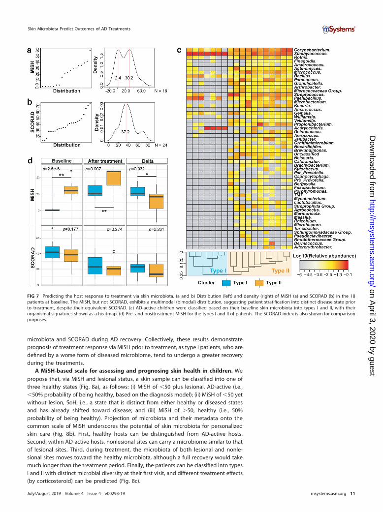
microbiota and SCORAD during AD recovery. Collectively, these results demonstrateprognosis of treatment response via MiSH prior to treatment, as type I patients, who aredefined by a worse form of diseased microbiome, tend to undergo a greater recoveryduring the treatments.
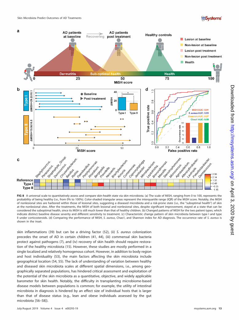
A MiSH-based scale for assessing and prognosing skin health in children. Wepropose that, via MiSH and lesional status, a skin sample can be classified into one ofthree healthy states (Fig. 8a), as follows: (i) MiSH of �50 plus lesional, AD-active (i.e.,�50% probability of being healthy, based on the diagnosis model); (ii) MiSH of �50 yetwithout lesion, SoH, i.e., a state that is distinct from either healthy or diseased statesand has already shifted toward disease; and (iii) MiSH of �50, healthy (i.e., 50%probability of being healthy). Projection of microbiota and their metadata onto thecommon scale of MiSH underscores the potential of skin microbiota for personalizedskin care (Fig. 8b). First, healthy hosts can be distinguished from AD-active hosts.Second, within AD-active hosts, nonlesional sites can carry a microbiome similar to thatof lesional sites. Third, during treatment, the microbiota of both lesional and nonle-sional sites moves toward the healthy microbiota, although a full recovery would takemuch longer than the treatment period. Finally, the patients can be classified into typesI and II with distinct microbial diversity at their first visit, and different treatment effects(by corticosteroid) can be predicted (Fig. 8c).
FIG 7 Predicting the host response to treatment via skin microbiota. (a and b) Distribution (left) and density (right) of MiSH (a) and SCORAD (b) in the 18patients at baseline. The MiSH, but not SCORAD, exhibits a multimodal (bimodal) distribution, suggesting patient stratification into distinct disease state priorto treatment, despite their equivalent SCORAD. (c) AD-active children were classified based on their baseline skin microbiota into types I and II, with theirorganismal signatures shown as a heatmap. (d) Pre- and posttreatment MiSH for the types I and II of patients. The SCORAD index is also shown for comparisonpurposes.
Skin Microbiota Predict Outcomes of AD Treatments
July/August 2019 Volume 4 Issue 4 e00293-19 msystems.asm.org 11
on April 3, 2020 by guest
http://msystem
s.asm.org/
Dow
nloaded from
At present, SCORAD is the prevalent clinical metric for AD diagnosis, yet it is limitedby subjectivity in judgment and inability to evaluate risk-prone states of skin thatexhibits no visible symptoms. Microbial � diversity is also linked to skin health (39), yetthe performance of Shannon index or Chao1 index for AD diagnosis is poor (AUC, 0.67and 0.59 separately; Fig. 8d), as no clear decision boundary can be obtained from eithermetric (Fig. S5a). S. aureus, to a certain extent, is considered a biomarker of AD (48), asit was enriched in AD while barely detected in healthy children (49). Moreover, it wasrecently reported that among Staphylococcus spp., S. aureus predominates in moresevere AD and S. epidermidis predominates in less severe AD (50). However, an ADdiagnosis model based on S. aureus alone carries an AUC of 0.83, much lower than thatof MiSH (0.90). This is due to the large interpersonal variation of S. aureus in AD patients(51); its occurrence rate can be rather low among all hosts (39% for healthy samples,87% for lesions, and 63% overall; Fig. 8d) and varies widely among the Beijing, Qingdao,and Denver cohorts (81%, 26%, and 95%, respectively; Fig. S5b). Therefore, a referencerange of S. aureus trained in one population would not apply in others, as the intercityvariation in its relevant abundance is quite high, even for healthy children (P � 0.012,Kruskal-Wallis test; Fig. S5c).
In our study, MiSH is significantly correlated with SCORAD (Fig. S6a; rho � 0.46,P � 6e�4, Pearson test) and with skin microbial � diversity (Fig. S6b; rho � 0.58,P � 2e�16, Pearson test). MiSH is also positively correlated with the relative abundanceof S. aureus (Fig. S6c; rho� 0.63, P � 2e�16, Pearson test). In the Qingdao cohort, theS. aureus-dominated group and an S. epidermidis-dominated group (ratios of S. aureusand S. epidermidis were determined by quantitative PCR [qPCR]; see Materials andMethods) were of significantly different MiSH, with S. aureus carrying lower MiSH(indicating more severe AD; P � 0.031). Thus, MiSH can be reconciled with existingbiomarkers of AD and can potentially serve as a clinically useful and generally appli-cable parameter.
These advantages of MiSH encourage us to establish an interactive website whichaccepts 16S rRNA-amplicon based data sets as input and returns a graphical report ofMiSH (http://bioinfo.single-cell.cn/mish/index.php/upload_mish) (see Materials andMethods). This online tool may be of potential value in personalized skin healthassessment, prediction of response to treatment, and comparison of skin care producteffects in both healthy and AD-active children.
DISCUSSION
Enthusiasm for diagnosis and therapy of skin disorders via skin microbiota has arisenfrom recent evidence that: (i) the dysbiosis of skin microbiota is not just associated with
TABLE 2 MiSH and SCORAD of the 18 AD patients in the BCP2 and B7U treatmentgroups, both prior to treatment and posttreatment
Sample ID Type
MiSH at: SCORAD at:
Baseline After treatment Baseline After treatment
1039 I 0.06 17.64 33.40 6.621069 I 0.14 19.88 55.80 36.501072 I 0.10 25.06 33.57 10.651052 I 0.22 25.84 35.12 22.821050 I 2.60 30.62 73.00 33.501001 I 2.48 46.08 41.83 16.521024 I 0.02 45.06 42.91 47.241056 II 30.54 12.66 48.81 41.481080 II 60.62 59.42 28.30 10.351089 II 34.20 35.88 28.88 20.311005 II 26.90 30.84 65.75 25.291070 II 41.58 62.14 13.20 1.951030 II 38.68 59.60 46.39 44.141060 II 35.04 59.46 44.82 20.621064 II 15.38 41.04 38.55 7.791077 II 30.16 65.84 31.20 19.941078 II 20.90 60.92 42.24 13.31
Sun et al.
July/August 2019 Volume 4 Issue 4 e00293-19 msystems.asm.org 12
on April 3, 2020 by guest
http://msystem
s.asm.org/
Dow
nloaded from
skin inflammations (39) but can be a driving factor (52), (ii) S. aureus colonizationprecedes the onset of AD in certain children (41, 44), (iii) commensal skin bacteriaprotect against pathogens (7), and (iv) recovery of skin health should require restora-tion of the healthy microbiota (15). However, these studies are mostly performed in asingle localized and relatively homogenous cohort. However, in addition to body regionand host individuality (53), the main factors affecting the skin microbiota includegeographical location (54, 55). The lack of understanding of variation between healthyand diseased skin microbiota scales at different spatial dimensions, i.e., among geo-graphically separated populations, has hindered critical assessment and exploitation ofthe potential of the skin microbiota as a quantitative, objective, and widely applicablebarometer for skin health. Notably, the difficulty in transplanting microbiome-baseddisease models between populations is common; for example, the utility of intestinalmicrobiota in diagnosis is hindered by an effect size of individual hosts that is largerthan that of disease status (e.g., lean and obese individuals assessed by the gutmicrobiota [56–58]).
FIG 8 A universal scale to quantitatively assess and compare skin-health state via skin microbiota. (a) The scale of MiSH, ranging from 0 to 100, represents theprobability of being healthy (i.e., from 0% to 100%). Color-shaded triangular areas represent the interquartile range (IQR) of the MiSH score. Notably, the MiSHof nonlesional sites are harbored within those of lesional sites, suggesting a diseased microbiota and a risk-prone state (i.e., the “suboptimal health”) of skinat the nonlesional sites. After the treatments, the MiSH of both lesional and nonlesional sites, despite significant improvement, stayed at a state that can beconsidered the suboptimal health, since its MiSH is still much lower than that of healthy children. (b) Changed patterns of MiSH for the two patient types, whichindicate distinct baseline disease severity and different sensitivity to treatment. (c) Characteristic change pattern of skin microbiota between type I and typeII under corticosteroids. (d) Comparing the performance of MiSH, S. aureus, Chao1, and Shannon index for AD diagnosis. The occurrence rate of S. aureus isshown in the inset.
Skin Microbiota Predict Outcomes of AD Treatments
July/August 2019 Volume 4 Issue 4 e00293-19 msystems.asm.org 13
on April 3, 2020 by guest
http://msystem
s.asm.org/
Dow
nloaded from
A comparison of the skin microbiome of AD and healthy pediatric cohorts fromthree cities, two Chinese and one American, revealed that, healthy and diseased skinmicrobiota from each city carried both city-specific signature (Fig. 3e) and AD-associated biomarkers (Fig. 4a), and there were significant overlaps among the healthyand diseased biomarkers. Although the two Chinese cities shared more disease bio-markers than did the intercontinental pairs, a significant core of AD-associated micro-biota was present, with its size and membership independent of geographic distancesamong populations. Therefore, despite the differences among pediatric cohorts, an ADdiagnosis model built from a single city can be applied across the three cities withacceptable accuracy. As a result, despite the large effect size of city and individualvariation, the MiSH model consisting of the top 25 bacterial skin genera can diagnoseAD with 86.4% accuracy (AUC, 0.90) across cities and continents, and it offers highsensitivity in assessing the efficacy of treatment products. Notably, although the bodylocation is one of the most important factors to the skin microbiome (54, 55), applica-tion of MiSH (which was generated based on all samples from the three cities) on theBeijing samples of various body locations revealed that, in each of the six bodylocations that include both moist (antecubital fossa and popliteal fossa) and dry (arm,knee, neck, and shank) ones, MiSH can reliably distinguish their health status (Fig. S6d).
Moreover, for nonlesional skin sites in AD-active children, the MiSH model revealeda distinct state of skin microbiota called suboptimal health, which is intermediatebetween those of lesional sites and healthy children, yet more similar to the former, andcarries a level of Staphylococcus spp. higher than that in healthy hosts but lower thanin lesional sites. This state was converted to a healthier state on the MiSH scale aftertopical medication. However, the degree of dysbiosis or its remediation does notcorrelate with physical distance to the lesional site. Although initial evidence for thealteration of microbiota on apparent healthy skin zones physically adjacent to thelesional sites has emerged (59, 60), the extent to which the skin microbiota respondacross the whole body is not known. Our findings here support AD as a topical effectbut with an underpinning microbiota dysbiosis that extends across the body (61, 62),and they underscore the dynamic interactions between global host immune responseand local skin microbiota (63). Therefore, MiSH can be used not only for AD severitymeasurement but also for assessing the healthy state and the risk-prone state of skinin AD-free children (whether this is applicable in adults is unknown, as AD skinmicrobiome is affected by age [64]).
Furthermore, pretreatment MiSH classifies children with clinically indistinguishableAD into two types with distinct disease severity and sensitivity to corticosteroidtherapy. These two types of patients feature distinct microbiota structures prior totreatment and exhibit characteristic patterns of microbiota change during treatment.Type I patients, with lower MiSH at baseline, carry a more disease-oriented microbiomethat features fewer genera yet much higher proportion of Staphylococcus spp., repre-sent a more severe AD form, and tend to have a more prominent response of recoveryduring treatment. In contrast, type II patients, with higher MiSH at baseline, carry a lessdisease-oriented microbiome characterized by a lower level of Staphylococcus spp. yethigher diversity of bacterial genera and represent a milder disease form of AD.Interestingly, in Qingdao and Denver, the MiSH of all AD patients also exhibit a bimodaldistribution (Fig. S6e; our current data do not allow testing of whether such clusteringis correlated with treatment effect in these two cities). Accordingly, the two typesshould be treated differently; for example, type I should be prioritized for treatmentwith higher drug dosage, since it represents a more severe form of AD yet is more likelyto respond to treatment in terms of skin microbiota recovery. Consistent with a recentstudy that suggests cross-modulation of the skin microbiome, skin surface microenvi-ronment and immune system underlie susceptibility to AD in adults (42), our findingshere support a microbial basis for the heterogeneity of response to AD treatment andfor the recovery of skin health in children. Notably, two distinct clusters of skinmicrobiome were discovered in the lesion samples from 51 adult psoriasis patientsfrom New York City (65), although whether their disease outcomes or treatment effects
Sun et al.
July/August 2019 Volume 4 Issue 4 e00293-19 msystems.asm.org 14
on April 3, 2020 by guest
http://msystem
s.asm.org/
Dow
nloaded from
are different remains to be tested. Therefore, it seems possible that such microbiome-defined cutaneotypes can be quite common in disease, and further characterization ofcutaneotypes within and across various kinds of skin inflammations might provide newinsights into disease diagnosis or treatment strategy.
At present, one limitation of MiSH is its inability to distinguish the various Staphy-lococcus species due to the genus-level resolution of 16S rRNA amplicon-based se-quencing in microbial identification. Recent reports suggested that different Staphylo-coccus species can play distinct roles in AD development; for example, S. epidermidisand Staphylococcus hominis, which predominantly reside on healthy human skin,actually contribute to cutaneous homeostasis and health (8); in addition, selectedStaphylococcus strains can either promote cutaneous antimicrobial activity or triggerinflammation in AD (7, 50). Thus, versions of MiSH that assess skin microbiota at thespecies or the strain levels should be developed, via either long-read sequencing of 16SrRNA amplicons or metagenome sequencing. Moreover, tools such as conditionally raretaxa (CRT) (66) can be used to probe the scope and origin of such city-specific bacterialtaxa, as they offered 97.6% to �100% accuracy in distinguishing the three cities and alevel of performance in distinguishing the AD status that is equivalent to that with MiSH(Materials and Methods).
On the other hand, as size of the treatment cohort here is relatively small, howgenerally applicable the microbiome-defined heterogeneity in treatment response isnot yet clear, and its mechanism is unknown. Future efforts tackling these questions arekey to more precise AD therapies (67, 68). Despite these limitations, once the costs ofsequencing are reduced to an acceptable level, MiSH is expected to contribute, inconjunction with SCORAD, for AD diagnosis and treatment in the clinical setting, wherethe state of skin microbiota is also taken into consideration.
MATERIALS AND METHODSStudy design. From the city of Beijing, China (the Beijing cohort), we established a cohort of 28
children age 4 to 12 years who were suffering from mild to moderate AD, plus 30 age-equivalent andsampling site-matched children with no personal or family history of AD and no history of chronic skinor systemic diseases (Table S1). To explore the link between AD treatment and skin microbiota alteration,an “AD-treatment cohort” was designed, in which AD-active children of the Beijing cohort underwent a4-week-long treatment regimen of corticosteroid administration, with each child using one of the threeskin care products of BCP2 (ultramild body wash with lipids), B7U (mild synthetic bar) or ZPT (ultramildbody wash with lipids and zinc pyrithione).
In addition, a second cohort of age-matched children was recruited from Qingdao (the Qingdaocohort), a coastal Chinese city at the West Pacific rim 650 kilometers southeast of Beijing. The Qingdaocohort consists of 29 pediatric patients suffering from moderate AD and 30 healthy subjects, who werescreened using selection criteria that are identical to those for the Beijing cohort.
Furthermore, to test whether healthy and AD-active skin microbiota patterns held true at evengreater geographic distances, a third cohort we previously published for Denver, an inland city of NorthAmerica (the Denver cohort), was also included into the three-way, cross-city comparison here. Thecohort consists of 59 AD-active and 13 healthy children that were 4 to 12 years old (60). Similar to thiswork, the Denver study employed MiSeq paired-end reads and the primer set of 27F/534R for profilingbacterial 16S rRNA amplicons (Table S3). Moreover, to ensure data comparability, samples from the threecities were computationally processed in an identical manner.
Exclusion criteria for all subjects include having a fever of �38.5°C, having bathed or showered aftermidnight before the day of sampling, using creams/lotions at the sites 24 h prior to sampling, havingreceived oral antibiotics, a bleach bath, or topical prescription medications (including but not limited toElidel, Protopic, topical corticosteroids, or topical antibiotics; more details below) 7 days prior tosampling, having taken systemic immunosuppressive drugs (including cyclosporine or oral steroids), andhaving experienced total body phototherapy (e.g., UV light B, psoralen plus UV light A, and tanning beds)within 20 days prior to sampling.
The study was conducted and all samples were collected with approval from the Procter & GambleBeijing Innovation Center institutional review board and in accordance with the World Medical Associ-ation Declaration of Helsinki (1996 amendment). ICH Guidelines for Good Clinical Practice (GCPs) werefollowed, and voluntary informed consent was provided with the approval of the Research Ethics Boardof P&G. Mothers who agreed to have their children participate in this study signed an informed consentform, and teenagers who agreed to participate signed an assent form.
Severity scoring of atopic dermatitis. Only subjects in the AD group would undergo dermatologicevaluations throughout the study. Subjects acclimated for a minimum of 30 minutes in an environmen-tally controlled room (maintained at 70°F and 30 to 45% relative humidity) prior to undergoing adermatologic evaluation from the study dermatologist at the following time points. At the baseline andweek 4 visits, the dermatologist would assess the subject’s atopic dermatitis lesional/measurement sites
Skin Microbiota Predict Outcomes of AD Treatments
July/August 2019 Volume 4 Issue 4 e00293-19 msystems.asm.org 15
on April 3, 2020 by guest
http://msystem
s.asm.org/
Dow
nloaded from
on their arms and/or legs only for the intensity of objective attributes. This evaluation along withthe extent of body surface involvement and subjective symptoms (pruritus and sleep loss) rated by thesubject was used to determine the SCORAD value (22). The SCORAD value is calculated using thefollowing formula:
SCORAD �Extent
5�
7 � Intensity
2� Subjective signs
where (i) “Extent” is the extent of body area affected; to determine the extent of affected area as apercentage of the whole body, the rule of nine is used. (ii) “Intensity” is the intensity grading scale; themarked lesional sites are graded for the intensity of each of the following signs: dryness, erythema,excoriation, weeping, induration, and lichenification. (iii) “Subjective signs” is the subjective symptoms,where itch and sleeplessness are each scored by the subjects or parent/guardian using a 10-cm visualanalogue scale where 0 is no itch (or no sleeplessness) and 10 is severe itch (or sleeplessness).
Skin microbiome sampling strategy. In each of the three cohorts, microbiota from skin zonescorresponding to the AD-active sites of patients were sampled in matched healthy individuals. Addi-tionally, for each AD-active child, skin microbiota from both lesional and nonlesional sites was collected.For the Beijing and Denver cohorts, nonlesional sites were taken from nonlesional skin site of asymmetric location on the body or the surrounding skin of the lesional sites, and for the Qingdao cohort,the nonlesional sites also included another two sites on the forearm and shank surface (Table S1).
At the inclusion visit and at the end of study, the same investigating dermatologist evaluated thechildren via the SCORAD (SCORing Atopic Dermatitis) index, which is a clinical tool for assessing ADseverity (22). Only individuals with a SCORAD index between 25 and 40 at baseline were included aspatients in the study. Skin microbiota samples of lesional skin were collected using aseptic techniquesunder sterile airflow generated by a portable hood. Similarly, samples were also collected from theunaffected symmetric and remote body skin area.
Sampling procedures were as follows. (i) Identify the designated sampling site being used for swabcollection (�10 cm2) and then use a ruler/template to mark an 8-cm2 area. (ii) Identify the designatednonlesion site being used for swab collection. (iii) Label all collection tubes. (iv) With gloved hands,remove DNA swab from packaging with care taken not to touch any surface. (v) Dip the swab tip intoNaCl plus Tween 20 solution, and press the swab to the inside of the tube to remove any excess liquid.(vi) Apply the swab in both horizontal and vertical directions (totally 50 times, for about 30 to �35 s) forsampling the marked area. (vii) Break the DNA extraction swab and put into the appropriately labeledempty 2-ml tube and cap. (viii) Store the tube in an ice box until samples can be stored at – 80°C. Finally,repeat steps iv through viii on a nonlesion site for each site.
Administration of medication for AD treatment. In the city of Beijing, for the 28 AD patients whowere sampled from both lesional skin sites and nonlesional sites at baseline and then again after 4 weeksof treatment via corticosteroids and bath products, only 24 of the patients were evaluated posttreatmentbecause four individuals failed to show up for the last visit. The treatment was via corticosteroid (0.1%hydrocortisone butyrate ointment), which was used on every patient based on doctor’s advice. Inaddition, one of three body wash products was used in bath, BCP2, ZPT, or B7U. Treatment assignmentwas randomized to subject to balance for baseline AD severity, age, biological sex, and body location ifpossible. Due to the complexity and smaller sample sizes, the balancing was prioritized in order ofimportance, with baseline disease severity, age, biological sex, and then body location (most of thesubjects had lesions on arms). Patients were instructed to apply the bath product once daily in theevening to their entire body. Patients were also asked not to change their hygiene practices or to applyany other bath products during the study.
Specifically, this is a 7-week, randomized, double-blind, parallel group in-home-use study amongmale and female subjects who are 4 to 12 years of age (inclusive) and having mild to moderate activeatopic dermatitis (AD), where three products were tested. Written informed consents were obtained fromthe parent/legal guardian of each subject and verbal assent from each subject according to ICH GCPsprior to screening based on the inclusion/exclusion criteria listed below. Qualified subjects (including thehealthy control group) completed a habits and practices questionnaire prior to starting the precondi-tioning phase. They completed a 7-day preconditioning phase where they used a provided bar soap forall body cleansing purposes and refrained from using any other personal cleansing products as well asany moisturizers, powders, topical medications, oils, or creams for the duration of the preconditioningphase. Subjects also refrained from using any body cleansing implements (e.g., wash cloths and bodypuffs) during the preconditioning phase of the study. Subjects were permitted to use their normal facialand hair care cleansing products, but they must refrain from using any products containing antibacterialingredients (i.e., acne products, salicylic acid-containing facial care products, and antidandruff shampoos)during the preconditioning phase.
Ingredients of the three body wash products tested. The ingredients for B7U (regular synthesizedbar soap) were sodium lauroyl isethionate, paraffin, sodium cocoglyceryl ether sulfonate, glycerin, water,talc, magnesium stearate, stearic acid, sodium isethionate, magnesium cocoate, sodium stearate, coconutacid, sodium chloride, sodium cocoate, fragrance/parfum, magnesium laurate, lauric acid, and titaniumdioxide.
The ingredients for BCP2 (ultramild moisturizing body wash) were water, petrolatum, sodiumtrideceth sulfate, sodium chloride, cocamidopropyl betaine, trideceth-3, guar hydroxypropyltrimoniumchloride, sodium benzoate, xanthan gum, glyceryl oleate, fragrance, disodium EDTA, citric acid, sodiumhydroxide, acrylates/c10-30 alkyl acrylate cross-polymer, Butyrospermum parkii (shea) butter, methyl-chloroisothiazolinone, and methylisothiazolinone.
Sun et al.
July/August 2019 Volume 4 Issue 4 e00293-19 msystems.asm.org 16
on April 3, 2020 by guest
http://msystem
s.asm.org/
Dow
nloaded from
The ingredients for ZPT (0.5% zinc pyrithione containing ultramild moisturizing body wash) werewater, petrolatum, sodium trideceth sulfate, sodium chloride, cocamidopropyl betaine, trideceth-3, zpt,guar hydroxypropyltrimonium chloride, sodium benzoate, xanthan gum, glyceryl oleate, fragrance, citricacid, sodium hydroxide, acrylates/c10-30 alkyl acrylate cross-polymer, Butyrospermum parkii (shea) butter,methylchloroisothiazolinone, and methylisothiazolinone.
DNA extraction, PCR amplification, and sequencing of skin microbiome. Genomic DNA wasextracted from each swab using the Qiagen tissue and blood DNA isolation kit, following the manufac-turer’s instructions, with slight modifications (69). PCR amplification of the V1-V3 region of 16S rRNAgenes was performed using the primer set (27F/534R) and followed the protocol developed by theHuman Microbiome Project. PCR amplification reaction mixtures in triplicate for each sample werepooled at approximately equal amounts and sequenced. For Qingdao and Denver, Illumina MiSeq wasemployed as the sequencing platform. For Beijing cohort, both MiSeq and Roche 454 FLX were used forsequencing each of the samples. Roche 454 sequencing data were used in building the RF model. MiSeqdata were used to calculate the effect size of factors, so as to avoid the bias due to difference insequencing platforms. For both healthy and lesional samples, the effect size of sequencing platform issmaller than that of city (Fig. S7). In addition, no positive PCR results were found in the negative controls(i.e., clean swabs), suggesting that no background bacterial contamination can be detected.
For quantitative PCR (qPCR) that measures the relative abundance between S. aureus and S.epidermidis, the primer pair is 5=-TAGTTGTAGTTTCAAGTCTAAGTAGCTCAGC and 3=-ATTTAACCGTATCACCATCAATCG for S. aureus and 5=-GGCAAATTTGTGGGTCAAGA and 3=-TGGCTAATGGTTTGTCACCA) for S.epidermidis (70). Gene copy number was calculated based on the standard curve of each primer systemusing the LightCycler 480 software 1.5 (Roche). Relative abundance is defined as gene copy number ofeach biomarker divided by 16S rRNA gene copy number of whole bacteria. Each qPCR reaction wasperformed in triplicate.
Sequence analyses of skin microbiomes. All sequences were preprocessed following the standardQIIME (v.1.9) pipeline. A total of 643,038 high-quality partial 16S rRNA sequences were obtained from the275 samples collected, with an average of 8,669 sequences per sample. Downstream bioinformaticsanalysis was performed using Parallel-Meta 3 (71), a software package for comprehensive taxonomicaland functional comparison of microbial communities. Clustering of OTUs was conducted at the 97%similarity level using a preclustered version of the GreenGenes database (72). To perform taxonomicclassifications at the species level for staphylococcal species, staphylococcal sequences were determinedto the species level by alignment to a curated collection of staphylococcal reference sequences fromcomplete genome sequences and type strains. Finally, each sequence was assigned a taxonomic label atthe species level (such as S. aureus, S. epidermidis, Staphylococcus capitis, and S. hominis) based on theconsensus call of sequence alignments with the lowest edit distance between a query and reference. The� diversity was calculated by Shannon index and Chao1, and the distance between each pairs of skinmicrobiota was computed based on the weighted Meta-Storms algorithm (73). For a certain genus to beconsidered “present,” it has to be of at least 0.01% abundance in at least 50% of the hosts within a city.Those genera with �0.01% abundance were merged together and referred to as “other genera”; on theother hand, those genera with �50% prevalence among the hosts within a city were not consideredfurther (74). As for �-diversity, Meta-Storms distance (which is integrated in PM3 [71]) was used toquantify the differences between any two samples. The Meta-Storms scoring function is a phylogeny-based algorithm that quantitatively evaluates the biological similarity/distance between the microbiomesamples on the OTU level (73). In parallel with above efforts, the contribution of conditionally rate taxa(CRT) to discrimination of originated city or AD status was quantified using CRT detection scripts (v1.0)with default parameters (75).
The other statistical analysis, e.g., Kruskal-Wallis test, Wilcoxon rank sum test, and permutationalmultivariate analysis of variance (PERMANOVA), were performed via the R scripts integrated in PM3. Thescripts take advantage of using standard functions of kruskal.test and wilcox.test, as well as the adonisfunction in the R package of vegan. The rarefaction analysis and Shannon diversity index were used toestimate the richness and diversity of species. The relative abundance of differential taxonomic groupswere visualized by “pheatmap” in the “pheatmap” R package. Differences in the relative abundance oftaxonomic groups at the genus level between samples were evaluated with Wilcoxon rank sum test.False-discovery rate (FDR) values were estimated using the Benjamini-Hochberg method to control formultiple testing. P values less than 0.05 were considered statistically significant.
Building the diagnostic models of atopic dermatitis. The N top-ranking AD-discriminatory taxathat led to reasonably good fit were identified based on “rfcv” function in the randomForest pack-age (https://cran.r-project.org/web/packages/randomForest/index.html). Random Forests models weretrained to identify disease status in the training set which included samples from the ‘‘healthy’’ and the‘‘lesional’’ groups using the taxonomy profiles. The results were evaluated with a 10-fold cross-validationapproach, and model performance was evaluated by ROC. Default parameters of the R implementationof algorithm were applied (ntree � 5,000, using default mtry of p/3, where p is the number of input taxa).To construct and optimize the MiSH, we tested how taxonomical levels influence the performance of RFmodel. Using the profiles of genus, the performance of models based on microbiota was furtherevaluated with a 10-fold cross-validation approach. In 10-fold cross-validation, the original samples wererandomly partitioned into 10 equal-sized subsamples. Of the 10 subsamples, a single subsample isretained as the validation data for testing the model, and the remaining nine subsamples were used astraining data. The cross-validation process was then repeated for 10 times, and the average of probabilitywas reported as the result. Based on optimization that selects the taxonomy level that maximizes modelperformance, Random Forest models were trained to identify disease status using the taxonomy profiles
Skin Microbiota Predict Outcomes of AD Treatments
July/August 2019 Volume 4 Issue 4 e00293-19 msystems.asm.org 17
on April 3, 2020 by guest
http://msystem
s.asm.org/
Dow
nloaded from
on the genus level. A receiver operating characteristic (ROC) curve was then used to illustrate thediagnostic performance of RF model (https://cran.r-project.org/web/packages/pROC/index.html). In theROC plots, x axis represents true-positive rate (TPR, or sensitivity), y axis stands for false-positive rate (FPR,or specificity), and area under the ROC curve (AUC) was calculated to summarize performance of the RFmodel.
Data availability. The sequence data in this study have been submitted to the Sequence ReadArchive (https://www.ncbi.nlm.nih.gov/sra) and can be accessed through the BioProject numbersPRJNA445780 and PRJNA268694.
SUPPLEMENTAL MATERIALSupplemental material for this article may be found at https://doi.org/10.1128/
mSystems.00293-19.FIG S1, TIF file, 0.5 MB.FIG S2, TIF file, 0.8 MB.FIG S3, TIF file, 0.5 MB.FIG S4, TIF file, 0.3 MB.FIG S5, TIF file, 0.2 MB.FIG S6, TIF file, 0.4 MB.FIG S7, TIF file, 0.9 MB.TABLE S1, DOCX file, 0.1 MB.TABLE S2, DOCX file, 0.1 MB.TABLE S3, DOCX file, 0.1 MB.
ACKNOWLEDGMENTSThis work was funded by grant 31425002 from the National Natural Science Foun-
dation of China, grants ZR2016QZ004 and ZR2017ZB0421 from the Natural Foundationof Shandong Province, and a joint research program between the Chinese Academy ofSciences and Procter & Gamble.
We declare no competing interests.We are grateful to Qingdao Women and Children’s Hospital for excellent technical
support in sampling of AD children in the Qingdao cohort.
REFERENCES1. Knights D, Parfrey LW, Zaneveld J, Lozupone C, Knight R. 2011. Human-
associated microbial signatures: examining their predictive value. CellHost Microbe 10:292–296. https://doi.org/10.1016/j.chom.2011.09.003.
2. Lozupone CA, Stombaugh JI, Gordon JI, Jansson JK, Knight R. 2012.Diversity, stability and resilience of the human gut microbiota. Nature489:220 –230. https://doi.org/10.1038/nature11550.
3. Huttenhower C, Gevers D, Knight R, Abubucker S, Badger JH, ChinwallaAT, Creasy HH, Earl AM, FitzGerald MG, Fulton RS, Giglio MG, Hallsworth-Pepin K, Lobos EA, Madupu R, Magrini V, Martin JC, Mitreva M, MuznyDM, Sodergren EJ, Versalovic J, Wollam AM, Worley KC, Wortman JR,Young SK, Zeng QD, Aagaard KM, Abolude OO, Allen-Vercoe E, Alm EJ,Alvarado L, Andersen GL, Anderson S, Appelbaum E, Arachchi HM,Armitage G, Arze CA, Ayvaz T, Baker CC, Begg L, Belachew T, BhonagiriV, Bihan M, Blaser MJ, Bloom T, Bonazzi V, Brooks JP, Buck GA, Buhay CJ,Busam DA, Campbell JL, et al. 2012. Structure, function and diversity ofthe healthy human microbiome. Nature 486:207–214. https://doi.org/10.1038/nature11234.
4. Costello EK, Lauber CL, Hamady M, Fierer N, Gordon JI, Knight R. 2009.Bacterial community variation in human body habitats across space andtime. Science 326:1694 –1697. https://doi.org/10.1126/science.1177486.
5. Edmonds-Wilson SL, Nurinova NI, Zapka CA, Fierer N, Wilson M. 2015.Review of human hand microbiome research. J Dermatol Sci 80:3–12.https://doi.org/10.1016/j.jdermsci.2015.07.006.
6. Kobayashi T, Glatz M, Horiuchi K, Kawasaki H, Akiyama H, Kaplan DH,Kong HH, Amagai M, Nagao K. 2015. Dysbiosis and Staphylococcusaureus colonization drives inflammation in atopic dermatitis. Immunity42:756 –766. https://doi.org/10.1016/j.immuni.2015.03.014.
7. Nakatsuji T, Chen TH, Narala S, Chun KA, Two AM, Yun T, Shafiq F, KotolPF, Bouslimani A, Melnik AV, Latif H, Kim JN, Lockhart A, Artis K, David G,Taylor P, Streib J, Dorrestein PC, Grier A, Gill SR, Zengler K, Hata TR,Leung DY, Gallo RL. 2017. Antimicrobials from human skin commensalbacteria protect against Staphylococcus aureus and are deficient in
atopic dermatitis. Sci Transl Med 9:eaah4680. https://doi.org/10.1126/scitranslmed.aah4680.
8. Nakatsuji T, Chen TH, Butcher AM, Trzoss LL, Nam SJ, Shirakawa KT, ZhouW, Oh J, Otto M, Fenical W, Gallo RL. 2018. A commensal strain ofStaphylococcus epidermidis protects against skin neoplasia. Sci Adv4:eaao4502. https://doi.org/10.1126/sciadv.aao4502.
9. Grice EA, Segre JA. 2011. The skin microbiome. Nat Rev Microbiol9:244 –253. https://doi.org/10.1038/nrmicro2537.
10. Blaser MJ, Dominguez-Bello MG, Contreras M, Magris M, Hidalgo G,Estrada I, Gao Z, Clemente JC, Costello EK, Knight R. 2013. Distinctcutaneous bacterial assemblages in a sampling of South AmericanAmerindians and US residents. ISME J 7:85–95. https://doi.org/10.1038/ismej.2012.81.
11. Clemente JC, Pehrsson EC, Blaser MJ, Sandhu K, Gao Z, Wang B, MagrisM, Hidalgo G, Contreras M, Noya-Alarcon O, Lander O, McDonald J, CoxM, Walter J, Oh PL, Ruiz JF, Rodriguez S, Shen N, Song SJ, Metcalf J,Knight R, Dantas G, Dominguez-Bello MG. 2015. The microbiome ofuncontacted Amerindians. Sci Adv 1:e1500183. https://doi.org/10.1126/sciadv.1500183.
12. Perez Perez GI, Gao Z, Jourdain R, Ramirez J, Gany F, Clavaud C, De-maude J, Breton L, Blaser MJ. 2016. Body site is a more determinantfactor than human population diversity in the healthy skin microbiome.PLoS One 11:e0151990. https://doi.org/10.1371/journal.pone.0151990.
13. Gilbert JA, Quinn RA, Debelius J, Xu ZJZ, Morton J, Garg N, Jansson JK,Dorrestein PC, Knight R. 2016. Microbiome-wide association studies linkdynamic microbial consortia to disease. Nature 535:94 –103. https://doi.org/10.1038/nature18850.
14. Dorrestein PC, Gallo RL, Knight R. 2016. Microbial skin inhabitants:friends forever. Cell 165:771–772. https://doi.org/10.1016/j.cell.2016.04.035.
15. Grice EA, Kong HH, Conlan S, Deming CB, Davis J, Young AC, Bouffard GG,Blakesley RW, Murray PR, Green ED, Turner ML, Segre JA, Progra N. 2009.
Sun et al.
July/August 2019 Volume 4 Issue 4 e00293-19 msystems.asm.org 18
on April 3, 2020 by guest
http://msystem
s.asm.org/
Dow
nloaded from
Topographical and temporal diversity of the human skin microbiome.Science 324:1190–1192. https://doi.org/10.1126/science.1171700.
16. Capone KA, Dowd SE, Stamatas GN, Nikolovski J. 2011. Diversity of thehuman skin microbiome early in life. J Invest Dermatol 131:2026 –2032.https://doi.org/10.1038/jid.2011.168.
17. Grice EA. 2014. The skin microbiome: potential for novel diagnostic andtherapeutic approaches to cutaneous disease. Semin Cutan Med Surg33:98 –103. https://doi.org/10.12788/j.sder.0087.
18. Rehman A, Rausch P, Wang J, Skieceviciene J, Kiudelis G, Bhagalia K,Amarapurkar D, Kupcinskas L, Schreiber S, Rosenstiel P, Baines JF, Ott S.2016. Geographical patterns of the standing and active human gutmicrobiome in health and IBD. Gut 65:238 –248. https://doi.org/10.1136/gutjnl-2014-308341.
19. Boguniewicz M, Leung D. 2010. Recent insights into atopic dermatitisand implications for management of infectious complications. J AllergyClin Immunol 125:4 –13. https://doi.org/10.1016/j.jaci.2009.11.027.
20. Galli E, Neri I, Ricci G, Baldo E, Barone M, Fortina AB, Bernardini R, BertiI, Caffarelli C, Calamelli E, Capra L, Carello R, Cipriani F, Comberiati P,Diociaiuti A, El Hachem M, Fontana E, Gruber M, Haddock E, Maiello N,Meglio P, Patrizi A, Peroni D, Scarponi D, Wielander I, Eichenfield LF.2016. Consensus conference on clinical management of pediatric atopicdermatitis. Ital J Pediatr 42:26. https://doi.org/10.1186/s13052-016-0229-8.
21. Nomura T, Honda T, Kabashima K. 2018. Multipolarity of cytokine axes inthe pathogenesis of atopic dermatitis in terms of age, race, species,disease stage and biomarkers. Int Immunol 30:419 – 428. https://doi.org/10.1093/intimm/dxy015.
22. Stalder JF, Taieb A, Atherton DJ, Bieber T, Bonifazi E, Broberg A, Calza A,Coleman R, Deprost Y, Diepgen TL, Gelmetti C, Giannetti A, Harper J,Kunz B, Lachapelle JM, Langeland T, Lever R, Oranje AP, Queilleroussel C,Revuz J, Ring J, Roujeau JC, Saurat JH, Song M, Tennstedt D, Vanneste D,Vieluf D, Poncet M. 1993. Severity scoring of atopic dermatitis: theSCORAD index. Consensus report of the European Task Force on AtopicDermatitis. Dermatology 186:23–31.
23. Vocks E, Plotz SG, Ring J. 1999. The dyshidrotic eczema area and severityindex–a score developed for the assessment of dyshidrotic eczema.Dermatology 198:265–269. https://doi.org/10.1159/000018127.
24. Oranje AP, Glazenburg EJ, Wolkerstorfer A, De Waard-van der Spek FB.2007. Practical issues on interpretation of scoring atopic dermatitis: theSCORAD index, objective SCORAD and the three-item severity score. BrJ Dermatol 157:645–648. https://doi.org/10.1111/j.1365-2133.2007.08112.x.
25. Ungar B, Garcet S, Gonzalez J, Dhingra N, Correa da Rosa J, Shemer A,Krueger JG, Suarez-Farinas M, Guttman-Yassky E. 2017. An integratedmodel of atopic dermatitis biomarkers highlights the systemic nature ofthe disease. J Invest Dermatol 137:603– 613. https://doi.org/10.1016/j.jid.2016.09.037.
26. Brunner PM, Emerson RO, Tipton C, Garcet S, Khattri S, Coats I, KruegerJG, Guttman-Yassky E. 2017. Nonlesional atopic dermatitis skin sharessimilar T-cell clones with lesional tissues. Allergy 72:2017–2025. https://doi.org/10.1111/all.13223.
27. Thijs JL, van Seggelen W, Bruijnzeel-Koomen C, de Bruin-Weller M,Hijnen D. 2015. New developments in biomarkers for atopic dermatitis.J Clin Med 4:479 – 487. https://doi.org/10.3390/jcm4030479.
28. Pivarcsi A, Gombert M, Dieu-Nosjean MC, Lauerma A, Kubitza R, Meller S,Rieker J, Muller A, Da Cunha L, Haahtela A, Sonkoly E, Fridman WH,Alenius H, Kemeny L, Ruzicka T, Zlotnik A, Homey B. 2004. CC chemokineligand 18, an atopic dermatitis-associated and dendritic cell-derivedchemokine, is regulated by staphylococcal products and allergen expo-sure. J Immunol 173:5810 –5817. https://doi.org/10.4049/jimmunol.173.9.5810.
29. Yoshioka T, Hikita I, Matsutani T, Yoshida R, Asakawa M, Toyosaki-MaedaT, Hirasawa T, Suzuki R, Arimura A, Horikawa T. 2003. DS-Nh as anexperimental model of atopic dermatitis induced by Staphylococcusaureus producing staphylococcal enterotoxin C. Immunology 108:562–569. https://doi.org/10.1046/j.1365-2567.2003.01588.x.
30. Paller AS, Kong HH, Seed P, Naik S, Scharschmidt TC, Gallo RL, Luger T,Irvine AD. 2019. The microbiome in atopic dermatitis. J Allergy ClinImmunol 143:26 –35. https://doi.org/10.1016/j.jaci.2018.11.015.
31. Hon KLE, Wang SS, Leung TF. 2012. What happens to the severitygrading by objective SCORAD if we over- or underestimate diseaseextent or intensity in patients with atopic dermatitis? Int J Dermatol51:295–299. https://doi.org/10.1111/j.1365-4632.2011.05004.x.
32. Eichenfield LF, Tom WL, Berger TG, Krol A, Paller AS, Schwarzenberger K,Bergman JN, Chamlin SL, Cohen DE, Cooper KD, Cordoro KM, Davis DM,Feldman SR, Hanifin JM, Margolis DJ, Silverman RA, Simpson EL, Williams
HC, Elmets CA, Block J, Harrod CG, Smith Begolka W, Sidbury R. 2014.Guidelines of care for the management of atopic dermatitis: section 2.Management and treatment of atopic dermatitis with topical therapies.J Am Acad Dermatol 71:116 –132. https://doi.org/10.1016/j.jaad.2014.03.023.
33. Sidbury R, Tom WL, Bergman JN, Cooper KD, Silverman RA, Berger TG,Chamlin SL, Cohen DE, Cordoro KM, Davis DM, Feldman SR, Hanifin JM,Krol A, Margolis DJ, Paller AS, Schwarzenberger K, Simpson EL, WilliamsHC, Elmets CA, Block J, Harrod CG, Smith Begolka W, Eichenfield LF.2014. Guidelines of care for the management of atopic dermatitis:section 4. Prevention of disease flares and use of adjunctive therapiesand approaches. J Am Acad Dermatol 71:1218 –1233. https://doi.org/10.1016/j.jaad.2014.08.038.
34. Berger TG, Duvic M, Van Voorhees AS, VanBeek MJ, Frieden IJ. 2006. Theuse of topical calcineurin inhibitors in dermatology: safety concerns.Report of the American Academy of Dermatology Association TaskForce. J Am Acad Dermatol 55:271–271. https://doi.org/10.1016/j.jaad.2006.01.054.
35. Paller AS, Tom WL, Lebwohl MG, Blumenthal RL, Boguniewicz M, Call RS,Eichenfield LF, Forsha DW, Rees WC, Simpson EL, Spellman MC, SteinGold LF, Zaenglein AL, Hughes MH, Zane LT, Hebert AA. 2016. Efficacyand safety of crisaborole ointment, a novel, nonsteroidal phosphodies-terase 4 (PDE4) inhibitor for the topical treatment of atopic dermatitis(AD) in children and adults. J Am Acad Dermatol 75:494 –503. https://doi.org/10.1016/j.jaad.2016.05.046.
36. Ahmed A, Solman L, Williams HC. 2018. Magnitude of benefit for topicalcrisaborole in the treatment of atopic dermatitis in children and adultsdoes not look promising: a critical appraisal. Br J Dermatol 178:659 – 662.https://doi.org/10.1111/bjd.16046.
37. Noordzij TC, Leunissen KML, Vanhooff JP. 1990. Cyclosporine-inducedhyperuricemia and gout. N Engl J Med 322:335–335. https://doi.org/10.1056/NEJM199002013220514.
38. Berke R, Singh K, Guralnick M. 2012. Atopic dermatitis: an overview. AmFam Physician 86:35– 42.
39. Kong HDH, Oh J, Deming C, Conlan S, Grice EA, Beatson MA, Nomicos E,Polley EC, Komarow HD, Murray PR, Turner ML, Segre JA, Program N.2012. Temporal shifts in the skin microbiome associated with diseaseflares and treatment in children with atopic dermatitis. Genome Res22:850 – 859. https://doi.org/10.1101/gr.131029.111.
40. Weiss S, Xu ZZ, Peddada S, Amir A, Bittinger K, Gonzalez A, Lozupone C,Zaneveld JR, Vazquez-Baeza Y, Birmingham A, Hyde ER, Knight R. 2017.Normalization and microbial differential abundance strategies dependupon data characteristics. Microbiome 5:27. https://doi.org/10.1186/s40168-017-0237-y.
41. Williams MR, Gallo RL. 2017. Evidence that human skin microbiomedysbiosis promotes atopic dermatitis. J Invest Dermatol 137:2460 –2461.https://doi.org/10.1016/j.jid.2017.09.010.
42. Chng KR, Tay AS, Li C, Ng AH, Wang J, Suri BK, Matta SA, McGovern N,Janela B, Wong XF, Sio YY, Au BV, Wilm A, De Sessions PF, Lim TC, TangMB, Ginhoux F, Connolly JE, Lane EB, Chew FT, Common JE, NagarajanN. 2016. Whole metagenome profiling reveals skin microbiome-dependent susceptibility to atopic dermatitis flare. Nat Microbiol1:16106. https://doi.org/10.1038/nmicrobiol.2016.106.
43. Lebon A, Labout JA, Verbrugh HA, Jaddoe VW, Hofman A, van WamelWJ, van Belkum A, Moll HA. 2009. Role of Staphylococcus aureusnasal colonization in atopic dermatitis in infants: the Generation RStudy. Arch Pediatr Adolesc Med 163:745–749. https://doi.org/10.1001/archpediatrics.2009.117.
44. Meylan P, Lang C, Mermoud S, Johannsen A, Norrenberg S, Hohl D, VialY, Prod’hom G, Greub G, Kypriotou M, Christen-Zaech S. 2017. Skincolonization by Staphylococcus aureus precedes the clinical diagnosis ofatopic dermatitis in infancy. J Invest Dermatol 137:2497–2504. https://doi.org/10.1016/j.jid.2017.07.834.
45. Oh J, Byrd AL, Park M, Kong HH, Segre JA, Sequencing NC. 2016.Temporal stability of the human skin microbiome. Cell 165:854 – 866.https://doi.org/10.1016/j.cell.2016.04.008.
46. Schwartz JR. 2016. Zinc pyrithione: a topical antimicrobial with complexpharmaceutics. J Drugs Dermatol 15:140 –144.
47. Reference deleted.48. Park HY, Kim CR, Huh IS, Jung MY, Seo EY, Park JH, Lee DY, Yang JM.
2013. Staphylococcus aureus colonization in acute and chronic skinlesions of patients with atopic dermatitis. Ann Dermatol 25:410 – 416.https://doi.org/10.5021/ad.2013.25.4.410.
Skin Microbiota Predict Outcomes of AD Treatments
July/August 2019 Volume 4 Issue 4 e00293-19 msystems.asm.org 19
on April 3, 2020 by guest
http://msystem
s.asm.org/
Dow
nloaded from
49. Bieber T. 2008. Mechanisms of disease: atopic dermatitis. N Engl J Med358:1483–1494. https://doi.org/10.1056/NEJMra074081.
50. Byrd AL, Deming C, Cassidy SKB, Harrison OJ, Ng WI, Conlan S, Belkaid Y,Segre JA, Kong HH, Progra N. 2017. Staphylococcus aureus and Staph-ylococcus epidermidis strain diversity underlying pediatric atopic der-matitis. Sci Transl Med 9:eaal4651. https://doi.org/10.1126/scitranslmed.aal4651.
51. Pezeshkpoor F, Miri S, Ghassemi R, Farid R, Ghenaat J. 2013. Skincolonization with Staphylococcus aureus in patients with atopic derma-titis. Allergy 68:441– 442.
52. Bjerre RD, Bandier J, Skov L, Engstrand L, Johansen JD. 2017. The role ofthe skin microbiome in atopic dermatitis: a systematic review. Br JDermatol 177:1272–1278. https://doi.org/10.1111/bjd.15390.
53. Oh J, Byrd AL, Deming C, Conlan S, Program NCS, Kong HH, Segre JA.2014. Biogeography and individuality shape function in the human skinmetagenome. Nature 514:59 – 64. https://doi.org/10.1038/nature13786.
54. Cundell AM. 2018. Microbial ecology of the human skin. Microb Ecol76:113–120. https://doi.org/10.1007/s00248-016-0789-6.
55. Chen YE, Tsao H. 2013. The skin microbiome: current perspectives andfuture challenges. J Am Acad Dermatol 69:143–155. https://doi.org/10.1016/j.jaad.2013.01.016.
56. Turnbaugh PJ, Gordon JI. 2009. The core gut microbiome, energy bal-ance and obesity. J Physiology 587:4153– 4158. https://doi.org/10.1113/jphysiol.2009.174136.
57. Wu GD, Chen J, Hoffmann C, Bittinger K, Chen YY, Keilbaugh SA, BewtraM, Knights D, Walters WA, Knight R, Sinha R, Gilroy E, Gupta K, Baldas-sano R, Nessel L, Li HZ, Bushman FD, Lewis JD. 2011. Linking long-termdietary patterns with gut microbial enterotypes. Science 334:105–108.https://doi.org/10.1126/science.1208344.
58. Caporaso JG, Lauber CL, Costello EK, Berg-Lyons D, Gonzalez A, Stom-baugh J, Knights D, Gajer P, Ravel J, Fierer N, Gordon JI, Knight R. 2011.Moving pictures of the human microbiome. Genome Biol 12:R50. https://doi.org/10.1186/gb-2011-12-5-r50.
59. Gonzalez ME, Schaffer JV, Orlow SJ, Gao Z, Li HL, Alekseyenko AV, BlaserMJ. 2016. Cutaneous microbiome effects of fluticasone propionatecream and adjunctive bleach baths in childhood atopic dermatitis. J AmAcad Dermatol 75:481– 493. https://doi.org/10.1016/j.jaad.2016.04.066.
60. Shi BC, Bangayan NJ, Curd E, Taylor PA, Gallo RL, Leung DYM, Li HY. 2016.The skin microbiome is different in pediatric versus adult atopic derma-titis. J Allergy Clin Immunol 138:1233–1236. https://doi.org/10.1016/j.jaci.2016.04.053.
61. Flores GE, Seite S, Henley JB, Martin R, Zelenkova H, Aguilar L, Fierer N.2014. Microbiome of affected and unaffected skin of patients with atopicdermatitis before and after emollient treatment. J Drugs Dermatol 13:1365–1372.
62. Lynde CW, Andriessen A, Bertucci V, McCuaig C, Skotnicki S, WeinsteinM, Wiseman M, Zip C. 2016. The skin microbiome in atopic dermatitis
and its relationship to emollients. J Cutan Med Surg 20:21–28. https://doi.org/10.1177/1203475415605498.
63. Zachariassen LF, Krych L, Engkilde K, Nielsen DS, Kot W, Hansen CHF,Hansen AK. 2017. Sensitivity to oxazolone induced dermatitis is trans-ferable with gut microbiota in mice. Sci Rep 7:44385. https://doi.org/10.1038/srep44385.
64. Shi BC, Bangayan NJ, Curd E, Taylor PA, Gallo RL, Leung DYM, Li HY. 2016.The skin microbiome differs with age in atopic dermatitis. J Allergy ClinImmunol 137:AB407. https://doi.org/10.1016/j.jaci.2015.12.1263.
65. Alekseyenko AV, Perez-Perez GI, De Souza A, Strober B, Gao Z, Bihan M,Li K, Methé BA, Blaser MJ. 2013. Community differentiation of thecutaneous microbiota in psoriasis. Microbiome 1:31. https://doi.org/10.1186/2049-2618-1-31.
66. Shade A, Jones SE, Caporaso JG, Handelsman J, Knight R, Fierer N, GilbertJA. 2014. Conditionally rare taxa disproportionately contribute to tem-poral changes in microbial diversity. mBio 5:e01371-14. https://doi.org/10.1128/mBio.01371-14.
67. Denby KS, Beck LA. 2012. Update on systemic therapies for atopicdermatitis. Curr Opin Allergy Clin Immunol 12:421– 426. https://doi.org/10.1097/ACI.0b013e3283551da5.
68. Raes J. 2016. Microbiome-based companion diagnostics: no longer sciencefiction? Gut 65:896–897. https://doi.org/10.1136/gutjnl-2015-311015.
69. Fierer N, Hamady M, Lauber CL, Knight R. 2008. The influence of sex,handedness, and washing on the diversity of hand surface bacteria. ProcNatl Acad Sci U S A 105:17994 –17999. https://doi.org/10.1073/pnas.0807920105.
70. Liang QY, Chan FK, Chiu J, Chen YX, Fang JY, Brim H, Ashktorab H, SungJ, Yu J. 2016. Fecal bacteria act as novel biomarkers for non-invasivediagnosis of colorectal cancer. Gastroenterology 150:S69. https://doi.org/10.1016/S0016-5085(16)30349-3.
71. Jing GC, Sun Z, Wang HL, Gong YH, Huang S, Ning K, Xu J, Su XQ. 2017.Parallel-meta 3: comprehensive taxonomical and functional analysisplatform for efficient comparison of microbial communities. Sci Rep7:40371. https://doi.org/10.1038/srep40371.
72. McDonald D, Price MN, Goodrich J, Nawrocki EP, DeSantis TZ, Probst A,Andersen GL, Knight R, Hugenholtz P. 2012. An improved Greengenestaxonomy with explicit ranks for ecological and evolutionary analyses ofbacteria and archaea. ISME J 6:610 – 618. https://doi.org/10.1038/ismej.2011.139.
73. Su XQ, Xu J, Ning K. 2012. Meta-Storms: efficient search for similarmicrobial communities based on a novel indexing scheme and similarityscore for metagenomic data. Bioinformatics 28:2493–2501. https://doi.org/10.1093/bioinformatics/bts470.
74. Salonen A, Salojarvi J, Lahti L, de Vos WM. 2012. The adult intestinal coremicrobiota is determined by analysis depth and health status. Clin MicrobiolInfect 18:16–20. https://doi.org/10.1111/j.1469-0691.2012.03855.x.
75. Shade A. 2014. Conditionally rare taxa detection scripts v1.0. EuropeanOrganization for Nuclear Research, Geneva, Switzerland.
Sun et al.
July/August 2019 Volume 4 Issue 4 e00293-19 msystems.asm.org 20
on April 3, 2020 by guest
http://msystem
s.asm.org/
Dow
nloaded from
![The Gut Microbiome Alterations and Inflammation-Driven ......to maintain host health [21]. The body sites colonized by microbiome include the oral cavity, skin, and gastrointestinal](https://img.pdfslide.us/doc/110x75/603952c23e69fe6cd3006f4b/the-gut-microbiome-alterations-and-inflammation-driven-to-maintain-host.jpg)