Embed Size (px)
Citation preview
METHODOLOGY ARTICLE Open Access
A method for culturing Gram-negative skinmicrobiotaIan A. Myles1*, Jensen D. Reckhow1†, Kelli W. Williams1†, Inka Sastalla1, Karen M. Frank2 and Sandip K. Datta1
Abstract
Background: Commensal Gram-negative (CGN) microbiota have been identified on human skin by DNA sequencing;however, methods to reliably culture viable Gram-negative skin organisms have not been previously described.
Results: Through the use of selective antibiotics and minimal media we developed methods to culture CGN from skinswabs. We identified several previously uncharacterized CGN at the species level by optimizing growth conditions andlimiting the inhibitory effects of nutrient shock, temperature, and bacterial competition, factors that may havepreviously limited CGN isolation from skin cultures.
Conclusions: Our protocol will permit future functional studies on the influences of CGN on skin homeostasis anddisease.
Keywords: Bacteriology, Skin, Microbiome, Culture techniques
BackgroundA wealth of recent work has identified the microbiomeas a major influence on human health and disease.Topographical surveys of the skin microbiota by 16Sribosomal RNA gene [1] and metagenomic shotgun [2]sequencing have highlighted the bacterial diversity foundon the human body. These studies confirmed the previ-ously appreciated prevalence of Gram-positive bacteriaon the skin, including various Staphylococcus speciesand Actinobacteria such as Propionibacterium and Cor-ynebacterium. Historically, low and inconsistent yields ofGram-negative bacteria from skin by culture led to theconclusion that Gram-negative species were absent ortransient inhabitants of human skin [3, 4]. However,genomic approaches have identified Gram-negative bac-teria as significant constituents of the skin biome,particularly at sites such as the antecubital fossa andvolar forearm [1, 2]. Limitations in DNA analysistechniques have largely prevented species-level identifica-tion of these Gram-negative bacteria in the skin micro-biome [1, 2, 5]. Here we describe novel methods to culture
viable commensal Gram-negative (CGN) skin bacteria. Thiswill allow species-level identification, whole genome se-quencing, design of gene primers for enhanced molecularidentification, and functional characterization of thesebacteria and their role in skin homeostasis and disease.
MethodsSubject selection and samplingThirteen healthy adults, with no history of skin disease,were seen in our outpatient clinic. The participants wereasked to refrain from bathing for the 24 h prior to theirvisits. For the nine participants who were amenable andable to return for repeat visits, isolation procedures wererepeated to assess temporal consistency of culture find-ings. The antecubital fossa and volar forearm were se-lected as culture sites due to their propensity to containCGN in published microbiome studies [1, 2] and theirrelevance as medically important sites for skin condi-tions such as atopic dermatitis.
Gram-negative bacterial isolationWe first moistened two FloqSwabs (Copan, Brescia,Italy) in sterile phosphate buffered saline (PBS;Corning Cellgro, Corning, NY). Both swabs weresimultaneously rubbed on the subject’s skin at theantecubital fossa and volar forearm vigorously for15–30 s. One swab was placed into a 15 mL conical
* Correspondence: [email protected]†Equal contributors1Bacterial Pathogenesis Unit, Laboratory of Clinical Infectious Diseases,National Institute of Allergy and Infectious Diseases, National Institutes ofHealth, Bethesda, MD, USAFull list of author information is available at the end of the article
© 2016 Myles et al. Open Access This article is distributed under the terms of the Creative Commons Attribution 4.0International License (http://creativecommons.org/licenses/by/4.0/), which permits unrestricted use, distribution, andreproduction in any medium, provided you give appropriate credit to the original author(s) and the source, provide a link tothe Creative Commons license, and indicate if changes were made. The Creative Commons Public Domain Dedication waiver(http://creativecommons.org/publicdomain/zero/1.0/) applies to the data made available in this article, unless otherwise stated.
Myles et al. BMC Microbiology (2016) 16:60 DOI 10.1186/s12866-016-0684-9
tube (Corning Life, Corning, NY) with 2 mL of ster-ile Hank’s balanced salt solution (HBSS; Sigma-Aldrich), vancomycin (300ug/mL), and amphotericinB (5ug/mL; Sigma-Aldrich, St. Louis, MO) to inhibitgrowth of Gram-positive bacteria and fungi. Theremaining swab was placed into a 15 mL conicaltube containing 2 mL of R2A broth (Teknova,Hollister, CA) with similar concentrations of vanco-mycin and amphotericin B. The tubes, with swabsleft in place, were then incubated at 32 °C with con-stant shaking for 48–72 h under aerobic conditionsbefore plating 100uL from each tube onto an R2A(Reasoner's 2A) agar plate (Remel, Lenexa, KS). R2Amedia is a relatively nutrient poor agar typically usedfor the isolation of slow-growing bacteria in potablewater [6] (Additional file 1: Supplemental methods).Colonies were appreciable 48–72 h later. Individualcolonies were then taken for species identification bymass spectrometry using matrix-assisted laser desorp-tion/ionization-time of flight (MALDI-TOF) analysis.Bacterial protein extraction for MALDI-TOF MS using
the BioTyper (v3.1, Bruker Daltonics Inc., Billerica,MA) was performed using previously describedmethods [7], instrument settings and calibration [8, 9].BioTyper identification was supplemented by add-itional mass spectra profiles provided by several NIHdeveloped databases [7, 10, 11]. Nine of the partici-pants were swabbed at three different times, separatedby at least 3 months over the course of a year andidentical species were isolated on these sequential cul-tures (participants 1–2, 5–9, 12–13).
Gram-positive bacterial isolationSkin swabs obtained as described above were plateddirectly on blood agar or brain heart infusion agar (BHI)and incubated under aerobic conditions at 37 °C.Staphylococcus aureus was initially distinguished fromother staphylococcal species by mannitol fermentationon mannitol salt agar. Speciation of suspected S. aureusisolates were confirmed by measuring coagulase activity(Fluka Chemicals, Switzerland).
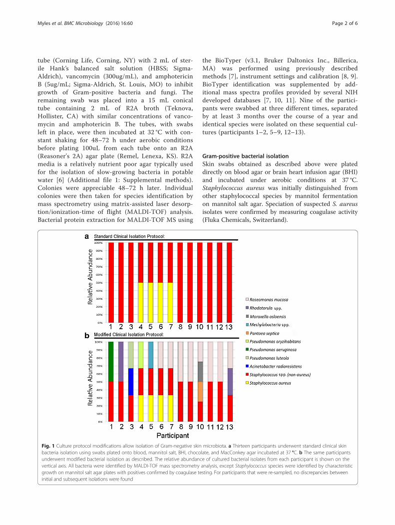
Fig. 1 Culture protocol modifications allow isolation of Gram-negative skin microbiota. a Thirteen participants underwent standard clinical skinbacteria isolation using swabs plated onto blood, mannitol salt, BHI, chocolate, and MacConkey agar incubated at 37 °C. b The same participantsunderwent modified bacterial isolation as described. The relative abundance of cultured bacterial isolates from each participant is shown on thevertical axis. All bacteria were identified by MALDI-TOF mass spectrometry analysis, except Staphylococcus species were identified by characteristicgrowth on mannitol salt agar plates with positives confirmed by coagulase testing. For participants that were re-sampled, no discrepancies betweeninitial and subsequent isolations were found
Myles et al. BMC Microbiology (2016) 16:60 Page 2 of 6
ConsentWritten informed consent was obtained for all partici-pants in this study. All participants were adults.
Results and discussionModification of media and temperature allows isolationof CGN from skinStandard culture techniques for Gram-negative bac-teria from sources other than skin involve incubationat 37 °C using liquid media such as tryptic soy broth(TSB), or on solid media such as chocolate, blood, orMacConkey [12]. We attempted to culture skin bac-teria from thirteen participants by plating forearmswabs onto 5 % sheep blood, mannitol salt, BHI,chocolate, and MacConkey agar incubated at 37 °C.Our use of these techniques readily isolated staphylo-coccal species from multiple healthy volunteers butfailed to culture any Gram-negative isolates, evenwith the use of a Gram-negative selective agar suchas MacConkey (Fig. 1a).The use of our modified Gram-negative bacteria iso-
lation protocol (see Methods) yielded several Gram-negative species from the forearm skin of healthy
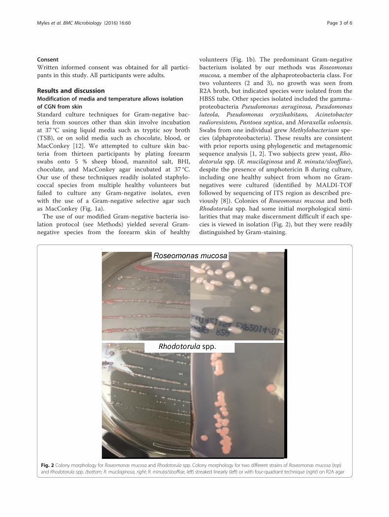
volunteers (Fig. 1b). The predominant Gram-negativebacterium isolated by our methods was Roseomonasmucosa, a member of the alphaproteobacteria class. Fortwo volunteers (2 and 3), no growth was seen fromR2A broth, but indicated species were isolated from theHBSS tube. Other species isolated included the gamma-proteobacteria Pseudomonas aeruginosa, Pseudomonasluteola, Pseudomonas oryzihabitans, Acinetobacterradioresistens, Pantoea septica, and Moraxella osloensis.Swabs from one individual grew Methylobacterium spe-cies (alphaproteobacteria). These results are consistentwith prior reports using phylogenetic and metagenomicsequence analysis [1, 2]. Two subjects grew yeast, Rho-dotorula spp. (R. mucilaginosa and R. minuta/slooffiae),despite the presence of amphotericin B during culture,including one healthy subject from whom no Gram-negatives were cultured (identified by MALDI-TOFfollowed by sequencing of ITS region as described pre-viously [8]). Colonies of Roseomonas mucosa and bothRhodotorula spp. had some initial morphological simi-larities that may make discernment difficult if each spe-cies is viewed in isolation (Fig. 2), but they were readilydistinguished by Gram-staining.
Fig. 2 Colony morphology for Roseomonas mucosa and Rhodotorula spp. Colony morphology for two different strains of Roseomonas mucosa (top)and Rhodotorula spp. (bottom; R. mucilaginosa, right; R. minuta/slooffiae, left) streaked linearly (left) or with four-quadrant technique (right) on R2A agar
Myles et al. BMC Microbiology (2016) 16:60 Page 3 of 6
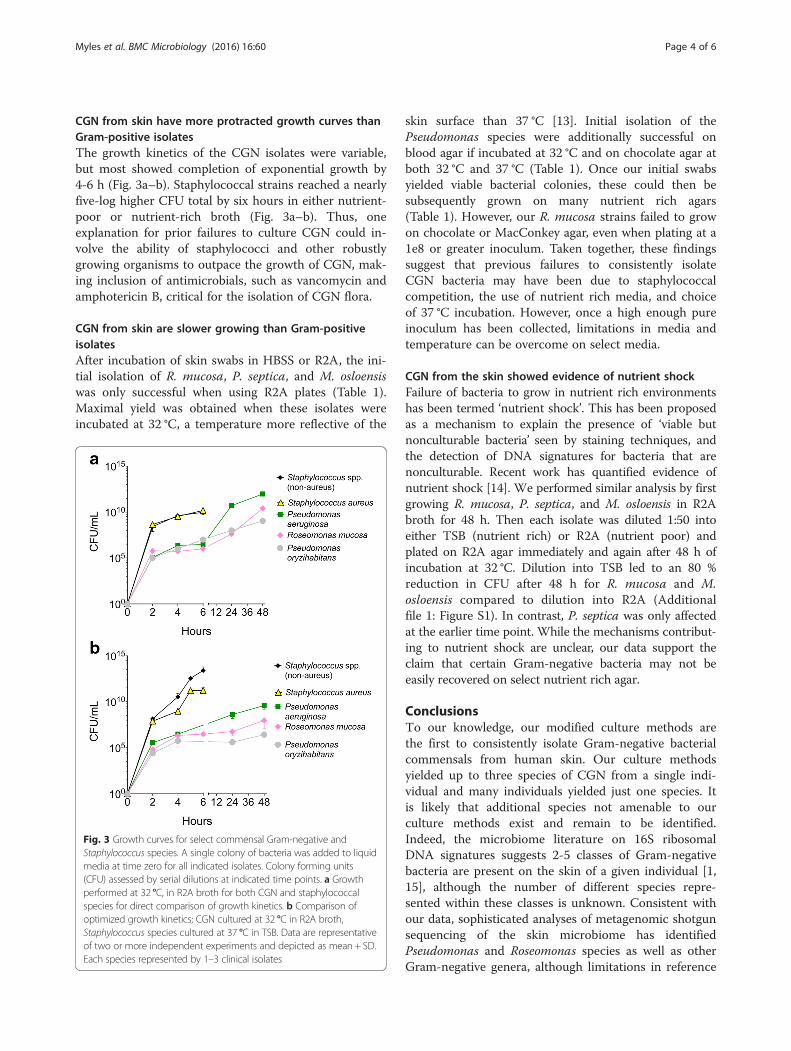
CGN from skin have more protracted growth curves thanGram-positive isolatesThe growth kinetics of the CGN isolates were variable,but most showed completion of exponential growth by4-6 h (Fig. 3a–b). Staphylococcal strains reached a nearlyfive-log higher CFU total by six hours in either nutrient-poor or nutrient-rich broth (Fig. 3a–b). Thus, oneexplanation for prior failures to culture CGN could in-volve the ability of staphylococci and other robustlygrowing organisms to outpace the growth of CGN, mak-ing inclusion of antimicrobials, such as vancomycin andamphotericin B, critical for the isolation of CGN flora.
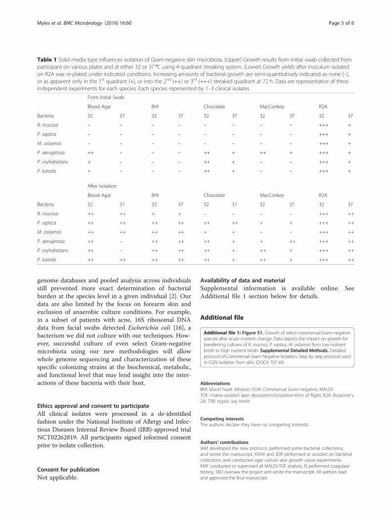
CGN from skin are slower growing than Gram-positiveisolatesAfter incubation of skin swabs in HBSS or R2A, the ini-tial isolation of R. mucosa, P. septica, and M. osloensiswas only successful when using R2A plates (Table 1).Maximal yield was obtained when these isolates wereincubated at 32 °C, a temperature more reflective of the
skin surface than 37 °C [13]. Initial isolation of thePseudomonas species were additionally successful onblood agar if incubated at 32 °C and on chocolate agar atboth 32 °C and 37 °C (Table 1). Once our initial swabsyielded viable bacterial colonies, these could then besubsequently grown on many nutrient rich agars(Table 1). However, our R. mucosa strains failed to growon chocolate or MacConkey agar, even when plating at a1e8 or greater inoculum. Taken together, these findingssuggest that previous failures to consistently isolateCGN bacteria may have been due to staphylococcalcompetition, the use of nutrient rich media, and choiceof 37 °C incubation. However, once a high enough pureinoculum has been collected, limitations in media andtemperature can be overcome on select media.
CGN from the skin showed evidence of nutrient shockFailure of bacteria to grow in nutrient rich environmentshas been termed ‘nutrient shock’. This has been proposedas a mechanism to explain the presence of ‘viable butnonculturable bacteria’ seen by staining techniques, andthe detection of DNA signatures for bacteria that arenonculturable. Recent work has quantified evidence ofnutrient shock [14]. We performed similar analysis by firstgrowing R. mucosa, P. septica, and M. osloensis in R2Abroth for 48 h. Then each isolate was diluted 1:50 intoeither TSB (nutrient rich) or R2A (nutrient poor) andplated on R2A agar immediately and again after 48 h ofincubation at 32 °C. Dilution into TSB led to an 80 %reduction in CFU after 48 h for R. mucosa and M.osloensis compared to dilution into R2A (Additionalfile 1: Figure S1). In contrast, P. septica was only affectedat the earlier time point. While the mechanisms contribut-ing to nutrient shock are unclear, our data support theclaim that certain Gram-negative bacteria may not beeasily recovered on select nutrient rich agar.
ConclusionsTo our knowledge, our modified culture methods arethe first to consistently isolate Gram-negative bacterialcommensals from human skin. Our culture methodsyielded up to three species of CGN from a single indi-vidual and many individuals yielded just one species. Itis likely that additional species not amenable to ourculture methods exist and remain to be identified.Indeed, the microbiome literature on 16S ribosomalDNA signatures suggests 2-5 classes of Gram-negativebacteria are present on the skin of a given individual [1,15], although the number of different species repre-sented within these classes is unknown. Consistent withour data, sophisticated analyses of metagenomic shotgunsequencing of the skin microbiome has identifiedPseudomonas and Roseomonas species as well as otherGram-negative genera, although limitations in reference
Fig. 3 Growth curves for select commensal Gram-negative andStaphylococcus species. A single colony of bacteria was added to liquidmedia at time zero for all indicated isolates. Colony forming units(CFU) assessed by serial dilutions at indicated time points. a Growthperformed at 32 °C, in R2A broth for both CGN and staphylococcalspecies for direct comparison of growth kinetics. b Comparison ofoptimized growth kinetics; CGN cultured at 32 °C in R2A broth,Staphylococcus species cultured at 37 °C in TSB. Data are representativeof two or more independent experiments and depicted as mean + SD.Each species represented by 1–3 clinical isolates
Myles et al. BMC Microbiology (2016) 16:60 Page 4 of 6
genome databases and pooled analysis across individualsstill prevented more exact determination of bacterialburden at the species level in a given individual [2]. Ourdata are also limited by the focus on forearm skin andexclusion of anaerobic culture conditions. For example,in a subset of patients with acne, 16S ribosomal DNAdata from facial swabs detected Escherichia coli [16], abacterium we did not culture with our techniques. How-ever, successful culture of even select Gram-negativemicrobiota using our new methodologies will allowwhole genome sequencing and characterization of thesespecific colonizing strains at the biochemical, metabolic,and functional level that may lend insight into the inter-actions of these bacteria with their host.
Ethics approval and consent to participateAll clinical isolates were processed in a de-identifiedfashion under the National Institute of Allergy and Infec-tious Diseases Internal Review Board (IRB)-approved trialNCT02262819. All participants signed informed consentprior to isolate collection.
Consent for publicationNot applicable.
Availability of data and materialSupplemental information is available online. SeeAdditional file 1 section below for details.
Additional file
Additional file 1: Figure S1. Growth of select commensal Gram-negativespecies after acute nutrient change. Data depicts the impact on growth fortransferring cultures of R. mucosa, P. septica, M. osloensis from low nutrientbroth to high nutrient broth. Supplemental Detailed Methods. Detailedprotocol of Commensal Gram Negative Isolation. Step by step protocol usedin CGN isolation from skin. (DOCX 707 kb)
AbbreviationsBHI: blood heart infusion; CGN: Commensal Gram-negative; MALDI-TOF: matrix-assisted laser desorption/ionization-time of flight; R2A: Reasoner's2A; TSB: tryptic soy broth.
Competing interestsThe authors declare they have no competing interests.
Authors’ contributionsIAM developed the new protocol, performed some bacterial collections,and wrote the manuscript. KWW and JDR performed or assisted on bacterialcollections and conducted agar culture and growth curve experiments.KMF conducted or supervised all MALDI-TOF analysis. IS performed coagulasetesting. SKD oversaw the project and wrote the manuscript. All authors readand approved the final manuscript.
Table 1 Solid media type influences isolation of Gram-negative skin microbiota. (Upper) Growth results from initial swab collected fromparticipant on various plates and at either 32 or 37 °C using 4-quadrant streaking system. (Lower) Growth yields after inoculum isolatedon R2A was re-plated under indicated conditions. Increasing amounts of bacterial growth are semi-quantitatively indicated as none (–),or as apparent only in the 1st quadrant (+), or into the 2nd (++) or 3rd (+++) streaked quadrant at 72 h. Data are representative of threeindependent experiments for each species. Each species represented by 1–3 clinical isolates
From Initial Swab
Blood Agar BHI Chocolate MacConkey R2A
Bacteria 32 37 32 37 32 37 32 37 32 37
R. mucosa – – – – – – – – +++ +
P. septica – – – – – – – – +++ +
M. osloensis – – – – – – – – +++ +
P. aeruginosa ++ – – – ++ + ++ + +++ +
P. oryhabiztans + – – – ++ + – – +++ +
P. luteola + – – – ++ + – – +++ +
After Isolation
Blood Agar BHI Chocolate MacConkey R2A
Bacteria 32 37 32 37 32 37 32 37 32 37
R. mucosa ++ ++ + + – – – – +++ ++
P. septica ++ ++ ++ ++ ++ ++ + + +++ ++
M. osloensis ++ ++ ++ ++ + + – – +++ ++
P. aeruginosa ++ – ++ ++ ++ + + ++ +++ ++
P. oryhabiztans ++ – ++ ++ ++ + ++ + +++ ++
P. luteola ++ ++ ++ ++ ++ + ++ + +++ ++
Myles et al. BMC Microbiology (2016) 16:60 Page 5 of 6
AcknowledgmentsWe would like to acknowledge the participants for their assistance in thisproject. We thank Frida Stock, Julia Shah, and Elim Cho for technicalassistance.
FundingThis work was supported by the Intramural Research Program of theNational Institutes of Health and the National Institute of Allergy andInfectious Disease.
Author details1Bacterial Pathogenesis Unit, Laboratory of Clinical Infectious Diseases,National Institute of Allergy and Infectious Diseases, National Institutes ofHealth, Bethesda, MD, USA. 2Department of Laboratory Medicine, NationalInstitutes of Health Clinical Center, Bethesda, MD, USA.
Received: 25 November 2015 Accepted: 29 March 2016
References1. Grice EA, Kong HH, Conlan S, Deming CB, Davis J, Young AC, Program NCS,
Bouffard GG, Blakesley RW, Murray PR, et al. Topographical and temporaldiversity of the human skin microbiome. Science. 2009;324(5931):1190–2.
2. Oh J, Byrd AL, Deming C, Conlan S, Program NCS, Kong HH, Segre JA.Biogeography and individuality shape function in the human skinmetagenome. Nature. 2014;514(7520):59–64.
3. Marples MJ. The normal flora of the human skin. Br J Dermatol. 1969;81Suppl 1:2–13.
4. Lowbury EJ. Gram-negative bacilli on the skin. Br J Dermatol. 1969;81 Suppl 1:55+.5. Conlan S, Kong HH, Segre JA. Species-level analysis of DNA sequence data
from the NIH Human Microbiome Project. PLoS One. 2012;7(10):e47075.6. Sandle T. An approach for the reporting of microbiological results from
water systems. PDA J Pharm Sci Technol. 2004;58(4):231–7.7. Lau AF, Wang H, Weingarten RA, Drake SK, Suffredini AF, Garfield MK, Chen Y,
Gucek M, Youn JH, Stock F, et al. A rapid matrix-assisted laser desorptionionization-time of flight mass spectrometry-based method for single-plasmidtracking in an outbreak of carbapenem-resistant Enterobacteriaceae. J ClinMicrobiol. 2014;52(8):2804–12.
8. Lau AF, Drake SK, Calhoun LB, Henderson CM, Zelazny AM. Development of aclinically comprehensive database and a simple procedure for identification ofmolds from solid media by matrix-assisted laser desorption ionization-time offlight mass spectrometry. J Clin Microbiol. 2013;51(3):828–34.
9. Youn JH, Drake SK, Weingarten RA, Frank KM, Dekker JP, Lau AF. ClinicalPerformance of a Matrix-Assisted Laser Desorption Ionization Time-of-FlightMass Spectrometry Method for the Detection of Certain blaKPC-containingPlasmids. J Clin Microbiol. 2016;54(1):35–42.
10. Stevenson LG, Drake SK, Shea YR, Zelazny AM, Murray PR. Evaluation ofmatrix-assisted laser desorption ionization-time of flight mass spectrometryfor identification of clinically important yeast species. J Clin Microbiol.2010;48(10):3482–6.
11. Saleeb PG, Drake SK, Murray PR, Zelazny AM. Identification of mycobacteriain solid-culture media by matrix-assisted laser desorption ionization-time offlight mass spectrometry. J Clin Microbiol. 2011;49(5):1790–4.
12. Wauters G, Vaneechoutte M. Chapter 33: Approaches to the Identification ofAerobicGram-Negative Bacteria. In: Jorgensen J, Pfaller M, Carroll K, Funke G,Landry M, Richter S, Warnock D, editors. Manual of Clinical Microbiology.Eleventh Edition. Washington: ASM Press; 2015. p. 613-634.
13. Liu Z, Wang L, Luo Z, Heusch AI, Cascioli V, McCarthy PW.Microenvironment temperature prediction between body and seat interfaceusing autoregressive data-driven model. J Tissue Viability. 2015;24(4):131–9.
14. Azevedo NF, Braganca SM, Simoes LC, Cerqueira L, Almeida C, Keevil CW,Vieira MJ. Proposal for a method to estimate nutrient shock effects inbacteria. BMC Res Notes. 2012;5:422.
15. Kong HH, Oh J, Deming C, Conlan S, Grice EA, Beatson MA, Nomicos E,Polley EC, Komarow HD, Program NCS, et al. Temporal shifts in the skinmicrobiome associated with disease flares and treatment in children withatopic dermatitis. Genome Res. 2012;22(5):850–9.
16. Fitz-Gibbon S, Tomida S, Chiu BH, Nguyen L, Du C, Liu M, Elashoff D, Erfe MC,Loncaric A, Kim J, et al. Propionibacterium acnes strain populations in thehuman skin microbiome associated with acne. J Invest Dermatol.2013;133(9):2152–60.
• We accept pre-submission inquiries
• Our selector tool helps you to find the most relevant journal
• We provide round the clock customer support
• Convenient online submission
• Thorough peer review
• Inclusion in PubMed and all major indexing services
• Maximum visibility for your research
Submit your manuscript atwww.biomedcentral.com/submit
Submit your next manuscript to BioMed Central and we will help you at every step:
Myles et al. BMC Microbiology (2016) 16:60 Page 6 of 6