Embed Size (px)
Citation preview

1
A melanosomal two-pore sodium channel regulates pigmentation.
Nicholas W. Bellono#1,2, Iliana E. Escobar#1, Elena Oancea1*
Supplementary Information
2
Supplementary Figures
Supplementary Figure 1. IPIP2 properties.
a) PI(3,5)P2 activates inwardly rectifying IPIP2 in the absence of OCA2-mediated Cl- currents
(representative of n = 2 melanosomes). OCA2 expression was reduced in Oa1-/- melanocytes, with
OCA2-targeted siRNA1.
b) In a RPE melanosome, IPIP2 is inhibited by 150 µM verapamil, similar to dermal melanosome IPIP2
(representative of n = 2 melanosomes).
c) IPIP2 current density (pA/pF) measured from Oa1-/- melanosomes was similar when the luminal (pipette)
pH was 4.6 or 6.8. Average current density (pA/pF) measured at -120 mV (± s.e.m., n = 4
melanosomes).
3
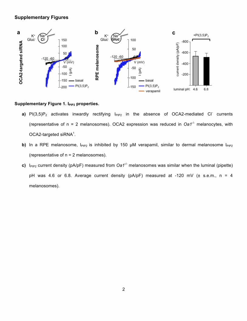
Supplementary Figure 2. TPC2 does not exhibit significant lysosomal localization in pigment cells.
a) In melan-a melanocytes GFP-tagged TPC2 (green) localizes to melanin (50.9 ± 3.2%) (bright field) and
TYRP1 (red)-positive compartments, but does not significantly overlap with structures immunolabeled
with antibodies against the lysosomal marker LAMP2 (red) (7.0 ± 2.7%, p < 0.0001 ) . Enlarged images
of outlined regions shown in lower panels. Scale bar = 10 µm.
b) Expression of the lysosomal protein LAMP1 tagged with GFP (green) localizes to LAMP2 (red)-positive
compartments in melan-a cells (71.4 ± 6.2%). LAMP1-GFP (green) does not localize to melanin (bright
field) and TYRP1 (red)-positive compartments LAMP1 (12.1 ± 2.1%, p < 0.0001).
4
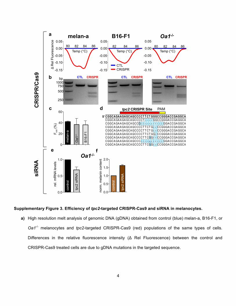
Supplementary Figure 3. Efficiency of tpc2-targeted CRISPR-Cas9 and siRNA in melanocytes.
a) High resolution melt analysis of genomic DNA (gDNA) obtained from control (blue) melan-a, B16-F1, or
Oa1-/- melanocytes and tpc2-targeted CRISPR-Cas9 (red) populations of the same types of cells.
Differences in the relative fluorescence intensity (Δ Rel Fluorescence) between the control and
CRISPR-Cas9 treated cells are due to gDNA mutations in the targeted sequence.
melan-a B16-F1 Oa1-/-
5’
tpc2 CRISPR Site PAM CT GT T ACAT T GGT GGGAT GGCGGCAGAAGAGCAGCCCCT T CT GGGCCGGGACCGAGGCAGT GGCCAGGT CCACT CCGGT GMajority
10 20 30 40 50 60 70 80
CT GT T ACAT T GGT GGGAT GGCGGCAGAAGAGCAGCCCCT T CT GGGCCGGGACCGAGGCAGT GGCCAGGT CCACT CCGGT G 80mTPCN2 Crisper1 Site.seqCT GT T ACAT T GGT GGGAT GGCGGCAGAAGAGCAGCCCCT T CT GGGCCGGGACCGAGGCAGT GGCCAGGT CCACT CCGGT G 80mTPCN2 Crisper1 Site.seq
10 20 30 40 50 60 70 80
CT GT T A CA T T GGT GGGA T GGCGGCA GA A GA GCA GCCCCT - - - - GGCCGGGA CCGA GGCA GT GGCCA GGT CCA CT CCGGT G Melan-a TPC2 Crisper1 TOPO_1CT GT T A CA T T GGT GGGA T GGCGGCA GA A GA GCA GCCG- - - - - - - - - - - GGA CCGA GGCA GT GGCCA GGT CCA CT CCGGT G Melan-a TPC2 Crisper1 TOPO_2CT GT T A CA T T GGT GGGA T GGCGGCA GA A GA GCA GCCCCT T CT G- - CCGGGA CCGA GGCA GT GGCCA GGT CCA CT CCGGT G Melan-a TPC2 Crisper1 TOPO_3CT GT T A CA T T GGT GGGA T GGCGGCA GA A GA GCA GCCCCT T CT G- - CCGGGA CCGA GGCA GT GGCCA GGT CCA CT CCGGT G Melan-a TPC2 Crisper1 TOPO_4CT GT T A CA T T GGT GGGA T GGCGGCA GA A GA GCA GCCCCT T CT GG- CCGGGA CCGA GGCA GT GGCCA GGT CCA CT CCGGT G Melan-a TPC2 Crisper1 TOPO_5CT GT T A CA T T GGT GGGA T GGCGGCA GA A GA GCA GCCCCT T CGG- - CCGGGA CCGA GGCA GT GGCCA GGT CCA CT CCGGT G Melan-a TPC2 Crisper1 TOPO_6CT GT T A CA T T GGT GGGA T GGCGGCA GA A GA GCA GCCC- - - - - - - - - - - GGA CCGA GGCA GT GGCCA GGT CCA CT CCGGT G Melan-a TPC2 Crisper1 TOPO_7CT GT T A CA T T GGT GGGA T GGCGGCA GA A GA GCA GCCCCT T CGG- - CCGGGA CCGA GGCA GT GGCCA GGT CCA CT CCGGT G Melan-a TPC2 Crisper1 TOPO_8CT GT T A CA T T GGT GGGA T GGCGGCA GA A GA GCA GCCCCT T - - - GGCCGGGA CCGA GGCA GT GGCCA GGT CCA CT CCGGT G Melan-a TPC2 Crisper1 TOPO_9CT GT T A CA T T GGT GGGA T GGCGGCA GA A GA GCA GCCCCT T CT GGGCCGGGA CCGA GGCA GT GGCCA GGT CCA CT CCGGT G mTPCN2 Crisper1 Site.seq
bp 1000 750 500
250
a
c
b
Temp (°C) Δ
Rel
Flu
ores
cenc
e
CTL CRISPR CTL CRISPR CTL CRISPR
0.05
0.00
-0.05
-0.15
-0.10
80 82 84 86 0.05
0.00
-0.05
-0.15
-0.10
82 84 86 0.05
0.00
-0.05
-0.15
-0.10
80 82 84 86 Temp (°C) Temp (°C)
f
CTLCRISPRCTL CRISPR
0
20
40
60
mel
an-a
Oa1
-/-
B16
-F1 F c
ut (%
)
d
0.0
0.5
1.0
e
rel.
mR
NA
leve
ls
cont
rol
tpc2
siR
NA
0.0
0.5
1.0
1.5
2.0
norm
. mel
anin
con
tent
cont
rol
tpc2
siR
NA si
RN
A C
RIS
PR/C
as9
Oa1-/-
5
b) Representative gels from mutation detection assay using gDNA from melan-a, B16-F1, or Oa1-/-
melanocytes expressing control or tpc2-targeted CRISPR-Cas9. Cleaved DNA fragments are due to
indels caused by CRISPR-Cas9-induced mutations.
c) Average fraction of cleaved fragments (Fcut) from Guide-it Resolvase assay determined from n = 3
independent experiments.
d) Identification of individual mutations in melanocytes treated with tpc2-targeted CRISPR-Cas9 using
single gDNA species cloned in the TOPO vector. The sequences from each clone were aligned with the
wild-type sequence (in bold) revealing a range of deletions and insertions the tpc2 gene (highlighted in
blue) at the CRISPR site.
e) Mouse tpc2-targeted siRNA stably expressed in Oa1-/- melanocytes reduced the TPC2 mRNA levels by
~30%, compared to control siRNA. (± s.e.m., n = 3, p < 0.01)
f) Melanocytes expressing TPC2-targeted siRNA have ~50% higher melanin content than control siRNA
expressing cells. (± s.e.m., n = 3, p < 0.001)
6
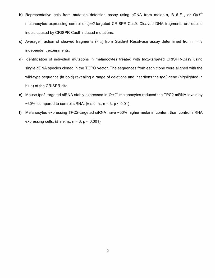
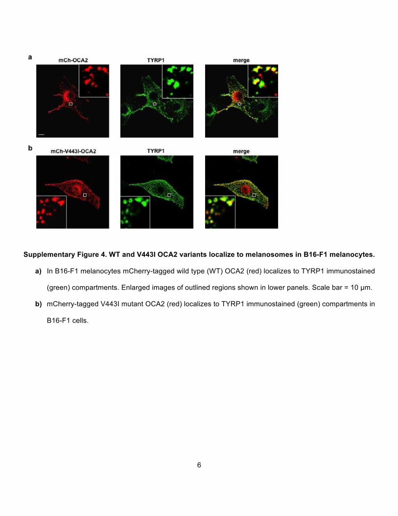
Supplementary Figure 4. WT and V443I OCA2 variants localize to melanosomes in B16-F1 melanocytes.
a) In B16-F1 melanocytes mCherry-tagged wild type (WT) OCA2 (red) localizes to TYRP1 immunostained
(green) compartments. Enlarged images of outlined regions shown in lower panels. Scale bar = 10 µm.
b) mCherry-tagged V443I mutant OCA2 (red) localizes to TYRP1 immunostained (green) compartments in
B16-F1 cells.

7
Supplementary reference
1 Bellono, N. W., Escobar, I. E., Lefkovith, A. J., Marks, M. S. & Oancea, E. An intracellular anion
channel critical for pigmentation. eLife 3 (2014).