Embed Size (px)
Citation preview
Available online at www.sciencedirect.com
www.elsevier.com/locate/jmbbm
j o u r n a l o f t h e m e c h a n i c a l b e h a v i o r o f b i o m e d i c a l m a t e r i a l s 5 4 ( 2 0 1 6 ) 9 3 – 1 0 5
http://dx.doi.org/101751-6161/& 2015 El
nCorrespondenceE-mail address:1Authors Prim a
Research Paper
A mechanical argument for the differentialperformance of coronary artery grafts
David A. Prima,1, Boran Zhoua,1, Adam Hartstone-Roseb,c, Mark J. Ulinea,d,Tarek Shazlya,e, John F. Ebertha,b,n
aUniversity of South Carolina, Biomedical Engineering Program, Columbia, SC, USAbUniversity of South Carolina School of Medicine, Department of Cell Biology and Anatomy, Columbia, SC, USAcUniversity of South Carolina, Department of Anthropology, Columbia, SC, USAdUniversity of South Carolina, Department of Chemical Engineering, Columbia, SC, USAeUniversity of South Carolina, Department of Mechanical Engineering, Columbia, SC, USA
a r t i c l e i n f o
Article history:
Received 2 June 2015
Received in revised form
3 September 2015
Accepted 14 September 2015
Available online 21 September 2015
Keywords:
CABG
Bypass graft
Stress homeostasis
Mechanical compatibility
.1016/j.jmbbm.2015.09.017sevier Ltd. All rights rese
to: USC SOM CBA, Bldg [email protected] Zhou contributed equ
a b s t r a c t
Coronary artery bypass grafting (CABG) acutely disturbs the homeostatic state of the trans-
planted vessel making retention of graft patency dependent on chronic remodeling processes.
The time course and extent to which remodeling restores vessel homeostasis will depend, in
part, on the nature and magnitude of the mechanical disturbances induced upon transplanta-
tion. In this investigation, biaxial mechanical testing and histology were performed on the
porcine left anterior descending artery (LAD) and analogs of common autografts, including the
internal thoracic artery (ITA), radial artery (RA), great saphenous vein (GSV) and lateral
saphenous vein (LSV). Experimental data were used to quantify the parameters of a
structure-based constitutive model enabling prediction of the acute vessel mechanical response
pre-transplantation and under coronary loading conditions. A novel metric Ξ was developed to
quantify mechanical differences between each graft vessel in situ and the LAD in situ, while a
second metric Ω compares the graft vessels in situ to their state under coronary loading. The
relative values of these metrics among candidate autograft sources are consistent with vessel-
specific variations in CABG clinical success rates with the ITA as the superior and GSV the
inferior graft choices based on mechanical performance. This approach can be used to evaluate
other candidate tissues for grafting or to aid in the development of synthetic and tissue
engineered alternatives.
& 2015 Elsevier Ltd. All rights reserved.
rved.
, RM C-36, 6439 Garners Ferry Road, Columbia, SC 29209, Tel.: þ1 803-216-3891.edu (J.F. Eberth).ally to this work.
1. Introduction
Coronary artery grafts bypass (CABG) can restore long-term
myocardial perfusion following advanced-stage coronary
artery disease (Sabik et al., 2011). Annually more than
400,000 CABG procedures are performed in the United States
alone. The health care cost of these CABG procedures is close
to 200 billion USD (Lloyd-Jones et al., 2010; US Department of
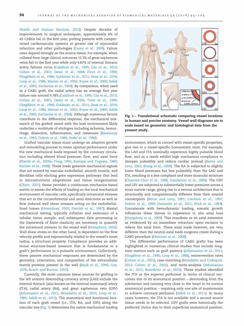
Fig. 1 – Translational schematic comparing vessel locationsin human and porcine anatomy. Vessel wall diagrams are toscale based on geometric and histological data from thepresent study.
j o u r n a l o f t h e m e c h a n i c a l b e h a v i o r o f b i o m e d i c a l m a t e r i a l s 5 4 ( 2 0 1 6 ) 9 3 – 1 0 594
Health and Human Services, 2013). Despite decades ofimprovements to surgical techniques, approximately 6% ofall CABGs fail in the first year, putting patients with compro-mised cardiovascular systems at greater risk of myocardialinfarction and other pathologies (Carey et al., 2009). Failurerates depend strongly on the source tissue. For example, whencollated from large clinical outcomes 15.3% of great saphenousveins fail in the first year while only 4.87% of internal thoracicartery failures occur (Calafiore et al., 1995; Cao et al., 2013;Cohen et al., 2001; Desai et al., 2004; Fiore et al., 1990;Fitzgibbon et al., 1996; Goldman et al., 2011; Hess et al., 2014;Loop et al., 1986; Maniar et al., 2002; Royse et al., 2000; Sabiket al., 2005; Zacharias et al., 2004). By comparison, when usedas a CABG graft, the radial artery has an average first yearfailure rate around 9.78% (Calafiore et al., 1995; Cao et al., 2013;Cohen et al., 2001; Desai et al., 2004; Fiore et al., 1990;Fitzgibbon et al., 1996; Goldman et al., 2011; Hess et al., 2014;Loop et al., 1986; Maniar et al., 2002; Royse et al., 2000; Sabiket al., 2005; Zacharias et al., 2004). Although numerous factorscontribute to the differential response, the mechanical mis-match of the grafted vessel with the host environment likelyunderlies a multitude of etiologies including ischemia, hemor-rhage, dissection, inflammation, and restenosis (Bassiounyet al., 1992; Dobrin et al., 1989; Hofer et al., 1996).
Grafted vascular tissue must undergo an adaptive growthand remodeling process to retain optimal performance underthe new mechanical loads imposed by the coronary circula-tion including altered blood pressure, flow, and axial force(Eberth et al., 2009a; Fung, 1991; Kamiya and Togawa, 1980;Rachev et al., 1998). These loads generate mechanical signalsthat are sensed by vascular endothelial, smooth muscle, andfibroblast cells eliciting gene expression pathways that leadto microstructural adaptations and tissue reorganization(Chien, 2007). Stress provides a continuum mechanics-basedmetric to assess the effects of loading on the local mechanicalenvironment of vascular cells, specifically intramural stressesthat act in the circumferential and axial directions as well asflow induced wall shear stresses acting on the endothelial-lined lumen (Humphrey, 2002; Zwolak et al., 1987). Biaxialmechanical testing, typically inflation and extension of atubular tissue sample, and subsequent data processing inthe framework of finite elasticity are necessary to quantifythe intramural stresses in the vessel wall (Humphrey, 2002).Wall shear stress on the other hand, is dependent on the flowvelocity profile and exponentially related to the vessel’s innerradius, a structural property. Compliance provides an addi-tional structure-based measure that is fundamental to agraft’s performance in a pulsatile environment. Collectively,these passive mechanical responses are determined by thegeometry, orientation, and composition of the extracellularmatrix proteins present in the wall (Bank et al., 1996; Cox,1978; Roach and Burton, 1957).
Currently, the most common tissue sources for grafting tothe left anterior descending coronary artery (LAD) include theinternal thoracic (also known as the internal mammary) artery(ITA), radial artery (RA), and great saphenous vein (GSV)(Athanasiou et al., 2011; Benedetto et al., 2015; Loop et al.,1986; Sabik et al., 2011). The anatomical and functional loca-tion of each graft vessel (i.e., ITA, RA, and GSV) along thevascular tree (Fig. 1) determines the native mechanical loading
environment, which in concert with vessel-specific properties,give rise to a vessel-specific homeostatic state. For example,the LAD and ITA nominally experience highly pulsatile bloodflow, and as a result exhibit high mechanical compliance todampen pulsatility and reduce cardiac preload (Berne andLevy, 2001; Konig et al., 2009). The RA is subjected to slightlylower blood pressures but less pulsatility than the LAD andITA, resulting in a less compliant and more muscular structure(Chamiot-Clerc et al., 1998; Zambanini et al., 2005). The GSVand LSV are subjected to substantially lower pressures across amore narrow range, giving rise to a venous architecture that isstructurally and compositionally different from their arterialcounterparts (Berne and Levy, 2001; Canham et al., 1997;Dobrin et al., 1990; Dummler et al., 2011; Stick et al., 1993).Concatenate with hemodynamic loading, somatic growthinfluences these tissues to experience in situ axial load(Humphrey et al., 2009). This manifests as an axial extensionas evidenced by an immediate retraction upon excision torelieve the axial force. These axial loads however, are verydifferent than the neutral axial loads surgeons create during aCABG procedure (Khonsari et al., 2008).
The differential performance of CABG grafts has beenhighlighted in numerous clinical studies that include long-term metrics such as graft patency (Athanasiou et al., 2011;Fitzgibbon et al., 1996; Loop et al., 1986), reintervention rates(Cohen et al., 2001), case-matching (Benedetto and Codispoti,2013; Cohen et al., 2001), and meta-analysis (Athanasiouet al., 2011; Benedetto et al., 2015). These studies identifiedthe ITA as the superior performer in terms of clinical out-comes due to its anatomical position – descending from thesubclavian and running very close to the heart in its normalanatomical position – requiring only one site of anastomosisto achieve coronary perfusion (Sabik et al., 2011). In manycases however, the ITA is not available and a second sourcetissue needs to be selected. GSV grafts were historically thepreferred choice due to their superficial anatomical position,
j o u r n a l o f t h e m e c h a n i c a l b e h a v i o r o f b i o m e d i c a l m a t e r i a l s 5 4 ( 2 0 1 6 ) 9 3 – 1 0 5 95
non-branching morphology, and comparable caliber to cor-onary arteries (Effler et al., 1970; Loop et al., 1986). Otherautologous source tissue options include RAs, and far lesscommonly the right gastroepiploic artery or inferior epigas-tric artery (Fitzgibbon et al., 1996; Hess et al., 2014). RAs havesimilar caliber and have superficial location, but RAs sufferfrom susceptibility to vasospasm, calcification, intimal hyper-plasia, and are relatively poor in patients with peripheralartery disease (Cheng and Slaughter, 2013).
Here we investigated if mechanical differences amongautologous vascular tissue sources, particularly under pre-and post-grafting loading conditions, correspond with differ-ences in clinical outcomes. To this end, we quantified thepassive mechanical response of the healthy porcine LAD and ahost of candidate vessels commonly used as coronary arterygrafts. Obtained mechanical data were processed in a con-tinuum mechanics-based framework, enabling prediction ofeach vessel's mechanical response under varied loading con-ditions. Vessel-specific mechanical metrics that account forchanges caused by grafting were developed and found tocorrelate with reported clinical outcomes. Our findings helpexplain differential outcomes following autologous grafting inthe coronary artery, and can be extended towards the devel-opment and assessment of other grafting alternatives (i.e.alternative tissue sources and tissue engineered materials).
2. Materials and methods
2.1. Tissue acquisition and handling
All porcine tissue was obtained fresh from a local abattoirand dissections were performed immediately following tissueacquisition. Isolated blood vessels included the LAD, ITA, RA,GSV, and an additional vessel, the lateral saphenous vein(LSV) (Fig. 1). While there is no true translational equivalentto the porcine lateral saphenous vein in humans, it isincluded due to its prevalence in other veterinary studiesand similar caliber to the porcine great saphenous vein.American Yorkshire pigs were used in this study and allanimals were 6 month old71 week. All target vessels weredissected as a set from the same animal (n¼6), and animalweights ranged from 102–113 kg. Upon dissection each vesselwas stored in a sterile solution of 1% phosphate bufferedsaline (PBS) and refrigerated until mechanical testing couldbe performed, which was always within 24 h of tissuedissection.
2.2. Biaxial mechanical testing
Mechanical testing was carried out using a Bose BioDynamic5270 biaxial mechanical testing device. Vessels were cut toapproximately 20 mm tubular sections, and initial measure-ments were taken of the unloaded length, outer diameter,and wall thickness. All measurements were made along themiddle section of each vessel to avoid extreme conditions atproximal and distal ends. Each vessel was mounted into thetesting chamber of the biaxial testing device; the chamberwas filled with Krebs–Heisenleit solution (37 1C and pH 7.4);and sodium nitroprusside (10�5 M) was flushed through the
vessel and device tubing at 60 mL/min to elicit the fullyrelaxed (passive) state of the SMCs and to remove all airfrom the flow loop.
Each vessel was axially extended by approximately 40% ofthe unloaded length at a displacement rate of 0.01 mm/s andpressures of 60 mmHg, 100 mmHg, and 140 mmHg. Thein vivo axial stretch ratio was then determined to be theintersection of resultant axial force versus displacementcurves. This phenomenological observation of in vivo axialstretch has been documented historically (Van Loon, 1976)and in our prior work (Eberth et al., 2009b). Each vessel thenunderwent five cycles of pressurization for preconditioning,helping to ensure reproducible results. For data collection,pressure was increased from 20 to 200 mmHg at a constantrate of approximately 1.3 mmHg/s while pressure-outer dia-meter and pressure-longitudinal force curves were recordedat an axial stretch ratio below the in vivo value. Datacollection was then carried out at the approximated in vivostretch ratio and again above the in vivo stretch ratio.Between tests, axial displacement was increased at a rate of0.01 mm/s, and the vessel was allowed to acclimate for15 min at the new stretch ratio. Each test was repeated threetimes (Eberth et al., 2009b; Zhou et al., 2013).
2.3. Zero stress state
A radial, stress-relieving cut was made into 1 mm thick ringsections taken from the middle region of each vessel follow-ing mechanical testing (Fung, 1991). This radial cut causesring sections to spring open into a sector. After allowing30 min in PBS for each vessel to equilibrate, a digital imagewas taken of the resultant open sector using a Nikon Coolpixs3500 (resolution of 20 μm/pixel). Image-Pro 6.0 image analy-sis software was used to measure the sector thickness H,inner arc length Li, and outer arc length Lo. From these data,cross-sectional area A and opening angle Ф of the sector werecalculated using
H¼ 2ALi þ Lo
and Ф¼ π� Lo�Li2H
: ð1Þ
Collectively, these data enable quantification of the zerostress state for each sample.
2.4. Data analysis
Vessels are assumed to be 3-D thick-walled cylindrical tubesthat experience an axisymmetric finite elastic deformationunder applied pressure and longitudinal extension. Neglect-ing the contribution of vascular smooth muscle cells, thepassive mechanical properties of vessels depend predomi-nantly on the properties, amount, and spatial arrangement ofcollagen and elastin in the vessel wall. Through inflation–extension mechanical testing, sample luminal pressure P andaxial stretch λz were controlled, and response data for thedeformed outer radius ro and axial force F were recorded.Under the assumption of tissue incompressibility, thedeformed inner radius ri is calculated as:
ri ¼ffiffiffiffiffiffiffiffiffiffiffiffiffiffiffiffiffir2o�
Aπλz
sð2Þ
j o u r n a l o f t h e m e c h a n i c a l b e h a v i o r o f b i o m e d i c a l m a t e r i a l s 5 4 ( 2 0 1 6 ) 9 3 – 1 0 596
Likewise, the lumen area compliance is calculated as:
c¼ πΔr2iΔP
ð3Þ
The average circumferential sθ and axial sz wall stressesare calculated as:
sθ ¼Pri
ro�ri; sz ¼
Fπ r2o�r2i� � : ð4Þ
The mid-wall circumferential λθ and axial λz stretch ratiosare calculated as:
λθ ¼2π ri þ roð ÞLi þ Lo
; λz ¼lL
ð5Þ
where l and L are the deformed and undeformed vessellengths, respectively.
2.5. Theoretical framework
For inflation and extension of an axisymmetric tube, itsdeformation is characterized by the right Cauchy–Greenstrain tensor
C½ � ¼ diagdrdR
� �2
; χrR
� �2; λ2z
( ); χ ¼ π
π�Фð6Þ
where r and R are the radial coordinates of an arbitrary pointwithin the vessel wall before and after deformation.
Due to the incompressibility of the vessel wall
drdR
rRχλz ¼ 1 ð7Þ
which after integration yields
r¼ffiffiffiffiffiffiffiffiffiffiffiffiffiffiffiffiffiffiffiffiffiffiffiffiffiffiffiffiffiffiffiffiffiffiffiffiffir2o�
1χλz
R2o�R2� �s
: ð8Þ
Given the zero-stress configuration, axial stretch ratio, andthe deformed outer radius, the components of the straintensor can be completely described. A diagrammatic repre-sentation of the zero-stress state and deformed configura-tions can be found in Zhou et al. (2013) and is consistent withthat of other researchers Matsumoto and Hayashi (1996).
We use an analytical form of the strain energy function,whereby stress and stretch are related by the energy stored inthe vessel wall as it is distended (Eberth et al., 2011; Zhouet al., 2013). This strain energy function was first describedby Holzapfel et al. (2000)) and later modified so that
W¼ ϕeb0 λ2θ þ λ2z þ λθλzð Þ−2� �−3
þ ϕc
4∑
k ¼ 1−4
bk12bk2
exp bk2
ffiffiffiffiffiffiffiffiffiffiffiffiffiffiffiffiffiffiffiffiffiffiffiffiffiffiffiffiffiffiffiffiffiffiffiffiffiffiffiffiffiffiffiffiffiffiffiλ2θ sin
2αk þ λ2z cos 2αk
q 2
−1
!224
35−1
8<:
9=;:
ð9ÞThe constitutive model of Eq. (9) is a structure-based
strain-energy function. The first collection of terms andmaterial constant b0 describes the isotropic, neo-hookeancontribution of an elastin-dominated, non-collagenous extra-cellular matrix (Bersi et al., 2014; Gundiah et al., 2007;Holzapfel et al., 2000; Zulliger et al., 2004). The second termdescribes the anisotropic contribution of four collagen fiberfamilies, where subscript k denotes a particular family offibers oriented at a mean angle of αk with respect to the
longitudinal axis with material constants bk1 and bk2. Thefour-fiber family model with 8 independent parameters givesan excellent representation of biaxial mechanical data with-out being over parameterized (Zeinali-Davarani et al., 2009).Histological evidence suggests that smooth muscle cell orien-tation is predominantly in the circumferential or diagonaldirections (Cheng et al., 2013). Therefore the passive contri-bution of smooth muscle is “lumped” into the correspondingcircumferential and diagonal fiber families (Ferruzzi et al.,2013). In this model α1¼901 represents circumferentiallyoriented fibers, and α2¼01 represents axially oriented fibers.Additionally α3 ¼ �α4 ¼ α represents diagonally orientedfibers, with the value for α obtained from the model. Accord-ingly the stress-like and dimensionless parameters for thesefiber families are equivalent so that b31 ¼ b41 and b32 ¼ b42respectively. ϕ is the area fraction of eð Þ elastin or cð Þ collagencompared to the total tissue as determined through histolo-gical analysis.
The following expressions are used to calculate the theo-retical values for pressure and axial force from a givendeformed configuration
P ¼Z ro
ri
λθ∂ W∂ λθ
drr; F¼ π
Z ro
ri
2λz∂ W∂ λz
�λθ∂ W∂ λθ
� �rdr: ð10Þ
Using data obtained through our mechanical testing andzero-stress state measurements, the material constants ofthe constitutive model were found for each vessel. Thematerial parameters associated with the constitutive modelwere determined via non-linear regression of Eq. (10) whichwas implemented in Matlab 2010b (Mathworks. Inc) using thelsqnonlin subroutine. The lower and upper limits of theparameters were prescribed as b0 and bk1A [0, 105], bk2 A
[0, 10], and α A [01, 901]. The constitutive model was thenused to predict the deformed configurations, stretch ratios,and average transmural stresses under prescribed, in situ andcoronary, loading conditions.
The in situ and grafted loads include both transmuralpressure and axial force. The values of in situ pressures forthe LAD, ITA, RA, GSV, and LSVs were taken from theliterature (Table 3) and represent rough estimates of themean values throughout the cardiac cycle (Canham et al.,1997; Chamiot-Clerc et al., 1998; Dummler et al., 2011; Koniget al., 2009). For example, a pressure of 100 mmHg wasselected as a loading condition for the LAD. The in situ axialforce, on the other hand, was determined as a result of biaxialtesting. The grafted coronary loads for the ITA, RA, GSV, andLSV were assumed to be the values of the pressure and axialforce of the LAD in subsequent calculations.
2.6. Histological analysis
Upon completion of mechanical testing, sections of eachvessel were fixed in 4% fresh paraformaldehyde, followedby embedding in paraffin wax. Sections approximately 5 μmthick were stained with a combination of Verhoeff's elasticand Masson's trichrome stain (O'Connor and Valle, 1982). Allvessels were sectioned and stained together to prevent batch-to-batch variations. Images were obtained using a Nikon E600microscope with CCD camera and computer interface with Q
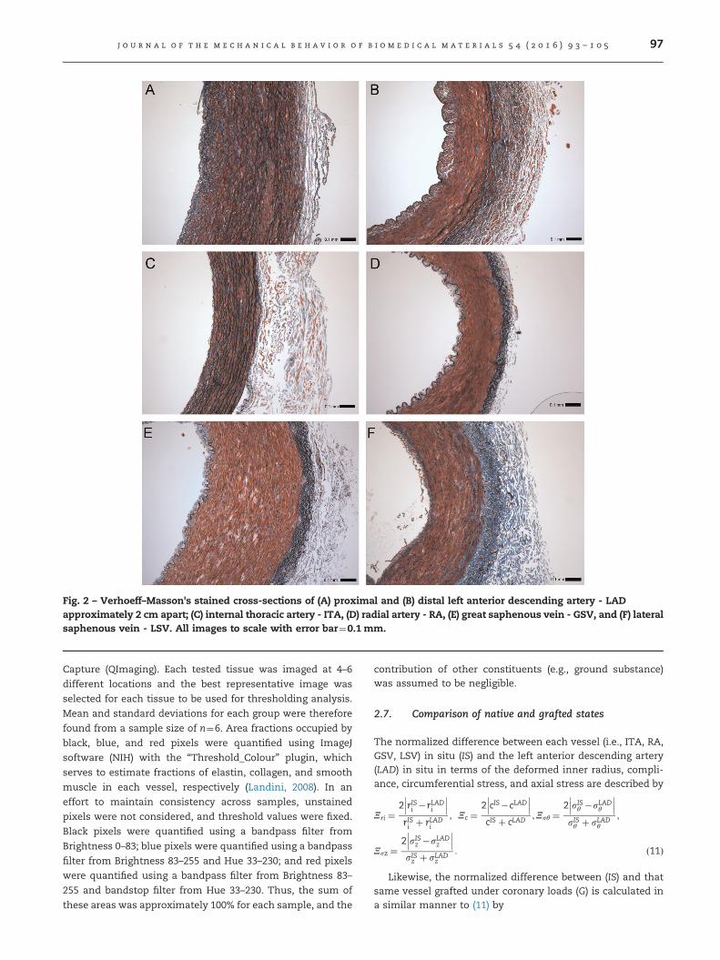
Fig. 2 – Verhoeff–Masson's stained cross-sections of (A) proximal and (B) distal left anterior descending artery - LADapproximately 2 cm apart; (C) internal thoracic artery - ITA, (D) radial artery - RA, (E) great saphenous vein - GSV, and (F) lateralsaphenous vein - LSV. All images to scale with error bar¼0.1 mm.
j o u r n a l o f t h e m e c h a n i c a l b e h a v i o r o f b i o m e d i c a l m a t e r i a l s 5 4 ( 2 0 1 6 ) 9 3 – 1 0 5 97
Capture (QImaging). Each tested tissue was imaged at 4–6
different locations and the best representative image was
selected for each tissue to be used for thresholding analysis.
Mean and standard deviations for each group were therefore
found from a sample size of n¼6. Area fractions occupied by
black, blue, and red pixels were quantified using ImageJ
software (NIH) with the “Threshold_Colour” plugin, which
serves to estimate fractions of elastin, collagen, and smooth
muscle in each vessel, respectively (Landini, 2008). In an
effort to maintain consistency across samples, unstained
pixels were not considered, and threshold values were fixed.
Black pixels were quantified using a bandpass filter from
Brightness 0–83; blue pixels were quantified using a bandpass
filter from Brightness 83–255 and Hue 33–230; and red pixels
were quantified using a bandpass filter from Brightness 83–
255 and bandstop filter from Hue 33–230. Thus, the sum of
these areas was approximately 100% for each sample, and the
contribution of other constituents (e.g., ground substance)was assumed to be negligible.
2.7. Comparison of native and grafted states
The normalized difference between each vessel (i.e., ITA, RA,GSV, LSV) in situ (IS) and the left anterior descending artery(LAD) in situ in terms of the deformed inner radius, compli-ance, circumferential stress, and axial stress are described by
Ξri ¼2���rISi �rLADi
���rISi þ rLADi
; Ξc ¼2���cIS�cLAD
���cIS þ cLAD
;Ξsθ ¼2���sISθ �sLADθ
���sISθ þ sLADθ
;
Ξsz ¼2���sISz �sLADz
���sISz þ sLADz
: ð11Þ
Likewise, the normalized difference between (IS) and thatsame vessel grafted under coronary loads (G) is calculated ina similar manner to (11) by
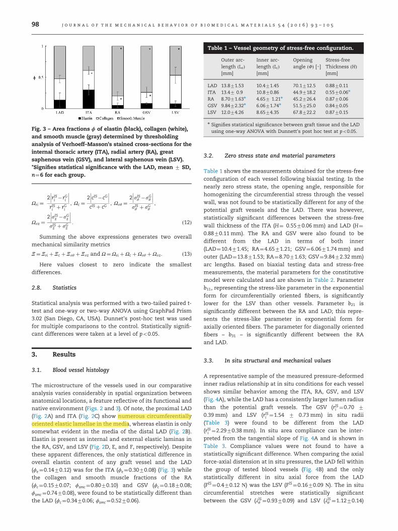
Fig. 3 – Area fractions ϕ of elastin (black), collagen (white),and smooth muscle (gray) determined by thresholdinganalysis of Verhoeff–Masson's stained cross-sections for theinternal thoracic artery (ITA), radial artery (RA), greatsaphenous vein (GSV), and lateral saphenous vein (LSV).*Signifies statistical significance with the LAD, mean 7 SD,n¼6 for each group.
Table 1 – Vessel geometry of stress-free configuration.
Outer arc-length ðLoÞ[mm]
Inner arc-length ðLiÞ[mm]
Openingangle ðΦÞ [◦]
Stress-freeThickness ðHÞ[mm]
LAD 13.871.53 10.471.45 70.1712.5 0.8870.11ITA 13.47 0.9 10.870.86 44.9718.2 0.5570.06n
RA 8.7071.63n 4.657 1.21n 45.2726.4 0.8770.06GSV 9.8472.32n 6.0671.74n 51.5725.0 0.8470.05LSV 12.074.26 8.6574.35 67.8722.2 0.8770.15
n Signifies statistical significance between graft tissue and the LADusing one-way ANOVA with Dunnett's post hoc test at po0.05.
j o u r n a l o f t h e m e c h a n i c a l b e h a v i o r o f b i o m e d i c a l m a t e r i a l s 5 4 ( 2 0 1 6 ) 9 3 – 1 0 598
Ωri ¼2���rISi �rGi
���rISi þ rGi
; Ωc ¼2���cIS�cG
���cIS þ cG
; Ωsθ ¼2���sISθ �sGθ
���sISθ þ sGθ
;
Ωsz ¼2���sISz �sGz
���sISz þ sGz
: ð12Þ
Summing the above expressions generates two overallmechanical similarity metrics
Ξ ¼ Ξri þ Ξc þ Ξsθ þ Ξsz and Ω¼Ωri þΩc þΩsθ þΩsz: ð13ÞHere values closest to zero indicate the smallest
differences.
2.8. Statistics
Statistical analysis was performed with a two-tailed paired t-test and one-way or two-way ANOVA using GraphPad Prism3.02 (San Diego, CA, USA). Dunnet's post-hoc test was usedfor multiple comparisons to the control. Statistically signifi-cant differences were taken at a level of po0.05.
3. Results
3.1. Blood vessel histology
The microstructure of the vessels used in our comparativeanalysis varies considerably in spatial organization betweenanatomical locations, a feature reflective of its functional andnative environment (Figs. 2 and 3). Of note, the proximal LAD(Fig. 2A) and ITA (Fig. 2C) show numerous circumferentiallyoriented elastic lamellae in the media, whereas elastin is onlysomewhat evident in the media of the distal LAD (Fig. 2B).Elastin is present as internal and external elastic laminas inthe RA, GSV, and LSV (Fig. 2D, E, and F, respectively). Despitethese apparent differences, the only statistical difference inoverall elastin content of any graft vessel and the LAD(ϕe¼0.1470.12) was for the ITA (ϕe¼0.3070.08) (Fig. 3) whilethe collagen and smooth muscle fractions of the RA(ϕc¼0.1570.07; ϕsmc¼0.8070.10) and GSV (ϕc¼0.1870.08;ϕsmc¼0.7470.08), were found to be statistically different thanthe LAD (ϕc¼0.3470.06; ϕsmc¼0.5270.06).
3.2. Zero stress state and material parameters
Table 1 shows the measurements obtained for the stress-freeconfiguration of each vessel following biaxial testing. In thenearly zero stress state, the opening angle, responsible forhomogenizing the circumferential stress through the vesselwall, was not found to be statistically different for any of thepotential graft vessels and the LAD. There was however,statistically significant differences between the stress-freewall thickness of the ITA (H¼ 0.5570.06 mm) and LAD (H¼0.8870.11 mm). The RA and GSV were also found to bedifferent from the LAD in terms of both inner(LAD¼10.471.45; RA¼4.6571.21; GSV¼6.0671.74 mm) andouter (LAD¼13.871.53; RA¼8.7071.63; GSV¼9.8472.32 mm)arc lengths. Based on biaxial testing data and stress-freemeasurements, the material parameters for the constitutivemodel were calculated and are shown in Table 2. Parameterb11, representing the stress-like parameter in the exponentialform for circumferentially oriented fibers, is significantlylower for the LSV than other vessels. Parameter b21 issignificantly different between the RA and LAD; this repre-sents the stress-like parameter in exponential form foraxially oriented fibers. The parameter for diagonally orientedfibers – b31 – is significantly different between the RAand LAD.
3.3. In situ structural and mechanical values
A representative sample of the measured pressure-deformedinner radius relationship at in situ conditions for each vesselshows similar behavior among the ITA, RA, GSV, and LSV(Fig. 4A), while the LAD has a consistently larger lumen radiusthan the potential graft vessels. The GSV (rISi ¼0.70 7
0.39 mm) and LSV (rISi ¼1.54 7 0.73 mm) in situ radii(Table 3) were found to be different from the LAD(rISi ¼2.2970.38 mm). In situ area compliance can be inter-preted from the tangential slope of Fig. 4A and is shown inTable 3. Compliance values were not found to have astatistically significant difference. When comparing the axialforce-axial distension at in situ pressures, the LAD fell withinthe group of tested blood vessels (Fig. 4B) and the onlystatistically different in situ axial force from the LAD(FIS¼0.470.12 Ν) was the LSV (FIS¼0.1670.09 Ν). The in situcircumferential stretches were statistically significantbetween the GSV (λISθ ¼0.9370.09) and LSV (λISθ ¼1.1270.14)
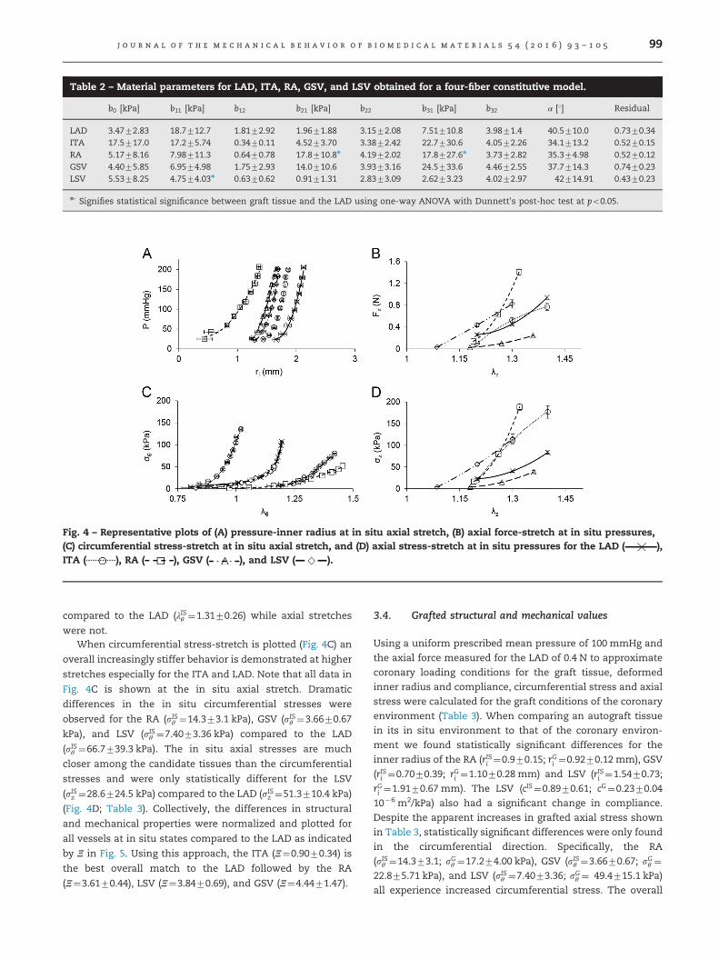
Table 2 – Material parameters for LAD, ITA, RA, GSV, and LSV obtained for a four-fiber constitutive model.
b0 [kPa] b11 [kPa] b12 b21 [kPa] b22 b31 [kPa] b32 α [1] Residual
LAD 3.4772.83 18.7712.7 1.8172.92 1.9671.88 3.1572.08 7.51710.8 3.9871.4 40.5710.0 0.7370.34ITA 17.5717.0 17.275.74 0.3470.11 4.5273.70 3.3872.42 22.7730.6 4.0572.26 34.1713.2 0.5270.15RA 5.1778.16 7.98711.3 0.6470.78 17.8710.8n 4.1972.02 17.8727.6n 3.7372.82 35.374.98 0.5270.12GSV 4.4075.85 6.9574.98 1.7572.93 14.0710.6 3.9373.16 24.5733.6 4.4672.55 37.7714.3 0.7470.23LSV 5.5378.25 4.7574.03n 0.6370.62 0.9171.31 2.8373.09 2.6273.23 4.0272.97 42714.91 0.4370.23
n- Signifies statistical significance between graft tissue and the LAD using one-way ANOVA with Dunnett's post-hoc test at po0.05.
Fig. 4 – Representative plots of (A) pressure-inner radius at in situ axial stretch, (B) axial force-stretch at in situ pressures,(C) circumferential stress-stretch at in situ axial stretch, and (D) axial stress-stretch at in situ pressures for the LAD ( ),ITA ( ), RA ( ), GSV ( ), and LSV ( ).
j o u r n a l o f t h e m e c h a n i c a l b e h a v i o r o f b i o m e d i c a l m a t e r i a l s 5 4 ( 2 0 1 6 ) 9 3 – 1 0 5 99
compared to the LAD (λISθ ¼1.3170.26) while axial stretches
were not.When circumferential stress-stretch is plotted (Fig. 4C) an
overall increasingly stiffer behavior is demonstrated at higher
stretches especially for the ITA and LAD. Note that all data in
Fig. 4C is shown at the in situ axial stretch. Dramatic
differences in the in situ circumferential stresses were
observed for the RA (sISθ ¼14.373.1 kPa), GSV (sISθ ¼3.6670.67
kPa), and LSV (sISθ ¼7.4073.36 kPa) compared to the LAD
(sISθ ¼66.7739.3 kPa). The in situ axial stresses are much
closer among the candidate tissues than the circumferential
stresses and were only statistically different for the LSV
(sISz ¼28.6724.5 kPa) compared to the LAD (sISz ¼51.3710.4 kPa)
(Fig. 4D; Table 3). Collectively, the differences in structural
and mechanical properties were normalized and plotted for
all vessels at in situ states compared to the LAD as indicated
by Ξ in Fig. 5. Using this approach, the ITA (Ξ¼0.9070.34) is
the best overall match to the LAD followed by the RA
(Ξ¼3.6170.44), LSV (Ξ¼3.8470.69), and GSV (Ξ¼4.4471.47).
3.4. Grafted structural and mechanical values
Using a uniform prescribed mean pressure of 100 mmHg and
the axial force measured for the LAD of 0.4 N to approximate
coronary loading conditions for the graft tissue, deformed
inner radius and compliance, circumferential stress and axial
stress were calculated for the graft conditions of the coronary
environment (Table 3). When comparing an autograft tissue
in its in situ environment to that of the coronary environ-
ment we found statistically significant differences for the
inner radius of the RA (rISi ¼0.970.15; rGi ¼0.9270.12 mm), GSV
(rISi ¼0.7070.39; rGi ¼1.1070.28 mm) and LSV (rISi ¼1.5470.73;
rGi ¼1.9170.67 mm). The LSV (cIS¼0.8970.61; cG¼0.2370.04
10�6 m2/kPa) also had a significant change in compliance.
Despite the apparent increases in grafted axial stress shown
in Table 3, statistically significant differences were only found
in the circumferential direction. Specifically, the RA
(sISθ ¼14.373.1; sGθ ¼17.274.00 kPa), GSV (sISθ ¼3.6670.67; sGθ ¼22.875.71 kPa), and LSV (sISθ ¼7.4073.36; sGθ ¼ 49.4715.1 kPa)
all experience increased circumferential stress. The overall
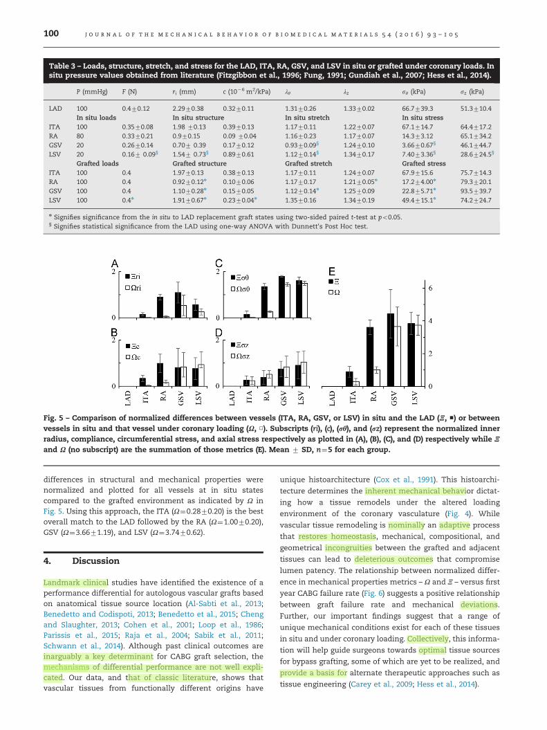
Table 3 – Loads, structure, stretch, and stress for the LAD, ITA, RA, GSV, and LSV in situ or grafted under coronary loads. Insitu pressure values obtained from literature (Fitzgibbon et al., 1996; Fung, 1991; Gundiah et al., 2007; Hess et al., 2014).
P (mmHg) F (N) ri (mm) c (10�6 m2/kPa) λθ λz sθ (kPa) sz (kPa)
LAD 100 0.470.12 2.2970.38 0.3270.11 1.3170.26 1.3370.02 66.7739.3 51.3710.4In situ loads In situ structure In situ stretch In situ stress
ITA 100 0.3570.08 1.98 70.13 0.3970.13 1.1770.11 1.2270.07 67.1714.7 64.4717.2RA 80 0.3370.21 0.970.15 0.09 70.04 1.1670.23 1.1770.07 14.373.12 65.1734.2GSV 20 0.2670.14 0.707 0.39 0.1770.12 0.9370.09§ 1.2470.10 3.6670.67§ 46.1744.7LSV 20 0.167 0.09§ 1.547 0.73§ 0.8970.61 1.1270.14§ 1.3470.17 7.4073.36§ 28.6724.5§
Grafted loads Grafted structure Grafted stretch Grafted stressITA 100 0.4 1.9770.13 0.3870.13 1.1770.11 1.2470.07 67.9715.6 75.7714.3RA 100 0.4 0.9270.12n 0.1070.06 1.1770.17 1.2170.05n 17.274.00n 79.3720.1GSV 100 0.4 1.1070.28n 0.1570.05 1.1270.14n 1.2570.09 22.875.71n 93.5739.7LSV 100 0.4n 1.9170.67n 0.2370.04n 1.3570.16 1.3470.19 49.4715.1n 74.2724.7
n Signifies significance from the in situ to LAD replacement graft states using two-sided paired t-test at po0.05.§ Signifies statistical significance from the LAD using one-way ANOVA with Dunnett's Post Hoc test.
Fig. 5 – Comparison of normalized differences between vessels (ITA, RA, GSV, or LSV) in situ and the LAD (Ξ, ■) or betweenvessels in situ and that vessel under coronary loading (Ω, □). Subscripts (ri), (c), (rθ), and (rz) represent the normalized innerradius, compliance, circumferential stress, and axial stress respectively as plotted in (A), (B), (C), and (D) respectively while Ξ
and Ω (no subscript) are the summation of those metrics (E). Mean 7 SD, n¼5 for each group.
j o u r n a l o f t h e m e c h a n i c a l b e h a v i o r o f b i o m e d i c a l m a t e r i a l s 5 4 ( 2 0 1 6 ) 9 3 – 1 0 5100
differences in structural and mechanical properties werenormalized and plotted for all vessels at in situ statescompared to the grafted environment as indicated by Ω inFig. 5. Using this approach, the ITA (Ω¼0.2870.20) is the bestoverall match to the LAD followed by the RA (Ω¼1.0070.20),GSV (Ω¼3.6671.19), and LSV (Ω¼3.7470.62).
4. Discussion
Landmark clinical studies have identified the existence of aperformance differential for autologous vascular grafts basedon anatomical tissue source location (Al-Sabti et al., 2013;Benedetto and Codispoti, 2013; Benedetto et al., 2015; Chengand Slaughter, 2013; Cohen et al., 2001; Loop et al., 1986;Parissis et al., 2015; Raja et al., 2004; Sabik et al., 2011;Schwann et al., 2014). Although past clinical outcomes areinarguably a key determinant for CABG graft selection, themechanisms of differential performance are not well expli-cated. Our data, and that of classic literature, shows thatvascular tissues from functionally different origins have
unique histoarchitecture (Cox et al., 1991). This histoarchi-
tecture determines the inherent mechanical behavior dictat-
ing how a tissue remodels under the altered loading
environment of the coronary vasculature (Fig. 4). While
vascular tissue remodeling is nominally an adaptive process
that restores homeostasis, mechanical, compositional, and
geometrical incongruities between the grafted and adjacent
tissues can lead to deleterious outcomes that compromise
lumen patency. The relationship between normalized differ-
ence in mechanical properties metrics – Ω and Ξ – versus first
year CABG failure rate (Fig. 6) suggests a positive relationship
between graft failure rate and mechanical deviations.
Further, our important findings suggest that a range of
unique mechanical conditions exist for each of these tissues
in situ and under coronary loading. Collectively, this informa-
tion will help guide surgeons towards optimal tissue sources
for bypass grafting, some of which are yet to be realized, and
provide a basis for alternate therapeutic approaches such as
tissue engineering (Carey et al., 2009; Hess et al., 2014).
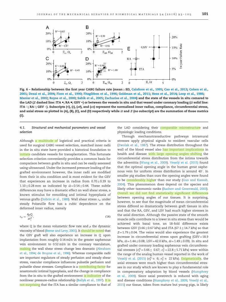
Fig. 6 – Relationship between the first year CABG failure rate (mean7SD, Calafiore et al., 1995; Cao et al., 2013; Cohen et al.,2001; Desai et al., 2004; Fiore et al., 1990; Fitzgibbon et al., 1996; Goldman et al., 2011; Hess et al., 2014; Loop et al., 1986;Maniar et al., 2002; Royse et al., 2000; Sabik et al., 2005; Zacharias et al., 2004) and the state of the vessels in situ comared tothe LAD (Ξ dashed line: ITA ●; RA ■; GSV ▲) or between the vessels in situ and that vessel under coronary loading (Ω solid line:ITA ◯; RA□; GSV △). Subscripts (ri), (c), (rθ), and (rz) represent the normalized inner radius, compliance, circumferential stress,and axial stress as plotted in (A), (B), (C), and (D) respectively while Ω and Ξ (no subscript) are the summation of those metrics(E).
j o u r n a l o f t h e m e c h a n i c a l b e h a v i o r o f b i o m e d i c a l m a t e r i a l s 5 4 ( 2 0 1 6 ) 9 3 – 1 0 5 101
4.1. Structural and mechanical parameters and vesselselection
Although a multitude of logistical and practical criteria is
used for surgical CABG vessel selection, matched inner radii
in the in situ state have provided a historical foundation to
initiate candidate vessels for transplantation. This fortunate
selection criterion conveniently provides a common basis for
comparison between grafts in situ and can be easily assessed
using ultrasound. Under the increased pressure loading of the
grafted environment however, the inner radii are modified
from their in situ condition and is most evident for the GSV
that experiences an increase in radius from 0.7070.39 to
1.1070.28 mm as indicated by Ω¼0.5470.44. These subtle
differences may have a dramatic effect on wall shear stress, a
known stimulus for remolding and intimal hyperplasia of
venous grafts (Dobrin et al., 1989). Wall shear stress τw under
steady Poiseuille flow has a cubic dependence on the
deformed inner radius, namely
τw ¼ 4μQπr3i
; ð14Þ
where Q is the mean volumetric flow rate and μ the dynamic
viscosity of blood (Berne and Levy, 2001). It should be noted that
the GSV graft will also experience an increase in Q upon
implantation from roughly 0.14ml/s in the greater saphenous
vein environment to 0.52ml/s in the coronary vasculature,
making the wall shear stress change less dramatic (Abraham
et al., 1994; de Bruyne et al., 1996). Whereas comparable radii
are important regulators of steady perfusion and steady shear
stress, vascular compliance influences pulsatile perfusion and
pulsatile shear stresses. Compliance is a contributing factor for
anastomotic intimal hyperplasia, and the change in compliance
from the in situ to the grafted environment is indicative of the
nonlinear pressure–radius relationship (Ballyk et al., 1997). It is
not surprising that the ITA has a similar compliance to that of
the LAD considering their comparable microstructure andphysiologic loading conditions.
Through mechanotransductive pathways intramuralstresses apply physical signals to resident vascular cells(Zwolak et al., 1987). The stress distribution throughout thewall of the blood vessel also has important implications inhealth and disease with large opening angles shifting thecircumferential stress distribution from the intima towardsthe adventitia (Wang et al., 2009). Veselý et al. (2015) foundthat the optimal opening angle in the human great saphe-nous vein for uniform stress distribution is around 401. Insmaller pig studies than ours the opening angles were foundto be considerably higher than our study (Guo and Kassab,2004). This phenomenon does depend on the species andlikely other taxonomic ranks (Rachev and Greenwald, 2003).Overall we did not find statistically significant differencesbetween opening angles of our tissues. It is surprising,however, to see that the magnitude of mean circumferentialstress differed so dramatically between graft tissues in situand that the RA, GSV, and LSV had much higher stresses inthe axial direction. Although the passive state of the smoothmuscle cells contribute to a lower in situ stress than would beachieved with basal tone, an 18-fold difference existsbetween GSV (3.6670.67 kPa) and ITA (67.1714.7 kPa) so thatΞ¼1.7970.04. The veins would also experience the greatestincrease in circumferential stress upon grafting (GSV¼18.9kPa, Ω¼1.4470.08; LSV¼42.0 kPa, Ω¼1.4970.09). In situ andgrafted under coronary loading saphenous vein circumferen-tial stresses (sISθ ¼3.667 0.67; sGθ ¼22.875.71 kPa) were withinthe range of the analog human vessel reported in the work ofVeselý et al. (2015) (sISθ E 6; sGθ E 22 kPa). Enigmatically, theaxial stresses were much higher than circumferential stres-ses in our study which are known to play a fundamental rolein compensatory adaptation by blood vessels (Humphreyet al., 2009). Since axial prestretch is reduced with agingand disease conditions (Humphrey et al., 2009; Veselý et al.,2015) our tissue, taken from mature but young pigs, is likely
j o u r n a l o f t h e m e c h a n i c a l b e h a v i o r o f b i o m e d i c a l m a t e r i a l s 5 4 ( 2 0 1 6 ) 9 3 – 1 0 5102
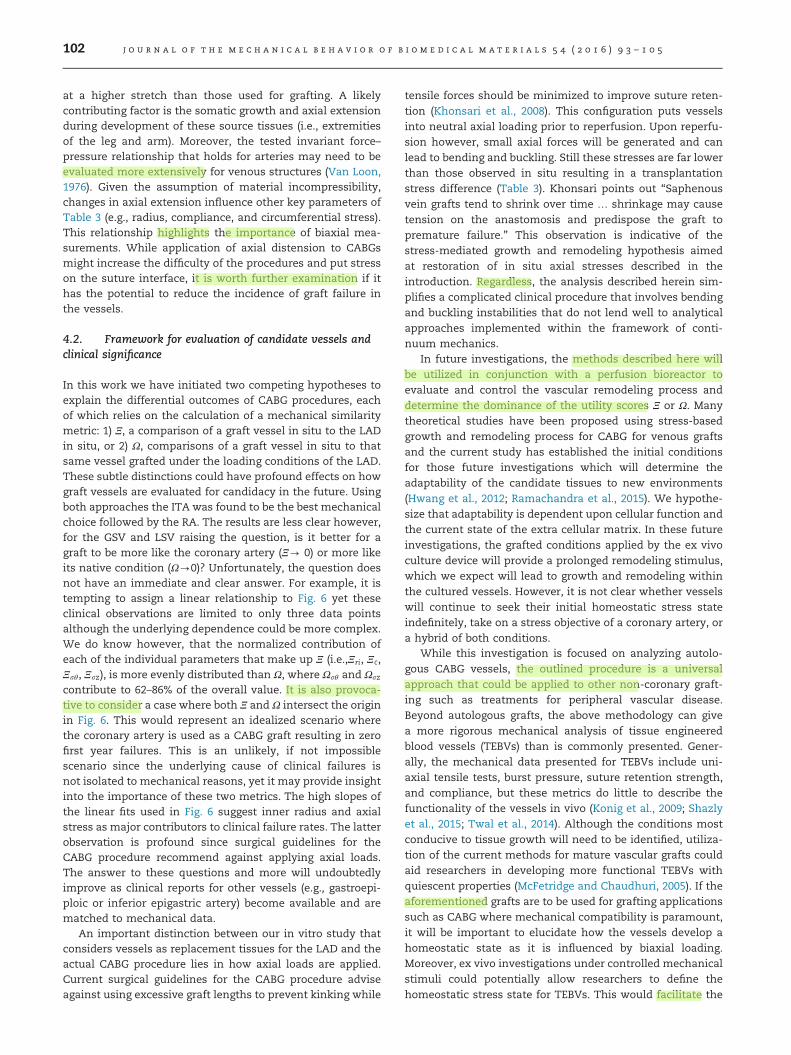
at a higher stretch than those used for grafting. A likelycontributing factor is the somatic growth and axial extensionduring development of these source tissues (i.e., extremitiesof the leg and arm). Moreover, the tested invariant force–pressure relationship that holds for arteries may need to beevaluated more extensively for venous structures (Van Loon,1976). Given the assumption of material incompressibility,changes in axial extension influence other key parameters ofTable 3 (e.g., radius, compliance, and circumferential stress).This relationship highlights the importance of biaxial mea-surements. While application of axial distension to CABGsmight increase the difficulty of the procedures and put stresson the suture interface, it is worth further examination if ithas the potential to reduce the incidence of graft failure inthe vessels.
4.2. Framework for evaluation of candidate vessels andclinical significance
In this work we have initiated two competing hypotheses toexplain the differential outcomes of CABG procedures, eachof which relies on the calculation of a mechanical similaritymetric: 1) Ξ, a comparison of a graft vessel in situ to the LADin situ, or 2) Ω, comparisons of a graft vessel in situ to thatsame vessel grafted under the loading conditions of the LAD.These subtle distinctions could have profound effects on howgraft vessels are evaluated for candidacy in the future. Usingboth approaches the ITA was found to be the best mechanicalchoice followed by the RA. The results are less clear however,for the GSV and LSV raising the question, is it better for agraft to be more like the coronary artery (Ξ- 0) or more likeits native condition (Ω-0)? Unfortunately, the question doesnot have an immediate and clear answer. For example, it istempting to assign a linear relationship to Fig. 6 yet theseclinical observations are limited to only three data pointsalthough the underlying dependence could be more complex.We do know however, that the normalized contribution ofeach of the individual parameters that make up Ξ (i.e.,Ξri, Ξc,Ξsθ, Ξsz), is more evenly distributed than Ω, where Ωsθ and Ωsz
contribute to 62–86% of the overall value. It is also provoca-tive to consider a case where both Ξ and Ω intersect the originin Fig. 6. This would represent an idealized scenario wherethe coronary artery is used as a CABG graft resulting in zerofirst year failures. This is an unlikely, if not impossiblescenario since the underlying cause of clinical failures isnot isolated to mechanical reasons, yet it may provide insightinto the importance of these two metrics. The high slopes ofthe linear fits used in Fig. 6 suggest inner radius and axialstress as major contributors to clinical failure rates. The latterobservation is profound since surgical guidelines for theCABG procedure recommend against applying axial loads.The answer to these questions and more will undoubtedlyimprove as clinical reports for other vessels (e.g., gastroepi-ploic or inferior epigastric artery) become available and arematched to mechanical data.
An important distinction between our in vitro study thatconsiders vessels as replacement tissues for the LAD and theactual CABG procedure lies in how axial loads are applied.Current surgical guidelines for the CABG procedure adviseagainst using excessive graft lengths to prevent kinking while
tensile forces should be minimized to improve suture reten-tion (Khonsari et al., 2008). This configuration puts vesselsinto neutral axial loading prior to reperfusion. Upon reperfu-sion however, small axial forces will be generated and canlead to bending and buckling. Still these stresses are far lowerthan those observed in situ resulting in a transplantationstress difference (Table 3). Khonsari points out “Saphenousvein grafts tend to shrink over time … shrinkage may causetension on the anastomosis and predispose the graft topremature failure.” This observation is indicative of thestress-mediated growth and remodeling hypothesis aimedat restoration of in situ axial stresses described in theintroduction. Regardless, the analysis described herein sim-plifies a complicated clinical procedure that involves bendingand buckling instabilities that do not lend well to analyticalapproaches implemented within the framework of conti-nuum mechanics.
In future investigations, the methods described here willbe utilized in conjunction with a perfusion bioreactor toevaluate and control the vascular remodeling process anddetermine the dominance of the utility scores Ξ or Ω. Manytheoretical studies have been proposed using stress-basedgrowth and remodeling process for CABG for venous graftsand the current study has established the initial conditionsfor those future investigations which will determine theadaptability of the candidate tissues to new environments(Hwang et al., 2012; Ramachandra et al., 2015). We hypothe-size that adaptability is dependent upon cellular function andthe current state of the extra cellular matrix. In these futureinvestigations, the grafted conditions applied by the ex vivoculture device will provide a prolonged remodeling stimulus,which we expect will lead to growth and remodeling withinthe cultured vessels. However, it is not clear whether vesselswill continue to seek their initial homeostatic stress stateindefinitely, take on a stress objective of a coronary artery, ora hybrid of both conditions.
While this investigation is focused on analyzing autolo-gous CABG vessels, the outlined procedure is a universalapproach that could be applied to other non-coronary graft-ing such as treatments for peripheral vascular disease.Beyond autologous grafts, the above methodology can givea more rigorous mechanical analysis of tissue engineeredblood vessels (TEBVs) than is commonly presented. Gener-ally, the mechanical data presented for TEBVs include uni-axial tensile tests, burst pressure, suture retention strength,and compliance, but these metrics do little to describe thefunctionality of the vessels in vivo (Konig et al., 2009; Shazlyet al., 2015; Twal et al., 2014). Although the conditions mostconducive to tissue growth will need to be identified, utiliza-tion of the current methods for mature vascular grafts couldaid researchers in developing more functional TEBVs withquiescent properties (McFetridge and Chaudhuri, 2005). If theaforementioned grafts are to be used for grafting applicationssuch as CABG where mechanical compatibility is paramount,it will be important to elucidate how the vessels develop ahomeostatic state as it is influenced by biaxial loading.Moreover, ex vivo investigations under controlled mechanicalstimuli could potentially allow researchers to define thehomeostatic stress state for TEBVs. This would facilitate the
j o u r n a l o f t h e m e c h a n i c a l b e h a v i o r o f b i o m e d i c a l m a t e r i a l s 5 4 ( 2 0 1 6 ) 9 3 – 1 0 5 103
creation of artificial grafts compatible with any mechanicalloading conditions and thus any anatomical location.
4.3. Limitations
We hold that this investigation represents a meaningfulcontribution to the collective knowledge of vascularmechanics, but acknowledge that there are limitations to thestudy that merit further consideration. First and foremost, weassume that uniform loading of the grafted vessels is equiva-lent to that of a native LAD artery. This implies that we areconsidering an end-to-end anastomosed graft replacing asection of the LAD rather than following the surgical guide-lines for a CABG procedure where vessels are initially underneutral axial loading. Furthermore, grafted conditions wereassumed to be 100 mmHg at an axial force of 0.4 N, approxi-mately representing the mean values experienced by the LAD.This does not account for the constant variation in stressesand strains caused by hemodynamic pulsatility and contrac-tion of the myocardium. We also point out that all vesselswere harvested from healthy, young pigs and that thesevessels which would likely experience altered mechanics indisease and aging (Horny et al., 2011; Kamenskiy et al., 2015).
Of further note, histological differences exist between theproximal (Fig. 2A) and distal (Fig. 2B) sections of the LADcoronary artery due to the significant structural variationsalong its length. Based on our previous assumption of vesselsorganizing to homogenize stress at their homeostatic state,this change in histology would indicate that the loadingenvironment changes significantly along the length of theLAD. As such, the optimal structure of a CABG graft of theLAD would vary according to where along its length it is beinggrafted. Moreover, the peripheral vessels of a quadraped are aless direct analogy to the human than the coronary vessels.Nowhere is this truer than for the LSV, a tissue that does notexist in the human but has applicability in veterinary med-icine and laboratory studies (Fig. 1). Moreover, the zero-stressstate measurements were performed following mechanicaltesting therefore the supra-physiological loading conditionscould potentially have an effect on opening angle measure-ments. This is likely a small contribution but one that shouldbe considered in future work. Lastly, this investigationfocused on the passive mechanics of the potential grafttissue. In highly muscular blood vessels such as the RA,GSV, or LSV, the stress state depends on smooth muscleactivation and likely contributes to the low native circumfer-ential stress values predicted in Table 3. An additional termcould be added to account for active smooth muscle behavior(Baek et al., 2007; Cheng et al., 2013).
5. Conclusion
Mechanical loading is an important factor driving vasculargrowth and remodeling in autologous grafting of matureblood vessels. Differences in structural and mechanical char-acteristics, as demonstrated by our novel mechanical simi-larity metrics Ξ and Ω, provide a reference to the extent thatremodeling must occur in the grafted environment andprovides supportive evidence for the differential performance
of CABG grafts. The results of the current study are consistent
with vessel-specific variations in clinical success rates and
provides the groundwork for engineering a better material for
CABG procedures. Further investigation and experimentation
is necessary to determine if these metrics can be manipu-
lated, acutely or chronically on native tissues, to improve
CABG outcomes.
Funding
This research was supported by NIH INBRE Grant for South
Carolina (P20GM103499) and through a provost sponsored
graduate scholarship from BMEN.
Acknowledgments
The authors would like to acknowledge the imaging and
histological assistance of Dr. Robert Price and Benny David-
son, dissection assistance of Brooks Lane and Carissa Leisch-
ner, image collection and processing assistance of Liam
McNamara, and Caughman’s Meat’n Place.
r e f e r e n c e s
Abraham, P., Leftheriotis, G., Desvaux, B., Saumet, M., Saumet, J.L., 1994. Diameter and blood velocity changes in the saphe-nous vein during thermal stress. Eur. J. Appl. Physiol. Occup.Physiol. 69, 305–308.
Al-Sabti, H.A., Al Kindi, A., Al-Rasadi, K., Banerjee, Y., Al-Hashmi,K., Al-Hinai, A., 2013. Saphenous vein graft vs. radial arterygraft searching for the best second coronary artery bypassgraft. J. Saudi Heart Assoc. 25, 247–254.
Athanasiou, T., Saso, S., Rao, C., Vecht, J., Grapsa, J., Dunning, J.,Lemma, M., Casula, R., 2011. Radial artery versus saphenousvein conduits for coronary artery bypass surgery: forty yearsof competition–which conduit offers better patency? A sys-tematic review and meta-analysis. Eur. J. Cardio-Thorac. Surg.40, 208–220.
Baek, S., Valentın, A., Humphrey, J.D., 2007. Biochemomechanicsof cerebral vasospasm and its resolution: II. Constitutiverelations and model simulations. Ann. Biomed. Eng. 35,1498–1509.
Ballyk, P.D., Walsh, C., Butany, J., Ojha, M., 1997. Compliancemismatch may promote graft–artery intimal hyperplasia byaltering suture-line stresses. J. Biomech. 31, 229–237.
Bank, A.J., Wang, H., Holte, J.E., Mullen, K., Shammas, R., Kubo, S.H., 1996. Contribution of collagen, elastin, and smooth muscleto in vivo human brachial artery wall stress and elasticmodulus. Circulation 94, 3263–3270.
Bassiouny, H.S., White, S., Glagov, S., Choi, E., Giddens, D.P.,Zarins, C.K., 1992. Anastomotic intimal hyperplasia: Mechan-ical injury or flow induced. J. Vasc. Surg. 15, 708–717.
Benedetto, U., Codispoti, M., 2013. Age cutoff for the loss ofsurvival benefit from use of radial artery in coronary arterybypass grafting. J. Thorac. Cardiovasc. Surg. 146, 1078–1084discussion 1084-1075.
Benedetto, U., Raja, S.G., Albanese, A., Amrani, M., Biondi-Zoccai, G.,Frati, G., 2015. Searching for the second best graft for coronaryartery bypass surgery: a network meta-analysis of randomizedcontrolled trials. Eur. J. Cardio-Thorac. Surg. 47, 59–65.
j o u r n a l o f t h e m e c h a n i c a l b e h a v i o r o f b i o m e d i c a l m a t e r i a l s 5 4 ( 2 0 1 6 ) 9 3 – 1 0 5104
Berne, R.M., Levy, M.N., 2001. Cardiovascular Physiology, 8th ed,
The Mosby Physiology Monograph Series, Mosby, Inc. St.
Louis, Missouri.Bersi, M.R., Ferruzzi, J., Eberth, J.F., Gleason Jr., R.L., Humphrey, J.
D., 2014. Consistent biomechanical phenotyping of common
carotid arteries from seven genetic, pharmacological, and
surgical mouse models. Ann. Biomed. Eng. 42, 1207–1223.Calafiore, A.M., Di Giammarco, G., Teodori, G., D’Annunzio, E.,
Vitolla, G., Fino, C., Maddestra, N., 1995. Radial artery and
inferior epigastric artery in composite grafts: improved mid-
term angiographic results. Ann. Thorac. Surg. 60, 517–524.Canham, P.B., Finlay, H.M., Boughner, D.R., 1997. Contrasting
structure of the saphenous vein and internal mammary artery
used as coronary bypass vessels. Cardiovasc. Res. 34, 557–567.Cao, C., Manganas, C., Horton, M., Bannon, P., Munkholm-Larsen,
S., Ang, S.C., Yan, T.D., 2013. Angiographic outcomes of radial
artery versus saphenous vein in coronary artery bypass graft
surgery: A meta-analysis of randomized controlled trials. J.
Thorac. Cardiovasc. Surg. 146, 255–261.Carey, J.S., Danielsen, B., Milliken, J., Li, Z., Stabile, B.E., 2009.
Narrowing the gap: early and intermediate outcomes after
percutaneous coronary intervention and coronary artery
bypass graft procedures in California, 1997 to 2006. J. Thorac.
Cardiovasc. Surg. 138, 1100–1107.Chamiot-Clerc, P., Copie, X., Renaud, J.-F., Safar, M., Girerd, X.,
1998. Comparative reactivity and mechanical properties of
human isolated internal mammary and radial arteries. Car-
diovasc. Res. 37, 811–819.Cheng, A., Slaughter, M.S., 2013. How I choose conduits and
configure grafts for my patients-rationales and practices. Ann.
Cardiothorac. Surg. 2, 527–532.Cheng, J., Stoilov, I., Mecham, R., Wagenseil, J., 2013. A fiber-based
constitutive model predicts changes in amount and organi-
zation of matrix proteins with development and disease in the
mouse aorta. Biomech. Model. Mechanobiol. 12, 497–510.Chien, S., 2007. Mechanotransduction and endothelial cell
homeostasis: the wisdom of the cell. Am. J. Physiol.: Heart
Circ. Physiol. 292, H1209–H1224.Cohen, G., Tamariz, M.G., Sever, J.Y., Liaghati, N., Guru, V.,
Christakis, G.T., Bhatnagar, G., Cutrara, C., Abouzahr, L.,
Goldman, B.S., Fremes, S.E., 2001. The radial artery versus the
saphenous vein graft in contemporary CABG: a case-matched
study. Ann. Thorac. Surg. 71, 180–185 discussion 185-186.Cox, J.L., Chiasson, D.A., Gotlieb, A.I., 1991. Stranger in a strange
land: the pathogenesis of saphenous vein graph stenosis with
emphasis on structural and functional differences between
veins and arteries. Prog. Cardiovasc. Dis. 34, 45–68.Cox, R.H., 1978. Passive mechanics and connective tissue com-
position of canine arteries. Am. J. Physiol. 234, H533–H541.de Bruyne, B., Bartunek, J., Sys, S.U., Pijls, N.H., Heyndrickx, G.R.,
Wijns, W., 1996. Simultaneous coronary pressure and flow
velocity measurements in humans feasibility, reproducibility,
and hemodynamic dependence of coronary flow velocity
reserve, hyperemic flow versus pressure slope index, and
fractional flow reserve. Circulation 94, 1842–1849.Desai, N.D., Cohen, E.A., Naylor, C.D., Fremes, S.E., 2004. A
randomized comparison of radial-artery and saphenous-vein
coronary bypass grafts. N. Engl. J. Med. 351, 2302–2309.Dobrin, P.B., Littooy, F.N., Endean, E.D., 1989. Mechanical factors
predisposing to intimal hyperplasia and medial thickening in
autogenous vein grafts. Surgery 105, 393–400.Dobrin, P.B., Schwarcz, T.H., Mrkvicka, R., 1990. Longitudinal
retractive force in pressurized dog and human arteries. J. Surg.
Res. 48, 116–120.Dummler, S., Eichhorn, S., Tesche, C., Schreiber, U., Voss, B.,
Deutsch, M.A., Hauner, H., Lahm, H., Lange, R., Krane, M.,
2011. Pulsatile ex vivo perfusion of human saphenous vein
grafts under controlled pressure conditions increases MMP-2expression. Biomed. Eng. Online 10, 62.
Eberth, J.F., Cardamone, L., Humphrey, J.D., 2011. Evolving biaxialmechanical properties of mouse carotid arteries in hyperten-sion. J. Biomech. 44, 2532–2537.
Eberth, J.F., Gresham, V.C., Reddy, A.K., Popovic, N., Wilson, E.,Humphrey, J.D., 2009a. Importance of pulsatility in hyperten-sive carotid artery growth and remodeling. J. Hypertens 27,2010–2021.
Eberth, J.F., Taucer, A.I., Wilson, E., Humphrey, J.D., 2009b.Mechanics of carotid arteries in a mouse model of Marfansyndrome. Ann. Biomed. Eng. 37, 1093–1104.
Effler, D., Favaloro, R., Groves, L., 1970. Coronary artery surgeryutilizing saphenous vein graft techniques. Clinical experiencewith 224 operations. J. Thorac. Cardiovasc. Surg. 59, 147.
Ferruzzi, J., Bersi, M.R., Humphrey, J.D., 2013. Biomechanicalphenotyping of central arteries in health and disease: advan-tages of and methods for murine models. Ann. Biomed. Eng.41, 1311–1330.
Fiore, A.C., Naunheim, K.S., Dean, P., Kaiser, G.C., Pennington, D.G., Willman, V.L., McBride, L.R., Barner, H.B., 1990. Results ofinternal thoracic artery grafting over 15 years: single versusdouble grafts. Ann. Thorac. Surg. 49, 202–209.
Fitzgibbon, G.M., Kafka, H.P., Leach, A.J., Keon, W.J., Hooper, G.D.,Burton, J.R., 1996. Coronary bypass graft fate and patientoutcome: angiographic follow-up of 5,065 grafts related tosurvival and reoperation in 1,388 patients during 25 years. J.Am. Coll. Cardiol. 28, 616–626.
Fung, Y.C., 1991. What are the residual stresses doing in our bloodvessels?. Ann. Biomed. Eng. 19, 237–249.
Goldman, S., Sethi, G.K., Holman, W., Thai, H., McFalls, E., Ward,H.B., Kelly, R.F., Rhenman, B., Tobler, G.H., Bakaeen, F.G., Huh,J., Soltero, E., Moursi, M., Haime, M., Crittenden, M., Kasirajan,V., Ratliff, M., Pett, S., Irimpen, A., Gunnar, W., Thomas, D.,Fremes, S., Moritz, T., Reda, D., Harrison, L., Wagner, T.H.,Wang, Y., Planting, L., Miller, M., Rodriguez, Y., Juneman, E.,Morrison, D., Pierce, M.K., Kreamer, S., Shih, M.C., Lee, K.,2011. Radial artery grafts vs saphenous vein grafts in coronaryartery bypass surgery: a randomized trial. JAMA 305, 167–174.
Gundiah, N., B Ratcliffe, M., Pruitt, L., A., 2007. Determination ofstrain energy function for arterial elastin: experiments usinghistology and mechanical tests. J. Biomech. 40, 586–594.
Guo, X., Kassab, G.S., 2004. Distribution of stress and strain alongthe porcine aorta and coronary arterial tree. Am. J. Physiol.-Heart Circ. Physiol. 286, H2361–H2368.
Hess, C.N., Lopes, R.D., Gibson, C.M., Hager, R., Wojdyla, D.M.,Englum, B.R., Mack, M.J., Califf, R.M., Kouchoukos, N.T.,Peterson, E.D., Alexander, J.H., 2014. Saphenous vein graftfailure after coronary artery bypass surgery: insights fromPREVENT IV. Circulation 130, 1445–1451.
Hofer, M., Rappitsch, G., Perktold, K., Trubel, W., Schima, H., 1996.Numerical study of wall mechanics and fluid dynamics inend-to-side anastomoses and correlation to intimal hyper-plasia. J. Biomech. 29, 1297–1308.
Holzapfel, G., Gasser, T., Ogden, R., 2000. A new constitutiveframework for arterial wall mechanics and a comparativestudy of material models. J. Elast. 61, 1–48.
Horny, L., Adamek, T., Gultova, E., Zitny, R., Vesely, J., Chlup, H.,Konvickova, S., 2011. Correlations between age, prestrain,diameter and atherosclerosis in the male abdominal aorta. J.Mech. Behav. Biomed. Mater. 4, 2128–2132.
Humphrey, J.D., 2002. Cardiovascular solid mechanics: cells,tissues, and organs. Springer, New York.
Humphrey, J.D., Eberth, J.F., Dye, W.W., Gleason, R.L., 2009.Fundamental role of axial stress in compensatory adaptationsby arteries. J. Biomech. 42, 1–8.
Hwang, M., Berceli, S.A., Garbey, M., Kim, N.H., Tran-Son-Tay, R.,2012. The dynamics of vein graft remodeling induced by
j o u r n a l o f t h e m e c h a n i c a l b e h a v i o r o f b i o m e d i c a l m a t e r i a l s 5 4 ( 2 0 1 6 ) 9 3 – 1 0 5 105
hemodynamic forces: a mathematical model. Biomech.Model. Mechanobiol. 11, 411–423.
Kamiya, A., Togawa, T., 1980. Adaptive regulation of wall shearstress to flow change in the canine carotid artery. Am. J.Physiol. 239, H14–H21.
Kamenskiy, A.V., Pipinos, I.I., Dzenis, Y.A., Phillips, N.Y., Desya-tova, A.S., Kitson, J., Bowen, R., MacTaggart, J.N., 2015. Effectsof age on the physiological and mechanical characteristics ofhuman femoropopliteal arteries. Acta Biomater. 11, 304–313.
Khonsari, S., Sintek, C., Ardehali, A., 2008. Cardiac Surgery:Safeguards and Pitfalls in Operative Technique. WoltersKluwer Health/Lippincott Williams & Wilkins, Philadelphia,PA.
Konig, G., McAllister, T.N., Dusserre, N., Garrido, S.A., Iyican, C.,Marini, A., Fiorillo, A., Avila, H., Wystrychowski, W., Zagalski,K., 2009. Mechanical properties of completely autologoushuman tissue engineered blood vessels compared to humansaphenous vein and mammary artery. Biomaterials 30,1542–1550.
Landini, G., 2008. Advanced shape analysis with ImageJ. In:Proceedings of the Second ImageJ User and Developer Con-ference, Luxembourg, pp. 116–121.
Lloyd-Jones, D., Adams, R.J., Brown, T.M., Carnethon, M., Dai, S.,De Simone, G., Ferguson, T.B., Ford, E., Furie, K., Gillespie, C.,Go, A., Greenlund, K., Haase, N., Hailpern, S., Ho, P.M., Howard,V., Kissela, B., Kittner, S., Lackland, D., Lisabeth, L., Marelli, A.,McDermott, M.M., Meigs, J., Mozaffarian, D., Mussolino, M.,Nichol, G., Roger, V.L., Rosamond, W., Sacco, R., Sorlie, P.,Stafford, R., Thom, T., Wasserthiel-Smoller, S., Wong, N.D.,Wylie-Rosett, J., 2010. Heart disease and stroke statistics—2010 update: a report from the American heart association.Circulation 121, e46–e215.
Loop, F.D., Lytle, B.W., Cosgrove, D.M., Stewart, R.W., Goormastic,M., Williams, G.W., Golding, L.A.R., Gill, C.C., Taylor, P.C.,Sheldon, W.C., Proudfit, W.L., 1986. Influence of the internal-mammary-artery graft on 10-year survival and other cardiacevents. N. Engl. J. Med. 314, 1–6.
Maniar, H.S., Sundt, T.M., Barner, H.B., Prasad, S.M., Peterson, L.,Absi, T., Moustakidis, P., 2002. Effect of target stenosis andlocation on radial artery graft patency. J. Thorac. Cardiovasc.Surg. 123, 45–52.
Matsumoto, T., Hayashi, K., 1996. Stress and strain distribution inhypertensive and normotensive rat aorta considering residualstrain. J. Biomech. Eng. 118, 62–73.
McFetridge, P.S., Chaudhuri, J.B., 2005. Design of vascular graftbioreactors. In: Chaudhuri, J.B., Al-Rubeai, M. (Eds.), Bioreac-tors for Tissue Engineering. Principles, Design, and Operation.Springer, Netherlands, pp. 269–283.
O’Connor, W.N., Valle, S., 1982. A combination Verhoeff’s elasticand Masson’s trichrome stain for routine histology. StainTechnol. 57, 207–210.
Parissis, H., Ramesh, B.C., Al-Alao, B., 2015. Which is the bestgraft for the right coronary artery?. Asian Cardiovasc. Thorac.Ann 23, 100–113.
Rachev, A., Greenwald, S., 2003. Residual strains in conduitarteries. J. Biomech. 36, 661–670.
Rachev, A., Stergiopulos, N., Meister, J.J., 1998. A model forgeometric and mechanical adaptation of arteries to sustainedhypertension. J. Biomech. Eng. 120, 9–17.
Raja, S.G., Haider, Z., Ahmad, M., Zaman, H., 2004. Saphenousvein grafts: to use or not to use?. Heart Lung Circ. 13, 150–156.
Ramachandra, A.B., Sankaran, S., Humphrey, J.D., Marsden, A.L.,2015. Computational Simulation of the Adaptive Capacity ofVein Grafts in Response to Increased Pressure. J. Biomech. Eng.137, 031009, http://dx.doi.org/10.1115/1.4029021.
Roach, M.R., Burton, A.C., 1957. The reason for the shape of thedistensibility curves of arteries. Can. J. Biochem. Physiol. 35,681–690.
Royse, A.G., Royse, C.F., Tatoulis, J., Grigg, L.E., Shah, P., Hunt, D.,Better, N., Marasco, S.F., 2000. Postoperative radial arteryangiography for coronary artery bypass surgery. Eur. J. Cardio-Thorac. Surg. 17, 294–304.
Sabik 3rd, J.F., Lytle, B.W., Blackstone, E.H., Houghtaling, P.L.,Cosgrove, D.M., 2005. Comparison of saphenous vein andinternal thoracic artery graft patency by coronary system.Ann. Thorac. Surg. 79, 544–551 discussion 544-551.
Sabik, J.F., Bansilal, S., Lytle, B.W., 2011. Chapter 65. Coronarybypass surgery. In: Fuster, V., Walsh, R.A., Harrington, R.A.(Eds.), Hurst’s The Heart, 13e. The McGraw-Hill Companies,New York, NY.
Schwann, T.A., Tranbaugh, R.F., Dimitrova, K.R., Engoren, M.C.,Kabour, A., Hoffman, D.M., Geller, C.M., Ko, W., Habib, R.H.,2014. Time-varying survival benefit of radial artery versus veingrafting: a multiinstitutional analysis. Ann. Thorac. Surg. 97,1328–1334 discussion 1334.
Shazly, T., Rachev, A., Lessner, S., Argraves, W.S., Ferdous, J., Zhou,B., Moreira, A.M., Sutton, M., 2015. On the uniaxial ring test oftissue engineered constructs. Exp. Mech. 55, 41–51.
Stick, C., Hiedl, U., Witzleb, E., 1993. Venous pressure in thesaphenous vein near the ankle during changes in posture andexercise at different ambient temperatures. Eur. J. Appl.Physiol. Occup. Physiol. 66, 434–438.
Twal, W.O., Klatt, S.C., Harikrishnan, K., Gerges, E., Cooley, M.A.,Trusk, T.C., Zhou, B., Gabr, M.G., Shazly, T., Lessner, S.M.,Markwald, R.R., Argraves, W.S., 2014. Cellularized microcar-riers as adhesive building blocks for fabrication of tubulartissue constructs. Ann. Biomed. Eng. 42, 1470–1481.
US Department of Health and Human Services, 2013. NationalHospital Discharge Survey 2010.
Van Loon, P., 1976. Length-force and volume-pressure relation-ships of arteries. Biorheology 14, 181–201.
Vesely, J., Horny, L., Chlup, H., Adamek, T., Krajıcek, M., Zitny, R.,2015. Constitutive modeling of human saphenous veins atoverloading pressures. J. Mech. Behav. Biomed. Mater. 45,101–108.
Wang, C., Guo, X., Kassab, G.S., 2009. A new observation on thestress distribution in the coronary artery wall. J. Biomech. Eng.131, 111011.
Zacharias, A., Habib, R.H., Schwann, T.A., Riordan, C.J., Durham,S.J., Shah, A., 2004. Improved survival with radial artery versusvein conduits in coronary bypass surgery with left internalthoracic artery to left anterior descending artery grafting.Circulation 109, 1489–1496.
Zambanini, A., Cunningham, S.L., Parker, K.H., Khir, A.W., Thom,S.M., Hughes, A.D., 2005. Wave–energy patterns in carotid,brachial, and radial arteries: a noninvasive approach usingwave-intensity analysis. Am. J. Physiol.-Heart Circ. Physiol.289, H270–H276.
Zeinali-Davarani, S., Choi, J., Baek, S., 2009. On parameter esti-mation for biaxial mechanical behavior of arteries. J. Biomech.42, 524–530.
Zhou, B., Wolf, L., Rachev, A., Shazly, T., 2013. A structure-motivated model of the passive mechanical response of theprimary porcine renal artery. J. Mech. Med. Biol. 14, 1450033.
Zulliger, M. a, Fridez, P., Hayashi, K., Stergiopulos, N., 2004. Astrain energy function for arteries accounting for wall com-position and structure. J. Biomech. 37, 989–1000.
Zwolak, R.M., Adams, M.C., Clowes, A.W., 1987. Kinetics of veingraft hyperplasia: association with tangential stress. J. Vasc.Surg. 5, 126–136.