Embed Size (px)
Citation preview

W&M ScholarWorks W&M ScholarWorks
Dissertations, Theses, and Masters Projects Theses, Dissertations, & Master Projects
2015
A Mathematical Model of Tau Protein Hyperphosphorylation: The A Mathematical Model of Tau Protein Hyperphosphorylation: The
Effects of Kinase Inhibitors as a Theoretical Alzheimer's Disease Effects of Kinase Inhibitors as a Theoretical Alzheimer's Disease
Therapy Therapy
Patrick Neil Blank College of William & Mary - Arts & Sciences
Follow this and additional works at: https://scholarworks.wm.edu/etd
Part of the Applied Mathematics Commons, and the Biochemistry Commons
Recommended Citation Recommended Citation Blank, Patrick Neil, "A Mathematical Model of Tau Protein Hyperphosphorylation: The Effects of Kinase Inhibitors as a Theoretical Alzheimer's Disease Therapy" (2015). Dissertations, Theses, and Masters Projects. Paper 1539626963. https://dx.doi.org/doi:10.21220/s2-gq0q-nt71
This Thesis is brought to you for free and open access by the Theses, Dissertations, & Master Projects at W&M ScholarWorks. It has been accepted for inclusion in Dissertations, Theses, and Masters Projects by an authorized administrator of W&M ScholarWorks. For more information, please contact [email protected].

A Mathematical Model of Tau Protein Hyperphosphorylation The Effects of Kinase Inhibitors as a Theoretical Alzheimer's Disease Therapy
Patrick Neil Blank .
Richmond, Virginia
BS, The College of William and Mary, 2013
A Thesis Presented to the Graduate Faculty of The College of William and Mary in Candidacy for the Degree of
Master of Science
Department of Chemistry
The College of William and Mary August, 2015
APPROVAL PAGE
This Thesis is Submitted in Partial Fulfillment of the Requirements for the Degree of
Master of Science
Patrick Neil Blank
Approved by the Committee, May 8th, 2015
I ( Qa a Pt f l j kv ut A'|' Committee Chair
Associate Professor Randolph A. Coleman, Chemistry The College of William and Mar
*
Chancellor Professor and'Department Chair Christopher J. Abelt, Chemistry The College of William and Mary
Professor and ndergraduate S tod iesjohn C. Poutsma, Chemistry The College of William and Mary

ABSTRACT
Using Biological Systems Theory (BST), our group builds intricate computer models of neurodegenerative diseases and associated processes to simulate the activities occurring within the cell regarding the disease of interest, utilizing CellDesigner 4.3 and MATLAB R2014a. This particular project primarily models Tau protein, as it is one of the most implicated proteins in Alzheimer’s disease. Intraneuronal hyperphosphorylated Tau protein leads to neurofibrillary tangles, which cause apoptosis. The model eventually resulted in the generation of optimal drug concentrations for molecules used theoretically as Tau protein kinase inhibitors, ultimately allowing the isolation of SP-600125 as the best mathematical solution to the biochemical problem presented in the form of our system of differential equations comprising aforementioned model. SP-600125 was determined as the best, but many other drugs performed well in this theoretical simulation of disease therapy.

TABLE OF CONTENTS
Acknowledgements ii
Dedication iii
List of Figures iv
History 1
Introduction and Background 2
Methods 35
Results 44
Discussion 56
Conclusion 59
Vita 60
Bibliography 61

ACKNOWLEDGEMENTS
I would like to formally acknowledge Randolph Coleman for his higher level contributions to this work and Alec Weech for his patience while enlightening me with the fundamentals and sometimes frustrating intricacies of coding in MATLAB.

This thesis is dedicated to my best friend, Matthew ‘Harvard’ Weber, foralways pushing
me to persevere and strive for excellence not only in academics, but in lifeitse lf...

LIST OF FIGURES
1. Chemical Structure of Aloisine 16
2. Chemical Structure of Hymenialdisine 16
3. Chemical Structure of 6-Bromoindirubin-3’-oxime 16
4. Chemical Structure of Flavopiridol 17
5. Chemical Structure of AR-014418 17
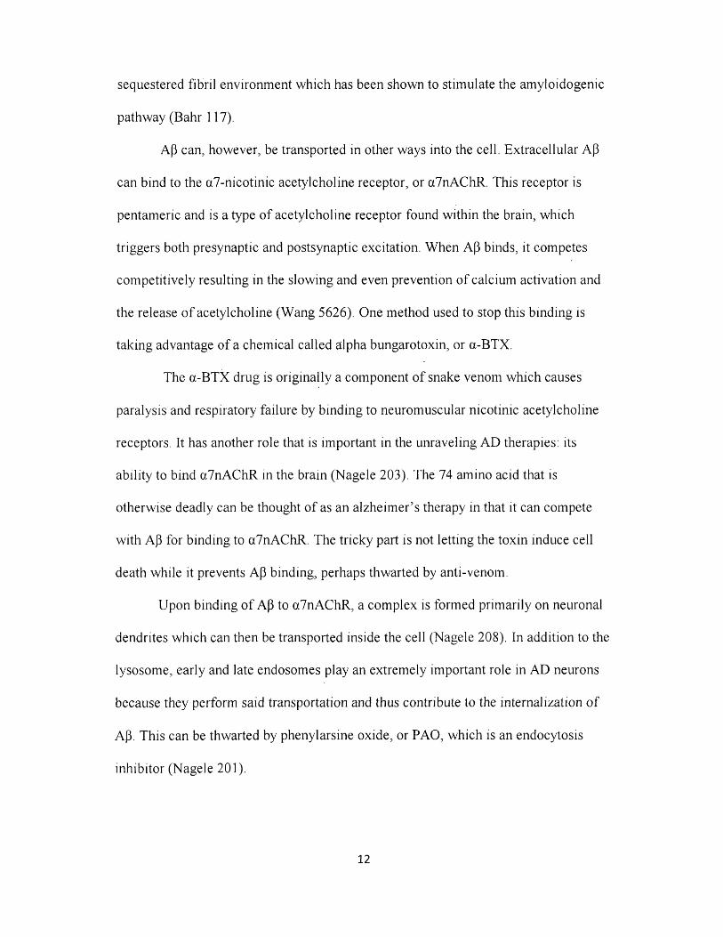
6. Chemical Structure of Olomoucine 18
7. Chemical Structure of Roscovitine 18
8. Chemical Structure of Purvalanol A 19
9. Chemical Structure of Purvalanol B 19
10. Chemical Structure of Paullones 20
11. Chemical Structure of R-CR8 20
12. Chemical Structure of Heparin 22
13. Chemical Structure of TBCA 22
14. Chemical Structure of IC-261 22
15. Chemical Structure of Harmine 24
16. Chemical Structure of Paclitaxel 25
17. Chemical Structure of FR180204 26
iv

18. Chemical Structure of SB-239063 26
19. Chemical Structure of SP600125 27
20. Chemical Structure of H-89 28
21. Chemical Structure of ATP 28
22. Two crystallographic bindings of H-89 to PKA 28
23. Chemical Structure of Akt VIII 29
24. Chemical Structure of Diacylglycerol 30
25. Chemical Structure of GF-1 31
26. Chemical Structure of Bryostatin 1 31
27. Chemical Structure of Melatonin 33
28. Chemical Structure of Okadaic Acid 33
29. Chemical Structure of Memantine 34
30. Graduate Version of Celldesigner Model 35
31. Cdk-5 Model Snapshot 36
32. Amyloidogenesis and Non-Amyloidogenesis Snapshot 37
33. Drug Concentration Values 39
34. Death Function Equation Sample 40
V

35. Kinetic Equation Sample 41
36. Plotting Function Sample 42
37. Healthy State 44
38. Diseased State 45
39. SP-600125 Concentration Range 0:700 nM (~7x IC50) 45
40. SP-600125 Concentration Range 210:235 nM with minimum 46
41. (R)-CR8 Concentration Range 0:900 nM (~7x IC50) 46
42. (R)-CR8 Concentration Range 360:430 nM with minimum 47
43.6-BIO Concentration Range 0:35 nM (~7x IC50) 47
44.6-BIO Concentration Range 9.8:10.7 nM with minimum 48
45.AR014418 Concentration Range 0:800 (~7x IC50) 48
46. AR014418 Concentration Range 220:245 nM with minimum 49
47. SB-239063 Concentration Range 0:4500 49
48. Paclitaxel Derivative Concentration Range 0:8000 50
49. Paclitaxel Derivative Concentration Range 2650:2900 w/ minimum 50
50. Heparin Concentration Range 0:7000 51
v i

51. Heparin Concentration Range 1650:2150 with minimum 51
52: Harmine Concentration Range 0:5000 52
53. Harmine Concentration Range 200:380 with minimum 52
54 Seleciclib Concentration Range 0:9000 (~30x IC50) 53
55. Seleciclib Concentration Range 400:900 with minimum 53
56. Bryostatin concentration range 0:2000 54
57. Alpha BTX Concentration 0:60 54
58. Table of Minima Compared to LD50 Values and Apoptosis Rankings 55
v i i

HISTORY
D em entia seem s to have been plaguing m ankind since A ncient Greece through
the m odern era, its m ost extrem e case discovered and thence classified by Alois
A lzheim er as A lzheim er’s disease in 1906 (AD 2013). Karl D eter, a m an o f Frankfurt,
G erm any brought his w ife, A uguste D eter, to a hospital for the m entally ill in 1901,
w here she w as brought im m ediately to the director o f said hospital, A lois A lzheim er.
A psychiatrist and neuropathologist, Dr. A lzheim er was quick to note in A uguste’s
patient file that she was very advanced in her dem entia (AD 2013), especially noting
her sense o f overw helm ing helplessness and inability to w rite despite her being a
literate, 50 year old. Five years later, she passed away after experiencing severely
w orsened sym ptom s including but not lim ited to illusions and hallucinations;
A lzheim er and others were quick to exam ine her brain. His specific findings w ould
lead to discovery o f neurofibrillary tangles, the m am biological association w ith the
disease (AD 2013). D espite this association, there is currently no know n cure . .
1

INTRODUCTION AND BACKGROUND
The nervous system is w hat allow s the hum an body to com plete both voluntary
and involuntary actions via the transm ission o f electric signals. The brain is part o f the
central nervous system , or CNS. The hum an brain is com m only thought o f as having
three m ain parts: the forebrain, m idbrain, and hindbrain generally responsible for
com plex thought processing, reflexes, and vital body functions respectively. The
cerebrum is also referred to as the cortex w hich is the outerm ost portion o f the brain,
consisting o f the frontal, parietal, occipital, and tem poral lobes. The cerebrum ,
thalam us, and hypothalam us com prise the forebrain; the tectum and tegm entum
com prise the m idbrain; and the cerebellum , pons, and m edulla com prise the hindbrain.
The thalam us and hypothalam us are part o f w hat is referred to as the lim bic system,
along w ith the am ygdala and m ost im portantly, the hippocam pus. The hippocam pus
resides w ithin the m edial tem poral lobe and is associated with mem ory, and it is for
th is reason that the cells o f this brain com ponent are heavily investigated by scientists
researching m em ory loss. A lzheim er’s disease researchers m ake particularly heavy
use o f hippocam pal cell lines, but that is not to say that these are the only types o f cells
affected by the disease.
A grey m atter type brain cell called a neuron has a cell body has a nucleus w ith
expected organelles such as a golgi apparatus, ribosom es, and m itochondria. This cell
2

body in turn receives inform ation from other neurons chem ically via attached
branching form ations called dendrites. D endrites, at specific points, are covered in
receptors w hich receive inform ation in the form o f neurotransm itters released from
neighboring neuron axonal synapses. Throughout the dendrites and axons alike are
m icrofilam ents and m icrotubules w hich m aintain the ce ll’s structural integrity, ju st
like that o f any eukaryotic cell. M icrotubules play the largest cytoskeletal role here,
and are kept together m ainly by m icrotubule-associated proteins, or M APs. One o f the
m ost im portant types o f M A Ps are the Tau variety, w hich play a m ajor factor in
prom oting tubulin assem bly into the m icrotubule m orphology (K oralova 2). H ow ever,
in the neurons o f people diagnosed w ith A lzheim er’s disease, tau proteins form
aggregates and develop an inability to bind tubulin through the norm al
phosphorylation-regulated method. The m icrotubules literally fall apart w ithout the
integration o f tau protein stabilizing them , resulting in neurodegeneration and cell
death, w hich causes m em ory loss prim arily.
Tau Protein
Tau proteins are particularly large, ranging in m olecular weight betw een
55,000 and 62,000 gram s per mole. They are rod-like in overall structure, and are tens
o f nanom eters in length on average. This length and size proves crucial in the binding
o f m icrotubules, especially in the rod-like shape o f a neuronal axon (H irokaw a 1449).
Tau proteins tend to act like short cross bridges, integrating them selves throughout the
m icrotubule via dissem inated binding to the point w here the m icrotubule ascertains a
3

slightly higher elasticity. This elasticity allow s cross linking o f m icrotubules, w hich
provides healthy neurons w ith augm ented structural integrity (H irokaw a 1451).
Tau is not the only im portant M AP, but is m ost interesting because it seem s to
have a large num ber o f inherent properties, such as the fact that it is a natively
unfolded protein (K oralova 2). This m eans that it can undergo m any transform ations
or m odifications w hich result in varying degrees o f conform ations. Researchers are
actively try ing to discover w hich relationship, w hether it be phosphorylation,
proteolysis, oxidation or glycosylation, is an active cause o f the neurofibrillary build
up as seen in AD patients.
Tau has m any phosphorylation sites on its unfolded native structure, m eaning
that hyper-phosphorylation, w hen m ore phosphate groups bind than the substrate
m olecule requires to perform a specific function, certainly can occur (M andelkow 8 ).
Phosphorylation o f Tau usually results in 2 phosphate groups per Tau m olecule in
healthy neurons, but AD diseased neurons typically show upw ards o f 8 . H ow ever, it
has been show n that this hyper-phosphorylation is not uncom m on in fetal neurons nor
in anim als w hom undergo hibernation, m arking it by m any as m ere coincidence. This
has been supported by sim ilar levels o f phosphatase and other enzym es who catalyze
phosphorylation in hyper-phosphorylated cellular environm ents (M andelkow 9).
H ow ever, it has also been shown that the hyper-phosphorylation o f Tau proteins
w eakens their affinity to m icrotubules, thus increasing the probability that Tau
aggregations will form w ith an increased num ber o f Tau - Tau collisions occurring
due to free Tau protein inside the cell. It is clear that both stances on Tau
phosphorylation still need further study (M andelkow 9).
4
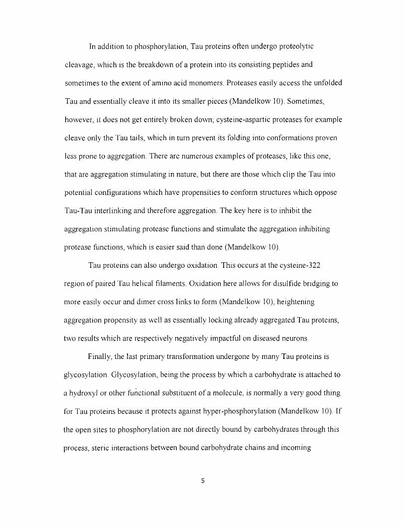
In addition to phosphorylation, Tau proteins often undergo proteolytic
cleavage, w hich is the breakdow n o f a protein into its consisting peptides and
som etim es to the extent o f am ino acid m onom ers. Proteases easily access the unfolded
Tau and essentially cleave it into its sm aller pieces (M andelkow 10). Som etim es,
how ever, it does not get entirely broken down; cysteine-aspartic proteases for exam ple
cleave only the Tau tails, w hich in turn prevent its folding into conform ations proven
less prone to aggregation. There are num erous exam ples o f proteases, like this one,
that are aggregation stim ulating in nature, but there are those w hich clip the Tau into
potential configurations w hich have propensities to conform structures w hich oppose
Tau-Tau interlinking and therefore aggregation. The key here is to inhibit the
aggregation stim ulating protease functions and stim ulate the aggregation inhibiting
protease functions, w hich is easier said than done (M andelkow 10).
Tau proteins can also undergo oxidation. This occurs at the cysteine-322
region o f paired Tau helical filam ents. O xidation here allow s for disulfide bridging to
m ore easily occur and dim er cross links to form (M andelkow 10), heightening
aggregation propensity as well as essentially locking already aggregated Tau proteins,
tw o results w hich are respectively negatively im pactful on diseased neurons.
Finally, the last prim ary transform ation undergone by m any Tau proteins is
glycosylation. G lycosylation, being the process by w hich a carbohydrate is attached to
a hydroxyl or o ther functional substituent o f a m olecule, is norm ally a very good thing
for Tau proteins because it protects against hyper-phosphorylation (M andelkow 10). I f
the open sites to phosphorylation are not directly bound by carbohydrates through this
process, steric interactions betw een bound carbohydrate chains and incom ing
5

phosphates m ay be w hat decreases the likelihood o f phosphate binding. H ow ever, in
AD diseased neurons, glycosylated Tau proteins becom e defective regarding
phosphorylation, stim ulating binding and thus increasing the propensity for hyper
phosphorylation (M andelkow 10). The reason for these defectives arising is a
currently researched, unsolved mystery.
Since Alois A lzheim er’s tim e, it has proven difficult to identify causes or at
least correlations betw een factors such as Tau proteins and A lzheim er’s disease. The
results appear to be sporadic, in that there is a lack o f com pelling evidence pointing
tow ard genetic involvem ent, as 1-5% o f all diagnosed have been deem ed so based on
heredity. It seem s the best w ay to identify the disease in general, and thereby have a
longer period to discover causes before death ensues, is through the seven stage
process endorsed by the A lzheim er’s Association.
Seven Stages of Alzheimer’s Disease
The first stage is that o f a norm al functioning hum an being w ho has zero
im pairm ent w hatsoever despite uncom m on absentm indedness or even forgetfulness
due to stress and other day to day factors. As previously described, som eone
experiencing said m inor m ishaps w ould not be diagnosed w ith dem entia by a m edical
professional, but perhaps som ething m ore along the lines o f issues regarding the
ability to focus, pay attention, et cetera.
Stage two goes beyond the uncom m on mental m ishap to envelop the
occurrences w hich involve im portant things such as the nam es o f fam iliar objects and
com m only used w ords o f the pertinent language. These are described as m em ory
6

lapses and are often noticed by the victim o f said lapses. Instances such as these would
still be unclassified as dem entia by m edical professionals, and usually not noticed by
relatives or persons o f everyday contact.
Stage three is that o f mild cognitive decline in w hich nam es are difficult to
recall o f new ly introduced persons, particularly valuable objects are lost in the same
proportion to those considered invaluable, and difficulty perform ing assigned tasks.
O ften m ild cognitive decline results in m em ory loss o f things that have ju st been read
or seen and has also been associated w ith increasing difficulty regarding
organizational skills and planning. M edical professionals w ould be able to diagnose
som e m ild cognitive declining persons w ith early onset A lzehim er’s disease, but not
all, some m ay be alternatively diagnosed w ith issues pertaining to concentration.
Stage four pertains to those persons w ith m oderate cognitive decline.
Sym ptom s here include forgetfulness o f recent events, inability to perform challenging
m ental arithm etic, m ore difficulty than m ild.cognitive declining persons regarding
planning or social m anagem ent, and m ost im portantly, forgetting facts about on e’s
own personal history. Previously, stages one through three involved potential
forgetfulness regarding others or even surroundings, but stage 4 is clearly identified by
rare forgetfulness o f topics that a person should always know about them selves.
M edical professionals w ould diagnose a stage four person w ith either early-onset or
m ild A lzheim er’s disease based on the com pilation o f symptom s.
Stage five pertains to those persons w ith m oderately severe cognitive decline,
m eaning that they have very externally noticeable gaps in m em ory and thinking
ability. Sym ptom s include but are not lim ited to recalling a personal address or phone
7

num ber, know ledge o f the day’s date, inability to perform non-challenging mental
arithm etic, and inability to choose clothing pertinent to the weather. D iscerning stage
five and beyond becom es increasingly difficult, so it is com m on to note things that
persons in these stages are still able to do: stage five persons still rem em ber personal
topics, how ever less so than stage four, and are still able to go to the bathroom
unassisted. M edical professionals w ould now be able to diagnose these persons w ith
m oderate or m id-stage A lzheim er’s disease.
Stage six pertains to those persons severe cognitive decline. The experiences
and sym ptom s that m ay be pertinent include forgetting w here one is or on e’s
surroundings, strange personality changes, needing help getting dressed entirely,
changes in sleep patterns, delusional behavior, and frequent loss o f bowel control.
H ow ever, stage six is m ost often identified by ability to still rem em ber o n e ’s own
nam e, despite loss o f alm ost all personal history recollection. M edical professionals
could diagnose a stage six person w ith m oderately severe A lzheim er’s disease.
The final stage is stage seven. Stage seven is the w orst step o f the disease, as
those afflicted experience very severe cognitive decline. Basically, in this stage, all
ability to establish a long term connection with their environm ent is lost. Some spurts
o f m em ory recollection have been noted, but are also all accom panied by another note
saying that the same m em ory recollection happens often. Further, a very well o ff stage
seven person m ay have an intriguing introduction and conversation w ith a healthy
person, but then im m ediately forget the events w hich m ay have taken place seconds
ago and attem pt to re-introduce his or herself as if nothing ever happened. This is the
best circum stance, w hereas the w orst o ff stage seven persons cannot cloth, feed, nor
clean them selves w ithout assistance; many o f the sym ptom s associated w ith
A lzheim er’s disease are associated with those o f an infant. This is not a coincidence,
as things such as daily hygiene are learned and upon stage seven onset o f AD, are fully
unlearned and essentially destroyed cognitions.
In addition to severe social sym ptom s, severe physical sym ptom s are
experienced by stage seven persons as well w hich delve beyond those experienced in
infanthood, including loss or at least im paired m otor control. For exam ple, m any lose
the ability to sw allow food, som ething that is done regularly and naturally by infants,
despite their still relying on being fed in the first place. O thers lose the ability to react
w ith reflexes, sm ile, and even keep a straight neck before ultim ately losing all
cognitive abilities and dying thenceforth.
Amyloid Beta Peptide
One o f the m ost im plicated m olecules in AD neurodegeneration is the
am yloid-beta (A(3) peptide. Ap is hydrophobic and can have a prim ary protein
structure over 40 am ino acids long. It is the proteolytic result o f A m yloid Precursor
Protein (APP) cleavage by proteases such as p-secretase (L ichtenthaler 10). Currently,
it is known that there are two m ajor com peting AD secretase pathways: the a-secretase
pathw ay and the p-secretase pathway. The m inor pathw ay is o f the y variety,
appropriately known as the y-secretase pathw ay. These pathw ays constitute w hat are c
called the non-am yloidogenic and the am yloidogenic pathways.
The non-am yloidogenic pathw ay is responsible for 90% o f the APP cleavage
activity, and is the pathw ay w hich does not lead to the generation o f Ap. This pathw ay
9

involves the m etalloprotease a-secretase cleaving APP to yield the shedding o f a
soluble APP fragm ent (sA PPa), leaving behind a carboxy term inal fragm ent (CTF)
com posed o f 83 am ino acids w ith a C -term inus (C83). This a-secretase cleavage is
follow ed by y-secretase cleavage o f C83, resulting in a peptide fragm ent (p3) and
another fragm ent referred to as the intracellular dom ain (AICD). These two resultant
fragm ents are not know n to lead to Ap generation in any way, so the stim ulation o f a-
secretase activity is thought o f as being highly therapeutic (L ichtenthaler 11).
The am yloidogem c pathw ay, correspondingly, is responsible for 10% o f the
A PP cleavage activity, and is the pathw ay w hich does in fact lead to the generation o f
Ap. This pathw ay instead involves p-secretase cleaving A PP to yield the shedding o f
sA PPp and C99. A fterw ards, y-secretase cleaves C99 in the same fashion as C83, but
now yields A ICD and Ap. U pon secretion, soluble Ap is not as neurotoxic as the
oligom ers w hich result from Ap m isfolding. These soluble, small oligom ers are
associated w ith inflam m ation, inhibition o f hippocam pal long term potentiation,
synaptic dysfunction, neuronal loss, and especially the form ation o f neurofibrillary
tangles (L ichtenthaler 10). Protofibrils are precursors to these tangles, w hich lead to
neuronal cell death either directly by form ing fibrils or indirectly through pathogenic
am yloid pores (PA P), w hich lead to reactive oxygen radical species (ROS). These
resulting high energy oxygen radicals are capable o f directly causing aforem entioned
neuronal cell death or indirectly causing it via activation o f kinases involved w ith Tau
protein activity.
Roles of the Endosomal System
10

One organelle that has not been portrayed as m uch in the A lzheim er’s
literature is the lysosome. The lysosom e is prim arily responsible for cellular waste
rem oval in addition to cellular digestion o f lingering, unw anted chem ical compounds.
It acts via hydrolytic enzym e secretion at a low, acidic pH that is enclosed via its
m em brane. This m em brane can fuse w ith other organelles and endosom e system s to
engu lf the biochem ical m atter o f interest, w hich can range from foreign m icrobes to
random particles or m olecular debris.
The lysosom es o f a neuron play a particularly im portant role in that they
com prise w hat are know n as the endocytic and autophagic pathways. These pathw ays
m ake synaptic transm ission o f chem ical signals and neurotransm itters as well as the
degradation o f excess organic m aterials possible. A cceleration o f endocytosis
essentially can overw helm the lysosom e and cause it to function improperly. This
acceleration is caused by APP gene duplication that is a result o f CTF accum ulation
and is m ediated by rab5 (Pim plikar 14947). Essentially, efficiency begins to wane and
proteolysis effectively halts, resulting in m assive debris buildup w hich visibly swells
neurons, changing their optim al conform ation. This buildup includes m olecules such
as Ap, RO S, as well as other toxins that w ould have otherw ise been rem oved, to an
extent, and thence results in neurodegeneration via m ultiple pathw ays (P im plikar
14948). It is clear that this role as well as others m akes the lysosom e an organelle that
should be considered m ore often w hen trying to unravel the m ysteries o f AD
neurodegenerative biochem istry. The internalized APP in particular participates in a
vicious cycle o f am yloidogenesis, generating m ore and m ore A p via the resultant
11
sequestered fibril environm ent w hich has been shown to stim ulate the am yloidogenic
pathw ay (B ahr 117).
A(3 can, however, be transported in other w ays into the cell. Extracellu lar Ap
can bind to the a7-nicotin ic acetylcholine receptor, or a7nA ChR. This receptor is
pentam eric and is a type o f acetylcholine receptor found w ithin the brain, w hich
triggers both presynaptic and postsynaptic excitation. W hen Ap binds, it com petes
com petitively resulting in the slow ing and even prevention o f calcium activation and
the release o f acetylcholine (W ang 5626). One m ethod used to stop this binding is
tak ing advantage o f a chem ical called alpha bungarotoxin, or a-BTX.
The a-B T X drug is originally a com ponent o f snake venom w hich causes
paralysis and respiratory failure by binding to neurom uscular nicotinic acetylcholine
receptors. It has another role that is im portant in the unraveling AD therapies: its
ability to bind a7nA C hR in the brain (N agele 203). The 74 am ino acid that is
otherw ise deadly can be thought o f as an a lzheim er’s therapy in that it can com pete
w ith Ap for b inding to a7nA C hR. The tricky part is not letting the toxin induce cell
death while it prevents Ap binding, perhaps thw arted by anti-venom .
U pon binding o f Ap to a7nA C hR , a com plex is form ed prim arily on neuronal
dendrites w hich can then be transported inside the cell (Nagele 208). In addition to the
lysosom e, early and late endosom es play an extrem ely im portant role in AD neurons
because they perform said transportation and thus contribute to the internalization o f
Ap. This can be thw arted by phenylarsine oxide, or PAO, w hich is an endocytosis
inhibitor (N agele 201).
12

Tau and Ap are indeed the two m ost im plicated proteins regarding the onset o f
A lzheim er’s disease. H yperphosphorylated Tau protein leads to the form ation o f
intraneuronal neurofibrillary tangles, w hich destabilize m icrotubules and result
inevitably in apoptosis. Ap leads to the form ation o f dim ers, protofibrils, and fibrils
w hich form interneuronal senile plaques, disruption interneuronal com m unication and
ultim ately inducing apoptosis as well. These two very im portant intraneuronal and
interneuronal pathw ays respectively are separated by the cell m em brane, but
u ltim ately connected by intracellular Ap that has been internalized via m ethods
described above. This connection arises and is founded on the fact that Ap stim ulates
casein kinase I and casein kinase II (C hauhan 50). These two enzym es are called
kinases because they perform phosphorylation, specifically here on tau protein. The
m ain focus o f my research project thus far has been to study and unravel the
relationships betw een such kinases and tau apoptosis by m athem atically portraying the
best way to lessen apoptosis both intraneuronally and m terneuronally. Before I talk
about the m ethods, 1 w ould like to go give an in depth review o f the m etals, kinases,
drugs, and other m olecules involved.
Roles of Metals
Let us not only look at the effects o f peptides and organelles on AD, but also at
the role m etals play in AD. I started w ith calcium and learned that it had significant
activity in aiding protein phosphatase 2A w ith dephosphorylating the
hyperphosphorylated tau proteins characteristic in AD brains. I found it interesting
that calcium being one o f the m ost readily accessible m etals in the body through
13

parathyroid horm one activity on bone cannot sim ply treat AD in this m anner. Aware
o f the new ‘cocktail’ approach where it is thought that no one m olecule or drug can
effectively cure a neurodegenerative disease, I knew there m ust be another type o f
m etal involved.
Zinc is a 3d transition m etal that is an essential m ineral to the hum an body.
Z inc deficiency is m ore com m on than that o f other m etals because the hum an body
lacks a specialized zinc storage system. Zinc has im portant roles in grow th and the
im m une system , but is also an im portant player w hen it com es to AD. Zinc has been
found to catalyze the conversion o f active PP2 A to inactive PP2A , as well as directly
catalyze the hyperphosphorylation o f Tau proteins (X iong 746). Thus, zinc seem s to
be in com petition w ith calcium , as they have antonym ic effects on Tau proteins.
H ow ever, rem em bering that it is an essential m ineral because o f its o ther functions
throughout the body, zinc is not to be elim inated from the diet as a potential
therapeutic treatm ent. Instead, zinc intake by those w ith AD should be m inim ized to
the low er end o f recom m ended daily dietary allow ance (RDA) range, here being about
9 m g/day. This com bined with other treatm ents could aid and indeed prove therapeutic
for those with AD, based on these recent zinc findings.
Lithium is the third elem ent o f the periodic table and is the lightest metal in
existence. It also is extrem ely reactive and flam m able. It has a w ide variety o f
inorganic applications, ranging from its incorporation into therm onuclear w eapon fuel
as lith ium - 6 deuteride to a source o f electrons in galvanic cell batteries. There are
som e bioorganic and m edicinal applications as well, including its use in the treatm ent
o f m ood disorders due to its involvem ent in the central nervous system as a cation
14

(Li+). In the brain, however, lithium has been show n to directly inhibit the beta
isoform o f G lycogen synthase kinase - 3 Beta (G SK -3p) by com peting with
m agnesium (G ong 2323). As a result, a relative increase in lithium could prove
therapeutic in AD brains.
Roles of Tau Protein Kinases and Their Respective Activity Modulators
In addition to m etals, A lzheim er’s disease involves a variety o f kinases and
other m olecules that can be catalytic regarding the hyperphosphorylation pathw ay, as
previously m entioned. Pharm acologically, it is m uch easier and m ore efficient to
develop drugs w ith the ability to inhibit these catalysts as opposed to developing drugs
w ith the ability to activate inhibitors such as aforem entioned PPA2. Two o f the m ost
im portant kinase catalysts in general are cdk-5 and the aforem entioned GSK-3p,
w hich are both proline directed protein kinases, or PDPKs.
GSK-3 is a protein kinase that is associated w ith adding phosphates to proteins
w ith threonine and serine prim ary protein structures, like those found in the tau
protein. This enzym e was discovered and appropriately nam ed in 1980 for its
regulatory involvem ent regarding glycogen synthase (Em bi 519). GSK-3 has an alpha
and a beta form , denoted by G SK -3a and G SK -3p respectively, G SK -3p appears to be
under m ore scrutiny than the alpha form for its im plication in A lzheim er’s disease
(G ong 2323). Both form s o f GSK-3 share m any inhibitors, including A loisine,
H ym enialdisine, Induribins, and Flavopiridol, and aforem entioned Lithium ju st to
nam e a few.
15

An aloisine has a m olecular form ula o f C 16H 17N 3O and is a potent, perm eableFigure 1
inhibitor o f m any kinases, including GSK-3 (Gong
2323). They are 6 -phenyl[5H ]pyrrolo[2,3-b]pyrazm es, / = \H 0 _ / j j
as seen in Figure 1, w hich act in an inhibitory fashion N %N
by com petitive ATP inhibition; this m eans that these m olecules inhibit the binding o f
A TP to the catalytic portion o f the kinase (M ettey 222). As a result, a relative increase
in aloisine could prove therapeutic in AD brains.
A m ong other m olecules that are A TP-com petitive HyNs
inhibitors o f GSK-3 are H ym enialdisine and Induribins, w hich HIM*
sure
•N
interestingly enough, are m arine- derived. H ym enialdisine, / /\
HO
N X_^NHportrayed by Figure 2, is a natural, m arine sponge m olecular h //
0
product that has been show n to be inhibitory o f GSK -3, am ong other kinases (M eijer
51). It has three constituent N -heterocyclic rings in its
structure that has m any analogues, a few o f w hich are
acetylated or contain brom ine substituents (M eijer 51).
Indirubins are active ingredients in the Danggui Longhui W an, which is an ancient
C hinese m edicinal recipe. These m olecules have four rings, as seen in Figure 3, two o f
w hich are N -heterocyclic, and are also inhibitors o f GSK-3 as well as other kinases
(Leclerc 251). One induribin in particular is 6 -B rom oindirubin-3’-oxim e, or 6 -BIO.
C rystallography o f both hym enialdisine and induribins reveals that these ringed
structures bind nicely to the ATP binding sites o f GSK-3, m aking them prim e targets
for AD therapeutic approaches.
16

HO OI!
HO'y* y
' o
C!'
Flavopiridol, seen in Figure 4 to the right,
is a decently large alcohol with four rings, one o f
w hich is N -heterocyclic. It has a chlorine
substituent as well as a ketone. Its com m on nam e
is A lvocidib, w here it and analogues are used toINI
inhibit kinases other than GSK-3. H ow ever, it is 1
Figure 4
still thought o f com m only as a GSK-3 inhibitor as well, proving as an effective
treatm ent for AD and other taupathies (M artinez 377).
A R -014418, seen in Figure 5, is a GSK-3 chem ical inhibitor o f the beta
isoform that has a urea foundation w ith term inal nitro and m ethoxybenzyl groups,
appropriately called N -(4-M ethoxybenzyl)- ? Jn-N O
N '-(5 -m tro-l,3 -th iazo l-2 -y l)u rea. Its
m olecular w eight is 308 .31 g/mol and it has OCH3
an IC50 value o f 104nM on GSK-3 beta. It is also active on CD K-5, but to a m uch ,
lesser extent, w ith an IC50 value o f over 100 m icrom olar (M artin 292).
All o f these inhibitors to the GSK-3 protein have been found to be therapeutic
in targeting tau protein phosphorylation and hyperphosphorylation respectively, but
m any o f these inhibitors and other m olecules are also associated w ith CD K-5, another
very im portant kinase involved in taupathy.
C yclin-dependent protein kinase 5 (cdk-5) is one o f m any cyclin-dependent
kinases (cdk), but is the m ost im plicated cdk in A lzheim er’s disease. This enzyme
binds cyclin, a regulatory protein w hich com es in m any varieties ranging from cyclin
17
A - cyclin G, and then becom es able to phosphorylate proteins like Tau. In addition to
aforem entioned inhibitors o f GSK-3 like Flavopiridol, A loisine, and Induribins, CDK-
5 has other inhibitors and inhibitor drugs, including O lom oucine, Roscovitine,
Purvalanol, and Paullones. It has activators, some o f w hich are p35 and p39 (Sausville
Cdk-5 cannot function m onom erically, for it depends specifically on activators.
These aforem entioned activators can each be proteolytically cleaved by calcium
dependent calpains resulting in their sm aller form s nam ed p35 and p25, respectively.
The cdk-5/p35 and cdk-5/p25 com plexes are m uch m ore active than the cdk-5/39 and
cdk-5/29 com plexes, contributing m oreso thusly to tau protein hyperpho.sphorylation
(M artin 292).
O lom oucine, or 2-(H ydroxyethylam ino)- Figure 6
been shown to regulate CDK-2 in the cell cycle, arresting cleavage in m any organism s
(A braham 105). O lom oucine is portrayed in Figure 6 .
32).
especially in the brain. Interestingly, it has also
m any cyclin dependent kinases, but CDK-5
C 15H 18N 6O and is a com petitive ATP inhibitor o f
6-benzylam ino-9-m ethylpurine, has form ula
butanol, 6-(B enzylam ino)-2(R )-[[l
very sim ilar m olecule in structure to olom oucine, is
o therw ise know n as 2-(R )-[[9 -(l-M ethy lethy l)-6 -
[(phenylm ethyl)am ino]-9H -purin-2-y l]am ino]-l-
R oscovitine (or Seliciclib), seen in Figure 7, is a
18

(hydroxym ethyl)propyl]ainino]-9-isopropylpurine with form ula C 19H 26N 6O. They can
be easily distinguished by the extra ethyl group o ff the benzylic nitrogen in
Roscovitine. It is also sim ilar in function to olom oucine, as it inhibits CDK-5
substantially and in a sim ilar m anner. Interestingly enough, roscovitine too plays a
role in the cell cycle, proving to be a useful antim itotic reagent (Borgne 527).
Purvalanol differs in structure w hen
com pared generally to olom oucine and roscovitine, Figure
but still m aintains the core double N -heterocyclic
ring system . The two form s, Purvalanol A and j p 1 1 /NO-, *
Purvalanol B, portrayed by Figures 8 and 9
"-N,
Cl
v > rI ^H , /
respectively, can be readily distinguished by observing that Purvalanol B has a
benzylic acid group in its chem ical structure,
as it is form ally nam ed 2-chloro-4-[(2-
{[(2R)- 1 -hydroxy-3-m ethylbutan-2-
yl]am ino}-9-(propan-2-yl)-9H -purin-6-
y l)am ino]benzoic acid w ith chem ical
form ula C 20H 25CIN 6O 3 Purvalanol A,
Figure 9
H~C.
C H
how ever, ju s t has a chlorine substitutent on that particular benzene group. Purvalanol
in general inhibits m any cyclin- dependent kinases (V illerbu 761), and has been used
specifically as a CDK-5 inhibitor. It has been shown to decrease Ser-522 and Thr-
514/409 levels specifically, the residues that have com m only been linked to tau
protein hyperphosphorylation sites (Cole 18230).
19

HN
NO
Fij [ure 10
Paullones are tetracyclic, fused
com pounds that cover a large range o f
derivatives w ith d ifferent purposes, many
o f w hich include being inhibitory regarding
cdks and GSK-3 (M artinez 377). They
generally are not sim ilar in structure to
olom oucine, roscovitine, nor purvalanol other than the fact that there are still two N -
heterocyclic rings. These two rings are not exactly the sam e as those found in the other
three m olecules due to one o f them being 8 m em bered instead o f 6 m em bered. A
specific type o f paullone called alsterpaullone, was show n to be specifically
im plicated in the reduction o f tau phosphorylation (Selem ca 959) and is portrayed
chem ically by Figure 10.
R -C R 8 , or (2-(R )-(l-ethyl-2-hydroxyethylam ino)-6-(4-(2-pyridyl)benzyl)-9- *
isopropylpurine) is an interesting
m olecule portrayed by Figure 11
w hich has the exact sam e structure as
roscovitine in that the only difference
is an extra quinolone substituent
attached to the benzene group. That
uH
being said, it is specifically a derivative o f (R )-roscovitine that is thought o f as a
versatile inhibitor because it acts on both CD K-5 and GSK-3 (M artin 297).
It seem s that generally, a double, fused quinolone N -heterocyclic ring sytem at
the core o f a chem ical is necessary or at least beneficial to its action as a CDK-5
20
inhibitor, especially for those w hich are ATP com petitive. This structure, or one that is
sim ilar, appears in m any o f the drugs that are being designed for this purpose, and
should be m aintained in the developm ent o f drugs specifically designed for AD
therapy to m inim ize tau protein phosphorylation and hyperphosphorylation.
CDK-5 and GSK-3 m ay be the m ost generally well know n kinases involved in
neurodegenerative diseases, but casein kinases are particularly im portant in
A lzheim er’s disease and my research project specifically, as previously m entioned.
C asein kinases are non-proline directed protein kinases, or non-PD PK , and have two
m am types each w ith its ow n isoforms.
Casein Kinase 1, or CK1 has 7 d ifferent isoform s know n as C K l-a , C K l-p ,
C K l-y l , C K l-y2 , C K l-y3 , CK 1- 5, and C K l-£ w hich range in m olecular w eight from
25 to 55 kDa. The C K l-a , CK 1- 5, and C K l-£ isoform s are involved specifically in
A lzheim er’s disease (M artin 298). These three isoform s are also referred to as
CSN K 1A 1, C SN K 1D , and CSN K 1E respectively and are know n to phosphorylate tau
protein.
Casein K inase 2, or CK2 has three isoform s, CK 2- a, C K 2 -a ’, and CK2-p.
CK 2- a and C K 2 -a ’ are involved in A lzheim er’s disease and are also referred to asi
CSNK2A1 and C SN K 2A 2 respectively. Both phosphorylate tau, while each also has
an interestingly unique additional function: CK 2- a activates PP2A and CK 2- a ’
inhibits PP2A (M artin 298). PP2A is a protein phosphatase, w hich effectively has a
reverse kinase effect on tau protein by rem oving phosphate.
21

H0X oXs0 0 oh'O'-t ^ qt5"
HNx ^ O
Both CK1 and CK2 proteins undergo aforem entioned activation by Ap, but are
also subject to inhibition by several chem icals including H eparin, TBCA, and IC261
w hich interestingly differ greatly in structure.
H eparin, seen structurally in Figure 12, is a
relatively large m olecular w eight drug averaging
around 13.5 kg/mol that has forever been used in a
sulfated form to act as an anticoagulant. However,
since the 1980s, H eparin has been show n to be an
effective inhibitor o f C K 2-a and C K 2 -a ’ CK-2
variants. (H athaw ay 8040).
Tetrabrom ocinnam ic acid, or TBCA, is portrayed by Figure 13 and is a small
drug com pared with the inhibitors acting on GSK-3 and CD K-5, with system atic nam e
(2E )-3-(2,3,4,5-Tetrabrom ophenyl)acrylic acid. Its
m ain features are the four brom ines on the phenyl
0
'OH
group w hich are com plem ented by the acid group on X X ^Br 'f Figure 13
the other end o f the m olecule. It is exceptionally m ore gj*
potent o f an inhibitor on CK2- a, C K 2 -a ’, and CK2-P than on C K l-a , CK 1- 8 , and
C K l-a , but is regardless an inhibitor o f both types o f casein kinases (M artin 298).
IC261, or l,3-D ihydro-3-[(2 ,4 ,6-
trim ethoxyphenyl)m ethylene]-2H -indol-2-one, is a
chem ical that effectively inhibits C K l-a , CK 1- 8 , and
C K l-e , but has a particularly potent activity on CK 1- 8 ,
w ith a rem arkable IC50 o f ju st 1.0 m icrom olar (M artin
O C H
uri
22

298). It is easily distinguished structurally by its sym m etrical trim ethoxyphenyl group
and has a m olecular w eight o f 311.33 g/mol.
Tau Tubulin K inase, or TTBK , is another non-proline directed protein kinase
that is also know n as B rain-D erived Tau Kinase, or BDTK. There are two varieties o f
TTBK, called Tau Tubulin K inase-1, or TTBK-1 and Tau Tubulin K inase-2, or
TTBK-2. TTBK-1 and TTB K -2 are very sim ilar in catalytic - dom ain structure and
are specific to the brain, expressed in cortical neurons and hippocam pal neurons
respectively (Sato 1573). Together these kinases phosphorylate tau at 10 different
phosphorylation sites, often catalytically rely ing on M g2+ and M n2+ cations to deposit
the phosphate (Sato 1580). TTBK-1 and TTBK-2 are indeed identified as im portant
targets for A D therapy, but no TTB K chem ical inhibitors have been discovered nor
synthesized to date.
D ual-Specificity Tyrosine-(Y )-Phosphorylation Regulated Kinase, or DYRK,
is yet another non-proline directed protein kinase that has 6 d ifferent isoforms. The
tw o isoform s actually involved in A lzheim er’s disease are D ual-Specificity Tyrosine-
(Y )-Phosphorylation Regulated K inase-1A (D Y R K 1 A) and D ual-Specificity
Tyrosine-(Y )-Phosphorylation R egulated K inase2 (DY RK 2). Interestingly,
chrom osom e 21 contains a gene on 21q22.2 w hich encodes DYRK1 A; this is very
im portant because chrom osom e 2 1 is involved in D ow n’s syndrom e and m ay be
linked to the neurofibrillary tangles found w ithin D ow n’s syndrom e patients (M artin
298). Both D Y R K 1A and D Y RK 2 phosphorylate tau protein but are relatively unique
23

regarding the other kinases m entioned in that they can prim e other kinases to act w ith
augm ented efficiency. D Y RK s are also able to undergo tyrosine auto-phosphorylation
to achieve activation. One very potent chem ical inhibitor is known, called Harmine.
H arm ine, seen in Figure 15, is an alkaloid chem ical that is also called
pyrido[3 ,4-b]-indole and has a m olecular w eight o f
212 .25 g/mol. It has a relatively small chem ical structure com posed m ainly o f three
fused rings. H arm ine is m ost com m only know n for its ability to inhibit m onoam ine
oxidase A (M A O -A ), w hich is an enzym e that breaks dow n m onoam ine com pounds.
Som e exam ples o f m onoam ines are serotonin, norepinephrine, and psilocybin w hich
are independently classified as a type o f neurotransm itter, horm one, and recreational
drug respectively. In addition to this effect, how ever, H arm ine has been used as an
anticancer as well as an inhibitor to D Y RK 1 A. Harm ine does in fact inhibit other
D Y R K s including D Y RK 2, but m ost effectively inhibits D Y R K 1A w ith an IC50 value
o f 80 nM (M artin 298).
E xtracellular signal-regulated kinases (ERK s) are m itogen-activated protein
kinases that are proline-directed. There are two isoforms, both o f w hich are involved
w ith AD: E xtracellu lar signal-regulated kinase-1, or ER K 1, and E xtracellular signal-
regulated kinase-2, or ERK2. They are also know n as M APK 3 and M A PK 1,
Telepathine. It is a naturally occurring com pound Figure 15
that is found in the M iddle Eastern plant Peganum
harmala. Its IUPA C nam e is 7 -M eO -l-M e-9H -
24

respectively. ER K s have one m ain activator and one m ain inhibitor, referred to as
Paclitaxel and FR 180204 respectively.
Paclitaxel, seen in Figure 16, is a chem ical activator o f ERKs that is also
com m only referred to as Taxol or Onxal. Its IUPA C nam e is (2a,4a,5p ,7p ,10p ,13a)-
4 ,10-B is(acety loxy)-13-{ [(2R,3S)-3-
large in size com pared w ith drugsFigure 16
discussed so far. It is a naturally occurring com pound, being isolated from tree bark o f
the organism Taxus brevifolia, but is currently developed com m ercially after being
totally synthesized in 1994. Paclitaxel is m ost com m only know n for its anticancer
properties, having been discovered officially in 1962 and thence utilized by the United
States N ational C ancer Institute and other H ealth O rganizations. It is technically a
cytoskeleton toxin, as cells treated w ith the drug exhibit serious deficiencies in
chrom osom e segregation and m itotic cell division. This is because in addition to its
activation o f ERK, w hich could lead to heightened phosphorylation and eventual
apoptosis, it over-stabilizes m icrotubule polym ers by binding beta-tubulin m icrotubule
subunits (Low e 1046). This m icrotubule stability seem s like a positive attribute
regarding the reduction o f cell death, but very stabilized m icrotubules have trouble
during disassem bly, thus disrupting mitosis. D isrupted m itosis eventually leads to cell
death. Therefore, paclitaxel would definitely have the negative effects o f cell death
en-2 -yl benzoate and is relatively
dihydroxy-9-oxo-5 ,20-epoxytax-11
phenylpropanoyl]oxy} -1,7-
(benzoylam ino)-2-hydroxy-3
25

that are situational ly taken advantage o f w hen fighting cancer, but i f adm inistered to a
person w ith AD, the m icrotubule stabilizing effects could be advantageous. Such
investigations into the efficacy o f paclitaxel on neurodegenerative m em ory loss are
being conducted at institutions across the country, including The U niversity o f
Pennsylvania.
ERKs also have an inhibitor, nam ed FR 180204.
Its IPUA C nam e is 5-(2-Phenyl-pyrazolo[l,5 -
a]pyridm -3-yl)-lH -pyrazolo[3 ,4-c]pyridazm -3-yIam m e
and its m olecular w eight is 327.34 g/mol. F R 180204 is
a potent inhibitor o f ERK1 w hen com pared to its
efficacy at inhibiting ERK2, w ith IC50 values o f 0.310 m icrom olar and 0.806
m icrom olar respectively (M artin 298). F R 180204 is portrayed by Figure 17.
P38s are m itogen-activated protein kinases that are involved in apoptosis and
differentiation o f cells. There are four isoform s, p38- a, p38- p, p38- y, p38- 5 and are
referred to as M A PK 14, M A P K 11, M A PK 12, and M APK 13 respectively. P38
phosphorylates tau at 2 1 different sites upon activation, which occurs via auto-
phosphorylation or in the presence o f extracellular stress
(M artin 297). P38 has a potent inhibitor called SB-239063,
show n structurally in Figure 18.
SB-239063, or trans-4-[4-(4-F luorophenyl)-5-(2-
m ethoxy-4 -py rim id iny l)-lH -im idazo l-l-y l]cyclohexanol, is a
selective inhibitor o f p38 w ith an IC50 value o f 0.044
j g u r e I B
OMe
2 6

m icrom olar and a m olecular w eight o f 368.41 g/mol.
C -Jun N -term inal Protein K inases (JN Ks) are m itogen activated protein
k inases (M A PK ) w ith 10 different isoforms. The three m ain types w hich collectively
house these ten isoform s are JNK1 (M A PK 8), JN K 2 (M A PK 9), and JN K
3(M A PK 10). The first two are found in m ost tissues throughout the body including the
brain, w hile JNK3 is alm ost exclusively found in brain tissue. JNKs range in size from
45 to 55 kD a and phosphorylate tau protein at 12 different sites, and is inhibited by
protein phosphatases and especially by a chem ical called SP600125 (M artin 297).
SP600125, show n in Figure 19, is a chem ical
inhibitor o f the three aforem entioned JN K types.
D epicted to the right, its formal nam e is A n th ra[l-9 -cd ]-
pyrazol-6(2H )-one, but is m ore com m only referred to as
1,9-Pyrazoloanthrone or sim ply A nthrapyrazolone. This
chem ical has a m olecular w eight o f 220.23 g/mol and has
an IC50 value o f 0.090 m icrom olar tow ard JNK3. It isFigure 19
very slightly m ore potent, how ever, at inhibiting JNK1 and JNK2 w ith respective
IC50 values each at 0.04 m icrom olar (M artin 297).
NH
Protein K inase A, or PICA, is an enzym e tetram er w hose regulatory subunits
require cyclic AM P, or cAM P, binding to induce enzym atic activation. It is for th is
reason that PK A is also com m only know n as cA M P D ependent Protein Kinase. PK A
phosphorylates 17 im portant sites along tau protein associated with AD brains, and
27

thusly can be considered an important target for AD therapy by inhibitors. The most
important inhibitor o f PKA activity is H-89 (Martin 299).
H-89, or N-[2-broinocinnamylamino)ethyl]-5-isoquinoline sulfonamide
dichloride hydrate, seen in Figure 20, is a
potent PKA inhibitor with an interesting
structure, roughly the size o f an ATP
molecule. Thusly, it inhibits PKA by
Figure 20
Br
.2HCI . 2 H . 0
nh2jl
T . I L - / 1
o -
HO OH
p ~ CT 0 ~I I i
.0 ™ p _ 0 - p - 0 - - P —O' li I! IIO O O
Figure 21
A T P
acting in competition with ATP with
isoquinoline mimicking adenosine,
sulfonamide m imicking ribose, and
bromobenzene m imicking the ATP
phosphates seen in Figure 21 (Lochner 265). The isoquinoline group o f FI-89 is
certainly not the exact same size as the
adenosine portion o f ATP, but
crystallography shows it is similar enough
to bind PKA, in place o f adenosine, around
the amino acid Val 123 (Engh 876). After
determining where the sulfonamide binds,
Engh et al. attempted but were not able to
identify the exact crystallographic position o f the bromobenzene electron density
portion o f H-89 binding, but they were in fact able to conclude that it was located
roughly within the region where the ATP phosphates otherwise would bind. They
depict the two best H-89 bindings to PKA in Figure 22.
I o —"
"‘""C /Figure 22
28

Protein Kinase B, or PKB, is also known as Akt and has three main isoforms,
Protein Kinase B - a (PKB-a), Protein Kinase B - (3 (PKB-(3), and Protein Kinase B - y
(PK B -y) or A k t-1, Akt-2, and Akt-3 respectively. PKB overexpression is known to
cause tau hyperphosphorylation at the main threonine and serine residues, threonine
212, serine 214, and 396 respectively (Martin 299). These three isoforms activate
GSK-3[3 and also have an inhibitor, Akt VIII trifluoroacetate hydrate salt.
Akt VIII, seen in Figure 23, inhibits all three isoforms but is more selective
toward Akt-1, with an IC50 value o f 58 nM
compared to the larger 2 lOnM IC50 value and
even larger 2100nM IC50 value for Akt-2 and
Akt-3. The salt is formally named 1,3-Dihydro-
1 -(1 -((4-(6-phenyl-1 H-imidazo[4,5-
g]qumoxalm-7-yl)phenyl)methyl)-4-piperidinyl)-
2H-benznnidazol-2-one, corresponding to its
large quinoxaline ringed chemical structure.
Interestingly, it is PKB specific in concentrations below 50 micromolar, but inhibits
PKA and PKC at concentrations above 50 micromolar (Martin 300).
Protein Kinase C, or PKC, is one o f the most diverse protein kinases in that it
has over fifteen isoforms, but for simplicity, but can be classified into three broad
categories: conventional, novel, and atypical. Conventional PKCs include the four
isoforms PKC-ol PKC-f31. PKC-p2, and PKC-y. These isoforms require DAG and
calcium ion to function. Novel PKCs include the seven isoforms PKC-8, PKC-51,
29

PKC-52, PKC-63, PKC-e, PKC-q, and PKC-O. These novel isoforms require DAG but
do not require calcium ion to function. The remaining five isoforms that are referred to
as atypical include the following: PKC-i, PKC-£, PK-N1, PK-N2, and finally, PK-N3.
These five are referred to as such because they are not like the rest, for they require
neither calcium ion nor DAG to function. In my model, DAG is incorporated
mathematically as an activator as it is required by the majority o f PKC isoforms to
function. Bisindolylmaleimide I is also incorporated, as it is a potent chemical
inhibitor o f PKC (Martin 300).
Diacylglycerol, or DAG, seen in Figure 24, is from a fundamental biological
perspective is a type o f molecule also called diglyceride that is involved in
?
o c'OH
biochemical signaling o f secondary messages. One particular DAG involved in PKC
signaling is a long lipid that is formally called l-palmitoyl-2-oleoyl-glycerol. The
D A G is found in the cell membrane, and the conventional and novel PKCs bind DA G
to achieve activation and thence ability to phosphorylate substrate, such as Tau
protein. I f the PKC is indeed conventional, calcium ions from the inositol
triphosphate, or IP3, intermembrane channel belonging to the endoplasmic reticulum
will need to be provided via release into the cytosol. IP3 is a chemical that will always
be present as long as DAG is present in the cell membrane, because it is a byproduct
o f Phospholipase C, which is an enzyme that cleaves IP3 from phosphatidylinositol
4,5-bisphosphate, or PIP 2 , to form DAG fundamentally. Ligands need to bind a
30

w"H
surface receptor to stimulate this entire pathway secondary messenger signaling
pathway.
Bisindolylmaleimide I, or
GF-1, seen in Figure 25 is a C}
chemical inhibitor o f PKC. Its
formal name is 3-[ 1 -[3-
(d im ethylam ino)propyl]-lH -indol-
3-yl J-4-( 1 H-indol-3-yl)-1H-
pyrrole-2,5-dione and the chemical y /\
has a truly fascinating structure . ..... \
with near symmetrical dual indole
substituents coming o ff o f a main unsaturated double y-lactam. GF-1 is a potent
inhibitor o f the conventional PKCs with 1C50 values less than 21 nM. It can also
inhibit the novel PKCs, exhibiting IC50 values below 200 nM. It does not do well
with the atypical PKCs, whereas GF-1 is only able to inhibit 50% o f PKC-^ with a
concentration o f 5800 nM (Martin
300). Another important modulator
o f PKC activity is Bryostatin I,
seen in Figure 26.
Bryostatins are very large
lactones that inhibit PKC.
Originally, Bryostatins were
naturally isolated from the Bugiila
H OH
31

neritina , which are a species o f aquatic invertebrates called bryozoan. There are
currently over 19 Bryostatins that have been isolated or synthesized. They were first
employed as anti-cancer agents, but are to date being investigated in
neurodegenerative diseases such as A lzheim er’s disease. Bryostatin 1, involved in AD,
has the formal name (1S,3S,5Z,7R,8E,11S,12S,13E,15S,17R,20R,23R,25S) -25-
A cetoxy-1,11,20-trihydroxy-17-[( 1 R)-l -hydroxyethyl]-5,13-bis(2-methoxy-2-
oxoethylidene)-10,10,26,26-tetramethyl-19-oxo-18,27,28,29-tetraoxatetracyclo-
[21.3.1.13,7.111,15] nonacos-8-en-12-yl (2E,4E)-2,4-octadienoate.
There are other kinases o f course, but these are the ones that I have targeted as
having the highest potential for proving therapeutic regarding the treatment o f
A lzhe im er’s disease.
Molecules involved in the phosphatase pathway:
Protein Phosphatase 2 (PP2A) was mentioned with regards to zinc and the
casein kinases above, but not fully explored. This protein is directly involved in
removing phosphate groups from proteins like Tau at the serine and threonine residue
regions; therefore, it is a catalyst o f tau dephosphorylation. It has been shown to be
upregulated by lithium in the rat frontal cortex and hippocampus (Tsuji 413). It is
heterotrimeric in structure, comprised o f a heterodimeric core enzyme and a subunit
that provides regulation. The heterodimeric portion is comprised o f a catalytic subunit
and a scaffolding subunit, respectively, in addition to being supported by M g2+
coenzyme (Xu 1239).
32
o
PP2A is the major tau phosphatase in theH
brain but should be used with caution as it is N
Hmore broad in its substrate specificities than N O
protein kinase inhibitors, which could lead to
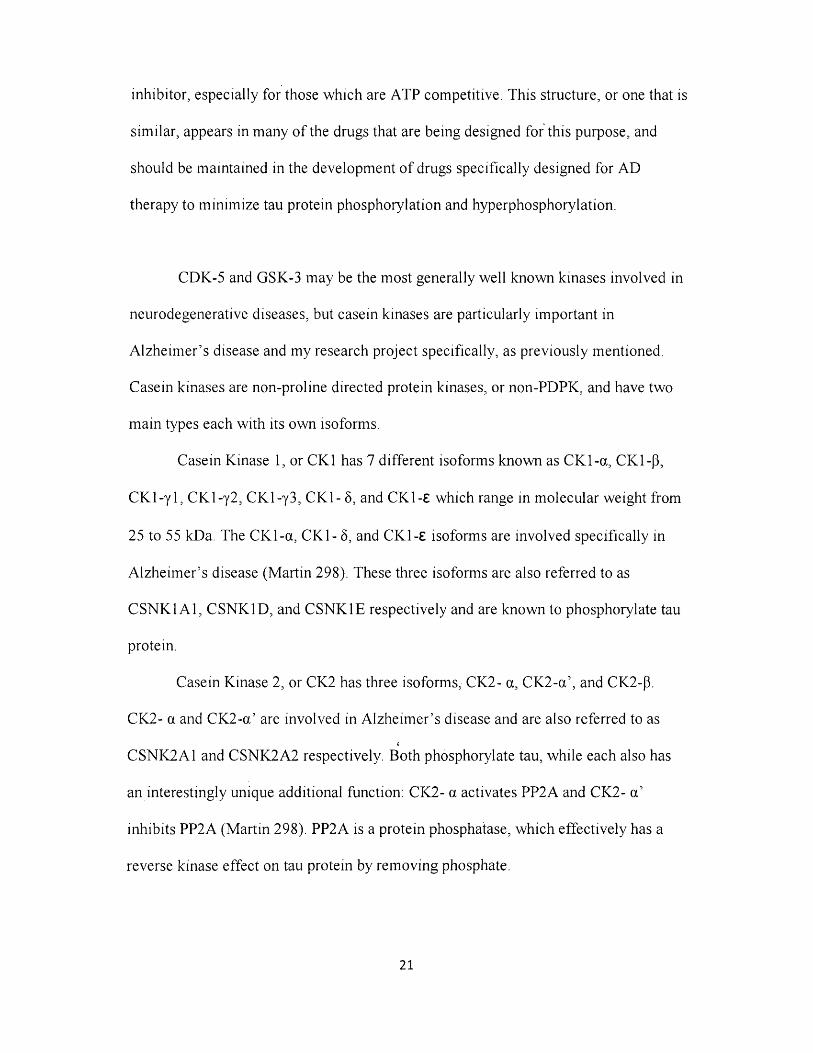
negative, unforeseen side effects (Gong 2326). Two inhibitors regulate PP2A activity,
known as I iPP2A and W P2A respectively. Melatonin, seen in Figure 27, is otherwise
known as N-acetyl-5-methoxytryptamine or N-[2-(5-methoxy-l H-indol-3-yl)ethyl],
and acts in a reverse manner and thus restoring PP2A activity (Gong 2327). However,
melatonin could have other side effects upon tau hyperphosphorylation reversal due to
its widespread hormonal abilities that have been prescribed to treat or subdue
circadian rhythm sleep disorders, immune disorders, and cardiovascular disorders, as
well as sexual dysfunction.
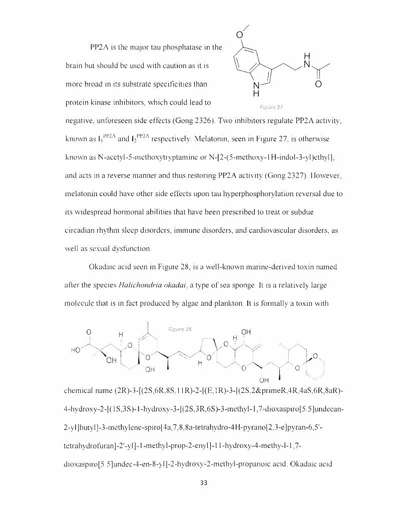
Okadaic acid seen in Figure 28, is a well-known marine-derived toxin named
after the species Hahchondria okadai, a type o f sea sponge. It is a relatively large
molecule that is in fact produced by algae and plankton. It is formally a toxin with
0 H X -e— « OH
HQ""V , , P tT '" V - w : w
H
v °
QH *chemical name (2R)-3-[(2S,6R,8S,l lR)-2-[(E ,lR)-3-[(2S,2& prnneR ,4R,4aS,6R,8aR)-
4-hydroxy-2-[( 1S,3S)-1 -hydroxy-3-[(2S,3R,6S)-3-methyl-l ,7-dioxaspiro[5.5]undecan-
2-yl]butyl]-3-methylene-spiro[4a,7,8,8a-tetrahydro-4H-pyrano[2,3-e]pyran-6,5'-
tetrahydrofuran]-2'-yl]-l -methyl-prop-2-enyl]-l 1 -hydroxy-4-methy-l-l ,7-
dioxaspiro[5.5]undec-4-en-8-yl]-2-hydroxy-2-methyl-propanoic acid. Okadaic acid
33

does not act directly on tau protein, but mathematically has the same effect as tau
phosphorylation by instead acting to non-competitively inhibit PPA2. Upon ingestion
o f the toxin, in addition to the inhibition o f Ser/Thr phosphatases such as PP2A in
places like brain hippocampal neurons, Okadaic acid causes diarrheal shellfish
poisoning, or DSP (Kamat 164). This occurs in the intestinal tract, in which Okadaic
acid inhibits the removal o f phosphate to induce more permeability along
gastrointestinal cell membranes, hence diarrhea as a symptom. One chemical which
seems able to reverse the negative effects o f Okadaic acid is memantine.
M emantine is an N-methyl-D-aspartate , orn h 9
N M D A , receptor agonist that modulates PP2A
signaling. Its IUPAC name is 3,5-
d im ethyltricyclo[3.3.1.13,7]decan-lam ine and is
named such because o f the triple fused saturated ring
system seen to the right in Figure 29.
It blocks over-activated N M D A receptors and thus prevents excite-toxicity (Li
268). In moderate to severe AD, memantine prevents tau hyperphosphorylation in
hippocampal neurons (Gong 2324).
34

METHODS
A sim plified m odel o f A lzheim er’s disease was m ade via the program Cell
D esigner4.2 using tau protein literature regarding m olecular links to
hyperphosphorylation. Below, in Figures 31 and 32, are snapshots o f two o f the most
im portant processes in the graduate student version o f the model. They are not true
Figure 30
snapshots, but rather cleaned up versions because the real model has becom e so
com plex that it is difficult to visually interpret, as evident in Figure 30 above.
35

dk-5 monomerdk-5 monomer
-5 m onom er:dk-5 m onom er)
£dk-5 m onom er) m onom er jZM
£dk-5 m onom er) Ip25:dk -5 mo nomem onomer :dk-5 m onom er
As seen here in Figure 31, CDK-5 complexes o f four varieties
hyperphosphorylate Tau protein. Generally throughout the whole model, the dark
green as seen here is consistent with the color for all o f the tau protein kinases, and the
dark blue also as seen here is consistent with the inactive forms o f said kinases. The
shiny red color o f the tau proteins here is consistent with the entire pathway from tau
to NFT formation, microtubule destabilization, and ultimately apoptosis. Lines that
jo in to form a single filled arrow were chosen to be indicative o f heterodimer
association or complex formation. Other regular filled arrows were chosen to be
indicative o f transition states from molecule to molecule, whereas lines with unfilled
circular ends were chosen to be indicative o f activation. The caveat here is that the
molecules exhibiting lines with unfilled circular ends activating the process o f
36

inactivation were later integrated as inhibitors o f the species undergoing said
inactivation. This is seen, as one might expect, with R-CR8 and Seleciclib. They are
both chemical inhibitors o f the four CDK-5 complexes, but it was more convenient in
CellDesigner to portray them as activating the process o f kinase inactivation.
Fibril'Protofibril /■
A p o p to s isS e n ile P la q u e
1 Intracellu lar A m yloid B eta 42 a 7 n A C h R .A B 4 2 C o m p le x:7n A C hR .A B 4 2 C o m p le x PAO
en d o cy to tic v e s ic leExtracellular Amyloid Beta 42
£EIM E reg u la tio n .
[ in tr a c e llu la r A m y lo id B eta 4 2 } - ' " 'C 59I
n eu ro n m e m b r a n esA P P bet; APR
B eta S e c r e t a s e j C 8 3
Upha S e c r e t a s e , ( G a m m a S e c r e t a s e )sA P P a lp h l
Figure 32
As seen here in Figure 32, the non-amyloidogenic pathway to the right o f APP
and the amyloidogenic pathway to the left o f APP, resulting in aforementioned
Extracellular Amyloid Beta 42 and eventual apoptosis via the senile plaque formation
cascade. The senile plaque cascade is portrayed in lime green while the extracellular
secretases are in a softer green. The amyloid is internalized via complex formation and
endocytotic vesicle transport throughout the cell. The resultant intracellular amyloid
then leads to the tau hyperphosphorylation pathway, thus triggering apoptosis. The
trigger arrow is the type with an unfilled arrowhead and perpendicular line just
beneath the arrowhead, signifying that there are aforementioned molecular pathways
37

taken in between to reach the molecule or process being triggered. The transport arrow
looks the exact same as the trigger arrow, but it has a filled arrowhead. The inhibitor
'a r ro w ’ is not quite an arrow as much as a line with a small perpendicular end It is
seen regarding bungarotoxin and PAO above.
After the molecular modeling, Biological Systems Theory (BST) was used to
make sense o f the above complexity exhibited by the model as a whole. Originally
developed by Eberhard Voit, BST allows theoretical biochemists to use ordinary
differential equations to model cell activity mathematically. Our group employed
m any o f the same BST methods described in a previous Coleman group, Sass et al.
(2009), but the main difference is that ours used Michaelis equations, Hill equations,
and real numerical values as inputs instead o f theorized estimated values for the first
time ever. We also used the more current and updated program M ATLAB version
R2014a instead o f PLAS
To acquire these values and thereby mathematically model the neuron with
true, real-life accuracy, large databases o f proteins and drugs alike such as BREND A
and M O PED were scoured that ultimately resulted in a whole spreadsheet full o f
formal name listings, LC50 values, IC50 values, initial concentrations, and rate
constant values. Most o f the time, these values were found cited as being from frontal
lobe brain tissue, which was especially helpful.
With a full set o f values and coded system o f equations, our group set out to
find the specific set o f drug concentrations which would be therapeutic in reducing the
am ount o f tau protein hyperphosphorylation and thereby a theoretical treatment for
38

A lzheim er’s disease. Computational healthy cell and diseased cell states were
constructed, then drugs were selected.
Figure 34 below is a snapshot o f the Hill equations implemented. Each line o f
code after the tdea th=’ portion is for a specific drug, utilizing a specific LD50 value.
These Hill equation variants are o f the form
l / ( l+ e A(Tox-[Drug])), where tox=LD50 and
Drug is the drug concentration from the list in
Figure 33 that uses dynamic_concentration as a
tool for calling from said list. Drugs taken into
account here were SP-600125, R-CR8, 6-BIO,
AR-A014418, SB-239063, paclitaxel, heparin,
bryostatin, harmine, seleciclib, and alpha BTX.
Other drugs were not included for various
reasons, ranging from our group deeming them
as drugs that are unfit for this study or simply
ones where LD50 values do not currently exist in
the literature due to lack o f clinical trial testing. The beauty here is that different drugs
can easily be added or subtracted from the equation when the need presents itself or
when one wants to change the parameters o f the study. Ultimately this set o f equations
uses lethality data to produce a semi-sigmoidal dose-response curve for every single
drug incorporated into the equation set.
(13 4) = SO; .
(13 6) = 5; % 6
(13 9) = 310;
(142) = 660;
( 1 4 5 ) = SO; %;
(14 9) = 10; %-
i j — z B i;
39
d rug ^jparams;i n i t _ o p c = X ; t o p t = [ 0 , 3 0 0 } ;
t i i r e s d _ - ^ r - . c _ ' c r e ' - r t _ " " s • ' m *'; i l r e o d e l , r o p e , i n i c c pc ) ;
; ^ en , v i d t n ; = 3 i z e i d y n a i a i c _ c o n c e n t r a t i c n s ) ;a e a t n " x/ ( - . - 2 . 7 ^ S 2 8 ' ' i ( i i 8 . 1 6 1 5 i £ l £ ' . . 0 ' ' 3 ) - d y n a m i c _ c c n c e r : - r a t . i o n s ( l e n , 99) ) * 1 / ( ( 1 5 . 1 8 1 1 / ( 1 - 2 . 7 - 5 2 5* ( ( 2 5 0 0 - d y n a m i c c o n c e n t r a t i o n s ( l e n , 1 3 3 ) ) * 1 / ( 2 5 0 0 / 4 ) ) } - . . . 7 1 / - 8 0 0 , . .1 / (1 - 2 ‘ i ( ( 6 . - d y n a r a i c _ c c n c e n t r a t i o n s ( l e n , 135)> * 1 / ( 6 . 9 6 ” 2 O'7 6 / 4 ) ) ) - . , . 01 / ( 1 - 2 . 0 - 525 ' ( ( 9 00 0 - d y n a i r i i c _ cc n c a n r . ra t . i o n s ( l e n , 136) ) * 1 / ( 9 0 0 0 / 4 ) ) ) -i- . . . «00ca 65 lOViyr 1 / ( 1 - 2 . 7 :5 2 8 * ( ( (32 0910) - d y n a i r i i c _c e nc e n t r a t i o n s ( l e n , 157 } ) ” 1 / ( 3 2 0 9 1 0 / 4 ) ) ) - . . . %//;<; 010 1/ ( 1 - 2 . 1 - 5 2 ” '■ ( ( ( 6 9 2 1 . 8 2 ) - d y n a r r i i c _ c o n c e n t r a t i c n 3 ( l e n , 138 ) ) ” 1 / (€ 9 21 . 8 2 / 4 } ) ) - . . . i i / e 1,; ( 2 - 2 . 7 i s 2 £ * ( ( (33 8 S-7. 71) - d y n a r c i c _ c c n c e n t r a t i c r . 3 i l e n , 190) ) ' I / (33 3 5 ~ .7 2 / 4 ) ) ) - . . . vyy 1 7 ( 2 - 2 . 7 i 8 i 3 ( ( ( 2 1 1 5 5 5 ) - d y n a m i c c o n c e n t r a t i o n s ( l e n , 142 i ) • 1 / ( 21155 5/ 4 ) ) t - . . . i/Ouu .v:.-1 / ( 2 - 2 . ~ i 5 2 5 ( ( ( 1 . 1 7 * 1 3 * 6 } - d y na i r i x c _ cc nc e nt r a t i o n s l i e n , 145 ) ) ” 1 / ( 2 . 1" ” 10 " 6 / 4} ) } - , . . )l / ( l - 2 . ? i 8 2 5' ( ( ( 1 . Gl* 10*6) - d y n a i r i i c _ c c n c e n t r a t l o n s ( l e n , 148} ) » ! / ( 1 . 0 1 - 1 0 ' ' 6 / 4 ) ) ) - . . .71 / ( 1 - 2 . 7 x 3 2 5 ' ( ( ( 2 . 6 2 ”10"S} - dynaira c _car i c e . t t r a t i o n s ( l e n , 149) ( ' I / ( 2 . 6 2 * 2 0 * 9 / 4 ) ) ) - . . ,7 1 / ( 1 - 2 . 7 1 8 2 8 '* ( ( (2 . 81 * 1 0 ” 9) - d y n a r t i i e _c cn ce nt r a t i o n s ( l e n , 250 5 ) ”1 / (2 . 8 1 *10 " 9 / 4) ) ) - . . ,72 / ( 1 - 2 . 71823' ' ( ( (1 . 2 3 * 2 0 *-7) - d y n a m i c c o n c e n t r a t i o n s ( l e n , 1 5 i > ) * 1 / ( 1 . 2 3 ’, 10*‘7 / 4 ) } ) -r . . . »2 / ( 1 - 2 . 7 2 8 2 8 ' ' ( ( ( 7 6 . 6 4 ) - d y n a i K i c _ c o n c e n t r a t i o n s ( l e n , 1 52) ) * 1 / ( 7 6 . 2 4 / 4 ) ) ) - . . . %//. - ir. ryi:2 / ( 1 - 2 . 7 1 8 2 S*( ( ( 1 0 . 2 2 ) - d y n a r e i c _ c G n c e n t r a t i o n 3 ( l e n , 153) ) * 2 / ( 1 0 . 2 2 / 4 ) ) i - . . . -V - . . V71 / ( 1 - 2 . 7 2 S 2 8 ~ ( ( ( 5 . 72 3 * 1 0 ' * 6 ) - d y n a r r i i e_ c c nc en t r a t i o n s ( l e n , 154) } * 1 / (5 . 7 2 3 ” 10*6 / 4) ) ) . . .7
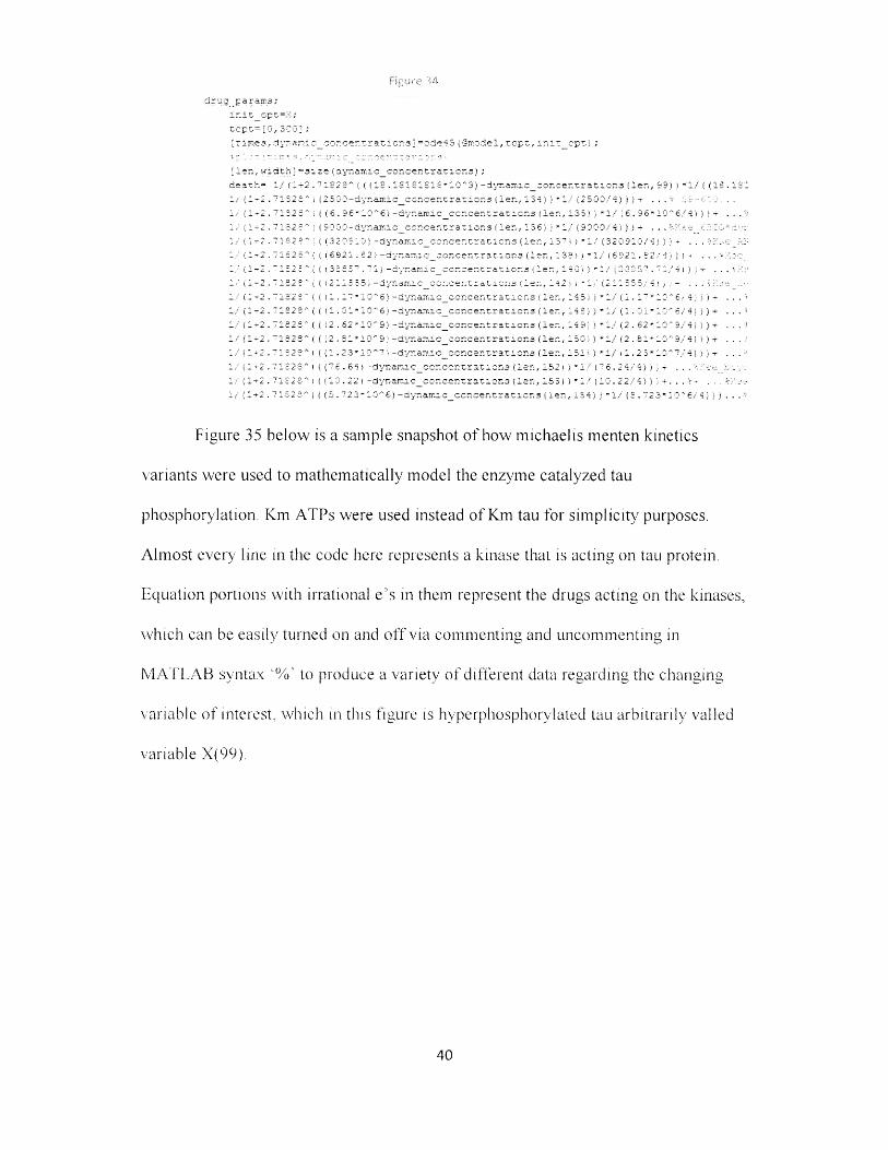
Figure 35 below is a sample snapshot o f how michaelis menten kinetics
variants were used to mathematically model the enzyme catalyzed tau
phosphorylation. Km ATPs were used instead o f Km tau for simplicity purposes.
A lm ost every line in the code here represents a kinase that is acting on tail protein.
Equation portions with irrational e ’s in them represent the drugs acting on the kinases,
which can be easily turned on and o ff via commenting and uncomm enting in
M ATLAB syntax "%' to produce a variety o f different data regarding the changing
variable o f interest, which in this figure is hyperphosphorylated tau arbitrarily vailed
variable X(99).
40

dX 99) = 0 . 0 1 : " M _ P. ( 1 0 0 ) ' ( ( (0 . OO017 0 3 5 * 3 ( 1 0 2 ) ) / ( 1 . 4 * i O 3 - X ( 1 0 0 ) ) ' (
* <■-1. 0 9 7 9 7 5 7 7 95 * _ (1 r * •*; ’“j 4*s3-KX (1 0 0 ) ) ) • %r:x; X.l
* ( 0 . 0 0 0 0 0 1 5 9 2 : iL * : .... . 1 2 1 " (19 * : ^ 3 - (1 0 0 ) ) ) ■ 7 ox XXX: , ::
- (0 . 3 2 1 4 5 5 - : - 2 _ / 1 4 . T* i : - 3 2 i ) ) * ( 1 / (2 ( ( • ( 1 3 5 ) - 4 4 ) * (1 / ( 9 4 / 4 ) )
- ( o . 3 0 l i * ; ; ( i 0 3) / 5 . 4 " .10 ' 3 - ( - 1 3 t * (1 / (--■: ~ ( (X ( 1 3 9 ) - 9 0 * ( 1 / ( 9 0 / 4 ) ) ) )
- ( (0 . 0 0 3 2 * ( 1 0 4 ) ; / (1 • 7 * .2 ' 5 — c. u o 0 J ) ) * ( 1 / (1 -*7 ’” ( ( ■ (04 7) - 1 0 0 0 ) * ( 1 / ( 1 0 0 0
-r ( 0 . 356 3 ’ 1 i 13 5 ) : / ( - * 13 ' 3 - i i : 0 1 ’ (2 / (1 ■" 1. ! 1. - 9 0 * ( 1 / ( 9 0 / 9 ) ) ) i
- i (0 . 0 1 3 3 ’ i - 0 6 ) / (1 - 9 * 3 ' 2 - (12 3 1 i 3 " ( 1 / (2 ( 1 1 _ _ . - ) - 9 0 ) * (1 / ( 9 0 / 4 ) )
T !' f 2 0 02 3 6 *: i 1 0 7 ’ 1 / I 3 9 0- 1 i o 0 i I 1* ( 1 / ( 2 - [ l . J i ) - 9 U ) * ( 1 / ( 9 0 / 4 ) 3 3) 3 3-r i 2 2 - ' W 2 1 2 = 5 * l . 12 2 0 - i 1 2 0 1 ) ) - (1 7 ( 1 ’ ' ( ( X ( 1 4 2 ) - 6 6 0 ) * ( 1 / ( 6 6 0 /- ! 1 I : : E 2 _ 32 - c~ ’ 1 _ 2 9 1 . t 5 5 0 3 - i 10 2 ) ) } " ( 1 / ( I - - - ( (X ( - 4 2 - 6 6 0 ) * ( 1 / ( 6 6 0
- 1 O . ■ n n f 7 -- <= p 5 9 ' X 1 1 0 ) ) / (1 6 0 0-r X ( 1 0 0 ) ) 1 7 W l e c a i . ::::
-r ( o . ~ " ~2 a ’ I - 1 ) ) ' 3 —: 1- 2 2 ) 1 ) 07 X 2 27 1:: 7
- ( o . : - - 12 } ) 7 I E - ’" 11 2 0 1 ) ) X :: k £.1CX:5.
- I tx-! u . j *5 4 ~j a .30 2 3 * ; . ( 1 1 3 ) }, ( 1 6 0 0 2 ■*■ 1 - 3 0 ) 5 ) * (1 / ( 1 - e ~ ( ( X ( 1 9 . } - S O ) * ( 1 / ( 8 0 /
T ( (0 0 1 5 7 5 3 1 2 59* ( 1 - 4 ’• ) .• t ~ ~ 0 0 - 1- - ■) > ) * ( 1 / ( 1 - - 7 ’" ( ( X ( 1 4 5 - S O ) * ( 1 / ( 5 0 / 9— t o - 3 0 5 0 5 7 4 " ■■ - * V 11 5 t / 9 2 2 0 - 1 _ 2 0 ! ) )- t o . * 3 0 3 5 2 5 3 3 5 3 * i; 2 1 6 1 ! / e " 0 0 0 -r.:, 1- 3 2 ) ) } ■ •- ( 0 . 0 02 3 5 - 2 * 1 ^ * _ _ ■ i ) / 3 EE^E 2 - i - O O ) ) ) ■— ( 0 . : 0 5 5 5 ’ l S . | I - 7 2 ) 1 ) . . ,-* { (0 0 0 65 6 '..,. U - 9 j j / ( 4 “1 ’J !- '• d o 3 } ) ) * ( 1 / f 1 - ( (X ( 1 5 2 ) - 2 S 1 * ( 1 / (2 5 1 / 4 ) ) )
- ( (0 01 : 0 E c 7 52 S - ’ 1 - - 1 2*10 5 - r 11 _ 1 i i ) * ( 1 / ( 1 ^ " ( C ( 1 3 5 ) - 1 3 0 ) * ( 1 .
- ( (0 *1 r, 7 ~ 5 p 7 2 9 9* ( - - - i - 2 * 10 1 * ( _ 4 0 1 t ) * ( 1 / ( 1 ^ ( ( ) ( 1 3 5 ) - 1 3 0 } * (1.
- { ( 0 . 0 -S I60 I3 -J4S6 ' - " i l ( 1 1 . 6 V C 0-r , ( 3a) } } "> : (99 j ;
To control the parameters o f the plots generated from the combined death
function equations and michaelis kinetics equations incorporated into the code, an
advanced plotting tool seen in Figure 35 was utilized, as seen partially in the snapshot
Figure 36 below. The “+, o, *, etc” are all strings used to denote different drugs,
assigned arbitrarily as the plotter receives them, allowing us to distinguish one drug
from another drug. The most important portions o f this code are the resolution and the
range.
The resolution is essentially the number o f points that are plotted,
appropriately giving enhanced resolution when this num ber is increased. This is
especially important for plots which do not appear contiguous upon initial inspection,
the resolution can be increased to reveal the activity o f the drug in areas unclear on the
graph. The catch here is that for every order o f magnitude one increases resolution, the
computational time for generating the plot o f interest increases proportionally. This
41

leads to some intensely long sessions o f plotting just to make sure some drugs do not
have extraneous local minimum cell death Y-axis values that do not initially appear.
Figure 36
X , ' p
xx f 3 5 , * d 5 , ' '
re3ciutron=1000;val_r r . ul t ip l i5r=l i ! ;3pace (0, n , r e so lu t i o n ) ; Fxrxx range :x rx,;X.: r c 1 r er X _ c= ze rc 3 ( r e s o l u t i o n ,15);Y_ g= ze r os ( r e so lu t i o n , 15);x v a lu e s = l i n s p a c e (0 ,50000 , r e s o l u t i o n ) ; xccn: fo r i = (-4j
for j = l : r e s o l u t i o nxva l*va l r e u l t i p l i e r ( j )*drug i n i t i a l s ( i ) ; I r a a l a >rralue.e t a aa a or:■IX''al-~'X'xalues •: j ) ;
drug used=drug params;drug u s e d (i ) =xva l ;yval=death_cutput(drug u se d ) ;X _g( j , i )=x va l ;Y_g(1 , i ) =yva l ; ox o x;; ■ =xr"-~ 1;VY a i a l . ;
legend e n t r y ( i ) = p lo t (X g (j , i ) , Y_g (j , 1 ; , s t y l e ( i ) ,* c o l o r c o l o r s ( i , : ) ) ;o o:aoo: a or:ox: xr ae
The range is simply computed using “xvalues=linspaee(0 ,15000,resolution)”
but can also be computed using “val_multiplier=linspaee(0,7,resolution);” , which
essentially automatically chooses a range from 0 to a multiplier o f the d rug’s IC50
value, in this case being 7 times. Changing the 0 and 15000 values in the first case
simply determines the concentration o f the drug on the X-axis. This allows one to
instantly locate the region o f the graph where the supposed LD50 value is and make
sure it is behaving properly. The range also works in tandem with the resolution, for
42
I r: .1:: r i X c ;i
er.dend

when the range is shortened more data can be plotted without changing the resolution
to a higher, computationally expensive value. The key here is to use both the
resolution and the range as efficiently as possible to find apoptosis minima.
43

RESULTS
The following Figures 37 - 58 are the results gathered from M ATLAB plotting
functions. Figures 37 and 38 show constructed healthy and diseased states, while
Figures 39 - 57 show graphs o f the selected drugs with concentration versus apoptosis
relative value. Figure 58 is essentially a table with all o f the important data tabulated
and further discussed in the discussion section.
Figure 37: Healthy State
4 0 0 0
2000
1 5 0 0
1000
5 0 0
00 5 0 10 0 15 0 2 0 0 .250 300
44

Figure 38: Diseased State
1 6 0 0 0 r 1 1 t-
1 4 0 0 0 -
12000 ‘
10000 "
8 0 0 0 -
6 0 0 0 -
4 0 0 0 -
2000 =■—— — — — —..........................
0 5 0 100 1 5 0 2 0 0 2 5 0 00 0
Drugs Plotted with Concentration(nM ) versus Apoptosis
SP-600125 Plots
Figure 39: SP-600125 Concentration Range 0:700 nM (~7x IC50)
0 .1 7 6 5
0 .1 7 5 5
0 .175
0.174
0 1 735
0 ,173
s 172 5
500 600 700200 300 4001000
45

Figure 40: SP-600125 Concentration Range 210:235 nM with minimum
0 .1 7 2 3
0 .1 7 2 3
0 1 7 2 3
0 .1 7 2 3
++ +
+ ++
+++ +
”+ + + + + B W .+ + + '_+ i
X: 2 2 5 .1 3 5 1 3 5 1 3 5 1 3 5 Y: 0 .1 7 2 3 1 6 5 5 6 1 6 2 8 3 8
0 .1 7 2 3
0 .1 7 2 3
0 .1 7 2 3210 2 1 5 220 2 2 5 2 3 0 2 3 5
(R)-CR8 Plots
Figure 41: (R)-CR8 Concentration Range 0:900 nM (~7x IC50)
0 .1 7 6 3
0 .1 7 6 3
0 .1 7 6 3
0 ,1 7 6 3
0 .1 7 6 3
'1 7 6 3
0 .1 7 6 3
0 .1 7 6 3
1763
L 1763100 2 0 0 300 4 0 0 500 6 0 0 7 00 800 900 10000
46

Figure 42: (R)-CR8 Concentration Range 360:430 nM with minimum
0 .1 7 6 3
0 .1 7 6 3
0 .1 7 6 3
'.1 7 6 3
0 .1 7 6 3
1.1763X: 4 0 0 .8 0 0 8 0 0 6 0 0 8 0 1 Yi 0 .1 7 6 3 0 6 6 1 8 5 3 0 9 6 9
0 .1 7 6 3
36 0 37 0 3 8 0 3 9 0 4 0 0 4 1 0 4 2 0 4 3 0
6-BIO Plots
Figure 43: 6-BIO Concentration Range 0:35 nM (~7x IC50)
0 .1 7 6 3 ~
0 .1 7 6 3 ~
0 .1 7 6 3 -
0 1 7 6 3 -
0 .1 7 6 3 -
0 .1 7 6 3 '
0 .1 7 6 3 ~
0 .1 7 6 3 -
0 .1 7 6 3 -
0 . 1 7 6 3 -------------------- 1--------------1-------------------- 1------------------1------------------ 1------------------1------------------ 10 5 10 1 5 2 0 2 5 3 0 3 5
47

Figure 44: 6-BIO Concentration Range 9.8:10.7 nM with minimum
0.1763
>.r
1 1 7 6 3
.1 7 6 3
0 . 1 7 6 3 -
0 .1 7 6 3
X: 10.3003003003003 Y: 0.176311216454931
J ___________ L. J ------------------1____________ i___________ L.
9 .9 1 0 1 0 .1 1 0 .2 1 0 .3 1 0 .4 1 0 .5 1 0 .6 1 0 .7
AR014418 Plots
Figure 45: AR014418 Concentration Range 0:800 (~7x IC50)
0 .1 7 6 3
0 ,1 7 6 3
0 .1 7 6 3
0 .1 7 6 3
0 .1 7 6 3
0 .1 7 6 3
0 .1 7 6 3
0 .1 7 6 3
0 .1 7 6 3
0 .1 7 6 3
0 .1 7 6 30 100 200 300 500 600 700 800
48

Figure 46: AR014418 Concentration Range 220:245 nM with minimum
0 .1763 -
0 .1763 *
0 .1763 -
0 .1763 -
0 .1763 ~
0 .1763 -
0 .1763 "
0 .1763 -
0 .1763 -
0 .1763 -
220 225 230 235 240 245
X: 2 2 8 .8 2 0 8 2 0 8 2 0 8 2 1 Y: 0 .1 7 6 3 1 0 9 9 1 1 4 9 1 0 3
SB-239063 Plots
Figure 47: SB-239063 Concentration Range 0:4500 (not effective)
0.22
0.21
0.2
0.18
0.16
0.15
0.14
0.13
0.12
3500 4 0 0 0 4 5 0 030001500 2 0 0 0 250010005000
49

Paclitaxel Derivative (Hypothetical) Plots
Figure 48: Paclitaxel Derivative Concentration Range 0:8000 (~x25 IC50)
D. 177
0 .1 7 6
(.175
(.174
(.173
0 .1 7 2
0 ,1 7 1
'.1 7
0 .1 6 90 1 0 0 0 2 0 0 0 3 0 0 0 4 0 0 0 5 0 0 0 6 0 0 0 7 0 0 0 8 0 0 0
Figure 49: Paclitaxel Derivative Concentration Range 2650:2900 with minimum
0 .1 6 9 3
0 .1 8 9 3
0 .1 8 9 3
0 .1 6 9 3
0 .1 6 9 3
0 .1 8 9 3
0 .1 6 9 3
0 .1 6 9 3
0 .1 8 9 3
0 .1 6 9 3
0 .1 6 9 3
□X: 2 7 6 9 .5 1 9 5 1 9 5 1 9 5 2
Y: 0 .1 6 9 3 0 4 6 0 6 9 5 5 8 8 3
2 6 5 0 2 7 0 0 2 7 5 0 2 8 5 0
50

Heparin Plots
Figure 50: Heparin Concentration Range 0:7000
0 ,1 7 6 4
0 .1 7 6 3
0 .1 7 6 2
0 .1761
>176
0 .1 7 5 9
0 .1 7 5 8
1 7 5 70 1000 2000 3000 4 0 0 0 5000 6000
Figure 51: Heparin Concentration Range 1650:2150 with minimum
0 .1 7 5 7
0 .1 7 5 7
0 .1 7 5 7
0 .1 7 5 7
0 .1 7 5 7
0 .1 7 5 7
0 .1 7 5 7
0 .1 7 5 7
0 .1 7 5 7
X 1 8 7 0 .8 7 0 8 7 0 8 7 0 8 7 Y: 0 .1 7 5 7 4 2 7 7 4 8 8 4 0 2 9
1 6 5 0 1 7 0 0 1 7 5 0 1 8 0 0 1 8 5 0 1 9 0 0 1 9 5 0 2 0 0 0 2 0 5 0 .2100 2 1 5 0
51

Harmine Plots
Figure 52: Harmine Concentration Range 0:5000
0 .1 7 6 3 r
0 .1 7 6 3
0 .1 7 6 2
L 1 7 6 2
0 .1 7 6 2
0 .1 7 6 2
0 ,1 7 6 2
0 .1 7 6 1
0 1000 2000 2 5 0 0 3 5 0 0
Figure 53: Harmine Concentration Range 200:380 with minimum
0 .1 7 6 1
0 .1 7 6 1
0 .1 7 6 1
0 .1 7 6 1
0 .1 7 6 1
0 .1 7 6 1
0 .1 7 6 1
0 .1 7 6 1
0 .1 7 6 1
v< '
X: 287.387387387387 Y: 0.176124126830265
2 0 0 2 2 0 2 4 0 2 6 0 2 8 0 3 0 0 3 2 0 3 4 0 3 6 0 3 8 0
52
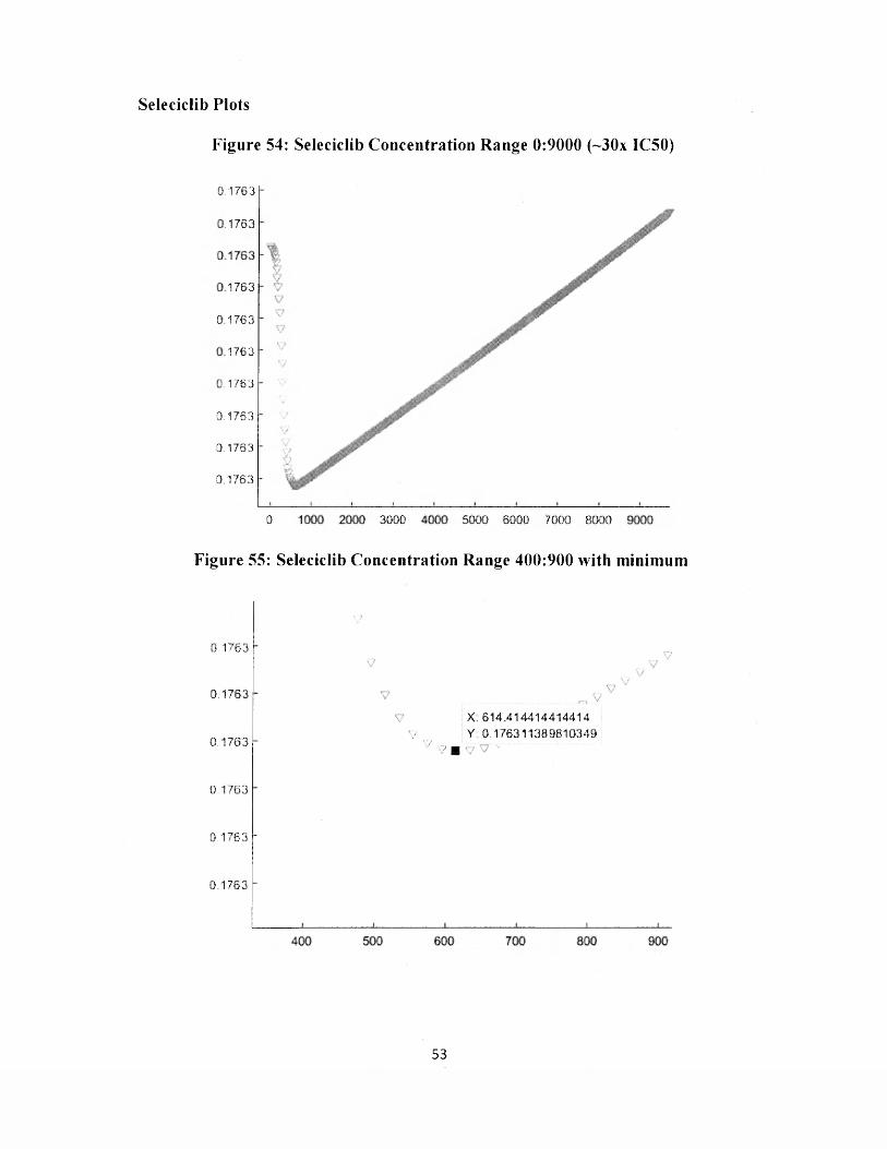
Seleciclib Plots
Figure 54: Seleciclib Concentration Range 0:9000 (~30x IC50)
0 .1 7 6 3
L1763
1 7 6 3
0 .1 7 6 3
1 7 6 3
M 7 6 3
0 .1 7 6 3
0 .1 7 6 3
0 .1 7 6 3
0 3 0 0 0 5 0 0 0 6 0 0 0 7 0 0 0 8 0 0 0
Figure 55: Seleciclib Concentration Range 400:900 with minimum
0 .1 7 6 3
0 .1 7 6 3
0 .1 7 6 3
0 .1 7 6 3
0 .1 7 6 3
0 .1 7 6 3
X; 6 1 4 .4 1 4 4 1 4 4 1 4 4 1 4
V r„. V : 0 .1 7 6 3 1 1 3 6 9 8 1 0 3 4 9V , 7 m v...
53

Brysostatin Plot (High Toxicity Plot)
Figure 56: Bryostatin Concentration Range 0:2000
0 0 0 0 0 0 0 0 0 0 0 0 0 0 0
1
9
8
0 .7
6
5
0 .4
3
2
1
0 200100 3 0 0 4 0 0 6 0 0 7 0 0
Alpha BTX Plot (Toxin Plot)
Figure 57: Alpha BTX Concentration Range -80:60
1 3
1.2 -
1.1-f
1 - +
0 .9 +
0 .8 - +
0 ,7 +•
0 .6 - +
0 .5 - ++
0 .4 +jr
■ff0 .3 -
■gUttHttHHii i ii 11 il I {_
-60 -40 -20 20 40 60
54
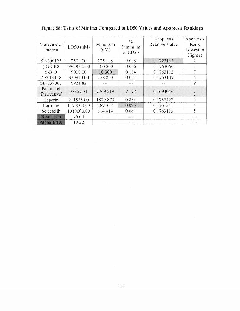
Figure 58: Table of Minima Compared to LD50 Values and Apoptosis Rankings
%Minimum o f LD50
Apoptosis ApoptosisM olecule o f
LD50 (nM)M inimum Relative Value Rank
Interest (nM) Lowest to Highest
SP-600125 2500.00 225.135 9.005 0.1723165 2(R)-CR8 6960000.00 400.800 0.006 0.1763066 5
6-BIO 9000.00 10.300 0.114 0.1763112 7AR014418 320910.00 228.820 0.071 0.1763109 6SB-239063 6921.82 _ _ _ _ _ _ — 9Paclitaxel
4 Derivative ’38857.71 2769.519 7.127 0.1693046
1Heparin 211555.00 1870.870 0.884 0.1757427 3Harmine 1170000.00 287.387 0.025 0.1761241 4
Seleciclib 1010000.00 614.414 0.061 0.1763113 8Brvostatin 76.64 _ _ _ _ _ _ _ _ _
Alpha-BTX 10.22 - - - _ _ _ — —
55

DISCUSSION
Figures 37 and 38 show the theoretical constructed healthy and disease states
respectively. Everything is within a few order o f magnitudes, which is accurate for
resting concentrations o f molecules within the cell. The disease state shows the value
o f hyper tau to be over almost 15x the value o f hyper tau in the normal state, at a
resounding 15.181818 nM. This is the hyper tau increase which is responsible for the
neurofibrillary tangles.
Figures 39 through 57 show the apoptosis versus concentration plots for each
drug, starting from relative apoptosis value at approximately 0.176325. This value is a
relatively arbitrary value used as a value that seems to make simple sense. All the cells
cannot die, but a large portion must die for the memory loss to occur. 0.176325, being
between 10 and 20% is a good estimation, seeing that AD brains shrink by
approximately that percentage compared to non-diseased patient brains. A tedious
algorithm o f finding the minimum apoptosis value was enacted via running the code
on different concentration ranges at varying resolutions to find the best minimum.
After many hours o f generating plots using some o f the highest resolutions that even a
computer with 16 individual core processors can handle, the minima were all located
on the drugs which worked and were tabulated in Figure 58.
Figure 58 shows each molecule o f interest’s determined minimum
concentration (X-coordinate) and database LD50 value. It also shows the apoptosis
m inimum value (Y-coordinate). None o f the apoptosis values diminish by a great
percentage. Some appear to not shrink at all from the mtitial 0.176325 value upon
56

initial inspection. This is because some o f our toxicity levels, tau in particular, are set
to counter the effects o f many drugs in tandem, not individually. This is a great way to
discover the drugs which do not work the best, seen by SB-239063 in Figure 47
because it does not even have a value that begins to decrease apoptosis under the
computationally imposed toxicity stresses. Thus the drugs do not appear effective
numerically until the differences between the values are taken into consideration;
everything is relative here. Additionally, rankings are assigned based on the lowest
and highest resultant apoptosis values.
The green shaded values under each column represent the best value in that
column. In the M inim um column, the green shaded value represents the lowest drug
concentration used. The shaded green values under the % M inim um o f LD50 column
represent the drug effectiveness in that those drugs shaded were effective at reducing
apoptosis with very low concentration percentage o f their LD50 value.
Paclitaxel is shaded blue because I wanted to implement a random neutral
hypothetical molecule into the model. Ignoring its potential therapies as a microtubule
stabilizer, if Paclitaxel was an inhibitor instead o f an activator, the values generated
are reported in Figure 58 The lowest apoptosis value mean that a very similar
derivative that functions oppositely to Paclitaxel may prove therapeutic regarding the
parameters and considerations o f this study. Essentially, this is demonstrating the
power o f BST m odeling and endless hypothetical possibilities that can be incorporated
into future work.
Alpha-BTX was incorporated into the model to show that it is indeed a toxin
and also to portray what the graph o f a toxin would look like compared to the other
57

drugs, as seen in Figure 57. Similarly, however, bryostatin 1 in Figure 56 shows
reverse activity, despite it being a potent PKC inhibitor. This is due to its very small
LD50 value, a value that is only lOx the LD50 o f A lpha-BTX at 77nM. Essentially the
cell dies before the therapeutic effects take place, so it portrays behavior similar to a
true toxin. Alpha-BTX and Bryostatin 1 and accordingly both shaded red in Figure 56,
m eaning that they are not therapeutic in the slightest.
The best mathematical solution to the problem o f A lzheim er’s disease
apoptosis presented by this model and set o f system o f differential equations is SP-
600125. It simply has the best apoptosis relative value, excluding hypothetical
paclitaxel derivative. Even though its % M inim um o f LD50 is the worst relatively, it is
still below 10% o f the LD50 value, coming in at 9.005. The second best choice is
Heparin, which has the second best apoptosis relative value excluding hypothetical
paclitaxel derivative, and interestingly enough, has one o f the lower % M inimum of
LD50 values. An alternative, if one is only interested in % o f LD50, is Harmine, as it
achieves the third lowest apoptosis relative value excluding hypothetical paclitaxel
derivative while maintaining the lowest % o f ED50 concentration required to achieve
said minimum.
58

CONCLUSION
Using biological systems theory (BST), it has been confirmed that most o f
these drugs perform their preconceived functions regarding inhibition and activation
o f tau protein kinases. It has been concluded that SP-600125 is the best mathematical
choice for A lzheim er’s disease therapy. It has also been concluded that Heparin and
Harmine are the alternative situational mathematical choices for A lzheim er’s disease
therapy. It has additionally been concluded that bryostatin is a pseudo toxin,
resembling alpha-BTX in that it is too toxic to portray therapeutic results
mathematically. It has also been concluded that SB-239063 is the worst choice for a
drug that does not act as a pseudo toxin.
Methods such as those conducted by this research group utilizing BST are
providing the essential theoretical information to start cutting edge research projects
around the globe in effort to slow the progression if find the elusive cure to this deadly
disease, as it can be one o f the worst to watch happen. It can occur very early in life
relative to the current life expectancies o f our era, and last for a long time thereafter,
proving challenging for caregivers. Caregiving is the last resort after treatments and
other m anagement techniques are applied. In terms o f medicated prevention, no
medication has been proven statistically, totally effective, resulting in alternative
methods, ranging from mentally stimulating activities to ultrasound technology.
59

VITA
Patrick Neil Blank
Patrick Neil Blank was born in Philadelphia, Pennsylvania on July 26, 1991.
He graduated from The Collegiate School o f Richmond, Virginia in June o f 2010 and
went on to study chemistry and mathematics at The College o f William and Mary. He
earned his B.S. in Chemistry in December o f 2013. He continued studying chemistry
at The College o f William and Mary, and defended his m aster’s thesis on May 8th,
2015. He will begin further studying o f chemistry at The University o f Pennsylvania
in August o f 2015. Essentially, he will be returning to his roots.
60

BIBLIOGRAPHY
References Cited:
(1) Abraham RT1, Acquarone M, Andersen A, Asensi A, Belle R, Berger F,
Bergoumoux C, Brunn G, Buquet-Fagot C, Fagot D, et al. (1995). “Cellular
effects o f olomoucine, an inhibitor o f cyclin-dependent kinases” Biol Cell.
83(2-3): 105-20.
(2) Alzheimer's Disease — History & Description. (2008). Retrieved April 14,
2013, from http://www.best-alzheimers-products.com/alzheimer's-disease.html
(3) Association, A. (n.d.). Seven Stages o f Alzheimer's. Retrieved from
https://www.alz.org/alzheimers_disease_stages_of_alzheimers.asp
(4) Bahr, B. A. (2003). Focus on Alzheimer's Disease Research (E. M. Welsh,
Ed ). Nova Biomedical Books.
(5) Borgne A, Meijer L 1, M ulner O, Chong JP, Blow JJ, Inagaki N, Inagaki M,
Delcros JG, M oulinoux JP. (1997). “Biochemical and cellular effects o f
roscovitine, a potent and selective inhibitor o f the cyclin-dependent kinases
cdc2, cdk2 and cdk5.” E u rJ Biochem.243(1 -2):527-36.
(6) Cole, Adam. Soutar, Marc P. M. (2008). “Relative Resistance o f Cdk5-
phosphorylated CRM P2 to Dephosphorylation.’V Biol Chem. 283(26). 18227-
18237.
(7) Eldar-Finkelman H, Martinez A (201 1). "GSK-3 Inhibitors. Preclinical and
Clinical Focus on CNS" Front Mol Neurosci 32(4).
61

(8) Embi N, Rylatt DB, Cohen P (June 1980). "Glycogen synthase kinase-3 from
rabbit skeletal muscle. Separation from cyclic-AM P-dependent protein kinase
and phosphorylase kinase" Ear J Biochem 107 (2): 519-27.
(9) Gong, C-X; Iqbal, K. (2008) “Elyperphosphorylation o f Microtubule-
Associated Protein Tau: A Promising Therapeutic Target for Alzheimer
Disease.” Current Medicinal Chemistry 23(15): 2321-8.
(10) Hirokawa, N. (1988). Tau Proteins: The Molecular Structure and Mode o f
Binding on Microtubules. Journal o f Cell Biology, 107, 1449-1459.
(11) Leclerc, Sophie (2000). Indirubins Inhibit Glycogen Synthase Kinase-3(3 and
CDK5/P25, Two Protein Kinases Involved in Abnormal Tau Phosphorylation
in Alzheimer's Disease. The Journal o f Biological Chemistiy, 276, 251-260.
(12) Lichtenthaler, S. F. (2010). Alpha-Secretase in Alzheimer's Disease:
molecular identity, regulation and therapeutic potential. Journal o f
Neurochemistry, 116(1).
(13) M andelkow, E M , & Mandelkow, E. (2012). Biochemistry and Cell
Biology o f Tau Protein in Neurofibrillary Degeneration. Cold Spring Harbor
Laboratory Press, 1-25.
(14) Martinez, Ana*; Castro, Ana; Dorronsoro, Isabel. Glycogen Synthase Kinase
3 (GSK-3)Inhibitors as N ew Promising Drugs for Diabetes,Neurodegeneration,
Cancer, and Inflammation.
(15) Meijer 1, A-M W H Thunnissen2*, AW White3, M Garnier 1, M Nikolic4f,L-
H Tsai4, J Walter5, KE Cleverley6, PC Sahnas6, Y-Z Wu7, J Biernat7, E-M
62

M andelkow7, S-H Kim2 and GR Pettit3. Inhibition o f cyclin-dependent
kinases, GSK-3b and CK1 by hymenialdisine, a marine sponge constituent.
(16) M ichala Kolarova, Francisco Garcia-Sierra, Ales Bartos, Jan Ricny, and
Daniela Ripova.(2012) Structure and Pathology o f Tau Protein in Alzheimer
Disease International Journal o f Alzheimer's Disease, 1-13.
(17) Pimplikar, S. W., Nixon, R. A., Robakis, N. K., Shen, J., & Tsai, L.-H.
(2010). Amyloid-Independent M echanisms in Alzheimer's Disease
Pathogenesis. The Journal o f Neuroscience, 30(45), 14946-14954.
(18) Sausville, Edward A. (2002) “Complexities in the development o f cyclin-
dependent kinase inhibitor drugs.” Trends in Molecular Medicine. 8:S32-S37
(19) Selemca M. L., Jensen H. S., Larsen A. K., Pedersen M. L., Helboe L., Leist
M., Lotharius J. (2007). Efficacy o f small-molecule glycogen synthase kinase-
3 inhibitors in the postnatal rat model o f tau hyperphosphorylation. Br. J
Pharmacol. 152, 959-979.10.1038
(20) Tsuji S, M orinobu S, Tanaka K, Kawano K, Yam awaki S. (2003). “Lithium,
but not valproate, induces the serine/threonine phosphatase activity o f protein
phosphatase 2A in the rat brain, without affecting its expression.” J Neural
TransmA 10(4) :413-25
(21) Villerbu N l , Gaben AM, Redeuilh G, Mester J. (2002).“ Cellular effects o f
purvalanol A: a specific inhibitor o f cyclin-dependent kinase activities. Int J
Cancer. 97(6) 761 -9
63

(22) Xu Y, Xing Y, Chen Y, Chao Y, Lin Z, Fan E, Yu JW, Strack S, Jeffrey PD,
Shi Y (2006) “ Structure o f the protein phosphatase 2A holoenzyme.” Cell.
127(6): 1239-51
(23) Yan Xiong, Xiao-Peng Jing, Xin-W en Zhou, Xiu-Lian Wang, Yang Yang,
Xu-Ying Sun, Mei Qiu, Fu-Yuan Cao, Y ou-M ing Lu, Rong Liu, Jian-Zhi
Wang, Zinc induces protein phosphatase 2A inactivation and tau
hyperphosphorylation through Src dependent PP2A (tyrosine 307)
phosphorylation, Neurobiology o f Aging, Volume 34, Issue 3, M arch 2013,
Pages 745-756, ISSN 0197-4580
(24) Yvette M ettey,f, Marie Gompel,{, Virginie Thom as,J , Matthieu G am ier,J ,
Maryse Leost,{, Irene Ceballos-Picot,§, Martin N oble,± , Jane Endicott,_i_,
Jean-michel Vierfond,f and, and Laurent Meijer*,J (2003). “Aloisines, a N ew
Family o f CDK/GSK-3 Inhibitors. SAR Study, Crystal Structure in Complex
with CDIC2, Enzyme Selectivity, and Cellular Effects ”Journal o f Medicinal
Chemistry. 46(2), 222-236
64