Embed Size (px)
Citation preview
1
Nanofibrillar cellulose hydrogels and reconstructed hydrogels as matrices for controlled drug
release
Heli Paukkonena, Mikko Kunnaria, Patrick Lauréna, Tiina Hakkarainena, Vili-Veli
Auvinena,b,
, Timo Oksanena, Raili Koivuniemia, Marjo Yliperttulaa,c, Timo Laaksonena,b
a Division of Pharmaceutical Biosciences, Faculty of Pharmacy, University of Helsinki, P.O. Box
56, FI-00014 Helsinki, Finland
b Department of Chemistry and Bioengineering, Tampere University of Technology, P.O. Box 541,
FI-33101 Tampere, Finland
c Department of Pharmaceutical and Pharmacological Sciences, via Marzalo 5, University of
Padova, Padova, Italy
Abstract
Concentrated 3 % and 6.5 % anionic nanofibrillar cellulose (ANFC) hydrogels were introduced as
matrix reservoirs for controlled delivery applications of small molecules and proteins. A further aim
was to study how the freeze-drying and subsequent rehydration of ANFC hydrogel affects the
rheological properties and drug release of selected model compounds from the reconstructed
hydrogels. It was demonstrated that the 3 % and 6.5 % ANFC hydrogels can be freeze-dried with
suitable excipients into highly porous aerogel structures and redispersed back into the hydrogel form
without significant change in the rheological properties. Freeze-drying did not affect the drug release
properties from redispersed ANFC hydrogels, indicating that these systems could be stored in the dry
form and only redispersed when needed. For large molecules, the diffusion coefficients were
significantly smaller when higher ANFC fiber content was used, indicating that the amount of ANFC
fibers in the hydrogel can be used to control the release rate. The release of small molecules was
controlled with the ANFC fiber content only to a moderate extent. The results indicate that ANFC
hydrogel can be used for controlled delivery of several types of molecules and that the hydrogel can
be successfully freeze-dried and redispersed.
Keywords: Nanofibrillar cellulose, drug release, diffusion, hydrogel, aerogel, freeze-drying,
rheology
Abbreviations: ANFC, Anionic nanofibrillar cellulose; BSA, bovine serum albumin; D, diffusion
coefficient; DSC, differential scanning calorimetry; exp, excipients trehalose and polyethylene
glycol; FITC-DEX, fluorescein isothiocyanate–dextran; KETO, ketoprofen; LZ, lysozyme; MZ,
metronidazole; NAD, nadolol; NFC, nanofibrillar cellulose; PEG, polyethylene glycol; TEMPO,
[(2,2,6,6-Tetramethylpiperidin-1-yl)oxyl]; tre, trehalose
2
1. Introduction
Hydrogels have various uses in biomedical applications and are well suited for regenerative medicine
(Peppas, et al. 2006; Slaughter, et al. 2009) and controlled drug delivery (Gupta, et al. 2002).
Nanofibrillated cellulose (NFC) hydrogel has been widely investigated in pharmaceutical and
biomedical fields, such as in drug delivery (Laurén, et al. 2014), scaffold synthesis (Borges, et al.
2011), and as cell and drug carriers (Thérien-Aubin, et al. 2016; Valo, et al. 2013). NFC hydrogel is
also applicable as a cell culture scaffold for three dimensional cell culturing (Bhattacharya, et al.
2012; Lou, et al. 2014; Lou, et al. 2015; Malinen, et al. 2014). It is produced from the natural
biopolymer cellulose, and wood pulp is a commonly used starting material in the production (Klemm,
et al. 2011). The surface of native NFC fibers can be further chemically modified with e.g. TEMPO
[(2,2,6,6-Tetramethylpiperidin-1-yl)oxyl] oxidation to contain negatively charged carboxyl groups
directly on the fiber surface (Saito, et al. 2006; Saito, et al. 2007). This way, it is possible to produce
anionic NFC (ANFC) fibers. Further, native as well as anionic TEMPO oxidized NFC grades have
been shown to be biocompatible and nontoxic in various in vitro cell models (Alexandrescu, et al.
2013; Bhattacharya, et al. 2012; Hannukainen, et al. 2012; Hua, et al. 2014; Vartiainen, et al. 2011),
making them appealing for pharmaceutical applications.
Since cellulose retains moisture and is both biocompatible (in humans) and biodegradable (in nature),
NFC has been investigated in wound treatment applications facing the need for faster and more
effective wound healing (Chinga-Carrasco and Syverud 2014; Lin and Dufresne 2014; Powell, et al.
2016; Zhang, et al. 2013). Previously, we have indicated the suitability of native NFC wound dressing
in clinical use in treatment of skin graft donor sites of burn patients (Hakkarainen, et al. 2016). NFC
hydrogels are also of special interest in the wound healing applications due to high moisture retaining
ability and biocompatibility (Lin and Dufresne 2014; Zhang, et al. 2013). Furthermore, NFC hydrogel
can be freeze-dried into solid aerogel structure, which may be utilized in drug delivery (Pääkkö, et al.
2008; Valo, et al. 2013). However, it is generally acknowledged that the drying of NFC promotes
irreversible hydrogen bonding between neighboring NFC nanofibrils, known as hornification (Diniz,
et al. 2004; Wan, et al. 2010). So far it has been a major manufacturing challenge to produce dry NFC
while maintaining the nano-scale dimensions, and several techniques such as oven drying, freeze-
drying, supercritical drying and spray-drying have been investigated (Kolakovic, et al. 2012; Peng,
et al. 2012; Peng, et al. 2013). Freeze-drying produces NFC aerogels with lamellar structures and
significant lamellar aggregation (Peng, et al. 2012).
In addition to the preservation of NFC nano-scale dimensions throughout drying process, the
preservation of rheological properties of the NFC hydrogels is important for drug delivery
3
applications. These should stay reasonably similar during processing in order to have reliable,
controlled and predictable drug release formulations. It has been shown that once-dried TEMPO-
oxidized ANFC fibers can be re-dispersed, reversing the hornification effect introduced by the drying,
by involving higher energy consumption (Kekäläinen, et al. 2014). This is probably due to the high
surface charge density present in the TEMPO-oxidized ANFC fibers, which is much lower in the
absence of carboxyl groups on the native NFC fiber surface. Furthermore, the electrostatic repulsion
forces working between the anionically-charged fibers stabilizes the aqueous suspension (Isogai, et
al. 2011). The high surface density of negative charges has been used to electrostatically immobilize
biomolecules (Weishaupt, et al. 2015). Therefore, the high aspect ratio as well as anionic charge of
ANFC fibers can be utilized as rate-controlling parameters in controlled drug release applications.
Rheological properties and redispersability of highly concentrated 3 - 6.5% ANFC hydrogels after
drying and consequent rehydration and re-gelling have not been studied extensively in the literature,
even though these might be especially useful in e.g. wound healing applications. Previous studies
have mainly focused on the evaluation of redispersibility and rheological properties of dilute 0.5 - 1%
NFC suspensions after spray drying or freeze-drying (Missoum, et al. 2012; Žepič, et al. 2014).
Freeze-drying is often used in the manufacturing of solid dosage forms of sensitive protein
pharmaceuticals to improve the storage stability (Carpenter, et al. 1997; Tang and Pikal 2004).
However, cryo- and lyoprotectants are usually required to prevent structural damage of protein
pharmaceuticals induced by freezing and drying steps in the freeze-drying process (Carpenter, et al.
1997; Franks 1998; Hubálek 2003). We studied the prospect that these excipients could retain the
structure of ANFC to a certain extent during freeze-drying. Furthermore, based on the literature
(Kekäläinen, et al. 2014), ANFC seems to be a more suitable grade than native NFC for the
preparation of freeze-dried formulations intended for consequent reconstruction and rehydration, and
was chosen as the hydrogel forming material.
We propose that with regard to preservation of ANFC structure it may be beneficial to use trehalose
to replace the hydrogens bonds that are lost due to the sublimation of water in the freeze drying of
ANFC. The suitability of trehalose as a lyo- and cryoprotectant has already been confirmed in the
freeze-drying of protein pharmaceuticals (Chang, et al. 2005; Jovanović, et al. 2006), red blood cells
(Han, et al. 2005), platelets (Crowe, et al. 2003; Wolkers, et al. 2001) and liposomes (Christensen, et
al. 2007). It has been proposed that the molecular mechanism of protein as well as cell membrane
stabilization with trehalose is achieved through water-trehalose hydrogen-bond replacement, coating
by a trapped water layer and/or mechanical inhibition of the conformational fluctuations (Lins, et al.
2004). Low trehalose concentration of 5-100 mM does not provide cryoprotection for proteins, but in
4
combination with 1% polyethylene glycol (PEG) stabilization can be achieved (Carpenter, et al. 1993;
Prestrelski, et al. 1993). Furthermore, PEG as hygroscopic polymer improves the water uptake (Jeon,
et al. 1991) which can be beneficial for reconstitution and rehydration of ANFC aerogels.
The aim of the current work was to characterize rheological as well as drug release properties of 3 %
and 6.5 % ANFC hydrogels prior to and after freeze-drying and reconstruction. Here, a combination
of trehalose and PEG 6000 was chosen as the cryo- and lyoprotectants in the freeze-drying of ANFC
hydrogels into aerogels in order to minimize the hornification of ANFC fibers and to preserve the
nano-scale structure of ANFC fibers. The freeze-dried aerogel formulations were rehydrated and re-
gelled into their original concentrations in order to reform the hydrogel structure prior to the release
testing and rheological measurements. Model compounds in the release studies included small
molecules metronidazole (MZ), nadolol (NAD) and ketoprofen (KETO) with molecular weight below
500 M. 4 kDa FITC-dextran (FITC-DEX), lysozyme (LZ) and bovine serum albumin (BSA)
represented high molecular weight compounds. The model compounds were selected based on their
size, wieght and different charges at pH 7.
The main release mechanism of small molecules and proteins from hydrogels can be characterized
by Fickian diffusion (Censi, et al. 2012), as the ANFC hydrogel does not exhibit swelling or any other
internal or external triggers for the release in PBS buffer solutions. The diffusion rate of solutes may
be influenced through weak interactions such as electrostatic, hydrophobic and hydrogen bonding
between the hydrogel polymer network and solutes. The permeation of small molecules and proteins
through hydrogels is further affected by their size, shape (Hoffman 2012) and the availability of free
water molecules to hydrate and dissolve the solute molecules in addition to hydrogel pore size, pore
size distribution and the pore interconnections affect the diffusion rate of molecules through the
hydrogel. It is therefore clear that the hydrogel structure plays an important role for the release rate
and must be rigorously controlled.
2. Materials and methods
2.1. Materials
3.2 % (11804-3) and 6.8 % (11815-3) anionic NFC (ANFC) hydrogels (FibDex™) were kindly
provided by UPM-Kymmene Corporation, Finland. Cellulose kraft pulp was chemically modified
and fibrillated to form ANFC hydrogels (Saito, et al. 2006; Saito, et al. 2007). A Carboxylic acid
content 1,06 mmol / g pulp was determined by conductometric titration according to the standard
SCAN-CM 65:02. The diameter of most of the fibrils is in the range of 4–10 nm and length 500 – 10
000 nm measured using electron microscopies. All model compounds and reagents were of analytical
5
grade. Bio-Rad Protein Assay reagent was purchased from Bio-Rad, USA. 4 and 10 kDa FITC-
dextran molecules were purchased from Sigma-Aldrich, Sweden. D-(+)-trehalose dihydrate was
purchased from Sigma-Aldrich, USA. Metronidazole was purchased from Sigma-Aldrich, China.
Nadolol was purchased from Sigma-Aldrich, Finland. Ketoprofen was purchased from Orion Pharma,
Finland. Lysozyme from hen egg white was purchased from Roche, Germany. Polyethylene glycol
6000 was purchased from Fluka, Switzerland. Dulbecco’s Phosphate Buffered Saline (10X)
concentrate without calcium and magnesium was purchased from Gibco, UK. Acetonitrile was of
analytical grade, Sigma-Aldrich, Germany.
2.2. Methods
2.2.1. Preparation of the ANFC hydrogel formulations
ANFC hydrogel formulations were prepared in 10 ml syringes. The hydrogels were homogenized
with model compounds by mixing for 10 min inside two attached syringes (200 times through the
syringe nozzle). Table 1 contains the formulation compositions of the physical mixtures of ANFC
hydrogels and model compounds. The ANFC amount in the final formulations was 3% or 6.5%
(m/m). The measured pH for pure ANFC hydrogels was 7. For MZ, NAD and KETO, an excess
amount of each drug was used in relation to their solubility in pH 7. Therefore, the drug containing
ANFC hydrogel formulations were monolithic dispersions. MZ, NAD and KETO were added as dry
powders into the ANFC hydrogels with final concentrations of 2 % for MZ, 1.7 % for NAD and 3.4
% for KETO. The amount of 4 kDa FITC-DEX, BSA and LZ in the formulations did not exceed the
solubility limit at pH 7. Therefore, these formulations were considered to be monolithic solutions. 4
kDa FITC-DEX was added into the ANFC hydrogel from 1 mg/ml stock solution with a 1 % final
concentration of FITC-DEX. BSA and LZ were added as dry powders into the ANFC hydrogels with
final concentrations of 1 % for BSA and 0.5 % for LZ. The 3 % and 6.5 % ANFC hydrogel
formulations with MZ, NAD, KETO and BSA were also prepared with 1 % of PEG6000 and 0.3 %
of trehalose for freeze-drying. Dilute 1.1% ANFC hydrogels were crosslinked with cations calcium
(Ca2+), aluminum (Al3+) and iron (Fe3+) and were used to study the effect of crosslinking on rheology
and drug release properties with 10 kDa FITC-DEX and MZ (detailed information in supplement).
6
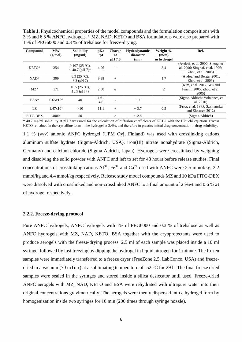
Table 1. Physicochemical properties of the model compounds and the formulation compositions with
3 % and 6.5 % ANFC hydrogels. * MZ, NAD, KETO and BSA formulations were also prepared with
1 % of PEG6000 and 0.3 % of trehalose for freeze-drying.
Compound MW
(g/mol)
Solubility
(mg/ml)
pKa
/pI
Charge
at
pH 7.0
Hydrodynamic
diameter
(nm)
Weight %
(m/m)
in hydrogel
Ref.
KETO* 254 0.107 (25 °C),
~ 40.7 (pH 7)† 4.06 - 3.4
(Avdeef, et al. 2000; Sheng, et
al. 2006; Singhai, et al. 1996;
Zhou, et al. 2005)
NAD* 309 8.3 (25 °C),
8.3 (pH 7) 9.28 + 1.7
(Avdeef and Berger 2001;
Zhou, et al. 2005)
MZ* 171 10.5 (25 °C),
10.5 (pH 7) 2.38 ø 2
(Kim, et al. 2012; Wu and
Fassihi 2005; Zhou, et al.
2005)
BSA* 6.65x104 40 4.6 -
4.8 - ~ 7 1
(Sigma-Aldrich; Yohannes, et
al. 2010)
LZ 1.47x10⁴ >10 11.1 + ~ 3.7 0.5 (Fritz, et al. 1995; Szymańska
and Ślósarek 2012)
FITC-DEX 4000 50 ø ~ 2.8 1 (Sigma-Aldrich)
† 40.7 mg/ml solubility at pH 7 was used for the calculation of diffusion coefficients of KETO with the Hiquchi equation. Excess
KETO remained in the crystalline form in the hydrogel at 3.4%, and therefore in practice initial drug concentration ˃ drug solubility.
1.1 % (w/v) anionic ANFC hydrogel (UPM Oyj, Finland) was used with crosslinking cations
aluminum sulfate hydrate (Sigma-Aldrich, USA), iron(III) nitrate nonahydrate (Sigma-Aldrich,
Germany) and calcium chloride (Sigma-Aldrich, Japan). Hydrogels were crosslinked by weighing
and dissolving the solid powder with ANFC and left to set for 48 hours before release studies. Final
concentrations of crosslinking cations Al3+, Fe3+ and Ca2+ used with ANFC were 2.5 mmol/kg, 2.2
mmol/kg and 4.4 mmol/kg respectively. Release study model compounds MZ and 10 kDa FITC-DEX
were dissolved with crosslinked and non-crosslinked ANFC to a final amount of 2 %wt and 0.6 %wt
of hydrogel respectively.
2.2.2. Freeze-drying protocol
Pure ANFC hydrogels, ANFC hydrogels with 1% of PEG6000 and 0.3 % of trehalose as well as
ANFC hydrogels with MZ, NAD, KETO, BSA together with the cryoprotectants were used to
produce aerogels with the freeze-drying process. 2.5 ml of each sample was placed inside a 10 ml
syringe, followed by fast freezing by dipping the hydrogel in liquid nitrogen for 1 minute. The frozen
samples were immediately transferred to a freeze dryer (FreeZone 2.5, LabConco, USA) and freeze-
dried in a vacuum (70 mTorr) at a sublimating temperature of -52 °C for 29 h. The final freeze dried
samples were sealed in the syringes and stored inside a silica desiccator until used. Freeze-dried
ANFC aerogels with MZ, NAD, KETO and BSA were rehydrated with ultrapure water into their
original concentrations gravimetrically. The aerogels were then redispersed into a hydrogel form by
homogenization inside two syringes for 10 min (200 times through syringe nozzle).
7
2.2.3. Scanning electron microscopy (SEM)
The morphology of the freeze-dried aerogels was imaged with a scanning electron microscope Quanta
FEG250 (SEM, FEI Company, USA). The aerogels were fractured manually for analysis of the inner
aerogel structure or cut with a scalpel for the analysis of the aerogel surface structure. Micrographs
of the cross-sections as well as surface structures of the aerogels were obtained. Before imaging the
samples were fixed onto a two-sided carbon tape and sputtered with platinum for 25s with an Agar
sputter device (Agar Scientific Ltd., UK).
2.2.4. Thermogravimetry analysis (TGA)
The residual moisture content of the aerogels was determined using TGA (TGA 850, Mettler-Toledo,
Switzerland). Samples were heated from 25 to 240°C at a heating rate of 10°C/min in nitrogen (40
ml/min) atmosphere. Residual moisture content of the aerogels was determined as a mass loss (%) of
evaporated water. TGA data was gathered from single measurement.
2.2.5. Differential scanning calorimetry (DSC)
Thermal analysis of the freeze dried ANFC aerogels were carried out using a differential scanning
calorimeter Mettler Toledo DSC 823e (Mettler Toledo, Giessen, Germany). The samples were placed
in sealed aluminum pans with closed lids and heated at a scanning rate of 10 °C/min between 25 and
200 °C in nitrogen atmosphere. The data was analyzed with STARe software (Mettler-Toledo,
Giessen, Germany). All DSC measurements were performed after one month storage period inside a
silica desiccator at 20 ⁰C. DSC data was gathered from single measurement.
2.2.6. Rheological measurements
The rheological measurements were performed at 37°C with HAAKE Viscotester iQ Rheometer
(Thermo Fisher Scientific, Karlsruhe, Germany) equipped with a Peltier system for temperature
control. Results were analyzed with HAAKE RheoWin 4.0 software (Thermo Fisher Scientific).
Parallel 35 mm diameter steel plate-and-plate geometry was used with a 1 mm gap in all
measurements. Before each measurement, the samples were allowed to rest for 5 min at 37°C.
Controlled stress amplitude sweeps were performed to determine the linear viscoelastic region for
different ANFC hydrogel formulations. Constant angular frequency ω = 1 Hz and oscillatory stress
between 1*10-4 – 500 Pa was used in all amplitude sweeps. The chosen oscillatory stresses for
frequency sweeps were τ = 20 Pa (crosslinked and non-crosslinked 1.1 % ANFC hydrogels), τ = 50
Pa (3% ANFC hydrogel) and τ = 100 Pa (6.5% ANFC hydrogel) and the angular frequency range
8
was 0.6 - 125.7 rads-1. Shear viscosity was measured by increasing the shear rate from 0.1 to 1000
1/s. All measurements were performed in triplicate.
2.2.7. In vitro release studies
Disc molds were filled with 1.07 g of ANFC hydrogel formulations. The mods had a constant flat
surface area of 1.33 cm2 exposed to the release buffer. Discs were placed in 150 ml amber glass
vessels on top of a holder. The vessels were filled to a final volume of 70 ml with pH 7.4 phosphate
buffered saline (1 x DPBS) and kept at 37°C under constant magnetic stirring (400 rpm) on top of a
multi-position magnetic stirrer IKA RT10 (IKA-Werke GmbH & Co KG, Germany). 1.5 ml samples
were collected from the vessels and replaced with fresh buffer up to 144 h. All model compounds
were found to be stable under the experimental test conditions. All experiments were done in
triplicate.
Established mathematical equations that take into consideration the structure of the device, the ratio
of initial drug concentration to drug solubility and the device geometry were used to quantitatively
describe the diffusion controlled drug release (Siepmann and Siepmann 2012). The diffusion
coefficients were calculated with Higuchi equation (1) for small molecules with a presumption that
these systems could be described as monolithic dispersions (initial drug concentration ˃ drug
solubility) with slab geometry (Higuchi 1963; Higuchi 1961; Siepmann and Peppas 2011; Thakkar,
et al. 2009):
𝑄 = √𝐷(2𝐶𝑖𝑛𝑖 − 𝐶𝑠)𝐶𝑠𝑡 (1)
𝐷 =𝑄2
2𝐶𝑖𝑛𝑖𝐶𝑠𝑡 (2)
where Q is the amount of drug released in time t per unit area, D is the diffusion coefficient of the
drug within the matrix system, Cini represents the initial drug concentration in the matrix system, Cs
is the drug solubility in the matrix media. Solubility values at pH 7 were used for KETO, NAD and
MZ as an estimate of Cs in the ANFC hydrogel matrix. For the calculation of diffusion coefficient the
equation can be reduced to (2) as in our system Cini ˃ Cs.
Diffusion coefficients for large molecules (Mw ≥ 1 kDa) were calculated with the unsteady-state form
of Fick’s second law of diffusion (3) with early values of time when 0 ˂ Mt/M∞ ˂ 0.6 (Siepmann and
Siepmann 2012). The assumption was that these systems could be described as monolithic solutions
(initial drug concentration ˂ drug solubility) with slab geometry:
9
𝑀𝑡
𝑀∞= 4 (
𝐷𝑡
𝜋𝐿2)
1
2 (3)
where Mt and M∞ denote the cumulative amounts of drug released at time t and infinite time ∞; D is
the diffusion coefficient of the drug within the matrix system; L represents the thickness (1 cm) of
the hydrogel. D was obtained from fitting the data of Mt/M∞ at selected time points.
2.2.8. Quantification of model compounds from in vitro release samples
KETO and NAD concentrations from the in vitro release samples were analyzed with Ultra
performance liquid chromatography (UPLC) instrument Acquity UPLC (Waters, USA). For KETO,
the used column was HSS-C18 1.8 µm (2.1 x 50 mm) (Waters, USA) at 30°C. The flow rate was 0.5
ml/min and the injection volume 5 µl. The KETO detection was performed at 255 nm wavelength.
During the gradient run, the mobile phase consisted of a mixture of acetonitrile and 15 mM phosphate
buffer pH 2 in 25:75 ratio at 0-3 min. After 3 min, the mobile phase composition was changed to
75:25 ratio. The retention time of KETO was 1.69 min. The linear concentration of KETO was
established in the range of 0.1- 25 µg/ml and LOQ for KETO was 0.03 µg/ml. For NAD, the used
column was HSS-T3 1.8 µm (2.1 x 50 mm) (Waters,USA) at 30°C. The flow rate was 0.5 ml/min and
the injection volume 5 µl. The NAD detection was performed at 215 nm wavelength. During the
gradient run the mobile phase consisted of a mixture of acetonitrile and 15 mM phosphate buffer pH
2 in 10:90 ratio at 0-3 min. After 3 min the mobile phase composition was changed to 50:50 ratio.
The retention time of NAD was 0.92 min. The linear concentration of NAD was established in the
range of 0.1 - 50 µg/ml and LOQ for NAD was 0.1 µg/ml.
FITC-DEX quantification was performed by fluorescence intensity measurements with an automated
multimode plate reader Varioskan Flash (Thermo Fisher Scientific, Finland). 200 µl of samples were
pipetted into wells of 96-well OptiPlate plates (PerkinElmer, Finland). Excitation wavelength 490
nm, emission wavelength 520 nm and 12 nm bandwidth were used. The established linear range for
FITC-DEX calibration curves was 0.005-0.15 µg/ml with squared correlation coefficients above 0.99
(R ˃ 0.99).
MZ and LZ quantification was performed by spectrophotometric analysis with Cary 100 UV-Vis
spectrophotometer (Varian Inc., USA). For MZ, absorbance was measured in 1 cm cell at 320 nm
with 12 nm bandwidth against blank. The established linear range for MZ calibration curve was 3 -
35 µg/ml with squared correlation coefficient above 0.99 (R ˃ 0.99). For LZ, absorbance was
10
measured in 1 cm cell at 280 nm with 12 nm bandwidth against blank. The established linear range
for LZ calibration curve was 1 - 17 µg/ml with squared correlation coefficient above 0.99 (R ˃ 0.99).
BSA quantification was performed by colorimetric Bio-Rad protein assay, which is based on
Bradford dye-binding method (Bradford 1976; González-González, et al. 2011). Shortly, 150 µl of
each BSA standard sample and BSA sample was pipetted in triplicate into separate microtiter well
plates and 50 µl of diluted Bradford dye reagent was added. Sample and reagent were mixed
thoroughly with a multi-channel pipet and incubated for 20 minutes at 20 ⁰C before absorbance
measurement. Absorbance was measured with a well plate reader Varioskan Flash ((Thermo Fisher
Scientific, Finland) at 470 nm and 595 nm with 5 nm bandwidth. The established linear range for
BSA calibration curves was 1 - 40 µg/ml with squared correlation coefficients above 0.99 (R ˃ 0.99).
Linearization of the assay was used to improve accuracy and sensitivity (Ernst and Zor 2010; Zor and
Selinger 1996).
3. Results and discussion
3.1. Morphology of ANFC aerogels
The ANFC hydrogel formulations were rapidly frozen with liquid nitrogen prior to freeze-drying.
Fast supercooling with liquid nitrogen typically produces a large number of small ice crystals (Tang
and Pikal 2004). It has been reported that the NFC aerogel structure formation is directly related to
the size and distribution of ice crystals in the frozen state (Aulin, et al. 2010). Furthermore, the extent
of protonation on the surface carboxyl groups of ANFC produced by TEMPO oxidation affects the
aerogel structure (Jiang and Hsieh 2016). High protonation reportedly results in the formation of a
porous ultrathin lamellar aerogel structures and high water uptake upon rehydration. PEG6000
present in the aerogels likely improved the wettability and re-gelling of the aerogels as it is generally
recognized as a hygroscopic polymer that improves the water uptake (Jeon, et al. 1991).
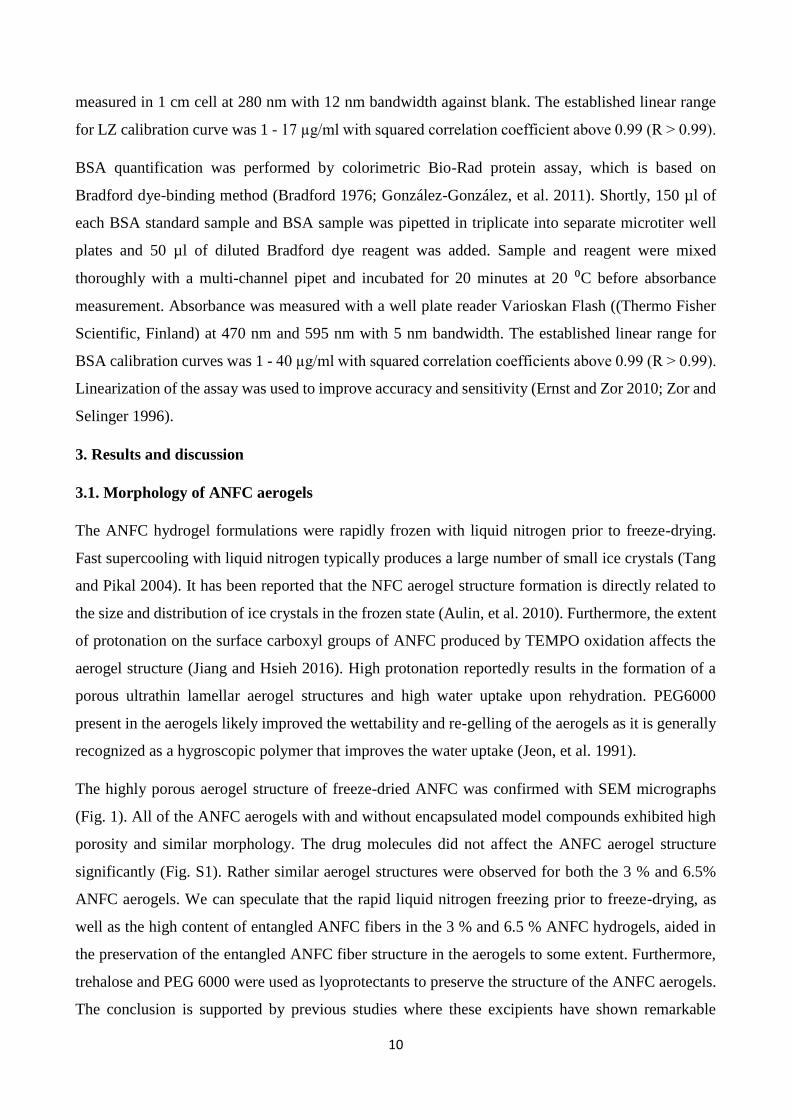
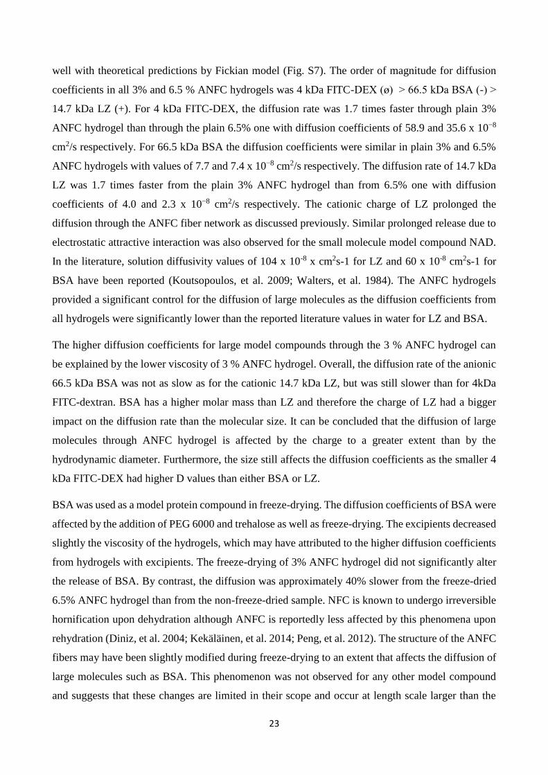
The highly porous aerogel structure of freeze-dried ANFC was confirmed with SEM micrographs
(Fig. 1). All of the ANFC aerogels with and without encapsulated model compounds exhibited high
porosity and similar morphology. The drug molecules did not affect the ANFC aerogel structure
significantly (Fig. S1). Rather similar aerogel structures were observed for both the 3 % and 6.5%
ANFC aerogels. We can speculate that the rapid liquid nitrogen freezing prior to freeze-drying, as
well as the high content of entangled ANFC fibers in the 3 % and 6.5 % ANFC hydrogels, aided in
the preservation of the entangled ANFC fiber structure in the aerogels to some extent. Furthermore,
trehalose and PEG 6000 were used as lyoprotectants to preserve the structure of the ANFC aerogels.
The conclusion is supported by previous studies where these excipients have shown remarkable
11
synergistic capacity to retain the structure and activity of proteins during freeze-drying (Carpenter, et
al. 1993; Prestrelski, et al. 1993). Although the morphology of the aerogels was rather similar,
differences were observed in the rheological properties before and after freeze-drying.
Figure 1. SEM micrographs of freeze-dried highly porous ANFC aerogels. 3% and 6.5% ANFC
hydrogels were freeze-dried without (left) and with the excipients (right). Scale bar is 200 µm.
Abbreviation exp refers to 0.3 % trehalose and 1 % PEG 6000.
3.2. Thermogravimetry analysis (TGA) and differential scanning calorimetry (DSC)
Residual moisture content of the aerogels was determined with TGA and mass change (%) for all
formulations is presented in Table 2. The aerogel produced from 6.5 % ANFC hydrogel contained
7.5 % of residual moisture, while the aerogel produced from 3 % ANFC hydrogel contained 6.8 % of
residual moisture. As there are more fibers in the more concentrated ANFC aerogel, there is also more
tightly bound surface water resulting in slightly higher residual moisture content when the same
processing parameters were used for both aerogels. When trehalose and PEG were used with ANFC,
the corresponding residual moisture was decreased for both 3 % and 6.5 % ANFC aerogels. In
previous studies trehalose and PEG have been used to protect proteins from undergoing
conformational charges during freeze-drying due to favorable interactions with PEG (Mi, et al. 2004)
and trehalose (Chang, et al. 2005; Jovanović, et al. 2006). One of the speculated stabilization
mechanisms of trehalose in the literature is the replacement of water molecules hydrogen bonded to
polar groups on the surface of materials during freeze-drying (Lins, et al. 2004; Wolkers, et al. 2002).
12
Due to this mechanism the amount of tightly bound surface water on ANFC fibers is reduced and
lower residual moisture content could be observed when trehalose and PEG were used in the
formulations.
Table 2. TGA and DSC analysis of different ANFC aerogel formulations after freeze-drying (n=1).
Residual moisture content of the aerogels was determined as a mass loss (%) of evaporated water.
Melting points of model compounds are reported as onset temperatures. All formulations except pure
3 % and 6.5 % ANFC contained 1 % PEG 6000 / 0.3 % trehalose in the final aerogel. Abbreviation
exp refers to 1 % PEG 6000 / 0.3 % trehalose.
Formulation Water content (%) Tm (⁰C) for drug or ANFC
ANFC 3 % 6.77 171.5
ANFC 3 % + exp 4.11 170 (ANFC/trehalose)
ANFC 6.5 % 7.48 174.0
ANFC 6.5 % + exp 5.81 187 (ANFC/trehalose)
BSA 1 % / ANFC 3 % 4.79 nd
BSA 1 % / ANFC 6.5 % 6.05 nd
MZ 100 % reference nd 159.4
MZ 2 % / ANFC 3 % 2.42 148.7
MZ 2 % / ANFC 6.5 % 4.98 143.5
NAD 100 % reference nd 128.8
NAD 1.7 % / ANFC 3 % 2.90 127.3
NAD 1.7 % / ANFC 6.5 % 4.79 128.3
KETO 100 % reference nd 94.6
KETO 3.4 % / ANFC 3 % 1.96 81
KETO 3.4 % / ANFC 6.5 % 3.84 84
For drug containing aerogels, the hydrophilicity (Table 1 in materials and methods) of the model
compounds affected the residual moisture content of the aerogels in addition to ANFC fiber amount.
The residual moisture content was higher for formulations with BSA, NAD and MZ, whereas KETO
as the most hydrophobic compound tested had the lowest values. All model compounds decreased
the moisture content when compared to the plain hydrogels. It might be possible to optimize the
freeze-drying process for constant residual moisture content, but this was not seen as necessary here,
as the main goal was to later redisperse the aerogels into hydrogels. In future studies more research
will be focused on the duration of primary drying and the secondary drying steps that removes the
tightly bound surface water. However, the low residual moisture content indicated that the
sublimation of water was effective and the process parameters were suitable for the aerogel
production.
The DSC thermograms of aerogels showed a reduction in the melting points of model compounds
with regard to references (Table 2). It has been shown in earlier studies that the melting peaks may
disappear from DSC thermograms if the drug and polymer matrix are well integrated. Valo et al. have
previously shown that the NFC grade and source affect the magnitude of drug interactions in freeze-
13
dried cellulose formulations (Valo, et al. 2013). Therefore, the observed differences in the melting
points of model compounds with regard to pure references indicate interactions between the ANFC
fibers and the model compounds. It has been shown in a previous study that the binding of drugs into
NFC fibers is pH dependent and the electrostatic forces are the main binding mechanism thus
resulting in the strongest binding between cationic molecules and anionic NFC fibers (Kolakovic et
al., 2013). Furthermore, it has been suggested that π stacking and hydrogen bonding may take place
as binding mechanisms in addition to electrostatic interactions. The structure of NFC and ANFC is
rather similar and therefore the pH dependent electrostatic binding of molecules into ANFC fibers
was expected to be the main interaction for binding of positively charged model compounds. For
negatively charged and neutral model compounds, we expected to observe only minimal binding with
ANFC fibers. The in vitro release studies were performed in order to evaluate more specifically the
effect of molecule charge on the release profiles.
3.3. Rheology
The numerous microscopic pores in the aerogel structure caused by the sublimated ice crystals
ensured enhanced water penetration into the aerogel structure upon rehydration (Jiang and Hsieh
2016). Furthermore, ANFC is known to have high water absorption and retention capacity (Jiang and
Hsieh 2014). The rehydration and hydrogel formation was further enhanced by mechanical mixing
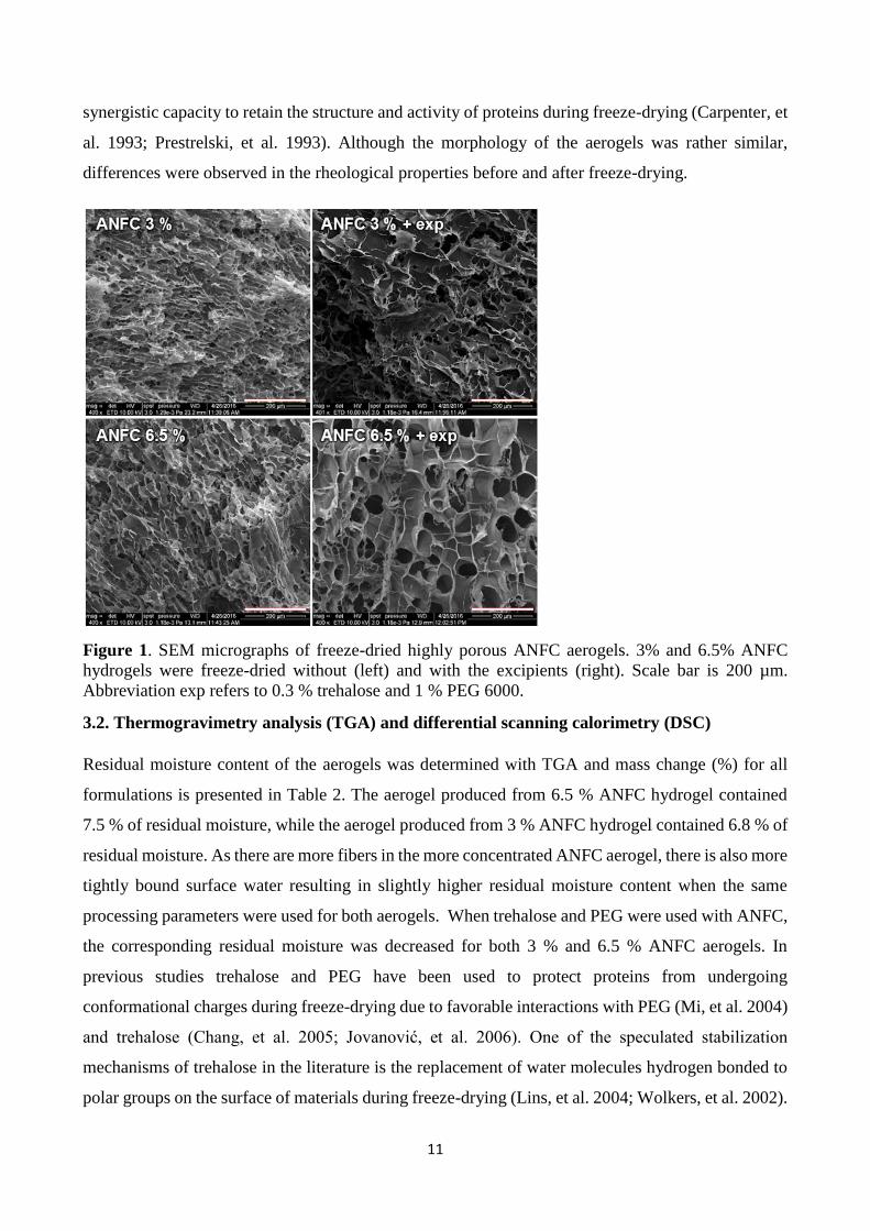
(Figure S1). However, freeze-drying of plain 3 % and 6.5 % ANFC without excipients reduced the
viscosity values of redispersed gels significantly (Fig. 2). Both grades showed ~150 % higher
viscosities before freeze-drying than their respective rehydrated samples at shear rates 0.1 – 40 (1/s).
The presence of excipients significantly reduced the gap to ~30 % by increasing the viscosity of the
freeze-dried samples and slightly lowering the plain hydrogel viscosities. This was more notable for
the 6.5 % hydrogels. The slight decrease in viscosity with ANFC hydrogels with excipients, when
compared to plain hydrogels without excipients, was probably due to the stabilization effect of
lowering the surface tension of the system (Gilányi, et al. 2006). Therefore, the excipients themselves
had minimal impact in the overall viscosity of the system. However, the main difference between the
rehydrated hydrogels (with vs. without excipients) was most likely due to larger amount of
aggregation in the samples dried and rehydrated without excipients. Therefore, the increase in
viscosity of the rehydrated samples with the excipients was attributed to the preservation of structural
integrity, limiting the formation of aggregates. The addition of model compounds did not significantly
change the viscosity when compared to the ANFC hydrogels with excipients, regardless of the
excessive (i.e. undissolved) amount of MZ, KETO and NAD. For BSA and MZ however, the gap
between the viscosities was slightly wider prior to and after freeze-drying than with the ANFC
14
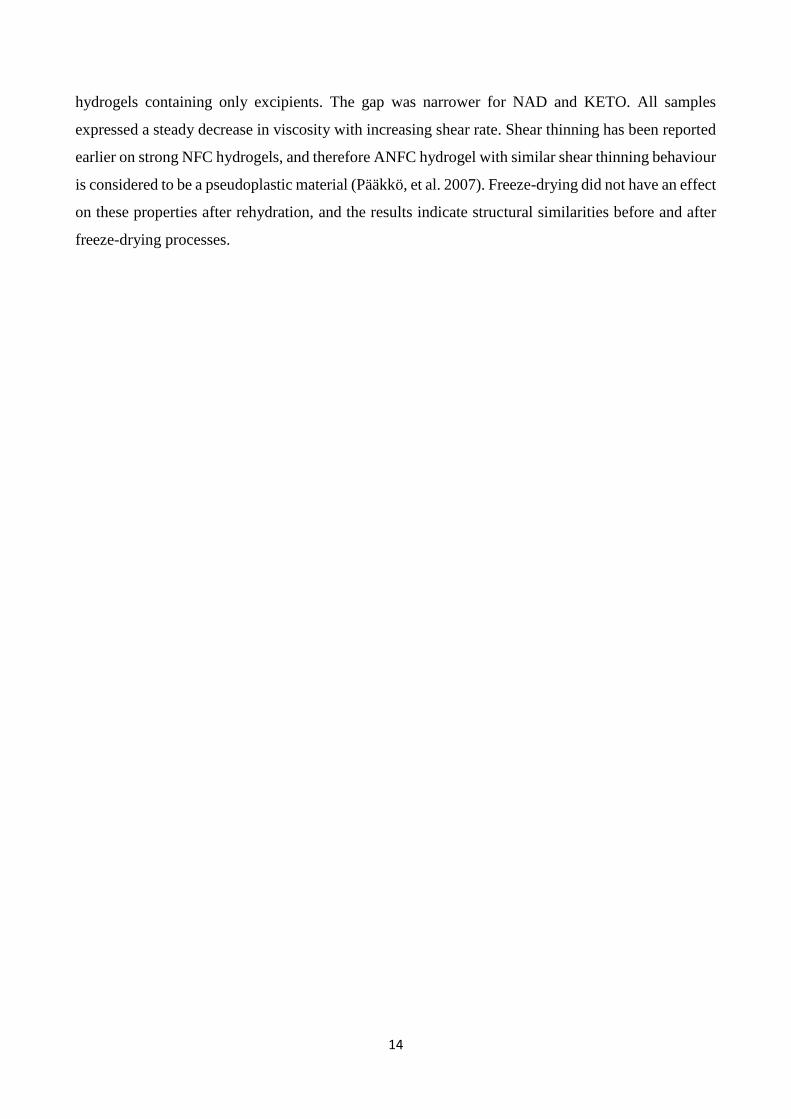
hydrogels containing only excipients. The gap was narrower for NAD and KETO. All samples
expressed a steady decrease in viscosity with increasing shear rate. Shear thinning has been reported
earlier on strong NFC hydrogels, and therefore ANFC hydrogel with similar shear thinning behaviour
is considered to be a pseudoplastic material (Pääkkö, et al. 2007). Freeze-drying did not have an effect
on these properties after rehydration, and the results indicate structural similarities before and after
freeze-drying processes.
15
Figure 2. Shear rate viscosity of plain ANFC (3 % and 6.5 %) as well as different ANFC hydrogel
formulations before and after freeze-drying and redispersion (mean ± S.D., n=3) . ANFC hydrogels
contained 1% BSA, 2% metronidazole (MZ), 3.4 % ketoprofen (KETO) or 1.7 % NAD (NAD). Abbreviations: FD = freeze-drying, exp = trehalose, PEG6000.
16
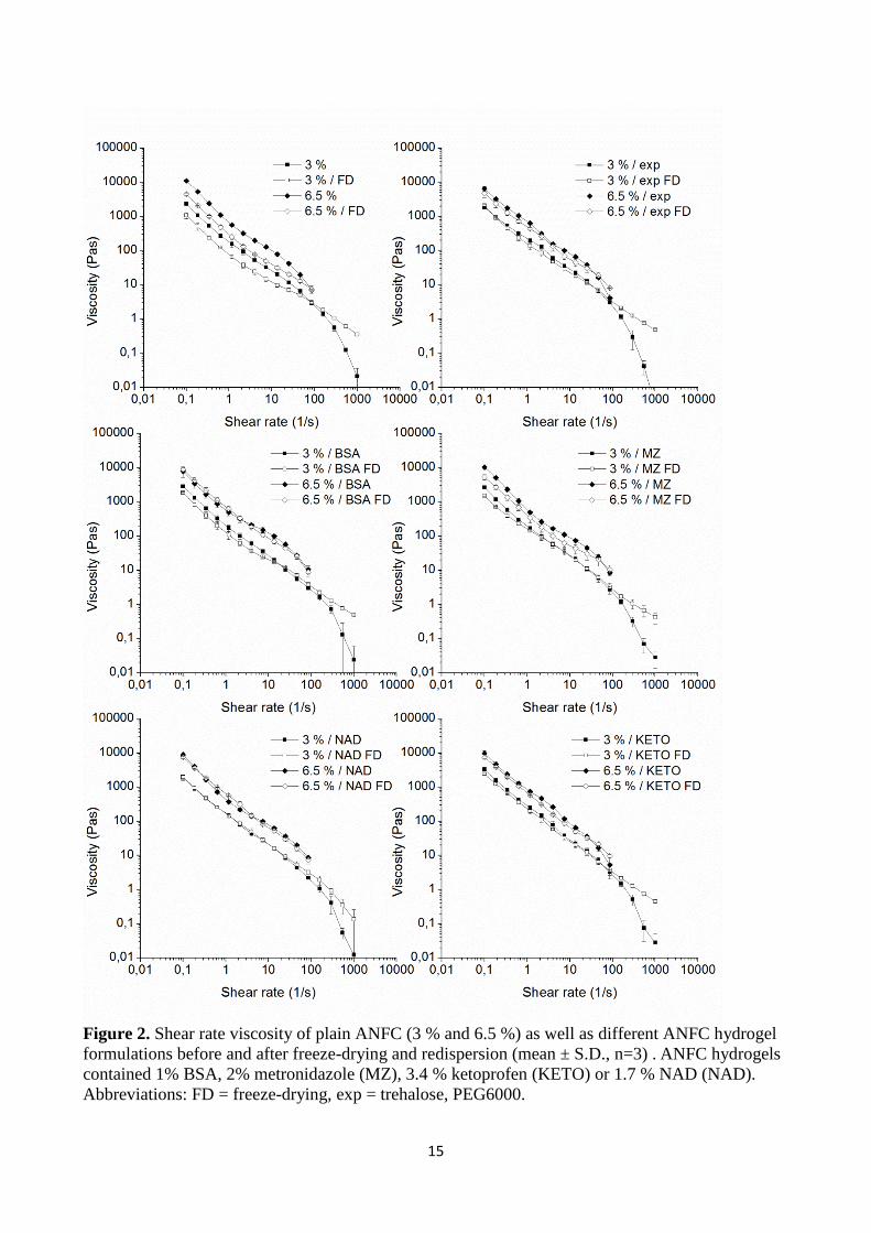
The effect of freeze-drying on the viscoelastic properties of ANFC hydrogels with and without
excipients was studied. Freeze-drying lowered the storage modulus (G’) values of all samples with
and without excipients (Fig. 3); however, the gap between the G’ values of the non-freeze-dried
hydrogels and freeze-dried (i.e. rehydrated) hydrogels becomes significantly narrower with
excipients. Therefore, the G’ values of rehydrated ANFC hydrogel samples with excipients resembled
the original values of the hydrogels without excipients before freeze-drying on both grades (3 % and
6.5 %). All samples, including mixtures with model compounds (Fig. S3, S4), were relatively
independent of the angular frequency at investigated ranges. Additionally, G’ values were higher than
loss modulus (G’’) values, which indicates an elastic behavior in strong ANFC hydrogels (Pääkkö, et
al. 2007). Therefore, the elastic properties of hydrogels remained after freeze-drying, indicating
structural similarities as was shown with viscosity measurements.
Figure 3. Storage (G’) and loss modulus (G’’) of ANFC hydrogels before and after freeze-drying and
redispersion (mean ± S.D., n=3). Abbreviations: FD= freeze-drying, exp = trehalose, PEG6000.
A slight increase in G’ can be observed with the stronger 6.5 % hydrogels towards higher frequencies.
For the less viscous 3 % ANFC the increase was not as prominent. Similarly, the G’’ values increased
towards higher frequencies on both grades of ANFC hydrogels; however, the effect of freeze-drying
varies. In contrast to 6.5 % ANFC hydrogel the G’’ of 3 % the freeze-dried ANFC samples had higher
values than the non-freeze-dried hydrogels, especially with the samples not containing excipients.
The G’’ of 6.5 % rehydrated hydrogels was slightly lower in comparison to non-freeze-dried samples
(as was observed with G’). However, the presence of excipients had a significant impact in reducing
the effects of freeze-drying in both concentrations of rehydrated hydrogels. Therefore, it can be
concluded that at higher frequencies, the excipients attributed to the elastic properties of ANFC
hydrogel after rehydration, i.e. with the absence of excipients, G’’ increases while G’ decreases and
17
the viscous characteristics become more predominant over the elastic properties (Dimić-Mišić, et al.
2013).
Based on the results of rheological measurements it was evident that the rheological properties were
preserved to a greater extent when excipients were used as stabilizers during freeze-drying. In dilution
studies of the re-gelled ANFC hydrogels, we observed that freeze-dried ANFC with excipients
remained suspended without aggregates (data not shown). On the contrary, visible aggregates were
observed when ANFC hydrogel was freeze-dried without excipients followed by re-gelling and
dilution. This may further indicate that the excipients reduced the irreversible hydrogen bonding
between the ANFC fibers in hydrogel as the freeze-dried ANFC fibers with excipients remained
hydrated more effectively.
Additionally, we investigated the effects of crosslinking cations on the rheological properties of
1.1 % ANFC hydrogel. The ANFC hydrogels were crosslinked with cations Al3+, Fe3+ or Ca2+. All
crosslinked systems showed an increase in viscosity, more notably with the trivalent cations Al3+ and
Fe3+ (Fig. S5, top). Similar increase in G’ and G’’ was observed (Fig. S5, bottom). The results indicate
a form of stabilization effect as with excipients; however, this was due to a different mechanism of
stiffening the hydrogel structure, i.e strengthening the bonds within the hydrophilic segments of the
hydrogel. More detailed information can be found within the supplementary material.
3.4. In vitro drug release
The in vitro release studies were performed in order the estimate the effect of ANFC fiber
concentration as well as freeze-drying on the release profiles of a variety of model compounds from
the hydrogels. The ANFC hydrogel matrix did not swell in the PBS buffer used in the release studies
thus retaining the original shape and dimensions of the hydrogel matrix throughout the release
experiments. Therefore, it was hypothesized that the slower diffusion in the non-swelling hydrogel
matrix would control the drug release rates. This property as well as high water retention capacity of
ANFC can be utilized beneficially in controlled drug delivery applications.
The pH of the ANFC hydrogels was 7 prior to the addition of model compounds, and the pH of the
release medium was kept at 7.4 with PBS. Due to moderate buffering capacity of ANFC, the pH of
the hydrogel matrix exposed to PBS buffer was estimated to be approximately 7 after the addition of
model compounds and excipients. Physical mixtures of KETO, NAD, and MZ with ANFC contained
excess amounts of drug in relation to their solubility at pH 7 and were defined as monolithic
dispersions (Table 1, Materials and methods). The amount of KETO at 34 mg/ml in the ANFC
18
hydrogels was below the pH 7 solubility limit of 40 mg/ml. However, in practice excess amount of
undissolved KETO remained in the ANFC hydrogel matrix resulting in a monolithic dispersion (Fig.
S2). 4 kDa FITC-DEX, LZ, and BSA were all completely dissolved in the ANFC hydrogels. By
definition these formulations were monolithic solutions.
The addition of trehalose and PEG 6000 lowered the viscosity of 3 % and 6.5 % ANFC hydrogels
slightly (Fig. 2 and 3). Therefore, the release studies were performed from plain ANFC hydrogels,
hydrogels with excipients and from freeze-dried and reconstructed hydrogels that also contained
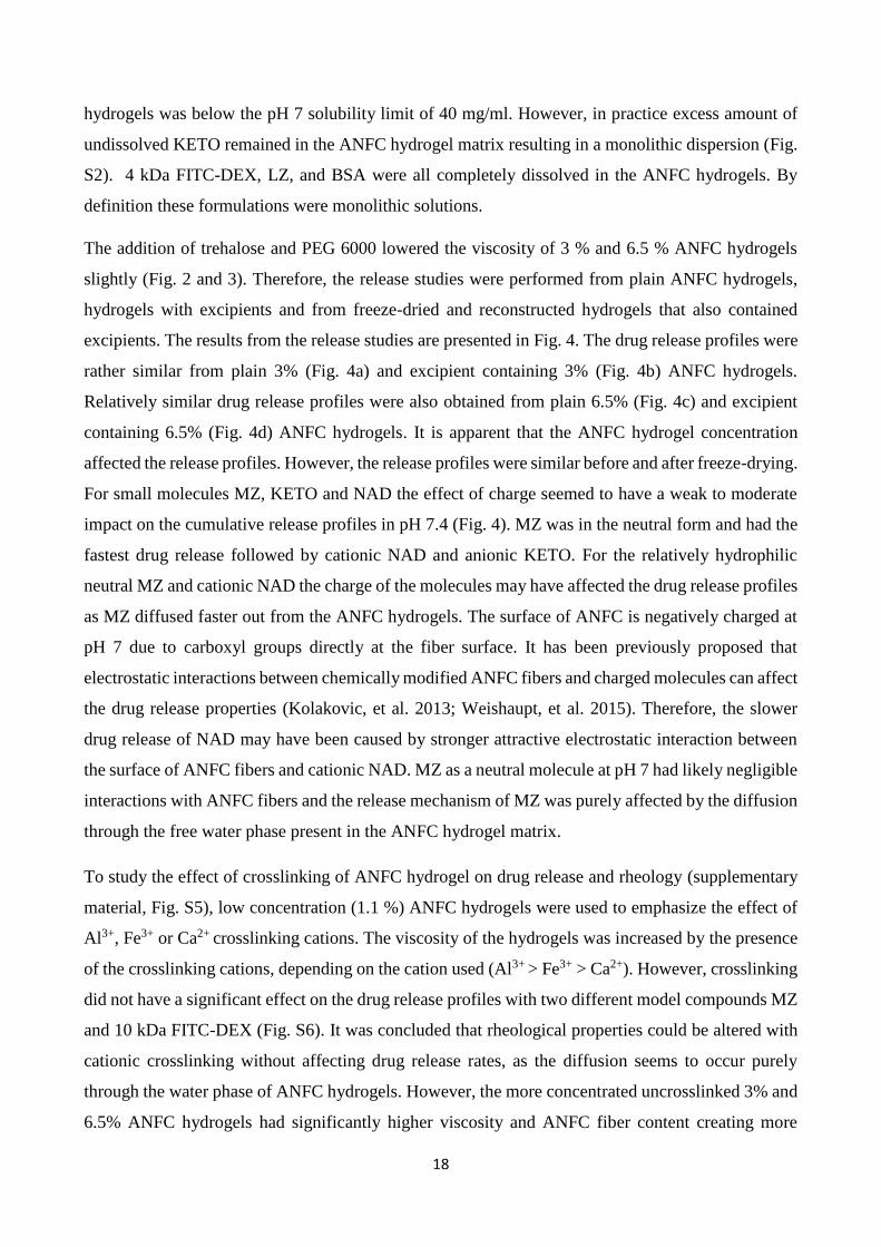
excipients. The results from the release studies are presented in Fig. 4. The drug release profiles were
rather similar from plain 3% (Fig. 4a) and excipient containing 3% (Fig. 4b) ANFC hydrogels.
Relatively similar drug release profiles were also obtained from plain 6.5% (Fig. 4c) and excipient
containing 6.5% (Fig. 4d) ANFC hydrogels. It is apparent that the ANFC hydrogel concentration
affected the release profiles. However, the release profiles were similar before and after freeze-drying.
For small molecules MZ, KETO and NAD the effect of charge seemed to have a weak to moderate
impact on the cumulative release profiles in pH 7.4 (Fig. 4). MZ was in the neutral form and had the
fastest drug release followed by cationic NAD and anionic KETO. For the relatively hydrophilic
neutral MZ and cationic NAD the charge of the molecules may have affected the drug release profiles
as MZ diffused faster out from the ANFC hydrogels. The surface of ANFC is negatively charged at
pH 7 due to carboxyl groups directly at the fiber surface. It has been previously proposed that
electrostatic interactions between chemically modified ANFC fibers and charged molecules can affect
the drug release properties (Kolakovic, et al. 2013; Weishaupt, et al. 2015). Therefore, the slower
drug release of NAD may have been caused by stronger attractive electrostatic interaction between
the surface of ANFC fibers and cationic NAD. MZ as a neutral molecule at pH 7 had likely negligible
interactions with ANFC fibers and the release mechanism of MZ was purely affected by the diffusion
through the free water phase present in the ANFC hydrogel matrix.
To study the effect of crosslinking of ANFC hydrogel on drug release and rheology (supplementary
material, Fig. S5), low concentration (1.1 %) ANFC hydrogels were used to emphasize the effect of
Al3+, Fe3+ or Ca2+ crosslinking cations. The viscosity of the hydrogels was increased by the presence
of the crosslinking cations, depending on the cation used (Al3+ > Fe3+ > Ca2+). However, crosslinking
did not have a significant effect on the drug release profiles with two different model compounds MZ
and 10 kDa FITC-DEX (Fig. S6). It was concluded that rheological properties could be altered with
cationic crosslinking without affecting drug release rates, as the diffusion seems to occur purely
through the water phase of ANFC hydrogels. However, the more concentrated uncrosslinked 3% and
6.5% ANFC hydrogels had significantly higher viscosity and ANFC fiber content creating more
19
barrier for the diffusion. The release of MZ reached 100% after 50 h from the 1.1 % ANFC hydrogel
as well as from 3% and 6.5% ANFC hydrogels. The finding supports the conclusion that the diffusion
of small molecules was affected by the availability of free water molecules in the 3 and 6.5 % ANFC.
Figure 4. The effect of ANFC hydrogel concentration and freeze-drying on the release profiles of
MZ, NAD, KETO, BSA, LZ and 4 kDa FITC-DEX(mean ± S.D., n=3) . Formulations were based
on plain 3% ANFC hydrogel (a), 3% ANFC hydrogel with PEG6000 and trehalose before and after
freeze-drying (b), plain 6.5% ANFC hydrogel (c), and 6.5% ANFC hydrogel with PEG6000 and
trehalose before and after freeze-drying (d). Release experiments were performed in PBS (pH 7.4)
at 37°C.

20
The dissolution rate and the excess amount of the small model compounds in the ANFC hydrogels
may have influenced the drug release profiles in addition to the charge of the molecules even though
the release experiments were performed under sink conditions. This was observed as prolonged drug
release rate for anionic KETO, which based solely on its charge should have diffused faster out from
the hydrogel matrix due to electrostatic repulsion with ANFC fibers. Overall, the cumulative release
of KETO was below 70% for all tested hydrogels at day 6, whereas MZ reached 100% and NAD
nearly 100% cumulative release. The slower drug release rate of KETO may be attributed to amount
dependent dissolution rate of KETO inside the ANFC hydrogel matrix as it has been previously
reported that the dissolution rate of KETO is strongly dose dependent even under sink conditions
(Yazdanian, et al. 2004). Overall, the higher 6.5 % ANFC fiber content reduced the drug release of
small molecules only slightly more effectively with regard to corresponding release profiles from 3
% ANFC hydrogels. Therefore, the amount of ANFC fibers can be used to control the drug release
rate of small molecules only to a moderate extent. Furthermore, the freeze-drying did not significantly
affect drug release properties of small molecules as the re-gelled formulations exhibited rather similar
release behavior as the corresponding pre-freeze-dried formulations (Fig. 4b and 4d). The quality of
the ANFC hydrogels as a matrix for controlled drug release applications for small molecules remained
unaltered regardless of the drying step as the drug release properties were retained. It was therefore
clearly demonstrated that the drug containing ANFC hydrogels can be freeze-dried and re-gelled
successfully without any loss on drug release properties.
For large model compounds BSA, LZ, and FITC-DEX the net charge at pH 7, molecular size and
ANFC fiber content had a significant impact on the cumulative release profiles (Fig. 4a and 4c).
Neutral FITC-DEX had the fastest release rate followed by anionic BSA and cationic LZ. The 4 kDa
FITC-DEX reached 100% release from plain 3% ANFC hydrogel and 85% release from plain 6.5%
ANFC hydrogel at day 6. This indicates that the ANFC fiber content did control the release of FITC-
DEX, but not as effectively as for LZ and BSA. 4 kDa FITC-DEX has significantly smaller size than
14.7 kDa LZ and 66.5 kDa BSA and therefore it was expected that the release of FITC-DEX would
be faster. Cationic LZ had the slowest release of approximately 30% from 3% ANFC and below 20%
from 6.5% ANFC hydrogels during 6 days. The anionic BSA had approximately 40% release from
both hydrogel concentrations during 6 days.
It is clear that the cationic charge of LZ reduced the release rate though electrostatic attractive
interaction with ANFC fibers. The conclusion is supported by the physicochemical properties of LZ
and BSA (Table 1, Materials and methods) as well as similar behavior and slow release of cationic
NAD. The molecule size and hydrodynamic diameter of LZ is smaller than for BSA and without any
21
electrostatic interaction the release of LZ would have likely been faster. The results indicate that for
large molecules (above 14 kDa) the charge of the molecule was more significant than the molar mass
and hydrodynamic diameter in terms of impact on the cumulative release profile.
Furthermore, the cumulative release of none of the large molecules from the ANFC hydrogel matrixes
reached 100 %. The physical entrapment of these large molecules in the highly entangled ANFC
fibers’ network likely allowed less free motion for diffusion than for small molecules, which was
observed as sustained release for BSA and LZ. BSA was the only large model compound that was
freeze-dried with ANFC and excipients. The release profiles of BSA from 3.5% and 6.5 % ANFC
hydrogels were similar before and after freeze-drying (Fig. 4b and 4d) indicating that the release
properties of large molecules may be retained even after the drying and re-gelling. This is a highly
beneficial property for drug delivery applications of protein pharmaceuticals in addition to small
molecules inside a hydrogel matrix as the formulation can be stored at dry state for prolonged storage
and rehydrated upon need. For example, the use of ANFC as a hydrogel matrix for the delivery of
drugs and growth factors into wound areas could have promoting effects on wound healing by
increasing the healing rate and efficacy.
3.5. The diffusivity in the hydrogel matrix
For the evaluation of diffusion rate without the impact of solubility, the Higuchi equation (Eq.2, see
Materials and Methods) was used to calculate the diffusion coefficients (D) for small molecules
(Table 3). We suggest that this is a more suitable way to evaluate the diffusivity of small molecules
inside the hydrogels when excess drug with regard to solubility is used in the hydrogel matrix. As
described previously literature values of solubility at pH 7 for MZ, NAD and KETO (Table 1) were
used for the determination of diffusion coefficients. It should be noted that the pH 7 is a region where
the solubility of KETO drastically improves due to ionization and the amount of KETO may affect
the dissolution rate (Avdeef, et al. 2000; Yazdanian, et al. 2004). The approximations of diffusion
coefficients should be used to compare the D values of small molecules in different hydrogel
formulations prior to and after freeze-drying.
The order of the magnitude of diffusion coefficients in all 3% and 6.5 % ANFC hydrogels was MZ
(ø) ˃ NAD (+) ˃ KETO (-). Furthermore, the diffusion coefficients from 3 % ANFC hydrogels were
only slightly higher than the corresponding values from 6.5 % hydrogels. This can be explained by
the lower viscosity, G’ (elastic response) and G’’(viscous behavior) of 3% ANFC hydrogels when
compared to the 6.5% hydrogels (see Fig. 2 and 3). The diffusion coefficients were rather similar for
both hydrogels without excipients, with excipients and after freeze-drying. Therefore, freeze-drying
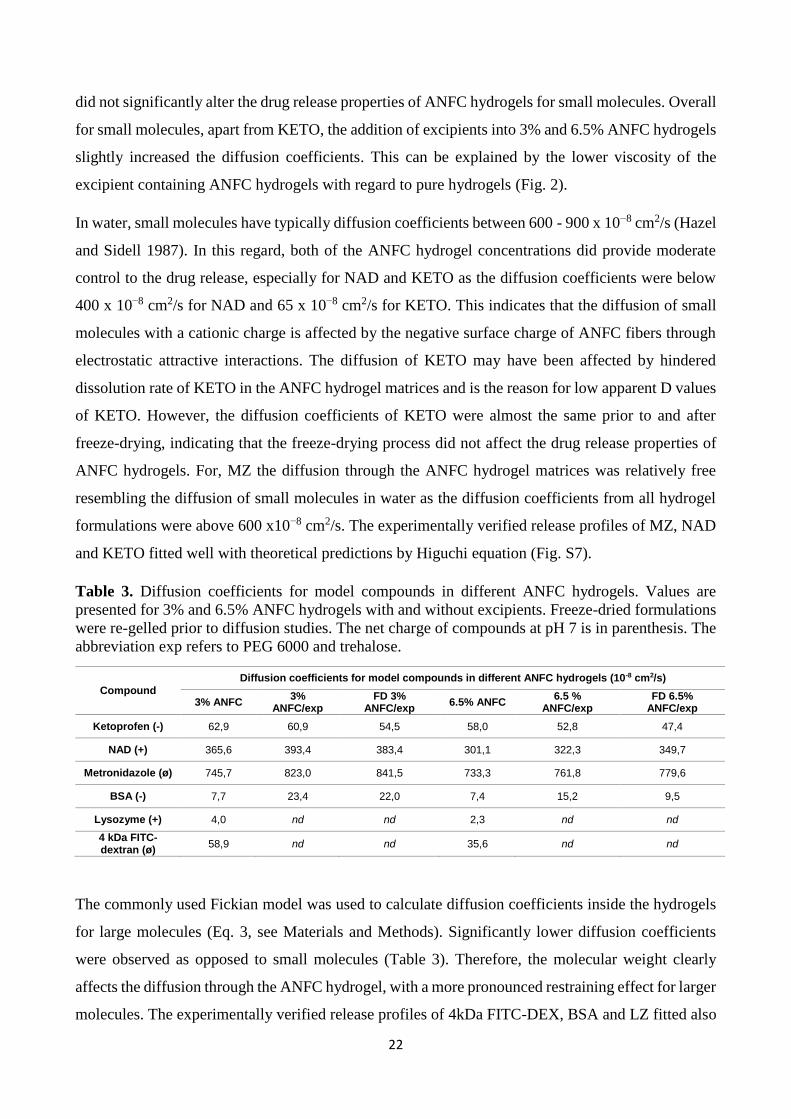
22
did not significantly alter the drug release properties of ANFC hydrogels for small molecules. Overall
for small molecules, apart from KETO, the addition of excipients into 3% and 6.5% ANFC hydrogels
slightly increased the diffusion coefficients. This can be explained by the lower viscosity of the
excipient containing ANFC hydrogels with regard to pure hydrogels (Fig. 2).
In water, small molecules have typically diffusion coefficients between 600 - 900 x 10−8 cm2/s (Hazel
and Sidell 1987). In this regard, both of the ANFC hydrogel concentrations did provide moderate
control to the drug release, especially for NAD and KETO as the diffusion coefficients were below
400 x 10−8 cm2/s for NAD and 65 x 10−8 cm2/s for KETO. This indicates that the diffusion of small
molecules with a cationic charge is affected by the negative surface charge of ANFC fibers through
electrostatic attractive interactions. The diffusion of KETO may have been affected by hindered
dissolution rate of KETO in the ANFC hydrogel matrices and is the reason for low apparent D values
of KETO. However, the diffusion coefficients of KETO were almost the same prior to and after
freeze-drying, indicating that the freeze-drying process did not affect the drug release properties of
ANFC hydrogels. For, MZ the diffusion through the ANFC hydrogel matrices was relatively free
resembling the diffusion of small molecules in water as the diffusion coefficients from all hydrogel
formulations were above 600 x10−8 cm2/s. The experimentally verified release profiles of MZ, NAD
and KETO fitted well with theoretical predictions by Higuchi equation (Fig. S7).
Table 3. Diffusion coefficients for model compounds in different ANFC hydrogels. Values are
presented for 3% and 6.5% ANFC hydrogels with and without excipients. Freeze-dried formulations
were re-gelled prior to diffusion studies. The net charge of compounds at pH 7 is in parenthesis. The
abbreviation exp refers to PEG 6000 and trehalose.
Compound
Diffusion coefficients for model compounds in different ANFC hydrogels (10-8 cm2/s)
3% ANFC 3%
ANFC/exp FD 3%
ANFC/exp 6.5% ANFC
6.5 % ANFC/exp
FD 6.5% ANFC/exp
Ketoprofen (-) 62,9 60,9 54,5 58,0 52,8 47,4
NAD (+) 365,6 393,4 383,4 301,1 322,3 349,7
Metronidazole (ø) 745,7 823,0 841,5 733,3 761,8 779,6
BSA (-) 7,7 23,4 22,0 7,4 15,2 9,5
Lysozyme (+) 4,0 nd nd 2,3 nd nd
4 kDa FITC-dextran (ø)
58,9 nd nd 35,6 nd nd
The commonly used Fickian model was used to calculate diffusion coefficients inside the hydrogels
for large molecules (Eq. 3, see Materials and Methods). Significantly lower diffusion coefficients
were observed as opposed to small molecules (Table 3). Therefore, the molecular weight clearly
affects the diffusion through the ANFC hydrogel, with a more pronounced restraining effect for larger
molecules. The experimentally verified release profiles of 4kDa FITC-DEX, BSA and LZ fitted also
23
well with theoretical predictions by Fickian model (Fig. S7). The order of magnitude for diffusion
coefficients in all 3% and 6.5 % ANFC hydrogels was 4 kDa FITC-DEX (ø) ˃ 66.5 kDa BSA (-) ˃
14.7 kDa LZ (+). For 4 kDa FITC-DEX, the diffusion rate was 1.7 times faster through plain 3%
ANFC hydrogel than through the plain 6.5% one with diffusion coefficients of 58.9 and 35.6 x 10⁻⁸
cm2/s respectively. For 66.5 kDa BSA the diffusion coefficients were similar in plain 3% and 6.5%
ANFC hydrogels with values of 7.7 and 7.4 x 10⁻⁸ cm2/s respectively. The diffusion rate of 14.7 kDa
LZ was 1.7 times faster from the plain 3% ANFC hydrogel than from 6.5% one with diffusion
coefficients of 4.0 and 2.3 x 10⁻⁸ cm2/s respectively. The cationic charge of LZ prolonged the
diffusion through the ANFC fiber network as discussed previously. Similar prolonged release due to
electrostatic attractive interaction was also observed for the small molecule model compound NAD.
In the literature, solution diffusivity values of 104 x 10-8 x cm2s-1 for LZ and 60 x 10-8 cm2s-1 for
BSA have been reported (Koutsopoulos, et al. 2009; Walters, et al. 1984). The ANFC hydrogels
provided a significant control for the diffusion of large molecules as the diffusion coefficients from
all hydrogels were significantly lower than the reported literature values in water for LZ and BSA.
The higher diffusion coefficients for large model compounds through the 3 % ANFC hydrogel can
be explained by the lower viscosity of 3 % ANFC hydrogel. Overall, the diffusion rate of the anionic
66.5 kDa BSA was not as slow as for the cationic 14.7 kDa LZ, but was still slower than for 4kDa
FITC-dextran. BSA has a higher molar mass than LZ and therefore the charge of LZ had a bigger
impact on the diffusion rate than the molecular size. It can be concluded that the diffusion of large
molecules through ANFC hydrogel is affected by the charge to a greater extent than by the
hydrodynamic diameter. Furthermore, the size still affects the diffusion coefficients as the smaller 4
kDa FITC-DEX had higher D values than either BSA or LZ.
BSA was used as a model protein compound in freeze-drying. The diffusion coefficients of BSA were
affected by the addition of PEG 6000 and trehalose as well as freeze-drying. The excipients decreased
slightly the viscosity of the hydrogels, which may have attributed to the higher diffusion coefficients
from hydrogels with excipients. The freeze-drying of 3% ANFC hydrogel did not significantly alter
the release of BSA. By contrast, the diffusion was approximately 40% slower from the freeze-dried
6.5% ANFC hydrogel than from the non-freeze-dried sample. NFC is known to undergo irreversible
hornification upon dehydration although ANFC is reportedly less affected by this phenomena upon
rehydration (Diniz, et al. 2004; Kekäläinen, et al. 2014; Peng, et al. 2012). The structure of the ANFC
fibers may have been slightly modified during freeze-drying to an extent that affects the diffusion of
large molecules such as BSA. This phenomenon was not observed for any other model compound
and suggests that these changes are limited in their scope and occur at length scale larger than the
24
small molecules. We conclude that the release properties of all model compounds were preserved in
3% hydrogels even after freeze-drying. As for the freeze-dried 6.5% hydrogels, only the release of
66.5 kDa BSA was altered, but the diffusion coefficients were still approximately the same as from
plain 6.5 % hydrogel without excipients.
4. Conclusions
The chemically modified ANFC hydrogels can be successfully freeze-dried into aerogels and
redispersed into the hydrogel form. Cryoprotectants were effectively used to preserve the rheological
properties of the rehydrated aerogels and the release profiles of the model compounds were similar
before and after freeze-drying. This is a highly desirable feature for processing of pharmaceutical
formulations as the shelf-life of hydrolysis sensitive compounds can be increased by the dry state of
an aerogel. Based on the results, it can be concluded that the freeze-drying did not significantly affect
the drug release properties from reconstructed ANFC hydrogels. Possible structural changes in the
nano-scale dimensions of ANFC fibers during freeze-drying did not affect the quality on ANFC as a
hydrogel matrix platform for controlled drug delivery of small molecules and large proteins. The
potential of ANFC hydrogels for controlled drug delivery applications at high fiber concentrations of
3% and 6.5% has been clearly demonstrated.
The ANFC fiber concentration and the high surface area of the individual ANFC fibers can be used
to control the release rate of small as well as large molecule size pharmaceutical compounds. The
ANFC hydrogel concentration affected the diffusion of small molecules moderately. The underlying
release mechanism of diffusion for small molecules was mainly affected by the availability of free
water molecules present in the ANFC hydrogels. Diffusivities of large proteins were decreased with
increasing ANFC fiber content in the hydrogels providing significant control for release kinetics. For
large protein molecules, cationic charge of the molecule may be more significant factor than size with
regard to the diffusion through ANFC hydrogel. The results indicate that ANFC is capable for
controlled delivery of several types of molecules. Furthermore, the aerogels can be easily rehydrated
and administered upon need.
Acknowledgements
The financial support from Academy of Finland (Grant no. 264988 and 258114) is gratefully
acknowledged. Orion Foundation of the Professor pool, Finland is greatly acknowledged by M.Y.
This project has been supported by Tekes-Industry funded GrowDex III-project.
References
25
Alexandrescu, L., Syverud, K., Gatti, A., Chinga-Carrasco, G., 2013. Cytotoxicity tests of cellulose
nanofibril-based structures. Cellulose, 20, 1765-1775.
Aulin, C., Netrval, J., Wågberg, L., Lindström, T., 2010. Aerogels from nanofibrillated cellulose with
tunable oleophobicity. Soft Matter, 6, 3298-3305.
Avdeef, A., Berger, C.M., Brownell, C., 2000. pH-metric solubility. 2: correlation between the acid-
base titration and the saturation shake-flask solubility-pH methods. Pharm. Res., 17, 85-89.
Avdeef, A., Berger, C.M., 2001. pH-metric solubility: 3. Dissolution titration template method for
solubility determination. Eur. J. Pharm. Sci., 14, 281-291.
Bhattacharya, M., Malinen, M.M., Lauren, P., Lou, Y., Kuisma, S.W., Kanninen, L., Lille, M., Corlu,
A., GuGuen-Guillouzo, C., Ikkala, O., Laukkanen, A., Urtti, A., Yliperttula, M., 2012. Nanofibrillar
cellulose hydrogel promotes three-dimensional liver cell culture. J. Control. Release, 164, 291-298.
Borges, A.C., Eyholzer, C., Duc, F., Bourban, P., Tingaut, P., Zimmermann, T., Pioletti, D.P.,
Månson, J.E., 2011. Nanofibrillated cellulose composite hydrogel for the replacement of the nucleus
pulposus. Acta Biomater., 7, 3412-3421.
Bradford, M.M., 1976. A rapid and sensitive method for the quantitation of microgram quantities of
protein utilizing the principle of protein-dye binding. Anal. Biochem., 72, 248-254.
Carpenter, J., Prestrelski, S., Arakawa, T., 1993. Separation of freezing-and drying-induced
denaturation of lyophilized proteins using stress-specific stabilization: I. Enzyme activity and
calorimetric studies. Arch. Biochem. Biophys., 303, 456-464.
Carpenter, J.F., Pikal, M.J., Chang, B.S., Randolph, T.W., 1997. Rational design of stable lyophilized
protein formulations: some practical advice. Pharm. Res., 14, 969-975.
Censi, R., Di Martino, P., Vermonden, T., Hennink, W.E., 2012. Hydrogels for protein delivery in
tissue engineering. J. Control. Release, 161, 680-692.
Chang, L.L., Shepherd, D., Sun, J., Ouellette, D., Grant, K.L., Tang, X.C., Pikal, M.J., 2005.
Mechanism of protein stabilization by sugars during freeze‐drying and storage: Native structure
preservation, specific interaction, and/or immobilization in a glassy matrix? J. Pharm. Sci., 94, 1427-
1444.
Chinga-Carrasco, G., Syverud, K., 2014. Pretreatment-dependent surface chemistry of wood
nanocellulose for pH-sensitive hydrogels. J. Biomater. Appl., 29, 423-432.
Christensen, D., Foged, C., Rosenkrands, I., Nielsen, H.M., Andersen, P., Agger, E.M., 2007.
Trehalose preserves DDA/TDB liposomes and their adjuvant effect during freeze-drying. Biochim.
Biophys. Acta, 1768, 2120-2129.
Crowe, J.H., Tablin, F., Wolkers, W.F., Gousset, K., Tsvetkova, N.M., Ricker, J., 2003. Stabilization
of membranes in human platelets freeze-dried with trehalose. Chem. Phys. Lipids, 122, 41-52.
Dimić-Mišić, K., Sanavane, Y., Paltakari, J., Maloney, T., 2013. Small scale rheological observation
of high consistency nanofibrillar material based furnishes. Journal of Applied Engineering Science,
11, 145-151.
Diniz, J.F., Gil, M., Castro, J., 2004. Hornification—its origin and interpretation in wood pulps. Wood
Sci. Technol., 37, 489-494.
Ernst, O., Zor, T., 2010. Linearization of the bradford protein assay. J. Vis. Exp., 12, e1918.
26
Franks, F., 1998. Freeze-drying of bioproducts: putting principles into practice. Eur. J. Pharm.
Biopharm., 45, 221-229.
Fritz, M., Radmacher, M., Cleveland, J.P., Allersma, M.W., Stewart, R.J., Gieselmann, R., Janmey,
P., Schmidt, C.F., Hansma, P.K., 1995. Imaging globular and filamentous proteins in physiological
buffer solutions with tapping mode atomic force microscopy. Langmuir, 11, 3529-3535.
Gilányi, T., Varga, I., Gilányi, M., Mészáros, R., 2006. Adsorption of poly(ethylene oxide) at the
air/water interface: A dynamic and static surface tension study. J Colloid Interface Sci., 301, 428-
435.
González-González, M., Mayolo-Deloisa, K., Rito-Palomares, M., Winkler, R., 2011. Colorimetric
protein quantification in aqueous two-phase systems. Process Biochem., 46, 413-417.
Gupta, P., Vermani, K., Garg, S., 2002. Hydrogels: from controlled release to pH-responsive drug
delivery. Drug Discov. Today, 7, 569-579.
Hakkarainen, T., Koivuniemi, R., Kosonen, M., Escobedo-Lucea, C., Sanz-Garcia, A., Vuola, J.,
Valtonen, J., Tammela, P., Mäkitie, A., Luukko, K., Yliperttula, M., Kavola, H., 2016. Nanofibrillar
cellulose wound dressing in skin graft donor site treatment. J. Control. Release, 244, 292-301.
Han, Y., Quan, G.B., Liu, X.Z., Ma, E.P., Liu, A., Jin, P., Cao, W., 2005. Improved preservation of
human red blood cells by lyophilization. Cryobiology, 51, 152-164.
Hannukainen, K., Suhonen, S., Savolainen, K., Norppa, H., 2012. Genotoxicity of nanofibrillated
cellulose in vitro as measured by enzyme comet assay. Toxicol. Lett., 211, S71.
Hazel, J.R., Sidell, B.D., 1987. A method for the determination of diffusion coefficients for small
molecules in aqueous solution. Anal. Biochem., 166, 335-341.
Higuchi, T., 1963. Mechanism of sustained‐action medication. Theoretical analysis of rate of release
of solid drugs dispersed in solid matrices. J. Pharm. Sci., 52, 1145-1149.
Higuchi, T., 1961. Rate of release of medicaments from ointment bases containing drugs in
suspension. J. Pharm. Sci., 50, 874-875.
Hoffman, A.S., 2012. Hydrogels for biomedical applications. Adv. Drug Deliv. Rev., 64, Supplement
18-23.
Hua, K., Carlsson, D.O., Ålander, E., Lindström, T., Strømme, M., Mihranyan, A., Ferraz, N., 2014.
Translational study between structure and biological response of nanocellulose from wood and green
algae. RSC Adv., 4, 2892-2903.
Hubálek, Z., 2003. Protectants used in the cryopreservation of microorganisms. Cryobiology, 46,
205-229.
Isogai, A., Saito, T., Fukuzumi, H., 2011. TEMPO-oxidized cellulose nanofibers. Nanoscale, 3, 71-
85.
Jeon, S., Lee, J., Andrade, J., De Gennes, P., 1991. Protein—surface interactions in the presence of
polyethylene oxide: I. Simplified theory. J. Colloid Interface Sci., 142, 149-158.
Jiang, F., Hsieh, Y., 2014. Super water absorbing and shape memory nanocellulose aerogels from
TEMPO-oxidized cellulose nanofibrils via cyclic freezing–thawing. J. Mater. Chem. A, 2, 350-359.
27
Jiang, F., Hsieh, Y., 2016. Self-assembling of TEMPO Oxidized Cellulose Nanofibrils As Affected
by Protonation of Surface Carboxyls and Drying Methods. ACS Sustainable Chem. Eng., 4, 1041-
1049.
Jovanović, N., Bouchard, A., Hofland, G.W., Witkamp, G., Crommelin, D.J.A., Jiskoot, W., 2006.
Distinct effects of sucrose and trehalose on protein stability during supercritical fluid drying and
freeze-drying. Eur. J. Pharm. Sci, 27, 336-345.
Kekäläinen, K., Liimatainen, H., Illikainen, M., Maloney, T.C., Niinimäki, J., 2014. The role of
hornification in the disintegration behaviour of TEMPO-oxidized bleached hardwood fibres in a high-
shear homogenizer. Cellulose, 21, 1163-1174.
Kim, H., Lee, Y., Yoo, H., Kim, J., Kong, H., Yoon, J., Jung, Y., Kim, Y.M., 2012. Synthesis and
evaluation of sulfate conjugated metronidazole as a colon-specific prodrug of metronidazole. J. Drug
Target., 20, 255-263.
Klemm, D., Kramer, F., Moritz, S., Lindström, T., Ankerfors, M., Gray, D., Dorris, A., 2011.
Nanocelluloses: A new family of nature‐based materials. Angewandte Chemie Int. Ed., 50, 5438-
5466.
Kolakovic, R., Laaksonen, T., Peltonen, L., Laukkanen, A., Hirvonen, J., 2012. Spray-dried
nanofibrillar cellulose microparticles for sustained drug release. Int. J. Pharm., 430, 47-55.
Kolakovic, R., Peltonen, L., Laukkanen, A., Hellman, M., Laaksonen, P., Linder, M.B., Hirvonen, J.,
Laaksonen, T., 2013. Evaluation of drug interactions with nanofibrillar cellulose. Eur. J. Pharm.
Biopharm., 85, 1238-1244.
Koutsopoulos, S., Unsworth, L.D., Nagai, Y., Zhang, S., 2009. Controlled release of functional
proteins through designer self-assembling peptide nanofiber hydrogel scaffold. Proc. Natl. Acad. Sci.
U. S. A., 106, 4623-4628.
Laurén, P., Lou, Y., Raki, M., Urtti, A., Bergström, K., Yliperttula, M., 2014. Technetium-99m-
labeled nanofibrillar cellulose hydrogel for in vivo drug release. Eur. J. Pharm. Sci., 65, 79-88.
Lin, N., Dufresne, A., 2014. Nanocellulose in biomedicine: Current status and future prospect. Eur.
Polym. J., 59, 302-325.
Lins, R.D., Pereira, C.S., Hünenberger, P.H., 2004. Trehalose–protein interaction in aqueous solution.
Proteins, 55, 177-186.
Lou, Y., Kanninen, L., Kuisma, T., Niklander, J., Noon, L.A., Burks, D., Urtti, A., Yliperttula, M.,
2014. The use of nanofibrillar cellulose hydrogel as a flexible three-dimensional model to culture
human pluripotent stem cells. Stem Cells Dev., 23, 380-392.
Lou, Y.R., Kanninen, L., Kaehr, B., Townson, J.L., Niklander, J., Harjumaki, R., Jeffrey Brinker, C.,
Yliperttula, M., 2015. Silica bioreplication preserves three-dimensional spheroid structures of human
pluripotent stem cells and HepG2 cells. Sci. Rep., 5, 13635.
Malinen, M.M., Kanninen, L.K., Corlu, A., Isoniemi, H.M., Lou, Y., Yliperttula, M.L., Urtti, A.O.,
2014. Differentiation of liver progenitor cell line to functional organotypic cultures in 3D
nanofibrillar cellulose and hyaluronan-gelatin hydrogels. Biomaterials, 35, 5110-5121.
Mi, Y., Wood, G., Thoma, L., 2004. Cryoprotection mechanisms of polyethylene glycols on lactate
dehydrogenase during freeze-thawing. AAPS J., 6, 45-54.
Missoum, K., Bras, J., Belgacem, M.N., 2012. Water redispersible dried nanofibrillated cellulose by
adding sodium chloride. Biomacromolecules, 13, 4118-4125.
28
Pääkkö, M., Ankerfors, M., Kosonen, H., Nykänen, A., Ahola, S., Österberg, M., Ruokolainen, J.,
Laine, J., Larsson, P.T., Ikkala, O., Lindström, T., 2007. Enzymatic hydrolysis combined with
mechanical shearing and high-pressure homogenization for nanoscale cellulose fibrils and strong
gels. Biomacromolecules, 8, 1934-1941.
Pääkkö, M., Vapaavuori, J., Silvennoinen, R., Kosonen, H., Ankerfors, M., Lindström, T., Berglund,
L.A., Ikkala, O., 2008. Long and entangled native cellulose I nanofibers allow flexible aerogels and
hierarchically porous templates for functionalities. Soft Matter, 4, 2492-2499.
Peng, Y., Gardner, D.J., Han, Y., 2012. Drying cellulose nanofibrils: in search of a suitable method.
Cellulose, 19, 91-102.
Peng, Y., Gardner, D.J., Han, Y., Kiziltas, A., Cai, Z., Tshabalala, M.A., 2013. Influence of drying
method on the material properties of nanocellulose I: thermostability and crystallinity. Cellulose, 20,
2379-2392.
Peppas, N.A., Hilt, J.Z., Khademhosseini, A., Langer, R., 2006. Hydrogels in biology and medicine:
from molecular principles to bionanotechnology. Adv Mater, 18, 1345-1360.
Powell, L.C., Khan, S., Chinga-Carrasco, G., Wright, C.J., Hill, K.E., Thomas, D.W., 2016. An
investigation of Pseudomonas aeruginosa biofilm growth on novel nanocellulose fibre dressings.
Carbohydr. Polym., 137, 191-197.
Prestrelski, S., Arakawa, T., Carpenter, J., 1993. Separation of freezing-and drying-induced
denaturation of lyophilized proteins using stress-specific stabilization: II. Structural studies using
infrared spectroscopy. Arch. Biochem. Biophys., 303, 465-473.
Saito, T., Kimura, S., Nishiyama, Y., Isogai, A., 2007. Cellulose nanofibers prepared by TEMPO-
mediated oxidation of native cellulose. Biomacromolecules, 8, 2485-2491.
Saito, T., Nishiyama, Y., Putaux, J., Vignon, M., Isogai, A., 2006. Homogeneous suspensions of
individualized microfibrils from TEMPO-catalyzed oxidation of native cellulose.
Biomacromolecules, 7, 1687-1691.
Sheng, J.J., Kasim, N.A., Chandrasekharan, R., Amidon, G.L., 2006. Solubilization and dissolution
of insoluble weak acid, ketoprofen: Effects of pH combined with surfactant. Eur. J. Pharm. Sci., 29,
306-314.
Siepmann, J., Peppas, N.A., 2011. Higuchi equation: derivation, applications, use and misuse. Int. J.
Pharm., 418, 6-12.
Siepmann, J., Siepmann, F., 2012. Modeling of diffusion controlled drug delivery. J. Control.
Release, 161, 351-362.
Sigma-Aldrich, Supplier's data.
Singhai, A.K., Jain, S., Jain, N.K., 1996. Cosolvent solubilization and formulation of an aqueous
injection of ketoprofen. Pharmazie, 51, 737-740.
Slaughter, B.V., Khurshid, S.S., Fisher, O.Z., Khademhosseini, A., Peppas, N.A., 2009. Hydrogels in
regenerative medicine. Adv. Mater, 21, 3307-3329.
Szymańska, A., Ślósarek, G., 2012. Light Scattering Studies of Hydration and Structural
Transformations of Lysozyme. Acta Physica Polonica, A., 121, 694-698.
Tang, X.C., Pikal, M.J., 2004. Design of freeze-drying processes for pharmaceuticals: practical
advice. Pharm. Res., 21, 191-200.
29
Thakkar, V., Shah, P., Soni, T., Parmar, M., Gohel, M., Gandhi, T., 2009. Goodness-of-fit model-
dependent approach for release kinetics of levofloxacin hemihydrates floating tablet. Dissolut.
Technol., 16, 35-39.
Thérien-Aubin, H., Wang, Y., Nothdurft, K., Prince, E., Cho, S., Kumacheva, E., 2016. Temperature-
Responsive Nanofibrillar Hydrogels for Cell Encapsulation. Biomacromolecules, 17, 3244-3251.
Valo, H., Arola, S., Laaksonen, P., Torkkeli, M., Peltonen, L., Linder, M.B., Serimaa, R., Kuga, S.,
Hirvonen, J., Laaksonen, T., 2013. Drug release from nanoparticles embedded in four different
nanofibrillar cellulose aerogels. Eur. J. Pharm. Sci., 50, 69-77.
Vartiainen, J., Pöhler, T., Sirola, K., Pylkkänen, L., Alenius, H., Hokkinen, J., Tapper, U., Lahtinen,
P., Kapanen, A., Putkisto, K., Hiekkataipale, P., Eronen, P., Ruokolainen, J., Laukkanen, A., 2011.
Health and environmental safety aspects of friction grinding and spray drying of microfibrillated
cellulose. Cellulose, 18, 775-786.
Walters, R.R., Graham, J.F., Moore, R.M., Anderson, D.J., 1984. Protein diffusion coefficient
measurements by laminar flow analysis: method and applications. Anal. Biochem., 140, 190-195.
Wan, J., Wang, Y., Xiao, Q., 2010. Effects of hemicellulose removal on cellulose fiber structure and
recycling characteristics of eucalyptus pulp. Bioresour. Technol., 101, 4577-4583.
Weishaupt, R., Siqueira, G., Schubert, M., Tingaut, P., Maniura-Weber, K., Zimmermann, T., Thony-
Meyer, L., Faccio, G., Ihssen, J., 2015. TEMPO-oxidized nanofibrillated cellulose as a high density
carrier for bioactive molecules. Biomacromolecules, 16, 3640-3650.
Wolkers, W.F., Tablin, F., Crowe, J.H., 2002. From anhydrobiosis to freeze-drying of eukaryotic
cells. Comp. Biochem. Physiol. A Mol. Integr. Physiol., 131, 535-543.
Wolkers, W.F., Walker, N.J., Tablin, F., Crowe, J.H., 2001. Human Platelets Loaded with Trehalose
Survive Freeze-Drying. Cryobiology, 42, 79-87.
Wu, Y., Fassihi, R., 2005. Stability of metronidazole, tetracycline HCl and famotidine alone and in
combination. Int. J. Pharm., 290, 1-13.
Yazdanian, M., Briggs, K., Jankovsky, C., Hawi, A., 2004. The “high solubility” definition of the
current FDA guidance on biopharmaceutical classification system may be too strict for acidic drugs.
Pharm. Res., 21, 293-299.
Yohannes, G., Wiedmer, S.K., Elomaa, M., Jussila, M., Aseyev, V., Riekkola, M., 2010. Thermal
aggregation of bovine serum albumin studied by asymmetrical flow field-flow fractionation. Anal.
Chim. Acta, 675, 191-198.
Žepič, V., Fabjan, E.Š, Kasunič, M., Korošec, R.C., Hančič, A., Oven, P., Perše, L.S., Poljanšek, I.,
2014. Morphological, thermal, and structural aspects of dried and redispersed nanofibrillated
cellulose (NFC). Holzforschung, 68, 657-667.
Zhang, Y., Nypelö, T., Salas, C., Arboleda, J., Hoeger, I.C., Rojas, O.J., 2013. Cellulose nanofibrils.
J. Renew. Mater., 1, 195-211.
Zhou, C., Jin, Y., Kenseth, J.R., Stella, M., Wehmeyer, K.R., Heineman, W.R., 2005. Rapid pKa
estimation using vacuum‐assisted multiplexed capillary electrophoresis (VAMCE) with ultraviolet
detection. J. Pharm. Sci., 94, 576-589.
Zor, T., Selinger, Z., 1996. Linearization of the Bradford protein assay increases its sensitivity:
theoretical and experimental studies. Anal. Biochem., 236, 302-308.

30