Embed Size (px)
Citation preview
THE JOURNAL OF BIOLOGICAL CHEMISTRY Q 1992 by The American Society for Biochemistry and Molecular Biology, Inc.
Vol. 26'7, No. 10. Issue of April 5, pp. 7053-7059.1992 Printed in U.S.A.
A Major Estrogen-regulated Protein Secreted from the Liver of Xenopus Zuevis Is a Member of the Serpin Superfamily NUCLEOTIDE SEQUENCE OF cDNA AND HORMONAL INDUCTION OF mRNA*
(Received for publication, September 26,1991)
Lene J. Holland$Q!l, Chanatip Suksangg, Amy A. Wall%, Lewis R. Roberts$Q)I, David R. Moser**, and Anuradha BhattacharyaQSS From the $Department of Physiology, University of Missouri School of Medicine, Columbia, Missouri 65212, the $Department of Physiology and Biophysics, University of Iowa College of Medicine, Iowa City and the **Diabetes and Endocrinology Research Center, University of Iowa College of Medicine, Iowa City, Iowa 52242
Estrogen treatment of Xenopus frogs causes four mRNAs to become highly abundant in the liver. Three of these mRNAs have been previously identified as coding for vitellogenin, ferritin, and serum retinol binding protein. We show here that the fourth abun- dant liver messenger RNA comprises about 1600 nu- cleotides and codes for a 46-kDa secreted protein, des- ignated Ep46. A clone complementary to Ep46 mRNA was isolated, and its identity was confirmed by hybrid- ization selection of mRNA that translated in vitro into the Ep46 precursor. Nucleotide sequence analysis of the nearly full length cDNA revealed a total length of 1464 base pairs consisting of: 36 nucleotides of the 6' noncoding region, 1308 base pairs encoding an open reading frame of 436 amino acids, and 110 nucleotides of the 3' untranslated region. Ep46 mRNA may origi- nate from as many as four closely spaced transcription start sites, which are 16 to 21 bases upstream of the first nucleotide of the cDNA clone. The Xenopus laevis genome appears to contain a single Ep46 gene. The deduced amino acid sequence indicates that Ep46 has features typical of a secreted protein, including a signal peptide of 16 amino acids and three potential sites for N-linked glycosylation, and is related to the serine protease inhibitors, a large family of proteins with very diverse physiological functions. Ep46 mRNA was absent in the liver of normal male frogs and increased at least 100-fold in response to estradiol-17/3. Thus, both Ep46 and vitellogenin mRNAs are switched from undetectable to very high levels, a pattern of expres- sion not found for any other mRNAs in Xenopus liver.
* This work was funded by Grant R01-HL39095 from the National Institutes of Health and grants from the Digestive Diseases Core Center, University of Iowa College of Medicine; the Weldon Spring Fund; and the Medical School Research Council, University of Mis- souri School of Medicine. Support facilities and programs of the Diabetes and Endocrinology Research Center, University of Iowa College of Medicine (NIH DK25295) and the DNA Core Facility of the Molecular Biology Program, University of Missouri were used. The costs of publication of this article were defrayed in part by the payment of page charges. This article must therefore be hereby marked "advertisement" in accordance with 18 U.S.C. Section 1734 solely to indicate this fact.
The nucleotide sequencefs) reported in thispaper has been submitted
M76410. to the GenBankTM/EMBL Data Bank with accession number(s)
ll To whom correspondence and reprint requests should be ad- dressed Dept. of Physiology, University of Missouri School of Med- icine, Columbia, MO 65212. Tel.: 314-882-5373. Fax: 314-884-4276.
11 Recipient of an Iowa Graduate Fellowship. $$ Present address: Dept. of Cell Biology, Baylor College of Medi-
cine, Houston, TX 11030.
The female sex steroid hormones, estrogens, dramatically affect several processes in the liver of the frog Xenopus laeuis. The most thoroughly characterized response is induction of synthesis of vitellogenin, the egg yolk protein precursor that is secreted from the liver and taken up by developing oocytes (1). The messenger RNA coding for vitellogenin is not present in untreated male frogs, but becomes the most abundant mRNA species in the liver after estrogen administration (2, 3). At the same time, estrogens inhibit synthesis of other liver proteins, particularly albumin (4, 5). In addition to modula- tion of specific mRNA levels, estrogens stimulate more global events, such as synthesis of total RNA (6, 7), elaboration of the rough endoplasmic reticulum (8), and proliferation of liver parenchymal cells (9).
The variety of estrogen effects in Xenopus liver provides the opportunity to compare different underlying mechanisms of steroid hormone action in a single cell type. Thus, we have undertaken to characterize multiple hormone-dependent processes in frog liver at the molecular level. We have shown previously that treatment of Xenopus frogs with repeated injections of estradiol-l7a causes three mRNAs, in addition to vitellogenin mRNA, to become highly abundant in the liver (6). When translated in u m s e mRNAs yield the follow- ing polypeptides: a translation product of approximately 45,000 daltons, called pre-Ep45, and two translation products in the 20,000-dalton range, termed pre-Ep20. The messenger RNAs coding for the two pre-Ep20 proteins have been iden- tified as ferritin and serum retinol binding protein mRNAs (7). Both of these mRNAs are approximately 1000 nucleotides in length and are abundant in the liver even before estrogen treatment; after hormone administration, they increase gen- erally in parallel with the total RNA content in the liver (7). Ep45 mRNA codes for a secreted protein that appears at elevated levels in frog plasma after exposure to estrogens (6). Ep45 mRNA is initially undetectable in the liver and is substantially induced by hormone treatment. Thus, unlike ferritin and serum retinol binding protein, induction of Ep45 is very similar to that of vitellogenin. To analyze the mecha- nisms controlling the profound effect of estrogen on expres- sion of the Ep45 gene, we have isolated cDNA clones comple- mentary to Ep45 mRNA. In this communication, we describe the characterization of these clones and the hormonal induc- tion of Ep45 mRNA levels in the liver of male frogs.
Ep45 is identified here as a member of the serine protease inhibitor (serpin) superfamily. The proteins of this large family are involved in many vital processes in inflammation, blood coagulation, fibrinolysis, complement activation, tissue invasion, and reproduction. Included are several plasma pro-
7053
7054 A Major Estrogen-induced Serpin from Xenopus laevis Liver
tease inhibitors synthesized and secreted by the liver, such as al-antitrypsin, al-antichymotrypsin, and protein C inhibitor (10-12). Some active serine protease inhibitors are synthe- sized in other tissues, for example neurite-promoting factor from glial cells (13) and plasminogen activator inhibitors from endothelial cells, monocytes, and placenta (14,15). The serpin family also includes many proteins with no known enzyme inhibitory activity, such as hormone binding globulins (16, 17), ovalbumin (18), uterine secreted proteins (19, 20), and angiotensinogen (21).
MATERIALS AND METHODS
Construction and Screening of cDNA Clones and Purification of Phage and Plnsmid cDNA-The X g t l O phage cDNA library was constructed as described (22), using X. laeuis liver poly(A)+ RNA from a male that was injected with 2 mg of estradiol-17B 8 days before use. The complexity of the initial library was 1.5 X 10' and of the amplified library, 3.5 X lo6. Plasmid DNA clones complementary to Ep45-enriched mRNA were constructed as previously (7), with mRNA fraction 21 (6) as the initial template, and inserted into the PstI site of pBR322 by G/C tailing (7). The insert cDNA from one of the plasmid clones was used to screen the amplified phage library under previously described conditions (22).
Purification of phage DNA and subcloning inserts into Bluescript SK- (Stratagene) is described elsewhere (22). Plasmid DNA was purified as outlined (7). For restriction enzyme mapping, digestions were carried out generally as recommended by the suppliers. DNA was resolved on agarose gels in Tris acetate buffer, stained with 1 pg/ ml ethidium bromide in HzO, and photographed with Polaroid 667 film (23).
Messenger RNA Selection-Either the purified cDNA insert of XIEp45/2 or PstI-cut pBR322 was used to select complementary RNA from total Xenopus liver RNA (7), using procedures of hybrid- ization, in vitro translation, and protein gel electrophoresis described earlier (22).
Primer Extension-Oligonucleotides were synthesized and labeled at the 5' end (7), and primer extension was carried out as described (22).
Nucleotide Sequence Determination and Analysis-The nucleotide sequence of XIEp45/2 was determined by dideoxynucleotide termi- nation as before (7). PC Gene programs from Intelligenetics were used to predict the signal peptide (24) of the deduced Ep45 protein and to calculate protein molecular weight. Searches for related se- quences in GenBank (Release 67.0) and the Protein Identification Resource (PIR), National Biomedical Research Foundation (Release 271, were carried out with a EuGene program (Baylor College of Medicine) utilizing the FASTA algorithm of Pearson and Lipman (25). Individual comparisons of Ep45 with the most closely related protein sequences from the database searches were performed with a program from the Molecular Biology Computer Research Resource (Harvard School of Public Health), using the FASTP algorithm of Lipman and Pearson (26). The multiple alignment of several serpins was accomplished with a program from the Genetics Computer Group, based on the methods of Feng and Doolittle (27) and Needleman and Wunsch (28).
Radioactive Labeling of cDNA and Molecular Hybridization to Ge- nomic DNA and Liuer RNA-DNA was radioactively labeled by random priming (29) as follows. Incubation was carried out a t room temperature for 3-24 h in 25 pl containing 50 mM Tris-HCI, pH 8.0, 5 mM MgC12,400 pg/ml crystalline bovine serum albumin (Behring), 200 mM Hepesl-NaOH, pH 6.6, 12 mM 0-mercaptoethanol, 0.3 mg/ ml hexanucleotides (Pharmacia LKB Biotechnology Inc.), 20 p~ concentration each of dCTP, dGTP, and dTTP, 50 pCi of [LU-~*P] dATP (3000 Ci/mmol, Du Pont-New England Nuclear), 5 units of Klenow enzyme (Boehringer Mannheim), and 0.1 pg of DNA, which was denatured by boiling for 8 min and cooled on ice before addition to the reaction. Reactions were terminated and processed as before (7).
Genomic DNA was purified from a female Xenopus laeuis frog, digested with restriction enzymes, and resolved by gel electrophoresis (22). The DNA was transferrred to Immobilon-N membrane (Milli- pore) according the the manufacturer's instructions. Hybridization
The abbreviations used are: Hepes, 4-(2-hydroxyethyl)-l-pipera- zineethanesulfonic acid; SDS, sodium dodecyl sulfate, bp, base pair($.
with random-primed Ep45 cDNA followed our standard procedures (22) a t 60 "C in 2 X SSC, 0.1% Ficoll, 0.1% polyvinylpyrrolidone, 0.1% bovine serum albumin, 0.5% SDS, and 100 pg/ml carrier yeast RNA. Washes were done as before (22) with 2 X SSC, 0.1% SDS at room temperature and 0.2 X SSC, 0.1% SDS at 60 "C.
Total Xenopus liver RNA was purified (23), resolved by electro- phoresis in an agarose gel containing formaldehyde (7), transferred to Genescreen (7), and hybridized with random-primed Ep45 cDNA under our standard conditions with 0.2 X SSC (22).
RESULTS
Isolation of cDNA Clones Complementary to Ep45 mRNA- To prepare cDNA clones to Ep45 mRNA, we took advantage of Ep45 mRNA being a predominant messenger RNA in the liver of estrogen-treated frogs. Total Xenopus liver mRNA was subjected to very high resolution size fractionation, and each fraction was analyzed by in vitro translation (6). The 1500-base Ep45 mRNA was well separated from the other major mRNA species: vitellogenin mRNA at about 6500 bases and two abundant 1000-nucleotide mRNAs. Double-stranded cDNA was synthesized from the fraction containing Ep45 mRNA and was inserted into the cloning vector pBR322. One of the resulting clones, designated XlEp45/1, was approxi- mately 300 bp in length, representing about 20% of the total Ep45 mRNA. To obtain larger clones, XlEp45/1 was used to screen a total Xenopus liver cDNA library in the phage vector XgtlO, which is more likely to contain longer cDNAs. A nearly full length clone was identified, and the insert was subcloned into the plasmid vector Bluescript SK-, generating the clone named XlEp45/2.
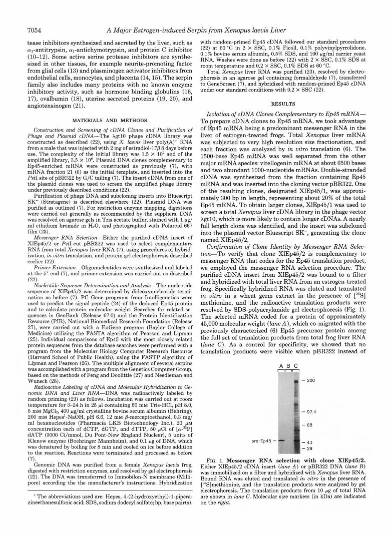
Confirmation of Clone Identity by Messenger RNA Selec- tion-To verify that clone XlEp45/2 is complementary to messenger RNA that codes for the Ep45 translation product, we employed the messenger RNA selection procedure. The purified cDNA insert from XlEp45/2 was bound to a filter and hybridized with total liver RNA from an estrogen-treated frog. Specifically hybridized RNA was eluted and translated in vitro in a wheat germ extract in the presence of [35S] methionine, and the radioactive translation products were resolved by SDS-polyacrylamide gel electrophoresis (Fig. 1). The selected mRNA coded for a protein of approximately 45,000 molecular weight (lane A ) , which co-migrated with the previously characterized (6) Ep45 precursor protein among the full set of translation products from total frog liver RNA (lane C ) . As a control for specificity, we showed that no translation products were visible when pBR322 instead of
nnn A B C
- 200
- 97.4
- 68
pre-Ep45 - - FIG. 1. Messenger RNA selection with clone XlEp45/2.
Either XIEp45/2 cDNA insert (lane A ) or pBR322 DNA (lane B ) was immobilized on a filter and hybridized with Xenopus liver RNA. Bound RNA was eluted and translated in uitro in the presence of [35S]methionine, and the translation products were analyzed by gel electrophoresis. The translation products from 10 pg of total RNA are shown in lane C. Molecular size markers (in kDa) are indicated on the right.
A Major Estrogen-induced Serpin from Xenopus &vis Liver 7055
Ep45 cDNA was used to select mRNA ( l a n e B ) . Analysis of the Structure and Sequence of cDNA Clone
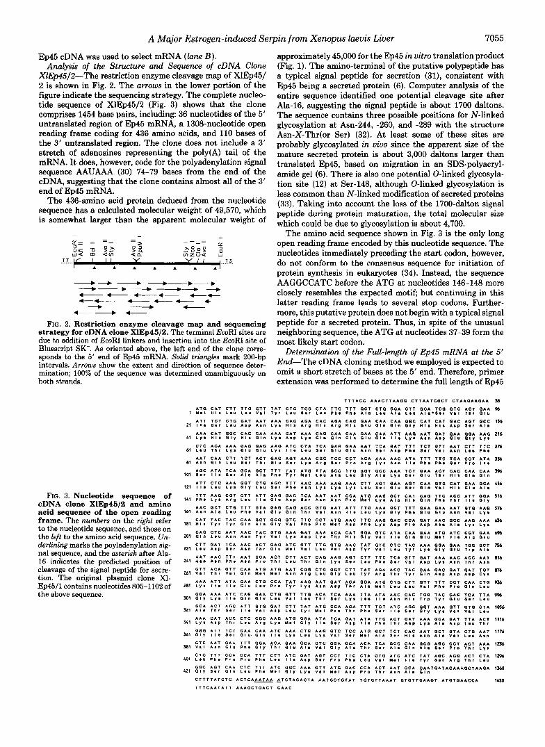
XlEp45/2"The restriction enzyme cleavage map of XlEp451 2 is shown in Fig. 2. The arrows in the lower portion of the figure indicate the sequencing strategy. The complete nucleo- tide sequence of XlEp45/2 (Fig. 3) shows that the clone comprises 1454 base pairs, including: 36 nucleotides of the 5' untranslated region of Ep45 mRNA, a 1308-nucleotide open reading frame coding for 436 amino acids, and 110 bases of the 3' untranslated region. The clone does not include a 3' stretch of adenosines representing the poly(A) tail of the mRNA. It does, however, code for the polyadenylation signal sequence AAUAAA (30) 74-79 bases from the end of the cDNA, suggesting that the clone contains almost all of the 3' end of Ep45 mRNA.
The 436-amino acid protein deduced from the nucleotide sequence has a calculated molecular weight of 49,570, which is somewhat larger than the apparent molecular weight of
"-d 4dd "---"--*"--*"---, "* -+4-"-G " ++ -4---"- + "+ __* +
&"--, 4"-
FIG. 2. Restriction enzyme cleavage map and sequencing strategy for cDNA clone XIEp46/2. The terminal EcoRI sites are due to addition of EcoRI linkers and insertion into the EcoRI site of Bluescript SK-. As oriented above, the left end of the clone corre- sponds to the 5' end of Ep45 mRNA. Solid triangles mark 200-bp intervals. Arrows show the extent and direction of sequence deter- mination; 100% of the sequence was determined unambiguously on both strands.
FIG. 3. Nucleotide sequence of cDNA clone XlEp45/2 and amino acid sequence of the open reading frame. The numbers on the right refer to the nucleotide sequence, and those on the left to the amino acid sequence. Un- derlining marks the poyladenylation sig- nal sequence, and the asterisk after Ala- 16 indicates the predicted position of cleavage of the signal peptide for secre- tion. The original plasmid clone XI- Ep45/1 contains nucleotides 805-1102 of the above sequence.
1
21
41
61
81
101
121
141
161
181
201
221
24 1
261
281
301
321
X 1
361
381
401
421
approximately 45,000 for the Ep45 in vitro translation product (Fig. 1). The amino-terminal of the putative polypeptide has a typical signal peptide for secretion (31), consistent with Ep45 being a secreted protein (6). Computer analysis of the entire sequence identified one potential cleavage site after Ala-16, suggesting the signal peptide is about 1700 daltons. The sequence contains three possible positions for N-linked glycosylation at Asn-244, -260, and -289 with the structure Asn-X-Thr(or Ser) (32). At least some of these sites are probably glycosylated in vivo since the apparent size of the mature secreted protein is about 3,000 daltons larger than translated Ep45, based on migration in an SDS-polyacryl- amide gel (6). There is also one potential 0-linked glycosyla- tion site (12) at Ser-148, although 0-linked glycosylation is less common than N-linked modification of secreted proteins (33). Taking into account the loss of the 1700-dalton signal peptide during protein maturation, the total molecular size which could be due to glycosylation is about 4,700.
The amino acid sequence shown in Fig. 3 is the only long open reading frame encoded by this nucleotide sequence. The nucleotides immediately preceding the start codon, however, do not conform to the consensus sequence for initiation of protein synthesis in eukaryotes (34). Instead, the sequence AAGGCCATC before the ATG at nucleotides 146-148 more closely resembles the expected motif; but continuing in this latter reading frame leads to several stop codons. Further- more, this putative protein does not begin with a typical signal peptide for a secreted protein. Thus, in spite of the unusual neighboring sequence, the ATG at nucleotides 37-39 form the most likely start codon.
Determination of t& Full-length of Ep45 mRNA at the 5' End-The cDNA cloning method we employed is expected to omit a short stretch of bases at the 5' end. Therefore, primer extension was performed to determine the full length of Ep45
T T T A C C A A A C T T A A G G C T T A A T C G C T C T A A G A A G A A 56
7056 A Major Estrogen-induced Serpin from Xenopus laevis Liver
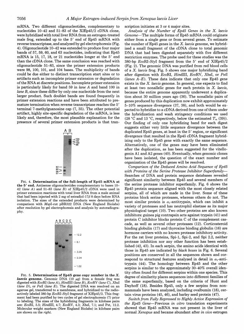
mRNA. Two different oligonucleotides, complementary to nucleotides 10-42 and 51-83 of the XlEp45/2 cDNA clone, were hybridized with total liver RNA from an estrogen-treated male frog, extended up to the 5‘ end of Ep45 mRNA with reverse transcriptase, and analyzed by gel electrophoresis (Fig. 4). Oligonucleotide 10-42 was extended to produce four major bands of 57, 59, 60, and 63 nucleotides, indicating that Ep45 mRNA is 15, 17, 18, or 21 nucleotides longer at the 5’ end than the cDNA clone. The same conclusion was reached with oligonucleotide 51-83, since the primer extension products were 98, 100, 101, and 104 bases. The multiplicity of bands could be due either to distinct transcription start sites or to artifacts such as incomplete primer extension or degradation of the RNA at discrete positions. Incomplete primer extension is particularly likely for band 59 in lune A and band 100 in lane B, since these differ by only one nucleotide from the next larger product. Such doublet bands are found frequently in primer extension reactions and have been attributed to pre- mature termination when reverse transcriptase reaches the 5’ terminal 7-methylguanosine cap (7, 35). The other potential artifact, highly site-specific degradation of the mRNA, is less likely and, therefore, the most plausible explanation for the presence of several primer extension products is that tran-
A B nn 1””11 _/lo4 #:E
FIG. 4. Determination of the full-length of Ep45 mRNA at the 5’ end. Antisense oligonucleotides complementary to bases 10- 42 ( l a n e A ) and 51-83 (lane B ) of XlEp45/2 cDNA were used in primer extension reactions with t o t a l liver RNA from a male animal that had been injected with 2 mg of estradiol-17@ 8 days before RNA isolation. The sizes of the extended products were determined by comparison with MspI-cut pBR322 DNA (New England Biolabs) after resolution by gel electrophoresis and analysis by autoradiogra- phy.
nnnnn A B C D E
- 12.2
- 6.1
- 3.05
- 2.04
- 1.02
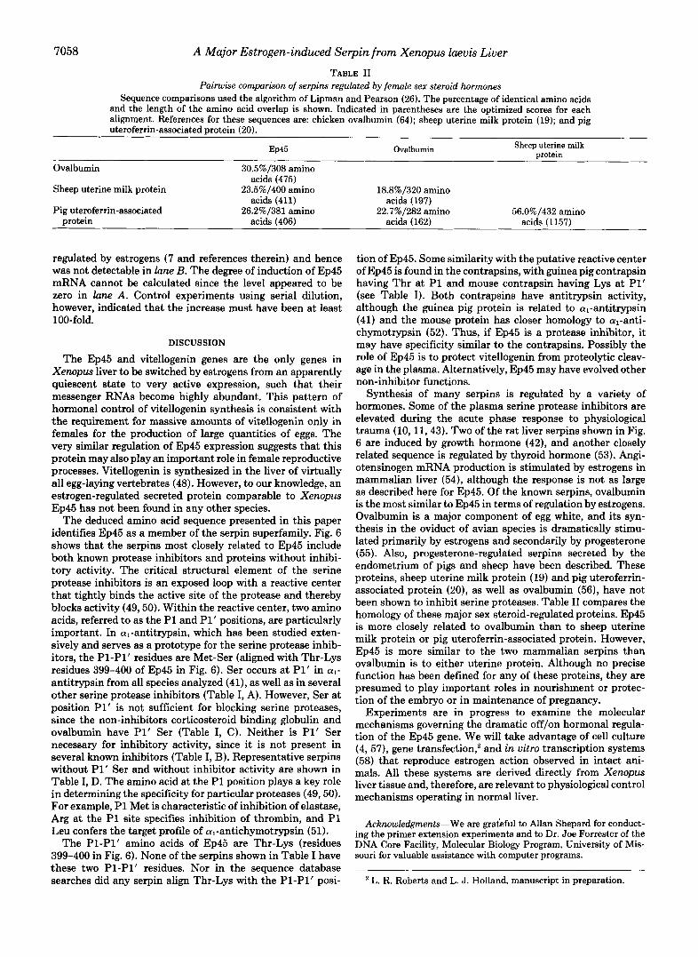
FIG. 5. Determination of Ep45 gene copy number in the X. laevis genome. Genomic DNA (10 pg) from a female frog was digested with EcoRI ( l a n e A ) , HindIII (lane B ) , EcoRV (lane C), XbaI (lane D), or PstI ( l a n e E ) . The digested DNA was resolved on an agarose gel, transferred to a membrane, and hybridized to the radio- actively labeled 380-bp EcoRI-Sty1 fragment of XIEp45/2. This frag- ment had been purified by two cycles of gel electrophoresis (7) prior to labeling. The sizes of the hybridizing fragments in kilobase pairs are: EcoRI, 5.5; HindIII, 4.7; EcoRV, 4.3; XbaI, 7.1; and Pst, 12.2. Molecular weight markers (New England Biolabs) in kilobase pairs are shown on the right.
scription initiates at 3 or 4 major sites. Analysis of the Number of Ep45 Genes in the X. laevis
Genome-The multiple forms of Ep45 mRNA could originate either from a single gene or from several genes. To estimate the number of Ep45 genes in the X. laevis genome, we hybrid- ized a small fragment of the cDNA clone to total genomic DNA that had been digested separately with five different restriction enzymes. The probe used for these studies was the 380-bp EcoRI-Sty1 fragment from the 5’ end of XlEp45/2 (Fig. 2). The genomic DNA was purified from red blood cells of a X. laevis frog. Fig. 5 shows one major hybridizing band after digestion with EcoRI, HindIII, EcoRV, XbaI, or PstI (lunes A-E). These data indicate that only one Ep45 gene exists in the X. laevis genome. In general, one expects to find a t least two nonallelic genes for each protein in X. laevis, because the entire genome apparently underwent a duplica- tion about 30 million years ago (36). The nonallelic pairs of genes produced by this duplication now exhibit approximately 5-10% sequence divergence (37, 38), and both would be ex- pected to hybridize to a cDNA derived from one of them under the hybridization and wash stringency conditions we used (28 “C and 13 “C, respectively, below the estimated T,,, (39)). Our finding of only one hybridizing band for each digest suggests either very little sequence divergence between the duplicated Ep45 genes, at least in the 5’ region, or significant divergence that resulted in the Ep45 cDNA fragment hybrid- izing only to the Ep45 gene with exactly the same sequence. Alternatively, one of the genes may have been eliminated after the duplication, as has been suggested for the vitello- genin A1 and A2 genes (40). Eventually, when genomic clones have been isolated, the question of the exact number and organization of the Ep45 genes will be resolved.
Comparison of the Deduced Amino Acid Sequence of Ep45 with Proteins of the Serine Protease Inhibitor Superfamily- Searches of DNA and protein sequence databases revealed significant similarity between Ep45 and several members of the serine protease inhibitor superfamily. Fig. 6 shows the Ep45 protein sequence aligned with the most closely related serpins, all of which are made in the liver. Some of these proteins block serine proteases, whereas others do not. The most similar protein is a1-antitrypsin, which can inhibit a variety of proteases and has neutrophil elastase as its major physiological target (10). Two other proteins are also known inhibitors: guinea pig contrapsin acts against trypsin (41) and protein C inhibitor blocks protein C of the complement cas- cade, as well as several other proteases (12). Corticosteroid binding globulin (17) and thyroxine binding globulin (16) are hormone carriers with no known protease inhibitory activity. For the rat liver proteins, Spi-1, Spi-2, and Spi 2.2, neither protease inhibition nor any other function has been estab- lished (42,43). In each serpin, the amino acids identical with those in Ep45 are indicated by black boxes in Fig. 6. Several positions are conserved in all the sequences shown and cor- respond to structural features analyzed in detail in al-anti- trypsin (44). The homology between Ep45 and the other serpins is similar to the approximately 30-40% overall iden- tity often found for different serpins within one species. This degree of similarity places sequences into different families of the same superfamily, based on the criteria of Hunt and Dayhoff (18). Besides Ep45, only a few serpins from non- mammals have been analyzed, including ovalbumin (18), cer- tain virus proteins (45,46), and barley seed protein (47).
Switch from Fully Repressed to Highly Active Expression of the Ep45 Gene-Previous in vitro translation experiments showed that Ep45 mRNA was not present in the liver of normal Xenopus and became abundant after in vivo estrogen

A Major Estrogen-induced Serpin from Xenopus luevis Liver 7057
FIG. 6. Comparison of Xenopus Ep45 deduced amino acid sequence with diverse members of the ser ine protease inhibitor superfamily. The Ep45 sequence is compared with the most closely related serpins from the data base sequence searches. The super- script numbers at the end of each seg- ment indicate the amino acid number for the Ep45 sequence. The multiple align- ment was generated as described under "Materials and Methods." The numbers at the end of each sequence show the percentage of identical residues over the indicated stretch of amino acids and the optimized score of each alignment, from pairwise comparisons of Ep45 with each serpin using the Lipman and Pearson algorithm (26). The rank order is based on the optimized scores. The references for the sequences are: human al-anti- trypsin (AT) , 10; human corticosteroid binding globulin (CBG), 17; guinea pig contrapsin, 41; rat Spi 2.2, 43 and Pro- tein Identification Resource accession number S08100; rat Spi-1 and Spi-2, 42 and Protein Identification Resource accession number S08102; human pro- tein C inhibitor, 12; and human thyrox- ine binding globulin (TBG), 16. al-An- titrypsin from other species is also sim- ilar to Ep45, but only the human sequence is shown.
albumin - Ep45 -
A B nn
1-
- 4110
- 1825
ferritin - # I ) -
FIG. '/. Induction of Ep45 mRNA levels in the liver of Xen- opus frogs treated with estrogens. Total liver RNA was purified from either an untreated male frog ( l a n e A ) or one that had received a 2-mg injection of estradiol-17P 4 days before use ( l u n e B) . The RNA was size-fractionated on an agarose/formaldehyde gel, trans- ferred to Genescreen, hybridized with "P-labeled XlEp45/1 cDNA and a ferritin mRNA-specific probe termed Ep20/70 (7), and sub- jected to autoradiography. Xenopus 28 S (4110 bases) and 18 S (1825 bases) ribosomal RNA present in our preparations of liver RNA were used as size markers (59).
stimulation (6). In vitro translation, however, is not as sensi- tive as molecular hybridization for detecting mRNA. There- fore, we used the Ep45 cDNA clone to analyze hepatic Ep45 mRNA levels before and after estrogen treatment. Total liver RNA was isolated from either an untreated male frog or one that had received a 2-mg injection of estradiol-170 4 days before. The RNA was resolved by gel electrophoresis and hybridized both with Ep45 cDNA and with ferritin cDNA as a control (Fig. 7). We have previously established that the ferritin probe hybridizes with only one mRNA band (7). The Ep45 probe hybridized exclusively with one messenger RNA
TABLE I Comparison of PI-PI' amino acids of serpins
Serpin P1-P1' Reference
A. Inhibitor/Pl' serine al-Antitrypsin Met-Ser 10 Protein C inhibitor Arg-Ser 12 a,-Antichymotrypsin Leu-Ser 11,60
B. Inhibitor/Pl' not serine Mouse contrapsin Arg-Lys 43 Guinea pig contrapsin Thr-Ala 41 Plasminogen activator inhibitor-1 Arg-Met 61 Plasminogen activator inhibitor-2 Arg-Thr 62 Complement C1 inhibitor Arg-Thr 63
C. Not inhibitor/Pl' serine Corticosteroid binding globulin Thr-Ser 17 Ovalbumin Ala-Ser 56
D. Not inhibitor/Pl' not serine Thyroxine binding globulin Gln-Pro 16 Uterine milk protein Ala-Asn 19 Uteroferrin-associated protein Val-Asp 20
in the sample from the estrogen-stimulated animal ( l a n e B ) . This mRNA comprised about 1500 bases, the expected size of Ep45 mRNA from earlier mRNA size fractionation and in vitro translation studies (6). There was no trace of Ep45 mRNA in the liver of the normal frog ( l a n e A ) . The ferritin cDNA hybridized with a 1000-base mRNA that decreased relative to total RNA after estrogen administration, consist- ent with our previous findings (7). The faint radioactive band at approximately 2000 bases in lane A was due to previous hybridization of the membrane with 32P-labeled Xenopus al- bumin cDNA (23). Albumin mRNA is known to be negatively
7058 A Major Estrogen-induced Serpin from Xenopus laevis Liver TABLE I1
Pairwise comparison of serpins regulated by female sex steroid hormones Sequence comparisons used the algorithm of Lipman and Pearson (26). The percentage of identical amino acids
and the length of the amino acid overlap is shown. Indicated in parentheses are the optimized scores for each alignment. References for these sequences are: chicken ovalbumin (64); sheep uterine milk protein (19); and pig uteroferrin-associated protein (20).
EP45 Ovalbumin Sheep uterine milk protein
Ovalbumin 30.5%/308 amino
Sheep uterine milk protein 23.5%/400 amino 18.8%/320 amino
Pig uteroferrin-associated 26.2%/381 amino 22.7%/282 amino 56.0%/432 amino protein acids (406) acids (162) acids (1157)
acids (475)
acids (411) acids (197)
regulated by estrogens (7 and references therein) and hence was not detectable in lane B. The degree of induction of Ep45 mRNA cannot be calculated since the level appeared to be zero in lune A. Control experiments using serial dilution, however, indicated that the increase must have been at least 100-fold.
DISCUSSION
The Ep45 and vitellogenin genes are the only genes in Xenopus liver to be switched by estrogens from an apparently quiescent state to very active expression, such that their messenger RNAs become highly abundant. This pattern of hormonal control of vitellogenin synthesis is consistent with the requirement for massive amounts of vitellogenin only in females for the production of large quantities of eggs. The very similar regulation of Ep45 expression suggests that this protein may also play an important role in female reproductive processes. Vitellogenin is synthesized in the liver of virtually all egg-laying vertebrates (48). However, to our knowledge, an estrogen-regulated secreted protein comparable to Xenopus Ep45 has not been found in any other species.
The deduced amino acid sequence presented in this paper identifies Ep45 as a member of the serpin superfamily. Fig. 6 shows that the serpins most closely related to Ep45 include both known protease inhibitors and proteins without inhibi- tory activity. The critical structural element of the serine protease inhibitors is an exposed loop with a reactive center that tightly binds the active site of the protease and thereby blocks activity (49,50). Within the reactive center, two amino acids, referred to as the P1 and P1’ positions, are particularly important. In a,-antitrypsin, which has been studied exten- sively and serves as a prototype for the serine protease inhib- itors, the P1-P1’ residues are Met-Ser (aligned with Thr-Lys residues 399-400 of Ep45 in Fig. 6). Ser occurs at P1’ in al- antitrypsin from all species analyzed (41), as well as in several other serine protease inhibitors (Table I, A). However, Ser at position P1’ is not sufficient for blocking serine proteases, since the non-inhibitors corticosteroid binding globulin and ovalbumin have P1’ Ser (Table I, C). Neither is P1‘ Ser necessary for inhibitory activity, since it is not present in several known inhibitors (Table I, B). Representative serpins without P1‘ Ser and without inhibitor activity are shown in Table I, D. The amino acid at the P1 position plays a key role in determining the specificity for particular proteases (49,50). For example, P1 Met is characteristic of inhibition of elastase, Arg at the P1 site specifies inhibition of thrombin, and P1 Leu confers the target profile of al-antichymotrypsin (51).
The P1-P1’ amino acids of Ep45 are Thr-Lys (residues 399-400 in Fig. 6). None of the serpins shown in Table I have these two P1-P1’ residues. Nor in the sequence database searches did any serpin align Thr-Lys with the P1-P1’ posi-
tion of Ep45. Some similarity with the putative reactive center of Ep45 is found in the contrapsins, with guinea pig contrapsin having Thr at P1 and mouse contrapsin having Lys at P1’ (see Table I). Both contrapsins have antitrypsin activity, although the guinea pig protein is related to cq-antitrypsin (41) and the mouse protein has closer homology to al-anti- chymotrypsin (52). Thus, if Ep45 is a protease inhibitor, it may have specificity similar to the contrapsins. Possibly the role of Ep45 is to protect vitellogenin from proteolytic cleav- age in the plasma. Alternatively, Ep45 may have evolved other non-inhibitor functions.
Synthesis of many serpins is regulated by a variety of hormones. Some of the plasma serine protease inhibitors are elevated during the acute phase response to physiological trauma (10, 11,43). Two of the rat liver serpins shown in Fig. 6 are induced by growth hormone (42), and another closely related sequence is regulated by thyroid hormone (53). Angi- otensinogen mRNA production is stimulated by estrogens in mammalian liver (54), although the response is not as large as described here for Ep45. Of the known serpins, ovalbumin is the most similar to Ep45 in terms of regulation by estrogens. Ovalbumin is a major component of egg white, and its syn- thesis in the oviduct of avian species is dramatically stimu- lated primarily by estrogens and secondarily by progesterone (55). Also, progesterone-regulated serpins secreted by the endometrium of pigs and sheep have been described. These proteins, sheep uterine milk protein (19) and pig uteroferrin- associated protein (20), as well as ovalbumin (56), have not been shown to inhibit serine proteases. Table I1 compares the homology of these major sex steroid-regulated proteins. Ep45 is more closely related to ovalbumin than to sheep uterine milk protein or pig uteroferrin-associated protein. However, Ep45 is more similar to the two mammalian serpins than ovalbumin is to either uterine protein. Although no precise function has been defined for any of these proteins, they are presumed to play important roles in nourishment or protec- tion of the embryo or in maintenance of pregnancy.
Experiments are in progress to examine the molecular mechanisms governing the dramatic off/on hormonal regula- tion of the Ep45 gene. We will take advantage of cell culture (4, 57), gene transfection,* and in vitro transcription systems (58) that reproduce estrogen action observed in intact ani- mals. All these systems are derived directly from Xenopus liver tissue and, therefore, are relevant to physiological control mechanisms operating in normal liver.
Acknowledgments-We are grateful to Allan Shepard for conduct- ing the primer extension experiments and to Dr. Joe Forrester of the DNA Core Facility, Molecular Biology Program, University of Mis- souri for valuable assistance with computer programs.
L. R. Roberts and L. J. Holland, manuscript in preparation.
A Major Estrogen-induced Serpin from Xenopus laevis Liver 7059
REFERENCES A. H., and Lingappa, V. R. (1979) in Secretory Mechanisms (Hopkins, C. R., and Duncan, C. J., eds) Vol. 33, pp. 9-36, Cambridge University Press, London 1. Clemens, M. J. (1974) Prog. Biophys. Mol. Biol. 28,69-108
2. Ryffel, G. u., Wahh w., and Weber, R. (1977) Cell 11,213-221 32. Marshall, R. D. (1974) ~ i o ~ h e ~ , sot, symp. 40, 17-26 3. Baker, H. J . 9 and ShaPiro, D. J. (1g77) J. Chem. 252p 8428- 33. Alberts, B., Dennis, B., Lewis, J., Raff, M., Roberts, K., and
4. Wangh, L. J., Osborne, J. A., Hentschel, C. C., and Tilly, R. Watson, J. D. (1983) Molecular Biology of the Cell, p. 345, Garland Publishing Inc., New York
5. May, F* E. B., RYffely G. u., Weber, R . ~ and Westley, B. R. 35. Bhattacharya, A., Shepard, A. R., Moser, D. R., and Holland, L.
6. Holland, L. J., and Wangh, L. J. (1987) Mol. cell. Endocr id . 36. Bisbee, c. A., Baker, M. A., Wilson, A. C., Hadji-Azimi, I., and
7. Holland, L. J., Wall, A. A., and Bhattacharya, A. (1991) Biochem- 37. Chien, y.-H., and Dawis, I. B. (1984) Mol. Cell. Biol. 4, 507-513
8. Lewis, J. A., Clemens, M. J., and Tata, J. R. (1976) Mol. Cell. and Schoenberg, D. R. (1989) Mol. Endocrinol. 3, 464-473
9. Spolski, R. J., Schneider, W., and Wangh, L. J. (1985) Deu. Biol. Kafatos, F. C. (1983) Methods Enzymol. 100,266-285
8434
(1979) Deu. Biol. 70,479-499
J. Biol. Chem. 257,13919-13923
34. Kozak, M. (1987) Nucleic Acids Res. 15,8125-8148
J. (1990) Mol. Cell. Endocrinol. 72, 213-220
Fischberg, M. (1977) Science 195, 785-787 49,63-73
istry 30,1965-1972 38. Moskaitis, J. E., Sargent, T. D., Smith, L. H. Jr., Pastori, R. L.,
Endocrinol. 4, 311-329 39. Beltz, G. A., Jacobs, K. A., Eickbush, T. H., Cherbas, P. T., and
40. Schubiger, J.-L., and Wahli, W. (1986) Nucleic Acids Res. 14,
Kurachi, K. (1984) Biochemistry 23,4828-4837 41. Suzuki, Y., Yoshida, K., Honda, E., and Sinohara, H. (1991) J.
Woo, S. L. C. (1983) Biochemistry 22,5055-5061 42. Yoon, J.-B., Towle, H. C., and Seelig, S. (1987) J. Biol. Chem.
Yamamoto, S., and Hashimoto, S. (1987) J. Bwl. Chem. 262, 43. Hill, R. E., and Hastie, N. D. (1987) Nature 326,96-99
108,332-340 10. Long, G. L., Chandra, T., Woo, S. L. C., Davie, E. W., and 8723-8734
11. Chandra, T., Stackhouse, R., Kidd, V. J., Robson, K. J. H., and Biol. Chem. 266,928-932
12. Suzuki, K., Deyashiki, Y., Nishioka, J., Kurachi, K., Akira, M., 262,4284-4289
611-616 44. Huber, R., and Carrell, R. W. (1989) Biochemistry 28,8951-8966 13. Sommer, J., Gloor, S. M., Rovelli, G. F., Hofsteenge, J., Nick, H., 45. Boursnell, M. E. G., Foulds, I. J., Campbell, J. I., and Binns, M.
14. Ginsburg, D., Zeheb, R., Yang, A. Y., Rafferty, U. M., Andreasen, 46. Upton, C., and McFadden, G. (1986) Mol. Cell. Biol. 6,265-276 Meier, R., and Monard, D. (1987) Biochemistry 26,6407-6410 M (1988) J. Gen. Virol. 69, 2995-3003
P. A., Nielson, L., Dano, K., Lebo, R. V., and Gelehrter, T. D. 47. Hejgaard, J., Rasmussen, S. K., Brandt, A., and Svendsen, I. (1986) J. Clin. Invest. 78,1673-1680 (1985) FEBS Lett. 180,89-94
15. Ye, R. D., Wun, T.-C., and Sadler, J. E. (1987) J. Biol. Chem. 48. Wallace, R. A. (1985) in Developmental Biology (Browder, L. W., 262,3718-3725 ed) Vol. I, pp. 127-177, Plenum Publishing Corp., New York
16. Flink, I. L., Bailey, T. J., Gustafson, T. A., Markham, B. E., and 49. Carrell, R. w., Pemberton, p. and Boswell, D- R. (1987) CoZd Morkin, E. (1986) Proc. Natl.Acad. Sci. U. S. A. 83,7708-7712 Spring Harbor Symp. Quunt. Biol. 52, 527-535
17. Hammond, G. L., Smith, C. L., Goping, 1. S., Underhill, D. A,, 50. Boswell, D. R.9 and Carrel19 R. w. (1988) BioessaYs 8~83-87 Harley, M. J., Reventos, J., Must.0, N. A., Gunsalus, G. L., and 51. Carrell, R., and Travis, J. (1985) Trends Biochem. SCi. 10,20-24 Bardin, C. W. (1987) Proc. Natl. A ~ & . Sei. U. S. A. 84, 5153- 52. Hill, R. E., Shaw, p. H., Boyd, p. A., Baumann, H., and Hastie, 5157 N. D. (1984) Nature 311, 175-177
18. Hunt, L. T., and Dayhoff, M. 0. (1980) Biochem. Bwphys. Res. 53- Tecce, M. F., Dozin, B., Magnuson, M- A., and Nikodem, v. M. Commun. 95,864-871 (1986) Biochemistry 26, 5831-5834
19. Ing, N. H., and Roberts, R. M. (1989) J. ~ i ~ l . C k m . 264, 3372- 54. Kunapuli, s. p., Benedct, c. R., and Kumar, A. (1987) Arch. 3379
M. (1990) Mol. Endocrinol. 4,428-440
Biochem. Biophys. 254,642-646 20. Malathy, p.-V., Imakawa, K., Simmen, R. C. M., and Roberts, R. 55. T., and Schimke, R. T. (1969) J. cell B i d 439 123-137
56. Stein, P. E., Leslie, A. G. W., Finch, J. T., Turnell, W. G., Laughlin, P. J., and Carrell, R. W. (1990) Nature 347, 99-102 21. Doolittle, R. F. (1983) Science 222, 417-419
22. Bhattacharya, A., Shepard, A. R., h,foser, D. R., Robeds, L. R., 57. Bhattacharya, A.9 and Holland, L. J. (1991) Mol. Endocrinol. 5 9
and L. J. (1991) Endocrinol. 76* '11-12' 58. Corthesy, B., Hipskind, R., Theulaz, I., and Wahli, W. (1988)
59. Holland, L. J., and Wangh, L. J. (1984) Mol. Cell. Biol. 4, 2543-
587-597 23. Holland, L. J., and Wangh, L. J. (1983) Nucleic Acids Res. 11,
24. von Heijne, G. (1986) Nucleic Acids Res. 14,4683-4690 3283-3300 Science 239, 1137-1139
2548 25. Pearson, w. R.7 and Lipman, D. J. (1988) PrOC. Natl. Acid. SCi. 60. r,.forii, M., and ~ ~ ~ ~ i ~ , J, (1983) J, ~ i ~ l . chern. 258,12749-12752
U. S. A. 85,2444-2448 - . ._ D. J. (1986) Proc. Natl. Acad. Sei. U. S. A. 83,6776-6780 26. Lipman, D. J., and Pearson, W. R. (1985) Science 227, 1435- 61. Ny, T., Sawdye, M., Lawrence, D., Millan, J. L., and Loskutoff,
1441 27. Feng, D.-F., and Doolittle, R. F. (1987) J. Mol. Euol. 25,351-360
62. Kiso, U., Kaudewitz, H., Henschen, A., Astedt,~B., Kruithof, E.
28. Need*eman, s. B., and WunSch, c. D- 489 63. Bock, S. C., Skriver, K., Nielsen, E., Thogersen, H.-C., Wiman, K. O., and Bachmann, F. (1988) FEBS Lett. 230,51-56
29. Feinberg, A. P., and Vogelstein, B. (1983) Anal. Biochem. 132, B., Donaldson, V. H., Eddy, R. L., Marrinan, J., Radziejewska, E., Huber, R., Shows, T. B., and Magnusson, S. (1986) Bio-
30. Proudfoot, N. J., and Brownlee, G. G. (1976) Nature 263, 211- 64. McReynolds, L., O'Malley, B. W., Nisbet, A. D., Fothergill, J. E., chemistry 25,4292-4301
214 Givol, D., Fields, S., Robertson, M., and Brownlee, G. G. (1978) 31. Blobel, G., Walter, P., Chang, C. N., Goldman, B. M., Erickson, Nature 273,723-728
443-453
6-13


![Study of Estrogen Receptor, Progesterone Receptor, …...[CANCER RESEARCH 49,4298-4304, August 1. 1989] Study of Estrogen Receptor, Progesterone Receptor, and the Estrogen-regulated](https://img.pdfslide.us/doc/110x75/5f95792bbdbd5e0915333803/study-of-estrogen-receptor-progesterone-receptor-cancer-research-494298-4304.jpg)