Embed Size (px)
Citation preview
Introduction
Among radiation-induced normal tissue injuries, earlyside effects appear within a few days to weeks after high-dose irradiation while late effects occur months to yearsafter irradiation with a latent period. Mitosis-linked deathis the main cause of the loss of cells in the early period.1,2
The radiation dose and radiosensitivity of the targettissue are two important factors in the degree of impact of
irradiation. Target tissues such as bone marrow, lymphoidtissues, and oral mucous membranes that proliferate con-tinuously tend to be sensitive to radiation, while little orno short-term effect of radiation occurs in muscle.2
A dose of 5 to 10 Gy of radiation can decrease the DNAcontent of osteoblasts in the proliferation, confluence, andpost-proliferation stages.3 Furthermore, it has been shownthat 10 to 20 Gy irradiation is capable of reducing bonemarrow cellularity for a week, although the effect is rever-sible and gradually returns to normal within 8 weeks.4 Themajor pathologic changes following fractionated radio-therapy from day 1 to 7 include edema and lack of hemato-poiesis, while intense hematopoietic activity and persistentedema are observed from day 10 to 14.5
─ 43 ─
A magnetic resonance imaging study on changes in rat mandibular bone marrow and pulptissue after high-dose irradiation
Wan Lee1, Byung-Do Lee1, Kang-Kyoo Lee2, Kwang-Joon Koh3,*1Department of Oral and Maxillofacial Radiology and Wonkwang Dental Research Institute, College of Dentistry, Wonkwang University,Iksan, Korea2Department of Radiation Oncology, School of Medicine, Wonkwang University, Iksan, Korea3Department of Oral and Maxillofacial Radiology, School of Dentistry and Institute of Oral Bioscience, Chonbuk National University,Jeonju, Korea
ABSTRACT
Purpose: This study was designed to evaluate whether magnetic resonance imaging (MRI) is appropriate fordetecting early changes in the mandibular bone marrow and pulp tissue of rats after high-dose irradiation.Materials and Methods: The right mandibles of Sprague-Dawley rats were irradiated with 10 Gy (Group 1, n==5)and 20 Gy (Group 2, n==5). Five non-irradiated animals were used as controls. The MR images of rat mandibleswere obtained before irradiation and once a week until week 4 after irradiation. From the MR images, the signalintensity (SI) of the mandibular bone marrow and pulp tissue of the incisor was interpreted. The MR images werecompared with the histopathologic findings. Results: The SI of the mandibular bone marrow had decreased on T2-weighted MR images. There was littledifference between Groups 1 and 2. The SI of the irradiated groups appeared to be lower than that of the controlgroup. The histopathologic findings showed that the trabecular bone in the irradiated group had increased. The SI ofthe irradiated pulp tissue had decreased on T2-weighted MR images. However, the SI of the MR images in Group 2was high in the atrophic pulp of the incisor apex at week 2 after irradiation. Conclusion: These patterns seen on MRI in rat bone marrow and pulp tissue were consistent with histopathologicfindings. They may be useful to assess radiogenic sclerotic changes in rat mandibular bone marrow. (Imaging SciDent 2014; 44 : 43-52)
KEY WORDS: Magnetic Resonance Imaging; Mandible; Bone Marrow; Irradiation
*This paper was supported by Wonkwang University in 2013.Received September 2, 2013; Revised September 25, 2013; Accepted October 6, 2013*Correspondence to : Prof. Kwang-Joon KohDepartment of Oral and Maxillofacial Radiology, School of Dentistry, 634-18 Keum-am-dong, Dukjin-gu, Jeonju 561-712, KoreaTel) 82-63-250-2023, Fax) 82-63-250-2081, E-mail) [email protected]
Imaging Science in Dentistry 2014; 44: 43-52http://dx.doi.org/10.5624/isd.2014.44.1.43
Copyright ⓒ 2014 by Korean Academy of Oral and Maxillofacial RadiologyThis is an Open Access article distributed under the terms of the Creative Commons Attribution Non-Commercial License (http://creativecommons.org/licenses/by-nc/3.0)
which permits unrestricted non-commercial use, distribution, and reproduction in any medium, provided the original work is properly cited.
Imaging Science in Dentistry∙pISSN 2233-7822 eISSN 2233-7830
The effects of dental irradiation, specifically, have alsobeen noted. For example, 20 Gy irradiation leads to cessa-tion of normal dentin deposition,6 and nuclear alterationsin the dental pulp tissue of rats can occur after fractionat-ed irradiation with a total dose of 60 Gy.7 Niehoff et al6
reported a significant decline in vertical bone appositionto the outer surface of irradiated mandibles. In childrenwho undergo irradiation, the growth retardation of thefacial skeleton occurs primarily in the jaws and teeth. Inthe mandible, growth retardation specifically occurs in theregion of the condylar head.8
Although computed tomography (CT) has been used forbone volume analysis of the irradiated mandible,9 MRI hasbeen known to be useful for detecting signs of radiogenicbone and soft tissue damage. Recognizing mandibularradiogenic bone change early is possible with MRI, allow-ing for early diagnosis before the onset of clinical symp-toms and bone destruction10 and can be used to evaluateearly bone marrow changes during radiotherapy.11 Eviden-ce of edema on MR images was found to consist of abnor-malities of the bone marrow with a decreased or increasedsignal intensity (SI).12,13
Comparing among imaging modalities, although CTbetter illustrates the degree and extent of cortical defects,14
it is not suitable for evaluating bone marrow in the earlypathologic stage, while radioisotope scanning has the crit-ical limitation of showing a high diagnostic sensitivity buta low specificity.10
After high-dose irradiation, identifying changes in themandibular bone marrow and adjacent soft tissues is impor-tant in order to evaluate tissue sensitivity and abnormality.The data available on early bone marrow and dental pulpchanges of irradiated mandibular bone visible by MRI arelimited.
Thus this study was designed to evaluate whether MRIis appropriate for detecting the early changes in the mandi-bular bone marrow and pulp tissue of rats after high-doseirradiation. Under different dosages of radiation, the ani-mals were monitored using MR T2-weighted images andthe data was confirmed by conventional staining methodswith tissues isolated from the rats. Thus, this paper pro-vides new insight into MRI diagnosis to predict early sideeffects of radiation.
Materials and Methods
Animals
The study was performed in accordance with the Guide
for Animal Experimentation by Wonkwang University andapproved by the Institutional Animal Care and Use Com-mittee of Wonkwang University.
Fifteen 4-week-old Sprague-Dawley rats were obtainedfrom Hubet Laboratories (Iksan, South Korea). Before theexperiment, the rats were allowed 7 days to acclimatize tothe conditions.
Five rats were not irradiated to use as controls. Ten ratswere randomly divided into two groups of five rats each.Group 1 was given a single dose of external irradiation of10 Gy, and Group 2 was given a single dose of 20 Gy.
MRI examination
MRI examinations were performed with a 4.7-T animalMRI instrument (Bruker, Ettlingen, Germany) using a lin-ear volume coil for rats. Axial T2-weighted images (T2WI)were obtained with the use of the Turbo RARE T2 tech-nique. The imaging parameters were repetition time (TR)11776.3 ms, echo time (TE) 36 ms, flip angle 180 degrees,average 6, 256×256 matrix, 0.0117×0.0117 cm pixel size,0.5 mm slice thickness, and 100 slices. Under inhalationalisoflurane anesthesia, an MRI of the rat’s head was takenbefore irradiation and once a week for 4 weeks after irra-diation.
Radiation delivery procedure
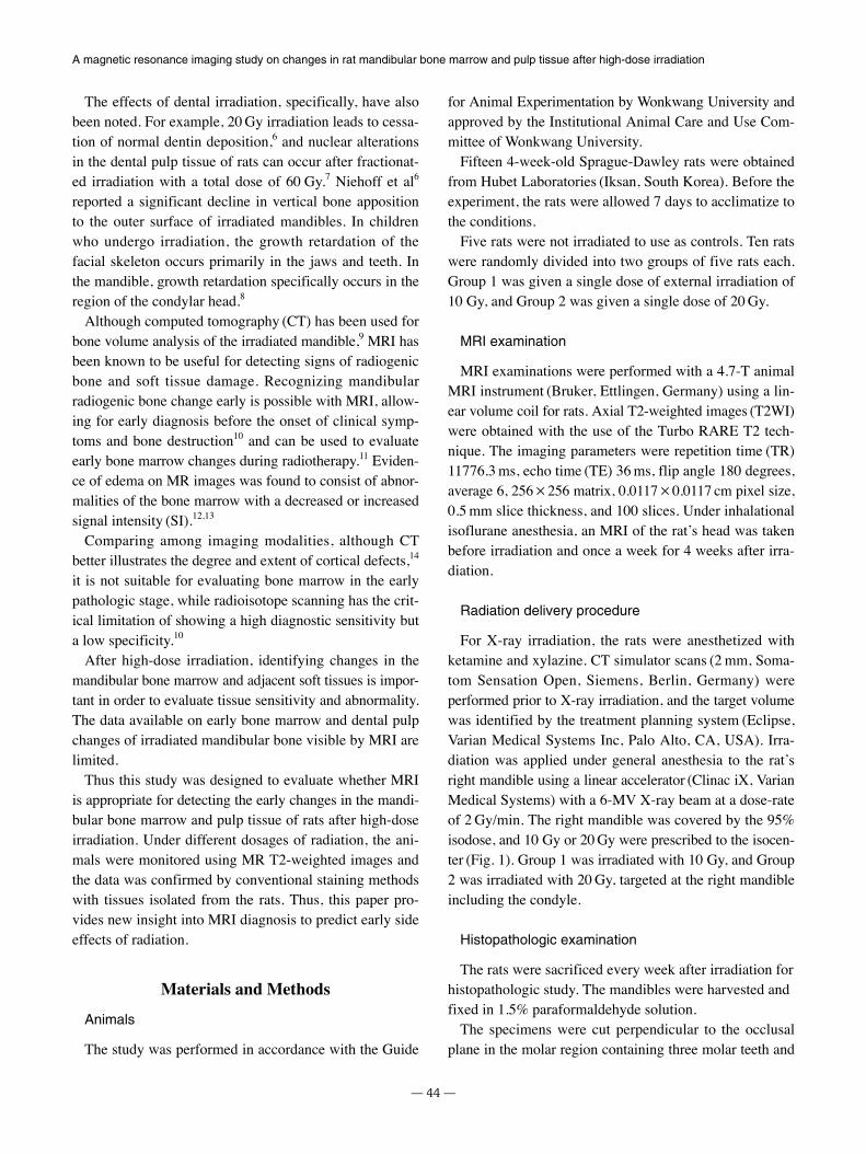
For X-ray irradiation, the rats were anesthetized withketamine and xylazine. CT simulator scans (2 mm, Soma-tom Sensation Open, Siemens, Berlin, Germany) wereperformed prior to X-ray irradiation, and the target volumewas identified by the treatment planning system (Eclipse,Varian Medical Systems Inc, Palo Alto, CA, USA). Irra-diation was applied under general anesthesia to the rat’sright mandible using a linear accelerator (Clinac iX, VarianMedical Systems) with a 6-MV X-ray beam at a dose-rateof 2 Gy/min. The right mandible was covered by the 95%isodose, and 10 Gy or 20 Gy were prescribed to the isocen-ter (Fig. 1). Group 1 was irradiated with 10 Gy, and Group2 was irradiated with 20 Gy, targeted at the right mandibleincluding the condyle.
Histopathologic examination
The rats were sacrificed every week after irradiation forhistopathologic study. The mandibles were harvested andfixed in 1.5% paraformaldehyde solution.
The specimens were cut perpendicular to the occlusalplane in the molar region containing three molar teeth and
─ 44 ─
A magnetic resonance imaging study on changes in rat mandibular bone marrow and pulp tissue after high-dose irradiation
the middle third of the incisor. The explanted bone speci-mens were fixed for 24 h in a 10% formol-saline solutionand then decalcified in a 10% nitric acid bath for 48 h. Thedecalcified bone specimens were dehydrated with alcohol(100% ethanol solutions) followed by toluene. The speci-mens of bone were then embedded in paraffin. Serial sec-tions of 5 μm were cut using a microtome. The sectionswere stained with hematoxylin-eosin (HE) and were thenexamined under a light microscope.
Image analysis
In order to focus accurately in the same plane of the bonemarrow every week, the plane was adjusted. The axialplane was modified to the pulp floor of the molar teeth,the sagittal plane to the midline of the face, and the coro-nal plane to the first molar including both roots. For inci-sor pulp evaluation, the sagittal plane was modified to fol-low the long axis of the incisor.
The MR SI of the region of interest (ROI) in the rat man-dible was evaluated using the OnDemand3D program
─ 45 ─
Wan Lee et al
Fig. 1. Target volume of the right mandible including the ramus and condyle in CT scans are set prior to high-dose irradiation. 100%isodoses are marked in yellow. Pre-irradiated planning enables setting a one-sided application of radiation and minimizing the change inadjacent tissues, especially the brain.
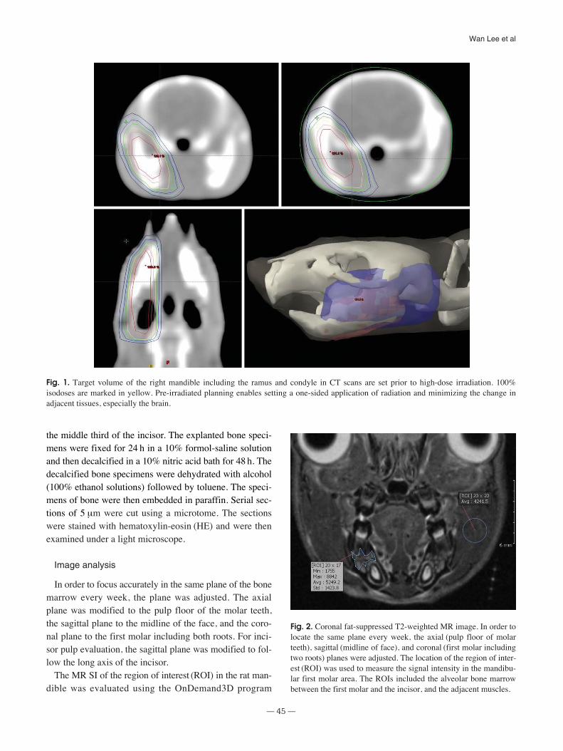
Fig. 2. Coronal fat-suppressed T2-weighted MR image. In order tolocate the same plane every week, the axial (pulp floor of molarteeth), sagittal (midline of face), and coronal (first molar includingtwo roots) planes were adjusted. The location of the region of inter-est (ROI) was used to measure the signal intensity in the mandibu-lar first molar area. The ROIs included the alveolar bone marrowbetween the first molar and the incisor, and the adjacent muscles.
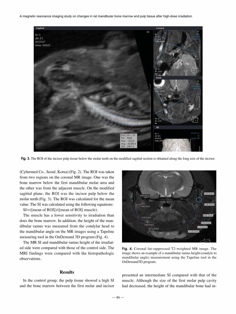
(Cybermed Co., Seoul, Korea) (Fig. 2). The ROI was takenfrom two regions on the coronal MR image. One was thebone marrow below the first mandibular molar area andthe other was from the adjacent muscle. On the modifiedsagittal plane, the ROI was the incisor pulp below themolar teeth (Fig. 3). The ROI was calculated for the meanvalue. The SI was calculated using the following equations:
SI==([mean of ROI])/([mean of ROI] muscle). The muscle has a lower sensitivity to irradiation than
does the bone marrow. In addition, the height of the man-dibular ramus was measured from the condylar head tothe mandibular angle on the MR images using a Tapelinemeasuring tool in the OnDemand 3D program (Fig. 4).
The MR SI and mandibular ramus height of the irradiat-ed side were compared with those of the control side. TheMRI findings were compared with the histopathologicobservations.
Results
In the control group, the pulp tissue showed a high SIand the bone marrow between the first molar and incisor
presented an intermediate SI compared with that of themuscle. Although the size of the first molar pulp cavityhad decreased, the height of the mandibular bone had in-
─ 46 ─
A magnetic resonance imaging study on changes in rat mandibular bone marrow and pulp tissue after high-dose irradiation
Fig. 3. The ROI of the incisor pulp tissue below the molar teeth on the modified sagittal section is obtained along the long axis of the incisor.
Fig. 4. Coronal fat-suppressed T2-weighted MR image. Theimage shows an example of a mandibular ramus height (condyle tomandibular angle) measurement using the Tapeline tool in theOnDemand3D program.
creased during the period. The SI of the bone marrow hadfallen continuously from week to week after irradiation.
In the irradiated groups, both Groups 1 and 2, the pat-terns of SI in the pulp and the bone marrow were very sim-ilar to those of the control group. The SI of the right irra-diated bone marrow had decreased. The SI of the irradiat-ed groups was lower than that of the control group. Thesedifferences occurred in the first week after irradiation.Based on these results, it can be concluded that bone mar-row is very sensitive to X-rays. However, only a small dif-ference was found between Groups 1 and 2.
The height of the mandibular bone and size of the pulphad decreased more than those in the control group, butthe SI in the muscle was slightly higher than that in thecontrol and contralateral side.
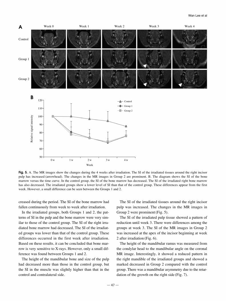
The SI of the irradiated tissues around the right incisorpulp was increased. The changes in the MR images inGroup 2 were prominent (Fig. 5).
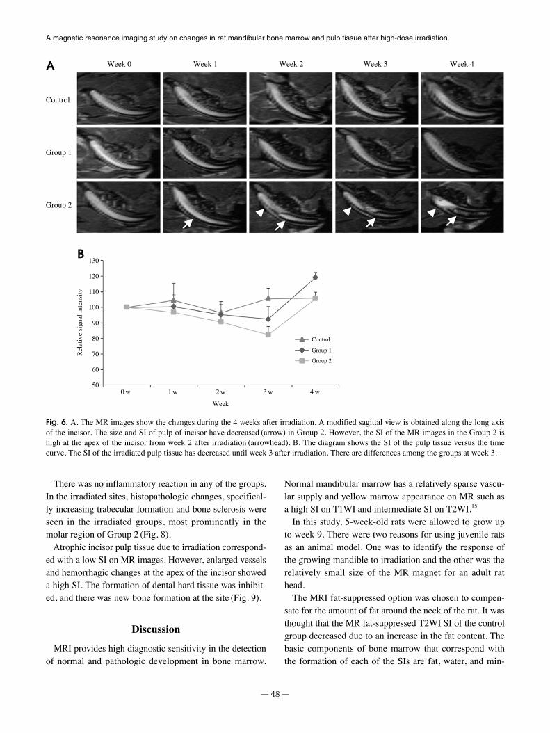
The SI of the irradiated pulp tissue showed a pattern ofreduction until week 3. There were differences among thegroups at week 3. The SI of the MR images in Group 2was increased at the apex of the incisor beginning at week2 after irradiation (Fig. 6).
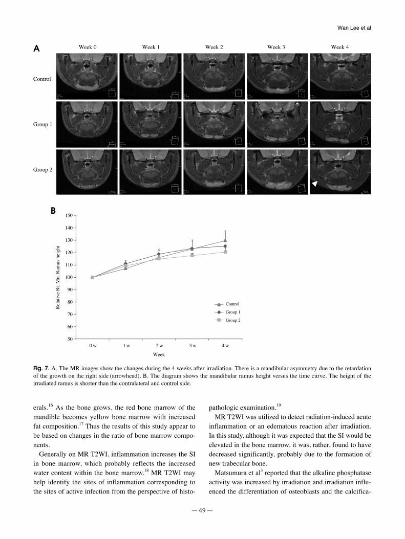
The height of the mandibular ramus was measured fromthe condylar head to the mandibular angle on the coronalMR image. Interestingly, it showed a reduced pattern inthe right mandible of the irradiated groups and showed amarked decreased in Group 2 compared with the controlgroup. There was a mandibular asymmetry due to the retar-dation of the growth on the right side (Fig. 7).
─ 47 ─
Wan Lee et al
Fig. 5. A. The MR images show the changes during the 4 weeks after irradiation. The SI of the irradiated tissues around the right incisorpulp has increased (arrowhead). The changes in the MR images in Group 2 are prominent. B. The diagram shows the SI of the bonemarrow versus the time curve. In the control group, the SI of the bone marrow has decreased. The SI of the irradiated right bone marrowhas also decreased. The irradiated groups show a lower level of SI than that of the control group. These differences appear from the firstweek. However, a small difference can be seen between the Groups 1 and 2.
Week 0 Week 1 Week 2 Week 3 Week 4
Control
Group 1
Group 2
120
110
100
90
80
70
60
50
Control
Group 1
Group 2
0 w 1 w 2 w 3 w 4 w
Week
Rel
ativ
e si
gnal
inte
nsity
A
B
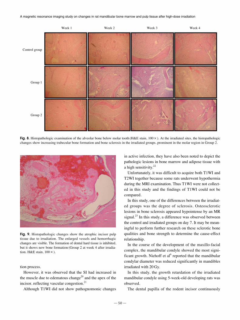
There was no inflammatory reaction in any of the groups.In the irradiated sites, histopathologic changes, specifical-ly increasing trabecular formation and bone sclerosis wereseen in the irradiated groups, most prominently in themolar region of Group 2 (Fig. 8).
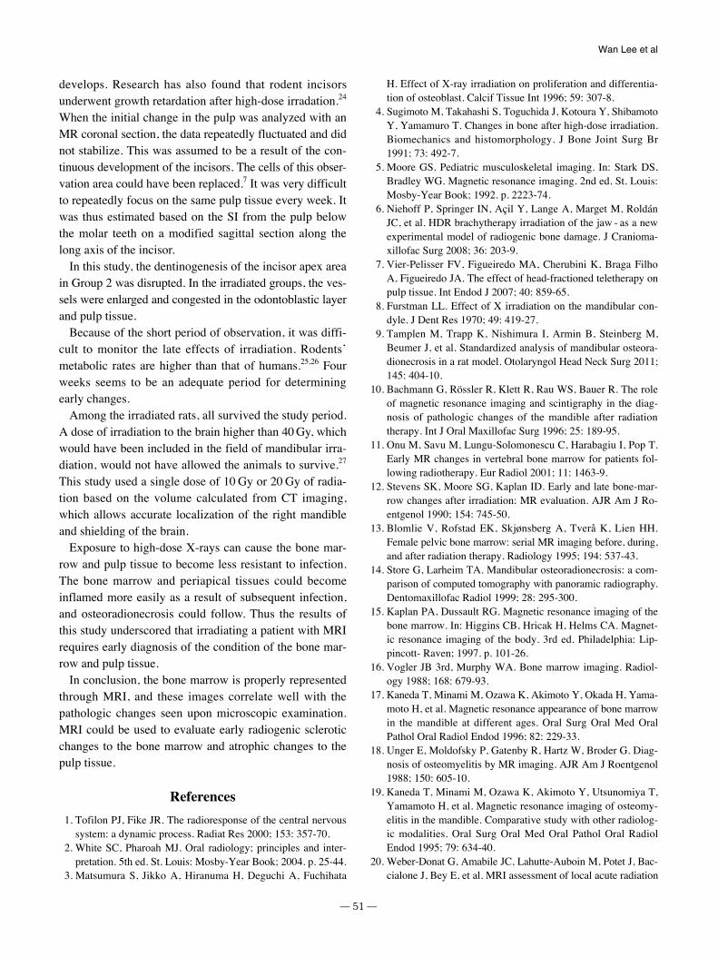
Atrophic incisor pulp tissue due to irradiation correspond-ed with a low SI on MR images. However, enlarged vesselsand hemorrhagic changes at the apex of the incisor showeda high SI. The formation of dental hard tissue was inhibit-ed, and there was new bone formation at the site (Fig. 9).
Discussion
MRI provides high diagnostic sensitivity in the detectionof normal and pathologic development in bone marrow.
Normal mandibular marrow has a relatively sparse vascu-lar supply and yellow marrow appearance on MR such asa high SI on T1WI and intermediate SI on T2WI.15
In this study, 5-week-old rats were allowed to grow upto week 9. There were two reasons for using juvenile ratsas an animal model. One was to identify the response ofthe growing mandible to irradiation and the other was therelatively small size of the MR magnet for an adult rathead.
The MRI fat-suppressed option was chosen to compen-sate for the amount of fat around the neck of the rat. It wasthought that the MR fat-suppressed T2WI SI of the controlgroup decreased due to an increase in the fat content. Thebasic components of bone marrow that correspond withthe formation of each of the SIs are fat, water, and min-
─ 48 ─
A magnetic resonance imaging study on changes in rat mandibular bone marrow and pulp tissue after high-dose irradiation
Fig. 6. A. The MR images show the changes during the 4 weeks after irradiation. A modified sagittal view is obtained along the long axisof the incisor. The size and SI of pulp of incisor have decreased (arrow) in Group 2. However, the SI of the MR images in the Group 2 ishigh at the apex of the incisor from week 2 after irradiation (arrowhead). B. The diagram shows the SI of the pulp tissue versus the timecurve. The SI of the irradiated pulp tissue has decreased until week 3 after irradiation. There are differences among the groups at week 3.
Week 0 Week 1 Week 2 Week 3 Week 4
130
120
110
100
90
80
70
60
50
Control
Group 1
Group 2
0 w 1 w 2 w 3 w 4 w
Week
Rel
ativ
e si
gnal
inte
nsity
Control
Group 1
Group 2
A
B
erals.16 As the bone grows, the red bone marrow of themandible becomes yellow bone marrow with increasedfat composition.17 Thus the results of this study appear tobe based on changes in the ratio of bone marrow compo-nents.
Generally on MR T2WI, inflammation increases the SIin bone marrow, which probably reflects the increasedwater content within the bone marrow.18 MR T2WI mayhelp identify the sites of inflammation corresponding tothe sites of active infection from the perspective of histo-
pathologic examination.19
MR T2WI was utilized to detect radiation-induced acuteinflammation or an edematous reaction after irradiation.In this study, although it was expected that the SI would beelevated in the bone marrow, it was, rather, found to havedecreased significantly, probably due to the formation ofnew trabecular bone.
Matsumura et al3 reported that the alkaline phosphataseactivity was increased by irradiation and irradiation influ-enced the differentiation of osteoblasts and the calcifica-
─ 49 ─
Wan Lee et al
Fig. 7. A. The MR images show the changes during the 4 weeks after irradiation. There is a mandibular asymmetry due to the retardationof the growth on the right side (arrowhead). B. The diagram shows the mandibular ramus height versus the time curve. The height of theirradiated ramus is shorter than the contralateral and control side.
Week 0 Week 1 Week 2 Week 3 Week 4
150
140
130
120
110
100
90
80
70
60
50
Control
Group 1
Group 2
0 w 1 w 2 w 3 w 4 w
Week
Rel
ativ
e R
t. M
n. R
amus
hei
ght
Control
Group 1
Group 2
A
B
tion process.However, it was observed that the SI had increased in
the muscle due to edematous change20 and the apex of theincisor, reflecting vascular congestion.21
Although T1WI did not show pathognomonic changes
in active infection, they have also been noted to depict thepathologic lesions in bone marrow and adipose tissue witha high sensitivity.22
Unfortunately, it was difficult to acquire both T1WI andT2WI together because some rats underwent hypothermiaduring the MRI examination. Thus T1WI were not collect-ed in this study and the findings of T1WI could not becompared.
In this study, one of the differences between the irradiat-ed groups was the degree of sclerosis. Osteoscleroticlesions in bone sclerosis appeared hypointense by an MRsignal.23 In this study, a difference was observed betweenthe control and irradiated groups on day 7. It may be mean-ingful to perform further research on these sclerotic bonequalities and bone strength to determine the cause-effectrelationship.
In the course of the development of the maxillo-facialcomplex, the mandibular condyle showed the most signi-ficant growth. Niehoff et al6 reported that the mandibularcondylar diameter was reduced significantly in mandiblesirradiated with 20 Gy.
In this study, the growth retardation of the irradiatedmandibular condyle using 5-week-old developing rats wasobserved.
The dental papilla of the rodent incisor continuously
─ 50 ─
A magnetic resonance imaging study on changes in rat mandibular bone marrow and pulp tissue after high-dose irradiation
Fig. 8. Histopathologic examination of the alveolar bone below molar tooth (H&E stain, 100×). At the irradiated sites, the histopathologicchanges show increasing trabecular bone formation and bone sclerosis in the irradiated groups, prominent in the molar region in Group 2.
Control group
Group 1
Group 2
Week 1 Week 2 Week 3 Week 4
Fig. 9. Histopathologic changes show the atrophic incisor pulptissue due to irradiation. The enlarged vessels and hemorrhagicchanges are visible. The formation of dental hard tissue is inhibited,but it shows new bone formation (Group 2 at week 4 after irradia-tion. H&E stain, 100×).
develops. Research has also found that rodent incisorsunderwent growth retardation after high-dose irradation.24
When the initial change in the pulp was analyzed with anMR coronal section, the data repeatedly fluctuated and didnot stabilize. This was assumed to be a result of the con-tinuous development of the incisors. The cells of this obser-vation area could have been replaced.7 It was very difficultto repeatedly focus on the same pulp tissue every week. Itwas thus estimated based on the SI from the pulp belowthe molar teeth on a modified sagittal section along thelong axis of the incisor.
In this study, the dentinogenesis of the incisor apex areain Group 2 was disrupted. In the irradiated groups, the ves-sels were enlarged and congested in the odontoblastic layerand pulp tissue.
Because of the short period of observation, it was diffi-cult to monitor the late effects of irradiation. Rodents’metabolic rates are higher than that of humans.25,26 Fourweeks seems to be an adequate period for determiningearly changes.
Among the irradiated rats, all survived the study period.A dose of irradiation to the brain higher than 40 Gy, whichwould have been included in the field of mandibular irra-diation, would not have allowed the animals to survive.27
This study used a single dose of 10 Gy or 20 Gy of radia-tion based on the volume calculated from CT imaging,which allows accurate localization of the right mandibleand shielding of the brain.
Exposure to high-dose X-rays can cause the bone mar-row and pulp tissue to become less resistant to infection.The bone marrow and periapical tissues could becomeinflamed more easily as a result of subsequent infection,and osteoradionecrosis could follow. Thus the results ofthis study underscored that irradiating a patient with MRIrequires early diagnosis of the condition of the bone mar-row and pulp tissue.
In conclusion, the bone marrow is properly representedthrough MRI, and these images correlate well with thepathologic changes seen upon microscopic examination.MRI could be used to evaluate early radiogenic scleroticchanges to the bone marrow and atrophic changes to thepulp tissue.
References
1. Tofilon PJ, Fike JR. The radioresponse of the central nervoussystem: a dynamic process. Radiat Res 2000; 153: 357-70.
2. White SC, Pharoah MJ. Oral radiology; principles and inter-pretation. 5th ed. St. Louis: Mosby-Year Book; 2004. p. 25-44.
3. Matsumura S, Jikko A, Hiranuma H, Deguchi A, Fuchihata
H. Effect of X-ray irradiation on proliferation and differentia-tion of osteoblast. Calcif Tissue Int 1996; 59: 307-8.
4. Sugimoto M, Takahashi S, Toguchida J, Kotoura Y, ShibamotoY, Yamamuro T. Changes in bone after high-dose irradiation.Biomechanics and histomorphology. J Bone Joint Surg Br1991; 73: 492-7.
5. Moore GS. Pediatric musculoskeletal imaging. In: Stark DS,Bradley WG. Magnetic resonance imaging. 2nd ed. St. Louis:Mosby-Year Book; 1992. p. 2223-74.
6. Niehoff P, Springer IN, Açil Y, Lange A, Marget M, RoldánJC, et al. HDR brachytherapy irradiation of the jaw - as a newexperimental model of radiogenic bone damage. J Cranioma-xillofac Surg 2008; 36: 203-9.
7. Vier-Pelisser FV, Figueiredo MA, Cherubini K, Braga FilhoA, Figueiredo JA. The effect of head-fractioned teletherapy onpulp tissue. Int Endod J 2007; 40: 859-65.
8. Furstman LL. Effect of X irradiation on the mandibular con-dyle. J Dent Res 1970; 49: 419-27.
9. Tamplen M, Trapp K, Nishimura I, Armin B, Steinberg M,Beumer J, et al. Standardized analysis of mandibular osteora-dionecrosis in a rat model. Otolaryngol Head Neck Surg 2011;145: 404-10.
10. Bachmann G, Rössler R, Klett R, Rau WS, Bauer R. The roleof magnetic resonance imaging and scintigraphy in the diag-nosis of pathologic changes of the mandible after radiationtherapy. Int J Oral Maxillofac Surg 1996; 25: 189-95.
11. Onu M, Savu M, Lungu-Solomonescu C, Harabagiu I, Pop T.Early MR changes in vertebral bone marrow for patients fol-lowing radiotherapy. Eur Radiol 2001; 11: 1463-9.
12. Stevens SK, Moore SG, Kaplan ID. Early and late bone-mar-row changes after irradiation: MR evaluation. AJR Am J Ro-entgenol 1990; 154: 745-50.
13. Blomlie V, Rofstad EK, Skjønsberg A, Tverå K, Lien HH.Female pelvic bone marrow: serial MR imaging before, during,and after radiation therapy. Radiology 1995; 194: 537-43.
14. Store G, Larheim TA. Mandibular osteoradionecrosis: a com-parison of computed tomography with panoramic radiography.Dentomaxillofac Radiol 1999; 28: 295-300.
15. Kaplan PA, Dussault RG. Magnetic resonance imaging of thebone marrow. In: Higgins CB, Hricak H, Helms CA. Magnet-ic resonance imaging of the body. 3rd ed. Philadelphia: Lip-pincott- Raven; 1997. p. 101-26.
16. Vogler JB 3rd, Murphy WA. Bone marrow imaging. Radiol-ogy 1988; 168: 679-93.
17. Kaneda T, Minami M, Ozawa K, Akimoto Y, Okada H, Yama-moto H, et al. Magnetic resonance appearance of bone marrowin the mandible at different ages. Oral Surg Oral Med OralPathol Oral Radiol Endod 1996; 82: 229-33.
18. Unger E, Moldofsky P, Gatenby R, Hartz W, Broder G. Diag-nosis of osteomyelitis by MR imaging. AJR Am J Roentgenol1988; 150: 605-10.
19. Kaneda T, Minami M, Ozawa K, Akimoto Y, Utsunomiya T,Yamamoto H, et al. Magnetic resonance imaging of osteomy-elitis in the mandible. Comparative study with other radiolog-ic modalities. Oral Surg Oral Med Oral Pathol Oral RadiolEndod 1995; 79: 634-40.
20. Weber-Donat G, Amabile JC, Lahutte-Auboin M, Potet J, Bac-cialone J, Bey E, et al. MRI assessment of local acute radiation
─ 51 ─
Wan Lee et al
syndrome. Eur Radiol 2012; 22: 2814-21.21. Sugimura H, Kisanuki A, Tamura S, Kihara Y, Watanabe K,
Sumiyoshi A. Magnetic resonance imaging of bone marrowchanges after irradiation. Invest Radiol 1994; 29: 35-41.
22. Tang JS, Gold RH, Bassett LW, Seeger LL. Musculoskeletalinfection of the extremities: evaluation with MR imaging.Radiology 1988; 166: 205-9.
23. Nolte-Ernsting CC, Adam G, Bühne M, Prescher A, GüntherRW. MRI of degenerative bone marrow lesions in experimen-tal osteoarthritis of canine knee joints. Skeletal Radiol 1996;25: 413-20.
24. Springer IN, Niehoff P, Açil Y, Marget M, Lange A, WarnkePH, et al. BMP-2 and bFGF in an irradiated bone model. J
Craniomaxillofac Surg 2008; 36: 210-7.25. Schultze-Mosgau S, Lehner B, Rödel F, Wehrhan F, Amann
K, Kopp J, et al. Expression of bone morphogenic protein 2/4,transforming growth factor-beta1, and bone matrix protein ex-pression in healing area between vascular tibia grafts and irra-diated bone-experimental model of osteonecrosis. Int J RadiatOncol Biol Phys 2005; 61: 1189-96.
26. Cohen M, Nishimura I, Tamplen M, Hokugo A, Beumer J,Steinberg ML, et al. Animal model of radiogenic bone damageto study mandibular osteoradionecrosis. Am J Otolaryngol2011; 32: 291-300.
27. Little JB. Cellular, molecular, and carcinogenic effects of radi-ation. Hematol Oncol Clin North Am 1993; 7: 337-52.
─ 52 ─
A magnetic resonance imaging study on changes in rat mandibular bone marrow and pulp tissue after high-dose irradiation