Embed Size (px)
Citation preview
125
A journey in bioinspired supramolecular chemistry:from molecular tweezers to small molecules that targetmyotonic dystrophySteven C. Zimmerman
Review Open Access
Address:Department of Chemistry, University of Illinois at Urbana-Champaign,Urbana, Illinois 61801, United States
Email:Steven C. Zimmerman - [email protected]
Keywords:catenanes; intercalation; macrocycles; multi-target drug discovery;RNA recognition; RNase mimic
Beilstein J. Org. Chem. 2016, 12, 125–138.doi:10.3762/bjoc.12.14
Received: 11 November 2015Accepted: 06 January 2016Published: 25 January 2016
This article is part of the Thematic Series "Supramolecular chemistry atthe interface of biology, materials and medicine".
Editor-in-Chief: P. H. Seeberger
© 2016 Zimmerman; licensee Beilstein-Institut.License and terms: see end of document.
AbstractThis review summarizes part of the author’s research in the area of supramolecular chemistry, beginning with his early life influ-
ences and early career efforts in molecular recognition, especially molecular tweezers. Although designed to complex DNA, these
hosts proved more applicable to the field of host–guest chemistry. This early experience and interest in intercalation ultimately led
to the current efforts to develop small molecule therapeutic agents for myotonic dystrophy using a rational design approach that
heavily relies on principles of supramolecular chemistry. How this work was influenced by that of others in the field and the evolu-
tion of each area of research is highlighted with selected examples.
125
ReviewEarly childhood and overviewI was born on October 8, 1957 in Evanston, Illinois, the second
of three boys. Our parents, Howard E. Zimmerman and Jane
Zimmerman, née Kirschenheiter, were very much in love and
also remarkably different people. My mother was a rebellious,
direct-speaking, very liberal, yet religious Christian who never
graduated from high school. My father was a soft spoken, politi-
cally conservative, nonpracticing Jew, who not only obtained a
B.S. and Ph.D. from Yale University, but went on to do post-
doctoral work with Robert Burns (R.B.) Woodward at Harvard
University. He was the first on either side of the family to get a
college degree. With the exception of my father, my family was
primarily working class (mailman, construction worker, Navy
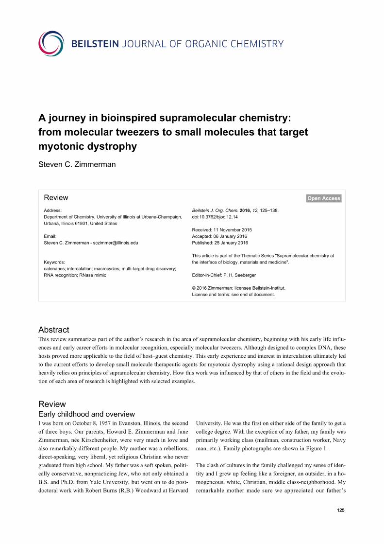
man, etc.). Family photographs are shown in Figure 1.
The clash of cultures in the family challenged my sense of iden-
tity and I grew up feeling like a foreigner, an outsider, in a ho-
mogeneous, white, Christian, middle class-neighborhood. My
remarkable mother made sure we appreciated our father’s
Beilstein J. Org. Chem. 2016, 12, 125–138.
126
Figure 1: Photographs of Howard E. Zimmerman (July 5, 1926–February 12, 2012) (left) and Jane Zimmerman (née Kirschenheiter) (December 24,1928–January 21, 1975) (center) as young adults, and the author (right) giving a Breslow group meeting presentation in graduate school (HavemeyerHall, Columbia University).
heritage. Although we were raised with traditional Christian
holidays, in early adulthood I recognized in myself a sense of
humor and an outlook on life that was distinctly Jewish. I was
drawn to the books of Saul Bellow and Isaac Bashevis Singer,
and New York City. What does any of this have to do with
chemistry? Chemistry provided a sense of belonging and iden-
tity from an early age simply because my father was immersed
in an occupation I didn't understand but recognized as being
very exciting. His passion for his work was obvious, so as a
young boy I told people I also wanted to be a chemist. Further-
more, the long line of remarkably talented chemistry graduate
and postdoctoral students that came to my house for Z-group
parties were like an extended family. They were all “cool”
people and some, for example, John McCall and Laren Tolbert
even served as babysitters. This was a family I wanted to join.
The occasional visiting faculty member solidified my choice of
chemistry as a career. What child wouldn’t be excited by Koji
Nakanishi cutting ropes in two only to have them magically
reconnect!
Undergraduate and graduate studies and anNSF-NATO postdocThe Department of Chemistry at the University of Wisconsin
was an extraordinarily stimulating place in the mid to late
1970s. I was fortunate to do undergraduate research with
Professor Hans J. Reich, investigating the mechanism of the
singlet oxygen reaction with alkenes and studying the oxidation
of selenide/sulfide mixtures using ozone and singlet oxygen [1].
In my senior year, I took three graduate level courses, which
was an amazing experience. Professors Charles P. (Chuck)
Casey and Harlan L. Goering taught the physical organic course
(Chem 641), Professor Barry M. Trost and Edwin Vedejs taught
the synthesis course (Chem 841) and Hans Reich a more
informal, once-a-week, mechanisms (arrow-pushing) class.
For each lecture, Trost or Vedejs passed out several pages
describing various methods of synthesizing several natural
product substructures with a rather lengthy bibliography. I
naively thought that this bibliography was an assigned reading
list rather than a list for future reference. It was a wonderful
mistake that led to my learning an enormous amount of exciting
synthetic chemistry. Indeed, in going to Columbia University
for graduate school I had every intention of working for
Professor Gilbert Stork or W. Clark Still. But Professor Ronald
Breslow’s enzyme reaction mechanisms course, my first class
ever on anything resembling biology or biochemistry, was so
exciting that I decided to join the Breslow group and work on
pyridoxal/pyridoxamine enzyme analogs [2-4]. Not only was
Ron Breslow a wonderful and inspirational mentor, he had built
an extraordinarily stimulating group of coworkers that he
himself described as “people that you will hear from in the
future, not people who will disappear into the woodwork.”
Indeed, my labmates during the period from 1979 to 1983
included Jik Chin, Robert Corcoran, Tony Czarnik, Sam
Gellman, Don Hilvert, Uday Maitra, Dave Okrongley, Russ
Petter, Darryl Rideout (coincidentally a Madison West High
School classmate), Alanna Schepartz, Alan Schwabacher,
George Trainor, Craig Wilcox, and Jeff Winkler.
Ron Breslow had broad interests, with projects ranging from
developing artificial enzymes, to novel anti-aromatic com-
pounds, to remote C–H activation of steroids, to determining
hydrocarbon pKa values using electrochemistry. The lesson
learned, and one I tried to put into practice in my independent
career (see below), is that it is very much possible to run a
research group focused in quite different areas of chemistry.
With an NSF-NATO postdoctoral fellowship, I spent just under
two years with Sir Alan Battersby at the University of
Cambridge where we completed the total synthesis of sirohy-
Beilstein J. Org. Chem. 2016, 12, 125–138.
127
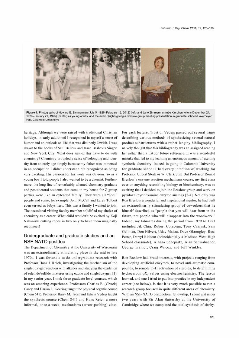
Figure 2: (a) Schematic double helix fully saturated with intercalator (in purple) according to the neighbor exclusion principle (NEP). (b) Bisinterca-lator with spermine linker. (c,d) Bisintercalator with long linker spanning two base-pairs and short linker preferring mono-intercalation to obey NEP.(e) Whitlock’s “rigid” molecular tweezer.
drochlorin, an intermediate in the biosynthesis of vitamin B12.
Then in July 1985, it was off to the University of Illinois at
Urbana-Champaign.
Molecular tweezers and a paradigm shift inhost–guest chemistryDeveloping molecular tweezers was one of the main projects I
started in my independent academic career at Illinois. The idea
originated at Columbia when I began to teach myself the
biochemistry and biology lacking in any of my formal course-
work. For example, one summer that process involved taking
J. D. Watson’s “Molecular Biology of the Gene” [5] on the
subway to a Long Island beach on weekends. The beautiful
structure of DNA and its intercalation complexes of aromatic
dyes were especially intriguing. In broader reading, I sought to
understand better the so-called nearest-neighbor exclusion prin-
ciple (NEP), wherein intercalators at full saturation of a DNA
helix bind every other site (i.e., one intercalator per two base-
pairs) [6].
Figure 2a schematically shows how insertion of monointercala-
tors at full saturation leads to a DNA helix with intercalation
sites only half occupied. Le Pecq and coworkers studied bisac-
ridines such as 1 (Figure 2b) [7]. Consistent with the NEP, 1
formed a very tight bisintercalation complex with its spermine-
derived linker chain spanning two base-pairs (Figure 2c). How-
ever, with shorter linkers that can only span a single base-pair, a
monointercalation complex forms (Figure 2d). In fact, with
bisintercalators the situation is considerably more complicated
with the apparent width of the intercalator determining whether
the nearest neighbor exclusion principle is obeyed. Although
the principle remains poorly understood even today, my idea as
a graduate student was to make a bisintercalator that was so
rigid it could not form the mono-intercalated complex in
Figure 2d. The ultimate goal was to develop a small molecule
ligand that might intercalate at sites that lack a conventional
neighboring intercalation site, for example, the ends of DNA
double helices, replication forks, or abasic sites.
My original research proposal was submitted August 16, 1983
and was entitled “Synthesis of a Rigid ‘Molecular Tweezer’
with Novel DNA Binding Potential.” The name “molecular
tweezer” was inspired by Howard Whitlock’s 1978 report [8] of
compound 2 containing two caffeine units linked by a rigid
diyne spacer (Figure 2e). Whitlock noted that conventional
bisintercalators such as 1 would have their affinity for oligo-
nucleotides significantly reduced as a result of intramolecular
π–π aromatic stacking. Whereas the diyne spacer would prevent
such stacking, it would not prevent mono-intercalation; there-
fore, we sought a spacer that would enforce a C-shape on the
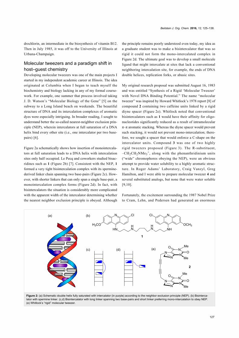
intercalator units. Compound 3 was one of two highly
rigid tweezers proposed (Figure 3). The R-substituent,
–CH2CH2NMe3+, along with the phenanthridinium units
(“wide” chromophores obeying the NEP), were an obvious
attempt to provide water solubility to a highly aromatic struc-
ture. In Roger Adams’ Laboratory, Craig Vanzyl, Greg
Hamilton, and I were able to prepare molecular tweezer 4 and
several substituted analogs, but none that were water soluble
[9,10].
Fortunately, the excitement surrounding the 1987 Nobel Prize
to Cram, Lehn, and Pedersen had generated an enormous
Beilstein J. Org. Chem. 2016, 12, 125–138.
128
Figure 3: Bismethidium molecular tweezer 3 proposed as a graduate student at Columbia University, which was quite close in structure to 4, synthe-sized and studied at Illinois. Chemically bonded stationary phase 5 used for HPLC assay of nitrated aromatics and for quantitative enthalpy determi-nations.
interest in host–guest chemistry and there was at this time a
move to go beyond cyclic crown ethers. In particular, the
groups of Rebek [11] and Hamilton [12] and many others were
developing hosts capable of complexing more structurally chal-
lenging organic guests such as nucleobases. We quickly discov-
ered that in chloroform solution, 4 and its analogs could bind
nitrated aromatic compounds, such as 2,4,7-trinitrofluorenone.
Nitrated polycyclic aromatics and polynitrated fluorenones were
known pollutants so Kurt Saionz covalently linked the molecu-
lar tweezers to silica gel (see 5, Figure 3), making chemically
bonded stationary phases which were packed into HPLC
columns that selectively retained and separated nitrated
aromatics [13]. The HPLC columns proved to be very useful for
quickly measuring ΔΗ° of complexation and, indeed, multiple
guests could be measured at once [14]. The HPLC method of
determining complexation ΔΗ° values was extended by Vincent
Kwan to hydrogen bonding host–guest complexes [15].
Molecular tweezers that complex adenineand analysis of binding interactionsThe idea of incorporating hydrogen bonding functionality into
the molecular tweezer was appealing because it meant that aro-
matic stacking and hydrogen bonding might cooperate to give
higher binding constants and guest selectivity. However, the
preparation of a rigid aromatic spacer with a functional group
converging on the binding cleft was not only a significant syn-
thetic challenge, I questioned whether the group might not dis-
tort the spacer, thereby altering its dimensions. I was able to
prepare small quantities (<100 mg) of 6 and 7 and crystallize
both for X-ray analysis (Figure 4) [16]. The solid state structure
of 6 revealed significant distortion and, indeed, a highly
nonplanar aromatic spacer that was not suitable for the desired
molecular tweezer. In contrast, analog 7 with nitrogen atoms in
the peri-positions was a much more planar and suitable candi-
date.
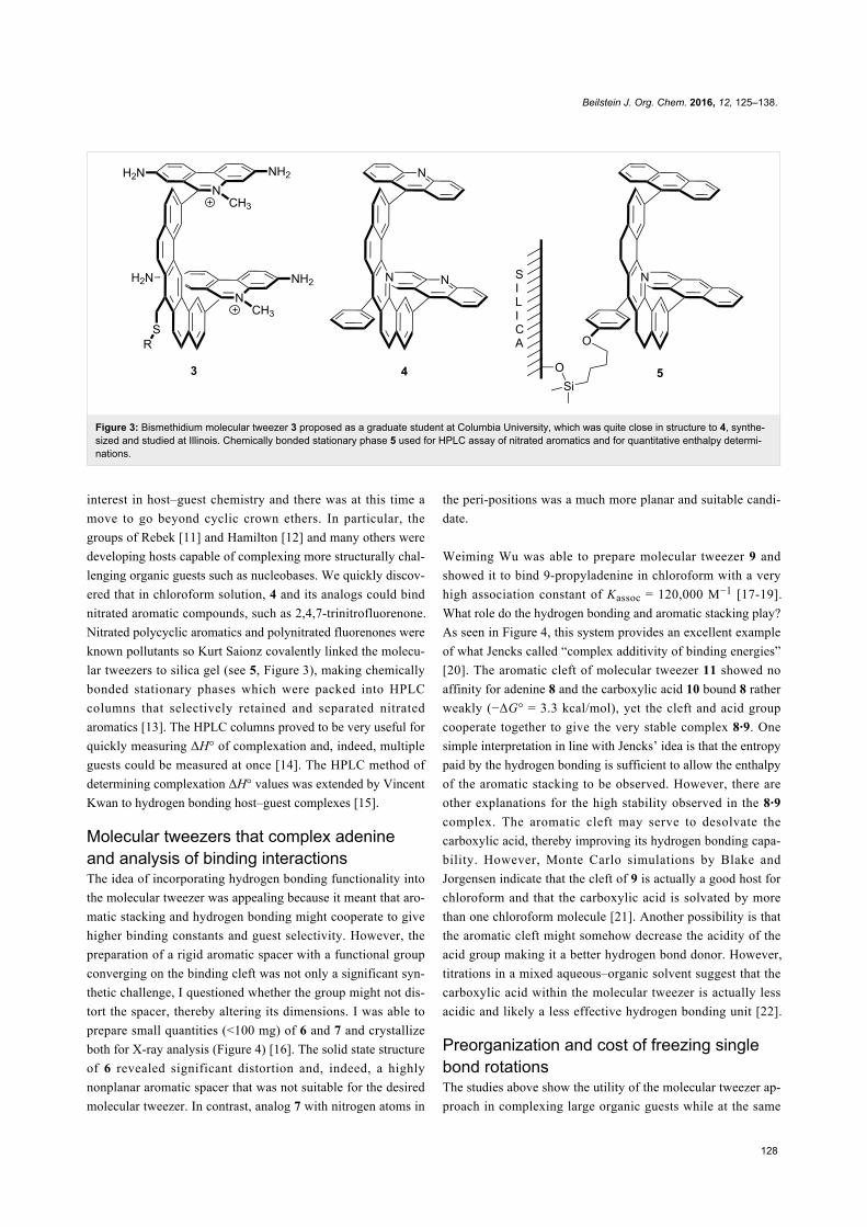
Weiming Wu was able to prepare molecular tweezer 9 and
showed it to bind 9-propyladenine in chloroform with a very
high association constant of Kassoc = 120,000 M−1 [17-19].
What role do the hydrogen bonding and aromatic stacking play?
As seen in Figure 4, this system provides an excellent example
of what Jencks called “complex additivity of binding energies”
[20]. The aromatic cleft of molecular tweezer 11 showed no
affinity for adenine 8 and the carboxylic acid 10 bound 8 rather
weakly (−ΔG° = 3.3 kcal/mol), yet the cleft and acid group
cooperate together to give the very stable complex 8·9. One
simple interpretation in line with Jencks’ idea is that the entropy
paid by the hydrogen bonding is sufficient to allow the enthalpy
of the aromatic stacking to be observed. However, there are
other explanations for the high stability observed in the 8·9
complex. The aromatic cleft may serve to desolvate the
carboxylic acid, thereby improving its hydrogen bonding capa-
bility. However, Monte Carlo simulations by Blake and
Jorgensen indicate that the cleft of 9 is actually a good host for
chloroform and that the carboxylic acid is solvated by more
than one chloroform molecule [21]. Another possibility is that
the aromatic cleft might somehow decrease the acidity of the
acid group making it a better hydrogen bond donor. However,
titrations in a mixed aqueous–organic solvent suggest that the
carboxylic acid within the molecular tweezer is actually less
acidic and likely a less effective hydrogen bonding unit [22].
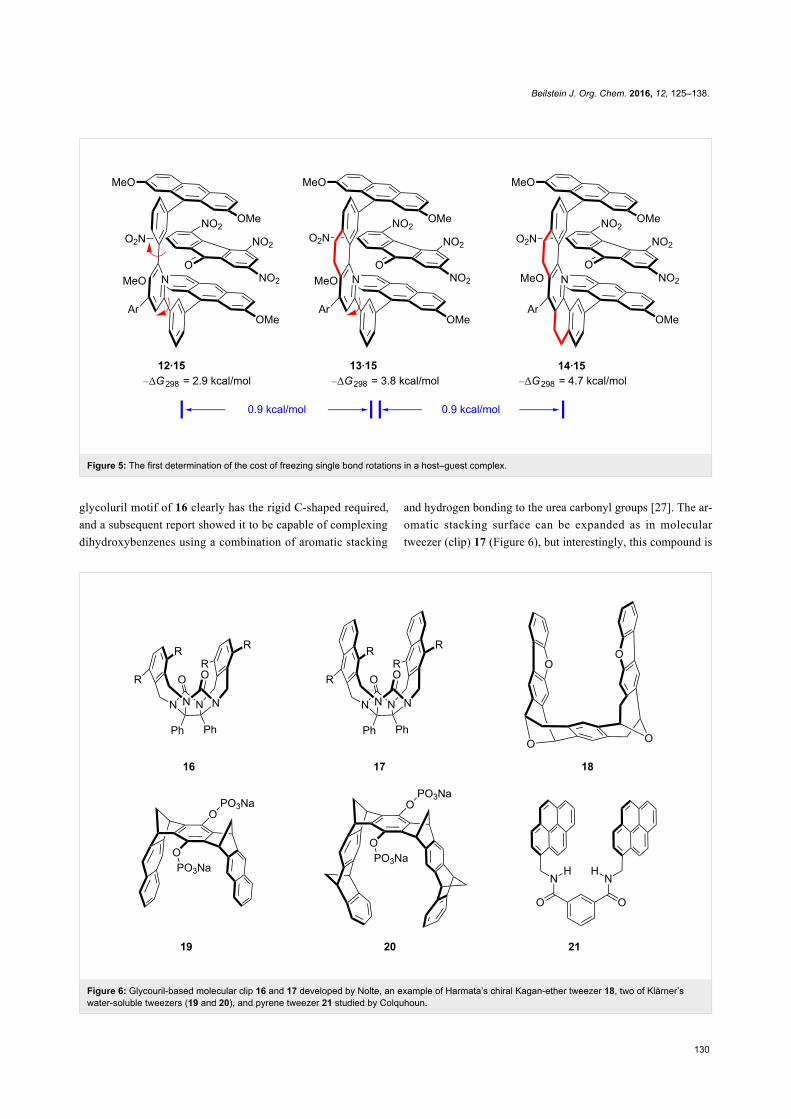
Preorganization and cost of freezing singlebond rotationsThe studies above show the utility of the molecular tweezer ap-
proach in complexing large organic guests while at the same

Beilstein J. Org. Chem. 2016, 12, 125–138.
129
Figure 4: Adenine 8 recognition by carboxylic acid containing tweezer 9 and a component analysis showing “complex additivity” of binding energies.
time uncovering important design criteria in host–guest chem-
istry. Regarding design criteria, the ability to synthesize struc-
turally analogous molecular tweezers provides an unprece-
dented opportunity to develop a wide range of important struc-
ture–property relationships. For example, a lot had been written
about the importance of Cram’s preorganization principle in
host design, but no one had measured the energy cost of locking
a single bond rotation in a host–guest complex. Monica Baloga,
Milan Mrksich, and I prepared three new molecular tweezers
12–14, where the spacer units possess zero, one, and two
aryl–aryl single bonds (i.e., 14 → 13 → 12) [23]. Their Kassoc
values with 2,4,5,7-tetranitrofluorenone (15) in chloroform
were then measured. As seen in Figure 5, freezing each single
bond rotation increases complex stability by about 0.9 kcal/mol.
This value is in line with a value of TΔS° = 0.6 to 1.2 kcal/mol
previously suggested by Jencks and Page as the cost paid to
freeze out each single bond rotation in a ring-forming reaction
[24]. Later Dudley J. Williams, in the context of analyzing the
vancomycin complex with D–Ala–D–Ala containing peptides,
suggested the cost of freezing a free rotation to be between 0.4
to 0.9 kcal/mol – a value that is also close to what we had
measured [25]. In Williams’ case, the value was derived from
the entropy of fusion within a homologous series of alkanes, not
an analysis of a host–guest system. All these values suggest that
freezing out a single bond rotation is not terribly costly but as-
sociation constants can drop significantly if too much flexi-
bility exists; freezing five single bonds within a complex would
lower its stability by as much as 104-fold.
Research on molecular tweezers goes main-streamSince our early studies on molecular tweezers, numerous exam-
ples have appeared in the literature. A comprehensive review is
not possible, but a few examples from other investigators are
presented to illustrate the breadth of structure and function that
has been achieved over the past three decades. One of the
earliest examples is a molecular tweezer developed contempora-
neously with our own efforts. Roeland Nolte and his team first
reported the synthesis and X-ray structure of 16 (Figure 6,
R = OH) as the basic building block [26]. The now familiar
Beilstein J. Org. Chem. 2016, 12, 125–138.
130
Figure 5: The first determination of the cost of freezing single bond rotations in a host–guest complex.
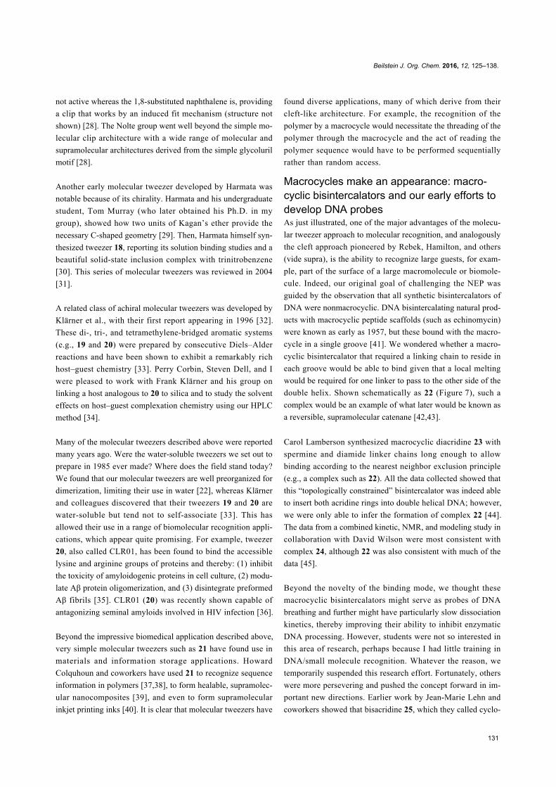
Figure 6: Glycouril-based molecular clip 16 and 17 developed by Nolte, an example of Harmata’s chiral Kagan-ether tweezer 18, two of Klärner’swater-soluble tweezers (19 and 20), and pyrene tweezer 21 studied by Colquhoun.
glycoluril motif of 16 clearly has the rigid C-shaped required,
and a subsequent report showed it to be capable of complexing
dihydroxybenzenes using a combination of aromatic stacking
and hydrogen bonding to the urea carbonyl groups [27]. The ar-
omatic stacking surface can be expanded as in molecular
tweezer (clip) 17 (Figure 6), but interestingly, this compound is
Beilstein J. Org. Chem. 2016, 12, 125–138.
131
not active whereas the 1,8-substituted naphthalene is, providing
a clip that works by an induced fit mechanism (structure not
shown) [28]. The Nolte group went well beyond the simple mo-
lecular clip architecture with a wide range of molecular and
supramolecular architectures derived from the simple glycoluril
motif [28].
Another early molecular tweezer developed by Harmata was
notable because of its chirality. Harmata and his undergraduate
student, Tom Murray (who later obtained his Ph.D. in my
group), showed how two units of Kagan’s ether provide the
necessary C-shaped geometry [29]. Then, Harmata himself syn-
thesized tweezer 18, reporting its solution binding studies and a
beautiful solid-state inclusion complex with trinitrobenzene
[30]. This series of molecular tweezers was reviewed in 2004
[31].
A related class of achiral molecular tweezers was developed by
Klärner et al., with their first report appearing in 1996 [32].
These di-, tri-, and tetramethylene-bridged aromatic systems
(e.g., 19 and 20) were prepared by consecutive Diels–Alder
reactions and have been shown to exhibit a remarkably rich
host–guest chemistry [33]. Perry Corbin, Steven Dell, and I
were pleased to work with Frank Klärner and his group on
linking a host analogous to 20 to silica and to study the solvent
effects on host–guest complexation chemistry using our HPLC
method [34].
Many of the molecular tweezers described above were reported
many years ago. Were the water-soluble tweezers we set out to
prepare in 1985 ever made? Where does the field stand today?
We found that our molecular tweezers are well preorganized for
dimerization, limiting their use in water [22], whereas Klärner
and colleagues discovered that their tweezers 19 and 20 are
water-soluble but tend not to self-associate [33]. This has
allowed their use in a range of biomolecular recognition appli-
cations, which appear quite promising. For example, tweezer
20, also called CLR01, has been found to bind the accessible
lysine and arginine groups of proteins and thereby: (1) inhibit
the toxicity of amyloidogenic proteins in cell culture, (2) modu-
late Aβ protein oligomerization, and (3) disintegrate preformed
Aβ fibrils [35]. CLR01 (20) was recently shown capable of
antagonizing seminal amyloids involved in HIV infection [36].
Beyond the impressive biomedical application described above,
very simple molecular tweezers such as 21 have found use in
materials and information storage applications. Howard
Colquhoun and coworkers have used 21 to recognize sequence
information in polymers [37,38], to form healable, supramolec-
ular nanocomposites [39], and even to form supramolecular
inkjet printing inks [40]. It is clear that molecular tweezers have
found diverse applications, many of which derive from their
cleft-like architecture. For example, the recognition of the
polymer by a macrocycle would necessitate the threading of the
polymer through the macrocycle and the act of reading the
polymer sequence would have to be performed sequentially
rather than random access.
Macrocycles make an appearance: macro-cyclic bisintercalators and our early efforts todevelop DNA probesAs just illustrated, one of the major advantages of the molecu-
lar tweezer approach to molecular recognition, and analogously
the cleft approach pioneered by Rebek, Hamilton, and others
(vide supra), is the ability to recognize large guests, for exam-
ple, part of the surface of a large macromolecule or biomole-
cule. Indeed, our original goal of challenging the NEP was
guided by the observation that all synthetic bisintercalators of
DNA were nonmacrocyclic. DNA bisintercalating natural prod-
ucts with macrocyclic peptide scaffolds (such as echinomycin)
were known as early as 1957, but these bound with the macro-
cycle in a single groove [41]. We wondered whether a macro-
cyclic bisintercalator that required a linking chain to reside in
each groove would be able to bind given that a local melting
would be required for one linker to pass to the other side of the
double helix. Shown schematically as 22 (Figure 7), such a
complex would be an example of what later would be known as
a reversible, supramolecular catenane [42,43].
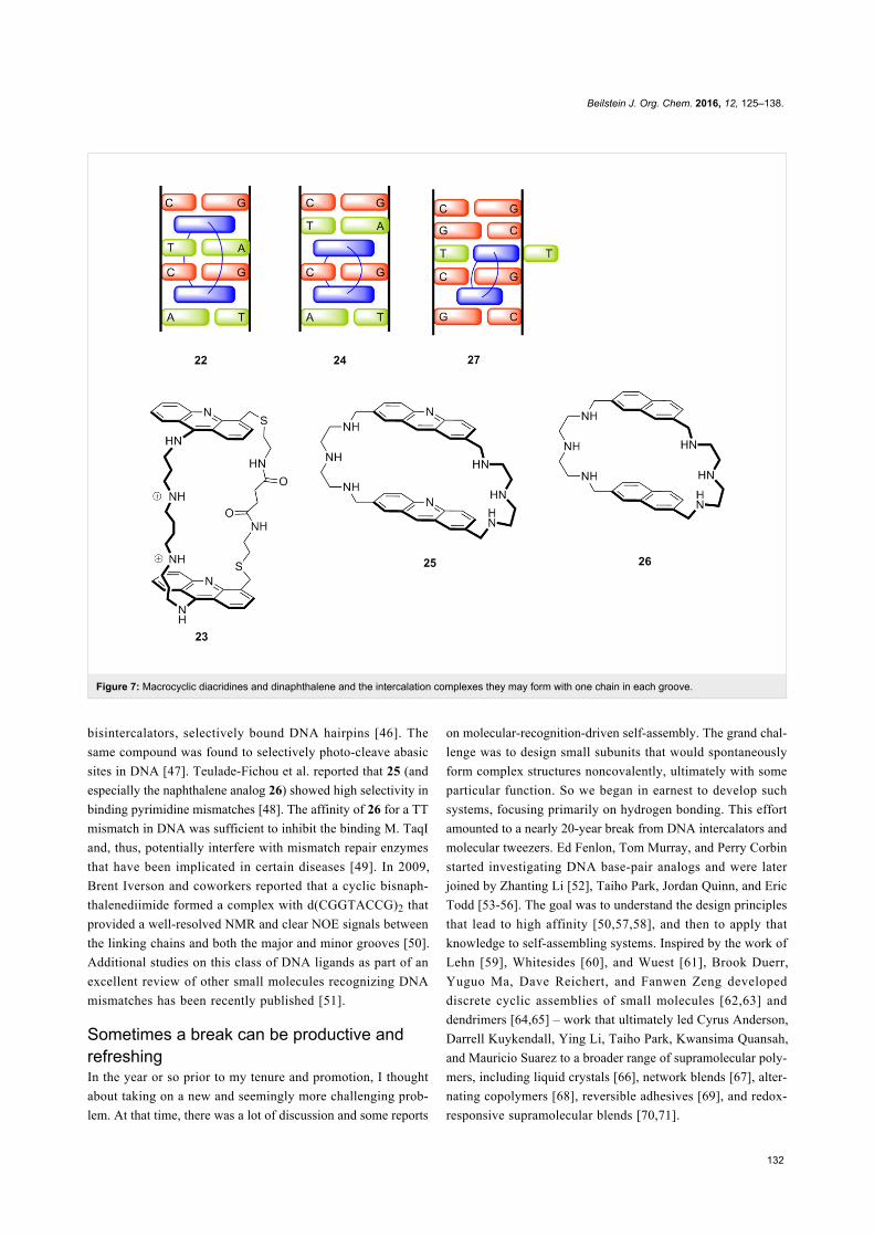
Carol Lamberson synthesized macrocyclic diacridine 23 with
spermine and diamide linker chains long enough to allow
binding according to the nearest neighbor exclusion principle
(e.g., a complex such as 22). All the data collected showed that
this “topologically constrained” bisintercalator was indeed able
to insert both acridine rings into double helical DNA; however,
we were only able to infer the formation of complex 22 [44].
The data from a combined kinetic, NMR, and modeling study in
collaboration with David Wilson were most consistent with
complex 24, although 22 was also consistent with much of the
data [45].
Beyond the novelty of the binding mode, we thought these
macrocyclic bisintercalators might serve as probes of DNA
breathing and further might have particularly slow dissociation
kinetics, thereby improving their ability to inhibit enzymatic
DNA processing. However, students were not so interested in
this area of research, perhaps because I had little training in
DNA/small molecule recognition. Whatever the reason, we
temporarily suspended this research effort. Fortunately, others
were more persevering and pushed the concept forward in im-
portant new directions. Earlier work by Jean-Marie Lehn and
coworkers showed that bisacridine 25, which they called cyclo-
Beilstein J. Org. Chem. 2016, 12, 125–138.
132
Figure 7: Macrocyclic diacridines and dinaphthalene and the intercalation complexes they may form with one chain in each groove.
bisintercalators, selectively bound DNA hairpins [46]. The
same compound was found to selectively photo-cleave abasic
sites in DNA [47]. Teulade-Fichou et al. reported that 25 (and
especially the naphthalene analog 26) showed high selectivity in
binding pyrimidine mismatches [48]. The affinity of 26 for a TT
mismatch in DNA was sufficient to inhibit the binding M. TaqI
and, thus, potentially interfere with mismatch repair enzymes
that have been implicated in certain diseases [49]. In 2009,
Brent Iverson and coworkers reported that a cyclic bisnaph-
thalenediimide formed a complex with d(CGGTACCG)2 that
provided a well-resolved NMR and clear NOE signals between
the linking chains and both the major and minor grooves [50].
Additional studies on this class of DNA ligands as part of an
excellent review of other small molecules recognizing DNA
mismatches has been recently published [51].
Sometimes a break can be productive andrefreshingIn the year or so prior to my tenure and promotion, I thought
about taking on a new and seemingly more challenging prob-
lem. At that time, there was a lot of discussion and some reports
on molecular-recognition-driven self-assembly. The grand chal-
lenge was to design small subunits that would spontaneously
form complex structures noncovalently, ultimately with some
particular function. So we began in earnest to develop such
systems, focusing primarily on hydrogen bonding. This effort
amounted to a nearly 20-year break from DNA intercalators and
molecular tweezers. Ed Fenlon, Tom Murray, and Perry Corbin
started investigating DNA base-pair analogs and were later
joined by Zhanting Li [52], Taiho Park, Jordan Quinn, and Eric
Todd [53-56]. The goal was to understand the design principles
that lead to high affinity [50,57,58], and then to apply that
knowledge to self-assembling systems. Inspired by the work of
Lehn [59], Whitesides [60], and Wuest [61], Brook Duerr,
Yuguo Ma, Dave Reichert, and Fanwen Zeng developed
discrete cyclic assemblies of small molecules [62,63] and
dendrimers [64,65] – work that ultimately led Cyrus Anderson,
Darrell Kuykendall, Ying Li, Taiho Park, Kwansima Quansah,
and Mauricio Suarez to a broader range of supramolecular poly-
mers, including liquid crystals [66], network blends [67], alter-
nating copolymers [68], reversible adhesives [69], and redox-
responsive supramolecular blends [70,71].
Beilstein J. Org. Chem. 2016, 12, 125–138.
133
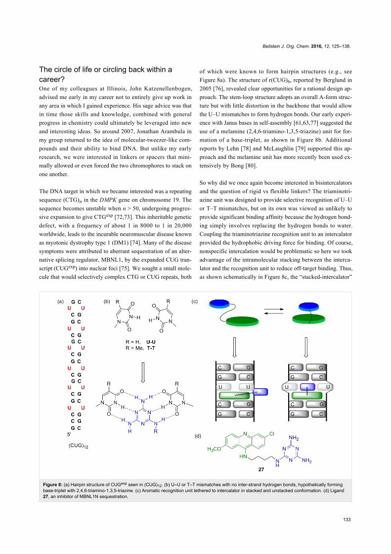
Figure 8: (a) Hairpin structure of CUGexp seen in (CUG)12. (b) U–U or T–T mismatches with no inter-strand hydrogen bonds, hypothetically formingbase-triplet with 2,4,6-triamino-1,3,5-triazine. (c) Aromatic recognition unit tethered to intercalator in stacked and unstacked conformation. (d) Ligand27, an inhibitor of MBNL1N sequestration.
The circle of life or circling back within acareer?One of my colleagues at Illinois, John Katzenellenbogen,
advised me early in my career not to entirely give up work in
any area in which I gained experience. His sage advice was that
in time those skills and knowledge, combined with general
progress in chemistry could ultimately be leveraged into new
and interesting ideas. So around 2007, Jonathan Arambula in
my group returned to the idea of molecular-tweezer-like com-
pounds and their ability to bind DNA. But unlike my early
research, we were interested in linkers or spacers that mini-
mally allowed or even forced the two chromophores to stack on
one another.
The DNA target in which we became interested was a repeating
sequence (CTG)n in the DMPK gene on chromosome 19. The
sequence becomes unstable when n > 50, undergoing progres-
sive expansion to give CTGexp [72,73]. This inheritable genetic
defect, with a frequency of about 1 in 8000 to 1 in 20,000
worldwide, leads to the incurable neuromuscular disease known
as myotonic dystrophy type 1 (DM1) [74]. Many of the disease
symptoms were attributed to aberrant sequestration of an alter-
native splicing regulator, MBNL1, by the expanded CUG tran-
script (CUGexp) into nuclear foci [75]. We sought a small mole-
cule that would selectively complex CTG or CUG repeats, both
of which were known to form hairpin structures (e.g., see
Figure 8a). The structure of r(CUG)6, reported by Berglund in
2005 [76], revealed clear opportunities for a rational design ap-
proach. The stem-loop structure adopts an overall A-form struc-
ture but with little distortion in the backbone that would allow
the U–U mismatches to form hydrogen bonds. Our early experi-
ence with Janus bases in self-assembly [61,63,77] suggested the
use of a melamine (2,4,6-triamino-1,3,5-triazine) unit for for-
mation of a base-triplet, as shown in Figure 8b. Additional
reports by Lehn [78] and McLaughlin [79] supported this ap-
proach and the melamine unit has more recently been used ex-
tensively by Bong [80].
So why did we once again become interested in bisintercalators
and the question of rigid vs flexible linkers? The triaminotri-
azine unit was designed to provide selective recognition of U–U
or T–T mismatches, but on its own was viewed as unlikely to
provide significant binding affinity because the hydrogen bond-
ing simply involves replacing the hydrogen bonds to water.
Coupling the triaminotriazine recognition unit to an intercalator
provided the hydrophobic driving force for binding. Of course,
nonspecific intercalation would be problematic so here we took
advantage of the intramolecular stacking between the interca-
lator and the recognition unit to reduce off-target binding. Thus,
as shown schematically in Figure 8c, the “stacked-intercalator”
Beilstein J. Org. Chem. 2016, 12, 125–138.
134
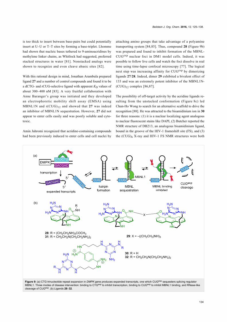
Figure 9: (a) CTG trinucleotide repeat expansion in DMPK gene produces expanded transcripts, one which CUGexp sequesters splicing regulatorMBNL1. Three modes of disease intervention: binding to CTGexp to inhibit transcription, binding to CUGexp to inhibit MBNL1 binding, and RNase-likecleavage of CUGexp. (b) Ligands 28–32.
is too thick to insert between base-pairs but could potentially
insert at U–U or T–T sites by forming a base-triplet. Lhomme
had shown that nucleic bases tethered to 9-aminoacridines by
methylene linker chains, as Whitlock had suggested, preferred
stacked structures in water [81]. Nonstacked analogs were
shown to recognize and even cleave abasic sites [82].
With this rational design in mind, Jonathan Arambula prepared
ligand 27 and a number of control compounds and found it to be
a dCTG- and rCUG-selective ligand with apparent KD values of
about 300–400 nM [83]. A very fruitful collaboration with
Anne Baranger’s group was initiated and they developed
an electrophoretic mobility shift assay (EMSA) using
MBNL1N and r(CUG)12 and showed that 27 was indeed
an inhibitor of MBNL1N sequestration. However, 27 did not
appear to enter cells easily and was poorly soluble and cyto-
toxic.
Amin Jahromi recognized that acridine-containing compounds
had been previously induced to enter cells and cell nuclei by
attaching amino groups that take advantage of a polyamine
transporting system [84,85]. Thus, compound 28 (Figure 9b)
was prepared and found to inhibit formation of the MBNL-
CUGexp nuclear foci in DM1 model cells. Indeed, it was
possible to follow live cells and watch the foci dissolve in real
time using time-lapse confocal microscopy [77]. The logical
next step was increasing affinity for CUGexp by dimerizing
ligands 27/28. Indeed, dimer 29 exhibited a bivalent effect of
133 and was an extremely potent inhibitor of the MBNL1N-
(CUG)12 complex [86,87].
The possibility of off-target activity by the acridine ligands re-
sulting from the unstacked conformation (Figure 8c) led
Chun-Ho Wong to search for an alternative scaffold to drive the
recognition [88]. He was attracted to the bisamidinium ion in 30
for three reasons: (1) it is a nuclear localizing agent analogous
to nuclear fluorescent stains like DAPI, (2) Butcher reported the
NMR structure of DB213, an analogous bisamidinium ligand,
bound in the groove of the HIV-1 frameshift site (FS), and (3)
the (CUG)6 X-ray and HIV-1 FS NMR structures were both
Beilstein J. Org. Chem. 2016, 12, 125–138.
135
A-form and similar suggesting replacement of the ammonium
ions in DB213 with triaminotriazine units. Ligand 30 was
studied in collaboration with both Anne Baranger and Paul
Hergenrother and it was found to have low cytotoxicity, enter
DM1 model cells, dissolved the MBNL1 foci and partially
corrected the missplicing of two key pre-mRNAs, cTNT and IR.
A terrific collaboration with Professor Edwin Chan’s group at
the Chinese University of Hong Kong allowed the compounds
to be tested in vivo using a DM1 Drosophila model that looked
at the rough eye phenotype with i(CUG)480 flies. Ligand 30
showed significant and dose-dependent improvement in the
rough eye phenotype, whereas the negative control, DB213
showed much weaker activity. Much less effort has been
devoted to developing small molecules to treat DM2, which
originates in a CCUG expansion, but Lien Nguyen, Chun-Ho
Wong, and JuYeon Lee used similar rational design approaches
and found lead agents that are selective for this RNA as well
[89].
Although the gain of function mechanism that has CUGexp
sequestering MBNL1 is well supported, it is clear that the
disease pathobiology is more complex. For example, Ranum
recently reported [90] that both the CUGexp and CAGexp
undergo repeat-associated non-ATG (RAN) translation to
produce up to nine homopeptides, some of which are known to
be toxic and involved in other disease [91]. Lien Nguyen and
Long Luu considered the possibility of rationally designing
ligands that could operate on multiple targets in the DM1 patho-
biology. They designed and studied ligands such as 31 and 32.
These ligands were shown to bind the DNA that causes DM1,
interacting with CTGexp to inhibit transcription to CUGexp, also
binding CUGexp that slips through inhibiting its sequestration of
MBNL1, and, with the catalytic amino/ammonium/imidazole
groups, slowly cleaving the CUG-RNA to prevent RAN transla-
tion [92]. Edwin Chan and his group again studied the in vivo
activity of our compounds, showing that 32 reversed two
separate CUGexp-induced phenotypes in transgenic DM1
Drosophila, specifically the rough eye phenotype and larvae
crawling mobility.
ConclusionDetours, perspectives, and future studiesI was Department Head or Interim Department Head for a total
of eight years (1999–2000 and 2005–2012). It was an honor to
serve our outstanding faculty, staff, and students and to follow
luminaries such as William A. Noyes, Roger Adams, Herb
Carter, Larry Faulkner, and Gary Schuster. Being department
head was without a doubt the most difficult thing I did during
my career. It was a period of extraordinary personal growth,
having learned how to work with a wide range of people and
manage a complex organization. The expressions “herding cats”
and “drinking from a fire hose” are apropos descriptors as it
was more than a full time job and extremely demanding in other
ways. Although it was hard to take time away from research and
teaching, the department head job was interesting, challenging,
and highly rewarding in seeing the department move forward,
especially with the help of our loyal alumni.
We entitled the thematic issue containing this contribution
“Supramolecular chemistry at the interface of biology, materi-
als and medicine.” Some of the examples presented herein illus-
trate the potential of the supramolecular approach to lead to ad-
vanced therapeutic agents. In particular the Klärner molecular
tweezers that complex lysine-containing peptides may lead to
agents that dissolve Alzheimers plaques or inhibit their forma-
tion. Our own efforts to create small molecules to target the
toxic RNA involved in myotonic dystrophy have expanded to
include multitarget drug-discovery approaches where supramo-
lecular design principles led to DNA and RNA-selective small
molecules that function even in the complex organisms (i.e.,
Drosophila).
Do we know so much about supramolecular interactions that all
of the focus now should be on applications in biology, materi-
als and medicine? The answer to this question is emphatically
“No!” At a recent NSF workshop comprised of physical
chemists and supramolecular chemists focused on water, the
supramolecular chemists mostly agreed that water was not a
special solvent, it just occupied an extreme, with low polariz-
ability and a high cohesive nature. In stark contrast, the physi-
cal chemists showed plots indicating that water was unlike any
other liquid and was clearly special. There also remain debates
about whether the π-cation or face-edge aromatic interactions
are unusual or even important. Therefore, model studies that
shed light on the strength and nature of supramolecular contacts
continue to be critically important.
One important emerging area I would like to highlight is the de-
velopment of complex supramolecular systems. So much of
supramolecular chemistry is inspired by biology, it is only
natural that the complexity of biological systems be modeled in
supramolecular systems. Thus, future developments will lead to
multicomponent supramolecular structures/systems that evolve
over time or communicate or respond to external or internal
stimuli. Dynamic covalent chemistry has moved in this direc-
tion [93], and some initial efforts in this area using supramolec-
ular chemistry have also appeared. For example, Andy Wilson’s
group reported a sequence of supramolecular recognition events
that proceed in a controlled and defined manner, the specific
pathway guided by what is present in solution [94]. This
primitive model of a signaling cascade points to what may be
possible as this area develops.

Beilstein J. Org. Chem. 2016, 12, 125–138.
136
AcknowledgementsThis work was supported by the National Institutes of Health
(R01 AR058361), the Muscular Dystrophy Association
(295229), and the National Science Foundation (CHE-
1307404). I want to thank all of my wonderful students who
have made this journey possible. Only some of their work could
be included in this review, but I am grateful to them all. This
review is dedicated to four remarkable women: my wife and
two daughters, and my mother who died much too young.
References1. Reich, H. J. Organoselenium Oxidations. In Oxidation in Organic
Chemistry; Trahanovsky, W. S., Ed.; Academic Press: New York, NY,USA, 1978; Vol. 5-C, pp 1–129.
2. Zimmerman, S. C.; Czarnik, A. W.; Breslow, R. J. Am. Chem. Soc.1983, 105, 1694–1695. doi:10.1021/ja00344a069
3. Zimmerman, S. C.; Breslow, R. J. Am. Chem. Soc. 1984, 106,1490–1491. doi:10.1021/ja00317a054
4. Weiner, W.; Winkler, J.; Zimmerman, S. C.; Czarnik, A. W.; Breslow, R.J. Am. Chem. Soc. 1985, 107, 4093–4094. doi:10.1021/ja00299a064
5. Watson, J. D. Molecular Biology of the Gene, 3rd ed.;Benjamin-Cummings Publishing Co.: Menlo Park, CA, USA, 1976.
6. Crothers, D. M. Biopolymers 1968, 6, 575–584.doi:10.1002/bip.1968.360060411
7. Le Pecq, J. B.; Le Bret, M.; Barbet, J.; Roques, B.Proc. Natl. Acad. Sci. U. S. A. 1975, 72, 2915–2919.doi:10.1073/pnas.72.8.2915
8. Chen, C. W.; Whitlock, H. W., Jr. J. Am. Chem. Soc. 1978, 100,4921–4922. doi:10.1021/ja00483a063
9. Zimmerman, S. C.; VanZyl, C. J. Am. Chem. Soc. 1987, 109,7894–7896. doi:10.1021/ja00259a055
10. Zimmerman, S. C.; VanZyl, C. M.; Hamilton, G. S. J. Am. Chem. Soc.1989, 111, 1373–1381. doi:10.1021/ja00186a035
11. Rebek, J., Jr. Angew. Chem., Int. Ed. Engl. 1990, 29, 245–255.doi:10.1002/anie.199002451
12. Chang, S. K.; Hamilton, A. D. J. Am. Chem. Soc. 1988, 110,1318–1319. doi:10.1021/ja00212a065
13. Zimmerman, S. C.; Saionz, K. W.; Zeng, Z.Proc. Natl. Acad. Sci. U. S. A. 1993, 90, 1190–1193.doi:10.1073/pnas.90.4.1190
14. Zimmerman, S.; Saionz, K. J. Am. Chem. Soc. 1995, 117, 1175–1176.doi:10.1021/ja00108a053
15. Zimmerman, S. C.; Kwan, W. Angew. Chem., Int. Ed. Engl. 1995, 34,2404–2406. doi:10.1002/anie.199524041
16. Zimmerman, S. C. Tetrahedron Lett. 1988, 29, 983–986.doi:10.1016/0040-4039(88)85314-0
17. Zimmerman, S. C.; Wu, W. J. Am. Chem. Soc. 1989, 111, 8054–8055.doi:10.1021/ja00202a077
18. Zimmerman, S. C.; Wu, W.; Zeng, Z. J. Am. Chem. Soc. 1991, 113,196–201. doi:10.1021/ja00001a028
19. Zimmerman, S. C.; Zeng, Z.; Wu, W.; Reichert, D. E. C.J. Am. Chem. Soc. 1991, 113, 183–196. doi:10.1021/ja00001a027
20. Jencks, W. P. Proc. Natl. Acad. Sci. U. S. A. 1981, 78, 4046–4050.doi:10.1073/pnas.78.7.4046
21. Blake, J. F.; Jorgensen, W. L. J. Am. Chem. Soc. 1990, 112,7269–7278. doi:10.1021/ja00176a029
22. Zimmerman, S. C. Top. Curr. Chem. 1993, 165, 71–102.doi:10.1007/BFb0111281
23. Zimmerman, S. C.; Mrksich, M.; Baloga, M. J. Am. Chem. Soc. 1989,111, 8528–8530. doi:10.1021/ja00204a041
24. Page, M. I.; Jencks, W. P. Proc. Natl. Acad. Sci. U. S. A. 1971, 68,1678–1683. doi:10.1073/pnas.68.8.1678
25. Searle, M. S.; Williams, D. H. J. Am. Chem. Soc. 1992, 114,10690–10697. doi:10.1021/ja00053a002
26. Smeets, J. W. H.; Sijbesma, R. P.; Niele, F. G. M.; Spek, A. L.;Smeets, W. J. J.; Nolte, R. J. M. J. Am. Chem. Soc. 1987, 109,928–929. doi:10.1021/ja00237a064
27. Sijbesma, R. P.; Kentgens, A. P. M.; Nolte, R. J. M. J. Org. Chem.1991, 56, 3199–3201. doi:10.1021/jo00010a001
28. Rowan, A. E.; Elemans, J. A. A. W.; Nolte, R. J. M. Acc. Chem. Res.1999, 32, 995–1006. doi:10.1021/ar9702684
29. Harmata, M.; Murray, T. J. Org. Chem. 1989, 54, 3761–3763.doi:10.1021/jo00277a001
30. Harmata, M.; Barnes, C. L. J. Am. Chem. Soc. 1990, 112, 5655–5657.doi:10.1021/ja00170a049
31. Harmata, M. Acc. Chem. Res. 2004, 37, 862–873.doi:10.1021/ar030164v
32. Klärner, F.-G.; Benkhoff, J.; Boese, R.; Burkert, U.; Kamieth, M.;Naatz, U. Angew. Chem., Int. Ed. Engl. 1996, 35, 1130–1133.doi:10.1002/anie.199611301
33. Klärner, F.-G.; Kahlert, B. Acc. Chem. Res. 2003, 36, 919–932.doi:10.1021/ar0200448
34. Kamieth, M.; Burkert, U.; Corbin, P. S.; Dell, S. J.; Zimmerman, S. C.;Klärner, F.-G. Eur. J. Org. Chem. 1999, 2741–2749.doi:10.1002/(SICI)1099-0690(199911)1999:11<2741::AID-EJOC2741>3.0.CO;2-K
35. Sinha, S.; Lopes, D. H. J.; Du, Z.; Pang, E. S.; Shanmugam, A.;Lomakin, A.; Talbiersky, P.; Tennstaedt, A.; McDaniel, K.; Bakshi, R.;Kuo, P.-Y.; Ehrmann, M.; Benedek, G. B.; Loo, J. A.; Klärner, F.-G.;Schrader, T.; Wang, C.; Bitan, G. J. Am. Chem. Soc. 2011, 133,16958–16969. doi:10.1021/ja206279b
36. Lump, E.; Castellano, L. M.; Meier, C.; Seeliger, J.; Erwin, N.;Sperlich, B.; Stürzel, C. M.; Usmani, S.; Hammond, R. M.;von Einem, J.; Gerold, G.; Kreppel, F.; Bravo-Rodriguez, K.;Pietschmann, T.; Holmes, V. M.; Palesch, D.; Zirafi, O.; Weissman, D.;Sowislok, A.; Wettig, B.; Heid, C.; Kirchhoff, F.; Weil, T.; Klärner, F.-G.;Schrader, T.; Bitan, G.; Sanchez-Garcia, E.; Winter, R.; Shorter, J.;Münch, J. eLife 2015, 4, e05397. doi:10.7554/eLife.05397
37. Colquhoun, H. M.; Zhu, Z. X. Angew. Chem., Int. Ed. 2004, 43,5040–5045. doi:10.1002/anie.200460382
38. Zhu, Z.; Cardin, C. J.; Gan, Y.; Colquhoun, H. M. Nat. Chem. 2010, 2,653–660. doi:10.1038/nchem.699
39. Burattini, S.; Greenland, B. W.; Hayes, W.; Mackay, M. E.;Rowan, S. J.; Colquhoun, H. M. Chem. Mater. 2011, 23, 6–8.doi:10.1021/cm102963k
40. Hart, L. R.; Harries, J. L.; Greenland, B. W.; Colquhoun, H. M.;Hayes, W. ACS Appl. Mater. Interfaces 2015, 7, 8906–8914.doi:10.1021/acsami.5b01569
41. Zolova, O. E.; Mady, A. S. A.; Garneau-Tsodikova, S. Biopolymers2010, 93, 777–790. doi:10.1002/bip.21489
42. Gil-Ramirez, G.; Leigh, D. A.; Stephens, A. J. Angew. Chem., Int. Ed.2015, 54, 6110–6150. doi:10.1002/anie.201411619
43. Flood, A. H.; Ramirez, R. J. A.; Deng, W. Q.; Muller, R. P.;Goddard, W. A., III; Stoddart, J. F. Aust. J. Chem. 2004, 57, 301–322.doi:10.1071/CH03307
44. Zimmerman, S. C.; Lamberson, C. R.; Cory, M.; Fairley, T. A.J. Am. Chem. Soc. 1989, 111, 6805–6809. doi:10.1021/ja00199a047
Beilstein J. Org. Chem. 2016, 12, 125–138.
137
45. Veal, J. M.; Li, Y.; Zimmerman, S. C.; Lamberson, C. R.; Cory, M.;Zon, G.; Wilson, W. D. Biochemistry 1990, 29, 10918–10927.doi:10.1021/bi00501a009
46. Slama-Schwok, A.; Teulade-Fichou, M.-P.; Vigneron, J.-P.;Taillandier, E.; Lehn, J.-M. J. Am. Chem. Soc. 1995, 117, 6822–6830.doi:10.1021/ja00131a003
47. Jourdan, M.; Garcia, J.; Lhomme, J.; Teulade-Fichou, M.-P.;Vigneron, J.-P.; Lehn, J.-M. Biochemistry 1999, 38, 14205–14213.doi:10.1021/bi991111h
48. David, A.; Bleimling, N.; Beuck, C.; Lehn, J. M.; Weinhold, E.;Teulade-Fichou, M. P. ChemBioChem 2003, 4, 1326–1331.doi:10.1002/cbic.200300693
49. Bahr, M.; Gabelica, V.; Granzhan, A.; Teulade-Fichou, M.-P.;Weinhold, E. Nucleic Acids Res. 2008, 36, 5000–5012.doi:10.1093/nar/gkn392
50. Chu, Y. J.; Hoffman, D. W.; Iverson, B. L. J. Am. Chem. Soc. 2009,131, 3499–3508. doi:10.1021/ja805676w
51. Granzhan, A.; Kotera, N.; Teulade-Fichou, M.-P. Chem. Soc. Rev.2014, 43, 3630–3665. doi:10.1039/c3cs60455a
52. Li, Z.-T. Beilstein J. Org. Chem. 2015, 11, 2057–2071.doi:10.3762/bjoc.11.222
53. Murray, T. J.; Zimmerman, S. C. J. Am. Chem. Soc. 1992, 114,4010–4011. doi:10.1021/ja00036a079
54. Fenlon, E. E.; Murray, T. J.; Baloga, M. H.; Zimmerman, S. C.J. Org. Chem. 1993, 58, 6625–6628. doi:10.1021/jo00076a021
55. Corbin, P. S.; Zimmerman, S. C. J. Am. Chem. Soc. 1998, 120,9710–9711. doi:10.1021/ja981884d
56. Park, T.; Todd, E. M.; Nakashima, S.; Zimmerman, S. C.J. Am. Chem. Soc. 2005, 127, 18133–18142. doi:10.1021/ja0545517
57. Murray, T. J.; Zimmerman, S. C.; Kolotuchin, S. V. Tetrahedron 1995,51, 635–648. doi:10.1016/0040-4020(94)00922-H
58. Quinn, J. R.; Zimmerman, S. C.; Del Bene, J. E.; Shavitt, I.J. Am. Chem. Soc. 2007, 129, 934–941. doi:10.1021/ja066341f
59. Lehn, J.-M.; Mascal, M.; Decian, A.; Fischer, J.J. Chem. Soc., Chem. Commun. 1990, 479–481.doi:10.1039/c39900000479
60. Seto, C. T.; Whitesides, G. M. J. Am. Chem. Soc. 1990, 112,6409–6411. doi:10.1021/ja00173a046
61. Ducharme, Y.; Wuest, J. D. J. Org. Chem. 1988, 53, 5787–5789.doi:10.1021/jo00259a037
62. Zimmerman, S. C.; Duerr, B. F. J. Org. Chem. 1992, 57, 2215–2217.doi:10.1021/jo00034a005
63. Kolotuchin, S. V.; Zimmerman, S. C. J. Am. Chem. Soc. 1998, 120,9092–9093. doi:10.1021/ja981862r
64. Zimmerman, S. C.; Zeng, F. W.; Reichert, D. E. C.; Kolotuchin, S. V.Science 1996, 271, 1095–1098. doi:10.1126/science.271.5252.1095
65. Ma, Y.; Kolotuchin, S. V.; Zimmerman, S. C. J. Am. Chem. Soc. 2002,124, 13757–13769. doi:10.1021/ja0202006
66. Suárez, M.; Lehn, J.-M.; Zimmerman, S. C.; Skoulios, A.; Heinrich, B.J. Am. Chem. Soc. 1998, 120, 9526–9532. doi:10.1021/ja981722h
67. Park, T.; Zimmerman, S. C.; Nakashima, S. J. Am. Chem. Soc. 2005,127, 6520–6521. doi:10.1021/ja050996j
68. Park, T.; Zimmerman, S. C. J. Am. Chem. Soc. 2006, 128,13986–13987. doi:10.1021/ja064116s
69. Li, Y.; Park, T.; Quansah, J. K.; Zimmerman, S. C. J. Am. Chem. Soc.2011, 133, 17118–17121. doi:10.1021/ja2069278
70. Anderson, C. A.; Jones, A. R.; Briggs, E. M.; Novitsky, E. J.;Kuykendall, D. W.; Sottos, N. R.; Zimmerman, S. C. J. Am. Chem. Soc.2013, 135, 7288–7295. doi:10.1021/ja4005283
71. Ji, X.; Jie, K.; Zimmerman, S. C.; Huang, F. Polym. Chem. 2015, 6,1912–1917. doi:10.1039/C4PY01715C
72. Fu, Y. H.; Pizzuti, A.; Fenwick, R.; King, J.; Rajnarayan, S.;Dunne, P. W.; Dubel, J.; Nasser, G. A.; Ashizawa, T.; De Jong, J.Science 1992, 255, 1256–1258. doi:10.1126/science.1546326
73. Brook, J. D.; McCurrach, M. E.; Harley, H. G.; Buckler, A. J.;Church, D.; Aburatani, H.; Hunter, K.; Stanton, V. P.; Thirion, J.-P.;Hudson, T.; Sohn, R.; Zemelman, B.; Snell, R. G.; Rundle, S. A.;Crow, S.; Davies, J.; Shelbourne, P.; Buxton, J.; Jones, C.;Juvonen, V.; Johnson, K.; Harper, P. S.; Shaw, D. J.; Housman, D. E.Cell 1992, 68, 799–808. doi:10.1016/0092-8674(92)90154-5
74. Cooper, T. A. N. Engl. J. Med. 2006, 355, 1825–1827.doi:10.1056/NEJMcibr064708
75. Fernandez-Costa, J. M.; Llamusi, M. B.; Garcia-Lopez, A.; Artero, R.Biol. Rev. Cambridge Philos. Soc. 2011, 86, 947–958.doi:10.1111/j.1469-185X.2011.00180.x
76. Mooers, B. H. M.; Logue, J. S.; Berglund, J. A.Proc. Natl. Acad. Sci. U. S. A. 2005, 102, 16626–16631.doi:10.1073/pnas.0505873102
77. Petersen, P. M.; Wu, W.; Fenlon, E. E.; Kim, S.; Zimmerman, S. C.Bioorg. Med. Chem. 1996, 4, 1107–1112.doi:10.1016/0968-0896(96)00103-4
78. Branda, N.; Kurz, G.; Lehn, J.-M. Chem. Commun. 1996, 2443–2444.doi:10.1039/cc9960002443
79. Chen, H.; McLaughlin, L. W. J. Am. Chem. Soc. 2008, 130,13190–13191. doi:10.1021/ja804607v
80. Zeng, Y.; Pratumyot, Y.; Piao, X.; Bong, D. J. Am. Chem. Soc. 2012,134, 832–835. doi:10.1021/ja2099326
81. Constant, J. F.; Laugaa, P.; Roques, B. P.; Lhomme, J. Biochemistry1988, 27, 3997–4003. doi:10.1021/bi00411a016
82. Lhomme, J.; Constant, J. F.; Demeunynck, M. Biopolymers 1999, 52,65–83. doi:10.1002/1097-0282(1999)52:2<65::AID-BIP1>3.0.CO;2-U
83. Arambula, J. F.; Ramisetty, S. R.; Baranger, A. M.; Zimmerman, S. C.Proc. Natl. Acad. Sci. U. S. A. 2009, 106, 16068–16073.doi:10.1073/pnas.0901824106
84. Jahromi, A. H.; Nguyen, L.; Fu, Y.; Miller, K. A.; Baranger, A. M.;Zimmerman, S. C. ACS Chem. Biol. 2013, 8, 1037–1043.doi:10.1021/cb400046u
85. Delcros, J.-G.; Tomasi, S.; Carrington, S.; Martin, B.; Renault, J.;Blagbrough, I. S.; Uriac, P. J. Med. Chem. 2002, 45, 5098–5111.doi:10.1021/jm020843w
86. Jahromi, A. H.; Fu, Y.; Miller, K. A.; Nguyen, L.; Luu, L. M.;Baranger, A. M.; Zimmerman, S. C. J. Med. Chem. 2013, 56,9471–9481. doi:10.1021/jm400794z
87. Jahromi, A. H.; Honda, M.; Zimmerman, S. C.; Spies, M.Nucleic Acids Res. 2013, 41, 6687–6697. doi:10.1093/nar/gkt330
88. Wong, C.-H.; Nguyen, L.; Peh, J.; Luu, L. M.; Sanchez, J. S.;Richardson, S. L.; Tuccinardi, T.; Tsoi, H.; Chan, W. Y.; Chan, H. Y. E.;Baranger, A. M.; Hergenrother, P. J.; Zimmerman, S. C.J. Am. Chem. Soc. 2014, 136, 6355–6361. doi:10.1021/ja5012146
89. Wong, C.-H.; Fu, Y.; Ramisetty, S. R.; Baranger, A. M.;Zimmerman, S. C. Nucleic Acids Res. 2011, 39, 8881–8890.doi:10.1093/nar/gkr415
90. Zu, T.; Gibbens, B.; Doty, N. S.; Gomes-Pereira, M.; Huguet, A.;Stone, M. D.; Margolis, J.; Peterson, M.; Markowski, T. W.;Ingram, M. A. C.; Nan, Z.; Forster, C.; Low, W. C.; Schoser, B.;Somia, N. V.; Clark, H. B.; Schmechel, S.; Bitterman, P. B.;Gourdon, G.; Swanson, M. S.; Moseley, M.; Ranum, L. P. W.Proc. Natl. Acad. Sci. U. S. A. 2011, 108, 260–265.doi:10.1073/pnas.1013343108
Beilstein J. Org. Chem. 2016, 12, 125–138.
138
91. Pearson, C. E. PLoS Genet. 2011, 7, e1002018.doi:10.1371/journal.pgen.1002018
92. Nguyen, L.; Luu, L. M.; Peng, S.; Serrano, J. F.; Chan, H. Y. E.;Zimmerman, S. C. J. Am. Chem. Soc. 2015, 137, 14180–14189.doi:10.1021/jacs.5b09266
93. Ludlow, R. F.; Otto, S. Chem. Soc. Rev. 2008, 37, 101–108.doi:10.1039/B611921M
94. Pellizzaro, M. L.; Houton, K. A.; Wilson, A. J. Chem. Sci. 2013, 4,1825–1829. doi:10.1039/c3sc22194f
License and TermsThis is an Open Access article under the terms of the
Creative Commons Attribution License
(http://creativecommons.org/licenses/by/2.0), which
permits unrestricted use, distribution, and reproduction in
any medium, provided the original work is properly cited.
The license is subject to the Beilstein Journal of Organic
Chemistry terms and conditions:
(http://www.beilstein-journals.org/bjoc)
The definitive version of this article is the electronic one
which can be found at:
doi:10.3762/bjoc.12.14