Embed Size (px)
Citation preview
Brain Tumors: Epidemiology and Current Trends in TreatmentMichael J Strong1,2, Juanita Garces3, Juan Carlos Vera4, Mansour Mathkour4, Noah Emerson5 and Marcus L Ware4,6*
1Department of Pathology, Tulane University School of Medicine, New Orleans, LA, USA2Tulane Cancer Center, Tulane University School of Medicine, New Orleans, LA, USA3Department of Neurological Surgery, Tulane University School of Medicine, New Orleans, LA, USA4Department of Neurological Surgery, Ochsner Clinic Foundation, New Orleans, LA, USA5Department of Radiology, Ochsner Clinic Foundation, New Orleans, LA, USA6The University of Queensland School of Medicine, Ochsner Clinical School, New Orleans, LA, USA
*Corresponding author: Marcus L. Ware, Department of Neurological Surgery, Ochsner Clinic Foundation, 1514 Jefferson Hwy., New Orleans, LA 70121, USA, Tel:(504) 842-4033; E-mail: [email protected]
Rec. Date: Sep 20, 2015; Acc. Date: Sep 26, 2015; Pub. Date: Sep 30, 2015
Copyright: © 2015 Strong MJ, et al. This is an open-access article distributed under the terms of the Creative Commons Attribution License, which permits unrestricteduse, distribution, and reproduction in any medium, provided the original author and source are credited
Abstract
Background: Brain tumors represent a group of neoplasms arising from brain tissue, each with their own uniquebiology, prognosis, and treatment. Included in this group are neoplasms not arising from brain parenchyma, whichencompass meningiomas, lymphomas, and metastatic disease from other primary sources (often referred to assecondary brain tumors). Despite the diverse group of neoplasms represented, most intracranial tumors followsimilar clinical presentations and diagnostic workups.
Methods: This review focuses on primary and secondary brain tumor epidemiology, imaging, and treatmentmodalities. In addition, we will highlight molecular genetic advances in the field that will help shape future treatmentapproaches.
Results: Although tumors affecting the Central Nervous System (CNS) are relatively uncommon, they are oftenvery difficult to treat and cause disproportionate morbidity and mortality. Many of these neoplasms are universallyfatal and our ability to treat both benign and malignant tumors is still in its infancy. Our lack of effective treatmentleaves many of our patients with few options.
Conclusions: The combination of poor prognosis and lack of therapeutic options make further innovation andinvestigation a priority to improve clinical outcomes for patients suffering from CNS malignancies.
Keywords: Brain tumors; Epidemiology; Treatment; Imaging;Neoplasm metastasis; Radiosurgery; Chemotherapy
Primary Brain Tumors
ClassificationThe World Health Organization (WHO) classifies brain tumors
based histologic features and presumed cellular origin. In 2007, theWHO updated its Central Nervous System (CNS) classification systemto reflect a grading scheme in which the histologic diagnosis directlycorrelates with the histologic grade of the tumor [1]. Seven majorcategories of tumors in the CNS have been identified by the WHOclassification system (Table 1) and include tumors of neuroepithelialtissue, tumors of cranial and paraspinal nerves, tumors of themeninges, lymphomas and hematopoietic neoplasms, germ celltumors, tumors of the sellar region, and metastatic tumors.
EpidemiologyApproximately 69,720 new cases of primary CNS tumors are
expected to be diagnosed in the United States this year [1]. Of theselesions, roughly 24,620 will be malignant [1]. Although the incidence
of primary brain tumors is relatively low compared to other cancertypes, primary brain tumors give rise to a disproportionate amount ofmorbidity and mortality, often causing debilitating impairment topatients’ movement and speech [2,3]. Although primary CNS tumorscomprise only 1.4% of all cancers, they are among the most aggressivetumors and result in a combined mortality rate of about 60% [2]. Infact, the five-year survival rate for primary malignant brain and centralnervous system tumors is the sixth lowest among all types of cancersafter pancreatic, liver & intrahepatic bile duct, lung, stomach, andesophageal [2].
The majority of primary brain tumors fall under the WHOclassification scheme of tumors of neuroepithelial tissue. Malignantgliomas are the most common primary brain tumor, comprising morethan 80 percent of all primary brain neoplasms [4]. Gliomas can bedivided into astrocytomas, oligodendrogliomas, ependymomas, andoligo-astrocytomas (mixed gliomas). These neuroglial tumors can befurther divided based on grade. Astrocytomas are subdivided intogrades I-IV as follows: pilocytic, grade I; diffuse, grade II; anaplastic,grade III; and Glioblastoma Multiforme (GBM), grade IV.Ependymomas are subdivided into grades I-III. Oligodendrogliomasare typically grade II and oligoastrocytomas are usually grade III.
Tumors of Neuroepithelial Tissue Microcystic (WHO grade I)
Strong, et al., Brain Tumors Neurooncol 2015, 1:1
Research Article Open Access
Volume 1 • Issue 1 • 1000102
DOI: 10.4172/2475-3203.1000102
J Brain Tumors Neurooncol, an open access journalISSN:2475-3203
Journal of Brain Tumors & NeurooncologyJo
urna
l of B
rain Tumors & Neurooncology
ISSN: 2475-3203
Astrocytic tumors Secretory (WHO grade I)
Pilocytic astrocytoma (WHO
grade I)
Lymphoplasmacyte-rich (WHO
grade I)
Diffuse astrocytoma (WHO
grade II)
Metaplastic (WHO grade I)
Anaplastic astrocytoma (WHO
grade III)
Chordoid (WHO grade II)
Glioblastoma (WHO grade IV) Clear cell (WHO grade II)
Oligodendroglial tumors Atypical (WHO grade II)
Oligodendroglioma (WHO grade
II)
Papillary (WHO grade III)
Anaplastic oligodendroglioma
(WHO grade III)
Rhabdoid (WHO grade III)
Ependymomas Anaplastic meningioma (WHO
grade III)
Myxopapillary (WHO grade I)
Subependymoma (WHO grade I) Tumors of the Sellar Region
Classic ependymoma (WHO
grade II)
Pituicytoma (posterior pituitary tumor)(WHO grade I)
Cellular Spindle cell oncocytoma of theadenohypophysis (anterior pituitarytumor) (WHO grade II)
Papillary
Clear Lymphomas and HaematopoieticNeoplasms
Anaplastic ependymomas (WHO
grade III)
Malignant lymphomas
Mixed Gliomas
Oligoastrocytomas (WHO grade
II)
Tumors of Cranial and ParaspinalNerves
Anaplastic oligoastrocytoma
(WHO grade III)
Schwannoma
Medulloblastoma (WHO grade IV) Neurofibroma
Dermal (WHO grade I)
Tumors of the Meninges Plexiform (WHO grade I)
Meningioma
Meningothelial (WHO grade I) Germ Cell Tumors
Fibrous (WHO grade I)
Transitional (WHO grade I) Metastatic Tumors
Psammomatous (WHO grade I)
Angiomatous (WHO grade I)
Table 1: Classification of brain tumors.
Radiation
**Ionizing
Electromagnetic radiation
Cell phones and radiofrequency radiation
Head trauma
Allergies
Diet and vitamins
N-nitroso compounds
Fat intake
Aspartame ingestion
Tobacco
Alcohol
Chemicals
Hair dyes and sprays
Traffic-related air pollution
Infection
Simian Virus 40
Human Cytomegalovirus
Polyomaviruses (e.g. JC and BK)
Toxoplasma infection
Varicella zoster – protective role
**Genetics
Neurofibromatosis type 1
Neurofibromatosis type 2
Von Hippel-Lindau syndrome
Li-Fraumeni syndrome
Turcot syndrome
Basal cell nevus syndrome
Adenomatous polyposis syndrome
Occupational Exposure
Electrical workers and electromagnetic fields
Agriculture workers exposed to pesticides, herbicides and fungicides
Other industries (vinyl chloride, petrochemical, and rubber industries
**Only proven risk factors
Table 2: Risk factors associated with brain tumors
Of the gliomas, GBMs (WHO-grade IV) account for 60-70%,anaplastic astrocytomas (WHO-grade III) account for 10-15%,anaplastic oligodendrogliomas (WHO-grade II) and anaplastic
Citation: Strong MJ, Garces J, Vera JC, Mathkour M, Emerson N, et al. (2015) Brain Tumors: Epidemiology and Current Trends in Treatment.Brain Tumors Neurooncol 1: 102.
Page 2 of 21
Volume 1 • Issue 1 • 1000102J Brain Tumors Neurooncol, an open access journalISSN:2475-3203
doi:10.4172/2475-3203.1000102

oligoastrocytomas (WHO-grade III) together comprise 10%. The last5-20% represent tumor types that are less common includinganaplastic ependymomas and anaplastic gangliogliomas [5].
Risk FactorsSeveral studies have investigated risk factors for brain tumors, but
our knowledge of their etiology remains limited. While the only clearrisk factor that has been identified for glial and meningeal neoplasms isionizing radiation [6], some investigators have observed associations tosupport potential risk factors for primary brain tumors (Table 2). Manyoccupational studies have been conducted to determine the relativerisk of brain tumors with no definitive association to specific chemicalsor exposures [7,8]. Evidence of radiation exposure (e.g.,electromagnetic radiation and cellular telephones and radiofrequencyradiation), other than ionizing, increasing the risk for developing aprimary brain tumor is inconclusive [7]. Assessment of additional riskfactors including head trauma, allergies, diet, tobacco, and alcoholhave also yielded conflicting results with increased risk for developingprimary brain tumors [7]. In parallel with these studies, exploration ofviral and genetic causes is burgeoning.
VirusesThe relationship between viruses and the development of primary
brain tumors is complex and unclear. Several viral families, includingPolyomaviruses and Herpesviruses have been associated with braintumor development. An incident with SV40 contamination in poliovaccines between 1955 and 1963 spurred investigations into SV40 andcancer risk [9]. Interest in SV40 and brain tumor development wasmotivated by both animal experiments demonstrating brain tumorformation after inoculation with SV40 and the observation of SV40isolated from human brain tumor tissue [10]. However, a studyconducted by Strickler and colleagues showed no difference in the riskof brain tumors between people who received vaccines contaminatedwith SV40 and those who did not receive the vaccinations [11]. Otherpolyomaviruses studied in brain tumors include JC and BK viruses [7].In recent years, an association of Human Cytomegalovirus (HCMV)and GBMs has been investigated with mixed results [12-17]. Due tothe high degree of discordance in the literature, the role of HCMV andGBMs is still unclear.
GeneticsSeveral inherited genetic syndromes have been associated with
primary brain tumor development. According to a review conductedby Bondy and colleagues several hereditary syndromes includingneurofibromatosis types 1 and 2 (NF1 and NF2), tuberous sclerosis,nevoid basal cell carcinoma syndrome, Turcot syndrome, Li-Fraumenisyndrome and von-Hippel-Lindau (VHL) syndrome all pose a geneticpredisposition to brain tumor development [18]. Both NF1 and NF2will be discussed in more detail in the vestibular schwannoma section.Tuberous sclerosis is a genetic disease affecting multiple systems and isassociated with benign tumors of the brain and other vital organs [19].Nevoid basal cell carcinoma syndrome also affects multiple systems inthe human body and is associated with increased risk ofmedulloblastoma [20]. Turcot syndrome, also known as mismatchrepair cancer syndrome due to the association of biallelic DNAmismatch repair mutations, is associated with several symptomsincluding brain tumors (medulloblastoms and gliomas) and colonicpolyposis [21]. Li-Fraumeni syndrome is an autosomal dominant traitthat is usually associated with an inherited mutation in the TP53 gene
[22]. Patients with Li-Fraumeni syndrome have an increased risk fordeveloping cancers including brain tumors [22]. Germline TP53mutations have been observed more frequently in patients who presentwith multifocal gliomas, glioma and another primary malignancy, orglioma associated with a family history of cancer than in patients withother brain tumors [23]. The VHL syndrome is an autosomaldominant disorder associated with a mutation in the VHL tumorsuppressor. The resulting mutation leads to hemangioblastomas,pancreatic cysts and neuroendocrine tumors, renal tumors, andpheochromocytomas [24]. Lastly, increased incidence of gliomaswithin families has been documented in several reports [25-27]. Anestimated 5% of all glioma cases may be familial [28]. However, thepattern of glioma occurrence in many families suggests environmentalcauses rather than a predisposing hereditary disease [29].
Clinical PresentationPatients with primary brain tumors can present with generalized or
focal signs and symptoms [30]. Typically, generalized symptoms occurlater in the disease pathogenesis as the tumor grows and causesincreased intracranial pressure leading to headache, seizure, nausea,vomiting, and altered mental function [30]. Focal symptoms such asfocal neurological deficit (e.g., hemiparesis and aphasia) are attributedto low-grade or high-grade tumors and reflect the intracranial locationof the tumor. Roughly 77% of all patients with primary brain tumorsreport a dull tension-type headache [31] that can persist for more thansix months in 50% of patients [4]. Although headache is the mostcommon initial presenting symptom [32,33], other symptoms are oftenassociated with headaches including seizures in 50% of patients, visualdisturbances in 40% of patients, and nausea and vomiting in 38% ofpatients [31].
Seizures are common in patients with brain tumors with 15-95% ofpatients presenting with at least one seizure during the course of theirillness [34]. Patients with low-grade gliomas present with seizuresmore frequently (65-95%) than patients with GBMs (15-25%). In onelarge study, 23% of patients had experienced at least one seizure beforetumor diagnosis [35]. Eighteen percent of patients with GBM initiallypresented with seizures [32,33]. Patient age, tumor location andhistology are associated with seizure occurrence. Patients aged 30-50years experienced seizures more frequently [35]. Tumor involvementof the frontal, frontoparietal, temporal, and frontotemporal lobes weremore often accompanied with seizures [35]. Finally, based onhistological diagnosis, patients with mixed gliomas,oligodendrogliomas, and astrocytomas experienced seizures at 62%,53%, and 42%, respectively [35].
ImagingThe scope of neuroradiology tumor imaging has continued to
evolve. Within the past decade we have seen imaging move fromindirect diagnosis of lesion using cerebral angiography to precise lesiondiagnosis with multi planar CT and MRI. Continued advancementshave furthered the imaging role to include not only precise lesionlocalization and diagnosis but also surveillance of treatment responseand lesion recurrence. A comparison of CT to MRI reflects that whileCT has many advantages (faster, cheaper and more readily available)MRI remains the modality of choice given its superior soft tissuecontrast resolution [36].
Institutional MRI brain tumor protocols may vary slightly but mostwill include the following conventional sequences: T1 pre and post-
Citation: Strong MJ, Garces J, Vera JC, Mathkour M, Emerson N, et al. (2015) Brain Tumors: Epidemiology and Current Trends in Treatment.Brain Tumors Neurooncol 1: 102.
Page 3 of 21
Volume 1 • Issue 1 • 1000102J Brain Tumors Neurooncol, an open access journalISSN:2475-3203
doi:10.4172/2475-3203.1000102
contrast, T2, FLAIR, Gradient Recall Echo (GRE) and DiffusionWeighted Imaging (DWI) [36]. T2 and FLAIR are fluid weightedsequences helping to identify tumoral cystic changes, necrosis, as wellas cytotoxic and vasogenic edema. The DWI sequence can revealelevated nuclear to cytoplasmic ratios. GRE capitalizes on the intrinsicartifact of magnetic substances to reveal the presence of hemorrhage orcalcification even at a microscopic level. T1 pre and post contrastimaging assesses the integrity of the blood brain barrier. Cumulatively,a lesion's conventional MRI characteristics not only help in diagnosisbut also in assessing tumor histological grade.
As the complexities of tumor genesis are discovered, new treatmentregimes will be designed. Unfortunately, these new therapies can resultin unpredictable and often confusing imaging appearances onconventional MRI sequences. Specifically with GBM therapies thephenomena coined pseudo progression and pseudo response haveemerged [37]. These, as well as long known phenomena of radiationnecrosis, are often indistinguishable from tumor progression onroutine conventional MRI sequences. While serial follow-up imagingremains the general standard for identification of recurrent tumorand/or its progression, new and old advanced imaging techniques arebeing applied to help define treatment effects versus recurrent tumor.Primary amongst these are Dynamic Susceptibility Contrast (DSC)Perfusion, Permeability, and Spectroscopy.
Perfusion helps define degrees of angiogenesis by measuring therelative cerebral blood volume; Permeability can assess the leakiness ofmicroscopic junctions; and Spectroscopy can reveal a lesion’smolecular composition. Variable success has been observed with thesemodalities and a rising school of thought is to assess the tumor bothpre and post therapy not only to have a baseline of a lesion’s behavior,but also to help define a lesion’s grade and therefore guide appropriatetherapy decisions.
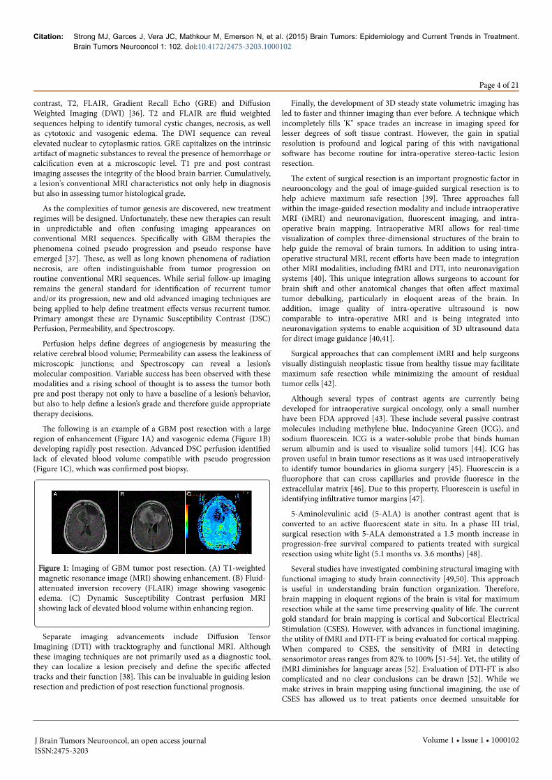
The following is an example of a GBM post resection with a largeregion of enhancement (Figure 1A) and vasogenic edema (Figure 1B)developing rapidly post resection. Advanced DSC perfusion identifiedlack of elevated blood volume compatible with pseudo progression(Figure 1C), which was confirmed post biopsy.
Figure 1: Imaging of GBM tumor post resection. (A) T1-weightedmagnetic resonance image (MRI) showing enhancement. (B) Fluid-attenuated inversion recovery (FLAIR) image showing vasogenicedema. (C) Dynamic Susceptibility Contrast perfusion MRIshowing lack of elevated blood volume within enhancing region.
Separate imaging advancements include Diffusion TensorImagining (DTI) with tracktography and functional MRI. Althoughthese imaging techniques are not primarily used as a diagnostic tool,they can localize a lesion precisely and define the specific affectedtracks and their function [38]. This can be invaluable in guiding lesionresection and prediction of post resection functional prognosis.
Finally, the development of 3D steady state volumetric imaging hasled to faster and thinner imaging than ever before. A technique whichincompletely fills 'K" space trades an increase in imaging speed forlesser degrees of soft tissue contrast. However, the gain in spatialresolution is profound and logical paring of this with navigationalsoftware has become routine for intra-operative stereo-tactic lesionresection.
The extent of surgical resection is an important prognostic factor inneurooncology and the goal of image-guided surgical resection is tohelp achieve maximum safe resection [39]. Three approaches fallwithin the image-guided resection modality and include intraoperativeMRI (iMRI) and neuronavigation, fluorescent imaging, and intra-operative brain mapping. Intraoperative MRI allows for real-timevisualization of complex three-dimensional structures of the brain tohelp guide the removal of brain tumors. In addition to using intra-operative structural MRI, recent efforts have been made to integrationother MRI modalities, including fMRI and DTI, into neuronavigationsystems [40]. This unique integration allows surgeons to account forbrain shift and other anatomical changes that often affect maximaltumor debulking, particularly in eloquent areas of the brain. Inaddition, image quality of intra-operative ultrasound is nowcomparable to intra-operative MRI and is being integrated intoneuronavigation systems to enable acquisition of 3D ultrasound datafor direct image guidance [40,41].
Surgical approaches that can complement iMRI and help surgeonsvisually distinguish neoplastic tissue from healthy tissue may facilitatemaximum safe resection while minimizing the amount of residualtumor cells [42].
Although several types of contrast agents are currently beingdeveloped for intraoperative surgical oncology, only a small numberhave been FDA approved [43]. These include several passive contrastmolecules including methylene blue, Indocyanine Green (ICG), andsodium fluorescein. ICG is a water-soluble probe that binds humanserum albumin and is used to visualize solid tumors [44]. ICG hasproven useful in brain tumor resections as it was used intraoperativelyto identify tumor boundaries in glioma surgery [45]. Fluorescein is afluorophore that can cross capillaries and provide fluoresce in theextracellular matrix [46]. Due to this property, Fluorescein is useful inidentifying infiltrative tumor margins [47].
5-Aminolevulinic acid (5-ALA) is another contrast agent that isconverted to an active fluorescent state in situ. In a phase III trial,surgical resection with 5-ALA demonstrated a 1.5 month increase inprogression-free survival compared to patients treated with surgicalresection using white light (5.1 months vs. 3.6 months) [48].
Several studies have investigated combining structural imaging withfunctional imaging to study brain connectivity [49,50]. This approachis useful in understanding brain function organization. Therefore,brain mapping in eloquent regions of the brain is vital for maximumresection while at the same time preserving quality of life. The currentgold standard for brain mapping is cortical and Subcortical ElectricalStimulation (CSES). However, with advances in functional imagining,the utility of fMRI and DTI-FT is being evaluated for cortical mapping.When compared to CSES, the sensitivity of fMRI in detectingsensorimotor areas ranges from 82% to 100% [51-54]. Yet, the utility offMRI diminishes for language areas [52]. Evaluation of DTI-FT is alsocomplicated and no clear conclusions can be drawn [52]. While wemake strives in brain mapping using functional imagining, the use ofCSES has allowed us to treat patients once deemed unsuitable for
Citation: Strong MJ, Garces J, Vera JC, Mathkour M, Emerson N, et al. (2015) Brain Tumors: Epidemiology and Current Trends in Treatment.Brain Tumors Neurooncol 1: 102.
Page 4 of 21
Volume 1 • Issue 1 • 1000102J Brain Tumors Neurooncol, an open access journalISSN:2475-3203
doi:10.4172/2475-3203.1000102
surgery. CSES demonstrates high sensitivity with the ability to map theentire exposed cortical region. Also, CSES provides excellent prognosiswith removal of negative cortical and subcortical areas leading to nopermanent neurological impairment [55].
Neuroepithelial tumorsAlthough the WHO classifies neuroepithelial tumors into nine
major groups, this review will only focus on the most frequent typesincluding astrocytic, oligodendroglial, ependymal and mixed. We willalso discuss the most common malignant neuroepithelial tumor inchildren, medulloblastomas.
Astrocytic NeoplasmsGlial-appearing cells give rise to most primary CNS neoplasms
(gliomas). Of these, astrocytomas are the most common.Astrocytomas, anaplastic astrocytomas, and glioblastomas are termeddiffusely infiltrating astrocytomas due to their range of diffuseinfiltration. The diffuse astrocytic neoplasms are most common in thecerebrum in adults and brain stem in children [56]. They have apropensity for progression with 50%-75% of astrocytomas progressingto anaplastic astrocytomas or GBMs [57]. Therefore, all patients withastrocytomas need regular followup. GBM has the highest incidence ofany primary neuroepithelial neoplasm, accounting for approximately50% of intracranial gliomas [58].
Histology
Pilocytic astrocytoma (WHO grade I)Macroscopically: Tumors are often cystic with discrete borders.
Microscopically: Neoplastic cells are usually bipolar with elongatedhairlike processes that are arranged in a parallel fashion. Rosenthalfibers, which are tapered corkscrew shaped eosinophilic hyalinemasses, are often present.
Diffuse Astrocytoma (WHO grade II)Macroscopically: Cerebral astrocytomas diffusely expand beyond
the white matter boundary oftentimes distorting the overlying graymatter. The neoplastic process is poorly demarcated.
Microscopically: Neoplastic cells show mild atypia. Fibrillaryastrocytomas may appear as bare nuclei. Astrocytomas show varyingdegrees of astrocytic differentiation. They may exhibit prominentfibrillary strands of eosinophilic cytoplasms, or plump cell bodies inwhich the nucleus is displaced by homogeneous eosinophiliccytoplasm, oftentimes referred to as the gemistocytic phenotype.
Anaplastic Astrocytoma (WHO grade III)Macroscopically: Anaplastic transformation may be associated with
little macroscopic change from astrocytomas. Although on MRI, areasundergoing anaplastic progression often show contrast enhancement.
Microscopically: Cytological and nuclear pleomorphism may bemore pronounced. Nuclear to cytoplasm ratio is increased. Mitoticactivity distinguishes the anaplastic astrocytoma from diffuseastrocytoma variants. However, necrosis is not present.
GBM (WHO grade IV)Macroscopically: GBM distorts the normal anatomy of the brain.
Foci of cyst formation, necrosis and hemorrhage are mixed withmucoid gray neoplastic tissue. GBMs commonly appear as sphericalmasses with a necrotic center, which may be seen on MRI as a ringenhancing mass. Their growth is not restricted to one hemisphere asthey often track along the corpus callosum affecting the contralateralhemisphere, commonly referred to as a butterfly glioma. GBMs mayalso spread along CSF pathways.
Microscopically: Necrosis and a florid microvascular proliferationare the key features separating GBM from the two other diffuseastrocytic neoplasms. Thrombi are often found in these vessels and areresponsible for the foci of necrosis. Cellular pleomorphism is moreextreme than in anaplastic astrocytomas. Finally, palisading of cellsaround necrotic areas is often seen.
Molecular GeneticsAlthough many important genetic alterations have been known in
gliomas, new technologies have shed light onto novel discoveries inrecent years. These genetic alterations are currently being used asbiomarkers. A biomarker is a genetic or biochemical feature that canbe assessed to indicate a particular diagnosis, prognosis, or response totreatment. As technology advances along with our understanding ofthe complex molecular genetics underlying brain tumors, the numberof biomarkers will likely increase.
O6-methylguanine methyltransferase (MGMT) promotermethylation: MGMT is a DNA repair protein that repairs thechemotherapy-induced alkylation at the O-position of guanine, theessential mediator of alkylating drug cytotoxicity, and thus counteractsthe effects of alkylating chemotherapeutic drugs such as nitrosoureasor temozolomide. Hypermethylation of the MGMT gene promoter isone mechanism to silence the gene and thus reduce the proteinconcentration. As such, hypermethylation of MGMT is associated with20-40% of patients with GBM [59]. Several clinical trials and cohortstudies have shown that the MGMT promoter methylation phenotypeis associated with prolonged progression-free and overall survival inpatients with GBM who are being treated with alkylating class ofchemotherapy drugs [60-62]. In a randomized clinical trial assessingradiotherapy alone with radiotherapy combined with concomitant andadjuvant treatment with temozolomide in newly diagnosed patientswith glioblastoma, the benefit from chemotherapy was almostexclusively attributable to patients with tumors with a methylatedMGMT gene promoter [62]. In the same study, patients with amethylated MGMT promoter showed better overall survival thanpatient with an unmethylated MGMT promoter [62]. These resultssuggest that treatment strategies should be individualized dependenton MGMT status and that MGMT status has prognostic value.
Loss of chromosomes 1p and 19q: The combined loss ofchromosomal arms 1p and 19q is a cytogenetic aberration resultingfrom an unbalanced t(1;19)(q10;p10) translocation occurring in50%-90% of oliogodendrogliomas and 30%-50% of oligoastrocytomas[63]. Tumors with the 1p/19q deletion respond better to chemotherapyand radiotherapy resulting in prolonged progression free survival andoverall survival in patients, especially with anaplasticoligodendrogliomas [64,65]. Outside of oligodendrogliomas, 1p/19qcodeletions are rare and additional studies are necessary to determinewhether these tumors have a less aggressive natural course [66].
Citation: Strong MJ, Garces J, Vera JC, Mathkour M, Emerson N, et al. (2015) Brain Tumors: Epidemiology and Current Trends in Treatment.Brain Tumors Neurooncol 1: 102.
Page 5 of 21
Volume 1 • Issue 1 • 1000102J Brain Tumors Neurooncol, an open access journalISSN:2475-3203
doi:10.4172/2475-3203.1000102
Isocitrate dehydrogenase (IDH) mutation: Single point mutations inthe metabolic genes IDH 1 and 2 were recently discovered in gliomas[67,68]. Approximately 80% of grade 2 and grade 3 gliomas as well assecondary GBMs harbor a single amino acid missense mutation inIDH1 at arginine 132 [69]. The IDH2 mutation at arginine 172 is lesscommon and is mutually exclusive with mutations in IDH1 [69]. IDH1and 2 mutations promote a neomorphic reaction in which the normalproduct α-ketoglutarate is converted to 2-hydroxyglutarate (2-HG) (acandidate oncometabolite) in a reaction that consumes, rather thanproduces, NADPH [70]. The accumulation of high concentrations of 2-HG has been shown to contribute to the formation and malignantprogression of gliomas [70]. In addition, the IDH1 mutation has beenassociated with the CpG island methylator phenotype (G-CIMP) ingliomas, which is associated with younger patients, improved survival,and is more common in low and intermediate grade gliomas [71,72].
Epidermal Growth Factor Receptor (EGFR) variant III: EGFR is acell surface receptor involved in the control of cell proliferation. Acommon alteration of the EGFR locus observed in gliomas is agenomic rearrangement with amplification of EGFR resulting in an in-frame deletion of exons 2-7 from the extracellular domain, causing atruncated mutant receptor known as EGFR variant III (EGFRvII) [73].This truncated mutant receptor is therefore ligand-independent andconstitutively active which confers enhanced tumorigenicity on gliomacells by increasing proliferation and reducing apoptosis [74].Overexpression of EGFR is observed in 50%-60% of GBMs with themost common EGFR mutation (EGFRvIII) expressed in 24%-67% ofcases [75-79]. Since the EGFRvIII mutation creates a new surfaceepitope, vaccination strategies based on this unique peptide sequencehave been developed. Subsequent phase 2 clinical trials havedemonstrated promising results, thus warranting phase 3 trials [80,81].The prognostic relevance of EGFR overexpression and EGFRvIII isunclear, but long-term survival might be worse in patients whosetumors carry the EGFRvIII mutation than in those who do not [75,82].
BRAF fusion or point mutation: The BRAF gene encodes a proteincalled B-Raf that is involved in cell signaling that promotes cell growth.Tandem duplications of BRAF and KIAA15A9 results in a gene fusionproduct called KIAA15A9-BRAF, which has constitutive B-Raf kinaseactivity. This fusion event is frequently detected in pilocyticastrocytomas, pleomorphic xanthoastrocytomas, and malignantastrocytomas [14,40]. The detection of the KIAA15A9-BRAF fusion isoften used to help distinguish pilocytic astrocytomas from higher-grade astrocytic tumors. Other BRAF gene alterations includingactivating point mutations in BRAF (e.g., BRAFV600E) have also beenidentified in low-grade as well as higher grade (III/IV) gliomas [83,84].Small-molecule BRAF kinase inhibitors, such as CCT239065 andRG7204, have been evaluated in melanomas and may provide a newtherapeutic approach to treat brain tumors harboring BRAF mutations[85].
Recent advances in sequencing along with the vast data from TheCancer Genome Atlas have re-classified GBMs into four distinctgenetic subtypes including classical, mesenchymal, proneural, andneural [86].
Classical: The classical subtype features many of the common genealterations observed in GBMs including chromosome 7 amplificationsand chromosome 10 deletions. In addition, EGFR amplification wasobserved more frequently in classical GBMs than the other subtypes[86]. Other common abnormalities in genes, TP53, NF1, PDGFRA, orIDH1 are not found in this group [86]. In response to aggressive
treatment, patients in the classical group lived the longest compared tothose in the other groups [86].
Mesenchymal: The mesenchymal subtype features mutations in theNF1 gene including focal hemizygous deletions of the 17q11.2 region,which contains the NF1 gene [86]. Mutations in the PTEN and TP53tumor suppressor genes are also frequently observed in this subtype[86]. Interestingly, tumors of the mesenchymal subtype expressSchwann cell markers such as the family S100A as well as microglialmarkers [86]. Genes associated with inflammation are enriched in thissubgroup, which is evident in the observation that there is a higheroverall fraction of necrosis in these tumors [86]. Patients in thissubtype typically survive longer after aggressive treatment than thosein the proneural and neural groups [86].
Proneural: The proneural subtype features alterations of thePDGFRA gene and point mutations in the IDH1 gene [86]. Althoughfocal amplifications of the PDGFRA locus are seen in all GBMsubtypes, proneural tumors have the highest rate. TP53 mutations andloss of heterozygosity are frequently observed in this subtype, whilechromosome 7 amplification and chromosome 10 deletion are lessprevalent [86]. This subtype shows high expression of oligodendrocyticdevelopment genes (e.g., PDGFRA, NKX2-2 and OLIG2), which mayhelp explain its atypical GBM subtype status [87]. In addition, theproneural subtype contains several proneural development genes suchas SOX, DCX, DLL2, ASCL1, and TCF4 [88]. This subgroupconsistently represents younger patients than the other subgroups andis often associated with secondary GBMs [86]. Interestingly, patients inthe proneural group who received aggressive treatment did not have asignificant survival advantage compared with patients in the proneuralgroup who did not receive aggressive treatment [86].
Neural: The neural subtype features expression of neuron markerssuch as NEFL, GABRA1, STY1, and SLC12A5 [86]. Tumors of thissubtype are associated with neural, astrocytic and oligodendroctyicgene signatures [86]. Patients in the neural group had someimprovement in survival but not as significant as those patients in theclassical and mesenchymal groups [86].
Treatment and PrognosisAlthough pilocytic astrocytomas commonly arise in the first two
decades of life, late presentation in adults is reported and typically hasa less favorable outcome [89]. If there is a high level of suspicion at thetime of presentation for pilocytic astrocytoma, decision-making shouldconsider the following: obtaining tissue diagnosis, restoringcerebrospinal fluid flow and decompressing adjacent neural structures,and non-surgical alternatives when an invasive procedure is precludedor incomplete.
Gross total resection of pilocytic astrocytomas provides the greatestclinical outcomes [90]. When risk of surgery is too high or if gross totalresection cannot be performed, consideration for radio- andchemotherapy can be given. The long-term risks of radiotherapy inchildren suggest it be employed only in cases of recurrence or pilocyticastrocytomas with aggressive nature. While various case series havefound prolonged survival after radiation therapy in children [91-93],the rate of recurrence after 5 years remains high. In a 10-yearprospective trial where practitioners felt irradiation posed a high-riskfor neurocognitive injury, a carboplatin and vincristine (CV) regimenwas compared to a thioguanine, procarbazine, lomustine, andvincristine (TPCV) regimen. Although five-year event-free survivalappeared more favorable using TPCV, toxicity observed with both
Citation: Strong MJ, Garces J, Vera JC, Mathkour M, Emerson N, et al. (2015) Brain Tumors: Epidemiology and Current Trends in Treatment.Brain Tumors Neurooncol 1: 102.
Page 6 of 21
Volume 1 • Issue 1 • 1000102J Brain Tumors Neurooncol, an open access journalISSN:2475-3203
doi:10.4172/2475-3203.1000102
regimens presented a major limitation for long-term effectiveness ineither group [94].
A multimodality approach consisting of surgical resection, radiationtherapy, and chemotherapy is used in the treatment of malignantgliomas. Surgery plays a key role in the treatment of malignant gliomasas it allows for both cytoreduction and confirmation of diagnosis.Furthermore, there is growing evidence in the literature that achievinggross total resection is important in prolonging survival [95,96]. Asdiscussed in the imaging section, advances in neuroimaging and brainmapping has allowed for increased gross total resection.
Radiation therapy is currently recommended for all patients withgrade III and IV gliomas (anaplastic astrocytomas and GBM,respectively). Only infants, young moribund children, and patientsdeclining treatment in favor of supportive care would not berecommended to receive some form of radiation therapy shortly afterdiagnosis. Radiation therapy alone has been shown to improve mediansurvival from 3-4 months to 9-12 months [97,98]. Although radiationtherapy has shown a clear improvement in survival for patients withprimary GBMs, radiotherapy for recurrent GBMs is controversialmainly due to the serious risks associated with reirradiation includingnecrosis of healthy brain tissue [99]. Despite these serious risks, studiessuggest there may be a benefit to a more focused radiation therapythrough the use of stereotactic radiosurgery or fractionated stereotacticreirradiation [100,101].
Alkylating agents are the most frequently used chemotherapy drugsin treating anaplastic astrocytomas and GBMs. The cytotoxic effectsare mediated primarily through DNA cross-linking, leading to celldeath by apoptosis. Carmustine (BCNU) has been shown to prolongsurvival of GBM patients in two clinical trials [102,103], however, dueto the post-operative complications; the addition of BCNU to apatient’s treatment protocol is unclear and ultimately determined bythe treating physician. In 2005, temozolomide was shown to improvemedian survival and increase the likelihood of long-term survival withnewly diagnosed GBM when given concurrently with RT and thenfollowing RT [98]. Since this publication, the Strupp protocol,involving surgery followed by radiotherapy plus concomitant andadjuvant temozolomide for the treatment of malignant gliomas is thecurrent gold standard [98].
Additional chemotherapeutic agents targeting specific moleculescurrently used in treating malignant gliomas include inhibitors ofEpidermal Growth Factor Receptor (EGFR), Platelet-Derived GrowthFactor Receptor (PDGFR), Vascular Endothelial Growth Factor(VEGFR), mammalian Target of Rapamycin (mTOR), Protein KinaseC (PKC), RAF-MEK-ERK pathway, and integrins [104]. With newchemotherapeutic agents being developed the gold standard forevaluating these agents remain randomized clinical trials. Recently, ananti-VEGF monoclonal antibody, bevacizumab, was evaluated in tworandomized phase 3 clinical trials in which it was concluded thatalthough there was improved progression-free survival, the overallsurvival in patients was not improved [105,106]. Although overallsurvival was not improved in these studies, progression-free survivalincreased 3-4 months and bevacizumab may still hold promise as anoption for treating GBMs. With the development of novelchemotherapeutic agents, a combined multimodality approachincluding both chemotherapy and radiotherapy is needed to overcometumor resistance through the use of multi-targeted strategies.
Since survival for patients with GBMs is relatively short, identifyingprognostic indicators in order to stratify patients into risk groups may
provide valuable in determining the best therapeutic approachincluding enrollment into active clinical trials. Lamborn andcolleagues surveyed 832 GBM patients enrolled into prospectiveclinical trials in order to identify potential risk groups [107]. Fromtheir analysis, they identified four risk groups in which the two lowerrisk groups included patients under the age of 40 with the lowestoverall risk group being young patients with frontal lobe tumors only[107]. The intermediate risk group included patients aged 40-65 withKPS >70 who underwent subtotal or total resection of the tumor.Finally, the highest risk group included patients over the age of 65 orpatients aged 40-65 with either KPS <80 or who only underwentbiopsy [107].
In addition to the age of the patient and the KPS score, other clinicalparameters serve as prognostic indicators of long-term survival. Tumorsize and location are also important indicators as extent of resection isdictated not only by tumor size but also by location of the tumor.Finally, grade of tumor is an important indicator of long term survivalas the higher the grade the more malignant the tumor is, whichdirectly results in a poorer prognosis.
Oligodendroglial tumorsHistorically, oligodendrogliomas (ODG) were thought to account
for only about 4% of primary brain tumors [108], however, with betterunderstanding of the tumor biology, it is thought that ODG maycomprise as much as 25-33% of gliomas [109,110]. Up to 57-87% ofpatients with ODGs present with seizures [108,111] with 22% ofpatients presenting with headaches [108].
HistologyCalcifications are typically seen histologically with 73% of tumors
having microscopic calcifications [112]. Histopathologic analysis ofthese tumors demonstrates a lucent perinuclear halo typically referredto as a “fried egg” appearance along with a “chicken-wire” vascularpattern [110]. Although these features are common to ODG, they arenot pathognomonic. Since most ODGs contain microtubules and notglial filaments, they typically do not stain for Glial Fibrillary AcidicProtein (GFAP) [113]. Attempts for a grading system has been metwith controversy, mainly surrounding the lack of prognosticsignificance [114]. Therefore, for prognostic reasons, a twoclassification system has been proposed that includes low grade;oligodendroglioma (WHO grade II) and high grade; anaplasticoligodendroglioma (WHO grade III) [114,115].
Treatment and PrognosisSurgery is mainly reserved for low grade ODGs, while surgical data
for high grade ODGs is less convincing. Radiation therapy for ODGs isalso unclear [116] with one retrospective study showing a 10-yearsurvival rate of 56% in those patients receiving postoperative radiation>45 Gy [117], while in another study, no difference in 5-year survivalwas observed in patients with or without postoperative radiation [118].Chemotherapy is therefore the primary modality for treating ODGs asmost respond to chemotherapy [119]. The 10-year survival rate forODGs is 10-30% with pure ODGs having a higher survival rate thanmixed ODGs [108,112]. The presence of calcifications on imaging as aprognostic indicator was evaluated in one series, however, additionalstudies are needed before conclusions can be drawn [108]. Finally,chromosomal 1p and 19q loss is associated with longer survival rates[120].
Citation: Strong MJ, Garces J, Vera JC, Mathkour M, Emerson N, et al. (2015) Brain Tumors: Epidemiology and Current Trends in Treatment.Brain Tumors Neurooncol 1: 102.
Page 7 of 21
Volume 1 • Issue 1 • 1000102J Brain Tumors Neurooncol, an open access journalISSN:2475-3203
doi:10.4172/2475-3203.1000102
EpendymomasEpendymomas are a rare type of glial tumor that is believed to arise
from ependymal cells lining the cerebral ventricles and along thecentral canal of the spinal cord. A large portion of intracranialependymomas (36-60%) occur in children, making ependymomas thesecond most common malignant brain tumor in this population [121].Spinal ependymomas are more common in adults, occurring in 96% ofcases [122].
ClassificationEpendymomas are usually well circumscribed and benign but they
have been known to be invasive. Ependymomas are divided into fourmajor subgroups including myxopapillary (WHO grade I),subependymoma (WHO grade I), classic ependymoma (WHO gradeII), and anaplastic ependymomas (WHO grade III). Within classicependymomas there are an additional four variants including cellular,papillary, clear cell, and tanycytic. Lastly, it is worth noting that onceconsidered a variant, ependymoblastoma, is now being regarded as arare childhood primitive neuroectodermal tumor with abundantmitotic figures and true rosettes [123].
HistologySimilar to all brain tumors, the diagnosis of ependymoma requires
histological confirmation. Classical ependymomas are characterized bydark small nuclei. They also show two cytoplasmic patterns:perivascular pseudorosettes and true rosettes. Perivascularpseudorosettes are areas of radiating neoplastic cells that lack nucleiand surround blood vessels. True rosettes are areas of ependymaltubules around a central blood vessel.
Treatment and PrognosisSince ependymomas are highly radiosensitive, the best approach for
treatment of ependymomas is gross total resection followed byradiation therapy. The role of chemotherapy for the treatment ofependymomas is currently unclear [124].
Mixed gliomasMixed gliomas are tumors that contain both oligodendroglioma and
astrocytoma cells. These tumors can be classically divided intooligoastrocytoma (WHO grade II) and anaplastic oligoastrocytoma(WHO grade III). Oligoastrocytomas comprise 10-19% of low-gradegliomas and usually develops in middle-aged adults [125]. Treatment isusually surgical resection. If however, these tumors recur, thetreatment approach is surgery followed by radiotherapy andchemotherapy.
MedulloblastomaMedulloblastomas are the second most frequent childhood brain
tumor after Pilocytic astrocytomas, and the most common malignantbrain tumor in children comprising roughly 25% of intracranialtumors [126]. They occur exclusively in the posterior fossa with a peakincidence between 4 and 7 years [127]. Patients with medulloblastomasoften present with symptoms of increased intracranial pressure(hydrocephalus) [128]. Since these tumors grow in the posterior fossa,gait ataxia, truncal instability, vomiting, dizziness and vision problemsare also common symptoms caused by involvement of the cerebellum,
brainstem, or cranial nerves. Since metastases along the cranio-spinalaxis are present in roughly 33% of patients [126], evaluation formetastases is recommended.
Classification and HistologyAlthough all medulloblastomas are classified as WHO grade IV,
within this classification scheme, there are currently five variantsincluding classic, desmoplastic/nodular, Medulloblastoma withExtensive Nodularity (MBEN), Large Cell (LC), and AnaplasticMedulloblastoma [129].
Molecular pathways: While the etiology is unknown, roughly 2-5%of medulloblastomas are associated with nevoid basal cell carcinomasyndrome (Gorlin syndrome), which is caused by mutations in thepatched-1 gene (PTCH-1), and familial adenomatous polyposis, whichis caused by inactivating mutations in the adenomatous polyposis coligene [130,131]. In addition, great strides have been made in ourunderstanding of the oncogenesis of medulloblastomas. Based on geneexpression profiles using tissue microarrays and substantiated usingwhole genome and whole exome sequencing, medulloblastomas havebeen separated into four distinct subgroups based on their uniquemolecular profiles [132-135]. These groups include sonic hedgehog(SSH), wingless (WNT), group 3, and group 4.
SHH medulloblastomas: This subgroup comprises roughly 25-30%of medulloblastomas and is characterized by high desmoplasia[132,134-137]. The overexpression of SHH pathway leads to thebinding and inactivation of PTCH-1, which normally blocks activationof a number of transcription factors through the inhibition ofsmoothened (SMO). SHH also upregulates MYCN, which is involvedin the cell cycle. In addition to mutations in PTCH-1, mutations inSMO and Suppressor of Fused Homolog (SUFU) have also beenobserved in SHH medulloblastomas [138,139]. Chromosomalaberrations have also been associated with this subgroup including lossof 9q (accounting for 21-47%), 10q, 20p, 21p and gain of chromosome3q and 9p [132,136,140]. The majority of SHH medulloblastomasoccur in infants under the age of 3 and again in adults above age 16[136,141]. In fact, nearly half of all adult medulloblastomas are of theSHH variant [142]. The overall survival is good in infants andintermediate in adults [143].
WNT medulloblastomas: The WNT medulloblastoma subgroupharbors mutations in essential genes of the WNT pathway includingAPC, β-catenin, and axin 1 [144-146]. Roughly 10-15% ofmedulloblastomas fall within this subgroup [137]. They arecharacterized by classic histology, more than 90% of WNT tumors,affecting patients above 3 years old, good prognosis, and infrequentmetastasis [137]. Unlike SHH tumors, WNT tumors rarely affectinfants and the overall survival is generally good [141,143].
Group 3 and 4 medulloblastomas: Both of these subgroups presentwith common clinical features and share similar molecular profiles.The age of onset for both groups vary with Group 3 peaking inchildhood (3-10 years), while Group 4 has a more distributed age ofonset from infancy to adulthood [136,141]. The majority of tumors inboth groups display classical histology. Chromosomal aberrations arecommon to both groups with isochromosome 17q representing themost frequent structural alteration [135]. Gain of 7 and 18q along withloss of 8 and 11p are also common abnormalities [136,137,147]. MYCamplification in group 3 is the main difference between these groupssince MYC amplification is rarely observed in group 4 [147,148].Conversely, enrichment of chromosome X loss is more common in
Citation: Strong MJ, Garces J, Vera JC, Mathkour M, Emerson N, et al. (2015) Brain Tumors: Epidemiology and Current Trends in Treatment.Brain Tumors Neurooncol 1: 102.
Page 8 of 21
Volume 1 • Issue 1 • 1000102J Brain Tumors Neurooncol, an open access journalISSN:2475-3203
doi:10.4172/2475-3203.1000102
group 4 as observed in 80% of females in this group [147]. Lastly, bothgroups have similar metastatic rates but group 3 has a poorerprognosis than group 4, which shows intermediate prognosis [148].
TreatmentMedulloblastomas are among the most radiosensitive tumor of the
central nervous system and are moderately chemosensitive. Therefore,the recommended therapeutic approach is surgical debulking followedby radiation therapy. 36 Gy to the entire craniospinal axis with a boostto the tumor bed for a total of 55 Gy is the recommended radiationtherapy dosage [149]. Medulloblastomas are moderatelychemosensitive, and as such, chemotherapy is now an integral part ofthe treatment of these tumors, including recurrent disease [150]. Somechemotherapy agents used include lomustin, cisplatin and vincristine.Finally, placement of permanent ventriculoperitoneal shunts isrequired in 30-40% of patients after tumor resection of the posteriorfossa [151,152].
PrognosisPatients with medulloblastomas are classified into three risk groups
that help facilitate treatment and provide predictions on prognosis.These groups include standard risk, intermediate risk, and poor risk.Patients with no residual tumor demonstrated on post-operative MRIalong with negative CSF results are classified as standard risk and carrya 5-year survival rate of 73% [153]. Residual tumor measuring greaterthan 1.5 cm2 on post-operative MRI and presence of tumor cells in thebrain, spine, or CSF are characteristic of patients in the poor riskgroup. These patients have a poor prognosis with a 5-year disease freesurvival of 36-52% [154]. The intermediate risk group is poorlycharacterized leaving the other two groups as primary predictors.Interestingly, females have a better prognosis than males [155].
MeningiomaMeningiomas arise from the layer of tissue covering the brain and
spinal cord. Meningiomas are the most common benign intracranialtumor accounting for about 13-26% of all primary brain tumors [156].Most meningiomas are intracranial, however, spinal meningiomas mayoccur accounting for an estimated 7.5 to 12.7% of all meningiomas[157]. The vast majority of meningiomas rarely metastasize with a rateof less than 1 in every 1,000 meningiomas [158]. The most commonsites of metastasis include lung and pleura, liver, lymph nodes, andbone [159]. Due to the slow growing nature of meningiomas, mostremain asymptomatic. Meningiomas rarely affect children with anincidence rate of roughly 2.2% [160] and of these, prevalence ofneurofibromatosis was 14.5% [161]. There have been reportsdocumenting prevalence of meningiomas in roughly 50% of NF2patients [162] with one study reporting prevalence as high as 58%[163]. If multiple meningiomas are observed, suspicion ofneurofibromatosis type 2 is high. Both neurofibromatosis types 1 and2 will be discussed in the vestibular schwannoma section.
Classification and HistologyMeningiomas have a complex and broad range of histological
patterns. The WHO currently recognizes 16 different variants groupedinto three grade designations (Table 1) [129]. Meningiomas falling inthe WHO grade I classification include: meningothelial, fibrous,transitional, psammomatous, angiomatous, microcystic, secretory,lymphoplasmacyte-rich, and metaplastic. Although each subtype has a
distinct histological pattern, all WHO grade I meningiomas have a lowrecurrence rate of 9% with no evidence of brain invasion [164]. Threesubtypes of meningiomas fall within WHO grade II and these include:chordoid, clear cell, and atypical. One major difference between the2000 WHO classification scheme and the recent 2007 WHOclassification scheme is the recognition that meningiomas withevidence of brain invasion should be classified as WHO grade IIregardless of a benign histological appearance [165,166]. WHO gradeII meningiomas have a recurrence at of 29% [164]. Lastly,meningiomas in the WHO grade III group include papillary, rhabdoid,and anaplastic with the anaplastic variant representing the majority ofWHO grade III cases [129]. All WHO grade III meningiomas haveincreased mitotic activity with a recurrence rate of 50% [164].
TreatmentIf the tumor is not causing symptoms, tumor growth may be
watched using serial MRIs. Otherwise, surgery is the standard of carefor treating meningiomas. Similar to other tumor types, extent oftumor resection is beneficial for minimizing the risk of tumorrecurrence. As a result, in 1957, Simpson established a classificationsystem consisting of five subdivisions to assess extent of resection ofmeningiomas and to correlate postoperative recurrence rates withextent of resection [167]. In his grading system, Simpson grade I isdefined as complete tumor resection with excision of the duralattachment and any abnormal bone. If the venous sinus is involved,complete resection of the sinus is also performed. Simpson grade II isdefined as complete tumor resection with coagulation of the duralattachment. Simpson grade III is defined as complete tumor resectionwithout resection or coagulation of the dural attachment. Simpsongrade IV is defined as a subtotal resection. Finally Simpson grade V isdefined as a simple decompression, with or without biopsy. The risk oftumor recurrence (minimum of 6 months of follow-up) for Simpsongrades I, II, III, and IV were 9%, 16%, 29%, and 39%, respectively[167].
With advancements in surgical techniques and treatment options,such as radiation therapy, relying solely on the Simpson grading systemof meningiomas in the modern era to predict recurrence isinconclusive. Therefore, cell proliferation markers, such as Ki-67(MIB-1 – monoclonal antibody) are being evaluated to complementthe Simpson grading system in predicting tumor recurrence [168]. In astudy conducted by Oya and colleagues, they determined that theMIB-1 index could differentiate meningiomas with a high risk ofrecurrence [168]. Further, the authors conclude that using the MIB-1index could be beneficial in planning optimal follow-up strategies witha shift from attempting aggressive resection to valuing the quality ofthe patient’s life [168].
Several retrospective studies have demonstrated that radiationtherapy (e.g., external beam radiation therapy and stereotacticradiosurgery) can provide improved and durable local control inselected patients with meningioma [169]. For WHO Grade I orpresumed Grade I meningiomas, radiation therapy achieved long-termlocal control in 68% to 100% of cases at 5 to 10 years, includingpatients treated postoperatively, primarily, or following tumorrecurrence.169 The use of stereotactic radiosurgery is considered mosteffective for patients with small meningiomas (usually less than 3 cmin diameter or 10 cm3 in volume), those with distinct margins, andthose at sufficient distance from functionally important brain, nerves,and other critical structures to permit safe delivery of an adequatetarget dose [169].
Citation: Strong MJ, Garces J, Vera JC, Mathkour M, Emerson N, et al. (2015) Brain Tumors: Epidemiology and Current Trends in Treatment.Brain Tumors Neurooncol 1: 102.
Page 9 of 21
Volume 1 • Issue 1 • 1000102J Brain Tumors Neurooncol, an open access journalISSN:2475-3203
doi:10.4172/2475-3203.1000102
For high-grade meningiomas, a multimodal approach usingradiation therapy and/or chemotherapy is usually given. Achievingmaximum resection and adjuvant radiotherapy have been shown to beindependent predictors of patient survival and disease-free survival inthe treatment of malignant meningioma [170]. Evaluating stereotacticradiosurgery in the setting of subtotal resection or recurrence, reportedlocal control rates (>2 years) range from 0% to 90%, with the majorityfalling within 50% to 80% for WHO grade II meningiomas [169].While some studies have suggested stereotactic radiosurgery is notindicated for malignant meningiomas [171], others have shownimproved local control rates of 17% at 15 months [172]. Finally, forrecurrent atypical or anaplastic meningiomas not suitable forradiosurgery, resection followed by permanent brachytherapy is apotential salvage therapy that has shown promise in the clinical setting[173,174]. In the largest series (n=21) to date examining brachytherapyfor therapy for the recurrence of aggressive atypical and anaplasticmeningiomas, Ware et al. reported a median survival of 1.6 years after[125] I implantation for atypical meningiomas and 2.4 years foranaplastic meningiomas [173] Due to the high complication ratesobserved including radiation necrosis occurring in 27% in one study,meticulous surgical technique and medical therapies to assist withwound healing after surgery is required [173].
For meningiomas that are inoperable and/or radiation-refractory,chemotherapy is often used with little to no effect. As a result of failedchemotherapeutic approaches, several studies have investigatedvarious chemotherapies in which all have been disappointing[175,176]. Although there is limited data, hydroxyurea, somatostatinanalogues and interferon-α have all been modestly successful inpatients with recurrent meningiomas [175]. Further, emerging targetedtherapies including sunitinib, may prove useful in refractorymeningiomas [175,176].
PrognosisPrognosis for patients with benign meningiomas is generally good
with a 5-year survival rate of 91.3% [177]. Recurrence depends onextent of surgical resection with a recurrence rate of 8% in cases with agross total resection, a 29% in cases with a subtotal resection [178].Atypical meningiomas have been reported to have a higher rate of localrecurrence and are associated with lower survival rates compared tobenign meningiomas [179]. Similar to benign meningiomas, achievinga gross total resection of atypical meningiomas was associated with alower recurrence rate (11%) compared to achieving a subtotal resection(100%) [179]. A similar trend of increased survival is also associatedwith (grade I) total resection of malignant mengiomas [180]. Lastly,Al-Mefty and colleagues investigating the malignant progression inmeningioma from a benign to a higher histological grade, andconcluded that the presence of complex genetic alterations (e.g.,increased MIB-1 staining and chromosomal aberrations such asalterations in chromosome 22 and deletion of chromosomes (1p, 14q,and 10q), even with a benign histological grade, may potentially havean aggressive phenotype and require closer follow up [181].
Pituitary TumorsThe majority of pituitary tumors are adenomas arising from the
anterior pituitary gland (adenohypophysis). In rare cases, pituitarycarcinomas have been described [182]. Pituitary tumors arising fromthe posterior pituitary gland (neurohypophyseal) are also rare [183].Pituitary adenomas are the fourth most common intracranial tumorafter gliomas, meningiomas and schwannomas [184]. Pituitary
adenomas are typically benign with even malignant pituitary tumorsrarely metastasizing [184]. These tumors may secrete abnormally highamounts of hormones that may lead to physiological dysfunctionresulting in patient morbidity. In addition to endocrinologicdisturbances, mass effect leading to bitemporal hemianopsia is oftenobserved in patients with pituitary neoplasms. Several risk factors havebeen identified including Multiple endocrine neoplasia type 1 (MEN1),Carney’s complex, and Familial isolated pituitary adenoma [185].
Classification and HistologyA functional classification scheme has been developed based upon
the secreted hormones and include lactotrophic adenomas(prolactinomas) which secrete prolactin, are the most common, andcauses amenorrhea-galactorrhea syndrome in women and impotencein men, somatotrophic adenomas which secrete growth hormone,corticotrophic adenomas which secrete adrenocorticotropic hormone,gonadotrophic adenomas which secrete luteinizing hormone andfollicle-stimulating hormone, thyrotropin-secreting adenomas whichsecrete thyroid-stimulating hormone, cause thyrotoxicosis, and arerare, and null cell adenomas which do not secrete hormones. Excessiveamounts of adrenocorticotropic hormone from pituitarycorticotrophic adenomas can lead to Cushing’s syndrome. As aconsequence of undergoing adrenalectomy for treatment of Cushing’ssyndrome, 8-43% of patients will develop hyperpigmentation referredto as Nelson’s syndrome [186,187]. An increased concentration ofgrowth hormone from somatotrophic adenomas can lead toacromegaly. Interestingly, more than 95% of cases of acromegaly aredue to a pituitary somatotroph adenoma [188].
TreatmentMR imaging with contrast on a pituitary protocol is the gold
standard for evaluating pituitary tumors. Since the normal pituitarygland also enhances, the timing of the contrast is important inachieving a high-spatial-resolution image that is able to discern normalpituitary tissue from a macroadenoma [189]. In patients with non-secreting tumors and without neurologically deficits, it is reasonable tofollow these patients with serial MRIs and visual field examinations.
Current treatment options for symptomatic pituitary adenomasinclude surgical resection, radiation therapy and medication therapy(first line for treating prolactinomas). Three dopamine agonists areroutinely given to treat prolactinomas and include bromocriptine,carbergoline, and pergolide. Surgery using a transsphenoidal approachis typically the first line treatment for the other subtypes of pituitaryadenomas [190,191]. Medical therapy is also used for patients withsomatotrophic adenomas and includes dopamine agonists (e.g.,bromocriptine), somatostatin analogues (e.g., octreotide), and growthhormone antagonists (e.g., pegvisomant). Medical treatment forthyrotropin-secreting tumors typically involves somatostatin analogues(e.g., octreotide).
For the treatment of pituitary adenomas, conventional radiationtherapy typically consists of 40-50 Gy administered in 20-25 fractionsover 4-6 weeks [192]. Although effective, complications associatedwith radiation therapy are high. One of the major post-radiationcomplications is hypopituitarism, which is both dose- and time-dependent. In addition, injury to the optic nerves and chiasm, lethargy,memory disturbances, cranial nerve palsies, and tumor necrosis withhemorrhage and apoplexy may also occur. The effects of radiationtherapy on somatotrophic adenomas is cumulative with time and may
Citation: Strong MJ, Garces J, Vera JC, Mathkour M, Emerson N, et al. (2015) Brain Tumors: Epidemiology and Current Trends in Treatment.Brain Tumors Neurooncol 1: 102.
Page 10 of 21
Volume 1 • Issue 1 • 1000102J Brain Tumors Neurooncol, an open access journalISSN:2475-3203
doi:10.4172/2475-3203.1000102
take 10 years to reduce the growth hormone levels into a “curative”range [193,194]. In contrast, for Cushing’s disease, radiation therapyrestores ACTH levels to normal range between 18 and 42 months[195].
The use of radiosurgery for the treatment of pituitary tumors is stilllimited to tertiary centers and protocols are not standardized. Yang etal. conducted a large aggregated
analysis of stereotactic radiosurgery treatment in patients withacromegaly and determined that the overall disease control rate wasapproximately 48%–53% for patients no longer taking suppressivemedications after radiosurgery for acromegaly [196]. The post-radiosurgery remission rates for Cushing’s disease reported in theliterature vary considerably from 0% to 100%, with most seriesdocumenting an approximately 50%–60% remission rate [197]. Long-term complications are thought to be similar to conventional radiationtherapy (except for optic symptoms).
PrognosisIn cases where the tumor is compressing the optic apparatus,
removal of the tumor improves vision in 90% of patients [198].Furthermore, in one case series, only 27% of patients with prolactin-secreting tumors and 20% of patients with growth hormone-secretingmacroadenomas returned to baseline hormone levels after surgicalresection [198]. In the same series, the recurrent rate was roughly 13%.However, the inclusion of post-operative radiation therapy as well asdegree of surgical resection of the tumor influenced the rate ofrecurrence. For example, for patients with partial surgical resectionwho did not receive post-operative radiation therapy, their recurrencerate was 50% [198]. On the other hand, there were no recurrencesobserved in patients with a gross total tumor removal who receivedpostoperative radiation therapy [198].
The posterior pituitary can sometimes be damaged during surgeryleading to a condition called central diabetes insipidus, which ischaracterized by excessive thirst and dilute urine.
Primary Central Nervous System LymphomaPrimary Central Nervous System Lymphomas (CNS lymphomas) is
a rare aggressive form of extranodal high-grade non-Hodgkinlymphoma that represents roughly 4% of intracranial neoplasms [199].Several risk factors have been identified for primary CNS lymphomasincluding collagen vascular diseases (e.g., systemic lupuserythematosus and Sjogren’s syndrome), immunosuppression,including AIDS, and Epstein-Barr virus, which is associated with manylymphoproliferative disorders with nearly 100% association withprimary CNS lymphomas [200], especially AIDS related [201]. On CTimaging, non-AIDS CNS lymphomas typically have a homogeneousenhancement pattern, whereas AIDS CNS lymphomas tend to have anecrotic center with the appearance of multifocal ring-enhancinglesions [202]. Surgery has a limited role in treatment and is usedmainly for tumor biopsy. Radiation therapy and chemotherapy areprimarily used to treat CNS lymphomas. Recent advances in treatmentoptions have resulted in the use of high-dose chemotherapy incombination with autologous stem cell transplantation as analternative treatment approach [199]. Whole-brain radiation therapy isoften used with 40-50Gy, especially when chemotherapy iscontraindicated [203].
For patients with non-AIDS CNS lymphomas the combination ofradiation therapy and chemotherapy has a better overall survival thanradiation therapy alone [204]. The median survival for patients notreceiving treatment is 1.8-3.3 months, with radiation therapy alonemedian survival increases to 10 months, and with intraventricularmethotrexate, the median time to recurrence increases to 41 months[205]. On the other hand, patients with AIDS-related CNS lymphomashave a much worse prognosis with a median survival of 2-5 monthseven after treatment [206,207].
Vestibular SchwannomaVestibular schwannomas, also known as acoustic neuromas, arise
from cells that produce the myelin sheath covering thevestibulocochlear nerve (CN VIII). They usually originate in thesuperior vestibular division of CNVIII. These tumors are generallybenign with an incidence rate of 1.67 per 100,000 person-years [1].Vestibular schwannomas have been linked to loss of NF2 (tumorsuppressor) on chromosome 22 [208,209]. Roughly 95% of vestibularschwannomas are unilateral, however, bilateral vestibularschwannomas is pathognomonic of neurofibromatosis type 2 (NF2)[210,211]. Most patients present with a classic clinical triad of hearingloss, tinnitus (high pitched), and disequilibrium [212]. Histologicallythese tumors contain Antoni A and B fibers [213]. Antoni A fibers arenarrow elongated bipolar cells that are tightly packed, while Antoni Bfibers are loosely packed cells with reticular fibers [213]. Verocaybodies (cellular areas surrounded by parallel arrangement of spindleshaped Schwann cells) are also seen histologically [214]. Since thesetumors involve CN VIII, audiometric evaluation is part of the initialworkup of this tumor and is used to help guide management. There arethree approaches to treating vestibular schwannomas. These includecomplete surgical resection, radiation therapy, or monitoring usingconsecutive MRIs. With complete surgical removal, the incidence ofrecurrence is minimal from 0%-3% [215]. There are currently threesurgical approaches used including retrosigmoid, which may preservehearing, translabyrinthine, which sacrifices hearing but increaseschances of preserving VII, and middle fossa approach, which is usuallyreserved for small lateral vestibular schwannomas [216]. The tumorprogression rate following subtotal resection is roughly 20% [215].
NeurofibromatosisAlthough there are six subtypes of neurofibromatosis, the most
common are type 1 (NF1) and type 2 (NF2) and are discussed below.
Neurofibromatosis type 1NF1 is an autosomal dominant disease that is linked with mutations
in the NF1 gene on chromosome 17 which codes for neurofibromin[217]. Loss of neurofibromin, which is a negative regulator of the Rasoncogene, leads to increased growth stimulating signaling.Neurofibromatosis 1 represents more than 90% of neurofibromatosis[218]. The diagnosis of NF1 is made by two or more of the following[219]:
• 6 café au lait spots
• 2 neurofibromas of any type or one plexiform neurofibroma
• hyperpigmentation in the axillary or inguinal areas
• optic glioma
• >2 Lisch nodules (pigmented iris hamartomas)
Citation: Strong MJ, Garces J, Vera JC, Mathkour M, Emerson N, et al. (2015) Brain Tumors: Epidemiology and Current Trends in Treatment.Brain Tumors Neurooncol 1: 102.
Page 11 of 21
Volume 1 • Issue 1 • 1000102J Brain Tumors Neurooncol, an open access journalISSN:2475-3203
doi:10.4172/2475-3203.1000102
• distinctive osseous abnormality (e.g., sphenoid dysplasia)
• a first degree relative with NF1
Neurofibromatosis type 2NF2 is an autosomal dominant disease linked with mutations in the
NF2 gene on chromosome 22, which codes for merlin (schwannomin)[211]. This tumor suppressor is typically produced in the centralnervous system particularly in Schwann cells. The diagnosis of NF2 ismade with either [220,221]:
• Bilateral vestibular schwannomas on imaging (MRI or CT)
• A first degree relative with NF2 and either:
• Unilateral vestibular schwannoma at age <30 years or
• Any two of the following: meningioma, glioma, posteriorsubcapsular lens opacity, neurofibroma
Additional criteria that carry less weight include:
• Unilateral vestibular schwannoma at age <30 and any of thefollowing: meningioma, schwannoma, glioma, posterior subcapsularlens opacity or
• Multiple meningiomas and either of the following: unilateralvestibular schwannoma or any two of the following: glioma,neurofibroma, schwannoma, or cataract
NeurofibromaNeurofibromas are benign nerve sheath neoplasms arising in the
peripheral nervous system. They are typically found in patients withneurofibromatosis. These tumors are divided into dermal (WHO gradeI) and plexiform (WHO grade I). Dermal neurofibromas are usuallyassociated with a single peripheral nerve and do not acquire malignantfeatures. However, plexiform neurofibromas are associated withmultiple nerve bundles and although low, have the ability to transforminto malignant tumors, making these tumors more difficult to treat.The clinical course usually dictates the treatment approach forneurofibromas. Since these tumors are intimately intertwined withfunctional nerve, surgery is only performed if there are symptomsfrom the neurofibroma leading to progressive morbidity [222]. Othertreatment options may include antihistamines, maturation agents (e.g.,retinoic acid), and antiangiogenic drugs [222]. Targeted therapeuticapproaches for specific molecular pathways vital to the tumor arepromising, but need to be evaluated in clinical trials [222].
Schwannoma vs. neurofibromaBoth of these tumors are very similar and hard to distinguish
without histological analysis. Schwannomas are typically wellcircumscribed and consist of Antoni A and B fibers. On the otherhand, neurofibromas are typically less cellular, not as wellcircumscribed, and consist of wavy collagen fibers with occasionalneuritis [223]. S-100 staining is oftentimes used to help distinguishthese tumors since schwannomas typically display a greater percentageof positive cells and the intensity of staining is higher. However, withboth tumors expressing some degree of S-100, this stain alone is notsufficient differentiating these tumors. Other stains that have beenstudied with varying sensitivities and specificities to help decipherthese two tumors include calretinin, CD34, CD56, glial fibrillary acidicprotein (GFAP), epithelial membrane antigen (EMA), factor XIIIa,Leu-7, myelin basic protein and Glut-1 [223,224]. A combination of
immunohistochemical stains provides the greatest support indetermining schwannoma versus neurofibroma.
Secondary Brain Tumors
EpidemiologySecondary, or metastatic brain tumors (MBTs), are the most
common malignancies of the central nervous system (CNS). Typically,MBTs arise from primary tumor cells that migrate hematogenously orvia direct invasion of adjacent tissue. According to population studies,the estimated prevalence of MBTs in the United States is 7-14 cases per100,000 people [225,226]. Given the considerable advancement ofdiagnostic imaging, preventive screening, and increasing life spans indeveloped countries, these national statistics likely underestimate theactual incidence [227].
In patients with previously diagnosed cancer, 10-30% will alsodevelop a brain metastasis [228]. This is partly due to the inherentcapacity of malignant tumor cells to invade and cross basementmembranes and migrate to healthy tissue. While patients typicallypresent with non-specific symptoms, the most frequently observedfindings include weakness, impaired balance, headaches, and seizures.Therefore, any patient with a history of a primary malignancy, whopresents with neurological symptoms, should be thoroughly evaluatedfor a CNS metastasis.
Data collected by CBTRUS have determined that MBTs most oftenoriginate from malignancies of the lung (39% to 56%), breast (13% to30%), skin, colorectal, kidney (2% to 6%), or unknown primaries (2%to 14%) [229,230]. Melanoma and primary colon cancers contributeapproximately 6%-11% and 3%-8%, respectively (Table 3). Of note, themalignancy potential among primary etiologies varies with respect tothe propensity to metastasize to the CNS. For instance malignantmelanoma, which represents only 6% of all cancers [229], has thehighest propensity of all systemic malignant tumors to metastasize tothe brain [231]. This is supported by incidence rates of brainmetastases secondary to malignant melanoma, which vary widely from6% to 43% in clinical series [232-234] to 12% to 90% in autopsy series[231]. Lung cancer ranks second in overall metastatic lesions withroughly 10% of lung cancer patients presenting with CNS metastases.The incidence of MBTs rises to 20% during treatment and finally,MBTs are observed in an estimated 50% of patients at autopsy[235-237].
Primary Tumor Source Frequency
Lung 39%-56%
Breast 13%-30%
Kidney 2%-6%
Melanoma 6%-11%
Colorectal 3%-8%
Ovarian 1.20%
Unknown 2%-14%
Table 3: Primary source of brain metastasis
It is important to note that the spectrum of metastasizing primarycancers and the risk of CNS involvement varies by patient age
Citation: Strong MJ, Garces J, Vera JC, Mathkour M, Emerson N, et al. (2015) Brain Tumors: Epidemiology and Current Trends in Treatment.Brain Tumors Neurooncol 1: 102.
Page 12 of 21
Volume 1 • Issue 1 • 1000102J Brain Tumors Neurooncol, an open access journalISSN:2475-3203
doi:10.4172/2475-3203.1000102
[238-240]. For instance, CNS metastases occur more frequently inadults, with the highest incidence seen in the fifth to seventh decadesof life [238,239]. As mentioned previously, the most common primarysources of brain metastases in adults are cancers arising from the lung,breast, kidney, gastrointestinal tract, or skin, but may originate fromany part of the body [229,230]. In children, the most common sourceof a brain metastasis is leukemia, followed by lymphomas and bone/soft tissue malignancies, including osteogenic sarcoma andrhabdomyosarcoma especially among children younger than 15 years[238]. Finally, germ cell tumors are the most frequent source of brainmetastases in patients 15 to 21 years old [238].
The incidence of the primary cancers between males and femalesleads to differences in the sources of CNS metastasis. For example,breast cancer is the most common source of CNS metastases inwomen, whereas lung cancer is the most common source in men [239].Comparing males and females diagnosed with melanoma, males aremore likely to experience tumor spread to the CNS, as their primarytumors develop in locations that make it easier to spread to the brain,such as the head, neck, or trunk [231,241].
ImagingMRI has high sensitivity to detect secondary CNS tumors. Namely,
T1- and T2- weighted imaging modalities with and without contrastare used as the gold standard for initial evaluation. Generally, MBTsare detectable by gadolinium contrast enhancement and appear asround, well-circumscribed lesions surrounded by disproportionateareas of vasogenic edema. However, MBTs may have minimal or absentedema [242]. Accordingly, MBT should be included in the differentialdiagnosis for ring-enhancing lesions, as 30% of ring-enhancing lesionsare diagnosed as secondary tumors [240].
Computed Tomography (CT) is a vital tool for initial work-up andperioperative management. On non-contrast enhanced CT (NECT),confined lesions may appear hypo-, iso-, or hyperdense relative tosurrounding brain parenchyma [242]. Hyperdensity without contrast,especially at the gray-white junction and watershed zones of majorvascular arteries, may suggest acute hemorrhagic lesions or CNSmelanoma [243,244]. As with MRI, though, contrast enhanced CT ispreferred to NECT for diagnostic purposes and is a viable alternativein cases where MRI is contraindicated.
Advanced MRI techniques such as Magnetic ResonanceSpectroscopy (MRS), Magnetic Resonance Perfusion (MRP), DiffusionWeighted Imaging (DWI) and Diffusion Tensor Imaging (DTI) areemployed for their specificity and to monitor treatment response. DTI,which hinges upon diffusion of water molecules along axons, is helpfulto evaluate the integrity of White Matter (WM), as well as the locationand orientation of WM tracts [245]. As an adjunct to other imaging,this technique has supplemented pre-surgical planning andcharacterization of intra-axial lesions to discern metastases from GBM[246]. MRP and DWI have utility in differentiating metastatic tumorsfrom cerebral abscesses. In MRP studies, relative Cerebral BloodVolume (rCBV) is decreased for cerebral abscess and vice versa formetastatic lesions [247]. On DWI, restricted diffusion will be evidentin the central non-enhancing portions of a cerebral abscess [248].
TreatmentCurrent approaches to the management of brain metastases are
driven by prognostic factors, including the Karnofsky PerformanceStatus (KPS), tumor histology, number of metastases, patient age, and
status of systemic disease [249]. These approaches include surgery,Whole Brain Radiotherapy (WBRT), Stereotactic Radiosurgery (SRS),and chemotherapy. The standard of care for treating these lesions isusually surgery and/or radiosurgery. In cases of multiple lesions,WBRT may be a viable option. Unfortunately for the vast majority ofmetastatic brain tumors, there are no good chemotherapeutic options.
Surgical Resection: Informed clinical judgment is imperative whenconsidering surgical resection of MBTs. Appropriate surgicalcandidates should be free of systemic cancer progression, havecontrolled primary disease, and independent function as evident by aKPS >70. Moreover, patients with solitary brain lesions generallybenefit more from surgical intervention than those with multiple brainlesions [250]. Negative predictors include extensive extracranialdisease and low karnofsky performance scores (KPS<70).
As our evidence and knowledge of metastatic disease expands,surgery is now being considered as one of the primary treatments forbrain metastases. As such, best practices for surgical resection arebeing evaluated. In two retrospective comparative studies from MDAnderson, en bloc resection was compared to piecemeal resection inpatients with supratentorial and infratentorial single metastases. Thesestudies demonstrated that there was a significant increase in the risk ofleptomeningeal dissemination in patients who underwent piecemealtumor resection compared with en bloc resection [251] and a higherrisk of local tumor recurrence with piecemeal resection than with enbloc resection [252].
When combined with post-operative WBRT, surgery has beenshown to dramatically improve survival, local control, and duration ofindependent function in selected patients with solitary tumors[253,254]. Comparing treatment strategies in patients with 1-3 MBTs,combined surgery + WBRT (2 year recurrence rate 23%-42%) hasoutperformed WBRT alone (recurrence rate at <1yr, 43%) [255,256].
Radiation: Radiation therapy is commonly used to treat cerebralmetastases. The addition of WBRT increases patient survival to 4 to 7months. Some studies have shown that focal radiation treatment inaddition to WBRT can increase survival for up to 12 months [250]. ARadiation Therapy Oncology Group trial (RTOG 9508) concludedseveral important aspects of radiation therapy in the treatment ofcerebral metastases [250,255]. For instance, in the case of a single brainmetastasis, SRS in addition to WBRT was shown to increase survival[250]. In the case of multiple brain metastases (limited in this case to1-3), The RTOG 95-08 trial concluded that SRS in addition to WBRTprovides palliative benefits and should be used for patients who have aKPS >70 [250,255]. For patients with more than 3 brain metastases,SRS has not been shown effective [250].
Chemotherapy: Several chemotherapeutic agents have been studiedin combination with WBRT for patients with brain metastases. Long-term benefits from treating MBTs using chemotherapy are yet to beseen [249], though, as positive clinical responses from anti-canceragents are quickly undermined by drug toxicity. This negativelyimpacts quality of life and results in minimal benefit to overall survival[249]. Incidentally, no chemotherapeutic agents have been approvedfor treatment of MBTs.
A major challenge to chemotherapeutic success is drug-deliveryacross the blood-brain barrier (BBB). Novel chemotherapeutic agentscapable of crossing the BBB may promote survival and ultimatelyimprove the bleak outlook posed by MBTs. Preliminary data from aninhibitor of topoisomerase I, topotecan, which crosses the BBB, hasshown to be effective in treating brain metastases from small cell lung
Citation: Strong MJ, Garces J, Vera JC, Mathkour M, Emerson N, et al. (2015) Brain Tumors: Epidemiology and Current Trends in Treatment.Brain Tumors Neurooncol 1: 102.
Page 13 of 21
Volume 1 • Issue 1 • 1000102J Brain Tumors Neurooncol, an open access journalISSN:2475-3203
doi:10.4172/2475-3203.1000102
and breast cancer [257]. An alkylating agent approved for treatment ofmalignant gliomas, temozolomide (TMZ), also has the capability tocross the BBB and may be effective against brain metastases secondaryto melanoma [258]. Additionally, TMZ may be combined withcisplatin to treat metastases from non-small cell lung cancer, breastcancer, and melanomas [259]. Finally, a novel lipid-conjugatedcompound, DM-CHOC-PEN, has shown promising results in Phase Iclinical trials [260-262]. After crossing the BBB, DM-CHOC-PEN ispreferentially taken up by tumor cells, resulting in cytotoxicity viapseudo-alkylation of N7-guanine, while sparing normal brainparenchyma.
PrognosisUntil recently, the mainstay of MBT treatment had been whole-
brain radiation therapy (WBRT) for symptom relief. When consideringall patients with CNS metastases, WBRT alone improved mediansurvival after diagnosis from 1 month to 3-6 months [228]. Given thepoor prognosis and the peak incidence in elderly populations, mostplans center on palliative options. To address this, the RTOG, acollaborative group funded by the National Cancer Instituteestablished to improve clinical outcomes and quality of life in cancerpatients, established the Recursive Partitioning Analysis (RPA)classification to guide selection of patients who would benefit fromintervention. The reliable predictors to determine short-term vs long-term survival include extracranial disease activity, number of CNS foci(e.g., solitary vs. multiple), and performance status (i.e., KPS).
Without treatment, metastatic brain disease confers median survivalof about one month. Whole-Brain Radiation Therapy (WBRT),however, only improves survival to 3-6 months, with most individualssuccumbing to systemic illness rather than to CNS metastasis.
The RPA classification has been validated in patients with breast,lung (NSCLC and SCLC), malignant melanoma, and unknownprimaries. Additionally, it has been applied to cases where surgicalresection and SRS have been main local treatment modalities. Table 4represents the 3 classes defined by RPA.
RPA Class Description
I Controlled primary tumor
Age < 65 years;
Karnofsky Performance Status (KPS) > 70
Absence of non-CNS metastases
II Patients that do not fit classes I or II
III KPS < 70
Table 4: Recursive partitioning analysis classification scheme.
Patients who received WBRT after surgery, which in general hadfavorable prognosis (RPA Class I) prior to any treatment, represent themost homogeneous cohorts studied. Differences in survival outcomesamong clinical trials, however, have been large. Overall mediansurvival in RPA Class I ranged from 15–29 months; Class II was 5.5–11months; Class III was 1.4–9 months [263-267].
Stereotactic Radiosurgery (SRS) may be another option fortreatment of solitary or multiple tumors less than 3 cm or as an adjunctto other therapies, respectively. Retrospective reports evaluating thebenefits of SRS in treating solitary metastasis have demonstrated a high
rate of local control at one year (80%-95%) and a reduced risk fordeveloping late side effects and severe neurologic complications(<10%) when administered without WBRT [268,269]. In a multi-center, retrospective analysis of radiosurgical outcomes from patientstreated with WBRT followed by adjunct radiosurgery, Auchter et al.observed a 86% local control rate, with an actuarial median survival of56 weeks and duration of functional independence (i.e., KPS >70) of 44weeks [270].
Though strides have been made to improve survival and prolongindependent function in patients with CNS metastases, the besttreatment modality or regimen for a large, heterogeneous group ofMBT patients is unknown [271]. Moreover, few options are available topatients with significantly impaired KPS. Indeed some retrospectivestudies have shown prolonged survival in patients with KPS <70 usingSRS as an adjunct to WBRT (median survival 8.7 mo) compared toWBRT-alone [272], but no prospective trials involving this patientpopulation have been performed. In most cases, the conservativeapproach is taken, which involves prescription of corticosteroids,namely dexamethasone, to mitigate the neurological symptomssecondary to intracranial mass effect. In patients treated withcorticosteroids-alone, survival rates may be extended up to two tothree months [273,274].
ConclusionsBrain metastases are a devastating complication of advanced
systemic cancer and remain the most common intracranial tumortype. Patients develop metastatic disease in the setting of advancedpathology and thus palliative care is often the primary therapy offered.However, there has been a change in recent years in diagnostics andtreatment strategies for systemic cancer with associated CNSinvolvement, in which, more effective treatments are allowing patientsto experience limited or stable systemic disease. Currently, surgery andSRS offer the best outcomes, but their combined impact on survivalleaves much to be desired. WBRT is still an important treatmentmodality for patients with multiple brain metastases as well as patientswho cannot tolerate surgery and SRS largely due to the palliativeeffectiveness in improving quality of life of patients with brainmetastasis. Continued research and development of novelchemotherapeutic agents may offer therapeutic advantages in thefuture, but to date none have led to significant gains for improvingclinical outcomes.
References1. http://www.cbtrus.org/factsheet/factsheet.html2. http://www.cancer.org/acs/groups/content/@editorial/documents/
document/acspc-044552.pdf3. Macdonald D, Kiebert G, Prados M, Yung A, Olson J (2005) Benefit of
temozolomide compared to procarbazine in treatment of glioblastomamultiforme at first relapse: effect on neurological functioning,performance status, and health related quality of life. Cancer Invest23:138-144.
4. Chandana SR, Movva S, Arora M, Singh T (2008) Primary brain tumorsin adults. Am Fam Physician 77: 1423-1430.
5. Wen PY, Kesari S (2008) Malignant gliomas in adults. N Engl J Med 359:492-507.
6. Fisher JL, Schwartzbaum JA, Wrensch M, Berger MS (2006) Evaluation ofepidemiologic evidence for primary adult brain tumor risk factors usingevidence-based medicine. Prog Neurol Surg 19: 54-79.
Citation: Strong MJ, Garces J, Vera JC, Mathkour M, Emerson N, et al. (2015) Brain Tumors: Epidemiology and Current Trends in Treatment.Brain Tumors Neurooncol 1: 102.
Page 14 of 21
Volume 1 • Issue 1 • 1000102J Brain Tumors Neurooncol, an open access journalISSN:2475-3203
doi:10.4172/2475-3203.1000102
7. Wrensch M, Minn Y, Chew T, Bondy M, Berger MS (2002) Epidemiologyof primary brain tumors: current concepts and review of the literature.Neuro Oncol 4: 278-299.
8. Bohnen NI, Kurland LT (1995) Brain tumor and exposure to pesticides inhumans: a review of the epidemiologic data. J Neurol Sci 132: 110-121.
9. Fisher SG, Weber L, Carbone M (1999) Cancer risk associated withsimian virus 40 contaminated polio vaccine. Anticancer Res 19:2173-2180.
10. Huang H, Reis R, Yonekawa Y, Lopes JM, Kleihues P, et al. (1999)Identification in human brain tumors of DNA sequences specific forSV40 large T antigen. Brain Pathol 9: 33-42.
11. Strickler HD, Rosenberg PS, Devesa SS, Hertel J, Fraumeni JF Jr, et al.(1998) Contamination of poliovirus vaccines with simian virus 40(1955-1963) and subsequent cancer rates. JAMA 279: 292-295.
12. Cobbs CS, Harkins L, Samanta M, Gillespie GY, Bharara S, et al. (2002)Human cytomegalovirus infection and expression in human malignantglioma. Cancer Res 62: 3347-3350.
13. Lau SK, Chen YY, Chen WG, Diamond DJ, Mamelak AN, et al. (2005)Lack of association of cytomegalovirus with human brain tumors. ModPathol 18: 838-843.
14. Mitchell DA, Xie W, Schmittling R, Learn C, Friedman A, et al (2008)Sensitive detection of human cytomegalovirus in tumors and peripheralblood of patients diagnosed with glioblastoma. Neuro-Oncology 10:10-18.
15. Poltermann S, Schlehofer B, Steindorf K, Schnitzler P, Geletneky K, et al.(2006) Lack of association of herpesviruses with brain tumors. JNeurovirol 12: 90-99.
16. Saddawi-Konefka R, Crawford J (2010) Chronic Viral Infection andPrimary Central Nervous System Malignancy. J NeuroimmunePharmacol 5: 387-403.
17. Scheurer ME, Bondy ML, Aldape KD, Albrecht T, El-Zein R (2008)Detection of human cytomegalovirus in different histological types ofgliomas. Acta Neuropathol 116: 79-86.
18. Bondy M, Wiencke J, Wrensch M, Kyritsis AP (1994) Genetics of primarybrain tumors: a review. J Neurooncol 18: 69-81.
19. Crino PB, Nathanson KL, Henske EP (2006) The tuberous sclerosiscomplex. N Engl J Med 355: 1345-1356.
20. Kijima C, Miyashita T, Suzuki M, Oka H, Fujii K (2012) Two cases ofnevoid basal cell carcinoma syndrome associated with meningiomacaused by a PTCH1 or SUFU germline mutation. Fam Cancer 11:565-570.
21. Hamilton SR, Liu B, Parsons RE, Papadopoulos N, Jen J, et al. (1995) Themolecular basis of Turcot's syndrome. N Engl J Med 332: 839-847.
22. Kamihara J, Rana HQ, Garber JE (2014) Germline TP53 mutations andthe changing landscape of Li-Fraumeni syndrome. Hum Mutat 35:654-662.
23. Kyritsis AP, Bondy ML, Xiao M, Berman EL, Cunningham JE, et al.(1994) Germline p53 gene mutations in subsets of glioma patients. J NatlCancer Inst 86: 344-349.
24. Maher ER, Neumann HP, Richard S (2011) von Hippel-Lindau disease: aclinical and scientific review. Eur J Hum Genet 19: 617-623.
25. Wrensch M, Lee M, Miike R, Newman B, Bargar G, et al (1997) Familialand Personal Medical History of Cancer and Nervous System Conditionsamong Adults with Glioma and Controls. American Journal ofEpidemiology 145: 581-593.
26. Malmer B, Grönberg H, Bergenheim AT, Lenner P, Henriksson R (1999)Familial aggregation of astrocytoma in northern Sweden: anepidemiological cohort study. Int J Cancer 81: 366-370.
27. Hemminki K, Li X, Vaittinen P, Dong C (2000) Cancers in the first-degreerelatives of children with brain tumours. Br J Cancer 83: 407-411.
28. Malmer B, Iselius L, Holmberg E, Collins A, Henriksson R, et al. (2001)Genetic epidemiology of glioma. Br J Cancer 84: 429-434.
29. Grossman SA, Osman M, Hruban R, Piantadosi S (1999) Central nervoussystem cancers in first-degree relatives and spouses. Cancer Invest 17:299-308.
30. Chang SM, Parney IF, Huang W, Anderson FA Jr, Asher AL, et al. (2005)Patterns of care for adults with newly diagnosed malignant glioma. JAMA293: 557-564.
31. Forsyth PA, Posner JB (1993) Headaches in patients with brain tumors: astudy of 111 patients. Neurology 43: 1678-1683.
32. FRANKEL SA, GERMAN WJ (1958) Glioblastoma multiforme; review of219 cases with regard to natural history, pathology, diagnostic methods,and treatment. J Neurosurg 15: 489-503.
33. ROTH JG, ELVIDGE AR (1960) Glioblastoma multiforme: a clinicalsurvey. J Neurosurg 17: 736-750.
34. DeAngelis LM1 (2001) Brain tumors. N Engl J Med 344: 114-123.35. Liigant A, Haldre S, Oun A, Linnamägi U, Saar A, et al. (2001) Seizure
disorders in patients with brain tumors. Eur Neurol 45: 46-51.36. Cha S1 (2009) Neuroimaging in neuro-oncology. Neurotherapeutics 6:
465-477.37. Brandsma D, Stalpers L, Taal W, Sminia P, van den Bent MJ (2008)
Clinical features, mechanisms, and management of pseudoprogression inmalignant gliomas. Lancet Oncol 9: 453-461.
38. Ito R, Mori S, Melhem ER (2002) Diffusion tensor brain imaging andtractography. Neuroimaging Clin N Am 12: 1-19.
39. Barone DG, Lawrie TA, Hart MG (2014) Image guided surgery for theresection of brain tumours. Cochrane Database Syst Rev 1: CD009685.
40. Rasmussen IA Jr., Lindseth F, Rygh OM, Berntsen EM, Selbekk T, et al.(2007) Functional neuronavigation combined with intra-operative 3Dultrasound: Initial experiences during surgical resections close toeloquent brain areas and future directions in automatic brain shiftcompensation of preoperative data. Acta Neurochirurgica 149: 365-378.
41. Unsgaard G, Rygh OM, Selbekk T, Müller TB, Kolstad F, et al. (2006)Intra-operative 3D ultrasound in neurosurgery. Acta Neurochir (Wien)148: 235-253.
42. Stummer W, Novotny A, Stepp H, Goetz C, Bise K, et al. (2000)Fluorescence-guided resection of glioblastoma multiforme by using 5-aminolevulinic acid-induced porphyrins: a prospective study in 52consecutive patients. J Neurosurg 93: 1003-1013.
43. Liu JT, Meza D, Sanai N (2014) Trends in fluorescence image-guidedsurgery for gliomas. Neurosurgery 75: 61-71.
44. Petrovsky A, Schellenberger E, Josephson L, Weissleder R, Bogdanov A Jr(2003) Near-infrared fluorescent imaging of tumor apoptosis. Cancer Res63: 1936-1942.
45. Behbahaninia M, Martirosyan NL, Georges J, Udovich JA, Kalani MY, etal. (2013) Intraoperative fluorescent imaging of intracranial tumors: areview. Clin Neurol Neurosurg 115: 517-528.
46. Neumann H, Kiesslich R, Wallace MB, Neurath MF (2010) Confocal laserendomicroscopy: technical advances and clinical applications.Gastroenterology 139: 388-392, 392.
47. Sankar T, Delaney PM, Ryan RW, Eschbacher J, Abdelwahab M, et al.(2010) Miniaturized Handheld Confocal Microscopy for Neurosurgery:Results in an Experimental Glioblastoma Model. Neurosurgery 66:410-418.
48. Stummer W, Pichlmeier U, Meinel T, Wiestler OD, Zanella F, et al. (2006)Fluorescence-guided surgery with 5-aminolevulinic acid for resection ofmalignant glioma: a randomised controlled multicentre phase III trial.Lancet Oncol 7: 392-401.
49. Ramnani N, Behrens TE, Penny W, Matthews PM (2004) Newapproaches for exploring anatomical and functional connectivity in thehuman brain. Biol Psychiatry 56: 613-619.
50. Rykhlevskaia E, Gratton G, Fabiani M (2008) Combining structural andfunctional neuroimaging data for studying brain connectivity: a review.Psychophysiology 45: 173-187.
51. Fandino J, Kollias SS, Wieser HG, Valavanis A, Yonekawa Y (1999)Intraoperative validation of functional magnetic resonance imaging andcortical reorganization patterns in patients with brain tumors involvingthe primary motor cortex. Journal of Neurosurgery 91: 238-250.
52. Spena G, Nava A, Cassini F, Pepoli A, Bruno M, D’Agata F, et al:Preoperative and intraoperative brain mapping for the resection of
Citation: Strong MJ, Garces J, Vera JC, Mathkour M, Emerson N, et al. (2015) Brain Tumors: Epidemiology and Current Trends in Treatment.Brain Tumors Neurooncol 1: 102.
Page 15 of 21
Volume 1 • Issue 1 • 1000102J Brain Tumors Neurooncol, an open access journalISSN:2475-3203
doi:10.4172/2475-3203.1000102
eloquent-area tumors. A prospective analysis of methodology,correlation, and usefulness based on clinical outcomes. ActaNeurochirurgica 152:1835-1846, 2010
53. Lehericy S, Duffau H, Cornu P, Capelle L, Pidoux B, Carpentier A, et al:Correspondence between functional magnetic resonance imagingsomatotopy and individual brain anatomy of the central region:comparison with intraoperative stimulation in patients with braintumors. Journal of Neurosurgery 92:589-598, 2000
54. Roux FE, Ibarrola D, Tremoulet M, Lazorthes Y, Henry P, et al. (2001)Methodological and technical issues for integrating functional magneticresonance imaging data in a neuronavigational system. Neurosurgery 49:1145-1156.
55. Kim SS, McCutcheon IE, Suki D, Weinberg JS, Sawaya R, et al. (2009)Awake Craniotomy for Brain Tumors Near Eloquent Cortex: Correlationof Intraoperative Cortical Mapping With Neurological Outcomes in 309Consecutive Patients. Neurosurgery 64: 836-846.
56. Ohgaki H, Kleihues P (2005) Population-Based Studies on Incidence,Survival Rates, and Genetic Alterations in Astrocytic andOligodendroglial Gliomas. J Neuropathol Exp Neurol 64: 479-489.
57. Chaichana KL, McGirt MJ, Laterra J, Olivi A, Quiñones-Hinojosa A(2010) Recurrence and malignant degeneration after resection of adulthemispheric low-grade gliomas. J Neurosurg 112: 10-17.
58. Adamson C, Kanu OO, Mehta AI, Di C, Lin N, et al. (2009) Glioblastomamultiforme: a review of where we have been and where we are going.Expert Opin Investig Drugs 18: 1061-1083.
59. Ostrom Q, Cohen ML, Ondracek A, Sloan A, Barnholtz-Sloan J (2013)Gene markers in brain tumors: what the epileptologist should know.Epilepsia 54 Suppl 9: 25-29.
60. Esteller M, Garcia-Foncillas J, Andion E, Goodman SN, Hidalgo OF, et al.(2000) Inactivation of the DNA-Repair Gene MGMT and the ClinicalResponse of Gliomas to Alkylating Agents. N Engl J Med 343: 1350-1354.
61. Hegi ME, Diserens A-C, Godard S, Dietrich P-Y, Regli Let al. (2004)Clinical Trial Substantiates the Predictive Value of O-6-Methylguanine-DNA Methyltransferase Promoter Methylation in Glioblastoma PatientsTreated with Temozolomide. Clin Cancer Res 10: 1871-1874.
62. Hegi ME, Diserens AC, Gorlia T, Hamou MF, de Tribolet N, et al. (2005)MGMT gene silencing and benefit from temozolomide in glioblastoma. NEngl J Med 352: 997-1003.
63. Mur P, Mollejo M, Ruano Y, de Lope ÁR, Fiaño C, et al. (2013)Codeletion of 1p and 19q determines distinct gene methylation andexpression profiles in IDH-mutated oligodendroglial tumors. ActaNeuropathol 126: 277-289.
64. Cairncross G, Berkey B, Shaw E, Jenkins R, Scheithauer B, et al. (2006)Phase III Trial of Chemotherapy Plus Radiotherapy Compared WithRadiotherapy Alone for Pure and Mixed Anaplastic Oligodendroglioma:Intergroup Radiation Therapy Oncology Group Trial 9402. J Clin Oncol24: 2707-2714.
65. van den Bent M, Carpentier A, Brandes A, Sanson M, Taphoorn M, et al.(2006) Adjuvant Procarbazine, Lomustine, and Vincristine ImprovesProgression-Free Survival but Not Overall Survival in Newly DiagnosedAnaplastic Oligodendrogliomas and Oligoastrocytomas: A RandomizedEuropean Organisation for Research and Treatment of Cancer Phase IIITrial. J Clin Oncol 24: 2715-2722.
66. Weller M, Stupp R, Hegi ME, van den Bent M, Tonn JC, et al. (2012)Personalized care in neuro-oncology coming of age: why we need MGMTand 1p/19q testing for malignant glioma patients in clinical practice.Neuro Oncol 14 Suppl 4: iv100-108.
67. Yan H, Parsons DW, Jin G, McLendon R, Rasheed BA, et al. (2009) IDH1and IDH2 mutations in gliomas. N Engl J Med 360: 765-773.
68. Parsons DW, Jones S, Zhang X, Lin JC, Leary RJ, et al. (2008) Anintegrated genomic analysis of human glioblastoma multiforme. Science321: 1807-1812.
69. Cohen AL, Holmen SL, Colman H (2013) IDH1 and IDH2 mutations ingliomas. Curr Neurol Neurosci Rep 13: 345.
70. Dang L, White DW, Gross S, Bennett BD, Bittinger MA, et al. (2009)Cancer-associated IDH1 mutations produce 2-hydroxyglutarate. Nature462: 739-744.
71. Noushmehr H, Weisenberger DJ, Diefes K, Phillips HS, Pujara K, et al.(2010) Identification of a CpG island methylator phenotype that defines adistinct subgroup of glioma. Cancer Cell 17: 510-522.
72. Turcan S, Rohle D, Goenka A, Walsh LA, Fang F, et al. (2012) IDH1mutation is sufficient to establish the glioma hypermethylator phenotype.Nature 483: 479-483.
73. Del Vecchio CA, Giacomini CP, Vogel H, Jensen KC, Florio T, et al.(2013) EGFRvIII gene rearrangement is an early event in glioblastomatumorigenesis and expression defines a hierarchy modulated byepigenetic mechanisms. Oncogene 32: 2670-2681.
74. Batra S, Castelino-Prabhu S, Wikstrand C, Zhu X, Humphrey P, et al.(1995) Epidermal growth factor ligand-independent, unregulated, cell-transforming potential of a naturally occurring human mutant EGFRvIIIgene. Cell Growth Differ 6: 1251-1259.
75. Heimberger AB, Hlatky R, Suki D, Yang D, Weinberg J, et al. (2005)Prognostic Effect of Epidermal Growth Factor Receptor and EGFRvIII inGlioblastoma Multiforme Patients. Clin Cancer Res 11: 1462-1466.
76. Ekstrand AJ, James CD, Cavenee WK, Seliger B, Pettersson RF, et al.(1991) Genes for Epidermal Growth Factor Receptor, TransformingGrowth Factor a, and Epidermal Growth Factor and Their Expression inHuman Gliomas in Vivo. Cancer Research 51: 2164-2172.
77. Wikstrand CJ, McLendon RE, Friedman AH, Bigner DD (1997) Cellsurface localization and density of the tumor-associated variant of theepidermal growth factor receptor, EGFRvIII. Cancer Res 57: 4130-4140.
78. Hegi ME, Rajakannu P, Weller M (2012) Epidermal growth factorreceptor: a re-emerging target in glioblastoma. Curr Opin Neurol 25:774-779.
79. Gan HK, Kaye AH, Luwor RB (2009) The EGFRvIII variant inglioblastoma multiforme. J Clin Neurosci 16: 748-754.
80. Sampson JH, Heimberger AB, Archer GE, Aldape KD, Friedman AH, etal. (2010) Immunologic Escape After Prolonged Progression-FreeSurvival With Epidermal Growth Factor Receptor Variant III PeptideVaccination in Patients With Newly Diagnosed Glioblastoma. J ClinOncol 28: 4722-4729.
81. Sampson JH, Aldape KD, Archer GE, Coan A, Desjardins A, et al. (2011)Greater chemotherapy-induced lymphopenia enhances tumor-specificimmune responses that eliminate EGFRvIII-expressing tumor cells inpatients with glioblastoma. Neuro-Oncol 13: 324-333.
82. Weller M, Kaulich K, Hentschel B, Felsberg J, Gramatzki D, et al. (2014)Assessment and prognostic significance of the epidermal growth factorreceptor vIII mutation in glioblastoma patients treated with concurrentand adjuvant temozolomide radiochemotherapy. Int J Cancer 134:2437-2447.
83. Schindler G, Capper D, Meyer J, Janzarik W, Omran H, et al. (2011)Analysis of BRAF V600E mutation in 1,320 nervous system tumorsreveals high mutation frequencies in pleomorphic xanthoastrocytoma,ganglioglioma and extra-cerebellar pilocytic astrocytoma. ActaNeuropathol 121: 397-405.
84. Nicolaides TP, Li H, Solomon DA, Hariono S, Hashizume R, et al. (2011)Targeted Therapy for BRAFV600E Malignant Astrocytoma. Clin CancerRes 17: 7595-7604.
85. Whittaker S, Ménard D, Kirk R, Ogilvie L, Hedley D, et al. (2010) A novel,selective, and efficacious nanomolar pyridopyrazinone inhibitor ofV600EBRAF. Cancer Res 70: 8036-8044.
86. Verhaak RG, Hoadley KA, Purdom E, Wang V, Qi Y, et al. (2010)Integrated genomic analysis identifies clinically relevant subtypes ofglioblastoma characterized by abnormalities in PDGFRA, IDH, EGFR,and NF1. Cancer Cell 17: 98-110.
87. Noble M, Pröschel C, Mayer-Pröschel M (2004) Getting a GR(i)P onoligodendrocyte development. Dev Biol 265: 33-52.
88. Phillips HS, Kharbanda S, Chen R, Forrest WF, Soriano RH, Wu TD, et al:Molecular subclasses of high-grade glioma predict prognosis, delineate a
Citation: Strong MJ, Garces J, Vera JC, Mathkour M, Emerson N, et al. (2015) Brain Tumors: Epidemiology and Current Trends in Treatment.Brain Tumors Neurooncol 1: 102.
Page 16 of 21
Volume 1 • Issue 1 • 1000102J Brain Tumors Neurooncol, an open access journalISSN:2475-3203
doi:10.4172/2475-3203.1000102
pattern of disease progression, and resemble stages in neurogenesis.Cancer Cell 9:157-173, 2006
89. Ye JM, Ye MJ2, Kranz S3, Lo P4 (2014) A 10 year retrospective study ofsurgical outcomes of adult intracranial pilocytic astrocytoma. J ClinNeurosci 21: 2160-2164.
90. Due-Tønnessen BJ, Helseth E, Scheie D, Skullerud K, Aamodt G, et al.(2002) Long-Term Outcome after Resection of Benign CerebellarAstrocytomas in Children and Young Adults (0–19 Years): Report of 110Consecutive Cases. Pediatr Neurosurg 37: 71-80.
91. Viano JC, Herrera EJ, Suárez JC (2001) Cerebellar astrocytomas: a 24-yearexperience. Childs Nerv Syst 17: 607-610.
92. Dirven CMF, Mooij JJA, Molenaar WM (1997) Cerebellar pilocyticastrocytoma: a treatment protocol based upon analysis of 73 cases and areview of the literature. Childs Nerv Syst 13: 17-23.
93. Hadjipanayis CG, Kondziolka D, Gardner P, Niranjan A, Dagam S, et al.(2002) Stereotactic radiosurgery for pilocytic astrocytomas whenmultimodal therapy is necessary. J Neurosurg 97: 56-64.
94. Ater JL, Zhou T, Holmes E, Mazewski CM, Booth TN, et al. (2012)Randomized Study of Two Chemotherapy Regimens for Treatment ofLow-Grade Glioma in Young Children: A Report From the Children'sOncology Group. J Clin Oncol 30: 2641-2647.
95. Kramm CM, Wagner S, Van Gool S, Schmid H, Strater R, et al. (2006)Improved Survival after Gross Total Resection of Malignant Gliomas inPediatric Patients from the HIT-GBM Studies. Anticancer Research 26:3773-3779.
96. Hentschel SJ, Sawaya R (2003) Optimizing outcomes with maximalsurgical resection of malignant gliomas. Cancer Control 10: 109-114.
97. Walker MD, Alexander E Jr, Hunt WE, MacCarty CS, Mahaley MS Jr, etal. (1978) Evaluation of BCNU and/or radiotherapy in the treatment ofanaplastic gliomas. A cooperative clinical trial. J Neurosurg 49: 333-343.
98. Stupp R, Mason WP, van den Bent MJ, Weller M, Fisher B, et al. (2005)Radiotherapy plus concomitant and adjuvant temozolomide forglioblastoma. N Engl J Med 352: 987-996.
99. Butowski NA, Sneed PK, Chang SM (2006) Diagnosis and treatment ofrecurrent high-grade astrocytoma. J Clin Oncol 24: 1273-1280.
100. Tsao M, Mehta M, Whelan T, Morris D, Hayman J, et al. (2005) TheAmerican Society for Therapeutic Radiology and Oncology (ASTRO)evidence-based review of the role of radiosurgery for malignant glioma.International journal of radiation oncology, biology, physics 63: 47-55.
101. Combs SE, Thilmann C, Edler L, Debus J, Schulz-Ertner D (2005) Efficacyof Fractionated Stereotactic Reirradiation in Recurrent Gliomas: Long-Term Results in 172 Patients Treated in a Single Institution. J Clin Oncol23: 8863-8869.
102. Brem H, Piantadosi S, Burger PC, Walker M, Selker R, et al. (1995)Placebo-controlled trial of safety and efficacy of intraoperative controlleddelivery by biodegradable polymers of chemotherapy for recurrentgliomas. The Lancet 345: 1008-1012.
103. Westphal M, Hilt DC, Bortey E, Delavault P, Olivares R, et al. (2003) Aphase 3 trial of local chemotherapy with biodegradable carmustine(BCNU) wafers (Gliadel wafers) in patients with primary malignantglioma. Neuro Oncol 5: 79-88.
104. Minniti G, Muni R, Lanzetta G, Marchetti P, Enrici RM (2009)Chemotherapy for glioblastoma: current treatment and futureperspectives for cytotoxic and targeted agents. Anticancer Res 29:5171-5184.
105. Gilbert MR, Dignam JJ, Armstrong TS, Wefel JS, Blumenthal DT, et al.(2014) A randomized trial of bevacizumab for newly diagnosedglioblastoma. N Engl J Med 370: 699-708.
106. Chinot OL, Wick W, Mason W, Henriksson R, Saran F, et al. (2014)Bevacizumab plus radiotherapy-temozolomide for newly diagnosedglioblastoma. N Engl J Med 370: 709-722.
107. Lamborn KR, Chang SM, Prados MD (2004) Prognostic factors forsurvival of patients with glioblastoma: recursive partitioning analysis.Neuro Oncol 6: 227-235.
108. Mørk SJ, Lindegaard KF, Halvorsen TB, Lehmann EH, Solgaard T, et al.(1985) Oligodendroglioma: incidence and biological behavior in adefined population. J Neurosurg 63: 881-889.
109. Coons SW, Johnson PC, Scheithauer BW, Yates AJ, Pearl DK (1997)Improving diagnostic accuracy and interobserver concordance in theclassification and grading of primary gliomas. Cancer 79: 1381-1393.
110. Daumas-Duport C, Varlet P, Tucker ML, Beuvon F, Cervera P, et al.(1997) Oligodendrogliomas. Part I: Patterns of growth, histologicaldiagnosis, clinical and imaging correlations: a study of 153 cases. JNeurooncol 34: 37-59.
111. Chin HW, Hazel JJ, Kim TH, Webster JH (1980) Oligodendrogliomas. I.A clinical study of cerebral oligodendrogliomas. Cancer 45: 1458-1466.
112. Roberts M, German WJ (1966) A long term study of patients witholigodendrogliomas. Follow-up of 50 cases, including Dr. HarveyCushing's series. J Neurosurg 24: 697-700.
113. Rutka JT, Murakami M, Dirks PB, Hubbard SL, Becker LE, et al. (1997)Role of glial filaments in cells and tumors of glial origin: a review. JNeurosurg 87: 420-430.
114. Fortin D, Cairncross GJ, Hammond RR (1999) Oligodendroglioma: anappraisal of recent data pertaining to diagnosis and treatment.Neurosurgery 45: 1279-1291.
115. Daumas-Duport C, Tucker ML, Kolles H, Cervera P, Beuvon F, et al.(1997) Oligodendrogliomas. Part II: A new grading system based onmorphological and imaging criteria. J Neurooncol 34: 61-78.
116. Paleologos NA, Cairncross JG (1999) Treatment of oligodendroglioma: anupdate. Neuro Oncol 1: 61-68.
117. Wallner KE, Gonzales M, Sheline GE (1988) Treatment ofoligodendrogliomas with or without postoperative irradiation. JNeurosurg 68: 684-688.
118. Reedy DP, Bay JW, Hahn JF (1983) Role of radiation therapy in thetreatment of cerebral oligodendroglioma: an analysis of 57 cases and aliterature review. Neurosurgery 13: 499-503.
119. Cairncross G, Macdonald D, Ludwin S, Lee D, Cascino T, et al. (1994)Chemotherapy for anaplastic oligodendroglioma. National CancerInstitute of Canada Clinical Trials Group. J Clin Oncol 12: 2013-2021.
120. Smith J, Perry A, Borell T, Lee H, O’Fallon J, et al. (2000) Alterations ofChromosome Arms 1p and 19q as Predictors of Survival inOligodendrogliomas, Astrocytomas, and Mixed Oligoastrocytomas. JClin Oncol 18: 636.
121. Dohrmann GJ, Farwell JR, Flannery JT (1976) Ependymomas andependymoblastomas in children. J Neurosurg 45: 273-283.
122. Mork SJ, Loken AC (1977) Ependymoma: a follow-up study of 101 cases.Cancer 40: 907-915.
123. Mørk SJ, Rubinstein LJ (1985) Ependymoblastoma. A reappraisal of a rareembryonal tumor. Cancer 55: 1536-1542.
124. Souweidane MM, Bouffet E, Finlay J (1998) The role of chemotherapy innewly diagnosed ependymoma of childhood. Pediatr Neurosurg 28:273-278.
125. Shaw EG, Scheithauer BW, O'Fallon JR, Davis DH (1994) Mixedoligoastrocytomas: a survival and prognostic factor analysis.Neurosurgery 34: 577-582.
126. Meyers SP, Wildenhain SL, Chang JK, Bourekas EC, Beattie PF, et al.(2000) Postoperative Evaluation for Disseminated MedulloblastomaInvolving the Spine: Contrast-enhanced MR Findings, CSF CytologicAnalysis, Timing of Disease Occurrence, and Patient Outcomes. AJNRAm J Neuroradiol 21: 1757-1765.
127. Gerber NU, Mynarek M2, von Hoff K3, Friedrich C4, Resch A5, et al.(2014) Recent developments and current concepts in medulloblastoma.Cancer Treat Rev 40: 356-365.
128. Park TS, Hoffman HJ, Hendrick EB, Humphreys RP, Becker LE (1983)Medulloblastoma: clinical presentation and management. Experience atthe hospital for sick children, toronto, 1950-1980. J Neurosurg 58:543-552.
Citation: Strong MJ, Garces J, Vera JC, Mathkour M, Emerson N, et al. (2015) Brain Tumors: Epidemiology and Current Trends in Treatment.Brain Tumors Neurooncol 1: 102.
Page 17 of 21
Volume 1 • Issue 1 • 1000102J Brain Tumors Neurooncol, an open access journalISSN:2475-3203
doi:10.4172/2475-3203.1000102

129. Louis DN, Ohgaki H, Wiestler OD, Cavenee WK, Burger PC, et al. (2007)The 2007 WHO classification of tumours of the central nervous system.Acta Neuropathol 114: 97-109.
130. Farndon PA, Del Mastro RG, Evans DG, Kilpatrick MW (1992) Locationof gene for Gorlin syndrome. Lancet 339: 581-582.
131. Wicking C, Shanley S, Smyth I, Gillies S, Negus K, et al. (1997) Mostgerm-line mutations in the nevoid basal cell carcinoma syndrome lead toa premature termination of the PATCHED protein, and no genotype-phenotype correlations are evident. Am J Hum Genet 60: 21-26.
132. Pugh TJ, Weeraratne SD, Archer TC, Pomeranz Krummel DA, Auclair D,et al. (2012) Medulloblastoma exome sequencing uncovers subtype-specific somatic mutations. Nature 488: 106-110.
133. Northcott PA, Jones DT, Kool M, Robinson GW, Gilbertson RJ, et al.(2012) Medulloblastomics: the end of the beginning. Nat Rev Cancer 12:818-834.
134. Robinson G, Parker M, Kranenburg TA, Lu C, Chen X, et al. (2012) Novelmutations target distinct subgroups of medulloblastoma. Nature 488:43-48.
135. Jones DT, Jäger N, Kool M, Zichner T, Hutter B, et al. (2012) Dissectingthe genomic complexity underlying medulloblastoma. Nature 488:100-105.
136. Kool M, Korshunov A, Remke M, Jones DT, Schlanstein M, et al. (2012)Molecular subgroups of medulloblastoma: an international meta-analysisof transcriptome, genetic aberrations, and clinical data of WNT, SHH,Group 3, and Group 4 medulloblastomas. Acta Neuropathol 123:473-484.
137. Li KK, Lau KM, Ng HK (2013) Signaling pathway and molecularsubgroups of medulloblastoma. Int J Clin Exp Pathol 6: 1211-1222.
138. Taylor MD, Liu L, Raffel C, Hui CC, Mainprize TG, et al. (2002)Mutations in SUFU predispose to medulloblastoma. Nat Genet 31:306-310.
139. Reifenberger J, Wolter M, Weber RG, Megahed M, Ruzicka T, et al. (1998)Missense Mutations in SMOH in Sporadic Basal Cell Carcinomas of theSkin and Primitive Neuroectodermal Tumors of the Central NervousSystem. Cancer Research 58: 1798-1803.
140. Kool M, Koster J, Bunt J, Hasselt NE, Lakeman A, et al. (2008) Integratedgenomics identifies five medulloblastoma subtypes with distinct geneticprofiles, pathway signatures and clinicopathological features. PLoS One 3:e3088.
141. Northcott PA, Korshunov A, Witt H, Hielscher T, Eberhart CG, et al.(2011) Medulloblastoma comprises four distinct molecular variants. JClin Oncol 29: 1408-1414.
142. Al-Halabi H, Nantel A, Klekner A, Guiot MC, Albrecht S, et al. (2011)Preponderance of sonic hedgehog pathway activation characterizes adultmedulloblastoma. Acta Neuropathol 121: 229-239.
143. Remke M, Hielscher T, Northcott PA, Witt H, Ryzhova M, et al. (2011)Adult medulloblastoma comprises three major molecular variants. J ClinOncol 29: 2717-2723.
144. Baeza N, Masuoka J, Kleihues P, Ohgaki H (2003) AXIN1 mutations butnot deletions in cerebellar medulloblastomas. Oncogene 22: 632-636.
145. Eberhart CG, Tihan T, Burger PC (2000) Nuclear localization andmutation of beta-catenin in medulloblastomas. J Neuropathol Exp Neurol59: 333-337.
146. Huang H, Mahler-Araujo BM, Sankila A, Chimelli L, Yonekawa Y, et al.(2000) APC mutations in sporadic medulloblastomas. Am J Pathol 156:433-437.
147. Taylor M, Northcott P, Korshunov A, Remke M, Cho YJ, et al. (2012)Molecular subgroups of medulloblastoma: the current consensus. ActaNeuropathol 123: 465-472.
148. Northcott PA, Korshunov A, Pfister SM, Taylor MD (2012) The clinicalimplications of medulloblastoma subgroups. Nat Rev Neurol 8: 340-351.
149. Packer RJ, Goldwein J, Nicholson HS, Vezina LG, Allen JC, et al. (1999)Treatment of children with medulloblastomas with reduced-dosecraniospinal radiation therapy and adjuvant chemotherapy: A Children'sCancer Group Study. J Clin Oncol 17: 2127-2136.
150. Packer RJ, Finlay JL (1996) Chemotherapy for ChildhoodMedulloblastoma and Primitive Neuroectodermal Tumors. Oncologist 1:381-393.
151. Culley DJ, Berger MS, Shaw D, Geyer R (1994) An Analysis of FactorsDetermining the Need for Ventriculoperitoneal Shunts after PosteriorFossa Tumor Surgery in Children. Neurosurgery 34: 402-408.
152. Gopalakrishnan CV, Dhakoji A, Menon G, Nair S (2012) Factorspredicting the need for cerebrospinal fluid diversion following posteriorfossa tumor surgery in children. Pediatr Neurosurg 48: 93-101.
153. David KM, Casey AT, Hayward RD, Harkness WF, Phipps K, et al. (1997)Medulloblastoma: is the 5-year survival rate improving? A review of 80cases from a single institution. J Neurosurg 86: 13-21.
154. Evans AE, Jenkin RD, Sposto R, Ortega JA, Wilson CB, et al. (1990) Thetreatment of medulloblastoma. Results of a prospective randomized trialof radiation therapy with and without CCNU, vincristine, andprednisone. J Neurosurg 72: 572-582.
155. Weil MD, Lamborn K, Edwards MS, Wara WM (1998) Influence of achild's sex on medulloblastoma outcome. JAMA 279: 1474-1476.
156. Whittle IR, Smith C, Navoo P, Collie D (2004) Meningiomas. Lancet 363:1535-1543.
157. Solero CL, Fornari M, Giombini S, Lasio G, Oliveri G, et al. (1989) Spinalmeningiomas: review of 174 operated cases. Neurosurgery 25: 153-160.
158. Som PM, Sacher M, Strenger SW, Biller HF, Malis LI (1987) "Benign"metastasizing meningiomas. AJNR Am J Neuroradiol 8: 127-130.
159. Karasick JL, Mullan SF (1974) A survey of metastatic meningiomas. JNeurosurg 40: 206-212.
160. Sheikh BY, Siqueira E, Dayel F (1996) Meningioma in children: a reportof nine cases and a review of the literature. Surg Neurol 45: 328-335.
161. Kotecha RS, Pascoe EM, Rushing EJ, Rorke-Adams LB, Zwerdling T, et al.(2011) Meningiomas in children and adolescents: a meta-analysis ofindividual patient data. Lancet Oncol 12: 1229-1239.
162. Goutagny S, Kalamarides M (2010) Meningiomas and neurofibromatosis.J Neurooncol 99: 341-347.
163. Mautner VF, Lindenau M, Baser ME, Hazim W, Tatagiba M, et al. (1996)The neuroimaging and clinical spectrum of neurofibromatosis 2.Neurosurgery 38: 880-885.
164. Kolles H, Niedermayer I, Schmitt C, Henn W, Feld R, et al. (1995) Tripleapproach for diagnosis and grading of meningiomas: histology,morphometry of Ki-67/Feulgen stainings, and cytogenetics. ActaNeurochir (Wien) 137: 174-181.
165. Perry A, Scheithauer BW, Stafford SL, Lohse CM, Wollan PC (1999)"Malignancy" in meningiomas: a clinicopathologic study of 116 patients,with grading implications. Cancer 85: 2046-2056.
166. Backer-Grøndahl T, Moen BH, Torp SH (2012) The histopathologicalspectrum of human meningiomas. Int J Clin Exp Pathol 5: 231-242.
167. SIMPSON D (1957) The recurrence of intracranial meningiomas aftersurgical treatment. J Neurol Neurosurg Psychiatry 20: 22-39.
168. Oya S, Kawai K, Nakatomi H, Saito N (2012) Significance of Simpsongrading system in modern meningioma surgery: integration of the gradewith MIB-1 labeling index as a key to predict the recurrence of WHOGrade I meningiomas. J Neurosurg 117: 121-128.
169. Rogers L, Barani I, Chamberlain M, Kaley TJ, McDermott M, et al. (2015)Meningiomas: knowledge base, treatment outcomes, and uncertainties. ARANO review. J Neurosurg 122: 4-23.
170. Dziuk TW, Woo S, Butler EB, Thornby J, Grossman R, et al. (1998)Malignant meningioma: an indication for initial aggressive surgery andadjuvant radiotherapy. J Neurooncol 37: 177-188.
171. Modha A, Gutin PH (2005) Diagnosis and treatment of atypical andanaplastic meningiomas: a review. Neurosurgery 57: 538-550.
172. Kondziolka D, Mathieu D, Lunsford LD, Martin JJ, Madhok R, et al.(2008) Radiosurgery as definitive management of intracranialmeningiomas. Neurosurgery 62: 53-58.
173. Ware ML, Larson DA, Sneed PK, Wara WW, McDermott MW (2004)Surgical resection and permanent brachytherapy for recurrent atypicaland malignant meningioma. Neurosurgery 54: 55-63.
Citation: Strong MJ, Garces J, Vera JC, Mathkour M, Emerson N, et al. (2015) Brain Tumors: Epidemiology and Current Trends in Treatment.Brain Tumors Neurooncol 1: 102.
Page 18 of 21
Volume 1 • Issue 1 • 1000102J Brain Tumors Neurooncol, an open access journalISSN:2475-3203
doi:10.4172/2475-3203.1000102
174. Walcott BP, Nahed BV, Brastianos PK, Loeffler JS (2013) RadiationTreatment for WHO Grade II and III Meningiomas. Front Oncol 3: 227.
175. Chamberlain MC1 (2012) The role of chemotherapy and targeted therapyin the treatment of intracranial meningioma. Curr Opin Oncol 24:666-671.
176. Kaley TJ, Wen P, Schiff D, Ligon K, Haidar S, et al. (2015) Phase II trial ofsunitinib for recurrent and progressive atypical and anaplasticmeningioma. Neuro Oncol 17: 116-121.
177. Mahaley MS Jr, Mettlin C, Natarajan N, Laws ER Jr, Peace BB (1989)National survey of patterns of care for brain-tumor patients. J Neurosurg71: 826-836.
178. Yamashita J, Handa H, Iwaki K, Abe M (1980) Recurrence of intracranialmeningiomas, with special reference to radiotherapy. Surg Neurol 14:33-40.
179. Ko KW, Nam DH, Kong DS, Lee JI, Park K, et al. (2007) Relationshipbetween malignant subtypes of meningioma and clinical outcome. J ClinNeurosci 14: 747-753.
180. Palma L, Celli P, Franco C, Cervoni L, Cantore G (1997) Long-termprognosis for atypical and malignant meningiomas: a study of 71 surgicalcases. J Neurosurg 86: 793-800.
181. Al-Mefty O, Kadri PA, Pravdenkova S, Sawyer JR, Stangeby C, et al.(2004) Malignant progression in meningioma: documentation of a seriesand analysis of cytogenetic findings. J Neurosurg 101: 210-218.
182. Ragel BT, Couldwell WT (2004) Pituitary carcinoma: a review of theliterature. Neurosurg Focus 16: E7.
183. Harris RD, Sung JH, Seljeskog EL (1979) Transnasal excision of aneurohypophyseal tumor. Surg Neurol 11: 53-56.
184. http://www.aans.org185. Beckers A, Daly AF (2007) The clinical, pathological, and genetic features
of familial isolated pituitary adenomas. Eur J Endocrinol 157: 371-382.186. Nagesser SK, van Seters AP, Kievit J, Hermans J, Krans HM, et al. (2000)
Long-term results of total adrenalectomy for Cushing's disease. World JSurg 24: 108-113.
187. Barber TM, Adams E, Ansorge O, Byrne JV, Karavitaki N, et al. (2010)Nelson's syndrome. Eur J Endocrinol 163: 495-507.
188. Chanson P, Salenave S (2008) Acromegaly. Orphanet J Rare Dis 3: 17.189. Chaudhary V, Bano S (2011) Imaging of the pituitary: Recent advances.
Indian J Endocrinol Metab 15 Suppl 3: S216-223.190. Sanno N, Teramoto A, Osamura RY (2000) Long-term surgical outcome
in 16 patients with thyrotropin pituitary adenoma. J Neurosurg 93:194-200.
191. Melmed S1 (2006) Medical progress: Acromegaly. N Engl J Med 355:2558-2573.
192. O'Halloran DJ, Shalet SM (1996) Radiotherapy for pituitary adenomas: anendocrinologist's perspective. Clin Oncol (R Coll Radiol) 8: 79-84.
193. Eastman RC, Gorden P, Roth J (1979) Conventional supervoltageirradiation is an effective treatment for acromegaly. J Clin EndocrinolMetab 48: 931-940.
194. Roth J, Gorden P, Brace K (1970) Efficacy of conventional pituitaryirradiation in acromegaly. N Engl J Med 282: 1385-1391.
195. Loeffler JS, Shih HA (2011) Radiation therapy in the management ofpituitary adenomas. J Clin Endocrinol Metab 96: 1992-2003.
196. Yang I, Kim W, De Salles A, Bergsneider M (2010) A systematic analysisof disease control in acromegaly treated with radiosurgery. NeurosurgFocus 29: E13.
197. Sheehan JP, Xu Z, Salvetti DJ, Schmitt PJ, Vance ML (2013) Results ofgamma knife surgery for Cushing's disease. J Neurosurg 119: 1486-1492.
198. Ciric I, Mikhael M, Stafford T, Lawson L, Garces R (1983)Transsphenoidal microsurgery of pituitary macroadenomas with long-term follow-up results. J Neurosurg 59: 395-401.
199. Ferreri AJ, Marturano E (2012) Primary CNS lymphoma. Best Pract ResClin Haematol 25: 119-130.
200. Hochberg FH, Miller G, Schooley RT, Hirsch MS, Feorino P, et al. (1983)Central-nervous-system lymphoma related to Epstein-Barr virus. N EnglJ Med 309: 745-748.
201. Guterman K, Hair L, Morgello S (1996) Epstein-Barr virus and AIDS-related primary central nervous system lymphoma. Viral detection byimmunohistochemistry, RNA in situ hybridization, and polymerase chainreaction. Clin Neuropathol 15: 79-86.
202. Poon T, Matoso I, Tchertkoff V, Weitzner I, Gade M (1989) CT features ofprimary cerebral lymphoma in AIDS and non-AIDS patients. J ComputAssist Tomogr 13: 6-9.
203. Ferreri AJ1 (2011) How I treat primary CNS lymphoma. Blood 118:510-522.
204. DeAngelis LM, Yahalom J, Heinemann MH, Cirrincione C, Thaler HT, etal. (1990) Primary CNS lymphoma: combined treatment withchemotherapy and radiotherapy. Neurology 40: 80-86.
205. DeAngelis LM, Yahalom J, Thaler HT, Kher U (1992) Combined modalitytherapy for primary CNS lymphoma. J Clin Oncol 10: 635-643.
206. Goldstein JD, Dickson DW, Moser FG, Hirschfeld AD, Freeman K, et al.(1991) Primary central nervous system lymphoma in acquired immunedeficiency syndrome. A clinical and pathologic study with results oftreatment with radiation. Cancer 67: 2756-2765.
207. Formenti SC, Gill PS, Lean E, Rarick M, Meyer PR, et al. (1989) Primarycentral nervous system lymphoma in AIDS. Results of radiation therapy.Cancer 63: 1101-1107.
208. Fong B, Barkhoudarian G, Pezeshkian P, Parsa AT, Gopen Q, et al. (2011)The molecular biology and novel treatments of vestibular schwannomas. JNeurosurg 115: 906-914.
209. Wolff RK, Frazer KA, Jackler RK, Lanser MJ, Pitts LH, et al. (1992)Analysis of chromosome 22 deletions in neurofibromatosis type 2-relatedtumors. Am J Hum Genet 51: 478-485.
210. Rouleau GA, Wertelecki W, Haines JL, Hobbs WJ, Trofatter JA, et al.(1987) Genetic linkage of bilateral acoustic neurofibromatosis to a DNAmarker on chromosome 22. Nature 329: 246-248.
211. Asthagiri AR, Parry DM, Butman JA, Kim HJ, Tsilou ET, et al. (2009)Neurofibromatosis type 2. Lancet 373: 1974-1986.
212. Harner SG, Laws ER Jr (1983) Clinical findings in patients with acousticneurinoma. Mayo Clin Proc 58: 721-728.
213. Wippold FJ, Lubner M, Perrin RJ, Lämmle M, Perry A (20007)Neuropathology for the Neuroradiologist: Antoni A and Antoni B TissuePatterns. AJNR 28: 1633-1638.
214. Ramzy I (1977) Benign schwannoma: demonstration of Verocay bodiesusing fine needle aspiration. Acta Cytol 21: 316-319.
215. Pollock BE, Lunsford LD, Flickinger JC, Clyde BL, Kondziolka D (1998)Vestibular schwannoma management. Part I. Failed microsurgery and therole of delayed stereotactic radiosurgery. J Neurosurg 89: 944-948.
216. Silk PS, Lane JI, Driscoll CL (2009) Surgical approaches to vestibularschwannomas: what the radiologist needs to know. Radiographics 29:1955-1970.
217. Karnes PS1 (1998) Neurofibromatosis: a common neurocutaneousdisorder. Mayo Clin Proc 73: 1071-1076.
218. Singhal S, Birch JM, Kerr B, Lashford L, Evans DG (2002)Neurofibromatosis type 1 and sporadic optic gliomas. Arch Dis Child 87:65-70.
219. [No authors listed] (1988) Neurofibromatosis. Conference statement.National Institutes of Health Consensus Development Conference. ArchNeurol 45: 575-578.
220. Evans DG1 (2009) Neurofibromatosis type 2 (NF2): a clinical andmolecular review. Orphanet J Rare Dis 4: 16.
221. Parry DM, Eldridge R, Kaiser-Kupfer MI, Bouzas EA, Pikus A, et al.(1994) Neurofibromatosis 2 (NF2): clinical characteristics of 63 affectedindividuals and clinical evidence for heterogeneity. Am J Med Genet 52:450-461.
222. Packer RJ, Gutmann DH, Rubenstein A, Viskochil D, Zimmerman RA, etal. (2002) Plexiform neurofibromas in NF1: toward biologic-basedtherapy. Neurology 58: 1461-1470.
Citation: Strong MJ, Garces J, Vera JC, Mathkour M, Emerson N, et al. (2015) Brain Tumors: Epidemiology and Current Trends in Treatment.Brain Tumors Neurooncol 1: 102.
Page 19 of 21
Volume 1 • Issue 1 • 1000102J Brain Tumors Neurooncol, an open access journalISSN:2475-3203
doi:10.4172/2475-3203.1000102
223. Fine SW, McClain SA, Li M (2004) Immunohistochemical staining forcalretinin is useful for differentiating schwannomas from neurofibromas.Am J Clin Pathol 122: 552-559.
224. Park JY, Park H, Park NJ, Park JS, Sung HJ, et al. (2011) Use of Calretinin,CD56, and CD34 for Differential Diagnosis of Schwannoma andNeurofibroma. Korean J Pathol 45: 30-35.
225. Owonikoko TK, Arbiser J2, Zelnak A, Shu HK3, Shim H3, et al. (2014)Current approaches to the treatment of metastatic brain tumours. NatRev Clin Oncol 11: 203-222.
226. Fox BD, Cheung VJ, Patel AJ, Suki D, Rao G (2011) Epidemiology ofmetastatic brain tumors. Neurosurg Clin N Am 22: 1-6, v.
227. Gavrilovic IT, Posner JB (2005) Brain metastases: epidemiology andpathophysiology. J Neurooncol 75: 5-14.
228. Norden AD, Wen PY, Kesari S (2005) Brain metastases. Curr Opin Neurol18: 654-661.
229. Nayak L, Lee EQ, Wen PY (2012) Epidemiology of brain metastases. CurrOncol Rep 14: 48-54.
230. Pectasides D, Aravantinos G, Fountzilas G, Kalofonos C, Efstathiou E, etal. (2005) Brain Metastases from Epithelial Ovarian Cancer. The HellenicCooperative Oncology Group (HeCOG) Experience and Review of theLiterature. Anticancer Res 25: 3553-3558.
231. Amer MH, Al-Sarraf M, Baker LH, Vaitkevicius VK (1978) Malignantmelanoma and central nervous system metastases. Incidence, diagnosis,treatment and survival. Cancer 42: 660-668.
232. Amer MH, Al-Sarraf M, Vaitkevicius VK (1979) Clinical presentation,natural history and prognostic factors in advanced malignant melanoma.Surg Gynecol Obstet 149: 687-692.
233. Atkinson L (1978) Melanoma of the central nervous system. Aust N Z JSurg 48: 14-16.
234. McNeer G, Das Gupta T (1965) Problem of recurrence in themanagement of melanoma. CA Cancer J Clin 15: 270-274.
235. Postmus PE, Smit EF (1999) Chemotherapy for brain metastases of lungcancer: a review. Ann Oncol 10: 753-759.
236. Burt M, Wronski M, Arbit E, Galicich JH (1992) Resection of brainmetastases from non-small-cell lung carcinoma. Results of therapy.Memorial Sloan-Kettering Cancer Center Thoracic Surgical Staff. JThorac Cardiovasc Surg 103: 399-410.
237. Nugent JL, Bunn PA Jr, Matthews MJ, Ihde DC, Cohen MH, et al. (1979)CNS metastases in small cell bronchogenic carcinoma: increasingfrequency and changing pattern with lengthening survival. Cancer 44:1885-1893.
238. Graus F, Walker RW, Allen JC (1983) Brain metastases in children. JPediatr 103: 558-561.
239. Walker AE, Robins M, Weinfeld FD (1985) Epidemiology of braintumors: the national survey of intracranial neoplasms. Neurology 35:219-226.
240. Garg RK, Sinha MK (2010) Multiple ring-enhancing lesions of the brain. JPostgrad Med 56: 307-316.
241. Joosse A, de Vries E, Eckel R, Nijsten T, Eggermont AM, et al. (2011)Gender differences in melanoma survival: female patients have adecreased risk of metastasis. J Invest Dermatol 131: 719-726.
242. Potts DG, Abbott GF, von Sneidern JV (1980) National Cancer Institutestudy: evaluation of computed tomography in the diagnosis ofintracranial neoplasms. III. Metastatic tumors. Radiology 136: 657-664.
243. Delattre JY, Krol G, Thaler HT, Posner JB (1988) Distribution of brainmetastases. Arch Neurol 45: 741-744.
244. Hwang TL, Close TP, Grego JM, Brannon WL, Gonzales F (1996)Predilection of brain metastasis in gray and white matter junction andvascular border zones. Cancer 77: 1551-1555.
245. Douek P, Turner R, Pekar J, Patronas N, Le Bihan D (1991) MR colormapping of myelin fiber orientation. J Comput Assist Tomogr 15:923-929.
246. Lu S, Ahn D, Johnson G, Cha S (2003) Peritumoral diffusion tensorimaging of high-grade gliomas and metastatic brain tumors. AJNR Am JNeuroradiol 24: 937-941.
247. Hakyemez B, Erdogan C, Bolca N, Yildirim N, Gokalp G, et al. (2006)Evaluation of different cerebral mass lesions by perfusion-weighted MRimaging. J Magn Reson Imaging 24: 817-824.
248. Nadal Desbarats L, Herlidou S, de Marco G, Gondry-Jouet C, Le Gars D,et al. (2003) Differential MRI diagnosis between brain abscesses andnecrotic or cystic brain tumors using the apparent diffusion coefficientand normalized diffusion-weighted images. Magn Reson Imaging 21:645-650.
249. Eichler AF, Loeffler JS (2007) Multidisciplinary management of brainmetastases. Oncologist 12: 884-898.
250. Hazard LJ, Jensen RL, Shrieve DC (2005) Role of stereotacticradiosurgery in the treatment of brain metastases. Am J Clin Oncol 28:403-410.
251. Suki D, Hatiboglu MA, Patel AJ, Weinberg JS, Groves MD, et al. (2009)Comparative risk of leptomeningeal dissemination of cancer after surgeryor stereotactic radiosurgery for a single supratentorial solid tumormetastasis. Neurosurgery 64: 664-676.
252. Patel AJ, Suki D, Hatiboglu MA, Abouassi H, Shi W, et al. (2010) Factorsinfluencing the risk of local recurrence after resection of a single brainmetastasis. J Neurosurg 113: 181-189.
253. Vecht CJ, Haaxma-Reiche H, Noordijk EM, Padberg GW, Voormolen JH,et al. (1993) Treatment of single brain metastasis: radiotherapy alone orcombined with neurosurgery? Ann Neurol 33: 583-590.
254. Patchell RA, Tibbs PA, Walsh JW, Dempsey RJ, Maruyama Y, et al. (1990)A randomized trial of surgery in the treatment of single metastases to thebrain. N Engl J Med 322: 494-500.
255. Andrews DW, Scott CB, Sperduto PW, Flanders AE, Gaspar LE, et al.(2004) Whole brain radiation therapy with or without stereotacticradiosurgery boost for patients with one to three brain metastases: phaseIII results of the RTOG 9508 randomised trial. Lancet 363: 1665-1672.
256. Kocher M, Soffietti R, Abacioglu U, Villà S, Fauchon F, et al. (2011)Adjuvant whole-brain radiotherapy versus observation after radiosurgeryor surgical resection of one to three cerebral metastases: results of theEORTC 22952-26001 study. J Clin Oncol 29: 134-141.
257. Wong ET, Berkenblit A (2004) The role of topotecan in the treatment ofbrain metastases. Oncologist 9: 68-79.
258. Agarwala SS, Kirkwood JM, Gore M, Dreno B, Thatcher N, et al. (2004)Temozolomide for the treatment of brain metastases associated withmetastatic melanoma: a phase II study. J Clin Oncol 22: 2101-2107.
259. Christodoulou C, Bafaloukos D, Linardou H, Aravantinos G, Bamias A,et al. (2005) Temozolomide (TMZ) combined with cisplatin (CDDP) inpatients with brain metastases from solid tumors: a Hellenic CooperativeOncology Group (HeCOG) Phase II study. J Neurooncol 71: 61-65.
260. Morgan L, Struck R, Waud W, LeBlanc B, Rodgers A, et al. (2009)Carbonate and carbamate derivatives of 4-demethylpenclomedine asnovel anticancer agents. Cancer Chemother Pharmacol 64: 829-835.
261. Weiner R, Ware M, Friedlander P, Gordon C, Saenger Y, et al. (2013) Afirst-in-humans Phase I cancer clinical trial for 4-demethyl-4-cholesteryloxycarbonylpenclomedine (DM-CHOC-PEN) in humans.Cancer Res 73: 73.
262. Weiner RS, Ware ML, Bastian G, Urien S, Rodgers AH, et al. (2012)Comparative pharmacokinetics for 4-demethyl-4-cholesteryloxy-carbonylpenclomedine (DM-CHOC-PEN) in humans. Proceedings of the103rd Annual Meeting of the American Association for Cancer Research,Chicago, IL, USA.
263. Agboola O, Benoit B, Cross P, Da Silva V, Esche B, et al. (1998) Prognosticfactors derived from recursive partition analysis (RPA) of radiationtherapy oncology group (RTOG) brain metastases trials applied tosurgically resected and irradiated brain metastatic cases. InternationalJournal of Radiation Oncology • Biology • Physics 42: 155-159.
264. Nieder C, Geinitz H, Molls M (2008) Validation of the graded prognosticassessment index for surgically treated patients with brain metastases.Anticancer Res 28: 3015-3017.
265. Paek SH, Audu PB, Sperling MR, Cho J, Andrews DW (2005)Reevaluation of surgery for the treatment of brain metastases: review of208 patients with single or multiple brain metastases treated at one
Citation: Strong MJ, Garces J, Vera JC, Mathkour M, Emerson N, et al. (2015) Brain Tumors: Epidemiology and Current Trends in Treatment.Brain Tumors Neurooncol 1: 102.
Page 20 of 21
Volume 1 • Issue 1 • 1000102J Brain Tumors Neurooncol, an open access journalISSN:2475-3203
doi:10.4172/2475-3203.1000102

institution with modern neurosurgical techniques. Neurosurgery 56:1021-1034.
266. Rades D, Pluemer A, Veninga T, Dunst J, Schild SE (2007) A boost inaddition to whole-brain radiotherapy improves patient outcome afterresection of 1 or 2 brain metastases in recursive partitioning analysis class1 and 2 patients. Cancer 110: 1551-1559.
267. Tendulkar RD, Liu SW, Barnett GH, Vogelbaum MA, Toms SA, et al.(2006) RPA classification has prognostic significance for surgicallyresected single brain metastasis. Int J Radiat Oncol Biol Phys 66: 810-817.
268. Serizawa T1 (2009) Radiosurgery for metastatic brain tumors. Int J ClinOncol 14: 289-298.
269. Tsao M, Xu W, Sahgal A (2012) A meta-analysis evaluating stereotacticradiosurgery, whole-brain radiotherapy, or both for patients presentingwith a limited number of brain metastases. Cancer 118: 2486-2493.
270. Auchter RM, Lamond JP, Alexander Iii E, Buatti JM, Chappell R, et al.(1996) A multiinstitutional outcome and prognostic factor analysis of
radiosurgery for resectable single brain metastasis. Int J Radiat Oncol BiolPhys 35: 27-35.
271. Tsao MN, Sultanem K, Chiu D, Copps F, Dixon P, et al. (2003) Supportivecare management of brain metastases: what is known and what we needto know. Conference proceedings of the National Cancer Institute ofCanada (NCIC) Workshop on Symptom Control in Radiation Oncology.Clin Oncol (R Coll Radiol) 15: 429-434.
272. Linskey M, Andrews D, Asher A, Burri S, Kondziolka D, et al. (2010) Therole of stereotactic radiosurgery in the management of patients withnewly diagnosed brain metastases: a systematic review and evidence-based clinical practice guideline. J Neurooncol 96: 45-68.
273. kofman S, Garvin JS, Nagamani D, Taylor SG 3rd (1957) Treatment ofcerebral metastases from breast carcinoma with prednisolone. J Am MedAssoc 163: 1473-1476.
274. Ruderman NB, Hall TC (1965) use of glucocorticoids in the palliativetreatment of metastatic brain tumors. Cancer 18: 298-306.
Citation: Strong MJ, Garces J, Vera JC, Mathkour M, Emerson N, et al. (2015) Brain Tumors: Epidemiology and Current Trends in Treatment.Brain Tumors Neurooncol 1: 102.
Page 21 of 21
Volume 1 • Issue 1 • 1000102J Brain Tumors Neurooncol, an open access journalISSN:2475-3203
doi:10.4172/2475-3203.1000102