Embed Size (px)
Citation preview

A HISTOLOGICAL STUDY OF HUMAN OLFACTORY MUCOSA:
REGIONAL DISTRIBUTION AND AGE RELATED CHANGES
by
FAHIM HAIDER JAFARI
M.B.,B.S., M.Phil
A THESIS SUBMITTED IN PARTIAL FULFILLMENT OF THE REQUIREMENTS FOR THE DEGREE OF
DOCTOR OF PHILOSOPHY
in
ANATOMY
UNIVERSITY OF HEALTH SCIENCES, LAHORE
December 2006
Fahim Haider Jafari, 2006

Dedicated to Late Prof. Mahmood Ahmad (HI, SI)
For his moral support, strong motivation and sympathetic attitude
and
To my Father, Dr. Mehram Khan Jafari, my wife Naheed Akhtar and to our
children; Ayesha, Babar, Zain and Zeeshan
For their support, understanding and patience.

ACKNOWLEDGMENTS
I am extremely indebted to my learned supervisor, Prof. Dr. Amir Ali Shoro, for
sharing his vast share of knowledge, extending prudent advice and positive criticism
throughout the course of my studies. His astute sense of direction helped me a lot in
understanding the inefficacies and prerequisites of Ph.D research work. I am grateful to
my Co-Supervisor Prof. Dr. M. Tahir, Chairman Anatomy Department, University of
Health Sciences, Lahore, for providing the necessary research facilities.
I feel immense pleasure in expressing my cordial gratitude to our esteemed
learned research guide and my mentor Late Prof. Dr. Mahmood Ahmed (HI,SI),
Chairman BoG, University of Health Sciences, Lahore, for his sympathetic attitude,
moral support, inspiring comments and strong motivation to address the problems
encountered during research work. I owe a lot to him.
I am thankful to my learned Prof. Dr. Malik Hussain Mubbashar (HI,SI), Vice
Chancellor, University of Health Sciences, Lahore, for professional guidance, support
and research orientation which inculcated in me the desire to carry out this research work
with diligence.
I am always highly indebted to Prof. Dr. M. A. Hafeez, Fellow, Pakistan
Academy of Sciences, Islamabad, for his assistance during the course of my studies. His
insight made me understand many of the important and essential aspects of my research.
I gratefully acknowledge the able guidance, encouragement and concern of Prof.
M. Arslan, Head Physiology Department and Prof. Naseer A. Chaudhry, Pathology
Department, who not only helped me in research but also were a continuous source of
inspiration throughout my project. I also pay my gratitude to Prof. Dr. Zafar Iqbal,
Registrar University of Health Sciences, Lahore, for his concern and caring attitude,
which always encouraged me to complete my work. I am thankful for the help and
suggestions extended by Drs. Wajid Barki and Uruj Zehra, my M.Phil colleagues and Dr.
M. Saad Khilji, my M.Phil colleague and room mate for the last two years, for their moral
support. They were always there to help me whenever I faced obstacles in my work.
These acknowledgments would be incomplete without mentioning the name of
Dr. Mehram Khan Jafari, my father, whose moral support and deep concern were my
constant inspiration. I firmly believe that I would not have been able to complete this
thesis with out continuous unconditional support, understating and patience of my wife
Naheed Akhtar; I certainly have no words to thank her in this manner that she so rightly
deserves. At the same time I would like to thank my children Ayesha, Babar, Zain and
Zeeshan who tolerated my separation for long times at an age and time when they really
needed my attention.
ii
ABSTRACT
The present study on the morphology of human olfactory mucosa was carried out
with emphasis on its regional distribution, and changes related with age and gender.
Eighty tissue samples (forty for either sex) were collected from cadavers ranging from 30
to 82 years of age, available in the mortuary of King Edward Medical College, Lahore.
Individual age groups of males and females included 10 specimens from each sex. The
histological study of the mucosa included morphology, regional distribution, quantitative
analysis of all four major types of epithelial cells, height of epithelium and thickness of
lamina propria in the roof, medial and lateral walls of both nasal cavities. A detailed
study of the epithelium revealed the presence of classically known three cells: olfactory
cells, sustentacular cells and basal cells and a fourth type, microvillar cells. In the age
group 30-39 years (male and female) the mucosa was seen in the roof lying next to
cribriform plate of the ethmoid bone and extending on both sides of the nasal septum and
on the lateral walls of both nasal cavities. At places the respiratory epithelium was seen in
the area of the olfactory epithelium which was much thicker. In the age group of 40-49
years, early age related changes were observed in the shape of occasional short epithelial
invaginations, and disturbance of the zonal distribution of olfactory and supporting cells.
In the age group 50-59 years, major morphological changes were observed like
substantial reduction in the number of nuclei resulting in decreased height of the
epithelium, disturbance of zonal distribution and presence of epithelial invaginations. The
age group of 60 years onwards showed gradual thinning of the epithelium, epithelial
invaginations, and in few cases atrophied olfactory epithelium devoid of olfactory cells.
ANOVA showed significant age related decrease in the number of olfactory and
sustentacular cells and in the height of the olfactory epithelium among the male and
female groups. There was no significant age related decrease in the number of basal cells
and thickness of the lamina propria. The number of microvillar cells was markedly less
when compared to other cells of the epithelium. These results suggest that loss of
olfactory and sustentacular cells becomes pronounced in individuals of both sexes of 50+
years of age. The results of the present study suggest that the reduction in the number of
olfactory receptors and in the height of neuroepithelium with advancing age is associated

iii
with impairment of olfactory sensibility. There was no evidence of significant sex related
differences in the olfactory mucosa. These results are in the accordance with the previous
observations in humans and other mammals showing a decline in the olfactory capacity
with aging, mostly attributable to a decline in the number of olfactory cells. Contrary to
earlier observations, the present study did not reveal any conclusive evidence that
females had an increased sense of smell based on histological observations alone.
iv
TABLE OF CONTENTS
ABSTRACT …………………………………………………...…………………………ii
LIST OF TABLES ………………………………………………….…….………………v
LIST OF FIGURES ……………………………………………….…………..……..…..vi
INTRODUCTION ………………………………………………………………………..1
MATERIALS AND METHODS ………………………………………………………..12
RESULTS ……………………………………………………………………………….18
DISCUSSION …………………………………………………………………………...69
APPENDIX …..………………………………………………………………………….79
REFERENCES.……………………………………………………………………...…..81
v
LIST OF TABLES
1. Schedule for staining sections of specimens taken from the nasal cavity from male and female subjects………….………………………………...16
2. Multiple comparison of number of olfactory receptor cells among the age groups in males…………………………………………………………..20
3. Multiple comparison of number of olfactory receptor cells among the age groups in females……………………………………………………...…21
4. Comparison of number of olfactory receptor cells among the age groups of males and females…………………………………………….22
5. Multiple comparison of number of sustentacular cells among the age groups in males……………………………………………………..……23
6. Multiple comparison of number of sustentacular cells among the age groups in females………………………………………………..……….24
7. Comparison of number of sustentacular cells among the age groups of males and females…………………………………………….25
8. Multiple comparison of number of basal cells among the age groups in males…………………………………………………………..26
9. Multiple comparison of number of basal cells among the age groups in females………………………………………………………...27
10. Comparison of number of basal cells among the age groups of males and females…………………………………………….28
11. Multiple comparison of the thickness of lamina propria among the age groups in males…………………………………………………………..30
12. Multiple comparison of the thickness of lamina propria among the age groups in females………………………………………………………...31
13. Comparison of the thickness of lamina propria among the age groups of males and females…………………………………………….32
14. Multiple comparison of the height of epithelium among the age groups in males…………………………………………..………………34
15. Multiple comparison of the height of epithelium among the age groups in females…………………………………..…………………….36
16. Comparison of the height of epithelium among the age groups of males and females…………………………………………….37

vi
LIST OF FIGURES
1. Location of olfactory area at the lateral wall of the nose………………...………38
2. Location of olfactory area at the medial wall of the nose……………..................39
3. Passage of olfactory nerves through the cribriform plate into the olfactory bulb……………………………………………………………40
4. Area of anterior cranial fossa removed en-bloc as a specimen…………………..41
5. Superior view of the specimen of olfactory area………………………...............42
6. Inferior view of the specimen of olfactory area………………………………….43
7. Photomicrograph showing different components of human olfactory mucosa………………………………………………................44
8. Photomicrograph of human olfactory mucosa showing transition from respiratory to olfactory epithelium………………………………45
9. Photomicrograph of human olfactory mucosa showing goblet cells in the respiratory epithelium……………………………………………………..46
10. Photomicrograph of human olfactory mucosa showing patches of respiratory epithelium replacing the olfactory epithelium……………………47
11. Photomicrograph of human olfactory mucosa showing classical cell types………………………………………………………………..48
12. Photomicrograph of human olfactory mucosa showing olfactory receptor cells with processes……………………………………………………..49
13. Photomicrograph of human olfactory mucosa showing the distribution of olfactory receptor cells…………………………………………...50
14. Graph showing the mean number of olfactory receptor cells in the four age groups of males…………………………………………………………19
15. Graph showing the mean number of olfactory receptor cells in the four age groups of females……………………………………………………….21
16. Photomicrograph of human olfactory mucosa showing the four classical cell types…………………………………………………………..51
17. Photomicrograph of human olfactory mucosa showing the basement membrane……………………………………………………...............52
18. Photomicrograph of lamina propria of human olfactory mucosa………………..53
19. Photomicrograph of serous acini and nerve bundles in the lamina propria……………………………………………………………………54
20. Photomicrograph of human olfactory mucosa showing a duct of Bowman’s gland piercing the basement membrane……………………..............55

vii
LIST OF FIGURES 21. Photomicrograph of human olfactory mucosa showing a duct of Bowman’s gland traversing through the epithelium to open at the surface………………….56
22. Photomicrograph of human olfactory mucosa showing mucous acini with serous demilunes……………………………………………..57
23. Photomicrograph of bundles of nerve fibres passing through the bone in the roof of nasal cavity………………………………………………58
24. Photomicrograph of human olfactory mucosa showing early age related changes………………………………………………………………59
25. Photomicrograph of human olfactory mucosa showing disturbance of the zonal distribution of receptor and supporting cells………………………..60
26. Photomicrograph of human olfactory mucosa showing age-related changes at the roof of nasal cavity…………………………..............61
27. Photomicrograph of human olfactory mucosa showing morphological changes in the olfactory epithelium……………………………...62
28. Photomicrograph of human olfactory mucosa showing the reduced height of olfactory epithelium…………………………………..............63
29. Photomicrograph of human olfactory mucosa showing epithelial invaginations in the lamina propria…...……………………………….64
30. Photomicrograph of human olfactory mucosa showing gradual thinning of olfactory epithelium………………………………………...65
31. Photomicrograph of human olfactory mucosa showing deep and numerous epithelial invaginations……………………………..............66
32. Photomicrograph of human olfactory mucosa showing atrophy and absence of receptor cells……………………………………………67
33. Photomicrograph of human olfactory mucosa showing gradual thinning of epithelium and substantial decrease in the number of nuclei………..68
34. Graph showing the mean height of epithelium in the four age groups of males…………………………………………………………34
35. Graph showing the mean height of epithelium in the four age groups of females……..…..…………………………………………….35
1
INTRODUCTION
General:
We all have a unique sense of smell (Wysocki and Preti, 2004). The sense of
smell is the act of perceiving and distinguishing odours (Anderson, 2003; Krieger and
Breer, 1999). It is one of the primitive senses (Dominy et al., 2004; Buck, 2000; Laska
and Teubner, 1998). Smell allows organisms with olfactory receptors to identify mates
(Keller et al., 2006; Wedekind and Furi, 1997; Stengl, 1993), food, predators, and
provides both sensual pleasure (e.g., odour of flowers) as well as warns of sources of
danger (e.g., enemies) (Mandal et al., 2005; Preti et al., 2003; Belanger et al., 2003). It is
one of the important means by which our environment communicates with us (Laska et
al., 2005; Shepherd, 2004; Leffingwell, 2002; Lledo et al., 2005).
We perceive the external world around us via five separate modalities: touch,
vision, taste, hearing and smell. Smells are all around us, and it is probably true to say
that life is made rich by them (Lewis, 2006; Chen and Dalton, 2005; Miwa et al., 2001).
Memory is also often associated with smell as smell evokes memories (Richardson and
Zucco, 1989). Smell is involved in relaying emotions such as fear and anxiety (Chen et
al., 2006; Caruso et al., 2004). Smell can provoke mating behavior (Jacob et al., 2002;
Beauchamp et al., 1985) and is useful in identification of kin in a variety of species (Preti
et al., 1997). Human mothers of newborn babies can recognize their offspring by odour
alone (Beauchamp et al., 1995; Kaitz et al., 1987). The infants are attracted to breast
odours of their mothers and move in the direction of the odour (Varendi and Porter,
2001). Smell receptors have been identified in human sperm, which function in sperm
chemotaxis and may be a critical component of the fertilization process (Spehr et al.,
2003). In certain diseases the smell given off by the patient can lead to a diagnosis. The
reason that a person's body odour changes in disease is because there is an alteration in
his normal physiology. For instance, the fruity, sweet odour of the breath of diabetics is
caused by an excess of ketone, an aminoacid breakdown product of proteins that are
being burnt off as fuel by the body (Kasper et al., 2005).
2
The nasal cavity, which contains the olfactory mucosa, extends from the nares,
through the external nose and between the bones of the face as far back as the posterior
nasal apertures or choanae, where the nasal cavity communicates with the nasopharynx
(Agur and Dalley, 2005; Cummings et al., 1990). The nasal cavity consists of floor,
medial and lateral walls and roof (Sing, 2003). It is divided sagittally into right and left
halves by the nasal septum (Snell, 2004). Each half of the nasal cavity is approximately
5 cm in height, and 5-7 cm in length. It is narrow transversely, measuring approximately
1.5 cm at the floor, and only 1-2 cm at the roof (Sinnatamby, 1999). It is divisible into
three regions, the nasal vestibule anteriorly, the respiratory region and the olfactory area
(Bradbury, 1973). The vestibule forms the beginning of the nasal cavity anteriorly; the
respiratory region constitutes the majority of the nasal cavity, while the limited and
variable olfactory area is confined mainly to its posterosuperior parts including the upper
regions of lateral and medial walls (Telford and Bridgman, 1990; Morrison and Costanzo,
1990; Basmajian, 1989).
The respiratory membrane extends from the limen nasi throughout the nose and
into the upper half of the nasopharynx. It also extends into the sinuses, through their
ostia, and is thinner there. It is also continuous with the epithelia of the nasolacrimal duct
and Eustachian tube. Above, it is continuous with the olfactory mucosa of the nose.
Anteriorly, at the limen nasi, it becomes continuous with the skin of the nasal vestibule
(Gray and Hawthorne, 1992).
The interior of the nose is lined by four types of epithelium. The stratified
squamous epithelium of the skin continues through the nares into the vestibule, where a
few large stiff hairs project into the airway. These are believed to help exclude large dust
particles in the inspired air. A few millimeters into the vestibule, stratified squamous
epithelium gives way to a narrow transitional band of nonciliated cuboidal or columnar
epithelium. This is continuous with ciliated pseudostratified columnar epithelium that
lines the remainder of the nasal cavity, save for a small area in the dorsal wall, where it is
replaced by the sensory olfactory epithelium (Kelly et al., 1984; Jafek et al., 2002; Gross
et al., 1982). The respiratory epithelium consists of ciliated columnar cells, goblet cells,
3
and small basophilic cells that are regarded as stem cells for replacement of the more
differentiated cell types. Beneath the epithelium there is a thick lamina propria containing
glands made up of both mucous and serous cells (Fawcett, 1994; Kratzing, 1984).
The olfactory mucosa occupies an area of approximately 10 cm2 covering the
posterior upper parts of the lateral nasal walls, including the superior concha, the spheno-
ethmoidal recess, the upper part of perpendicular plate of the ethmoid (Fig. 1), and the
roof of the nose arching between the septum and the lateral wall, including the underside
of the cribriform plate of the ethmoid bone (Doty, 1990) (Fig. 2). It consists of a
pseudostratified olfactory epithelium (Naguro and Iwashita, 1992), which contains the
sensory receptors, and an underlying lamina propria, which overlies the dense connective
tissue forming the periosteum of the cribriform plate of the ethmoid bone (Lane et al.,
2002). The olfactory epithelium is considerably thicker (up to 100 µm) than the adjacent
respiratory epithelium (Standring et al., 2005). It is composed of four principal types of
cells: olfactory receptor cells (sensory cell), sustentacular cells (supporting cell), basal
cells (Nomura et al., 2004; Polyzonis et al., 1979) and microvillar cells (Moran et al.,
1982a,b; Jafek et al., 2002; Morrison and Costanzo, 1992).
The olfactory receptor cell is a bipolar neuron with its cell body generally located
deeper in the epithelium than the sustentacular cells and a nerve fiber (axon) extends
from its basal end. The nuclei of the sensory cells are elliptical and darkly stained. The
dendrites of these sensory cells ascend toward the surface in the crevices between
sustentacular cells terminate in a knob bearing olfactory cilia (Bloom and Engström,
1952; Graziadei, 1965; Telford and Bridgman, 1990). Gap junctions are present between
sensory neurons, facilitating in the continual turnover and development of olfactory
receptor cells (Delay and Dione, 2003). The olfactory nerve fibers enter the lamina
propria forming fila olfactoria, which pass through the cribriform plate of the ethmoid
bone to join the olfactory bulb (Fig. 3).
The sustentacular cells are irregular columnar cells separating and partially
ensheathing the olfactory receptor cells. Their large, vertically elongated, euchromatic

4
nuclei form a layer superficial to the receptor perikarya (Weiler and Farbman, 1998). At
the exposed surface of the epithelium they send numerous long, somewhat irregular
microvilli into the mucous layer (Polyzonis et al., 1979), among the long trailing ends of
the olfactory cilia. Their cytoplasm contains many mitochondria, granular and especially
much agranular endoplasmic reticulum. Near the epithelial surface fine microfilaments
attached to desmosomes give mechanical support to the epithelium. Tight junctions are
present between the sustentacular cells and the olfactory receptor cells (Ross et al., 2003;
Morrison and Costanzo, 1990).
The basal cells are small, round or cone-shaped cells that form a single layer
resting on the basal lamina. They are stem cells; the source of new olfactory receptors
and sustentacular cells that differentiate to replace cells lost during normal turnover or
injury (Schwob, 2005; Costanzo and Graziadei, 1983; Farbman et al., 1988; Goldstein
and Schwob, 1996; Ducray et al., 2002). There are two types of basal cells; basal cells
proper and globose basal cells (Williams et al., 1995).
The microvillar cells are flask shaped cells that exhibit large, blunt microvilli at
their apical surface, a feature that gives them their name. The nuclei of these cells lie
close to the apical surface. The basal surface of these cells is in synaptic contact with
nerve fibres that penetrate the basal lamina (Kwon et al., 2005; Moran et al., 1982a;
Kratzing, 1982).
The basement membrane (basal lamina) usually consists of a well defined,
homogenous structure, lying against the under surface of epithelial cells (Petruson, 1984;
Frisch, 1967).
The lamina propria contains numerous olfactory nerve fascicles (Lovel et al.,
1982) and sub-epithelial olfactory glands (glands of Bowman) which secrete through
ducts on to the epithelial surface (Polyzonis et al., 1979). It also includes some pigment
cells, lymphoid cells, and a rich plexus of blood capillaries. In its deeper portion, there is
a plexus of large veins and numerous lymphatics (Williams et al., 1995).
5
The Bowman’s glands are branched tubuloalveolar structures that lie beneath the
olfactory epithelium and secrete onto the epithelial surface through narrow, vertical ducts
(Nomura et al., 2004; Frisch, 1967). Their secretions, which include defensive
substances, lysozymes, lactoferrin, Immunoglobulin A (IgA) and sulphated proteoglycans
(Okamura et al., 1999), bathe the dendritic endings and cilia of the olfactory receptors
allowing their diffusion to the sensory receptors. The Bowman’s glands are confined to
the olfactory epithelium (Nakashima et al., 1984).
The dendrite of the sensory cells is the receptor portion. It terminates in a knob
bearing several long cilia extending to the surface of the mucosa. Each receptor cell has
10-20 cilia (Ganong, 2005). These cilia contain the binding sites for attachment of
odourants. A substance must be sufficiently volatile (easily vaporized) so that some of its
molecules can enter the nose in the inspired air and should be sufficiently water soluble
to dissolve in the mucous layer coating the olfactory mucosa (Sherwood, 2004; Johnson
et al., 1998). The olfactory mucosa contains as many as 1,000 different odourant binding
proteins (OBP) that concentrate the odourants and transfer them to the receptors (Fox,
2006). During smell detection, an odour is broken into various components. Each
receptor responds to only one discrete component of an odour rather than to the whole
odourant molecule. Accordingly, each of the various parts of an odour is detected by one
of the thousand different receptors, and a given receptor can respond to a particular odour
component shared in common by different scents (Sherwood, 2004). All the odourant
receptors are coupled to Guanine nucleotide binding proteins (G-proteins) (Silverthorn,
1998). Binding of an appropriate scent signal to an olfactory receptor activates a G-
protein, triggering a cascade of cyclic adenosine monophosphate (cAMP) dependant
intracellular reactions that lead to opening of Na+ channels (Bhandawat et al., 2005;
Waxman and deGroot, 1995). The resultant ion movement brings about a depolarizing
receptor potential that generates action potentials which are propagated through the axons
of olfactory receptor cells into the two olfactory bulbs (Johnson et al., 1998). The input
layer of each bulb contains about 2000 spherical structures called glomeruli. Within each
glomerulus, the endings of about 25,000 primary olfactory axons converge and terminate
on the dendrites of about 100 second-order neurons. Each glomerulus receives input only
6
from receptor cells expressing a particular receptor protein gene (Widmaier et al., 2006).
The output axons of the olfactory bulbs course through the olfactory tracts. Each
olfactory tract projects directly into the primitive regions of the cerebral cortex; from here
information passes to the thalamus and finally on to the neocortex (Fox, 2006).
There is growing evidence that as we get old, our sense of smell declines (Larsson
et al., 2000; Ship et al., 1996; Farbman, 1994; Graziadei and Monti-Graziadei, 1978;
Moulton, 1974). This also affects our sense of taste. Thus food loses its flavour (Duffy et
al., 1999). By the age of 80 years, 80% of people have some major smell dysfunction and
50% are "anosmic" by the standards of young people (Doty et al., 1984; Stevens et al.,
1982). Not only do we lose our sense of smell, but we also lose our ability to discriminate
between smells. There is also evidence that women, while also losing smell sensitivity
with age, perform better than men at all ages (Jacob, 2006), i.e. gender associated
changes are also on record.
Age – related changes:
Steady loss and replacement of receptor cells throughout life is a normal process,
and the stem cells undergo periodic mitotic division throughout life giving rise to new
olfactory receptors (Mumm et al., 1996; Caggiano et al., 1994; Farbman, 1994;
Graziadei, 1973). However as stated earlier, the olfactory function is markedly altered in
old age and in a number of age related diseases (Rombaux et al., 2005; Feng et al., 1997;
Doty, 1989) and aging can seriously blunt olfactory sensations mediated by the olfactory
receptor system (Stevens et al., 1982). Head and facial injuries (Zusho, 1982; Jafek et al.,
2000), medication (Douek et al., 1975) and environmental risk such as working in places
where exposure to caustic fumes (e.g., formaldehyde) is common, constitute long term
risk factors for odour identification (Elsner, 2001). Histological studies show that there is
an age – related tendency to loose olfactory receptor cells, the sensory epithelial surface
being replaced by ciliated respiratory epithelium (Legrier et al., 2001; Paik et al., 1992).
Distortions to olfactory sensation can cause great disturbance to our lives (Doty and
Mishra, 2001). There is a loss of quality of life and it can bring anxiety and loss of
appetite. For example, the inability to detect smoke can be dangerous and may lead to
7
subsequent harm while food poisoning is more prevalent in patients who cannot detect
rotten food. Age-related changes in the ability to perceive odours include deficits in
olfactory sensitivity, odour discrimination, odour identification, odour recognition, odour
memory, the perception of odour pleasantness and susceptibility to and recovery from
olfactory adaptation (Doty, 1990; Stevens et al., 1989; Murphy, 1998; Weiffenback,
1984). According to Doty et al. (1984), the average ability to identify odours declines
markedly after the seventh decade of life. This indicates that certain morphologic and or
physiologic changes may occur in the olfactory system as part of the aging process,
relating to lower probability of obtaining olfactory tissue from older subjects. There is an
age-related decrease in the ability to identify or recognize odourants. Olfactory decrement
is especially prominent in the elderly; as over three-fourths of individuals over 80 years
of age are nearly anosmic and over one half of those between the ages of 65 and 80 years
are similarly deficient (Doty et al., 1984). Such a decline may be universal, as it appears
to occur in persons from all walks of life in all cultures (Gilbert and Wysocki, 1987; Doty
et al., 1985; Doty, 1986).
A number of factors are likely to be responsible for age-related changes in human
odour perception, e.g., vascular and metabolic insufficiency and loss of specific
neurotropic factors leading to age related atrophy of the olfactory receptors, viral damage
(Jafek et al., 1990; Douek et al., 1975), nutritional deficiencies and air pollution (Hudson
et al., 2006). Anatomical changes in the olfactory region (Zhao et al., 2004; Mishra and
Doty, 2002; Yamagishi et al., 1988) and alterations in the highly vascularized nasal
respiratory epithelium, particularly the epithelium located on the nasal turbinates, can
alter airflow patterns within the nose and presumably the amount of odourized air
reaching the receptors through the small (<1 mm wide) superior meatus (Doty et al.,
1988; Ghorbanian et al., 1983). Thus, age-related alterations in factors which influence
airway patency, such as decreased mucosal thickness, nasal polyps, turbinal engorgement
and inflammation, could influence odour perception in elderly individuals (Ge et al.,
2002; Wolfenberger and Hummel, 2002; Larsson and Tjälve, 2000; Apter et al., 1999).
8
Environmentally induced damage to the receptor epithelium is probably the most
common basis for age-related alterations in the olfactory function (Hinds et al., 1984). It
is conceivable that age-related physiological and structural changes either directly
damage the neuroepithelium or predispose it to damage from environmental insults. The
olfactory receptor cells can differentiate from the basal cells of the olfactory epithelium
in normal adult vertebrates (Hinds et al., 1984; Graziadei et al., 1978; Graziadei and
DeHan, 1973). Such plasticity has been suggested to be an adaptation to the fact that the
apical processes of these neurons are exposed almost directly to the outside environment,
making them very susceptible to insult from bacteria, viruses and airborne toxins
(Bonfanti and Fasolo, 2002; Hummel et al., 1998). In humans, histological studies of the
olfactory epithelium suggest that the plasticity in receptor cell turnover fails to protect all
sectors of the olfactory epithelium from destruction.
Nakashima et al. (1984) examined, by light microscopy, the olfactory epithelia of
five aborted human fetuses and 21 adults ranging in age from 20 to 91 years at autopsy.
They observed regions of disorganization or degeneration in adult tissue and found
islands of respiratory epithelium in olfactory areas. The regions of disorganization and
mixing made it difficult to distinguish respiratory and olfactory epithelium. Invasion of
respiratory epithelium was more prominent in the roof of the nasal cavity compared with
the other olfactory regions. They noted zonal degeneration of receptor cells in specimens
of all ages, although the effect was more marked in the older specimens. The distribution
of the basal, sustentacular and sensory receptor cells was often disturbed. Metaplasia of
the respiratory epithelium was evident within the olfactory epithelium, suggesting that
regions of the damaged olfactory epithelium were replaced with respiratory epithelium.
In the fetus, the olfactory neuroepithelium extended from the roof of the nasal cavity to
the mid portion of the nasal septum and onto the superior turbinates in a continuous
fashion. In fetal life and early childhood, the olfactory epithelium was highly cellular and
thicker than the respiratory epithelium, but in the adults, the olfactory epithelium was
generally thinner than the respiratory epithelium (Naessen, 1970).
9
Specimens obtained from the olfactory region in human autopsy cases, sometimes
contained only respiratory epithelium. The border between the respiratory and olfactory
regions was irregular, with intermixed strands of respiratory and olfactory epithelia
(Engström and Bloom, 1953). Naessen (1971a) observed that the olfactory epithelium in
adults was mixed with the respiratory epithelium. There were alterations of the olfactory
neuroepithelium in humans who did not have intranasal or intracranial diseases
suggesting that these changes occur with aging. A reduction in thickness of the olfactory
epithelium was evident in aging humans, with a concomitant loss of normal zonal
distribution of sustentacular and sensory cell nuclei. A less well-defined boundary
between respiratory and olfactory epithelia and an increase in pigment granules within
the sustentacular cells were also seen. The olfactory epithelium was often intermixed
with islets of ciliated respiratory tissue. Paik et al. (1992) obtained the specimens by
biopsies and performed en-block removal of the olfactory area. They reported that the
olfactory mucosa was replaced by respiratory epithelium with aging. They observed
multiple patches of respiratory epithelium over the upper portion of nasal septum and
superior turbinates. Frequent metaplasia was suggested with increase in age. Out of 36
specimens, only 17 had olfactory epithelium.
Age-related changes have been described in rat olfactory bulb, with decline in
size in later life (Hinds and McNelly, 1981). However, this was thought to be secondary
to the changes within the olfactory epithelium as changes in number of olfactory receptor
neurons directly influence the size of the olfactory bulb. Thus the primary deficit in
declining olfactory function may reside in the olfactory epithelium. Proliferation density
in the olfactory epithelium of unperturbed rats declines dramatically with age
(irrespective of body weight) from the neonate to the age of 11 months (Weiler and
Farbman, 1997).
Sex – related changes:
Scores on the University of Pennsylvania smell identification test (UPSIT)
demonstrate that peak performance in odour identification ability occurs in the 3rd
through the 5th decades of life and markedly declines after the 7th decade, with women
10
generally out performing men at all ages. Women have greater sense of smell than men of
the same age group (Doty et al., 1984). Impairment in the ability to detect low
concentrations of odourants occurs in later life. On the average the decline in sensitivity
(i.e., rise in thresholds), while present in both sexes, begins at an earlier age in men than
in women (Deems and Doty, 1987). The lower discrimination ability for odour mixtures
indicates that human olfaction is reduced with age. Olofsson and Nordin (2004)
investigated chemosensory gender differences and showed that compared to men, women
gave higher intensity and unpleasantness ratings. Gender does not affect olfactory
detection thresholds and discrimination (Kaneda et al., 2000) and is not the definitive
factor in the age-related decline in olfactory function (Corwin et al., 1995). Women
outperformed men in the tasks involving verbal processing i.e. memory for familiar
odours and odour identification (Öberg et al., 2002).
Objectives of the study :
It is clear from the above review that olfactory function decreases significantly in
older persons, and sex-related changes are also on record. Such decline can be detected
by a variety of psychological tests, including the ones which assess odour detection,
identification, discrimination, memory and adaptation. Such perceptual alterations are
accompanied by changes in the anatomy and physiology of the olfactory mucosa.
However, most of the research on olfactory mucosa has been conducted in the United
States of America and Europe. There are marked geographical differences between these
countries and Pakistan. There is also difference in the life expectancy of the populations
of these countries. According to WHO health report 2006 for the year 2004, the life
expectancy in Pakistan was 62 years for males and 63 years for females, as compared to
USA where it was 75 years for males and 80 years for females. There is lack of studies
on human olfactory mucosa, its morphology, gender distribution and changes related with
age in this part of the world. Information generated through studies on populations in
other geographic regions of the world can hardly be applied universally without direct
evidence for local populations. While women have been considered in the existing
literature to possess a sharper sense of smell compared to men, there have been no stud ies
that show differences between men and women of the same age in the histology of the
11
olfactory mucosa. Moreover, most studies have been carried out on laboratory animals
and even studies on humans have been confined to a limited number of cases (Nomura et
al., 2004; Weiler and Farbman, 1997; Paik et al., 1992; Morrison and Costanzo, 1990;
Nakashima et al., 1984; Moran et al., 1982a,b).
With these considerations in mind, the present work was planned to study the
human olfactory mucosa in randomly selected indigenous male and female cadavers of
different age groups and, to compare our findings with those from other parts of the
world. It was also hoped that the study would provide base line data on age and sex
related differences in its morphology to correlate with the functional aspect. The plan of
work on the human olfactory mucosa included study of the following.
a. Morphology of the olfactory receptor cells, sustentacular cells, basal cells and
microvillar cells.
b. Population of all major type of cells in the roof, medial and lateral walls of right
and left nasal cavities.
c. Height of epithelium in the roof, medial and lateral walls of right and left nasal
cavities.
d. Histological study of the lamina propria and its various components.
e. Thickness of lamina propria in the roof, medial and lateral walls of right and left
nasal cavities.
12
MATERIALS AND METHODS
Tissue samples for study of nasal mucosa were collected from cadavers (The
Human Tissue Act, 1961) available in the mortuary of King Edward Medical College,
Lahore. The cadavers with the following conditions of nose were not included in the
study.
a) Nasal injury,
b) Inflammatory and neoplastic conditions of nose identified on gross appearance,
c) Nasal polyps, and
d) Evidence of major rhinal surgery.
The nasal mucosa of all those cadavers was presumed to be normal which did not
have any of the above mentioned conditions of the nose. Particulars of the cadavers
regarding age, sex and septal position were noted (Appendix). The cadavers were placed
in a cold room at 40C until the time of autopsy. The interval between death and collection
of specimens did not exceed six hours. Tissue samples were obtained from the nasal
septum, lateral wall and roof of the nasal cavity from 80 human adults. Eight groups of
10 specimens of each regional sample were made according to age and sex (Table 1).
Method of taking specimens :
Removal of scalp:
The scalp was removed after dividing it into four pieces by the following
incisions. The first incision was given in the sagittal plane, cutting through the scalp from
the root of the nose to the external occipital protuberance. The second incision was made
in the coronal plane from the middle of the first incision to the root of each auricle,
dividing the scalp into four pieces. All the five layers of these four pieces of the scalp
were peeled off to the ends of the cut.

13
Removal of calvaria:
A thread was tied around the skull 1 cm above the orbital margins and the
external occipital protuberance. A pencil mark was made on the bone all along the thread
and the thread was removed. A saw cut along this line was made, but no deeper than the
marrow cavity which was indicated by blood stained saw dust. The inner table of the
skull was broken with chisel and mallet by a series of short sharp strokes and the calvaria
was removed. This exposed the brain along with dura mater and endocranium with the
superior sagittal sinus in the midline.
En - bloc removal of brain along with its coverings:
The brain along its meninges was removed to expose the cranial fossae in the
following manner: The endocranium in the three cranial fossae was mobilized and
detached manually. Falx cerebri was severed from the crista galli and the foramen
caecum. The frontal lobes were pulled out with gentle force and attempt was made during
this maneuver to preserve the soft tissue in the region of cribriform plates and the
olfactory filaments. The following structures were cut carefully in sequence from front to
back.
a. Optic nerves and ophthalmic arteries,
b. Infundibulum,
c. Abducent, Trochlear and Trigeminal nerves,
d. Facial and Vestibulocochlear nerves, and
e. IX, X, XI and XII cranial nerves and Internal Jugular Vein.
Following this, the medulla oblongata was separated with a long knife along with
vertebral arteries as close to the foramen magnum as possible, and the brain was taken
out to expose the anterior cranial fossa.
En - bloc removal of olfactory area:
With a swab soaked in 10 % formalin, the anterior cranial fossa was cleaned to
remove any debris, especially in the vicinity of crista galli, cribriform plates and foramen

14
caecum. A rectangle measuring 4 x 3 centimeters was marked with lead pencil around the
cribriform plates with its longer axis directed anteroposteriorly (Fig. 4).
This area encompassed the following:
a. Crista galli,
b. Cribriform plates of the ethmoid,
c. Narrow edge of orbital plates of the frontal bones, and
d. Four mm of jugum sphenoidale.
With a sharp chisel and mallet, the bone was cut along the demarcated area
(Nakashima et al., 1984). This allowed access to the perpendicular plate of the ethmoid in
the middle and the ethmoidal air cells on either side. The incision was deepened using a
sharp scalpel to cut through the perpendicular plate of the ethmoid anteriorly and
posteriorly, and on either side the incision was continued through the ethmoidal air cells
about 4 centimeters to include the following:
a. Roof of the nasal cavity,
b. Sphenoethmoidal recess,
c. Superior concha,
d. Superior meatus, and
e. Upper part of the nasal septum.
The block of the tissue was separated by cutting the lateral nasal walls by repeated
medial excursions of the knife from both sides and then cutting the nasal septum in the
same dimensions and then it was lifted out (Figs. 5, 6). The bone on both sides adjacent
to the roof, which was not required, was cut off by a bone cutter.
Fixation:
The specimen was immediately immersed in 10% formol saline fixative. The
fixation time was seven days after which the specimen was processed for decalcification.
15
Decalcification of the bone:
The bone, which was part of the specimen, was decalcified in a glass jar
containing ethylene diamine tetracetic acid (EDTA) (5.5 g) in 10% formol saline solution
(Mukherjee, 1988). The solution was changed daily and the bone was manually checked
for decalcification. Manual check revealed that on the twenty fourth day the bone showed
flexibility and had the quality of a piece of cartilage indicating completion of
decalcification.
Dissection of decalcified specimen:
The decalcified specimen was washed in running water; the roof of the nose on
either side of crista galli was trimmed by a pair of scissors. Two coronal incisions,
parallel to each other were made, first 8 mm behind the anterior end and the other one cm
posterior to the first. These incisions were extended on each side to the extent of lateral
nasal walls, and were carried vertically. Specimens thus obtained included the roof and
both lateral walls and the septum of the nose. Anteroposterior identification of specimen
was achieved by tying a thread passed with the help of fine sewing needle through the
lower part of the posterior edge of lateral wall of the left nasal cavity.
Dehydration:
The tissue samples were dehydrated in ascending ethanol series, cleared in xylene
and embedded in paraffin wax (56-580C melting point) according to standard histological
procedures.
Embedding and orientation of the tissue:
For embedding, Leuckhart’s moulds were used. The orientation thread was
removed from the samples and a blunt nosed warm forceps was used for transferring
them to the mould. Melted paraffin was poured over the tissue and tissue was oriented in
the desired plane ensuring that the sections will be cut from the anterior surface.

16
Table 1. Schedule for staining sections of specimens taken from the nasal cavity of male
(M) and female (F) subjects.
Stains and Sections Examined 2 Group 1
H&E PAS Silver Stain
30-39 3 sections 3 sections 3 sections
40-49 3 sections 3 sections 3 sections
50-59 3 sections 3 sections 3 sections
60 & over 3 sections 3 sections 3 sections
1 Individual age groups (males & females) include 10 specimens from each sex.
2 Thirty sections from each sex per age group were stained as indicated in Table 1.
Sectioning and staining:
Six micron thick consecutive sections were made on a rotary microtome (Leica
RM 2125). The sections were affixed to precleaned albumenized glass slides and stained
with Haematoxylin and Eosin (H & E) for general histological study (A), periodic acid
Schiff (PAS) for demonstration of basement membrane and mucous cells (B), and
Bielschowsky’s silver stain, (Glees-Marsland modification) for demonstration of neurons
(C) (Bancroft and Gamble, 2002).
Microscopic study:
The sections (see scheme in Table I) were studied under a light microscope (Leica
DM 1000). Observations were made separately on sections taken from roof, medial and
lateral walls of the right and the left nasal cavities.
In addition to the morphology of the mucosa in the areas mentioned above,
quantitative measurements were made to determine.
a. Number of various cell types in the olfactory epithelium,
b. Height of the olfactory epithelium, and
c. Thickness of the lamina propria.
17
Quantitative measurements were made under appropriate magnification using a
precalibrated ocular micrometer (10X objective: 1 oc. div = 9.89 µm; 40X objective: 1
oc. div = 2.4 µm). The number of various cell types in the olfactory epithelium was
counted in H & E stained sections at the diameter of the field i.e., 0.4 mm (400 µm)
under 40X objective (Drury and Wallington, 1967). This area was selected randomly
from the epithelium. The number was counted from three different fields and mean was
calculated. The height was measured with the help of a 40X objective from three
different fields and mean was calculated. Thickness of the lamina propria was measured
with 10X objective from three different fields and mean was calculated.
Statistical analysis:
Statistical analyses were conducted using the statistical package for social
sciences (SPSS version 10.0). Age-related differences between the age groups of each sex
(Group I: 30-39 years; Group II: 40-49 years; Group III: 50-59 years and Group IV: 60
years onwards) were tested for significance separately in the males and females using
ANOVA (Analysis of variance). Tukey honestly significant difference was used as the
post-hoc test before calculating the differences, Kolmogorov-Smirnov test of normality
and Levene test of homogeneity of variance was calculated from the data to assess the
normality of the data (Kuzma and Bohnenblust, 2000).
Similarly gender related differences were tested for significance by using t-test,
probability less than 0.05 was considered as significant.
18
RESULTS
Observation of the Mucosa in the age group 30-39 years (male and female groups):
The olfactory mucosa was observed in the right and the left nasal cavities. At low
magnification, the mucosa was seen in the roof area of the nasal cavities lying next to the
cribriform plate of the ethmoid bone, extending on both sides of the nasal septum (medial
wall) and on the lateral walls of the right and the left nasal cavities. General histological
study of the olfactory mucosa revealed it to consist of the usua lly well known layers,
namely, pseudostratified epithelium, basement membrane beneath the epithelial cells and
lamina propria (Fig. 7).
The transition from the respiratory to the olfactory epithelium was observed in the
superior region of the nasal cavity (Fig. 8). At places, the respiratory epithelium was seen
in the area of the olfactory epithelium (Fig. 9). Patches of respiratory epithelium
characterized by the deeply stained goblet cells were seen replacing the olfactory
epithelium. Serous acini were also seen in the lamina propria (Fig. 10). The olfactory
epithelium was located in the roof, medial and lateral walls of the right and the left nasal
cavities resting upon a uniform basement membrane. It was observed that the olfactory
epithelium was much thicker compared to the respiratory epithelium. A detailed study of
the epithelium revealed the classically known four major types of cells (Fig. 11).
Olfactory receptor cells:
The cell bodies of the receptor cells were generally located in the middle and
lower half of the epithelium. The cell bodies appeared round or oval. The nuclei were
mostly elliptical and deeply stained; some of them were spherical and lightly stained as
observed in haematoxylin and eosin stained sections. From the apical portion of the
receptor cells a single extension was seen projecting towards the epithelial surface;
likewise, the basal cytoplasm of these cells was seen forming a basal process (Fig. 12). In
sections stained with Bielschowsky’s silver method, the dark brown oval cell bodies were
seen placed in the middle and basal two thirds of the epithelium. The apical and the basal
processes could be distinguished as dendrites and axons respectively giving the cells the
19
typical appearance of bipolar neurons. Olfactory vesicles (dendritic knobs) were evident
arising from the apical aspect of the dendrites of the receptor cells, projecting into the
surface layer lining the nasal cavity (Fig. 13).
Statistical analysis of age-related changes in the number of olfactory receptor cells in
males:
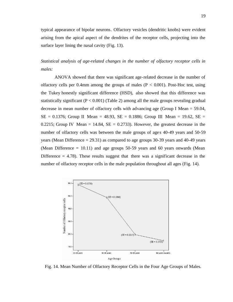
ANOVA showed that there was significant age-related decrease in the number of
olfactory cells per 0.4mm among the groups of males (P < 0.001). Post-Hoc test, using
the Tukey honestly significant difference (HSD), also showed that this difference was
statistically significant (P < 0.001) (Table 2) among all the male groups revealing gradual
decrease in mean number of olfactory cells with advancing age (Group I Mean = 59.04,
SE = 0.1376; Group II Mean = 48.93, SE = 0.1886; Group III Mean = 19.62, SE =
0.2215; Group IV Mean = 14.84, SE = 0.2733). However, the greatest decrease in the
number of olfactory cells was between the male groups of ages 40-49 years and 50-59
years (Mean Difference = 29.31) as compared to age groups 30-39 years and 40-49 years
(Mean Difference = 10.11) and age groups 50-59 years and 60 years onwards (Mean
Difference = 4.78). These results suggest that there was a significant decrease in the
number of olfactory receptor cells in the male population throughout all ages (Fig. 14).
Fig. 14. Mean Number of Olfactory Receptor Cells in the Four Age Groups of Males.
20
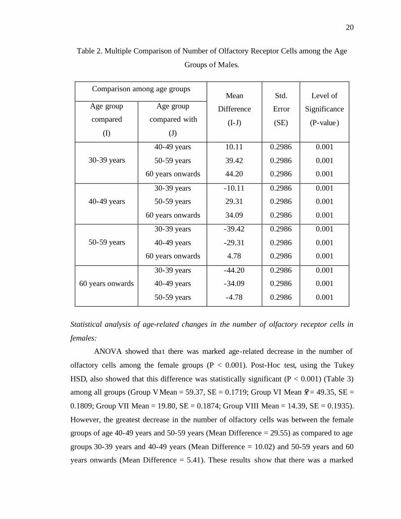
Table 2. Multiple Comparison of Number of Olfactory Receptor Cells among the Age
Groups of Males.
Statistical analysis of age-related changes in the number of olfactory receptor cells in
females:
ANOVA showed tha t there was marked age-related decrease in the number of
olfactory cells among the female groups (P < 0.001). Post-Hoc test, using the Tukey
HSD, also showed that this difference was statistically significant (P < 0.001) (Table 3)
among all groups (Group V Mean = 59.37, SE = 0.1719; Group VI Mean x? = 49.35, SE =
0.1809; Group VII Mean = 19.80, SE = 0.1874; Group VIII Mean = 14.39, SE = 0.1935).
However, the greatest decrease in the number of olfactory cells was between the female
groups of age 40-49 years and 50-59 years (Mean Difference = 29.55) as compared to age
groups 30-39 years and 40-49 years (Mean Difference = 10.02) and 50-59 years and 60
years onwards (Mean Difference = 5.41). These results show that there was a marked
Comparison among age groups
Age group
compared
(I)
Age group
compared with
(J)
Mean
Difference
(I-J)
Std.
Error
(SE)
Level of
Significance
(P-value)
30-39 years
40-49 years
50-59 years
60 years onwards
10.11
39.42
44.20
0.2986
0.2986
0.2986
0.001
0.001
0.001
40-49 years
30-39 years
50-59 years
60 years onwards
-10.11
29.31
34.09
0.2986
0.2986
0.2986
0.001
0.001
0.001
50-59 years
30-39 years
40-49 years
60 years onwards
-39.42
-29.31
4.78
0.2986
0.2986
0.2986
0.001
0.001
0.001
60 years onwards
30-39 years
40-49 years
50-59 years
-44.20
-34.09
-4.78
0.2986
0.2986
0.2986
0.001
0.001
0.001

21
decrease in the number of olfactory receptor cells in the female subjects throughout all
ages (Fig. 15).
Fig. 15. Mean Number of Olfactory Receptor Cells in the Four Age Groups of Females.
Table 3. Multiple Comparison of Number of Olfactory Receptor Cells among the Age
Groups of Females.
Comparison among age groups
Age group compared
(I)
Age group compared with
(J)
Mean Difference
(I-J)
Std. Error (SE)
Level of Significance
(P-value)
30-39 years 40-49 years 50-59 years
60 years onwards
10.02 39.57 44.98
0.2596 0.2596 0.2596
0.001 0.001 0.001
40-49 years 30-39 years 50-59 years
60 years onwards
-10.02 29.55 34.96
0.2596 0.2596 0.2596
0.001 0.001 0.001
50-59 years 30-39 years 40-49 years
60 years onwards
-39.57 -29.55 5.41
0.2596 0.2596 0.2596
0.001 0.001 0.001
60 years onwards 30-39 years 40-49 years 50-59 years
-44.98 -34.96 -5.41
0.2596 0.2596 0.2596
0.001 0.001 0.001

22
Statistical analysis of gender related differences in the number of olfactory receptor
cells:
The analysis of gender related differences was calculated using the independent
sample t-test between the corresponding male and female groups. Comparison of age
groups 30-39, 40-49, and 50-59 years showed that the numbers of olfactory cells in the
two sexes were not significant ly different (p-value > 0.05). However, in the age group 60
years and above the males had greater number of olfactory cells (Mean Difference = 0.45,
p-value > 0.05) (Table 4).
Table 4. Comparison of Number of Olfactory Receptor Cells among the Age Groups of
Males and Females.
Age group Mean
(Males)
Mean
(Females)
Standard
Error t-score p-value
30-39 years 59.04 59.37 0.22 -1.498 0.151
40-49 years 48.93 49.35 0.26 -1.607 0.125
50-59 years 19.62 19.80 0.29 -0.62 0.543
60 years
onwards 14.84 14.39 0.33 1.344 0.196
Sustentacular cells:
These cells were tall and cylindrical; lying perpendicular to the surface,
surrounding the olfactory receptor cells. Their large, spherical, lightly stained nuclei were
observed superficial to the nuclei of the receptor cells as seen in the haematoxylin and
eosin stained sections. Apical projections (microvilli) forming a brush border on the
surface of these cells were also observed (Fig. 16).
23
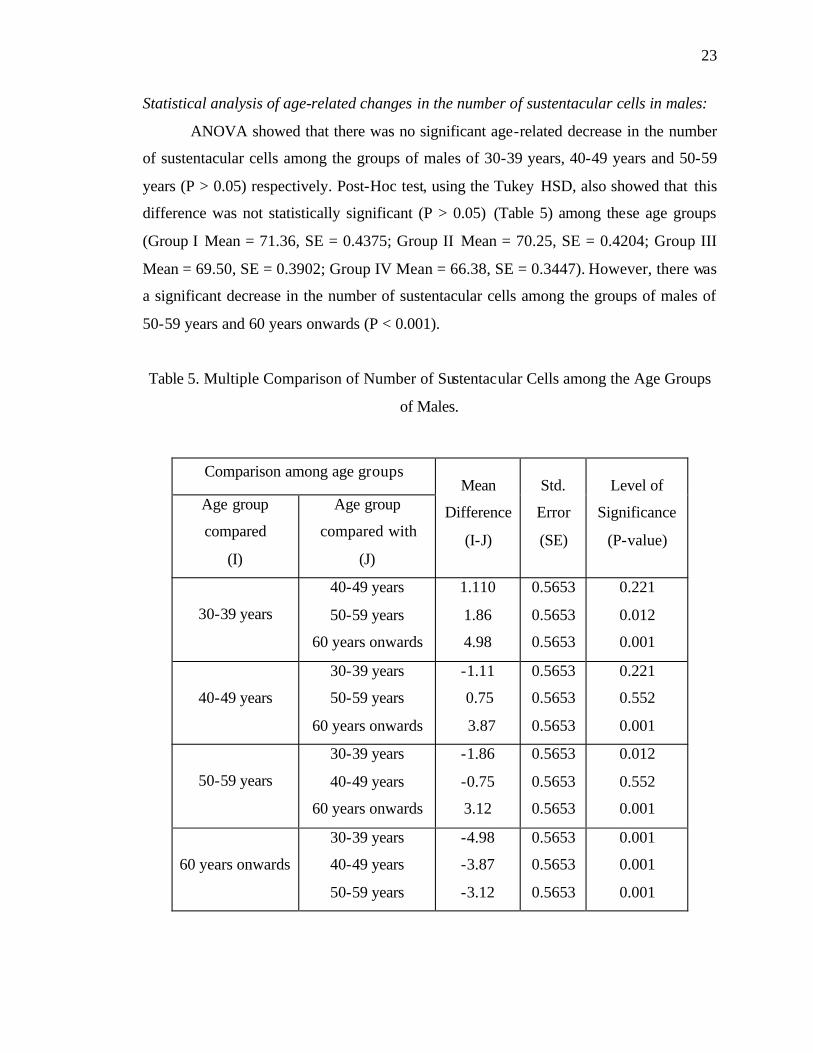
Statistical analysis of age-related changes in the number of sustentacular cells in males:
ANOVA showed that there was no significant age-related decrease in the number
of sustentacular cells among the groups of males of 30-39 years, 40-49 years and 50-59
years (P > 0.05) respectively. Post-Hoc test, using the Tukey HSD, also showed that this
difference was not statistically significant (P > 0.05) (Table 5) among these age groups
(Group I Mean = 71.36, SE = 0.4375; Group II Mean = 70.25, SE = 0.4204; Group III
Mean = 69.50, SE = 0.3902; Group IV Mean = 66.38, SE = 0.3447). However, there was
a significant decrease in the number of sustentacular cells among the groups of males of
50-59 years and 60 years onwards (P < 0.001).
Table 5. Multiple Comparison of Number of Sustentacular Cells among the Age Groups
of Males.
Comparison among age groups
Age group
compared
(I)
Age group
compared with
(J)
Mean
Difference
(I-J)
Std.
Error
(SE)
Level of
Significance
(P-value)
30-39 years
40-49 years
50-59 years
60 years onwards
1.110
1.86
4.98
0.5653
0.5653
0.5653
0.221
0.012
0.001
40-49 years
30-39 years
50-59 years
60 years onwards
-1.11
0.75
3.87
0.5653
0.5653
0.5653
0.221
0.552
0.001
50-59 years
30-39 years
40-49 years
60 years onwards
-1.86
-0.75
3.12
0.5653
0.5653
0.5653
0.012
0.552
0.001
60 years onwards
30-39 years
40-49 years
50-59 years
-4.98
-3.87
-3.12
0.5653
0.5653
0.5653
0.001
0.001
0.001
24
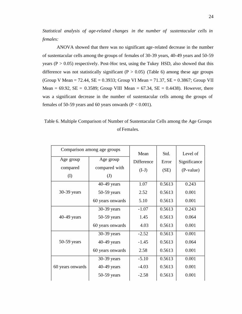
Statistical analysis of age-related changes in the number of sustentacular cells in
females:
ANOVA showed that there was no significant age-related decrease in the number
of sustentacular cells among the groups of females of 30-39 years, 40-49 years and 50-59
years (P > 0.05) respectively. Post-Hoc test, using the Tukey HSD, also showed that this
difference was not statistically significant (P > 0.05) (Table 6) among these age groups
(Group V Mean = 72.44, SE = 0.3933; Group VI Mean = 71.37, SE = 0.3867; Group VII
Mean = 69.92, SE = 0.3589; Group VIII Mean = 67.34, SE = 0.4438). However, there
was a significant decrease in the number of sustentacular cells among the groups of
females of 50-59 years and 60 years onwards (P < 0.001).
Table 6. Multiple Comparison of Number of Sustentacular Cells among the Age Groups
of Females.
Comparison among age groups
Age group
compared
(I)
Age group
compared with
(J)
Mean
Difference
(I-J)
Std.
Error
(SE)
Level of
Significance
(P-value)
30-39 years
40-49 years
50-59 years
60 years onwards
1.07
2.52
5.10
0.5613
0.5613
0.5613
0.243
0.001
0.001
40-49 years
30-39 years
50-59 years
60 years onwards
-1.07
1.45
4.03
0.5613
0.5613
0.5613
0.243
0.064
0.001
50-59 years
30-39 years
40-49 years
60 years onwards
-2.52
-1.45
2.58
0.5613
0.5613
0.5613
0.001
0.064
0.001
60 years onwards
30-39 years
40-49 years
50-59 years
-5.10
-4.03
-2.58
0.5613
0.5613
0.5613
0.001
0.001
0.001

25
Statistical analysis of gender related differences in the number of sustentacular cells:
The analysis of gender related differences was calculated using the independent
sample t-test between the corresponding male and female groups. Comparison of age
groups 30-39 years showed that although the females had greater number of sustentacular
cells as compared to the males (Mean Difference = 1.08) the difference was not
significant (p-value > 0.05). The same results were obtained for comparison of age
groups 40-49 years, 50-59 years and 60 year onwards (Mean Difference = 1.12, p-value >
0.05; Mean Difference = 0.42, p-value > 0.05; Mean Difference = 0.96, p-value > 0.05)
respectively (Table 7).
Table 7. Comparison of Number of Sustentacular Cells among the Age Groups of Males
and Females.
Age group Mean
(Males)
Mean
(Females)
Standard
Error t-score p-value
30-39 years 71.36 72.44 0.58 -1.836 0.083
40-49 years 70.25 71.37 0.57 -1.961 0.066
50-59 years 69.50 69.92 0.53 -0.792 0.439
60 years
onwards 66.38 67.34 0.56 -1.708 0.105
Basal cells:
These short and broad cells were seen disposed along the deeper part of the
epithelium, resting on the basement membrane and did not reach the apical surface. In
haematoxylin and eosin stained sections, the nuclei of these cells were oval to round,
some of them were stained lightly while others appeared deeply stained. The shape of
some of the cells was polygonal with darkly stained cytoplasm; however some of them
were rounded in appearance with pale cytoplasm (Fig. 16).
26
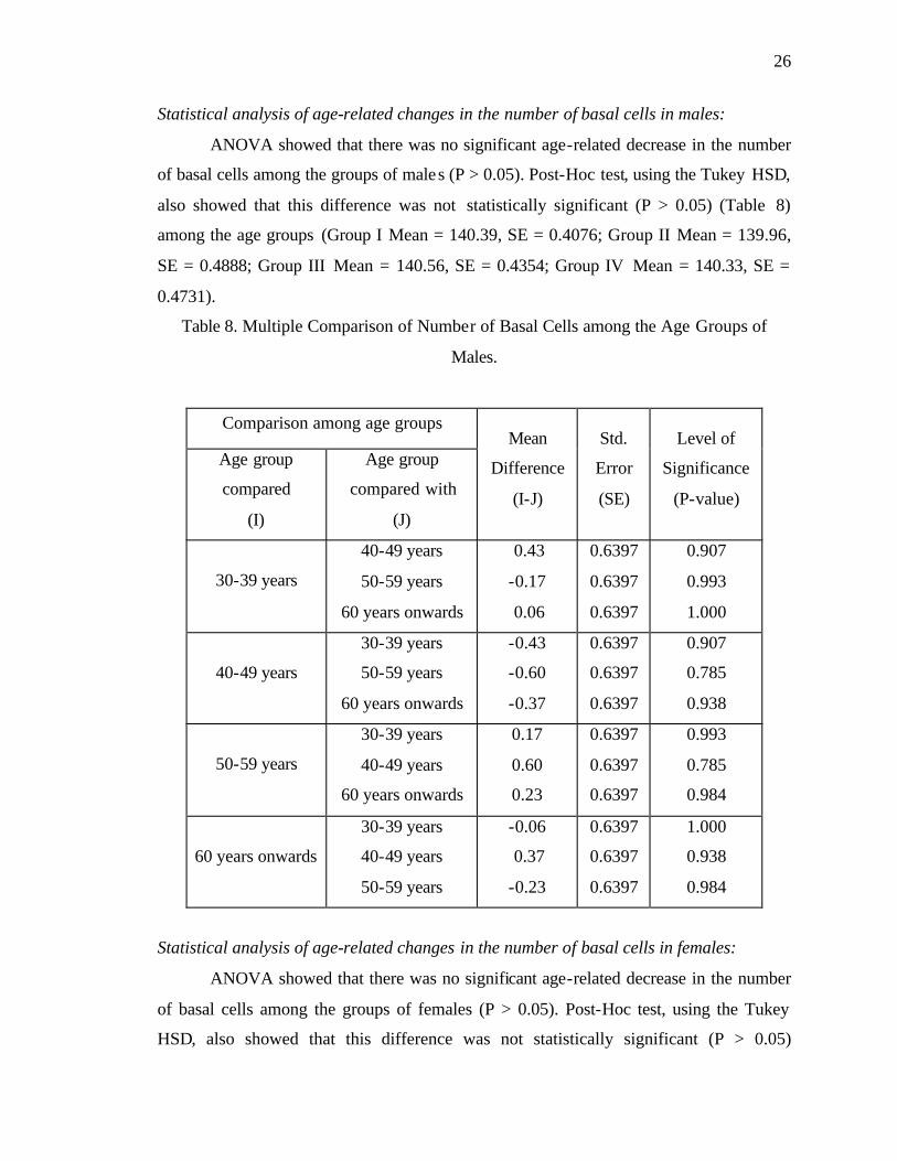
Statistical analysis of age-related changes in the number of basal cells in males:
ANOVA showed that there was no significant age-related decrease in the number
of basal cells among the groups of males (P > 0.05). Post-Hoc test, using the Tukey HSD,
also showed that this difference was not statistically significant (P > 0.05) (Table 8)
among the age groups (Group I Mean = 140.39, SE = 0.4076; Group II Mean = 139.96,
SE = 0.4888; Group III Mean = 140.56, SE = 0.4354; Group IV Mean = 140.33, SE =
0.4731).
Table 8. Multiple Comparison of Number of Basal Cells among the Age Groups of
Males.
Statistical analysis of age-related changes in the number of basal cells in females:
ANOVA showed that there was no significant age-related decrease in the number
of basal cells among the groups of females (P > 0.05). Post-Hoc test, using the Tukey
HSD, also showed that this difference was not statistically significant (P > 0.05)
Comparison among age groups
Age group
compared
(I)
Age group
compared with
(J)
Mean
Difference
(I-J)
Std.
Error
(SE)
Level of
Significance
(P-value)
30-39 years
40-49 years
50-59 years
60 years onwards
0.43
-0.17
0.06
0.6397
0.6397
0.6397
0.907
0.993
1.000
40-49 years
30-39 years
50-59 years
60 years onwards
-0.43
-0.60
-0.37
0.6397
0.6397
0.6397
0.907
0.785
0.938
50-59 years
30-39 years
40-49 years
60 years onwards
0.17
0.60
0.23
0.6397
0.6397
0.6397
0.993
0.785
0.984
60 years onwards
30-39 years
40-49 years
50-59 years
-0.06
0.37
-0.23
0.6397
0.6397
0.6397
1.000
0.938
0.984
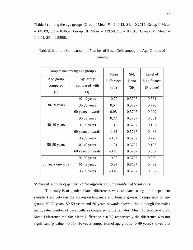
27
(Table 9) among the age groups (Group I Mean x? = 140.12, SE = 0.2715; Group II Mean
= 140.89, SE = 0.4632; Group III Mean = 139.58, SE = 0.4816; Group IV Mean =
140.04, SE = 0.3896).
Table 9. Multiple Comparison of Number of Basal Cells among the Age Groups of
Females.
Statistical analysis of gender related differences in the number of basal cells:
The analysis of gender related differences was calculated using the independent
sample t-test between the corresponding male and female groups. Comparison of age
groups 30-39 years, 50-59 years and 60 years onwards showed that although the males
had greater number of basal cells as compared to the females (Mean Difference = 0.27;
Mean Difference = 0.98; Mean Difference = 0.29) respectively the difference was not
significant (p-value > 0.05). However comparison of age groups 40-49 years showed that
Comparison among age groups
Age group
compared
(I)
Age group
compared with
(J)
Mean
Difference
(I-J)
Std.
Error
(SE)
Level of
Significance
(P-value)
30-39 years
40-49 years
50-59 years
60 years onwards
-0.77
0.54
0.08
0.5797
0.5797
0.5797
0.551
0.778
0.999
40-49 years
30-39 years
50-59 years
60 years onwards
0.77
1.31
0.85
0.5797
0.5797
0.5797
0.551
0.127
0.468
50-59 years
30-39 years
40-49 years
60 years onwards
-0.54
-1.31
-0.46
0.5797
0.5797
0.5797
0.778
0.127
0.857
60 years onwards
30-39 years
40-49 years
50-59 years
-0.08
-0.85
-0.46
0.5797
0.5797
0.5797
0.999
0.468
0.857

28
females had more number of basal cells as compared to males but the difference was not
significant (Mean Difference = 0.93, p-value > 0.05) (Table 10 ).
Table 10. Comparison of Number of Basal Cells among the Age Groups of Males and
Females.
Age group Mean
(Males)
Mean
(Females)
Standard
Error t-score p-value
30-39 years 140.39 140.12 0.48 0.551 0.588
40-49 years 139.96 140.89 0.67 -1.381 0.184
50-59 years 140.56 139.58 0.64 1.509 0.149
60 years
onwards 140.33 140.04 0.61 0.473 0.642
Microvillar cells:
These cells were quite distinct. In haematoxylin and eosin stained sections, large
nuclei were observed close to the epithe lial surface. The cells appeared pear-shaped
having a clear cytoplasm. Microvilli were present on the apical surface of these cells. The
number of microvillar cells was markedly less when compared to the other cells of the
epithelium (Fig. 16).
Basement membrane:
In routine H & E sections, the basement membrane appeared as a well-defined
homogenous structure (Figs. 7, 9), lying against the under surface of the epithelial cells
becoming more prominent in PAS stained sections (Figs. 10, 17).
Lamina Propria:
The lamina propria was quite thick. It consisted of a framework of collagenous
connective tissue containing fibroblasts, lymphocytes and neutrophils (Fig. 18), nerve
bundles, blood vessels and serous acini (Fig. 19).
29
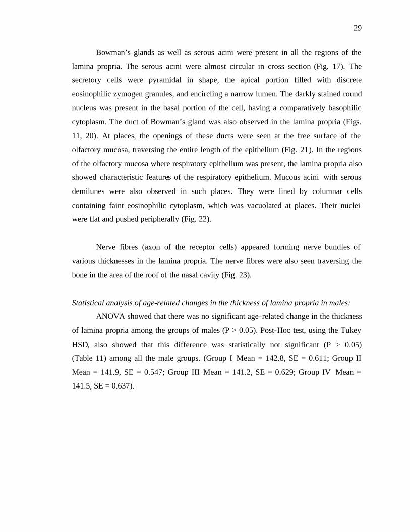
Bowman’s glands as well as serous acini were present in all the regions of the
lamina propria. The serous acini were almost circular in cross section (Fig. 17). The
secretory cells were pyramidal in shape, the apical portion filled with discrete
eosinophilic zymogen granules, and encircling a narrow lumen. The darkly stained round
nucleus was present in the basal portion of the cell, having a comparatively basophilic
cytoplasm. The duct of Bowman’s gland was also observed in the lamina propria (Figs.
11, 20). At places, the openings of these ducts were seen at the free surface of the
olfactory mucosa, traversing the entire length of the epithelium (Fig. 21). In the regions
of the olfactory mucosa where respiratory epithelium was present, the lamina propria also
showed characteristic features of the respiratory epithelium. Mucous acini with serous
demilunes were also observed in such places. They were lined by columnar cells
containing faint eosinophilic cytoplasm, which was vacuolated at places. Their nuclei
were flat and pushed peripherally (Fig. 22).
Nerve fibres (axon of the receptor cells) appeared forming nerve bundles of
various thicknesses in the lamina propria. The nerve fibres were also seen traversing the
bone in the area of the roof of the nasal cavity (Fig. 23).
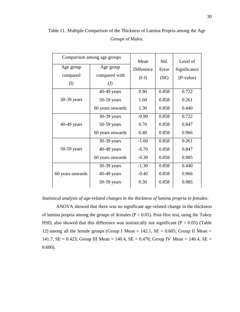
Statistical analysis of age-related changes in the thickness of lamina propria in males:
ANOVA showed that there was no significant age-related change in the thickness
of lamina propria among the groups of males (P > 0.05). Post-Hoc test, using the Tukey
HSD, also showed that this difference was statistically not significant (P > 0.05)
(Table 11) among all the male groups. (Group I Mean = 142.8, SE = 0.611; Group II
Mean = 141.9, SE = 0.547; Group III Mean = 141.2, SE = 0.629; Group IV Mean =
141.5, SE = 0.637).
30
Table 11. Multiple Comparison of the Thickness of Lamina Propria among the Age
Groups of Males.
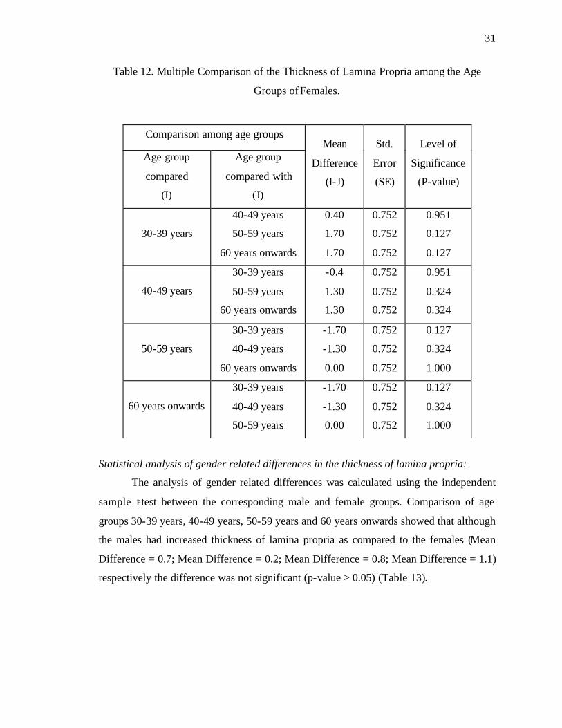
Statistical analysis of age-related changes in the thickness of lamina propria in females:
ANOVA showed that there was no significant age-related change in the thickness
of lamina propria among the groups of females (P > 0.05). Post-Hoc test, using the Tukey
HSD, also showed that this difference was statistically not significant (P > 0.05) (Table
12) among all the female groups (Group I Mean = 142.1, SE = 0.605; Group II Mean =
141.7, SE = 0.423; Group III Mean = 140.4, SE = 0.476; Group IV Mean = 140.4, SE =
0.600).
Comparison among age groups
Age group
compared
(I)
Age group
compared with
(J)
Mean
Difference
(I-J)
Std.
Error
(SE)
Level of
Significance
(P-value)
30-39 years
40-49 years
50-59 years
60 years onwards
0.90
1.60
1.30
0.858
0.858
0.858
0.722
0.261
0.440
40-49 years
30-39 years
50-59 years
60 years onwards
-0.90
0.70
0.40
0.858
0.858
0.858
0.722
0.847
0.966
50-59 years
30-39 years
40-49 years
60 years onwards
-1.60
-0.70
-0.30
0.858
0.858
0.858
0.261
0.847
0.985
60 years onwards
30-39 years
40-49 years
50-59 years
-1.30
-0.40
0.30
0.858
0.858
0.858
0.440
0.966
0.985
31
Table 12. Multiple Comparison of the Thickness of Lamina Propria among the Age
Groups of Females.
Statistical analysis of gender related differences in the thickness of lamina propria:
The analysis of gender related differences was calculated using the independent
sample t-test between the corresponding male and female groups. Comparison of age
groups 30-39 years, 40-49 years, 50-59 years and 60 years onwards showed that although
the males had increased thickness of lamina propria as compared to the females (Mean
Difference = 0.7; Mean Difference = 0.2; Mean Difference = 0.8; Mean Difference = 1.1)
respectively the difference was not significant (p-value > 0.05) (Table 13).
Comparison among age groups
Age group
compared
(I)
Age group
compared with
(J)
Mean
Difference
(I-J)
Std.
Error
(SE)
Level of
Significance
(P-value)
30-39 years
40-49 years
50-59 years
60 years onwards
0.40
1.70
1.70
0.752
0.752
0.752
0.951
0.127
0.127
40-49 years
30-39 years
50-59 years
60 years onwards
-0.4
1.30
1.30
0.752
0.752
0.752
0.951
0.324
0.324
50-59 years
30-39 years
40-49 years
60 years onwards
-1.70
-1.30
0.00
0.752
0.752
0.752
0.127
0.324
1.000
60 years onwards
30-39 years
40-49 years
50-59 years
-1.70
-1.30
0.00
0.752
0.752
0.752
0.127
0.324
1.000

32
Table 13. Comparison of the Thickness of Lamina Propria among the Age Groups of
Males and Females.
Age group Mean
(Males)
Mean
(Females)
Standard
Error t-score p-value
30-39 years 142.80 142.10 0.86 0.814 0.426
40-49 years 141.90 141.70 0.69 0.289 0.776
50-59 years 141.20 140.40 0.78 1.014 0.324
60 years
onwards 141.50 140.40 0.87 1.257 0.225
Age-related changes in the Mucosa in the age group 40-49 years (male and female
groups):
In this age group, morphological changes were evident in some of the specimens.
Early age-related changes were observed in both males and females in the shape of
occasional short epithelial invaginations and incidence of mucoserous glands instead of
pure serous glands. However, the olfactory epithelium appeared quite normal at this stage
(Fig. 24). Disturbance of the zonal distribution of the olfactory receptor cells and the
sustentacular cells was observed occasionally (Fig. 25).
Age-related changes in the Mucosa in the age group 50-59 years (male and female
groups):
Major morphological changes were observed in this group. In some places, a
substantial reduction in the number of nuclei (and hence cells) was noted, which resulted
in decreased height of the epithelium (Fig. 26). There were changes in the form of
reduction in the height of olfactory epithelium, and disturbance of zonal distribution (Fig.
27). The olfactory mucosa was seen in the roof of nasal cavity as a continuous sheet.
When viewed over the medial and lateral nasal walls, several patches of respiratory
epithelium were found distributed in the olfactory area. Another change observed in this
age group was decrease in the height of the olfactory epithelium compared to respiratory
33
epithelium (Fig. 28). Surface epithelial invaginations of the epithelium into the lamina
propria were also found quite frequently in this age group (Fig. 29).
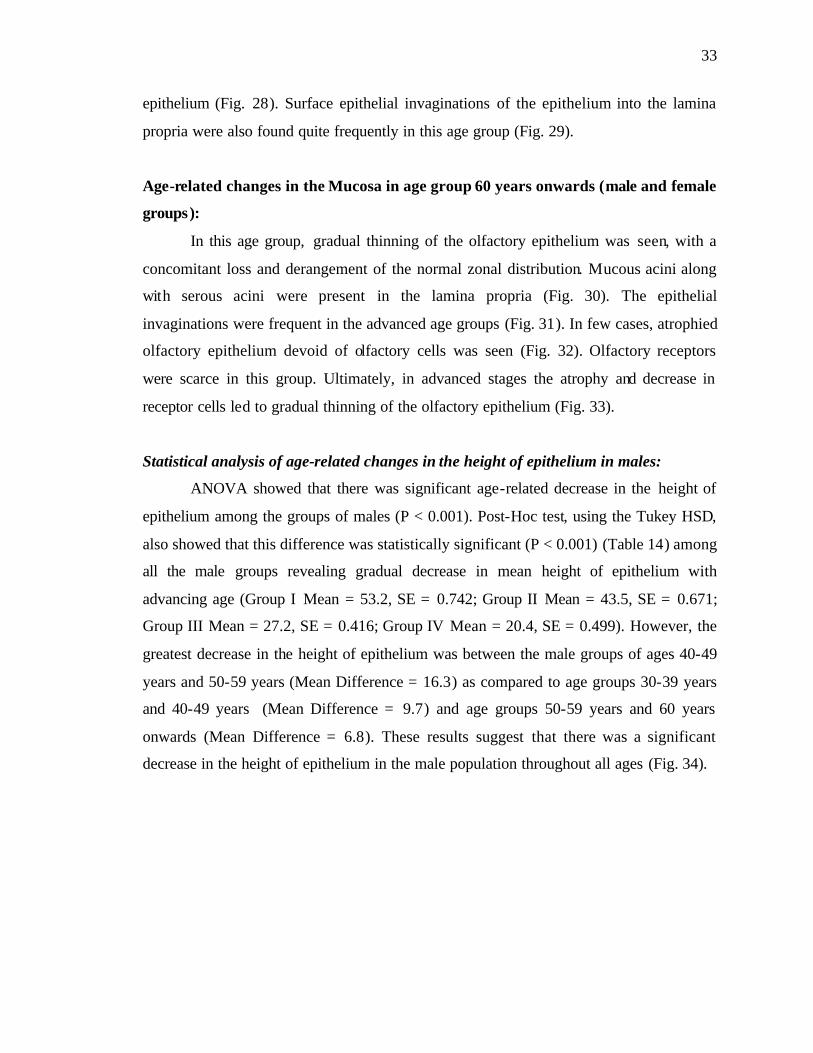
Age-related changes in the Mucosa in age group 60 years onwards (male and female
groups):
In this age group, gradual thinning of the olfactory epithelium was seen, with a
concomitant loss and derangement of the normal zonal distribution. Mucous acini along
with serous acini were present in the lamina propria (Fig. 30). The epithelial
invaginations were frequent in the advanced age groups (Fig. 31). In few cases, atrophied
olfactory epithelium devoid of olfactory cells was seen (Fig. 32). Olfactory receptors
were scarce in this group. Ultimately, in advanced stages the atrophy and decrease in
receptor cells led to gradual thinning of the olfactory epithelium (Fig. 33).
Statistical analysis of age-related changes in the height of epithelium in males:
ANOVA showed that there was significant age-related decrease in the height of
epithelium among the groups of males (P < 0.001). Post-Hoc test, using the Tukey HSD,
also showed that this difference was statistically significant (P < 0.001) (Table 14) among
all the male groups revealing gradual decrease in mean height of epithelium with
advancing age (Group I Mean = 53.2, SE = 0.742; Group II Mean = 43.5, SE = 0.671;
Group III Mean = 27.2, SE = 0.416; Group IV Mean = 20.4, SE = 0.499). However, the
greatest decrease in the height of epithelium was between the male groups of ages 40-49
years and 50-59 years (Mean Difference = 16.3) as compared to age groups 30-39 years
and 40-49 years (Mean Difference = 9.7) and age groups 50-59 years and 60 years
onwards (Mean Difference = 6.8). These results suggest that there was a significant
decrease in the height of epithelium in the male population throughout all ages (Fig. 34).

34
Fig. 34. Mean Height of Epithelium in the Four Age Groups of Males.
Table 14. Multiple Comparison of the Height of Epithelium among the Age Groups of
Males.
Comparison among age groups
Age group
compared
(I)
Age group
compared with
(J)
Mean
Difference
(I-J)
Std.
Error
(SE)
Level of
Significance
(P-value)
30-39 years
40-49 years
50-59 years
60 years onwards
9.70
26.0
32.8
0.844
0.844
0.844
0.001
0.001
0.001
40-49 years
30-39 years
50-59 years
60 years onwards
-9.7
16.3
23.1
0.844
0.844
0.844
0.001
0.001
0.001
50-59 years
30-39 years
40-49 years
60 years onwards
-26.0
-16.3
6.80
0.844
0.844
0.844
0.001
0.001
0.001
60 years onwards
30-39 years
40-49 years
50-59 years
-32.8
-23.1
-6.8
0.844
0.844
0.844
0.001
0.001
0.001
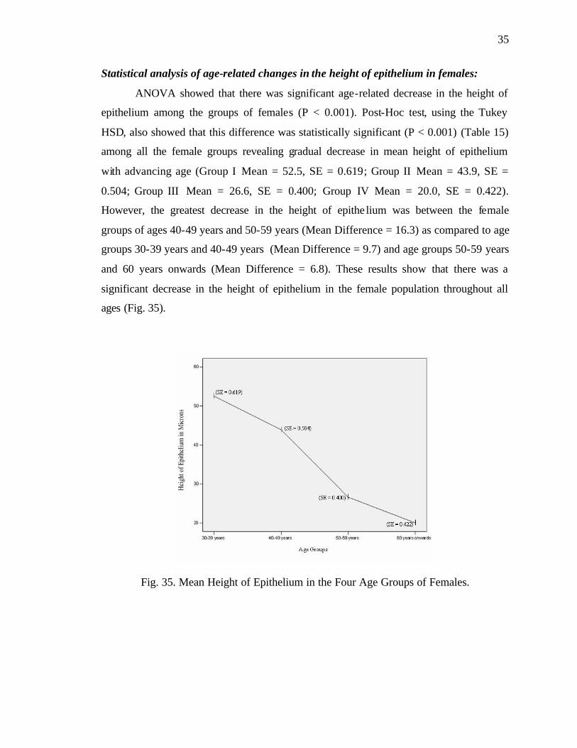
35
Statistical analysis of age-related changes in the height of epithelium in females:
ANOVA showed that there was significant age-related decrease in the height of
epithelium among the groups of females (P < 0.001). Post-Hoc test, using the Tukey
HSD, also showed that this difference was statistically significant (P < 0.001) (Table 15)
among all the female groups revealing gradual decrease in mean height of epithelium
with advancing age (Group I Mean = 52.5, SE = 0.619; Group II Mean = 43.9, SE =
0.504; Group III Mean = 26.6, SE = 0.400; Group IV Mean = 20.0, SE = 0.422).
However, the greatest decrease in the height of epithe lium was between the female
groups of ages 40-49 years and 50-59 years (Mean Difference = 16.3) as compared to age
groups 30-39 years and 40-49 years (Mean Difference = 9.7) and age groups 50-59 years
and 60 years onwards (Mean Difference = 6.8). These results show that there was a
significant decrease in the height of epithelium in the female population throughout all
ages (Fig. 35).
Fig. 35. Mean Height of Epithelium in the Four Age Groups of Females.
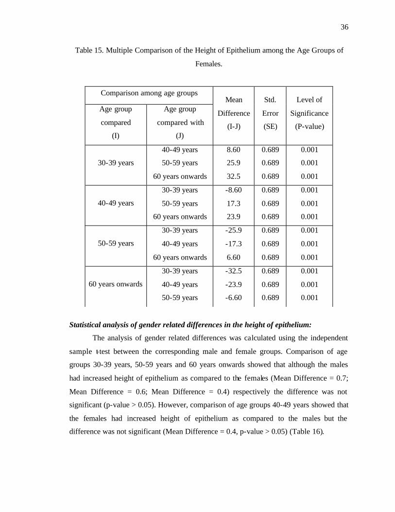
36
Table 15. Multiple Comparison of the Height of Epithelium among the Age Groups of
Females.
Statistical analysis of gender related differences in the height of epithelium:
The analysis of gender related differences was calculated using the independent
sample t-test between the corresponding male and female groups. Comparison of age
groups 30-39 years, 50-59 years and 60 years onwards showed that although the males
had increased height of epithelium as compared to the females (Mean Difference = 0.7;
Mean Difference = 0.6; Mean Difference = 0.4) respectively the difference was not
significant (p-value > 0.05). However, comparison of age groups 40-49 years showed that
the females had increased height of epithelium as compared to the males but the
difference was not significant (Mean Difference = 0.4, p-value > 0.05) (Table 16).
Comparison among age groups
Age group
compared
(I)
Age group
compared with
(J)
Mean
Difference
(I-J)
Std.
Error
(SE)
Level of
Significance
(P-value)
30-39 years
40-49 years
50-59 years
60 years onwards
8.60
25.9
32.5
0.689
0.689
0.689
0.001
0.001
0.001
40-49 years
30-39 years
50-59 years
60 years onwards
-8.60
17.3
23.9
0.689
0.689
0.689
0.001
0.001
0.001
50-59 years
30-39 years
40-49 years
60 years onwards
-25.9
-17.3
6.60
0.689
0.689
0.689
0.001
0.001
0.001
60 years onwards
30-39 years
40-49 years
50-59 years
-32.5
-23.9
-6.60
0.689
0.689
0.689
0.001
0.001
0.001
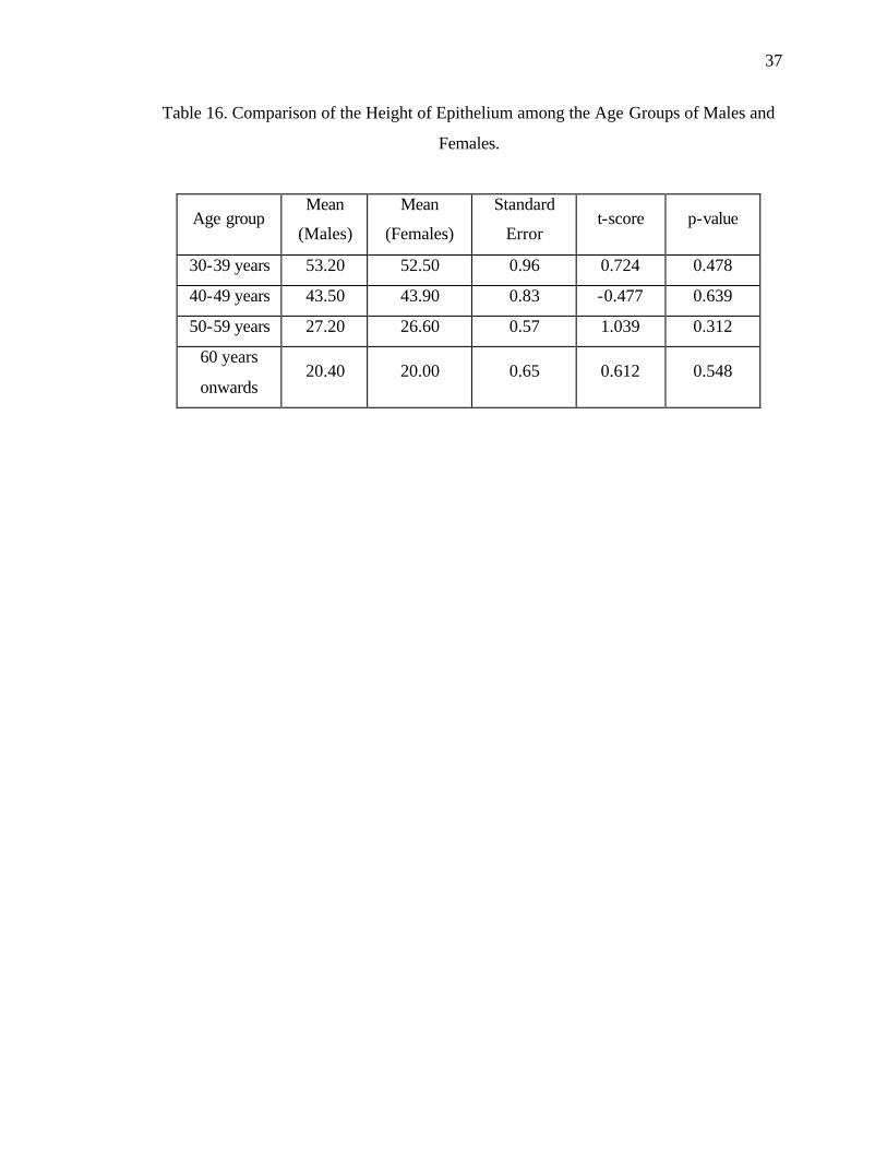
37
Table 16. Comparison of the Height of Epithelium among the Age Groups of Males and
Females.
Age group Mean
(Males)
Mean
(Females)
Standard
Error t-score p-value
30-39 years 53.20 52.50 0.96 0.724 0.478
40-49 years 43.50 43.90 0.83 -0.477 0.639
50-59 years 27.20 26.60 0.57 1.039 0.312
60 years
onwards 20.40 20.00 0.65 0.612 0.548

38
Fig. 1. Location of olfactory area (blue) at the lateral wall of the nose. Photograph showing: the superior turbinate (ST), middle turbinate (MT) and inferior turbinate (IT) at the lateral wall of the nose. Olfactory bulb (OB) lying above the cribriform plate (arrowhead) of the ethmoid bone, sphenoidal air sinus (SS) and hard palate (HP) are also visible. (Netter’s Atlas of Human Anatomy, 2003).
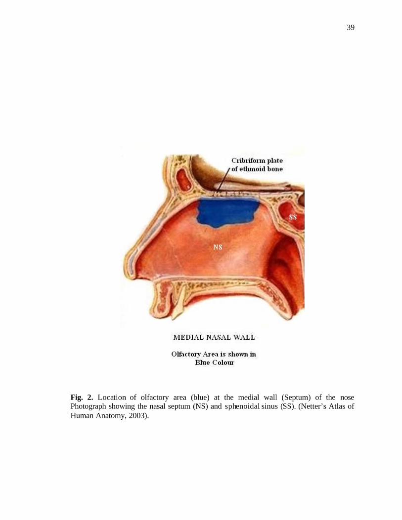
39
Fig. 2. Location of olfactory area (blue) at the medial wall (Septum) of the nose Photograph showing the nasal septum (NS) and sphenoidal sinus (SS). (Netter’s Atlas of Human Anatomy, 2003).
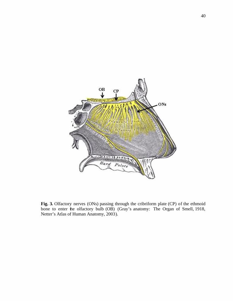
40
Fig. 3. Olfactory nerves (ONs) passing through the cribriform plate (CP) of the ethmoid bone to enter the olfactory bulb (OB) (Gray’s anatomy: The Organ of Smell, 1918, Netter’s Atlas of Human Anatomy, 2003).
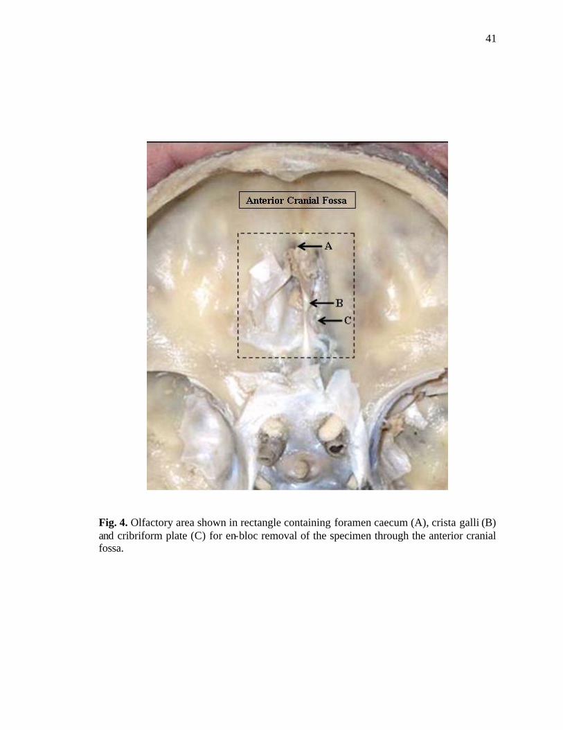
41
Fig. 4. Olfactory area shown in rectangle containing foramen caecum (A), crista galli (B) and cribriform plate (C) for en-bloc removal of the specimen through the anterior cranial fossa.
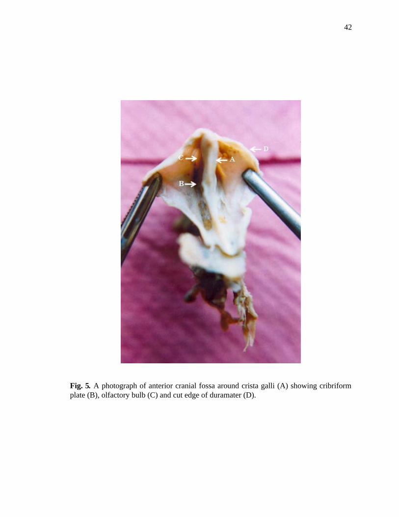
42
Fig. 5. A photograph of anterior cranial fossa around crista galli (A) showing cribriform plate (B), olfactory bulb (C) and cut edge of duramater (D).

43
Fig. 6. Photograph of inferior aspect of olfactory part of the nasal cavity; the lateral walls (C) are stretched out showing roof as cribriform plate (B) and nasal septum (A).
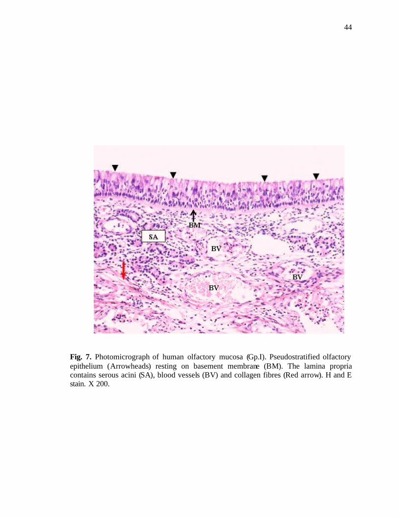
44
Fig. 7. Photomicrograph of human olfactory mucosa (Gp.I). Pseudostratified olfactory epithelium (Arrowheads) resting on basement membrane (BM). The lamina propria contains serous acini (SA), blood vessels (BV) and collagen fibres (Red arrow). H and E stain. X 200.
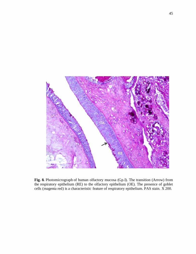
45
Fig. 8. Photomicrograph of human olfactory mucosa (Gp.I). The transition (Arrow) from the respiratory epithelium (RE) to the olfactory epithelium (OE). The presence of goblet cells (magenta red) is a characteristic feature of respiratory epithelium. PAS stain. X 200.

46
Fig. 9. Photomicrograph of human olfactory mucosa (Gp.I). Respiratory epithelium (left side) containing goblet cells (Arrows) are shown next to the olfactory epithelium (right side). Basement membrane (BM) is seen under the epithelium. In the lamina propria serous acini (SA) are present. PAS stain. X 200.
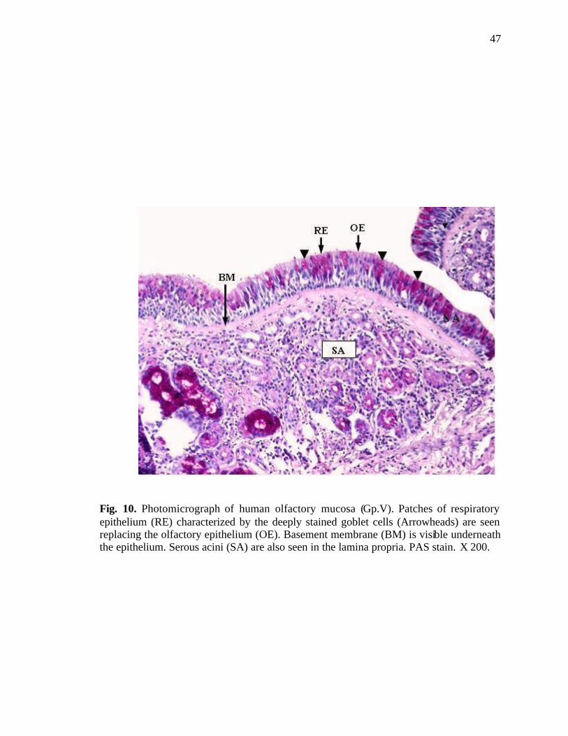
47
Fig. 10. Photomicrograph of human olfactory mucosa (Gp.V). Patches of respiratory epithelium (RE) characterized by the deeply stained goblet cells (Arrowheads) are seen replacing the olfactory epithelium (OE). Basement membrane (BM) is visible underneath the epithelium. Serous acini (SA) are also seen in the lamina propria. PAS stain. X 200.

48
Fig. 11. Photograph of human olfactory mucosa (Gp.I). The olfactory epithelium showing classical cell types. The lamina propria contains nerve bundles (NB), blood vessels (BV) and duct of the Bowman’s gland (D), lined by cuboidal epithelium. H and E stain. X 200. Inset showing receptor cell (RC), microvillar cell (MC), sustentacular cell (SC), and basal cell (BC). H and E stain. X 400.

49
Fig. 12. Photomicrograph of human olfactory mucosa (Gp.I). Darkly stained olfactory receptor cell nuclei (RC) are present at different levels. A dendrite (A) is seen running towards the epithelial surface ending as a dendritic knob (DK). An axon (B) is present at the basal end of the cell body. Also present are: sustentacular cell (SC) having a brush border (BB) at the apical surface and basal cells (BC) lying at the deepest part of epithelium. Lamina propria (LP) lies beneath the epithelium. H and E stain. X 1000.
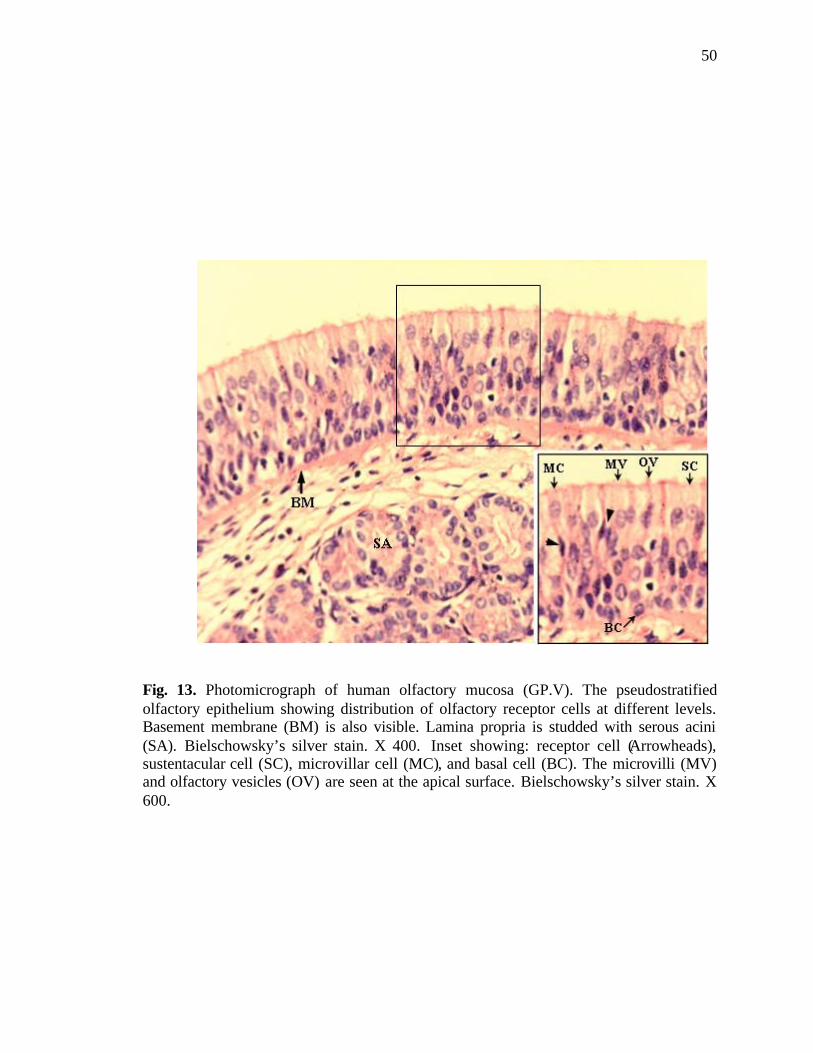
50
Fig. 13. Photomicrograph of human olfactory mucosa (GP.V). The pseudostratified olfactory epithelium showing distribution of olfactory receptor cells at different levels. Basement membrane (BM) is also visible. Lamina propria is studded with serous acini (SA). Bielschowsky’s silver stain. X 400. Inset showing: receptor cell (Arrowheads), sustentacular cell (SC), microvillar cell (MC), and basal cell (BC). The microvilli (MV) and olfactory vesicles (OV) are seen at the apical surface. Bielschowsky’s silver stain. X 600.
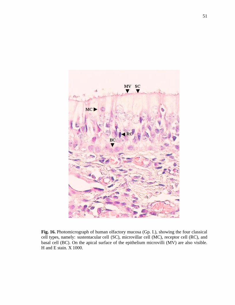
51
Fig. 16. Photomicrograph of human olfactory mucosa (Gp. I.), showing the four classical cell types, namely: sustentacular cell (SC), microvillar cell (MC), receptor cell (RC), and basal cell (BC). On the apical surface of the epithelium microvilli (MV) are also visible. H and E stain. X 1000.
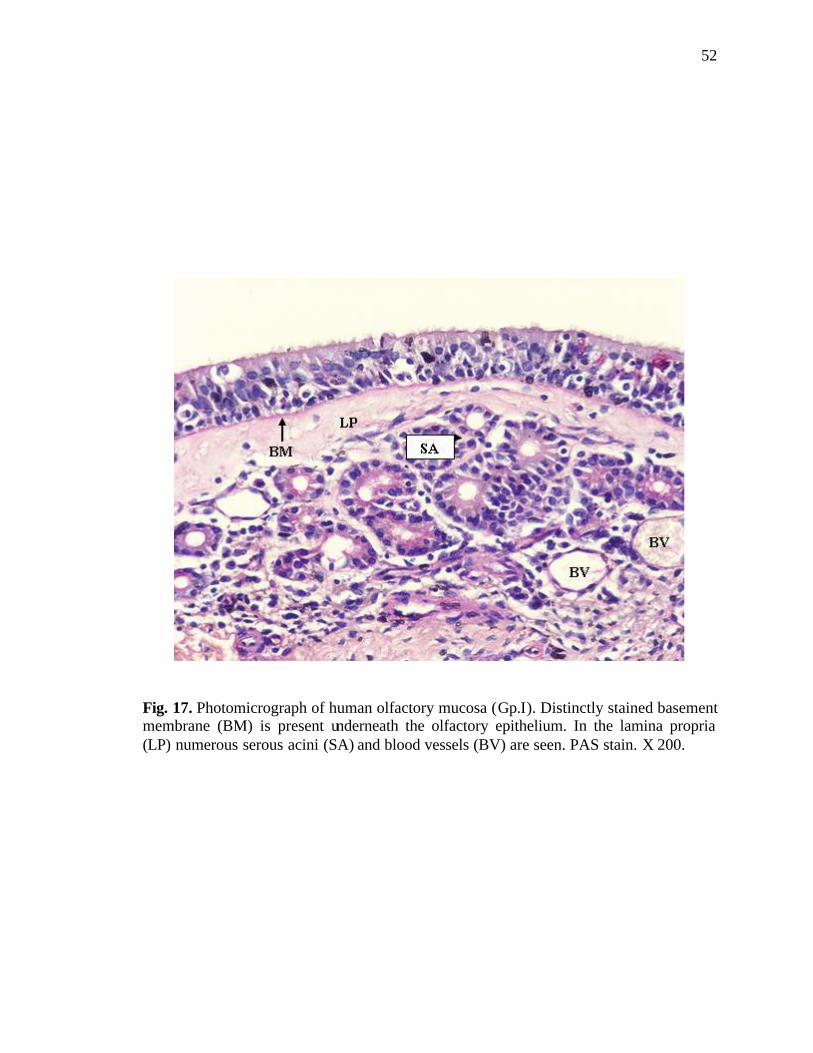
52
Fig. 17. Photomicrograph of human olfactory mucosa (Gp.I). Distinctly stained basement membrane (BM) is present underneath the olfactory epithelium. In the lamina propria (LP) numerous serous acini (SA) and blood vessels (BV) are seen. PAS stain. X 200.
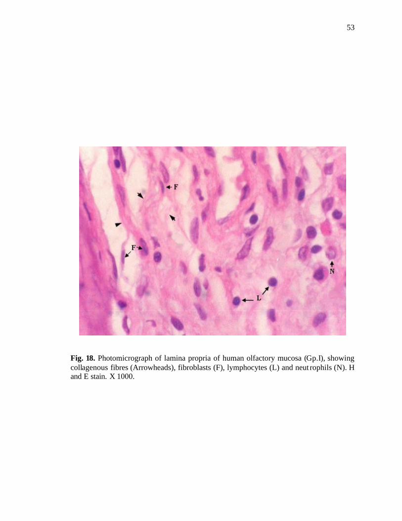
53
Fig. 18. Photomicrograph of lamina propria of human olfactory mucosa (Gp.I), showing collagenous fibres (Arrowheads), fibroblasts (F), lymphocytes (L) and neut rophils (N). H and E stain. X 1000.
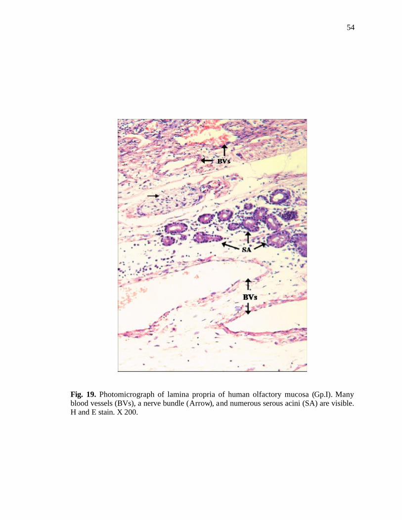
54
Fig. 19. Photomicrograph of lamina propria of human olfactory mucosa (Gp.I). Many blood vessels (BVs), a nerve bundle (Arrow), and numerous serous acini (SA) are visible. H and E stain. X 200.
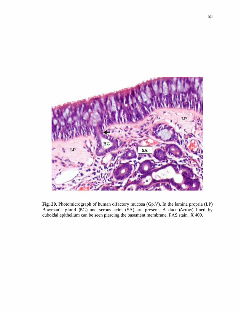
55
Fig. 20. Photomicrograph of human olfactory mucosa (Gp.V). In the lamina propria (LP) Bowman’s gland (BG) and serous acini (SA) are present. A duct (Arrow) lined by cuboidal epithelium can be seen piercing the basement membrane. PAS stain. X 400.
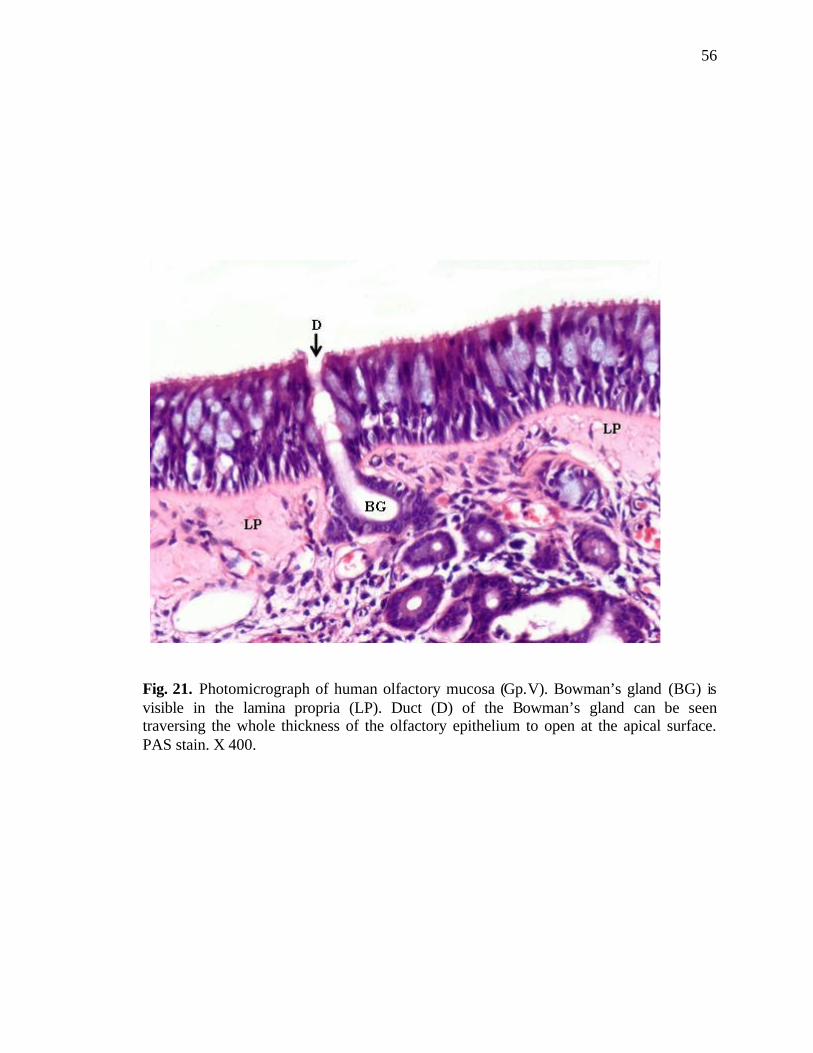
56
Fig. 21. Photomicrograph of human olfactory mucosa (Gp.V). Bowman’s gland (BG) is visible in the lamina propria (LP). Duct (D) of the Bowman’s gland can be seen traversing the whole thickness of the olfactory epithelium to open at the apical surface. PAS stain. X 400.
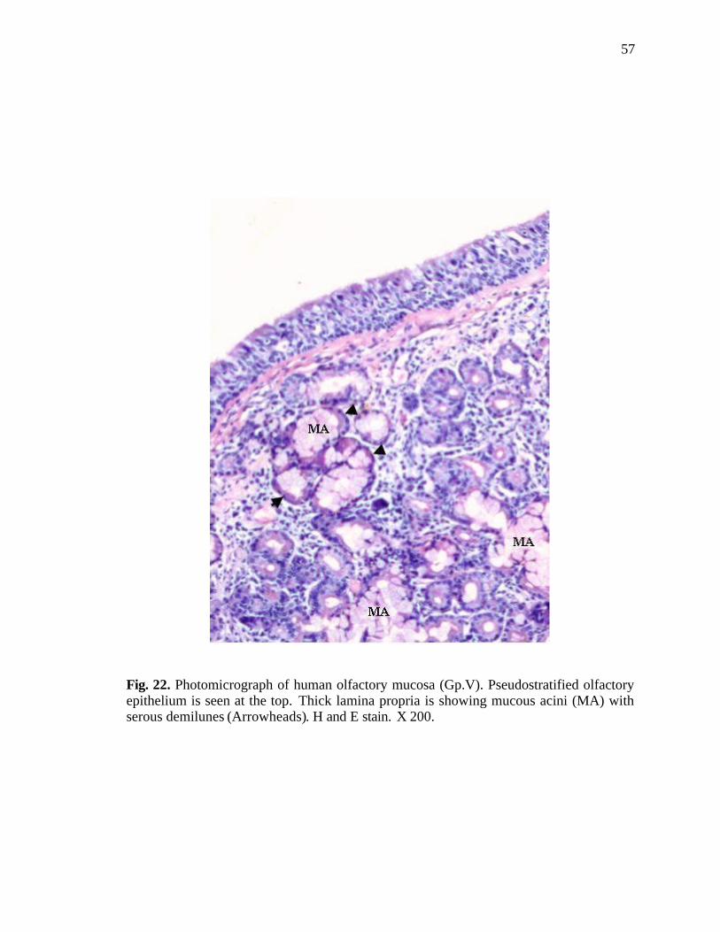
57
Fig. 22. Photomicrograph of human olfactory mucosa (Gp.V). Pseudostratified olfactory epithelium is seen at the top. Thick lamina propria is showing mucous acini (MA) with serous demilunes (Arrowheads). H and E stain. X 200.
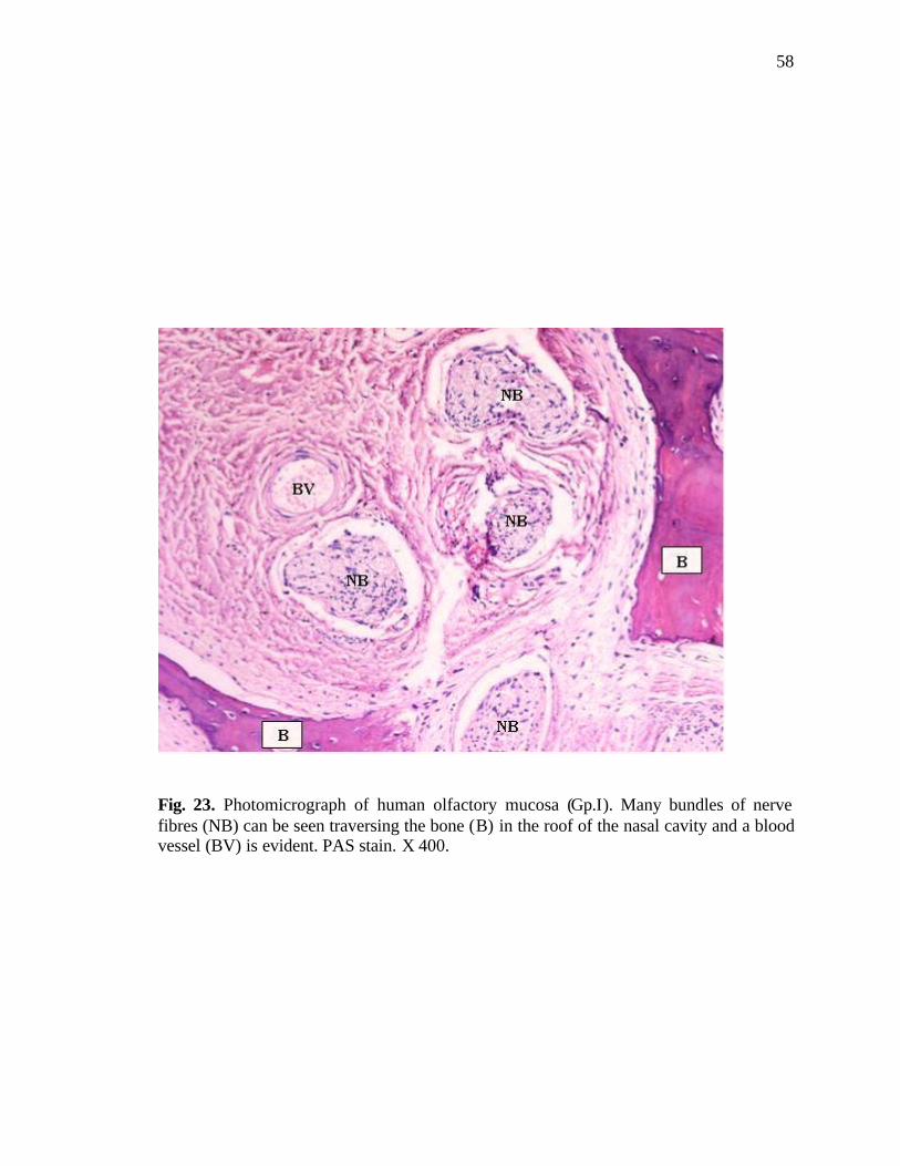
58
Fig. 23. Photomicrograph of human olfactory mucosa (Gp.I). Many bundles of nerve fibres (NB) can be seen traversing the bone (B) in the roof of the nasal cavity and a blood vessel (BV) is evident. PAS stain. X 400.
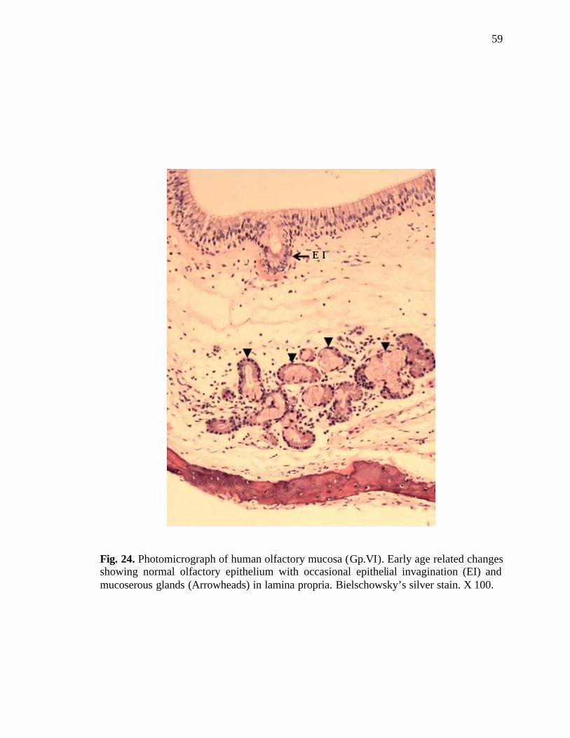
59
Fig. 24. Photomicrograph of human olfactory mucosa (Gp.VI). Early age related changes showing normal olfactory epithelium with occasional epithelial invagination (EI) and mucoserous glands (Arrowheads) in lamina propria. Bielschowsky’s silver stain. X 100.
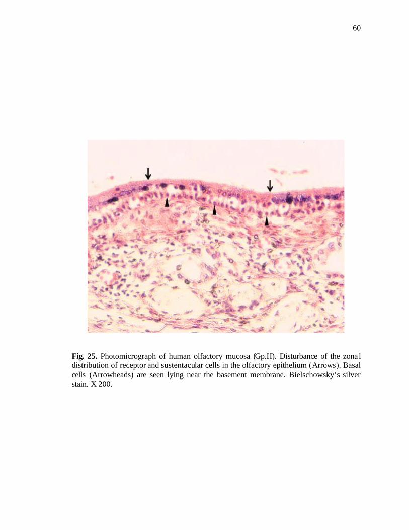
60
Fig. 25. Photomicrograph of human olfactory mucosa (Gp.II). Disturbance of the zona l distribution of receptor and sustentacular cells in the olfactory epithelium (Arrows). Basal cells (Arrowheads) are seen lying near the basement membrane. Bielschowsky’s silver stain. X 200.
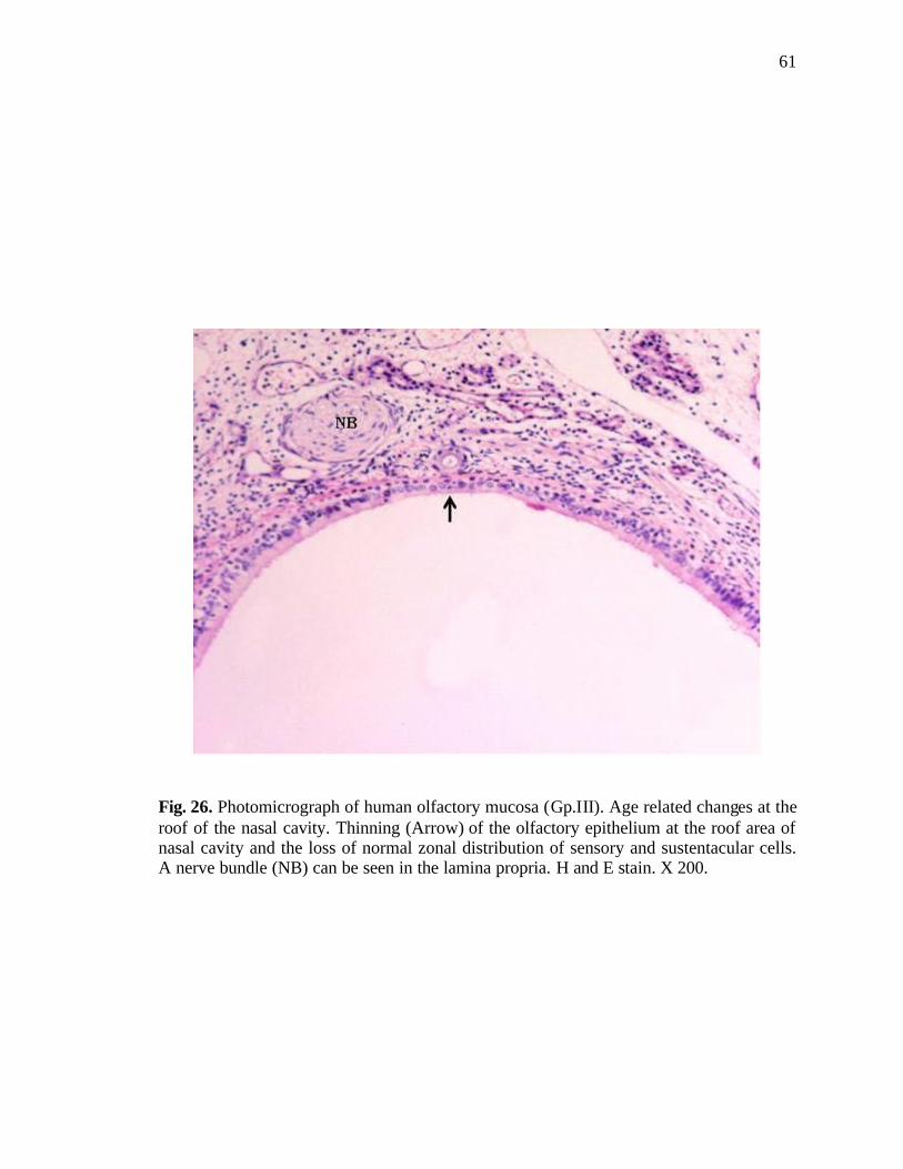
61
Fig. 26. Photomicrograph of human olfactory mucosa (Gp.III). Age related changes at the roof of the nasal cavity. Thinning (Arrow) of the olfactory epithelium at the roof area of nasal cavity and the loss of normal zonal distribution of sensory and sustentacular cells. A nerve bundle (NB) can be seen in the lamina propria. H and E stain. X 200.
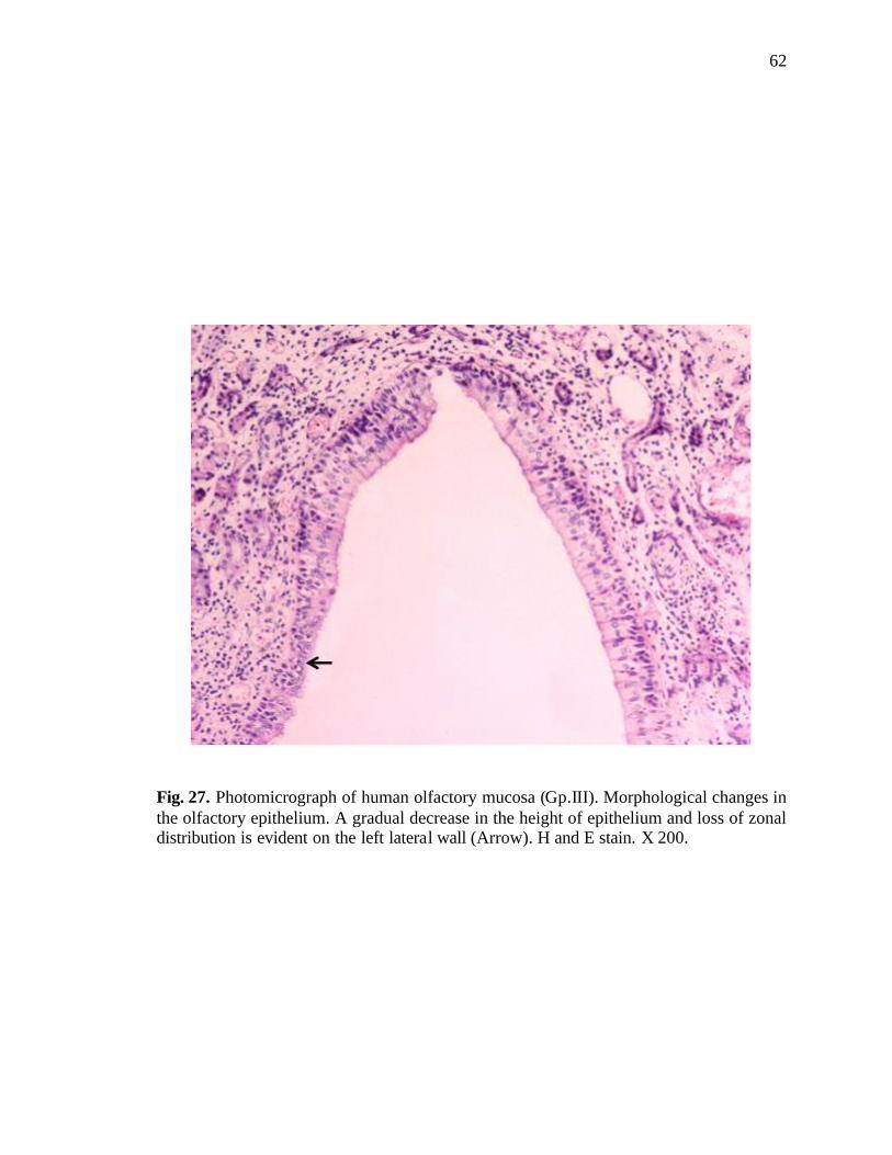
62
Fig. 27. Photomicrograph of human olfactory mucosa (Gp.III). Morphological changes in the olfactory epithelium. A gradual decrease in the height of epithelium and loss of zonal distribution is evident on the left lateral wall (Arrow). H and E stain. X 200.
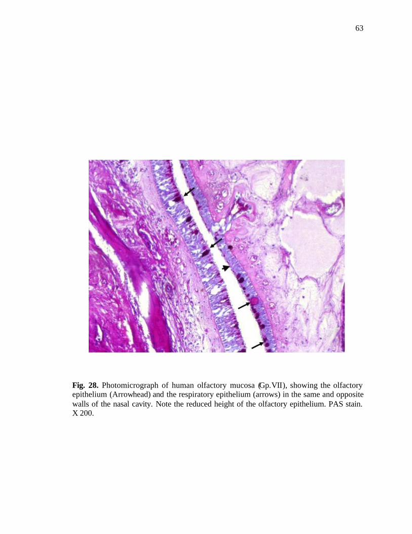
63
Fig. 28. Photomicrograph of human olfactory mucosa (Gp.VII), showing the olfactory epithelium (Arrowhead) and the respiratory epithelium (arrows) in the same and opposite walls of the nasal cavity. Note the reduced height of the olfactory epithelium. PAS stain. X 200.
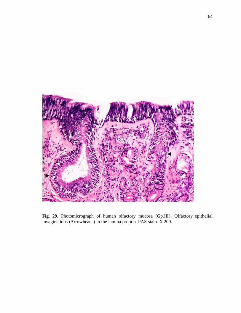
64
Fig. 29. Photomicrograph of human olfactory mucosa (Gp.III). Olfactory epithelial invaginations (Arrowheads) in the lamina propria. PAS stain. X 200.
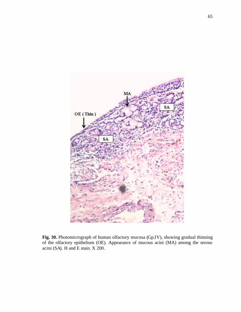
65
Fig. 30. Photomicrograph of human olfactory mucosa (Gp.IV), showing gradual thinning of the olfactory epithelium (OE). Appearance of mucous acini (MA) among the serous acini (SA). H and E stain. X 200.
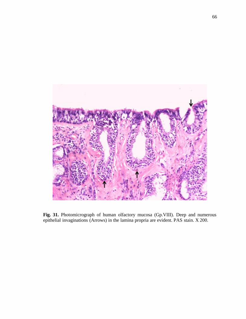
66
Fig. 31. Photomicrograph of human olfactory mucosa (Gp.VIII). Deep and numerous epithelial invaginations (Arrows) in the lamina propria are evident. PAS stain. X 200.
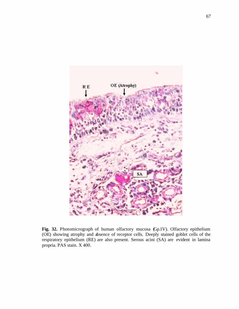
67
Fig. 32. Photomicrograph of human olfactory mucosa (Gp.IV). Olfactory epithelium (OE) showing atrophy and absence of receptor cells. Deeply stained goblet cells of the respiratory epithelium (RE) are also present. Serous acini (SA) are evident in lamina propria. PAS stain. X 400.
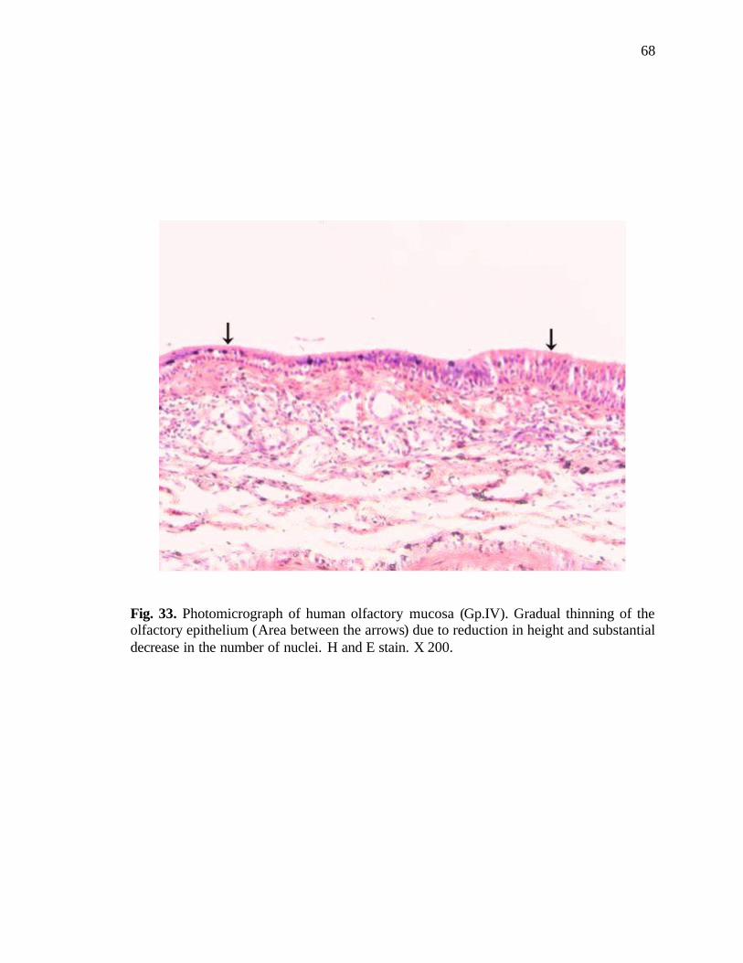
68
Fig. 33. Photomicrograph of human olfactory mucosa (Gp.IV). Gradual thinning of the olfactory epithelium (Area between the arrows) due to reduction in height and substantial decrease in the number of nuclei. H and E stain. X 200.
69
DISCUSSION
Although most of the previous light microscopic studies on the human olfactory
mucosa focused on the morphology of the olfactory epithelium, little attention has been
directed at age-related changes during adult life span. However, some evidence does exist
which shows that as we get old, our sense of smell declines (Larsson et al., 2000; Ship et
al., 1996; Farbman, 1994; Murphy et al., 2000; Stevens et al., 1982). Such perceptual
alterations are accompanied by changes in the anatomy and physiology of the olfactory
mucosa (Rombaux et al., 2005; Feng et al., 1997; Stevens et al., 1989; Elsener, 2001).
Histological studies show that there is an age-related tendency to lose olfactory receptor
cells, the sensory epithelial surface being replaced by respiratory epithelium (Legrier et
al., 2001; Nakashima et al., 1991; Paik et al., 1992; Feron et al., 1998). There is also
evidence that women, while losing smell sensitivity with age, perform better than men at
all ages (Jacob, 2006; Murphy et al., 2002). In the present study, the morphology of the
human olfactory mucosa was explored in male and female subjects ranging in age from
30 to 82 years in a Pakistani population. In this respect, this is a pioneer study from this
part of the world regarding the histology of human olfactory mucosa, its regional
distribut ion and age and sex related changes.
The olfactory epithelium of vertebrate species including humans has been
extensively studied using different morphological approaches (Cuschieri and Bannister,
1975; Graziadei and DeHan, 1973; Hinds et al., 1984; Belangar et al., 2003; Mandal et
al., 2005; Moran et al., 1992; McBride et al., 2003; Nomura et al., 2004; Kocianova et
al., 2003; Datta and Bandyopadhyay, 1997). Light and transmission electron microscopic
studies have shown that the human olfactory epithelium has morphological characteristics
similar to those in other vertebrates (Engström and Bloom, 1953; Naessen, 1970,
1971a,b; Nakashima et al., 1984; Naguro and Iwashita, 1992; Morrison and Costanzo,
1992; Kocianova et al., 2001). All of these studies agree that the olfactory epithelium is
pseudostratified and consists of olfactory receptor cells, sustentacular cells and basal
cells. In the present study, morphologically pseudostratified columnar epithelium resting
upon a uniform basement membrane was identified in the area of roof, medial and lateral

70
walls of right and left nasal cavities as has been shown by workers in the past. Four cell
types (olfactory receptor cells, sustentacular cells, basal cells and microvillar cells) in the
olfactory epithelium could be identified. Prior to 1982, most morphological studies of
human olfactory material (Smith, 1938; Bloom and Engström, 1952; Polyzonis et al.,
1979; Ohno et al., 1981) have described only three types of cells in the olfactory
epithelium. However, Moran et al. (1982a,b) consistently observed a fourth cell type in
electron microscopic studies on biopsies of normal human olfactory epithelium. They
named it as ‘microvillar cell’. Prior to the observations of Moran et al. (1982a), there was
no definitive description or identification of a mammalian microvillar cell. Monti
Graziadei and Graziadei (1979) had seen neurons located near the epithelial surface
showing a peculiar type of “degeneration”, characterized by a clear and swollen
cytoplasm but no definitive identity could be ascribed to these cells. Okano et al. (1967)
observed a fourth cell type with microvilli, having electron lucent cytoplasm, and Loo
(1977) observed microvillar cells in one of the four primates he investigated (slow loris),
but he called these cells, non-cilliated receptor cells. The observations of the microvillar
cell type by Moran and coworkers (1982a,b) received clear support for the first time from
more recent studies of Morrison and Costanzo (1990), Miller et al. (1995) and Jafek et al.
(2002) who also observed microvillar cells near the apical surface in the human olfactory
epithelium. Kwon et al. (2005) were able to note even age-related changes in the
microvillar cells of the rat olfactory epithelium on the basis of histological and
immunohistochemical methods. They observed that the microvillar cells in the olfactory
epithelium of aged rats were markedly hypertrophied with swollen end-feet reaching the
basement membrane. They did not observe such features in young cells. They noticed
that the ratio of the number of microvillar cells to that of the sustentacular cells increased
with aging and the total cell population decreased in the aged olfactory epithelium. They
suggested that microvillar cells are non-neuronal and are more resistant to aging
compared to the olfactory receptor cells and the sustentacular cells. The microvillar cells
are the only type of receptor cells found in the vomeronasal organ; this accessory
olfactory organ plays a significant role in olfaction in animals (Aujard, 1997; Keverne et
al., 1999; Smith et al., 2001; Smith et al., 2003; Dennis et al., 2003). The identity of the
71
cells designated as microvillar, in the present study, is in agreement with the above
mentioned studies.
The present study clearly shows age-related decrease in the number of olfactory
cells in both male and female subjects. The difference is statistically significant among
the various male groups and among the female groups. The decrease in the number of
olfactory cells occurs gradually with advancing age. However, the greatest difference in
the number of olfactory cells is between the male groups of ages 40-49 years and 50-59
years (Mean Difference = 29.31) as compared to age groups 30-39 years and 40-49 years
(Mean Difference = 10.11) and age groups 50-59 years and 60 years onwards (Mean
Difference = 4.78). These results suggest that the loss of olfactory receptors becomes
pronounced with advancing age of individuals of both sexes. Similarly there is significant
age-related decrease in the number of sustentacular cells among the groups of male and
female beyond the age of 50, with groups 60+ years showing the most distinct decrease.
The basal cells remain unaffected in groups of all ages. In the elderly human subjects,
Paik et al. (1992) observed a few olfactory receptor cells with tortuous dendrites, having
irregular nuclear margins and highly vacuolated cytoplasm suggesting age-related change
in the olfactory receptor population. Even degenerating cells were also seen and in a 76-
year old male, the respiratory epithelium seemed to have replaced the olfactory
epithelium. Nakashima et al. (1991) studied the characteristics of olfactory mucosa in
human fetuses and 40 subjects ranging in age from 31 years to 80 years. They observed
regular zonal distribution of the sustentacular cells, olfactory cells and basal cells in the
olfactory mucosa of the human fetus but in adults saw degenerated mucosa and decrease
or disappearance of the olfactory receptor cells. Rosli et al. (1999) has also shown age-
dependant reduction in both the olfactory receptor cells and the sustentacular cells.
Changes in both the number and size of the olfactory knobs at the surface and in the
density of olfactory cilia could be discerned. Weiler and Farbman (1997) have provided
evidence of dramatic decrease in the proliferative density of the olfactory epithelium in
rats from postnatal day 1 to post natal day 333. On postnatal day 1, the proliferative
density was 151 cells/mm, and on post natal day 333 it was only 8 cells/mm. Hinds and
McNelly (1981) observed that the number of olfactory receptors in the septal region of
72
rats, showed a marked increase from the age of 2 to 3 months, a steady increase at
somewhat lesser rate from three to eighteen months and then a decrease in the number of
receptor cells at subsequent ages, especially from twenty nine to thirty three months.
Information on the prevalence of disorders of the chemical senses has been rather
limited. A survey by the National Institute on Deafness and Other Communication
Disorders (NIDCD) in 1994 based on approximately 42,000 randomly-selected
households 1994 in the United States of America reported olfactory disorders in 2.7
million (1.4%) adults. This rate increased exponentially with age. It was observed that
almost 40% individuals with a chemosensory problem happened to be 65 years or older
(Hoffman et al., 1998). While gradual loss of olfactory capability with age is a common
phenomenon, mechanisms underlying this loss are not fully understood. Deficits occur
with age in both identification and detection thresholds for a variety of odours, with
changes becoming particularly evident after the fifth decade (Deems and Doty, 1987;
Schiffman and Pasternak, 1979; Schemper et al., 1981; Özdener and Rawson, 2004).
According to Cain and Stevens (1989) chronological age is strongly associated with
impairment of the sense of smell. On the basis of a special odour identification procedure,
Eskenazi et al. (1986) found that normal adults from 20 to 50 years of age could ident ify
85-100% of the odours presented but deterioration of odour identification ensued as age
advanced and at around 50 to 60 years, adult individuals could identify only 65-75% of
these odours. Murphy et al. (2002) examined odour perception in a population-based
study of 2,491 residents of Beaver Dam, Wisconsin, who ranged in age from 53 to 97
years. It was empirically determined that mean prevalence of olfactory impairment was
24.5% which increased with age to 62.5% among 80 to 97 year old individuals. In an
earlier study on 1955 individuals ranging from 5 to 99 years of age, it was found that
peak ability to identify odours was evident in the 3rd to the 5th decades but it declined
markedly after the 7th decade (Doty et al., 1984).
The present study also revealed random distribution of respiratory mucosa in the
olfactory area of the nasal cavity. These islands of respiratory mucosa in the olfactory
area could be identified by the typical respiratory nature of the epithelium and presence

73
of mucous glands in the lamina propria, confirmed by PAS staining. Such patches were
occasional in the age group 30-39 years, but increased gradually as age advanced from
40-49, through 50-59 year groups to 60 years onwards. In the 60+ year group, the patches
of respiratory epithelium could be seen in the roof, medial and lateral walls of both nasal
cavities; the boundary between the two types of epithelia being less defined. Naessen
(1970, 1971a) has described presence of respiratory epithelium in the olfactory area and
has suggested that these changes occur with aging. He studied the topographical
localization of the olfactory epithelium in human subjects and observed that the olfactory
margin displays a characteristic irregularity often associated with atrophy of the olfactory
epithelium that commonly occurs with age. The boundary line between the olfactory and
respiratory areas appeared clearly defined. The biological basis for changes in olfactory
function with age has been investigated in both animal and human models. There is
histological evidence that the neuroepithelium becomes patchy with age, with increased
infiltration by respiratory epithelium (Paik et al., 1992; Loo et al., 1996; Hirai et al.,
1996). These changes imply that the ability of the olfactory epithelium to regenerate
deteriorates with age (Walker et al., 1990; Doucette et al., 1983). In addition, axonal
degeneration and changes in the olfactory bulbs of elderly humans have been reported,
including loss of glomeruli (Meisami et al., 1998; Smith, 1942) and increase in glial cells
(Liss and Gomez, 1958). Collectively, these studies suggest that this loss of olfactory
receptor cells may explain the commonly reported age-related loss of olfactory
sensitivity. Paik et al. (1992) has also reported presence of various sized patches of
respiratory mucosa in the domain of the olfactory mucosa, with the transition sharply
defined between the olfactory and the respiratory mucosa over the septal and the conchal
mucosa as well as underneath the cribriform plate of the ethmoid bone. In old individuals,
they observed increase in respiratory patches, which were distributed throughout the
olfactory area, including the roof of the nasal cavity just underneath the cribriform plate
and along the upper 8 mm of the nasal septum. They observed that the probability of
obtaining olfactory specimens with the biopsy instrument decreased with advanced age,
reflecting loss of olfactory mucosa in aged subjects. Morrison and Costanzo (1990)
observed patches of respiratory epithelium in the superior nasal cavity, which is
considered a purely olfactory area. Nakashima et al. (1984) has also reported that

74
invasion by respiratory epithelium in the region of olfactory mucosa was more prominent
in the roof of the nasal cavity as compared with other regions and deduced that it is
characteristic of adult humans. Rosli et al. (1999) studied age related changes in the
mouse olfactory epithelium under transmission and scanning electron microscope. They
observed that the boundary between the respiratory and olfactory epithelium was sharp
and clearly delineated with an absence of intermixing of cells in the young and unaffected
aged mice. However, in the area of the septum of the aged mice the transition line
became interdigitated and tortuous. These findings by various researchers imply that
certain morphologic and physiologic changes may occur in the olfactory system as part of
the aging process. In the present study, this relates to the observation of the respiratory
mucosa in the region of the olfactory mucosa in cases of advanced age and together with
earlier studies suggest that age is an important factor leading to decline in the olfactory
function in humans. However, some studies have not reported any significant decline in
the ability of humans to smell odours. Elsner (2001) studied odour threshold, recognition,
discrimination and identification in centenarians. He tested the odour threshold in 21
centenarians (mean = 105.1 years) for phenylethyl alcohol (PEA) and menthol,
recognition and discrimination of lexically challenging odours, and identification of
common odours. Chronological age was not found to be a significant predictor of abilities
for any of the tasks performed by the centenarians in his study.
Age-related changes were also observed in the zonal distribution of cell nuclei in
the present study. The changes were quite drastic. Whereas the distribution of the cells
was normal in the age group 30-39 years, occasional disturbance of the position of
olfactory receptor cells and sustentacular cells was evident in age group 40-49 years
which became quite substantial in the age group 50-59 years, this resulted in decreased
height of the epithelium. In the age group 60 years onwards, reduction in thickness of the
olfactory epithelium was very significant with a concomitant loss of the normal zonal
distribution of sensory and sustentacular cell nuclei. These changes were evident in the
roof, medial and lateral walls of right and left nasal cavities. Naessen (1971a) and
Nakashima et al. (1984) have reported disturbance of the zonal distribution of the
olfactory receptor cells, sustentacular cells and epithelial degeneration, with the basal
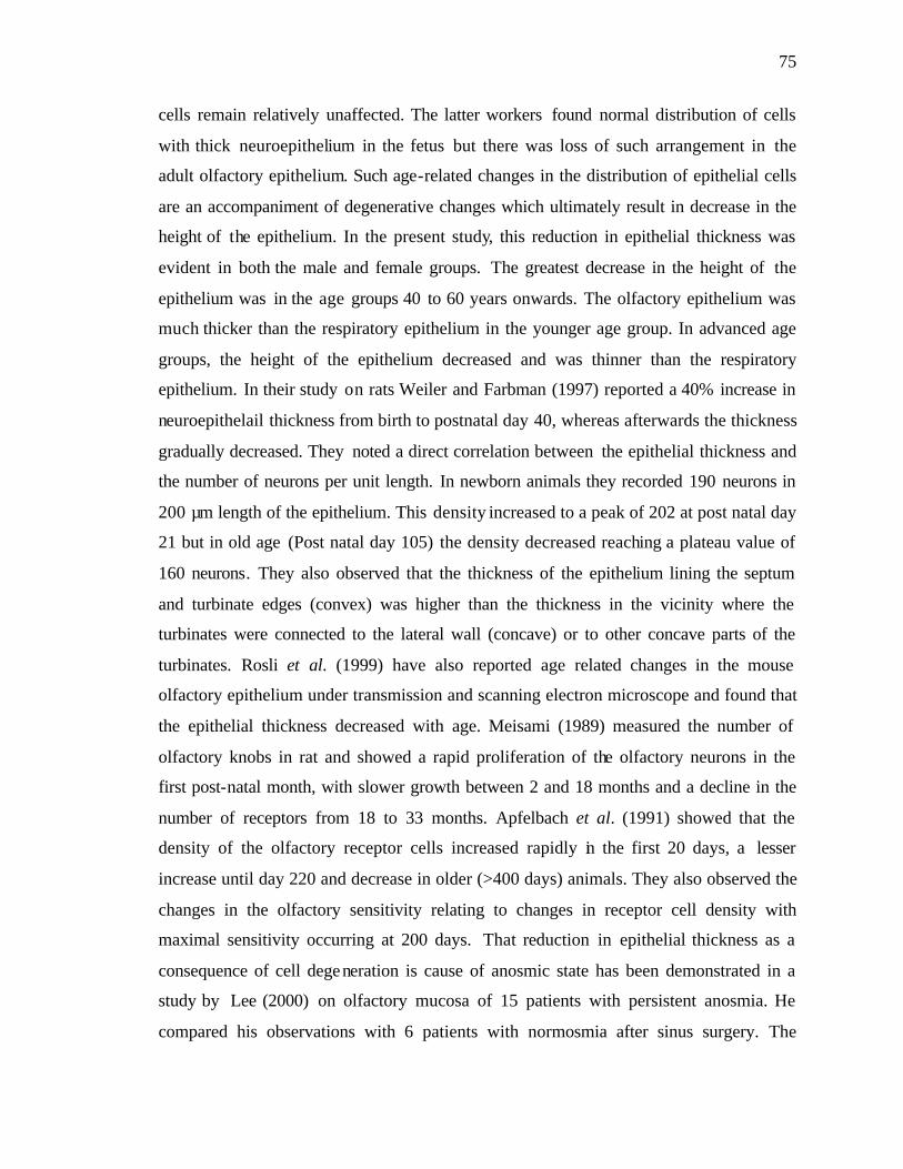
75
cells remain relatively unaffected. The latter workers found normal distribution of cells
with thick neuroepithelium in the fetus but there was loss of such arrangement in the
adult olfactory epithelium. Such age-related changes in the distribution of epithelial cells
are an accompaniment of degenerative changes which ultimately result in decrease in the
height of the epithelium. In the present study, this reduction in epithelial thickness was
evident in both the male and female groups. The greatest decrease in the height of the
epithelium was in the age groups 40 to 60 years onwards. The olfactory epithelium was
much thicker than the respiratory epithelium in the younger age group. In advanced age
groups, the height of the epithelium decreased and was thinner than the respiratory
epithelium. In their study on rats Weiler and Farbman (1997) reported a 40% increase in
neuroepithelail thickness from birth to postnatal day 40, whereas afterwards the thickness
gradually decreased. They noted a direct correlation between the epithelial thickness and
the number of neurons per unit length. In newborn animals they recorded 190 neurons in
200 µm length of the epithelium. This density increased to a peak of 202 at post natal day
21 but in old age (Post natal day 105) the density decreased reaching a plateau value of
160 neurons. They also observed that the thickness of the epithelium lining the septum
and turbinate edges (convex) was higher than the thickness in the vicinity where the
turbinates were connected to the lateral wall (concave) or to other concave parts of the
turbinates. Rosli et al. (1999) have also reported age related changes in the mouse
olfactory epithelium under transmission and scanning electron microscope and found that
the epithelial thickness decreased with age. Meisami (1989) measured the number of
olfactory knobs in rat and showed a rapid proliferation of the olfactory neurons in the
first post-natal month, with slower growth between 2 and 18 months and a decline in the
number of receptors from 18 to 33 months. Apfelbach et al. (1991) showed that the
density of the olfactory receptor cells increased rapidly in the first 20 days, a lesser
increase until day 220 and decrease in older (>400 days) animals. They also observed the
changes in the olfactory sensitivity relating to changes in receptor cell density with
maximal sensitivity occurring at 200 days. That reduction in epithelial thickness as a
consequence of cell degeneration is cause of anosmic state has been demonstrated in a
study by Lee (2000) on olfactory mucosa of 15 patients with persistent anosmia. He
compared his observations with 6 patients with normosmia after sinus surgery. The
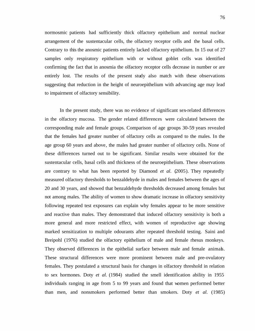
76
normosmic patients had sufficiently thick olfactory epithelium and normal nuclear
arrangement of the sustentacular cells, the olfactory receptor cells and the basal cells.
Contrary to this the anosmic patients entirely lacked olfactory epithelium. In 15 out of 27
samples only respiratory epithelium with or without goblet cells was identified
confirming the fact that in anosmia the olfactory receptor cells decrease in number or are
entirely lost. The results of the present study also match with these observations
suggesting that reduction in the height of neuroepithelium with advancing age may lead
to impairment of olfactory sensibility.
In the present study, there was no evidence of significant sex-related differences
in the olfactory mucosa. The gender related differences were calculated between the
corresponding male and female groups. Comparison of age groups 30-59 years revealed
that the females had greater number of olfactory cells as compared to the males. In the
age group 60 years and above, the males had greater number of olfactory cells. None of
these differences turned out to be significant. Similar results were obtained for the
sustentacular cells, basal cells and thickness of the neuroepithelium. These observations
are contrary to what has been reported by Diamond et al. (2005). They repeatedly
measured olfactory thresholds to benzaldehyde in males and females between the ages of
20 and 30 years, and showed that benzaldehyde thresholds decreased among females but
not among males. The ability of women to show dramatic increase in olfactory sensitivity
following repeated test exposures can explain why females appear to be more sensitive
and reactive than males. They demonstrated that induced olfactory sensitivity is both a
more general and more restricted effect, with women of reproductive age showing
marked sensitization to multiple odourants after repeated threshold testing. Saini and
Breipohl (1976) studied the olfactory epithelium of male and female rhesus monkeys.
They observed differences in the epithelial surface between male and female animals.
These structural differences were more prominent between male and pre-ovulatory
females. They postulated a structural basis for changes in olfactory threshold in relation
to sex hormones. Doty et al. (1984) studied the smell identification ability in 1955
individuals ranging in age from 5 to 99 years and found that women performed better
than men, and nonsmokers performed better than smokers. Doty et al. (1985)
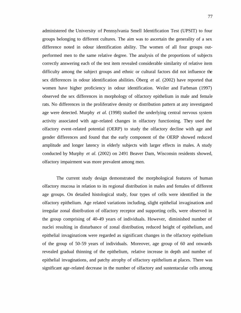
77
administered the University of Pennsylvania Smell Identification Test (UPSIT) to four
groups belonging to different cultures. The aim was to ascertain the generality of a sex
difference noted in odour identification ability. The women of all four groups out-
performed men to the same relative degree. The analysis of the proportions of subjects
correctly answering each of the test item revealed considerable similarity of relative item
difficulty among the subject groups and ethnic or cultural factors did not influence the
sex differences in odour identification abilities. Öberg et al. (2002) have reported that
women have higher proficiency in odour identification. Weiler and Farbman (1997)
observed the sex differences in morphology of olfactory epithelium in male and female
rats. No differences in the proliferative density or distribution pattern at any investigated
age were detected. Murphy et al. (1998) studied the underlying central nervous system
activity associated with age-related changes in olfactory functioning. They used the
olfactory event-related potential (OERP) to study the olfactory decline with age and
gender differences and found that the early component of the OERP showed reduced
amplitude and longer latency in elderly subjects with larger effects in males. A study
conducted by Murphy et al. (2002) on 2491 Beaver Dam, Wisconsin residents showed,
olfactory impairment was more prevalent among men.
The current study design demonstrated the morphological features of human
olfactory mucosa in relation to its regional distribution in males and females of different
age groups. On detailed histological study, four types of cells were identified in the
olfactory epithelium. Age related variations including, slight epithelial invaginations and
irregular zonal distribution of olfactory receptor and supporting cells, were observed in
the group comprising of 40-49 years of individuals. However, diminished number of
nuclei resulting in disturbance of zonal distribution, reduced height of epithelium, and
epithelial invaginations were regarded as significant changes in the olfactory epithelium
of the group of 50-59 years of individuals. Moreover, age group of 60 and onwards
revealed gradual thinning of the epithelium, relative increase in depth and number of
epithelial invaginations, and patchy atrophy of olfactory epithelium at places. There was
significant age-related decrease in the number of olfactory and sustentacular cells among

78
male and female groups, more pronounced in individuals of 50 years and above without
significant gender differences.
The analysis of these observations clearly pointed out decline in the sense of
olfaction with age possibly due to the mechanism of reduction in the number of olfactory
receptors; in addition no evident sex related differences were seen in these groups. A
number of age related disorders have been reported with alterations in olfactory function.
The most accepted evidence, of smell loss in age related diseases, is based on studies of
patients suffering from Alzheimer’s disease and Parkinsonism. Although advances have
been made in documenting the nature of age related changes in smell perception; causal
relations between the changes in the morphology of olfactory mucosa and the underlying
pathologic mechanism, are yet to be established. This study presents a perspective for
future research endeavors in the emerging area of “geriatric otolaryngology”. The
findings of this study have implications for understanding the association between aging
and morphology of olfactory mucosa.
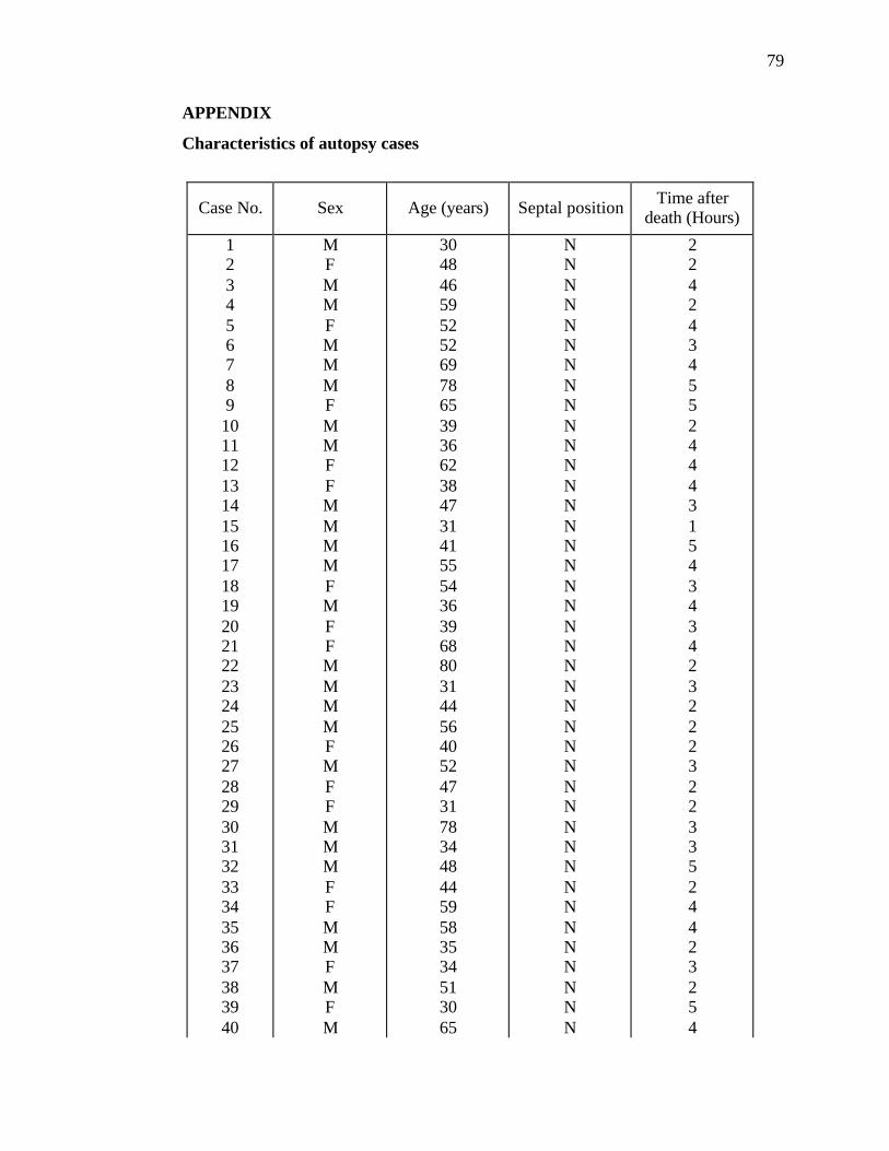
79
APPENDIX
Characteristics of autopsy cases
Case No. Sex Age (years) Septal position Time after death (Hours)
1 2 3 4 5 6 7 8 9 10 11 12 13 14 15 16 17 18 19 20 21 22 23 24 25 26 27 28 29 30 31 32 33 34 35 36 37 38 39 40
M F M M F M M M F M M F F M M M M F M F F M M M M F M F F M M M F F M M F M F M
30 48 46 59 52 52 69 78 65 39 36 62 38 47 31 41 55 54 36 39 68 80 31 44 56 40 52 47 31 78 34 48 44 59 58 35 34 51 30 65
N N N N N N N N N N N N N N N N N N N N N N N N N N N N N N N N N N N N N N N N
2 2 4 2 4 3 4 5 5 2 4 4 4 3 1 5 4 3 4 3 4 2 3 2 2 2 3 2 2 3 3 5 2 4 4 2 3 2 5 4
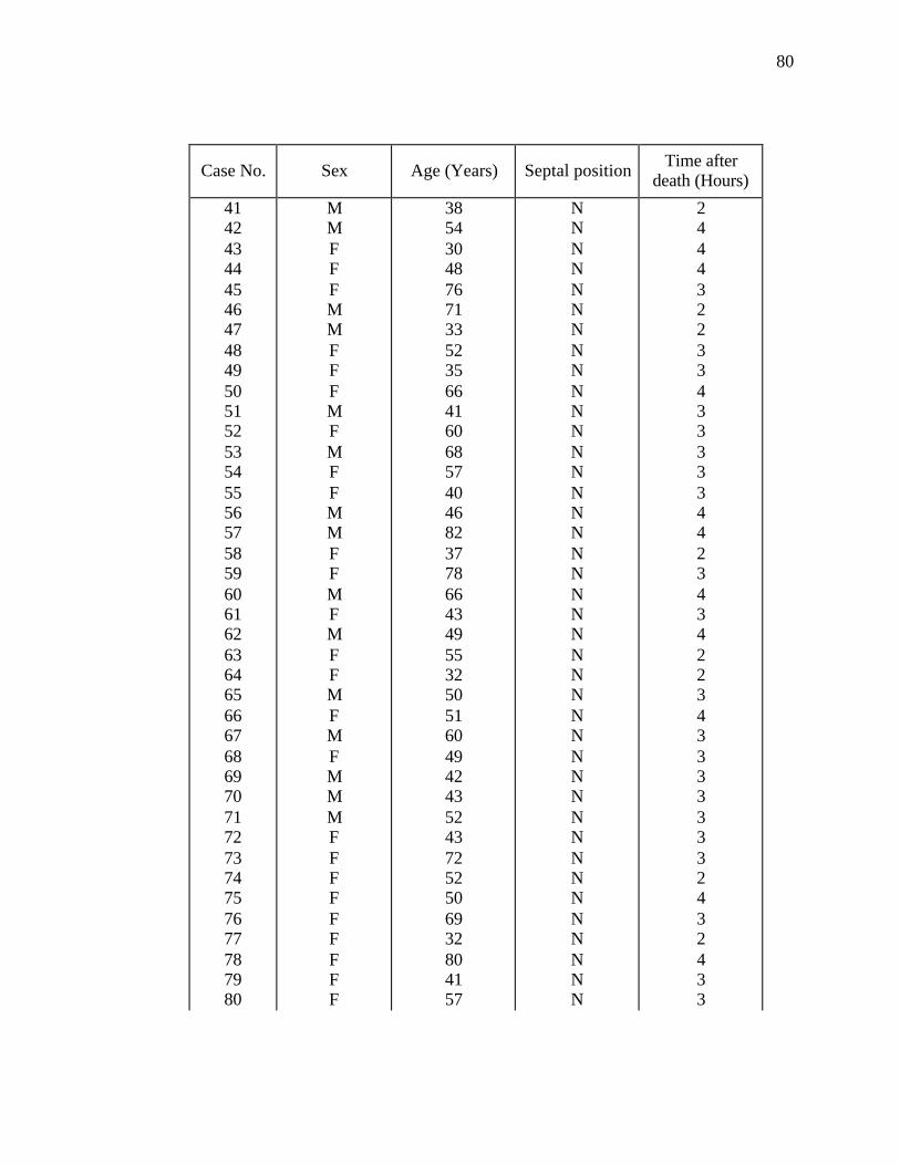
80
Case No. Sex Age (Years) Septal position Time after death (Hours)
41 42 43 44 45 46 47 48 49 50 51 52 53 54 55 56 57 58 59 60 61 62 63 64 65 66 67 68 69 70 71 72 73 74 75 76 77 78 79 80
M M F F F M M F F F M F M F F M M F F M F M F F M F M F M M M F F F F F F F F F
38 54 30 48 76 71 33 52 35 66 41 60 68 57 40 46 82 37 78 66 43 49 55 32 50 51 60 49 42 43 52 43 72 52 50 69 32 80 41 57
N N N N N N N N N N N N N N N N N N N N N N N N N N N N N N N N N N N N N N N N
2 4 4 4 3 2 2 3 3 4 3 3 3 3 3 4 4 2 3 4 3 4 2 2 3 4 3 3 3 3 3 3 3 2 4 3 2 4 3 3
81
REFERENCES
Agur, A.M.R., and Dalley, A.F. 2005. Nose, paranasal sinuses and pterygopalatine fossa. In: Grants Atlas of Anatomy. 11th ed. Philadelphia: Lippincott Williams and Wilkins, pp. 680-95. Anderson, A. 2003. Breastfeeding: societal encouragement needed. J. Hum. Nutr. Diet. 16(4): 217-8. Apfelbach, R., Russ, D., and Slotnick, B.M. 1991. Ontogenetic changes in odor sensitivity, olfactory receptor area and olfactory receptor density in the rat. Chem. Senses 16(3): 209-18. Apter, A.J., Gent, J.F., and Frank, M.E. 1999. Fluctuating olfactory sensitivity and distorted odor perception in allergic rhinitis. Arch. Otolaryngol. Head Neck Surg. 125: 1005-10. Aujard, F. 1997. Effect of vomeronasal organ removal on male socio-sexual responses to female in a prosimian primate (Microcebus murinus). Physiol. Behav. 62(5): 1003-8. Bancroft, J.D., and Gamble, M. 2002. Theory and Practice of Histological Techniques. 5th ed. Edinburgh: Churchill Livingstone, pp. 376-8. Basmajian, J.V., and Slonecker. 1989. Grant’s Method of Anatomy. 11th ed. Baltimore: Williams and Wilkins. Beauchamp, G.K., Katahira, K., Yamazaki, K., Mennella, J.A., Bard, J., and Boyse, E.A. 1995. Evidence suggesting that the odortypes of pregnant women are a compound of maternal and fetal odortypes. Proc. Natl. Acad. Sci. 92(7): 2617-21. Beauchamp, G.K., Wysocki, C.J., and Wellington, J.L. 1985. Extinction of response to urine odor as a consequence of vomeronasal organ removal in male guinea pigs. Behav. Neurosci. 99(5): 950-5. Belanger, R.M., Smith, C.M., Corkum, L.D., and Zielinski, B.S. 2003. Morphology and histochemistry of the peripheral olfactory organ in the round goby, Neogobius melanostomus (Teleostei: Gobiidae). J. Morphol. 257(1): 62-71. Bhandawat, V., Reisert, J., and Yau, K.W. 2005. Elementary response of olfactory receptor neurons to odorants. Science 308: 1931-4. Bloom, G., and Engström, H. 1952. The structure of the epithelial surface in the olfactory region. Exp. Cell. Res. 3: 669-71.
82
Bonfanti, M.L., and Fasolo, P.P.A. 2002. Adult structural plasticity and neurogenesis in the mammalian olfactory system. Rend. Fis. Acc. Lincei 13: 145-79. Bradbury, S. 1973. Hewer’s Text Book of Histology for Medical Students. 9th ed. London: William Heinemann. Buck, L.B. 2000. The molecular architecture of odor and pheromone sensing in mammals. Cell 100: 611-8. Caggiano, M., Kauer, J.S., and Hunter, D.D. 1994. Globose basal cells are neuronal progenitors in the olfactory epithelium: a lineage analysis using a replication- incompetent retrovirus. Neuron 13(2): 339-52. Cain, W.S., and Stevens, J.C. 1989. Uniformity of olfactory loss in aging. Ann. N. Y. Acad. Sci. 561: 29-38. Caruso, S., Grillo, C., Agnello, C., Mari, L.D., Farina, M., and Serra, A. 2004. Olfactometric and rhinomanometric outcomes in post-menopausal women treated with hormone therapy: a prospective study. Human Reprod. 19(12): 2959-64. Chen, D., and Dalton, P. 2005. The effect of emotion and personality on olfactory perception. Chem. Senses 30: 345-51. Chen, D., Katdare, A., and Lucas, N. 2006. Chemosignals of fear enhance cognitive performance in humans. Chem. Senses 31: 415-23. Corwin, J., Loury, M., and Gilbert, A.N. 1995. Workplace, age, and sex as mediators of olfactory function: data from the National Geographic Smell Survey. J. Gerontol. Psychol. Sci. 50: 179-86. Costanzo, R.M., and Graziadei, P.P.C. 1983. A quantitative analysis of changes in the olfactory epithelium following bulbectomy in hamsters. J. Comp. Neurol. 215: 370-81. Cummings, C.W., Fredrickson, J.M., Harker, L.A., Krause, C.J., and Schuller, D.E. (editors). 1990. Otolaryngology-Head and Neck Surgery. 1st ed. New Delhi: BI. Cuschieri, A., and Bannister, L.H. 1975. The development of the olfactory mucosa in the mouse: light microscopy. J. Anat. 119(2): 277-86. Datta, C., and Bandyopadhyay, S. 1997. Ultrastructure of cell types of the olfactory epithelium in a catfish, Heteropneustes fossilis (Bloch). J. Biosci. 22: 233-45. Deems, D.A., and Doty, R.L. 1987. The nature of age-related olfactory detection threshold changes in man. Trans. Pa. Acad. Ophthalmol. Otolaryngol. 39: 87-93. Delay, R.J., and Dionne, V.E. 2003. Coupling between sensory neurons in the olfactory epithelium. Chem. Senses 28: 807-15.
83
Dennis, J.C., Allgier, J.G., Desouza, L.S., Eward, W.C., and Morrison, E.E. 2003. Immunohistochemistry of the canine vomeronasal organ. J. Anat. 203: 329-38. Diamond, J., Dalton, P., Doolittle, N., and Breslin, P.A.S. 2005. Gender-specific olfactory sensitization: Hormonal and cognitive influences. Chem. Senses 30 (Suppl 1): 224-5. Dominy, N.J., Ross, C.F., and Smith, T.D. 2004. Evolution of the special senses in primates: past, present and future. Anat. Rec. 281A: 1078-82. Doty, R.L. 1986. Cross-cultural studies of taste and smell perception. In: Duvall, D., Muller-Schwarze, D., and Silverstein, R.M., (editors). Chemical Signals in Vertebrates 4. New York: Plenum Publishing Corporation, pp. 673-84. Doty, R.L. 1989. Age-related alterations in taste and smell function. In: Goldstein, J.C., Kashima, H.K., and Coopmann, C.F. Jr., (editors). Geriatric Otorhinolaryngology Burlington: B.C. Decker, pp. 97-104. Doty, R.L. 1990. Olfactory capacities in aging and Alzheimer’s disease: psychophysical and anatomic considerations. Ann. N. Y. Acad. Sci. 640: 20-7. Doty, R.L., and Mishra, A. 2001. Olfaction and its alteration by nasal obstruction, rhinitis and rhinosinusitis. Larngoscope 111(3): 409-23. Doty, R.L., Applebaum, S., Zusho, H., and Settle, G. 1985. Sex differences in odor identification ability: a cross-cultural analysis. Neuropsychologia 23(5): 667-72. Doty, R.L., Deems, D.A., Frye, R.E., Pelberg, R., and Shapiro, A. 1988. Olfactory sensitivity, nasal resistance and autonomic functions in patients with multiple chemical sensitivities. Arch. Otolaryngol. Head Neck Surg. 114: 1422-7. Doty, R.L., Shaman, P., Applebaum, S.L., Giberson, R., Siksorski, L., and Rosenberg, L. 1984. Smell ident ification ability: changes with age. Science 226: 1441-3. Doucette, J.R., Kiernan, J.A, and Flumerfelt, B.A. 1983. Two different patterns of retrograde degeneration in the olfactory epithelium following transaction of primary olfactory axons. J. Anat. 136(4): 673-89. Douek, E., Bannister, L.H., and Dodson, H.C. 1975. Recent advances in the pathology of olfaction. Proc. R. Soc. Med. 68: 467-70. Drury, R.A.B., and Wallington, E.A. 1967. Carleton’s Histological Technique. 4th ed. London: Oxford University Press, pp.13-15.

84
Ducray, A., Bondier, J.R., Michel, G., Bon, K., Propper, A., and Kastner, A. 2002. Recovery following peripheral destruction of olfactory neurons in young and adult mice. Eur. J. Neurosci. 15: 1907-17. Duffy, V.B., Cain, W.S., and Ferris, A.M. 1999. Measurement of sensitivity to olfactory flavor: application in a study of aging and dentures. Chem. Senses 24: 671-7. Elsner, R.J.F. 2001. Odor threshold, recognition, discrimination and identification in centenarians. Arch. Gerontol. Geriatr. 33: 81-94. Engström, H., and Bloom, G. 1953. The structure of the olfactory region in man. Acta. Otolaryngol. 43(1): 11-21. Eskenazi, B., Cain, W.S., Novelly, R.A., and Mattson, R. 1986. Odor perception in temporal lobe epilepsy patients with and without temporal lobectomy. Neuropsychologia 24: 553-62. Farbman, A.I., Brunjes, P.C., Rentfro, L., Michas, J., and Ritz, S. 1988. The effect of unilateral naris occlusion on cell dynamics in the developing rat olfactory epithelium. J. Neurosci. 8(9): 3290-5. Farbman, A.I. 1994. Developmental biology of olfactory sensory neurons. Semin. Cell Biol. 5: 3-10. Fawcett, D.W. 1994. A Text Book of Histology. 12th ed. New York: Chapman and Hall. Feng, W.H., Kauer, J.S., Adelman, L., and Talamo, B.R. 1997. New structure, the "olfactory pit," in human olfactory mucosa. J. Comp. Neurol. 378: 443-53. Feron, F., Perry, C., McGrath, J.J., and Mackay-Sim, A. 1998. New techniques for biopsy and culture of human olfactory epithelial neurons. Arch. Otolaryngol. Head Neck Surg. 124: 861-6. Fox, S.I. 2006. Sensory Physiology (Smell). In: Human Physiology. 9th ed. Boston: McGraw-Hill, pp. 247-9. Frisch, D. 1967. Ultrastructure of mouse olfactory mucosa. Am. J. Anat. 121: 87- 120. Ganong, W.F. 2005. Smell and Taste. In: Review of Medical Physiology. 22nd ed. Boston: McGraw-Hill, pp. 185-8. Ge, Y., Tsukatani, T., Nishimura, T., Furukawa, M., and Miwa, T. 2002. Cell death of olfactory receptor neurons in a rat with nasosinusitis infected artificially with staphylococcus. Chem. Senses 27: 521-7.
85
Ghorbanian, S.N., Paradise, J.L., and Doty, R.L. 1983. Odor perception in children in relation to nasal obstruction. Pediatrics 72(4): 510-6. Gilbert, A.N., and Wysocki, C.J. 1987. The smell survey. Natl. Geogr. Mag. 172: 514-25. Goldstein, B.J., and Schwob, J.E. 1996. Analysis of the globose basal cell compartment in rat olfactory epithelium using GBC-1, a new monoclonal antibody against globose basal cells. J. Neurosci. 16(12): 4005-16. Gray, R.F., and Hawthorne, M. 1992. Synopsis of Otolaryngology. 5th ed. Cambridge: Butterworth Heinemann. Graziadei, P. 1965. Electron microscopic observations on the olfactory mucosa of the cat. Experientia 21(5): 274-5. Graziadei, P.P.C., Levine, R.R., and Monti Graziadei, G.A. 1978. Regeneration of olfactory axons and synapse formation in the forebrain after bulbectomy in neonatal mice. Proc. Natl. Acad. Sci. 75(10): 5230-4. Graziadei, P.P.C. 1973. Cell dynamics in the olfactory mucosa. Tissue Cell 5: 113-31. Graziadei, P.P.C., and DeHan, R.S. 1973. Neuronal regeneration in frog olfactory system. J. Cell Biol. 59: 525-30. Graziadei, P.P.C., and Monti Graziadei, G.A. 1978. Continuous nerve cell renewal in the olfactory system. In: Jacobson, M., (editor). Handbook of Sensory Physiology: IX. Development of Sensory Systems. Berlin: Springer-Verlag, pp. 55-83. Gross, E.A., Swenberg, J.A., Fields, S., and Popp, J.A. 1982. Comparative morphometry of the nasal cavity in rats and mice. J. Anat. 135(1): 83-8. Hinds, J.W., and McNelly, N.A. 1981. Aging in the rat olfactory system: correlation of changes in the olfactory epithelium and olfactory bulb. J. Comp. Neurol. 203(3): 441-53. Hinds, J.W., Hinds, P.L., and McNelly, N.A. 1984. An autoradiographic study of the mouse olfactory epithelium: evidence for long- lived receptors. Anat. Rec. 210: 375-83. Hirai, T., Kojima, S., Shimada, A., Umemura, T., Sakai, M., and Itakura, C. 1996. Age-related changes in the olfactory system of dogs. Neuropathol. Appl. Neurobiol. 22: 531-9. Hoffman, H.J., Ishii, E.K., and Macturk, R.H. 1998. Age-related changes in the prevalence of smell/taste problems among the United States adult population: results of the 1994 disability supplement to the National Health Interview Survey (NHIS). Ann. N. Y. Acad. Sci. 855: 716-22.

86
Hudson, R., Arriola, A., Martinez-Gomez, M., and Distel, H. 2006. Effect of air pollution on olfactory function in residents of Mexico City. Chem. Senses 31: 79-85. Human Tissue Act 1961. 2006 [cited 2006 Dec] available from: http://www.uktransplant.org.uk/ukt/about_transplants/legislation/human_tissue_act/human_tissue_act.jsp Hummel, T., Rothbauer, C., Barz, S., Grosser, K., Pauli, E., and Kobal, G. 1998. Olfactory function in acute rhinitis. Ann. N. Y. Acad. Sci. 855: 616-24. Jacob, S., McClintock, M.K., Zelano, B., and Ober, C. 2002. Paternally inherited HLA alleles are associated with women's choice of male odor. Nat. Genet. 30(2):175-9. Jacob, T. Olfaction: A tutorial on the sense of smell [online]. Published 2006 [Cited 2006]. Available from: http://www.cf.ac.uk/biosi/staff/jacob/teaching/sensory/olfact1.html Jafek, B.W., Murrow, B., and Linschoten, M. 2000. Evaluation and treatment of anosmia. Curr. Opin. Otolaryngol. Head Neck Surg. 8: 63-7. Jafek, B.W., Hartman, D., Eller, P.M., Johnson, E.W., Strahan, R.C., and Moran, D.T. 1990. Postviral olfactory dysfunction. Am. J. Rhinol. 4: 91-100. Jafek, B.W., Murrow, B., Michaels, R., Restrepo, D., and Linschoten, M. 2002. Biopsies of human olfactory epithelium. Chem. Senses 27: 623-8. Johnson, L.R., Byrne, J.H. (editors). 1998. Essential Medical Physiology. 2nd ed. Philadelphia: Lippincott-Raven, 745-56. Kaitz, M., Good, A., Rokem, A.M., and Eidelman, A.I. 1987. Mothers' recognition of their newborns by olfactory cues. Dev. Psychobiol. 20(6): 587-91. Kaneda. H., Maeshima, K., Goto, N., Kobayakawa, T., Ayabe-Kanamura, S., and Siato, S. 2000. Decline in taste and odor discrimination abilities with age and relationship between gestation and olfaction. Chem. Senses 25: 331-7. Kasper, D.L., Braunwald, E., Fauci, A.S., Hauser, S.L., Longo, D.L., and Jameson, J.L. (editors). 2005. Harrison’s Principles of Internal Medicine. 16th ed. New York: McGraw-Hill. Keller, M., Douhard, Q., Baum, M.J., and Bakker, J. 2006. Destruction of the main olfactory epithelium reduces female sexual behavior and olfactory investigation in female mice. Chem. Senses 31: 315-23. Kelly, D.E., Wood, R.L., and Enders, A.C. 1984. Bailey’s Text Book of Microscopic Anatomy. 18th ed. Baltimore: Williams and Wilkins.

87
Keverne, E.B. 1999. The vomeronasal organ. Science 286: 716-20. Kocianova, I., Buchtova, M., Gorosova, A., Holubcova, S., and Cizek, P. 2003. The ontogeny of the olfactory epithelium: an ultrastructural study in sheep. Acta. Vet. Brno. 72: 3-10. Kocianova, I., Tichy, F., and Gorosova, A. 2001. Ultrastructure of the olfactory epithelium of kittens. Acta. Vet. Brno. 70: 375-9. Kratzing, J.E. 1982. Regional variation in respiratory epithelium of the nasal cavity of the bandicoot (Isoodon macrourus). J. Anat. 134(1): 1-9. Kratzing, JE. 1984. The anatomy and histology of the nasal cavity of the koala (Phascolarctos cinereus). J. Anat. 138: 55-65. Krieger, J., and Breer, H. 1999. Olfactory reception in invertebrates. Science 286: 720-3. Kuzma, J.W., and Bohnenblust, S.E. 2000. Basic Statistics for the Health Sciences. 4th ed. California: Mayfield Publishing Company. Kwon, B.S., Kim, M.K., Kim, W.H., Pyo, J.S., Cheon, Y.H., Cha. C.I., Nam, S.Y., Baik, T.K., and Lee, B.L. 2005. Age-related changes in microvillar cells of rat olfactory epithelium. Neurosci. Lett. 378: 65-9. Lane, A.P., Gomez, G., Dankulich, T., Wang, H., Bolger, W.E., and Rawson, N.E. 2002. The superior turbinate as a source of functional human olfactory receptor neurons. Laryngoscope 112(7):1183-9. Larsson, M., Finkel, D., and Pederson, N.L. 2000. Odor identification: influence of age, gender, cognition and personality. J. Gerontol. Psychol. Sci. 55B(5): 304-10. Larsson, P., and Tjälve, H. 2000. Intranasal installation of aflatoxin B1 in rats: bioactivation in the nasal mucosa and neuronal transport to the olfactory bulb. Toxicol. Sci. 55: 383-91. Laska, M., and Teubner, P. 1998. Odor structure-activity relationships of carboxylic acids correspond between squirrel monkeys and humans. Am. J. Physiol. 274: 1639-45. Laska, M., Wieser, A., and Salazar, L.T.H. 2005. Olfactory responsiveness to two odorous steroids in three species of nonhuman primates. Chem. Senses 30: 505-11. Lee, S.H., Lim, H.H., Lee, H.M., Park, H.J., and Choi, J.O. 2000. Olfactory mucosal findings in patients with persistent anosmia after endoscopic sinus surgery. Ann. Otol. Rhinol. Laryngol. 109: 720-5. Leffingwell, J.C. 2002. Olfaction-Update No. 5. Leffingwell Reports 2(1): 1-34.
88
Legrier, M.E., Ducray, A., Propper, A., Chao, M., and Kastner, A. 2001. Cell cycle regulation during mouse olfactory neurogenesis. Cell Growth Differ. 12: 591-601. Lewis, C.S. The Magician’s Nephew. 2006 Jan [cited 2006 Aug]. Available from: http://everything2.com/index.pl?node=smell Liss, L., Mich, A.A., and Gomez, F. 1958. The nature of senile changes of the human olfactory bulb and tract. Arch. Otolaryngol. 67: 167-71. Lledo, P.M., Gheusi, G., and Vincent, J.D. 2005. Information processing in the mammalian olfactory system. Physiol. Rev. 85: 281-317. Loo, A.T., Youngentob, S.L., Kent, P.F., and Schwob, J.E. 1996. The aging olfactory epithelium: neurogenesis, response to damage, and odorant induced activity. Int. J. Dev. Neurosci. 14: 881-900. Loo, S. K. 1977. Fine structure of the olfactory epithelium in some primates. J. Anat. 123: 135-45. Lovell, M.A., Jafek, B.W., Moran, D.T., and Rowley III, J.C. 1982. Biopsy of human olfactory mucosa. Arch. Otolaryngol. 108: 247-9. Mandal, D.K., Roy, D., and Ghosh, L. 2005. Structural organization of the olfactory epithelium of a spotted snakehead fish, Channa punctatus. Acta. Ichthyol. Piscat. 35(1): 45-50. McBride, K., Slotnik, B., and Margolis, F.L. 2003. Does intranasal application of zinc sulfate produce anosmia in the mouse? An olfactometric and anatomical study. Chem. Senses 28: 659-70. Meisami, E. 1989. A proposed relationship between increases in the number of olfactory receptor neurons, convergence ratio and sensitivity in the developing rat. Dev. Brain. Res. 46: 9-19. Meisami, E., Mikhail, L., Baim, D., and Bhatnagar, K.P. 1998. Human olfactory bulb: aging of glomeruli and mitral cells and a search for the accessory olfactory bulb. Ann. N. Y Acad. Sci. 855: 708-15. Miller, M.L., Andringa, A., Evans, J.E., and Hastings, L. 1995. Microvillar cells of the olfactory epithelium: morphology and regeneration following exposure to toxic compounds. Brain Res. 669: 1-9. Mishra, A., and Doty, R.L. 2002. Olfaction - A clinical approach. Indian J. Otolaryngol. Head Neck Surg. 54(2): 156-60.
89
Miwa, T., Furukawa, M., Tsukatani, T., Costanzo, R.M., DiNardo, L.J., and Reiter, E.R. 2001. Impact of olfactory impairment on quality of life and disability. Arch. Otolaryngol. Head Neck Surg. 127: 497-503. Monti Graziadei, G.A., and Graziadei, P.P.C. 1979. Neurogenesis and neuron regeneration in the olfactory system of mammals. II. Degeneration and reconstitution of the olfactory sensory neurons after axotomy. J. Neurocytol. 8: 197-213. Moran, D.T., Jafek, B.W., Eller, P.M., and Rowley III, J.C. 1992. Ultrastructural histopathology of human olfactory dysfunction. Microsc. Res. Tech. 23: 103-10. Moran, D.T., Rowley III, J.C., and Jafek, B.W. 1982a. Electron microscopy of human olfactory epithelium reveals a new cell type: the microvillar cell. Brain Res. 253(1-2): 39-46. Moran, D.T., Rowley III, J.C., Jafek, B.W., and Lovel, M.A. 1982b. The fine structure of olfactory mucosa in man. J. Neurocytol. 11: 721-46. Morrison, E.E., and Costanzo, R.M. 1990. Morphology of the human olfactory epithelium. J. Comp. Neurol. 297: 1-13. Morrison, E.E., and Costanzo, R.M. 1992. Morphology of olfactory epithelium in humans and other vertebrates. Microsc. Res. Tech. 23(1): 49-61. Moulton, D.G. 1974. Dynamics of cell populations in the olfactory epithelium. Ann. N. Y. Acad. Sci. 237: 52-61. Mukherjee, K.L. 1988. Medical Laboratory Technology: A Procedure Manual for Routine Diagnostic Tests. Vol III. New Delhi: Tata McGraw- Hill. Mumm, J.S., Shou, J., and Calof, A.L. 1996. Colony-forming progenitors from mouse olfactory epithelium: evidence fo r feedback regulation of neuron production. Proc. Natl. Acad. Sci. 93: 11167-72. Murphy, C., Morgan, C.D., Geisler, M.W., Wetter, S., Covington, J.W., Madowitz, M.D., Nordin, S., and Polich, J.M. 2000. Olfactory event-related potentials and aging: normative data. Int. J. Psychophysiol. 36: 133-45. Murphy, C., Schubert, C.R., Cruickshanks, K.J., Klein, B.E., Klein, R., and Nondahl, D.M. 2002. Prevalence of olfactory impairment in older adults. JAMA. 288(18): 2307-12. Murphy, C., Wetter, S., Morgan, C.D., Ellison, D.W., and Geisler, M.W. 1998. Age effects on central nervous system activity reflected in the olfactory event-related potential. Evidence for decline in middle age. Ann. N. Y. Acad. Sci. 855: 598-607.
90
Naessen, R. 1970. The identification and topographical localisation of the olfactory epithelium in man and other mammals. Acta. Otolaryngol. 70: 51-7. Naessen, R. 1971a. An enquiry on the morphological characteristics and possible changes with age in the olfactory region of man. Acta. Otolaryngol. 71: 49-62. Naessen, R. 1971b. The receptor surface of the olfactory organ (Epithelium) of man and guinea pig: a descriptive and experimental study. Acta. Otolaryngol. 71: 335-48. Naguro, T., and Iwashita, K. 1992. Olfactory epithelium in young adult and aging rats as seen with high-resolution scanning electron microscopy. Microsc. Res. Tech. 23: 62-75. Nakashima, T., Kimmelman, C.P., and Snow, J.B. 1984. Structure of human fetal and adult olfactory neuroepithelium. Arch. Otolaryngol. 110: 641-6. Nakashima, T., Tanaka, M., Inamitsu, M., and Uemura, T. 1991. Immunohistopathology of variations of human olfactory mucosa. Eur. Arch. Otorhinolaryngol. 248: 370-5. Netter, F.H. 2003. Nose. In: Atlas of Human Anatomy. Hansen, J, T., Kelly, P., and Surich, J. (editors). 3rd ed. New Jersey: Icon Learning Systems, pp. 32-40. Nomura, T., Takahashi, S., and Ushiki, T. 2004. Cytoarchitecture of the normal rat olfactory epithelium: light and scanning electron microscopic studies. Arch. Histol. Cytol. 67(2): 159-70. Öberg, C., Larsson, M., and Bäckman, L. 2002. Differential sex effects in olfactory functioning: the role of verbal processing. J. Int. Neuropsychol. Soc. 8: 691-8. Ohno, I., Ohyama, M., Hanamure, Y., and Ogawa, K. 1981. Comparative anatomy of olfactory epithelium. Biomed. Res. 2(Suppl): 455-8. Okamura, H., Sugai, N., and Suzuki, K. 1999. Localization of carbonic anhydrase in guinea pig Bowman’s glands. J. Histochem. Cytochem. 47(12): 1525-31. Okano, M., Weber, A.F., and Frommes, S.P. 1967. Electron microscopic studies of the distal border of the canine olfactory epithelium. J. Ultrastruct. Res. 17: 487-502. Olofsson, J.K., and Nordin, S. 2004. Gender differences in chemosensory perception and event related potentials. Chem. Senses 29: 629-37. Özdener, M.H., and Rawson, N.E. 2004. Olfactory dysfunction in neurodegenerative diseases. Eur. J. Gen. Med. 1(3): 1-11. Paik, S.I., Lehman, M.N., Seiden, A.M., Duncan, H.J., and Smith, D.V. 1992. Human olfactory biopsy: the influence of age and receptor distribut ion. Arch. Otolaryngol. Head Neck Surg. 118: 731-8.
91
Pakistan. World Health Organization. 2006 Jan [cited 2006 Aug]. Available from: http://www.who.int/countries/pak/en/ Petruson, B., Hansson, H.A., and Karlsson, G. 1984. Structural and functional aspects of cells in the nasal mucociliary system. Arch. Otolaryngol. 110: 576-81. Polyzonis, B.M., Kafandaris, P.M., Gigis, P.I., and Demetriou, T. 1979. An electron microscopic study of human olfactory mucosa. J. Anat. 128(1): 77-83. Preti, G., Spielman, A.I., and Wysocki, C.J. 1997. Vomeronasal organ and human chemical communication. In: Encyclopedia of Human Biology. Dulbecco, R. (editors). 2nd ed. San Diego: Academic Press, pp. 769-83. Preti, G., Wysocki, C.J., Barnhart, K.T., Sondheimer, S.J., and Leyden, J.J. 2003. Male axillary extracts contain pheromones that affect pulsatile secretion of luteinizing hormone and mood in women recipients. Biol. Reprod. 68: 2107-13. Richardson, J.T.E., and Zucco, G.M. 1989. Cognition and olfaction: a review. Psychol. Bull. 105(3): 352-60. Rombaux, Ph., Collet, S., Eloy, Ph., Ledeghen, S., and Bertrand, B. 2005. Smell disorders in ENT clinic. B. ENT 1 (Suppl): 97-109. Rosli, Y., Breckenridge, L.J., Smith, R.A. 1999. An ultrastructural study of age-related changes in mouse olfactory epithelium. J. Electron Microsc. 48: 77-84. Ross, M.H., Kaye, G.I., and Pawlina, W. (editors). 2003. Histology: A Text and Atlas with Cell and Molecular Biology. 4th ed. Philadelphia: Lippincott Williams and Wilkins. Saini, K.D., and Breipohl, W. 1976. Surface morphology in the olfactory epithelium of normal male and female rhesus monkeys. Am. J. Anat. 147(4): 433-45. Schemper, T., Voss, S., and Cain, W.S. 1981. Odor identification in young and elderly persons: sensory and cognitive limitations. J. Gerontol. 36: 446-52. Schiffman, S., and Pasternak, M. 1979. Decreased discrimination of food odors in the elderly. J. Gerontol. 34: 73-9. Schwob, J.E. 2005. Restoring olfaction: a view from the olfactory epithelium. Chem. Senses 30(Suppl 1): 131-2. Shepherd, G.M. 2004. The human sense of smell: are we better than we think? PloS. Biol. 2(5): 572-5. Sherwood, L. 2004. Chemical Senses. In: Human Physiology: From Cell to Systems. 5th ed. Wadsworth / Thomson: Belmont, pp. 224-31.
92
Ship, J.A., Pearson, J.D., Cruise, L.J., Brant, L.J., and Metter, E.J. 1996. Longitudinal changes in smell identification. J. Gerontol. A. Biol. Sci. Med. Sci. 51: M86-M91. Silverthorn, D.U. 1998. Human Physiology: An Integrated Approach. 2nd ed. New Jersey: Prentice Hall. Sing, I. 2003. The Nasal Cavity. In: Textbook of Anatomy. Vol. Three. 3rd ed. New Delhi: Jaypee, pp. 970-3. Sinnatamby, C.S. 1999. Last’s Anatomy: Regional and Applied. 10th ed. Edinburgh: Churchill Livingstone. Smith, C.G. 1942. Age incidence of atrophy of olfactory nerves in man. J. Comp. Neurol. 77: 589-95. Smith, C.G. 1938. Changes in the olfactory mucosa and the olfactory nerves following intranasal treatment with one percent zinc sulphate. Can. Med. Assoc. J. 39(2): 138-40. Smith, T.D., Siegel, M.I., and Bhatnagar, K.P. 2001. Reappraisal of the vomeronasal system of catarrhine primates: ontogeny, morphology, functionality, and persisting questions. Anat. Rec. 265(4): 176-92. Smith, T.D., Siegel, M.I., and Bhatnagar, K.P. 2003. Observations on the vomeronasal organ of prenatal Tarsius bancanus borneanus with implications for ancestral morphology. J. Anat. 203: 473-81. Snell, R.S. 2004. The Nose. In: Clinical Anatomy. 7th ed. Philadelphia: Lippincott Williams and Wilkins, pp. 859-62. Spehr, M., Gisselmann, G., Poplawski, A., Riffell, J.A., Wetzel, C.H., Zimmer, R.K., and Hatt, H. 2003. Identification of a testicular odorant receptor mediating human sperm chemotaxis. Science 299: 2054-8. Standring, S., Ellis, H., Healy, J.C., Johnson, D., Williams, A., Collins, P., Wigley, C., Berkovitz, B.K.B., Borley, N.R., Crossman, A.R., Davies, M.S., FitzGerald, M.J.T., Glass, J., Hackney, C.M., Ind, T., Mundy, A.R., Newell, R.L.M., Ruskell, G.L., and Shah, P. 2005. Gray’s Anatomy: The Anatomical Basis of Clinical Practice. 39th ed. Edinburgh: Elsevier Churchill Livingstone. Stengl, M. 1993. Intracellular-messenger-mediated cation channels in cultured olfactory receptor neurons. J. Exp. Biol. 178: 125-47. Stevens, J.C., Cain, W.S., Schiet, F.T., and Oatley, M.W. 1989. Olfactory adaptation and recovery in old age. Perception 18(2): 265-76.
93
Stevens, J.C., Plantinga, A., and Cain, W.S. 1982. Reduction of odor and nasal pungency associated with aging. Neurobiol. Aging 3: 125-32. Telford, I.R., and Bridgman, C.F. 1990. Introduction to Functional Histology. 1st ed. New York: Harper and Row. Varendi, H., and Porter, R.H. 2001. Breast odour as the only maternal stimulus elicits crawling towards the odour source. Acta. Paediatr. 90(4): 372-5. Walker, D.G., Breipohl, W., Simon-Taha, A., Lincoln, D., Lobie, P.E., and Aragon, J.G. 1990. Cell dynamics and maturation within the olfactory epithelium proper of the mouse – a morphometric analysis. Chem. Senses 15(6): 741-53. Waxman, S.G., and Groot, J.D. 1995. Correlative Neuroanatomy. 22nd ed. Connecticut: Appleton R Lange. Wedekind, C., and Füri, S. 1997. Body odour preferences in men and women: do they aim for specific MHC combinations or simply heterozygosity? Proc. R. Soc. Lond. B. 264: 1471-9. Weiffenbach, J.M. 1984. Taste and smell perception in aging. Gerodontology 3: 137-46. Weiler, E., and Farbman, A.I. 1997. Proliferation in the rat olfactory epithelium: age-dependant changes. J. Neurosci. 17(10): 3610-22. Weiler, E., and Farbman, A.I. 1998. Supporting cell proliferation in the olfactory epithelium decreases postnatally. Glia 22(4): 315-28. Widmaier, E.P., Raff, H., and Strang, K.T. 2006. Mechanism of Body Function. In: Vander’s Human Physiology. 10th ed. Boston: McGraw-Hill, pp. 248-9. Williams, P.L., Bannister, L.H., Berry, M.M., Collins, P., Dyson, M., and Dussek, J.E. 1995. Gray’s Anatomy: The Anatomical Basis of Medicine and Surgery. 38th ed. Edinburgh: Churchill Livingstone. Wolfensberger, M., and Hummel, T. 2002. Anti- inflammatory and surgical therapy of olfactory disorders related to sino-nasal disease. Chem. Senses 27: 617-22. Wysocki, C.J., and Preti, G. 2004. Facts, fallacies, fears, and frustrations with human pheromones. Anat. Rec. A. 281A: 1201-11. Yamagishi, M., Hasegawa, S., and Nakano, Y. 1988. Examination and classification of human olfactory mucosa in patients with clinical olfactory disturbances. Arch. Otolaryngol. 245: 316-20.

94
Zhao, K., Scherer, P.W., Hajiloo, S.A., and Dalton, P. 2004. Effect of anatomy on human nasal air flow and odorant transport patterns: implications for olfaction. Chem. Senses 29: 365-79. Zusho, H. 1982. Posttraumatic anosmia. Arch. Otolaryngol. 108: 90-2.


![Histological assessment of the palatal mucosa and greater ... · exposed root, alveolar ridge, or soft tissue around implants [2, 3]. Successful surgery depends on several factors,](https://img.pdfslide.us/doc/110x75/5e070bb6a07c0d7ce307230d/histological-assessment-of-the-palatal-mucosa-and-greater-exposed-root-alveolar.jpg)