Embed Size (px)
Citation preview
IntroductionThe pelvic floor muscles form a hammock at the base of the abdominal cavity and support the abdomino-pelvic viscera. They play an important role in generating and maintaining intra-abdominal pressure through their co-activation with the abdominal muscles and the diaphragm, controlling bladder and bowel continence and evacuation, and providing stability in the lumbo-pelvic region (Ashton-Miller et al 2001, Pool-Goudzwaard et al 2005, Sapsford and Hodges 2001). Therefore, disorders of the pelvic floor can have significant social and physical implications. Indeed, recent studies have revealed a link between pelvic floor dysfunction and low back pain and sacroiliac joint problems (Lukban et al 2001, Pool-Goudzwaard et al 2005, Smith et al 2006).
Assessment of pelvic floor muscle function has commonly included clinical observation, digital palpation, electromyography, manometry, dynamometry, magnetic resonance imaging, and vaginal cones (Bø and Sherburn 2005, Frawley et al 2006b). However, methods such as digital palpation, electromyography, and manometry are invasive; they need to be performed by specially trained physiotherapists and may not be appropriate for use in certain populations (Thompson et al 2005) or in a group setting. There has been recent interest in the use of transabdominal ultrasound by physiotherapists to assess the ‘lifting’ aspect of the pelvic floor. Movement of the bladder base can be observed as an indicator of pelvic floor muscle activity during pelvic floor exercises (Avery et al 2000, Bø et al 2003, O’Sullivan et al 2002a, Sherburn et al 2005, Thompson and O’Sullivan, 2003). Transabdominal ultrasound is also
Australian Journal of Physiotherapy 2007 Vol. 53 – © Australian Physiotherapy Association 2007 187
Kelly et al: Pelvic floor elevation in healthy adults
being used as a biofeedback modality to retrain pelvic floor musculature, not only for patients with incontinence, but also for patients with low back pain as an adjunct to strengthening programs. Transabdominal ultrasound has the advantage of being non-invasive, comfortable for the patient, and quick and easy to apply; by allowing visualisation of the movement of the bladder base as a result of pelvic floor muscle contractions, it provides useful visual biofeedback of pelvic floor muscle contractions (Bø and Sherburn 2005, Dietz et al 2001, Frawley et al 2006a, Petri et al 1999, Thompson and O’Sullivan 2003, Thompson et al 2005). Given these advantages, it may be a useful tool for the assessment and training of the pelvic floor for participants attending physiotherapy with musculoskeletal disorders.
Traditionally, the pelvic floor muscle has been assessed in lying. Although several studies have compared pelvic floor elevation in women in different positions using MRI (Constantinou et al 2002) and transabdominal ultrasound (Frawley et al 2006a), no research using transabdominal ultrasound has directly compared displacement of the pelvic floor and endurance of an elevating pelvic floor muscle contraction in standing and crook-lying using an asymptomatic population comprising both males and females. To utilise transabdominal ultrasound for rehabilitation effectively, it is important to understand the function of the pelvic floor muscles in male and female populations in different positions, to optimise intervention for patients with low back pain and sacroilaic joint pain and for those needing strengthening programs. Therefore, the research questions for this study were:
Healthy adults can more easily elevate the pelvic floor in standing than in crook-lying: an experimental study
Malina Kelly, B-K Tan, Judith Thompson, Sara Carroll, Melissa Follington, Alicia Arndt and Melissa Seet
Curtin University of Technology Australia
Questions: Are there any differences in the displacement and endurance of an elevating voluntary pelvic floor muscle contraction in standing and in crook-lying? Are there any differences in these variables between males and females in either test position? Design: An experimental study. Participants: Forty-five nulliparous female and 20 male participants aged 23 years (SD 3) with no symptoms of urinary incontinence or low back pain. Intervention: Voluntary pelvic floor muscle contraction was measured in both standing and crook-lying. Outcome measures: Transabdominal ultrasound was used to measure the displacement (mm) and endurance (s) of pelvic floor elevation. Results: Displacement was greater in standing than in crook-lying (mean difference 2.6 mm, 95% CI 1.5 to 3.7). There was no difference between males and females (mean difference 1.3 mm, 95% CI –0.5 to 3.2). Similarly, endurance of pelvic floor elevation was longer in standing than in crook-lying (mean difference 17.3 s, 95% CI 12.2 to 22.4). Again there was no difference between males and females (mean difference 0.5 s, 95% CI –9.3 to 8.3). Conclusion: Standing was found to be a more effective position for achieving and sustaining an elevation of the pelvic floor compared to crook-lying, regardless of sex, and this should be taken into account when assessing and training pelvic floor muscle contraction. [Kelly M, Tan B-K, Thompson J, Carroll S, Follington M, Arndt A, Seet M (2007) Healthy adults can more easily elevate the pelvic floor in standing than in crook-lying: an experimental study. Australian Journal of Physiotherapy 53: 187–191]
Key words: Muscle Strength, Pelvic Floor, Posture, Ultrasound
1. What is the difference in displacement and endurance of pelvic floor elevation between standing and crook-lying in healthy adults?
2. Is there any difference in displacement and endurance of pelvic floor elevation between males and nulliparous females?
3. Are displacement and endurance of pelvic floor elevation related?
Method
Design
An experimental study was employed to examine the displacement and endurance of elevation of the pelvic floor during standing and crook-lying using transabdominal ultrasound. Participants were randomised to be tested in either the crook-lying position or the standing position first and then were subsequently tested in the other position within the same data collection session. A continence and women’s health physiotherapist (JT) with expertise in operating real-time ultrasound instructed the participants and performed all measurements during the pelvic floor contractions. In both positions, the participants had the ultrasound image in view for visual biofeedback (Dietz et al 2001). This study was approved by Curtin University of Technology Human Research Ethics Committee. Participants gave informed written consent prior to participation in the study.
Participants
Healthy university students were recruited. Exclusion criteria included symptoms of incontinence as determined by the International Consultation on Incontinence Modular Questionnaire Urinary Incontinence short form questionnaire (Avery et al 2004), major pelvic floor surgery, back pain
affecting work or sport, and pregnancy. Participants were required to fill out a questionnaire on symptoms of urinary continence.
Intervention
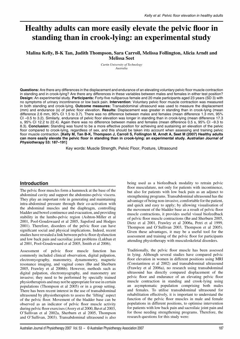
Pelvic floor elevation was measured in two positions. In standing, participants stood in a comfortable position (Figure 1A). In crook-lying, participants lay with their head supported on a pillow, their hips and knees flexed to 60 degrees, and the lumbar spine in neutral (Figure 1B).
Outcome measures
Pelvic floor elevation was measured using transabdominal ultrasound. Participants filled their bladder by consuming 3 to 4 glasses (600–750 ml) of water in half an hour, one hour before the measurement without voiding to allow optimal imaging of the base of the bladder. An Acoustic Imaging Performa ultrasound with a 3.75 MHz curved array transducer(a) was placed in the transverse plane immediately supra-pubically over the lower abdomen. Good intra- and inter-rater reliability of transabdominal ultrasound has been reported for measurement of bladder base movement during voluntary pelvic floor muscle contraction in both the transverse and sagittal plane; but there is poor agreement between the measurements in the sagittal and transverse plane, necessitating the use of one plane of measurement (Sherburn et al 2005). The reliability of pelvic floor muscle contraction has been established previously with intraclass correlation of 0.93 and standard error of measurement of 0.1 mm (Thompson et al 2005).
The ultrasound transducer was placed on the supra-pubic region in the transverse plane and angled in a posterior/caudal direction. The angle of the transducer was adjusted in each participant to obtain the clearest image of the inferior–posterior aspect of the bladder. This angle varied depending
Australian Journal of Physiotherapy 2007 Vol. 53 – © Australian Physiotherapy Association 2007188
Research
Figure1. Set up of the testing positions: (A) in standing and (B) in crook-lying. In both positions, the ultrasound image was in view to provide visual feedback.
A B
Australian Journal of Physiotherapy 2007 Vol. 53 – © Australian Physiotherapy Association 2007 189
Kelly et al: Pelvic floor elevation in healthy adults
on the fullness of participants’ bladders. Participants were directed to perform a pelvic floor muscle contraction using a standardised instruction: ‘Draw in through your pelvic floor muscles as best as you can whilst breathing normally’. A marker ‘X’ was placed on the image of the central portion of the bladder base at the junction of the hyper- and hypo-echoic structures in the region of the greatest displacement visualised during a pelvic floor muscle contraction using the technique previously described by Sherburn et al (2005).
For displacement of pelvic floor elevation, participants were asked to perform three pelvic floor muscle contractions and each image was captured at the point of maximal displacement and again marked with an ‘X’. Displacement was measured as the distance between the two ‘X’ points (mm). Participants held the contraction for the time it took to take the measurement which was no more than three seconds. If the base of the bladder was found to descend during a pelvic floor muscle contraction, the displacement was given a negative value.
For endurance of pelvic floor elevation, participants who were able to achieve elevation of the base of the bladder were then required to contract and hold the pelvic floor in the elevated position whilst breathing normally for a maximum of 60 seconds, timed using a stopwatch. Between each timed contraction, the participants were given a rest period equal to the duration of the hold time achieved (for example, a 60 second contraction was given a 60 second rest) to allow adequate recovery of the muscles. This process was repeated three times. If participants were unable to elevate the pelvic floor in one of the positions, their duration score for this position was entered as zero. However, if they were unable to elevate the pelvic floor in both positions, they were withdrawn from the study.
Data analysis
The mean of the three trials completed for each position was used for the analysis. Preliminary testing revealed that all Levene’s tests were non-significant, indicating that conditions for homogeneity of variance had been met. A two-way ANOVA with repeated measures was employed to compare position and sex differences for each of the dependent variables; displacement and endurance. The alpha level was set at 0.05. Effect sizes were calculated using the technique recommended by Field (2005) while power analyses were conducted on non-significant findings. As floor and ceiling effects were noted in the endurance data, nonparametric analyses (Wilcoxon test for displacement and Mann-Whitney U for sex) were used to confirm the ANOVA
results. The relationship between displacement and hold times in the two positions was assessed using Spearman’s Rank correlation.
Results
Characteristics of participants
Seventy-four healthy university students, 22 males and 52 nulliparous females, were recruited. Two male and seven female participants were excluded from the study: one for an excessively full bladder, which did not allow optimal viewing of the pelvic floor, one for having back pain that limited normal functional activities, one could not perform an elevating contraction in either crook-lying or standing, and six participants reported a recent event of urinary incontinence as determined by questionnaire. Thus the data of 65 participants aged 23 years (SD 3) were used for this study – 20 males aged 23 years (SD 5) and 45 females aged 23 years (SD 3).
Difference in displacement and endurance between standing and crook-lying
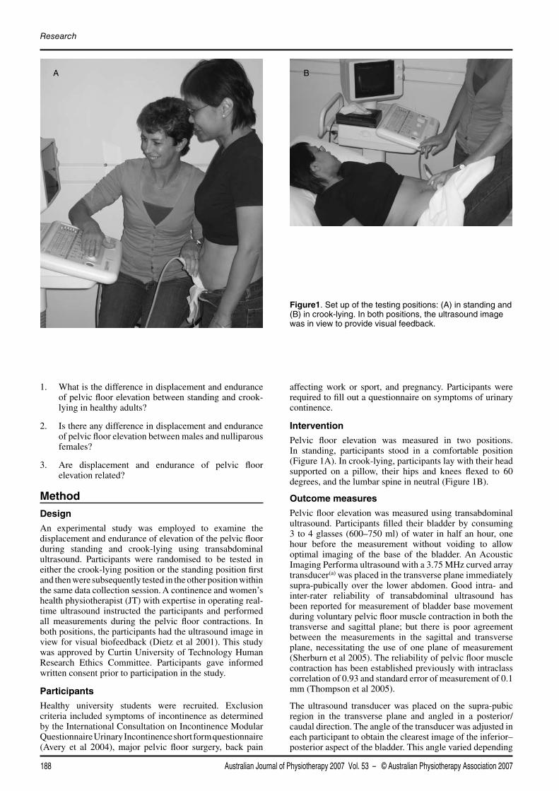
Position had a significant effect on both displacement and endurance of pelvic floor elevation (Table 1). Displacement was 2.6 mm (95% CI 1.5 to 3.7) greater in standing than in crook-lying. Endurance was 17.3 s (95% CI 12.2 to 22.4, p < 0.001) longer in standing than crook-lying.
Difference in displacement and endurance between sexes
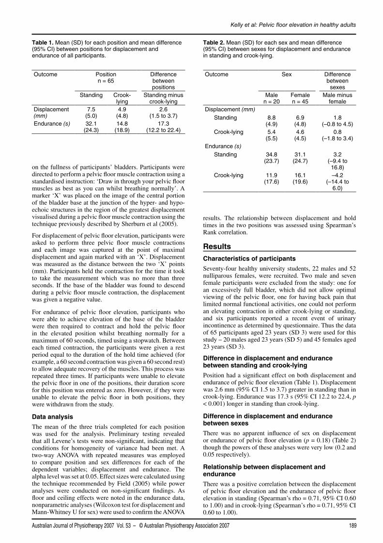
There was no apparent influence of sex on displacement or endurance of pelvic floor elevation (p = 0.18) (Table 2) though the powers of these analyses were very low (0.2 and 0.05 respectively).
Relationship between displacement and endurance
There was a positive correlation between the displacement of pelvic floor elevation and the endurance of pelvic floor elevation in standing (Spearman’s rho = 0.71, 95% CI 0.60 to 1.00) and in crook-lying (Spearman’s rho = 0.71, 95% CI 0.60 to 1.00).
Table 1. Mean (SD) for each position and mean difference (95% CI) between positions for displacement and endurance of all participants.
Outcome Position n = 65
Difference between positions
Standing Crook-lying
Standing minus crook-lying
Displacement (mm)
7.5 (5.0)
4.9 (4.8)
2.6 (1.5 to 3.7)
Endurance (s) 32.1 (24.3)
14.8 (18.9)
17.3 (12.2 to 22.4)
Table 2. Mean (SD) for each sex and mean difference (95% CI) between sexes for displacement and endurance in standing and crook-lying.
Outcome Sex Difference between
sexesMale
n = 20Female n = 45
Male minus female
Displacement (mm) Standing 8.8
(4.9)6.9
(4.8)1.8
(–0.8 to 4.5) Crook-lying 5.4
(5.5)4.6
(4.5)0.8
(–1.8 to 3.4)Endurance (s) Standing 34.8
(23.7)31.1
(24.7)3.2
(–9.4 to 16.8)
Crook-lying 11.9 (17.6)
16.1 (19.6)
–4.2 (–14.4 to
6.0)
Australian Journal of Physiotherapy 2007 Vol. 53 – © Australian Physiotherapy Association 2007190
Research
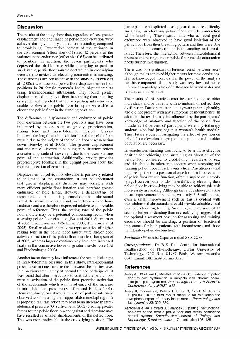
Discussion
The results of the study show that, regardless of sex, greater displacement and endurance of pelvic floor elevation were achieved during voluntary contraction in standing compared to crook-lying. Twenty-five percent of the variance in the displacement (effect size 0.51) and 42 percent of the variance in the endurance (effect size 0.65) can be attributed to position. In addition, the seven participants who depressed the bladder base while attempting to perform an elevating pelvic floor muscle contraction in crook-lying were able to achieve an elevating contraction in standing. These findings are consistent with the study by Frawley et al (2006a) who assessed pelvic floor displacement in four positions in 20 female women’s health physiotherapists using transabdominal ultrasound. They found greater displacement of the pelvic floor in standing than in sitting or supine, and reported that the two participants who were unable to elevate the pelvic floor in supine were able to elevate the pelvic floor in the standing position.
The difference in displacement and endurance of pelvic floor elevation between the two positions may have been influenced by factors such as gravity, proprioception, resting tone and intra-abdominal pressure. Gravity improves the length-tension relationship of the pelvic floor muscle due to the weight of the pelvic floor viscera acting down (Frawley et al 2006a). The greater displacement and endurance achieved in standing may therefore reflect a greater amplitude of movement due to the lower starting point of the contraction. Additionally, gravity provides proprioceptive feedback in the upright position about the required direction of contraction.
Displacement of pelvic floor elevation is positively related to endurance of the contraction. It can be speculated that greater displacement distances may correlate with more efficient pelvic floor function and therefore greater endurance or hold times. However a disadvantage of measurements made using transabdominal ultrasound is that the measurements are not taken from a fixed bony landmark and are therefore expressed relative to a moveable point of reference. Thus, the resting tone of the pelvic floor muscle may be a potential confounding factor when assessing pelvic floor elevation (Bø et al 2003, Sherburn et al 2005, Thompson and O’Sullivan 2003, Thompson et al 2005). Smaller elevations may be representative of higher resting tone in the pelvic floor musculature and/or poor active contraction of the pelvic floor muscle (Thompson et al 2005) whereas larger elevations may be due to increased laxity in the connective tissue or greater muscle force (Bø and Finckenhagen 2003).
Another factor that may have influenced the results is changes in intra-abdominal pressure. In this study, intra-abdominal pressure was not measured as the aim was to be non-invasive. In a previous small study of normal trained participants, it was found that after instructions to contract the pelvic floor muscle, activation of the pelvic floor preceded activation of the abdominals which was in advance of the increase in intra-abdominal pressure (Sapsford and Hodges 2001). However, during our study, a number of participants were observed to splint using their upper-abdomen/diaphragm. It is proposed that this action may lead to an increase in intra-abdominal pressure (O’Sullivan et al 2002) creating greater forces for the pelvic floor to work against and therefore may have resulted in smaller displacements of the pelvic floor. This was more noticeable in the crook-lying position. The
participants who splinted also appeared to have difficulty sustaining an elevating pelvic floor muscle contraction whilst breathing. Those participants who achieved good endurance were observed to have good isolation of the pelvic floor from their breathing pattern and thus were able to maintain the contraction in both standing and crook-lying. Therefore, the interaction between intra-abdominal pressure and resting tone on pelvic floor muscle contraction needs further investigation.
There was no significant difference found between sexes although males achieved higher means for most conditions. It is acknowledged however that the power of the analysis for this component of the study was very low and hence inferences regarding a lack of difference between males and females cannot be made.
The results of this study cannot be extrapolated to older individuals and/or patients with symptoms of pelvic floor dysfunction. Participants in this study were generally healthy and did not present with any symptoms of incontinence. In addition, the results may be influenced by the participants’ knowledge of anatomy and function of the pelvic floor muscle as 88 percent of participants were physiotherapy students who had just begun a women’s health module. Thus, future studies investigating the effect of position on pelvic floor elevation in symptomatic patients and in older population are necessary.
In conclusion, standing was found to be a more effective position for achieving and sustaining an elevation of the pelvic floor compared to crook-lying, regardless of sex, and this should be taken into account when assessing and training pelvic floor muscle contraction. Practitioners tend to place a patient in a position of ease for initial assessments of pelvic floor muscle function, often in supine or in crook-lying. However patients who have difficulty elevating their pelvic floor in crook-lying may be able to achieve this task more easily in standing. Although this study showed that the mean improvement in standing was only 2–3 millimetres, even a small improvement such as this is evident with transabdominal ultrasound and could provide valuable visual biofeedback during training. Similarly, an endurance of 17 seconds longer in standing than in crook-lying suggests that the optimal assessment position for assessing and training endurance should be standing, a position of functional importance for both patients with incontinence and those with lumbo-pelvic dysfunction.
Footnotes: (a)Toshiba Capasee model SSA 220A.
Correspondence: Dr B-K Tan, Centre for International Health/School of Physiotherapy, Curtin University of Technology, GPO Box U1987 Perth, Western Australia 6845. Email: [email protected]
ReferencesAvery A, O’Sullivan P, MacCallum M (2000) Evidence of pelvic
floor muscle dysfunction in subjects with chronic sacro-iliac joint pain syndrome. Proceedings of the 7th Scientific Conference of the IFOMT, p.35.
Avery K, Donovan J, Peters T, Shaw C, Gotoh M, Abrams P (2004) ICIQ: a brief robust measure for evaluation the symptoms impact of urinary incontinence. Neurourology and Urodynamics 23: 322–330.
Ashton-Miller JA, Howard D, Delancey JO (2001) The functional anatomy of the female pelvic floor and stress continence control system. Scandinavian Journal of Urology and Nephrology. Supplementum: 1–7; discussion 106–125.
Bø K, Finckenhagen HB (2003) Is there any difference in measurement of pelvic floor muscle strength in supine and standing position? ACTA Obstetricia et Gynecologica Scandinavica 82: 1120–1124.
Bø K, Sherburn M (2005) Evaluation of female pelvic-floor muscle function and strength. Physical Therapy 85: 269–282.
Bø K, Sherburn M, Allen T (2003) Transabdominal ultrasound measurement of pelvic floor muscle activity when activated directly or via a transversus abdominis muscle contraction. Neurourology and Urodynamics 22: 582–588.
Constantinou CE, Hvistendahl G, Ryhammer A, Nagel LL, Djurhuus JC (2002) Determining the displacement of the pelvic floor and pelvic organs during voluntary contractions using magnetic resonance imaging in younger and older women. BJU International 90: 408–414.
Dietz HP, Wilson PD, Clarke B (2001) The use of perineal ultrasound to quantify levator activity and teach pelvic floor muscle exercises. International Urogynecology Journal and Pelvic Floor Dysfunction 12: 166–168; discussion 168–169.
Field A (2005) Discovering Statistics Using SPSS (2nd edn). London: Sage.
Frawley HC, Galea MP, Phillips BA, Sherburn M, Bø K (2006a) Effect of test position on pelvic floor muscle assessment. International Urogynecology Journal and Pelvic Floor Dysfunction 17: 365–371.
Frawley HC, Galea MP, Phillips BA, Sherburn M, Bø K (2006b) Reliability of pelvic floor muscle strength assessment using different test positions and tools. Neurourology and Urodynamics 25: 236–242.
Lukban J, Whitmore K, Kellogg-Spadt S, Bologna R, Lesher A, Fletcher E (2001) The effect of manual physical therapy in patients diagnosed with interstitial cystitis, high-tone pelvic floor dysfunction, and sacroiliac dysfunction. Urology 57: 121–122.
O’Sullivan PB, Beales DJ, Beetham JA, Cripps J, Graf F, Lin IB, Tucker B, Avery A (2002) Altered motor control strategies in subjects with sacroiliac joint pain during the active straight-leg-raise test. Spine 27: E1–8.
Petri E, Koelbl H, Schaer G (1999) What is the place of ultrasound in urogynecology? A written panel. International Urogynecology Journal and Pelvic Floor Dysfunction 10: 262–273.
Pool-Goudzwaard AL, Slieker Ten Hove MC, Vierhout ME, Mulder PH, Pool JJ, Snijders CJ and Stoeckart R (2005) Relations between pregnancy-related low back pain, pelvic floor activity and pelvic floor dysfunction. International Urogynecology Journal and Pelvic Floor Dysfunction 16: 468–474.
Sapsford R, Hodges P (2001) Contraction of the pelvic floor muscles during abdominal manoeuvres. Archives of Physical Medicine and Rehabilitation 82: 1081–1088.
Sherburn M, Murphy CA, Carroll S, Allen TJ, Galea MP (2005) Investigation of transabdominal real-time ultrasound to visualise the muscles of the pelvic floor. Australian Journal of Physiotherapy 51: 167–170.
Smith M, Russell A, Hodges P (2006) Disorders of breathing and continence have a stronger association with back pain than obesity and physical activity. Australian Journal of Physiotherapy 52: 11–16.
Thompson J, O’Sullivan P (2003) Levator plate movement during voluntary pelvic floor muscle contraction in subjects with incontinence and prolapse: a cross–sectional study and review. International Urogynecology Journal and Pelvic Floor Dysfunction 14: 84–88.
Thompson J, O’Sullivan P, Briffa K, Neumann P, Court S (2005) Assessment of pelvic floor movement using transabdominal and transperineal ultrasound. International Urogynecology Journal and Pelvic Floor Dysfunction 16: 285–292.
Australian Journal of Physiotherapy 2007 Vol. 53 – © Australian Physiotherapy Association 2007 191
Kelly et al: Pelvic floor elevation in healthy adults
Statement regarding registration of clinical trials from the Editorial Board of Australian Journal of Physiotherapy
This journal is moving towards requiring that clinical trials whose results are submitted for publication in Australian Journal of Physiotherapy are registered. From January 2008, all clinical trials submitted to the journal must have been registered prospectively in a publicly-accessible trials register. We will accept any register that satisfies the International Committee of Medical Journal Editors requirements. Authors must provide the name and address of the register and the trial registration number on submission.