Embed Size (px)
Citation preview
A Handheld Point-of-Care Genomic Diagnostic SystemFrank B. Myers1,2, Richard H. Henrikson1,2, Jennifer Bone3, Luke P. Lee1,2*
1Department of Bioengineering, University of California, Berkeley, California, United States of America, 2 Berkeley Sensor and Actuator Center, University of California,
Berkeley, California, United States of America, 3Department of Physics, University of California, Berkeley, California, United States of America
Abstract
The rapid detection and identification of infectious disease pathogens is a critical need for healthcare in both developedand developing countries. As we gain more insight into the genomic basis of pathogen infectivity and drug resistance,point-of-care nucleic acid testing will likely become an important tool for global health. In this paper, we present aninexpensive, handheld, battery-powered instrument designed to enable pathogen genotyping in the developing world. OurMicrofluidic Biomolecular Amplification Reader (mBAR) represents the convergence of molecular biology, microfluidics,optics, and electronics technology. The mBAR is capable of carrying out isothermal nucleic acid amplification assays withreal-time fluorescence readout at a fraction of the cost of conventional benchtop thermocyclers. Additionally, the mBARfeatures cell phone data connectivity and GPS sample geotagging which can enable epidemiological surveying and remotehealthcare delivery. The mBAR controls assay temperature through an integrated resistive heater and monitors real-timefluorescence signals from 60 individual reaction chambers using LEDs and phototransistors. Assays are carried out on PDMSdisposable microfluidic cartridges which require no external power for sample loading. We characterize the fluorescencedetection limits, heater uniformity, and battery life of the instrument. As a proof-of-principle, we demonstrate the detectionof the HIV-1 integrase gene with the mBAR using the Loop-Mediated Isothermal Amplification (LAMP) assay. Although wefocus on the detection of purified DNA here, LAMP has previously been demonstrated with a range of clinical samples, andour eventual goal is to develop a microfluidic device which includes on-chip sample preparation from raw samples. ThemBAR is based entirely around open source hardware and software, and in the accompanying online supplement wepresent a full set of schematics, bill of materials, PCB layouts, CAD drawings, and source code for the mBAR instrument withthe goal of spurring further innovation toward low-cost genetic diagnostics.
Citation: Myers FB, Henrikson RH, Bone J, Lee LP (2013) A Handheld Point-of-Care Genomic Diagnostic System. PLoS ONE 8(8): e70266. doi:10.1371/journal.pone.0070266
Editor: Nitika Pant Pai, McGill University Health Centre, McGill University, Canada
Received December 25, 2012; Accepted June 18, 2013; Published August 1, 2013
Copyright: � 2013 Myers et al. This is an open-access article distributed under the terms of the Creative Commons Attribution License, which permitsunrestricted use, distribution, and reproduction in any medium, provided the original author and source are credited.
Funding: This work was supported by the DSO of Defense Advanced Research Project Agency (DxOD project) and the Bill & Melinda Gates Foundation (GrandChallenges). Additional support was provided by the NDSEG fellowship and the Siebel Scholarship. The funders had no role in study design, data collection andanalysis, decision to publish, or preparation of the manuscript.
Competing Interests: The authors have declared that no competing interests exist.
* E-mail: [email protected]
Introduction
Integrated microfluidic diagnostic systems present an unprece-
dented opportunity to facilitate high-quality, low-cost healthcare in
remote and resource-limited settings [1]. These systems show
promise for both acute infection discovery and chronic disease
monitoring and management [2]. In particular, a wide array of
nucleic acid tests (NATs) are emerging to identify pathogen species
as well as specific clinically-relevant characteristics such as
pathogenicity, origin, and drug susceptibility [3], [4]. The
importance of rapid diagnostics for infectious diseases is
highlighted by the threat of increasing drug resistance due to
indiscriminate treatment where a rapid test could improve patient
outcome and reduce the emergence of more deadly strains [5], [6].
Although developed economies have access to more accurate
molecular diagnostics, including polymerase chain reaction (PCR)
tests, these tests have yet to be translated into diagnostics that meet
the specific needs and requirements of point-of-care testing.
Lateral flow immunoassays have made a significant impact in the
diagnosis of diseases like HIV, but it is difficult to adapt this assay
format to situations where a quantitative readout or molecular
amplification is required. Recent efforts have focused on
automating standard sample preparation and PCR techniques;
however the significant assay cost and relatively low throughput
remain prohibitive barriers for wide-scale adoption [7].
Recently, several methods have been developed to enable
isothermal amplification, facilitating reduced power consumption
and expanded options for material and chemical selection [8].
Some leading isothermal nucleic acid amplification techniques
include Loop-Mediated Isothermal Amplification (LAMP) [9],
Recombinase Polymerase Amplification (RPA) [10], and Rolling
Circle Amplification (RCA) [11], among others. Furthermore,
these methods frequently offer improved performance over
standard PCR, achieving highly sensitive and specific results in
as little as 15 minutes. LAMP may be particularly well-suited for
blood-based detection because Bst polymerase is not inhibited by
hemoglobin, whereas Taq polymerase used in PCR is [12]. This
may allow LAMP to work with much ‘‘dirtier’’ samples.
Furthermore, the amount of DNA produced by LAMP (and
hence the fluorescence signal) is considerably higher than that
produced by PCR, allowing it to be used with inexpensive
detection hardware.
A number of recent efforts have aimed to miniaturize isothermal
amplification diagnostic assays for use in resource-poor settings
[8,13]. However, no isothermal amplification system yet provides
sample-in, answer-out capability. All of these systems presented to
PLOS ONE | www.plosone.org 1 August 2013 | Volume 8 | Issue 8 | e70266
date work with conventional PCR tubes which limits the degree to
which they can be expanded to include multi-step assays including
sample prep and multiplexed detection. Microfluidic assay
cartridges offer many compelling advantages over conventional
tubes, most notably the degree to which multiple assay steps can be
incorporated on one integrated device. Our goal in the present
work is to introduce a platform for isothermal amplification and
real-time detection on a multiplexed microfluidic cartridge. Such a
cartridge could be coupled with some of the microfluidic sample
preparation techniques already presented [14], [15], yielding
sample-in, answer-out diagnostic at a significantly reduced cost as
compared with PCR.
Loop-mediated isothermal amplification (LAMP) has been
adapted for a range of assays, including infectious pathogen
detection and genotyping of single nucleotide polymorphisms
(SNPs) [16]. There has recently been interest in leveraging this
assay in a miniaturized format, however a complete integrated
system for multiplexed sample analysis has yet to be established.
Fluorescent analysis offers a number of advantages over competing
optical or electrochemical detection methods, however imple-
menting sensitive fluorescence detection inexpensively remains a
challenge [17]. We have developed a microfluidic biomolecular
amplification reader (mBAR), coupled with a disposable assay
cartridge, for efficient and inexpensive disease analysis compatible
with isothermal amplification techniques [18]. We have charac-
terized the performance of the mBAR system and conducted initial
tests for LAMP-based detection of HIV (via the integrase gene).
Materials and Methods
A. Microfluidic CartridgeWe have developed a disposable microfluidic device which
passive degas-driven fluid actuation (Figure 1A) [14]. The chip
measures 5564265 mm and consists of 96 parallel reaction
chambers at 4.5 mm pitch supplied by 6 separate sample inlets for
multiplexed analysis. Each circular reaction chamber is 2 mm in
diameter and 200 mm in height, for a total volume of 628 nL.
The chambers are cast in polydimethylsiloxane (PDMS) using
standard soft lithography techniques [19] and bonded to a thin
transparent elastomer base using oxygen plasma [20]. The chip is
UV sterilized for 24 hours after bonding. After bonding, the device
is exposed to a vacuum of ,300 mTorr for .1 hour, and then
vacuum sealed using a commercial food sealer. Due to the
elastomeric pore structure of the PDMS, the chip will begin
drawing in sample introduced into the inlets immediately upon
exposure to atmospheric pressure. Samples can be completely
loaded in less than 1 hour (see Movie S1). This time can be
significantly reduced with smaller reaction chambers. Chips can be
stored vacuum sealed for at least one month (likely much longer)
without loss of vacuum. This degas-driven loading method enables
us to use dead-end reaction chambers which help ensure that
amplicons remain on chip and reduce the risk of contamination
between amplification runs. Although we have used the LAMP
method in the design of these primer sets, this chip is compatible
with other nucleic acid amplification techniques.
B. LAMP AssayWe have adapted the calcein metal indicator fluorophore as a
tool for both real-time fluorescence measurement and naked-eye
readout of signal based on initial target presence. Briefly, this
method involves the use of a calcein dye that is initially quenched
by manganese ions. As a byproduct of nucleic acid amplification,
pyrophosphate groups are produced in abundance and readily
precipitate out of solution as manganese pyrophosphate, removing
the quencher from calcein and yielding a bright fluorescent signal
[21].
For our proof-of-principle amplification assay, all reagents were
mixed under sterile conditions immediately prior to loading them
on the chip. A total reaction volume of 20 mL was loaded into each
inlet on the chip (this gets distributed among 16 reaction wells).
The same sample was simultaneously used with the PCR
thermocycler (20 mL reaction volume). The reaction mix consisted
of: 1.6 mM each of FIP/BIP primers, 0.8 mM each of Loop-F/
Loop-B primers, 0.2 mM each of F3/B3 primers, 27.3 mMCalcein, 0.8 mM Bst DNA Polymerase, 1.4 mM dNTPs, 20 mM
Tris Buffer, 10 mM KCl, 8 mM MgSO4, 1 mM (NH4)2SO4,
1 mM Tween-20, 0.8 M Betaine, and 1.49 mM MnCl2. The
sample template (purified DNA) was added last.
C. InstrumentFor quantitative readout of the assay, the chip is inserted into a
battery-powered instrument which maintains assay temperature,
illuminates the chip, and detects fluorescence emission from the
reaction chambers using an array of phototransistors (Figure 1C–
F). It does this without the use of costly optical components, and
without the need for alignment or focusing. The instrument is
automated with an Atmel Atmega2560 microcontroller imple-
menting the Arduino bootloader and USB interface. The
instrument features a Secure Digital (SD) Flash memory card
reader for storing assay parameters and results, and a 4.3 inch
color touchscreen user interface (Amulet Technologies). Addition-
ally, the instrument includes a GSM cell phone module and a GPS
module, thus enabling epidemiological surveillance and medical
coordination in remote locations. A block diagram is provided in
Figure 2.
To run an assay, the microfluidic chip is inserted directly on top
of a 47667 mm indium tin oxide (ITO) coated glass slide which
heats the chip to 60uC. The ITO is connected to a TPS61085
boost converter which delivers 1 A through the slide. The chip
reaches its set point temperature in less than 20 minutes. The
temperature is maintained by toggling the boost circuit with a solid
state relay. A thermistor in a half-bridge circuit configuration
provides temperature feedback. This thermistor is mounted
directly underneath the ITO slide. For LAMP and other
isothermal reactions, the temperature is held constant, whereas
for PCR it can be programmed to cycle.
Three blue InGaN LEDs (peak = 472 nm) illuminate the chip
from the top side through rectangular glass waveguides cladded
with black paint to minimize stray light. The waveguides promote
total internal reflection (TIR) of excitation light within the chip.
The refractive index of glass, PDMS, and water are similar enough
that reflections of excitation light off of internal surfaces of the chip
do not significantly contribute to background signal on the
phototransistors. The LEDs are driven by a second boost circuit
based on the MIC3289 which delivers a controlled current to the
LEDs which does not vary with battery charge state. This chip
provides 16 logarithmically-spaced intensity levels which are
digitally selected.
The calcein used in the LAMP reaction emits green fluores-
cence (peak excitation wavelength = 480 nm, peak emission
wavelength = 515 nm). This fluorescence is detected with a
phototransistor located directly underneath each chamber.
Importantly, there is a small air gap between the phototransistor
housings and the ITO heater, which ensures TIR and reduces
feedthrough of the excitation light into the phototransistors. Each
one of the 96 phototransistor is wired to one of three 32:1 analog
multiplexers (AD732). The microcontroller uses these multiplexers
to raster through the phototransistor array, selecting one at a time
A Handheld Point-of-Care Genomic Diagnostic System
PLOS ONE | www.plosone.org 2 August 2013 | Volume 8 | Issue 8 | e70266
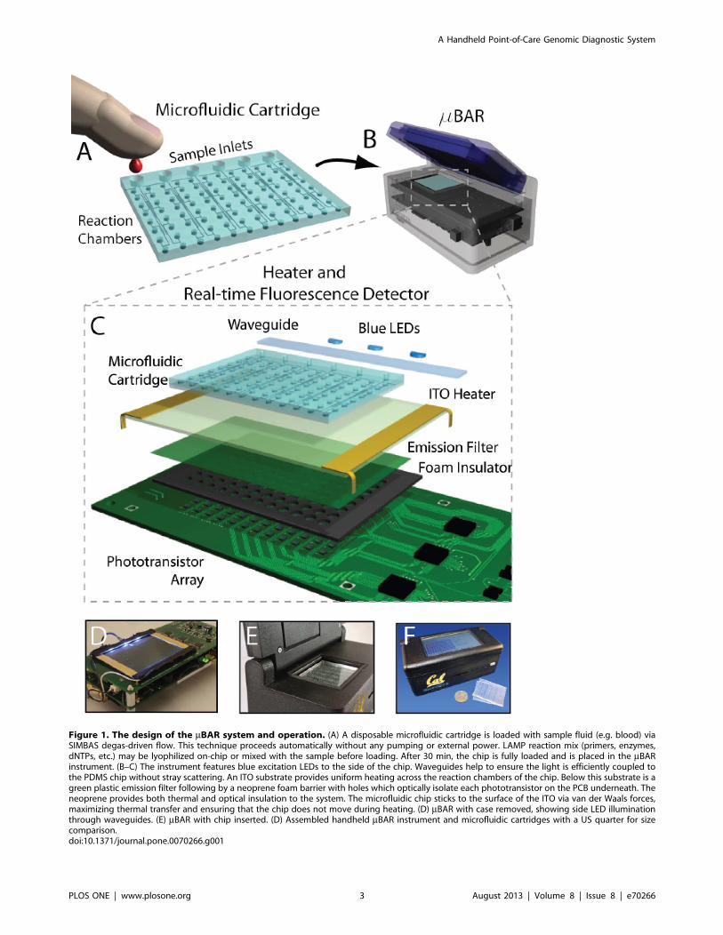
Figure 1. The design of the mBAR system and operation. (A) A disposable microfluidic cartridge is loaded with sample fluid (e.g. blood) viaSIMBAS degas-driven flow. This technique proceeds automatically without any pumping or external power. LAMP reaction mix (primers, enzymes,dNTPs, etc.) may be lyophilized on-chip or mixed with the sample before loading. After 30 min, the chip is fully loaded and is placed in the mBARinstrument. (B–C) The instrument features blue excitation LEDs to the side of the chip. Waveguides help to ensure the light is efficiently coupled tothe PDMS chip without stray scattering. An ITO substrate provides uniform heating across the reaction chambers of the chip. Below this substrate is agreen plastic emission filter following by a neoprene foam barrier with holes which optically isolate each phototransistor on the PCB underneath. Theneoprene provides both thermal and optical insulation to the system. The microfluidic chip sticks to the surface of the ITO via van der Waals forces,maximizing thermal transfer and ensuring that the chip does not move during heating. (D) mBAR with case removed, showing side LED illuminationthrough waveguides. (E) mBAR with chip inserted. (D) Assembled handheld mBAR instrument and microfluidic cartridges with a US quarter for sizecomparison.doi:10.1371/journal.pone.0070266.g001
A Handheld Point-of-Care Genomic Diagnostic System
PLOS ONE | www.plosone.org 3 August 2013 | Volume 8 | Issue 8 | e70266

for interrogation. The entire array is sampled at a specified
interval (2 seconds, typically). The outputs of the multiplexers are
connected to a transimpedance amplifier with an input biased to
2.5V. When a phototransistor is selected, this bias voltage allows
collector current to flow in proportion to the illuminance on the
phototransistor’s surface. Phototransistors, rather than photodi-
odes, were chosen because the leakage current of CMOS analog
multiplexers is significant compared to photodiode currents
leading to crosstalk problems. Phototransistors provide significant-
ly more current for the same illuminance and in this configuration
they are be biased through the multiplexer which eliminates the
possibility of crosstalk. The photocurrent from each phototransis-
tor is sampled over time and recorded to the SD card or to an
attached PC via USB. To render amplification plots, the raw data
is first baseline subtracted. The baseline is estimated based on a
linear regression of the raw data from the first 10–20 minutes of
the assay run (it is assumed that no amplification occurs before 20
minutes). The signal from each well is then normalized by that
well’s predetermined sensitivity coefficient (Figure 3C). Finally, the
signal is smoothed with a moving average span of 100 samples to
improve SNR.
The instrument is powered by a 3.7 V, 2000 mAh lithium
polymer battery. A third boost converter (LTC3525) delivers a 5V
supply to the digital electronics, LCD display, and transimpedance
amplifier. A typical assay run lasts approximately 2 hours and
consumes 500 mAh. The heater dominates power consumption.
Our ultimate goal is to create a fully-integrated, portable
instrument which addresses the needs of remote/resource poor
settings. The specific components included can be tailored for the
situation. At its simplest, this instrument would be a handheld
heater and LED illuminator which would allow qualitative naked
eye readout.
Results
A. Fluorescence SensitivityWe first characterized the fluorescence sensitivity of the mBAR
instrument by introducing different known concentrations of
calcein (the fluorophore used in our LAMP assays) into the
microfluidic device and observing the relative changes in
photoamplifier output across all reaction chambers (Figure 3A).
For this experiment, we developed a slightly modified chip which
included a serpentine channel connecting all of the reaction wells
in series. This allowed us to sequentially introduce higher calcein
concentrations (Figure 3B) while ensuring that the chip place-
ment/geometry was fixed. We repeated this experiment with 3
independent chips (marked with red, blue, and green circles) and
found that detector output as a function of concentration followed
a linear trend over four orders of magnitude. To determine the
sensitivity of the instrument to fluorescence changes, we calculated
the Instrument Detection Limit (IDL) as the concentration that
produces a signal three times greater than the noise standard
deviation of the instrument for a given time resolution. In the case
of realtime nucleic acid amplification signals, a time resolution
(BW21) of 2 min is sufficient. In that case, the noise standard
deviation of the mBAR is 5 mVrms, leading to an IDL of
approximately 600 pM with the mBAR. As most nucleic acid
amplification reactions involve fluorophore concentrations that are
orders of magnitude higher than this (typically 1–10 mM), this is
more than sufficient to carry out these kinds of assays. We further
examined the nonuniformity of photoamplifier sensitivity across
the detection array. Because the LED lighting does not lead to
uniform illumination, and due to some internal scattering within
the chips, the amount of light reaching each reaction chamber is
slightly different. By calculating the linear slope of amplitude
change as a function of concentration change (i.e. sensitivity) at
each well (Figure 3C), we can normalize subsequent assay runs so
that intensities can be directly compared across the chip. Although
Figure 2. Block diagram of the mBAR system. The system is controlled by an ATmega 2560 microcontroller which has been loaded with theArduino bootloader firmware. A GPS receiver and cell phone transceiver facilitate remote field diagnostics and epidemiological studies. The systemcan receive user input via either an LCD touchscreen or PC via USB. Data is stored to an SD card and also broadcast to the PC, if connected. Amonolithic LED driver supplies a constant current to 3 blue InGaAs LEDs (even as battery voltage decreases), and a booster circuit delivers a constantcurrent to the ITO heater. The microcontroller receives temperature feedback from a thermistor and turns the heater on and off accordingly. An arrayof 96 phototransistors are positioned underneath the heater and an analog multiplexer is used to raster across the array and read photocurrents fromeach location using a single photoamplifier.doi:10.1371/journal.pone.0070266.g002
A Handheld Point-of-Care Genomic Diagnostic System
PLOS ONE | www.plosone.org 4 August 2013 | Volume 8 | Issue 8 | e70266

the chip was designed with 96 reaction chambers in a 1268
configuration, we determined that it was not possible to use assay
results from the outermost chambers due to excitation light
scattering at the edges of the device. This limitation could easily be
overcome in subsequent designs (see discussion). We therefore
restrict our analysis to the 60 innermost reaction chambers (6610
array).
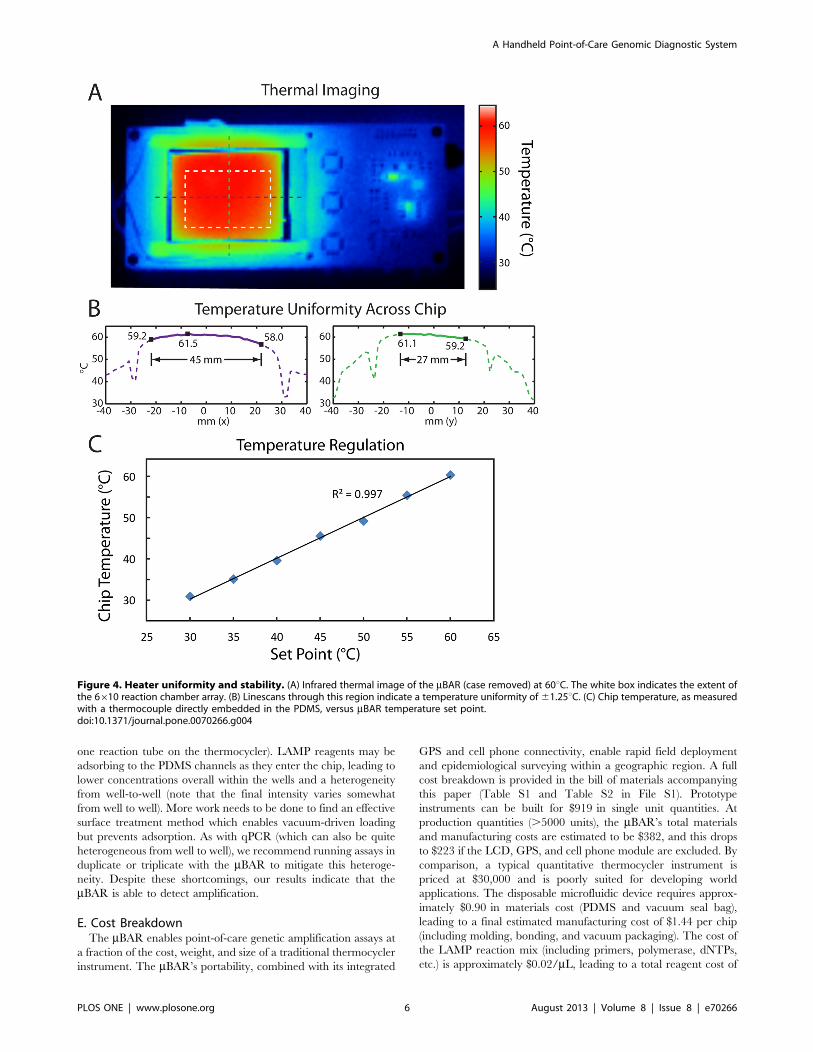
B. Heater PerformanceWe next examined the spatial uniformity and temperature
regulation performance of the mBAR’s internal ITO substrate
heater. We used an infrared thermal camera to examine the
surface of a PDMS device on top of the ITO heater with the case
removed (Figure 4A). For the 6610 array of reaction chambers in
the center of the chip, we found a temperature distribution of
6061.25uC (see linescans, Figure 4B). By comparison, modern
mid-range thermocyclers have a well-to-well temperature nonuni-
formity of 1–2uC during a plateau phase [22]. We then embedded
a commercial thermocouple temperature probe (Fluke 87V) within
the PDMS microfluidic device and calibrated the on-board
temperature feedback (from a thermistor positioned on the
underside of the ITO heater) with the actual chip temperature.
Following calibration, we found excellent agreement between the
mBAR’s temperature setpoint and the independently measured
chip temperature (Figure 4C).
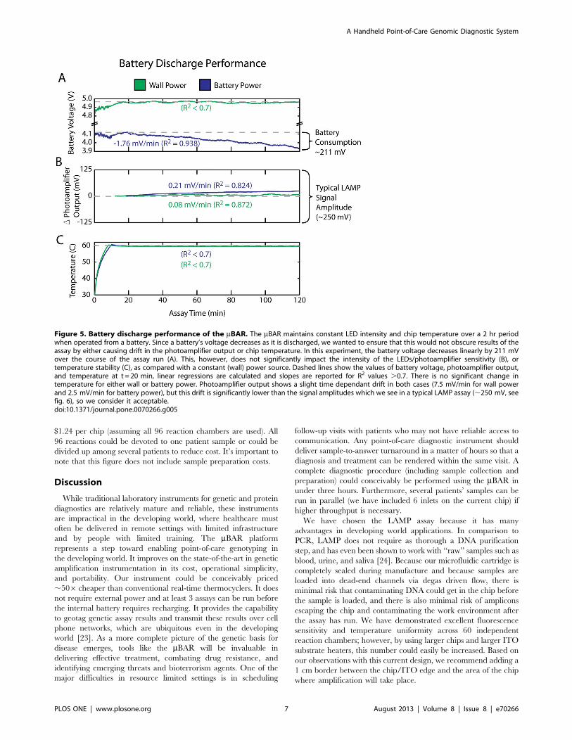
C. Battery LifeThe mBAR can either be operated from a 5V DC wall power
source or from a lithium polymer battery (3.7V nominal voltage).
During battery operation, the battery discharge leads to a
decreasing battery voltage, and it’s important that this not
significantly affect LED intensity, photoamplifier sensitivity, or
temperature over the course of a typical 2 hour assay run, because
any time-varying signal might obscure the assay results. Using a
chip with a constant dye concentration, we directly compared
photoamplifier output and temperature for wall power and battery
power (Figure 5). We found that both wall power and battery
power lead to a small linear drift in photoamplifier output (0.21
and 0.08 mV/min, respectively) which was well below the
amplitude of a typical LAMP assay (.10 mV/min, see Figure 6).
Both wall power and battery power resulted in a steady
temperature of 60C 60.1 (standard deviation).
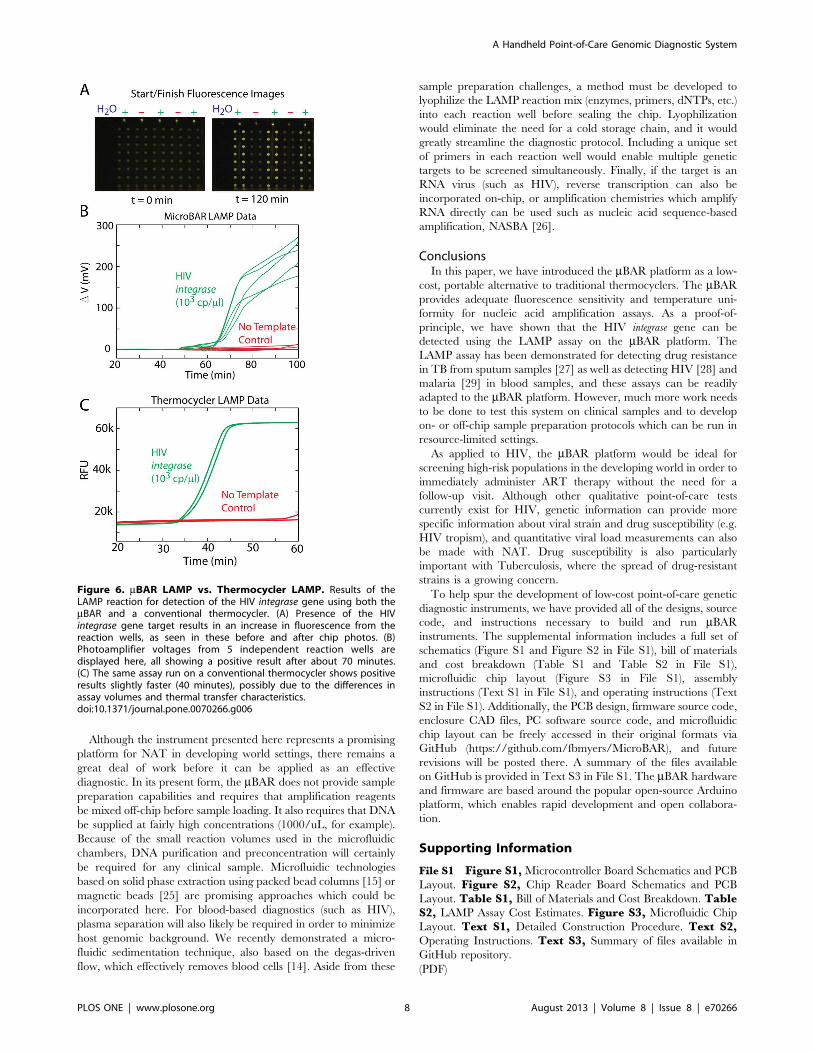
D. LAMP Genetic Amplification Assay PerformanceFigure 6 shows the results of a loop-mediated isothermal
amplification (LAMP) assay for the HIV integrase gene using our
microfluidic chip and electronic reader. Two independent samples
are shown, one containing a plasmid with the target gene at a
concentration of 103 copies/mL, and the other containing no DNA
(no template control, NTC). The chips were vacuum treated at
300 mTorr, samples were dropped onto the chip (20 mL per inlet),
and the chip was placed into the mBAR instrument upon
completion of sample loading. Figure 6A shows before and after
pictures of the microfluidic device under fluorescent illumination
(490 nm excitation with a long-pass filter). Amplification is clearly
visible in the chambers with the target HIV integrase plasmid as
compared with the NTC chambers. Figure 6B shows that within
70 minutes, the positive samples exhibit readily detectable signals
above the NTC background. When run on a conventional
quantitative PCR thermocycler (Bio-Rad CFX96), the amplifica-
tion occurs somewhat earlier (around 40 minutes). This discrep-
ancy in time may be due to the differences in reaction volumes and
surface-to-volume ratios of the two assays (each on-chip chamber
contains about 1.2 mL sample volume, whereas the full 20 mL is in
Figure 3. Fluorescence sensitivity and linearity. (A) Differentconcentrations of calcein were introduced into a microfluidic cartridgeand the corresponding change in photoamplifier voltage was recorded.The circles indicate the mean signal amplitude across all reaction wellsfor three different chips (red, green, and blue). As expected, therelationship between calcein concentration and photoamplifier outputis linear (R2 = 0.983). Limit of detection can be evaluated by determiningwhen this trend line crosses the noise floor of the instrument at a givenbandwidth. At a time resolution (BW21) of 2 min, the mBAR candistinguish changes in calcein concentration of 30 pM, which is ordersof magnitude below the fluorophore concentrations typically used innucleic acid amplification reactions. (B) 50 mM steps of calcein areclearly visible in the photoamplifier output and this output remainsstable for a constant concentration. (C) Due to the nature of theillumination scheme and the lack of optics, there is some nonuniformityof sensitivity across the 6610 reaction chamber array. However, we takethis into account and normalize each well by its mean sensitivity, shownhere. In this graphic, the illumination LEDs and sample inlets are locatedabove the topmost row.doi:10.1371/journal.pone.0070266.g003
A Handheld Point-of-Care Genomic Diagnostic System
PLOS ONE | www.plosone.org 5 August 2013 | Volume 8 | Issue 8 | e70266
one reaction tube on the thermocycler). LAMP reagents may be
adsorbing to the PDMS channels as they enter the chip, leading to
lower concentrations overall within the wells and a heterogeneity
from well-to-well (note that the final intensity varies somewhat
from well to well). More work needs to be done to find an effective
surface treatment method which enables vacuum-driven loading
but prevents adsorption. As with qPCR (which can also be quite
heterogeneous from well to well), we recommend running assays in
duplicate or triplicate with the mBAR to mitigate this heteroge-
neity. Despite these shortcomings, our results indicate that the
mBAR is able to detect amplification.
E. Cost BreakdownThe mBAR enables point-of-care genetic amplification assays at
a fraction of the cost, weight, and size of a traditional thermocycler
instrument. The mBAR’s portability, combined with its integrated
GPS and cell phone connectivity, enable rapid field deployment
and epidemiological surveying within a geographic region. A full
cost breakdown is provided in the bill of materials accompanying
this paper (Table S1 and Table S2 in File S1). Prototype
instruments can be built for $919 in single unit quantities. At
production quantities (.5000 units), the mBAR’s total materials
and manufacturing costs are estimated to be $382, and this drops
to $223 if the LCD, GPS, and cell phone module are excluded. By
comparison, a typical quantitative thermocycler instrument is
priced at $30,000 and is poorly suited for developing world
applications. The disposable microfluidic device requires approx-
imately $0.90 in materials cost (PDMS and vacuum seal bag),
leading to a final estimated manufacturing cost of $1.44 per chip
(including molding, bonding, and vacuum packaging). The cost of
the LAMP reaction mix (including primers, polymerase, dNTPs,
etc.) is approximately $0.02/mL, leading to a total reagent cost of
Figure 4. Heater uniformity and stability. (A) Infrared thermal image of the mBAR (case removed) at 60uC. The white box indicates the extent ofthe 6610 reaction chamber array. (B) Linescans through this region indicate a temperature uniformity of61.25uC. (C) Chip temperature, as measuredwith a thermocouple directly embedded in the PDMS, versus mBAR temperature set point.doi:10.1371/journal.pone.0070266.g004
A Handheld Point-of-Care Genomic Diagnostic System
PLOS ONE | www.plosone.org 6 August 2013 | Volume 8 | Issue 8 | e70266
$1.24 per chip (assuming all 96 reaction chambers are used). All
96 reactions could be devoted to one patient sample or could be
divided up among several patients to reduce cost. It’s important to
note that this figure does not include sample preparation costs.
Discussion
While traditional laboratory instruments for genetic and protein
diagnostics are relatively mature and reliable, these instruments
are impractical in the developing world, where healthcare must
often be delivered in remote settings with limited infrastructure
and by people with limited training. The mBAR platform
represents a step toward enabling point-of-care genotyping in
the developing world. It improves on the state-of-the-art in genetic
amplification instrumentation in its cost, operational simplicity,
and portability. Our instrument could be conceivably priced
,506cheaper than conventional real-time thermocyclers. It does
not require external power and at least 3 assays can be run before
the internal battery requires recharging. It provides the capability
to geotag genetic assay results and transmit these results over cell
phone networks, which are ubiquitous even in the developing
world [23]. As a more complete picture of the genetic basis for
disease emerges, tools like the mBAR will be invaluable in
delivering effective treatment, combating drug resistance, and
identifying emerging threats and bioterrorism agents. One of the
major difficulties in resource limited settings is in scheduling
follow-up visits with patients who may not have reliable access to
communication. Any point-of-care diagnostic instrument should
deliver sample-to-answer turnaround in a matter of hours so that a
diagnosis and treatment can be rendered within the same visit. A
complete diagnostic procedure (including sample collection and
preparation) could conceivably be performed using the mBAR in
under three hours. Furthermore, several patients’ samples can be
run in parallel (we have included 6 inlets on the current chip) if
higher throughput is necessary.
We have chosen the LAMP assay because it has many
advantages in developing world applications. In comparison to
PCR, LAMP does not require as thorough a DNA purification
step, and has even been shown to work with ‘‘raw’’ samples such as
blood, urine, and saliva [24]. Because our microfluidic cartridge is
completely sealed during manufacture and because samples are
loaded into dead-end channels via degas driven flow, there is
minimal risk that contaminating DNA could get in the chip before
the sample is loaded, and there is also minimal risk of amplicons
escaping the chip and contaminating the work environment after
the assay has run. We have demonstrated excellent fluorescence
sensitivity and temperature uniformity across 60 independent
reaction chambers; however, by using larger chips and larger ITO
substrate heaters, this number could easily be increased. Based on
our observations with this current design, we recommend adding a
1 cm border between the chip/ITO edge and the area of the chip
where amplification will take place.
Figure 5. Battery discharge performance of the mBAR. The mBAR maintains constant LED intensity and chip temperature over a 2 hr periodwhen operated from a battery. Since a battery’s voltage decreases as it is discharged, we wanted to ensure that this would not obscure results of theassay by either causing drift in the photoamplifier output or chip temperature. In this experiment, the battery voltage decreases linearly by 211 mVover the course of the assay run (A). This, however, does not significantly impact the intensity of the LEDs/photoamplifier sensitivity (B), ortemperature stability (C), as compared with a constant (wall) power source. Dashed lines show the values of battery voltage, photoamplifier output,and temperature at t = 20 min, linear regressions are calculated and slopes are reported for R2 values .0.7. There is no significant change intemperature for either wall or battery power. Photoamplifier output shows a slight time dependant drift in both cases (7.5 mV/min for wall powerand 2.5 mV/min for battery power), but this drift is significantly lower than the signal amplitudes which we see in a typical LAMP assay (,250 mV, seefig. 6), so we consider it acceptable.doi:10.1371/journal.pone.0070266.g005
A Handheld Point-of-Care Genomic Diagnostic System
PLOS ONE | www.plosone.org 7 August 2013 | Volume 8 | Issue 8 | e70266
Although the instrument presented here represents a promising
platform for NAT in developing world settings, there remains a
great deal of work before it can be applied as an effective
diagnostic. In its present form, the mBAR does not provide sample
preparation capabilities and requires that amplification reagents
be mixed off-chip before sample loading. It also requires that DNA
be supplied at fairly high concentrations (1000/uL, for example).
Because of the small reaction volumes used in the microfluidic
chambers, DNA purification and preconcentration will certainly
be required for any clinical sample. Microfluidic technologies
based on solid phase extraction using packed bead columns [15] or
magnetic beads [25] are promising approaches which could be
incorporated here. For blood-based diagnostics (such as HIV),
plasma separation will also likely be required in order to minimize
host genomic background. We recently demonstrated a micro-
fluidic sedimentation technique, also based on the degas-driven
flow, which effectively removes blood cells [14]. Aside from these
sample preparation challenges, a method must be developed to
lyophilize the LAMP reaction mix (enzymes, primers, dNTPs, etc.)
into each reaction well before sealing the chip. Lyophilization
would eliminate the need for a cold storage chain, and it would
greatly streamline the diagnostic protocol. Including a unique set
of primers in each reaction well would enable multiple genetic
targets to be screened simultaneously. Finally, if the target is an
RNA virus (such as HIV), reverse transcription can also be
incorporated on-chip, or amplification chemistries which amplify
RNA directly can be used such as nucleic acid sequence-based
amplification, NASBA [26].
ConclusionsIn this paper, we have introduced the mBAR platform as a low-
cost, portable alternative to traditional thermocyclers. The mBARprovides adequate fluorescence sensitivity and temperature uni-
formity for nucleic acid amplification assays. As a proof-of-
principle, we have shown that the HIV integrase gene can be
detected using the LAMP assay on the mBAR platform. The
LAMP assay has been demonstrated for detecting drug resistance
in TB from sputum samples [27] as well as detecting HIV [28] and
malaria [29] in blood samples, and these assays can be readily
adapted to the mBAR platform. However, much more work needs
to be done to test this system on clinical samples and to develop
on- or off-chip sample preparation protocols which can be run in
resource-limited settings.
As applied to HIV, the mBAR platform would be ideal for
screening high-risk populations in the developing world in order to
immediately administer ART therapy without the need for a
follow-up visit. Although other qualitative point-of-care tests
currently exist for HIV, genetic information can provide more
specific information about viral strain and drug susceptibility (e.g.
HIV tropism), and quantitative viral load measurements can also
be made with NAT. Drug susceptibility is also particularly
important with Tuberculosis, where the spread of drug-resistant
strains is a growing concern.
To help spur the development of low-cost point-of-care genetic
diagnostic instruments, we have provided all of the designs, source
code, and instructions necessary to build and run mBARinstruments. The supplemental information includes a full set of
schematics (Figure S1 and Figure S2 in File S1), bill of materials
and cost breakdown (Table S1 and Table S2 in File S1),
microfluidic chip layout (Figure S3 in File S1), assembly
instructions (Text S1 in File S1), and operating instructions (Text
S2 in File S1). Additionally, the PCB design, firmware source code,
enclosure CAD files, PC software source code, and microfluidic
chip layout can be freely accessed in their original formats via
GitHub (https://github.com/fbmyers/MicroBAR), and future
revisions will be posted there. A summary of the files available
on GitHub is provided in Text S3 in File S1. The mBAR hardware
and firmware are based around the popular open-source Arduino
platform, which enables rapid development and open collabora-
tion.
Supporting Information
File S1 Figure S1, Microcontroller Board Schematics and PCB
Layout. Figure S2, Chip Reader Board Schematics and PCB
Layout. Table S1, Bill of Materials and Cost Breakdown. TableS2, LAMP Assay Cost Estimates. Figure S3, Microfluidic Chip
Layout. Text S1, Detailed Construction Procedure. Text S2,Operating Instructions. Text S3, Summary of files available in
GitHub repository.
(PDF)
Figure 6. mBAR LAMP vs. Thermocycler LAMP. Results of theLAMP reaction for detection of the HIV integrase gene using both themBAR and a conventional thermocycler. (A) Presence of the HIVintegrase gene target results in an increase in fluorescence from thereaction wells, as seen in these before and after chip photos. (B)Photoamplifier voltages from 5 independent reaction wells aredisplayed here, all showing a positive result after about 70 minutes.(C) The same assay run on a conventional thermocycler shows positiveresults slightly faster (40 minutes), possibly due to the differences inassay volumes and thermal transfer characteristics.doi:10.1371/journal.pone.0070266.g006
A Handheld Point-of-Care Genomic Diagnostic System
PLOS ONE | www.plosone.org 8 August 2013 | Volume 8 | Issue 8 | e70266
Movie S1 Time lapse movie of degas driven sampleloading in a microfluidic cartridge. Initial loading is shown
at 10X speed and once all chambers have begun filling, the video
is sped up to 500X. The entire chip is fully loaded in about an
hour.
(WMV)
Acknowledgments
We would like to thank Paul van Helden, Rob Warren and colleagues at
the Centre of Excellence for Biomedical Tuberculosis Research at
Stellenbosch University, Patrick Goodwill and Neil Switz at UC Berkeley
for helpful discussions, and Paul Lum at the UC Berkeley Biomolecular
Nanotechnology Center.
Author Contributions
Conceived and designed the experiments: FM RH LL. Performed the
experiments: FM RH JB. Analyzed the data: FM. Contributed reagents/
materials/analysis tools: FM RH LL. Wrote the paper: FM RH JB LL.
References
1. Chin CD, Linder V, Sia SK (2007) Lab-on-a-chip devices for global health: Past
studies and future opportunities. Lab Chip 7: 41–57. doi:10.1039/b611455e.
2. Robertson BH, Nicholson JKA (2005) New microbiology tools for public health
and their implications. Annu Rev Public Health 26: 281–302. doi:10.1146/
annurev.publhealth.26.021304.144522.
3. Wilson PE, Alker AP, Meshnick SR (2005) Real-time PCR methods for
monitoring antimalarial drug resistance. Trends Parasitol 21: 278–283.
doi:10.1016/j.pt.2005.04.007.
4. Arnold C, Westland L, Mowat G, Underwood A, Magee J, et al. (2005) Single-
nucleotide polymorphism-based differentiation and drug resistance detection in
Mycobacterium tuberculosis from isolates or directly from sputum. Clin
Microbiol Infect 11: 122–130. doi:10.1111/j.1469-0691.2004.01034.x.
5. Perkins M, Cunningham J (2006) Diagnostics for Tuberculosis: Global Demand
and Market Potential. Word Health Organization. Available: http://www.
finddiagnostics.org/export/sites/default/resource-centre/find_documentation/
pdfs/tbdi_full.pdf.
6. Fiscus SA, Cheng B, Crowe SM, Demeter L, Jennings C, et al. (2006) HIV-1
Viral Load Assays for Resource-Limited Settings. PLoS Med 3: 1743–1749.
doi:10.1371/journal.pmed.0030417.
7. Helb D, Jones M, Story E, Boehme C, Wallace E, et al. (2010) Rapid Detection
of Mycobacterium tuberculosis and Rifampin Resistance by Use of On-
Demand, Near-Patient Technology. J Clin Microbiol 48: 229–237. doi:10.1128/
JCM.01463-09.
8. Asiello PJ, Baeumner AJ (2011) Miniaturized isothermal nucleic acid
amplification, a review. Lab Chip 11: 1420–1430. doi:10.1039/C0LC00666A.
9. Notomi T, Okayama H, Masubuchi H, Yonekawa T, Watanabe K, et al. (2000)
Loop-mediated isothermal amplification of DNA. Nucleic Acids Res 28: E63.
doi:10.1093/nar/28.12.e63.
10. Piepenburg O, Williams CH, Stemple DL, Armes NA (2006) DNA detection
using recombination proteins. PLoS Biol 4: e204. doi:10.1371/journal.-
pbio.0040204.
11. Liu D, Daubendiek SL, Zillman MA, Ryan K, Kool ET (1996) Rolling Circle
DNA Synthesis: Small Circular Oligonucleotides as Efficient Templates for
DNA Polymerases. J Am Chem Soc 118: 1587–1594. doi:10.1021/ja952786k.
12. Poon LLM, Wong BWY, Ma EHT, Chan KH, Chow LMC, et al. (2006)
Sensitive and inexpensive molecular test for falciparum malaria: detecting
Plasmodium falciparum DNA directly from heat-treated blood by loop-mediated
isothermal amplification. Clin Chem 52: 303–306. doi:10.1373/clin-
chem.2005.057901.
13. Niemz A, Ferguson TM, Boyle DS (2011) Point-of-care nucleic acid testing for
infectious diseases. Trends Biotechnol 29: 240–250. doi:10.1016/j.tib-
tech.2011.01.007.
14. Dimov IK, Basabe-Desmonts L, Garcia-Cordero JL, Ross BM, Ricco AJ, et al.
(2011) Stand-alone self-powered integrated microfluidic blood analysis system
(SIMBAS). Lab on a Chip 11: 845. doi:10.1039/c0lc00403k.
15. Dimov IK, Garcia-Cordero JL, O’Grady J, Poulsen CR, Viguier C, et al. (2008)Integrated microfluidic tmRNA purification and real-time NASBA device for
molecular diagnostics. Lab Chip 8: 2071–2078. doi:10.1039/B812515E.
16. Mori Y, Notomi T (2009) Loop-mediated isothermal amplification (LAMP): arapid, accurate, and cost-effective diagnostic method for infectious diseases.
J Infect Chemother 15: 62–69. doi:10.1007/s10156-009-0669-9.17. Myers FB, Lee LP (2008) Innovations in optical microfluidic technologies for
point-of-care diagnostics. Lab Chip 8: 2015–2031. doi:10.1039/b812343h.
18. Myers FB, Henrikson RH, Xu L, Lee LP (2011) A point-of-care instrument forrapid multiplexed pathogen genotyping. Conf Proc IEEE Eng Med Biol Soc
2011: 3668–3671. doi:10.1109/IEMBS.2011.6090619.19. Duffy DC, McDonald JC, Schueller OJA, Whitesides GM (1998) Rapid
Prototyping of Microfluidic Systems in Poly(dimethylsiloxane). AnalyticalChemistry 70: 4974–4984. doi:10.1021/ac980656z.
20. Eddings MA, Johnson MA, Gale BK (2008) Determining the optimal PDMS–
PDMS bonding technique for microfluidic devices. Journal of Micromechanicsand Microengineering 18: 067001. doi:10.1088/0960-1317/18/6/067001.
21. Tomita N, Mori Y, Kanda H, Notomi T (2008) Loop-mediated isothermalamplification (LAMP) of gene sequences and simple visual detection of products.
Nat Protocols 3: 877–882. doi:10.1038/nprot.2008.57.
22. Nolan T, Bustin SA (2013) PCR Technology: Current Innovations. PcrTechnology: Current Innovations. Taylor & Francis Group. p. p.135.
23. Breslauer DN, Maamari RN, Switz NA, Lam WA, Fletcher DA (2009) MobilePhone Based Clinical Microscopy for Global Health Applications. PLoS ONE 4:
e6320. doi:10.1371/journal.pone.0006320.24. Francois P, Tangomo M, Hibbs J, Bonetti E-J, Boehme CC, et al. (2011)
Robustness of a loop-mediated isothermal amplification reaction for diagnostic
applications. FEMS Immunol Med Microbiol 62: 41–48. doi:10.1111/j.1574-695X.2011.00785.x.
25. Wang J-H, Wang C-H, Ling W-S, Jheng L, Wang S-W, et al. (2012) Integratedmicrofluidic system for HIV detection. 2012 IEEE 25th International Confer-
ence on Micro Electro Mechanical Systems (MEMS). 961–964. doi:10.1109/
MEMSYS.2012.6170346.26. Compton J (1991) Nucleic acid sequence-based amplification. Nature 350: 91–
92. doi:10.1038/350091a0.27. Boehme CC, Nabeta P, Henostroza G, Raqib R, Rahim Z, et al. (2007)
Operational feasibility of using loop-mediated isothermal amplification fordiagnosis of pulmonary tuberculosis in microscopy centers of developing
countries. J Clin Microbiol 45: 1936–1940. doi:10.1128/JCM.02352-06.
28. Hosaka N, Ndembi N, Ishizaki A, Kageyama S, Numazaki K, et al. (2009)Rapid detection of human immunodeficiency virus type 1 group M by a reverse
transcription-loop-mediated isothermal amplification assay. J Virol Methods157: 195–199. doi:10.1016/j.jviromet.2009.01.004.
29. Poschl B, Waneesorn J, Thekisoe O, Chutipongvivate S, Karanis P, et al. (2010)
Comparative diagnosis of malaria infections by microscopy, nested PCR, andLAMP in northern Thailand. Am J Trop Med Hyg 83: 56–60. doi:10.4269/
ajtmh.2010.09-0630.
A Handheld Point-of-Care Genomic Diagnostic System
PLOS ONE | www.plosone.org 9 August 2013 | Volume 8 | Issue 8 | e70266