Embed Size (px)
Citation preview
ChemicalScience
EDGE ARTICLE
Ope
n A
cces
s A
rtic
le. P
ublis
hed
on 0
4 A
pril
2019
. Dow
nloa
ded
on 3
/17/
2022
8:0
5:30
PM
. T
his
artic
le is
lice
nsed
und
er a
Cre
ativ
e C
omm
ons
Attr
ibut
ion-
Non
Com
mer
cial
3.0
Unp
orte
d L
icen
ce.
View Article OnlineView Journal | View Issue
A H-bond strateg
aCAS Key Laboratory of Separation Science fo
Chemical Physics, Chinese Academy of Scien
dicp.ac.cnbSingapore University of Technology and Des
Singapore. E-mail: [email protected] Laboratory of Biomacromolecules,
of Sciences, Beijing 100101, China. E-mail:dDepartment of Chemistry and Nano Scienc
750, Korea. E-mail: [email protected]
† Electronic supplementary informationmolecular structures of rhodamine spiroinformation on synthesis and characteri(PDF), and Movies 1 and 2 showing thecells. CCDC 1582847, 1901370–1901372.CIF or other electronic format see DOI: 10
Cite this: Chem. Sci., 2019, 10, 4914
All publication charges for this articlehave been paid for by the Royal Societyof Chemistry
Received 15th March 2019Accepted 4th April 2019
DOI: 10.1039/c9sc01284b
rsc.li/chemical-science
4914 | Chem. Sci., 2019, 10, 4914–492
y to develop acid-resistantphotoswitchable rhodamine spirolactams forsuper-resolution single-molecule localizationmicroscopy†
Qingkai Qi,a Weijie Chi,b Yuanyuan Li,c Qinglong Qiao,a Jie Chen,a Lu Miao,a
Yi Zhang,a Jin Li,a Wei Ji,c Tao Xu,*c Xiaogang Liu,*b Juyoung Yoon *d
and Zhaochao Xu *a
Rhodamine spirolactam based photoswitches have been extensively applied in super-resolution single-
molecule localization microscopy (SMLM). However, the ring-opening reactions of spirolactams are
cross-sensitive to acid, limiting their photoswitch use to neutral pH conditions. In addition, the ring-
closing reactions of spirolactams are environment-sensitive and slow (up to hours), virtually making
rhodamine spirolactams caged fluorescent dyes instead of reversible photoswitches in SMLM. Herein, by
introducing hydrogen bonds to stabilize spirolactams, we report a series of acid-resistant rhodamine
spirolactams with accelerated ring-closing reactions from fluorescent xanthyliums to non-fluorescent
spirolactams, endowing them with good photoswitchable properties even in acidic environments. By
further substitution of 6-phenylethynyl naphthalimide on the spirolactam, we shifted the photoactivation
wavelength into the visible region (>400 nm). Subsequently, we have successfully applied these dyes in
labeling and imaging the cell surface of Bacillus subtilis at pH 4.5 using SMLM.
Introduction
Super-resolution uorescence microscopy breaks the diffrac-tion limit and achieves nanometre-scale resolution.1 Thefundamental principle underlying this technique relies on the“dark” and “uorescent” pair states of uorescent markers,enabling temporal separation of adjacent molecules in a sub-diffraction-sized region. The requirements for both dark/emitting state-switching and outstanding emission properties(including high brightness and photostability), particularly forsuper-resolution single-molecule localization microscopy(SMLM),2 have driven the creation of improved photoswitchable
r Analytical Chemistry, Dalian Institute of
ces, Dalian 116023, China. E-mail: zcxu@
ign, 8 Somapah Road, Singapore 487372,
sg
Institute of Biophysics, Chinese Academy
e, Ewha Womans University, Seoul 120-
(ESI) available: Data-mining of 3Damides, computational results, generalzation, Fig. S1–S26 and Tables S1–S73D-SMLM imaging of Bacillus subtilis
For ESI and crystallographic data in.1039/c9sc01284b
2
uorescent probes via two means: combining a photochromicmoiety with excellent uorophores3 and incorporating uores-cent fragments in photochromic compounds.4
Among various photoswitchable uorophores, rhodaminespirolactams have attracted considerable research interest,owing to their excellent photophysical properties and bio-compatibility. These dyes were widely applied in constructingvarious chemosensors for pH, metal ions and bioactive mole-cules, based on ring-opening reactions from colorless and non-uorescent spirolactams to colored and strongly uorescentxanthyliums.5 Since their photoswitching properties were rstreported in 1977, rhodamine spirolactams have been used asphotochromic materials.6 Recently, owing to dark/emittingstate switching, rhodamine spirolactams have also beensuccessfully deployed in SMLM.7 While many exciting super-resolution imaging applications have been demonstrated,8
two signicant challenges limit the application of rhodaminespirolactams. First, rhodamine spirolactams are strictly limitedto neutral pH environments, due to their considerable acid-activated switching to uorescent xanthyliums (the openform) across a wide acidic pH range (usually 2.0 < pH < 6.5)(Fig. 1a).5 As these dyes enter an acidic environment (such as inlysosomes (pH 4.5–6.0), endosomes (pH 5.0–6.5), or locationsnext to proton-donating groups in proteins), acid-activationbecomes substantial, leading to the disablement of photo-activation. Yet, low pH or acidic environments are ubiquitous in
This journal is © The Royal Society of Chemistry 2019
Edge Article Chemical Science
Ope
n A
cces
s A
rtic
le. P
ublis
hed
on 0
4 A
pril
2019
. Dow
nloa
ded
on 3
/17/
2022
8:0
5:30
PM
. T
his
artic
le is
lice
nsed
und
er a
Cre
ativ
e C
omm
ons
Attr
ibut
ion-
Non
Com
mer
cial
3.0
Unp
orte
d L
icen
ce.
View Article Online
biological systems. Intracellular acidication is associated withmany critical diseases (such as cancer).9 Second, environmentalchanges greatly alter the lifetime of the open form, whichranges frommilliseconds to hours.6,7a,c With a long “open form”
lifetime in aqueous solutions, photobleaching reactions couldconsiderably compete with thermal fading ring-closing reac-tions, compromising the photostability of rhodamine spi-rolactams.6c,10 Moreover, on the timescale of seconds or longer,the photoswitching reversibility of rhodamine spirolactams isvirtually negligible for image acquisition purposes; these dyeseffectively behave as typical caged uorescent compounds: theyare activated, imaged, and then bleached (Fig. 1a).7a By intro-ducing a lag-time in between frames to allow relaxation, thesedyes could be used as localized markers several times. Ina landmark paper, a spontaneously blinking uorophore basedon an intramolecular spirocyclization reaction was reported toincrease reversible photoswitching cycles and allow long-termdynamic studies in live cells.10 In a recent study, the photo-switch of rhodamine spirolactam was combined with uxion-ality to enable long time-lapse SMLM imaging in live cells.7i
However, these uorophores were still acid sensitive or pH-dependent, limiting their application to neutral pH environ-ments. Consequently, dedicated photoswitchable rhodaminespirolactams with acid immunity and short “open form” life-time are required for SMLM.
Herein, we report a series of acid-resistant rhodamine spi-rolactams with a dedicated photoswitchable property and short“open form” lifetime. These characteristics are attributed to the
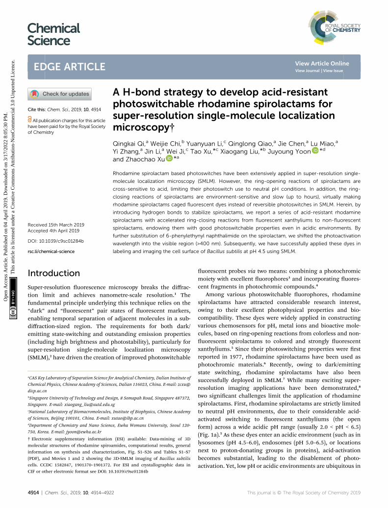
Fig. 1 (a) The switch of rhodamine spirolactams from the non-fluoresceThe intramolecular hydrogen bonding enables acid-resistant photoswrolactams without and with intramolecular hydrogen bonding.
This journal is © The Royal Society of Chemistry 2019
substitution of 3-amino groups in the carboxyphenyl ring andassociated intramolecular hydrogen bonding in spirolactams(Fig. 1b). Inspired by Moerner's work,7c we further extended thephotoactivation wavelength of our dyes into the visible region(>400 nm), via conjugation with 6-phenylethynyl naph-thalimide. The excellent acid-resistance and visible-light pho-toswitching make our rhodamine spirolactams suitable forSMLM imaging, and more importantly, generate reliable uo-rescence signals even in acidic environments.
Results and discussionMolecular design strategy
Our strategy of introducing intramolecular hydrogen bondingto increase the acid-resistance of spirolactams and shorten the“open form” lifetime originated from three facts: (1) the acid-activated ring-opening reaction is initiated with the proton-ation of the carbonyl oxygen (Fig. 1a);1 (2) the resulting ringopening reaction is associated with a rotation of the amidegroup (–CONR) with respect to the phenyl ring (Fig. 1a, S1;Tables S1 and S2†); (3) introducing an electron-donating groupinto spirolactams reduces the driving force of ring-openingreactions.11 We anticipated that the intramolecular hydrogenbonding would reduce the reactivity of carbonyl oxygen withexterior acid and lock the spirolactam group. In addition, boththe suppressed rotation of the amide group and the incorpo-ration of an electron-donating group contributed to the stabi-lization of the closed form. Accordingly, we synthesized
nt closed form to fluorescent open form activated by light or acid. (b)itchable rhodamine spirolactams. (c) The designed rhodamine spi-
Chem. Sci., 2019, 10, 4914–4922 | 4915
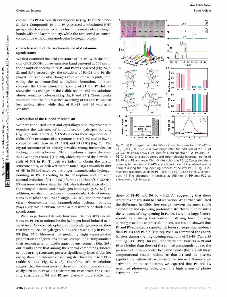
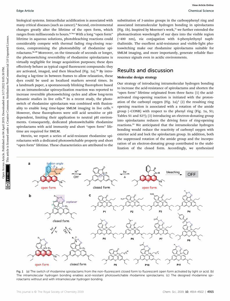
Fig. 2 (a) Photograph and (b) UV-vis absorption spectra of P1–P8 inCH2Cl2/CH3OH (9/1, v/v), two hours after the addition of 2.3 mL ofCF3COOH (1000 equiv.). (c) Local 1H NMR spectra of P2–P5 and P7–P8. (d) Single-crystal structures and intramolecular hydrogen bonds ofP4, P7 and P8 and weak CH/O interactions in P6. (e) Calculated ring-opening tendencies of P1–P6 in acidic solution. (f) Calculated energybarriers during the ring-opening process of neutral P1–P6. (g) Fluo-rescence quantum yields of P1–P8 in CH2Cl2/CH3OH (9/1, v/v) solu-tion. (h) The absorption intensities at 565 nm of P9 and P10 asa function of pH in water.
Chemical Science Edge Article
Ope
n A
cces
s A
rtic
le. P
ublis
hed
on 0
4 A
pril
2019
. Dow
nloa
ded
on 3
/17/
2022
8:0
5:30
PM
. T
his
artic
le is
lice
nsed
und
er a
Cre
ativ
e C
omm
ons
Attr
ibut
ion-
Non
Com
mer
cial
3.0
Unp
orte
d L
icen
ce.
View Article Online
compounds P1–P6 to verify our hypothesis (Fig. 1c and SchemesS1–S3†). Compounds P4 and P5 possessed 3-substituted NHRgroups which were expected to form intramolecular hydrogenbonds with the lactam moiety, while the rest served as controlcompounds without intramolecular hydrogen bonds.
Characterization of the acid-resistance of rhodaminespirolactams
We rst examined the acid resistance of P1–P6. With the addi-tion of CF3COOH, a new emission band centered at 560 nm inthe absorption spectra of P1–P3 and P6 was observed (Fig. 2a, b,S2 and S3†). Accordingly, the solutions of P1–P3 and P6 dis-played noticeable color changes from colorless to pink, indi-cating the acid-controlled xanthylium formation. In starkcontrast, the UV-vis absorption spectra of P4 and P5 did notshow obvious changes in the visible region, and the solutionsalmost remained colorless (Fig. 2a, b and S2†). These resultsindicated that the uorescence switching of P4 and P5 was farless acid-sensitive, while that of P1–P3 and P6 was acid-sensitive.
Verication of the H-bond mechanism
We next conducted NMR and crystallographic experiments toexamine the existence of intramolecular hydrogen bonding(Fig. 2c, d and Table S3†). 1H NMR spectra show large downeldshis of the resonance of NH protons in P4 (5.30) and P5 (6.75),compared with those in P2 (3.83) and P3 (3.83) (Fig. 2c). Thecrystal structure of P4 directly revealed strong intramolecularhydrogen bonding between NH and carbonyl oxygen (distance:2.347 A; angle: 128.61�) (Fig. 2d), which explained the downeldshi of NH in P4. Though we failed to obtain the crystalstructure of P5, we believed that the much larger downeld shiof NH in P5 indicated even stronger intramolecular hydrogenbonding in P5. According to the absorption and emissionspectral evolution of P4 and P5 (aer the addition of CF3COOH),P5 was more acid-resistant than P4, which should be ascribed tothe stronger intramolecular hydrogen bonding (Fig. S2–S4†). Inaddition, we also noticed weak intramolecular CH/O interac-tions in P6 (distance: 2.249 A; angle: 130.80�). The above resultsclearly demonstrate that intramolecular hydrogen bondingplays a key role in enhancing the acid-resistance of rhodaminespirolactams.
We also performed density functional theory (DFT) calcula-tions on P1–P6 to rationalize the hydrogen-bond induced acid-resistance. As expected, optimized molecular geometries showthat intramolecular hydrogen bonds are present only in P4 andP5 (Fig. S5†). Moreover, by modelling eight representativeprotonation congurations for each compound to fully simulatetheir responses in an acidic aqueous environment (Fig. S6†),our results show that among the control compounds, uores-cent open-ring structures possess signicantly lower Gibbs freeenergy than non-emissive closed-ring structures by up to 0.19 eV(Table S4 and Fig. S7–S12†). Therefore, DFT calculationssuggest that the emissions of these control compounds couldeasily turn on in an acidic environment. In contrast, the closed-ring structures of P4 and P5 are relatively more stable than
4916 | Chem. Sci., 2019, 10, 4914–4922
those of P1–P3 and P6 by �0.12 eV, suggesting that thesestructures are resistant to acid-activation. We further calculatedthe difference in Gibbs free energy between the most stableclosed-ring and open-ring protonated structures (d) to quantifythe tendency of ring-opening in P1–P6. Herein, a large d corre-sponds to a strong thermodynamic driving force for ring-opening reactions to proceed. Indeed, our results showed thatP4 and P5 exhibited a signicantly lower ring-opening tendencythan P1–P3 and P6 did (Fig. 2e). We also computed the energybarriers during the ring-opening reactions of P1–P6 (Table S5and Fig. S13–S19†). Our results show that the barriers in P4 andP5 are higher than those of the control compounds, due to thepresence of intramolecular hydrogen bonds (Fig. 2f). All thesecomputational results rationalize that P4 and P5 possesssignicantly enhanced acid-resistance towards uorescenceactivation. At the same time, we expected that P4 and P5remained photoswitchable, given the high energy of photo-activation light.
This journal is © The Royal Society of Chemistry 2019
Edge Article Chemical Science
Ope
n A
cces
s A
rtic
le. P
ublis
hed
on 0
4 A
pril
2019
. Dow
nloa
ded
on 3
/17/
2022
8:0
5:30
PM
. T
his
artic
le is
lice
nsed
und
er a
Cre
ativ
e C
omm
ons
Attr
ibut
ion-
Non
Com
mer
cial
3.0
Unp
orte
d L
icen
ce.
View Article Online
Improving quantum yields and water solubility of rhodaminespirolactams
Unfortunately, the quantum yields of P2–P6 are low (Fig. 2g),possibly quenched by photo-induced electron transfer (PET)from the 3-aminophenyl moiety to the xanthene scaffold. Toenhance brightness and preserve acid-resistance, we synthe-sized P7 and P8 (Scheme S4†) with 3-acetamido and 3-tolue-nesulfonamide substituents to inhibit PET, respectively. Asa result, the quantum yields of P7 and P8 were signicantlyimproved (Fig. 2g). At the same time, P7 and P8 remained acid-resistant (Fig. 2a and b). 1H NMR analysis and the single crystalstructures of P7 and P8 clearly showed the existence of intra-molecular hydrogen bonds (Fig. 2c and d). Notably, thestronger intramolecular hydrogen bonding in P7 than that inP8 resulted in stronger acid-resistance of P7 than that of P8(Fig. S2–S4†). Owing to its high brightness and excellentacid-resistance, we chose P7 for further derivations andapplications.
Subsequently, we synthesized water-soluble compounds P9and P10 with polyethylene glycol (PEG) groups to verify theapplicability of our strategy in water (Scheme S5†). As shownin Fig. 2h and S20,† the UV-vis absorption and emissionintensities of the open form of P9 substantially increasedwhen the pH was decreased from 7.40 to 3.89, and thendecreased when the pH was decreased from 3.89 to 1.44. Incontrast, P10 did not show any noticeable absorption anduorescence bands in the visible region in the pH range from1.44 to 7.40 (Fig. 2h and S20†). These results indicated thatour strategy to increase the acid-resistance of rhodaminespirolactams worked well in aqueous solutions and wassuitable for biological systems.
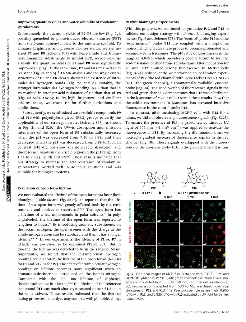
Fig. 3 Confocal images of MCF-7 cells stained with LTG (0.1 mM) and(a) P12 (10 mM) or (b) P13 (10 mM); green channel: excitation at 488 nm,emission collected from 500 to 550 nm; red channel: excitation at561 nm, emission collected from 580 to 653 nm. Insets: chemicalstructures of P12 and P13. The Pearson coefficients are high, 0.905(LTGwith P12) and 0.925 (LTGwith P13 activated by UV light for 5 min),respectively.
Evaluation of open form lifetime
We next evaluated the lifetime of the open forms via laser ashphotolysis (Table S6 and Fig. S21†). It's reported that the life-time of the open form was greatly affected both by the envi-ronment and molecular structures.6,7a,c The open form hasa lifetime of a few milliseconds in polar solvents.6 In poly-vinylalcohol, the lifetime of the open form was reported tolengthen to hours.7a By introducing aromatic substituents onthe lactam nitrogen, the open isomer with the charge at theamide nitrogen atom can be stabilized and then it has a longerlifetime.6b,7a,c In our experiments, the lifetime of P1 vs. P7 inCH2Cl2 was too short to be examined (Table S6†). But indioxane, the lifetime was detected to be in the range of 60 ns.Importantly, we found that the intramolecular hydrogenbonding could shorten the lifetime of the open forms (62.5 nsfor P1 and 59.7 ns for P7). The effect of intramolecular hydrogenbonding on lifetime becomes more signicant when anaromatic substituent is introduced on the lactam nitrogen.Compared with the 200 ms lifetime of N-phenyl-rhodaminelactame in dioxane,6a,b the lifetime of the referencecompound P11 was much shorter, measured to be �31.5 ns inthe same solvent. These results indicated that the thermalfading processes in our dyes may compete with photobleaching.
This journal is © The Royal Society of Chemistry 2019
In vitro bioimaging experiments
With this progress, we continued to synthesize P12 and P13 tovalidate our design strategy with in vitro bioimaging experi-ments (Fig. 3 and Scheme S7†). The “control” probe P12 and the“experimental” probe P13 are coupled with a morpholinemoiety, which enables these probes to become protonated andaccumulated in lysosomes. The pH value of lysosomes is in therange of 4.0–6.0, which provides a good platform to test theacid-resistance of rhodamine spirolactams. Aer incubation for30 min, P12 emitted strong uorescence in MCF-7 cells(Fig. S22†). Subsequently, we performed co-localization experi-ments of P12 (the red channel) with LysoTracker Green DND-26(LTG; the green channel), a commercially available lysosomeprobe (Fig. 3a). The good overlap of uorescence signals in thered and green channels demonstrates that P12 was distributedin the lysosomes of MCF-7 cells. Overall, these results show thatthe acidic environment in lysosomes has activated intensiveuorescence in the control probe P12.
In contrast, aer incubating MCF-7 cells with P13 for 2hours, we did not observe any uorescence signals (Fig. S22†).To ensure the presence of P13 in lysosomes, continuous UVlight of 375 nm (�1 mW cm�2) was applied to activate theuorescence of P13. By increasing the illumination time, wenoticed a gradual increase of uorescence signals in the redchannel (Fig. 3b). These signals overlapped with the uores-cence of the lysosome probe LTG in the green channel. It is thus
Chem. Sci., 2019, 10, 4914–4922 | 4917
Chemical Science Edge Article
Ope
n A
cces
s A
rtic
le. P
ublis
hed
on 0
4 A
pril
2019
. Dow
nloa
ded
on 3
/17/
2022
8:0
5:30
PM
. T
his
artic
le is
lice
nsed
und
er a
Cre
ativ
e C
omm
ons
Attr
ibut
ion-
Non
Com
mer
cial
3.0
Unp
orte
d L
icen
ce.
View Article Online
clear that P13 maintains acid-resistance in lysosomes andpossesses a dedicated photoactivation property for cellularimaging. It's worth mentioning that the uorescent form of P13in lysosomes did not thermally revert back to the closed form ina short time. Considering the environmental sensitivity of theopen form, this is probably because a large number of P13accumulated in lysosomes, and many xanthyliums formedunder continuous strong UV irradiation tended to stabilize eachother, thus slowing down the ring-close reactions. The photo-activation experiments of P1 and P7 in CH2Cl2 may support ourhypothesis. The open form lifetimes of P1 and P7 in CH2Cl2were too short to observe a large amount of open isomers. But ifwe excited these compounds in CH2Cl2 solution withcontinuous UV irradiation, simultaneously photoactivated
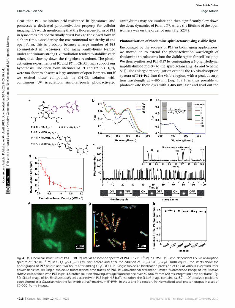
Fig. 4 (a) Chemical structures of P14–P18. (b) UV-vis absorption spectraspectra of P17 (10�5 M) in CH2Cl2/CH3OH (9/1, v/v) before and afterphotographs of P17 before and two hours after adding CF3COOH. (d) Spower densities. (e) Single-molecule fluorescence time traces of P18. (subtilis cells stained with P18 in pH 4.5 buffer solution showing average fl
3D-SMLM image of live Bacillus subtilis cells stained with P18 in pH 4.5 bueach plotted as a Gaussian with the full width at half-maximum (FHWM)30 000-frame images.
4918 | Chem. Sci., 2019, 10, 4914–4922
xanthyliums may accumulate and then signicantly slow downthe decay dynamics of P1 and P7, where the lifetime of the openisomers was on the order of min (Fig. S23†).
Photoactivation of rhodamine spirolactams using visible light
Encouraged by the success of P13 in bioimaging applications,we moved on to extend the photoactivation wavelength ofrhodamine spirolactams into the visible region for cell imaging.We thus synthesized P14–P17 by conjugating a 6-phenylethynylnaphthalimide moiety to the spirolactam (Fig. 4a and SchemeS8†). The enlarged p-conjugation extends the UV-vis absorptionspectra of P14–P17 into the visible region, with a peak absorp-tion wavelength at �400 nm (Fig. 4b). It is thus possible tophotoactivate these dyes with a 405 nm laser and read out the
of P14–P17 (10�5 M) in DMSO. (c) Time-dependent UV-vis absorptionthe addition of CF3COOH (2.3 mL, 1000 equiv.); the insets show theingle molecule localization precision of P17 at various excitation laserf) Conventional diffraction-limited fluorescence image of live Bacillusuorescence over 30 000 frames (20 ms integration time per frame). (g)ffer solution; the SMLM image contains ca. 5.7� 105 localized positions,in the X and Y direction. (h) Normalized total photon output in a set of
This journal is © The Royal Society of Chemistry 2019
Edge Article Chemical Science
Ope
n A
cces
s A
rtic
le. P
ublis
hed
on 0
4 A
pril
2019
. Dow
nloa
ded
on 3
/17/
2022
8:0
5:30
PM
. T
his
artic
le is
lice
nsed
und
er a
Cre
ativ
e C
omm
ons
Attr
ibut
ion-
Non
Com
mer
cial
3.0
Unp
orte
d L
icen
ce.
View Article Online
uorescence signals with an excitation laser of 561 nm. Thevisible photoactivation wavelength is critical for minimizingphototoxicity in live-cell imaging.
Among P14–P17, we selected P17 for further experiments,foreseeing its excellent acid-resistance and good quantum yield.Indeed, P17 remained colorless in acidic medium (Fig. 4c). Wethen doped P17 into a PMMA lm at a low concentration tocharacterize its single-molecule uorescence properties. Weapplied a continuous 405 nm laser light (60 W cm�2) to pho-toactivate P17 (Fig. S24†), and a 561 nm laser light to generateuorescence from activated P17. Our experiments showed thata laser excitation power of �1.2 kW cm�2 at 561 nm led toa localization precision of the single molecule of �12 nm inPMMA (Fig. 4d and S25†). Subsequent experiments showed thatgood imaging quality could be achieved by reducing the pho-toactivation light intensity at 405 nm down to 1.8 W cm�2 ina cellular environment.
Super-resolution imaging of Bacillus subtilis in acidicenvironments
To fully demonstrate the imaging applications of our photo-switchable dyes, we linked N-hydroxysuccinimide ester to P17,resulting in the formation of P18 (Fig. 4a and Scheme S9†).Laser ash photolysis showed that the open form lifetime of P18was 105 ns in aqueous solutions with 5% DMSO (Table S6 and
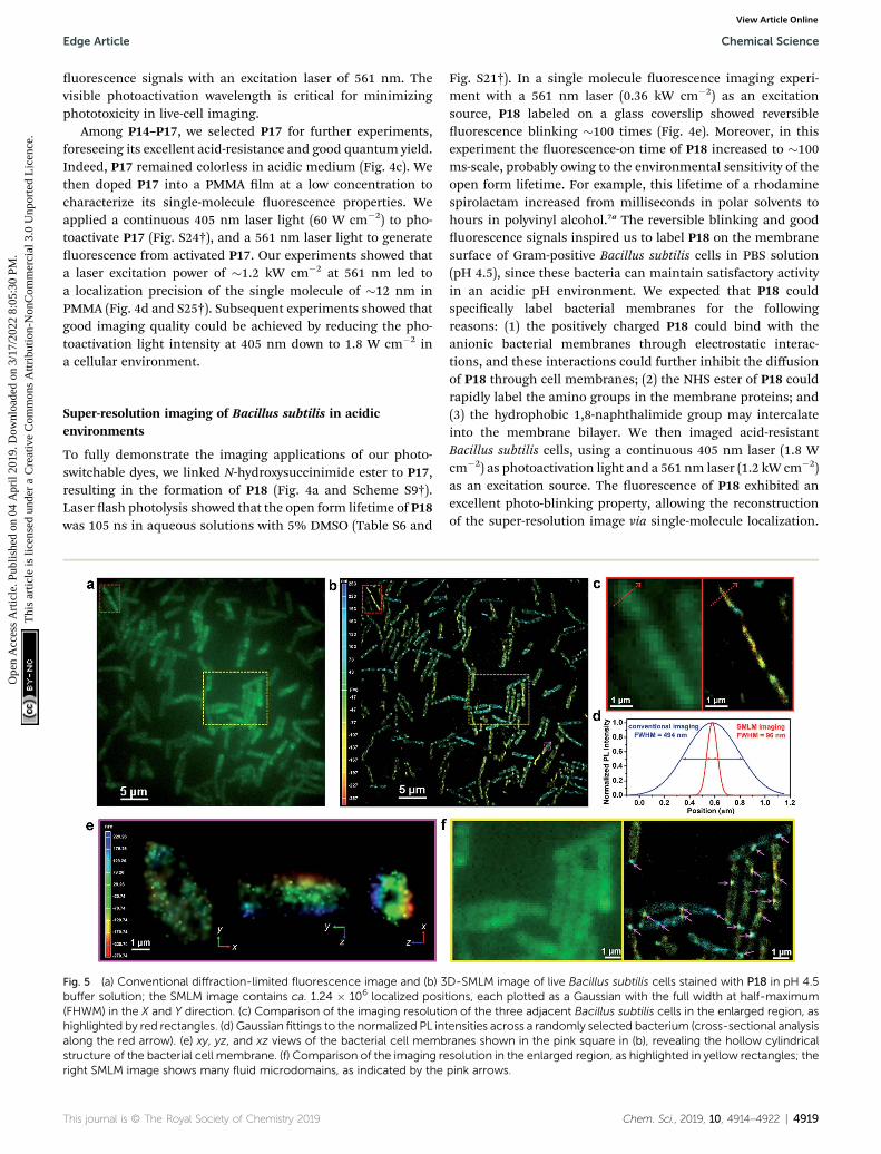
Fig. 5 (a) Conventional diffraction-limited fluorescence image and (b) 3buffer solution; the SMLM image contains ca. 1.24 � 106 localized posi(FHWM) in the X and Y direction. (c) Comparison of the imaging resolutiohighlighted by red rectangles. (d) Gaussian fittings to the normalized PL intalong the red arrow). (e) xy, yz, and xz views of the bacterial cell membstructure of the bacterial cell membrane. (f) Comparison of the imaging reright SMLM image shows many fluid microdomains, as indicated by the
This journal is © The Royal Society of Chemistry 2019
Fig. S21†). In a single molecule uorescence imaging experi-ment with a 561 nm laser (0.36 kW cm�2) as an excitationsource, P18 labeled on a glass coverslip showed reversibleuorescence blinking �100 times (Fig. 4e). Moreover, in thisexperiment the uorescence-on time of P18 increased to �100ms-scale, probably owing to the environmental sensitivity of theopen form lifetime. For example, this lifetime of a rhodaminespirolactam increased from milliseconds in polar solvents tohours in polyvinyl alcohol.7a The reversible blinking and gooduorescence signals inspired us to label P18 on the membranesurface of Gram-positive Bacillus subtilis cells in PBS solution(pH 4.5), since these bacteria can maintain satisfactory activityin an acidic pH environment. We expected that P18 couldspecically label bacterial membranes for the followingreasons: (1) the positively charged P18 could bind with theanionic bacterial membranes through electrostatic interac-tions, and these interactions could further inhibit the diffusionof P18 through cell membranes; (2) the NHS ester of P18 couldrapidly label the amino groups in the membrane proteins; and(3) the hydrophobic 1,8-naphthalimide group may intercalateinto the membrane bilayer. We then imaged acid-resistantBacillus subtilis cells, using a continuous 405 nm laser (1.8 Wcm�2) as photoactivation light and a 561 nm laser (1.2 kW cm�2)as an excitation source. The uorescence of P18 exhibited anexcellent photo-blinking property, allowing the reconstructionof the super-resolution image via single-molecule localization.
D-SMLM image of live Bacillus subtilis cells stained with P18 in pH 4.5tions, each plotted as a Gaussian with the full width at half-maximumn of the three adjacent Bacillus subtilis cells in the enlarged region, asensities across a randomly selected bacterium (cross-sectional analysisranes shown in the pink square in (b), revealing the hollow cylindricalsolution in the enlarged region, as highlighted in yellow rectangles; thepink arrows.
Chem. Sci., 2019, 10, 4914–4922 | 4919
Chemical Science Edge Article
Ope
n A
cces
s A
rtic
le. P
ublis
hed
on 0
4 A
pril
2019
. Dow
nloa
ded
on 3
/17/
2022
8:0
5:30
PM
. T
his
artic
le is
lice
nsed
und
er a
Cre
ativ
e C
omm
ons
Attr
ibut
ion-
Non
Com
mer
cial
3.0
Unp
orte
d L
icen
ce.
View Article Online
In comparison to a conventional diffraction-limited uores-cence image (Fig. 4f), the 3D-SMLM image of Bacillus subtilisdisplayed a greatly improved spatial resolution (Fig. 4g).Besides, owing to the low intensity of the 405 nm excitationlaser, and slow photobleaching rates of the 561 nm laser (asa result of fast ring-close reactions and a short open-form life-time), P18 exhibited excellent photostability. The number ofcollected photons per 3000 frames (20 ms per frame) remainedalmost constant throughout our experiments of >10 min(Fig. 4h), demonstrating the potential of P18 to perform longtime-lapse nanoscopy imaging in future studies.
We continued to investigate the staining properties andlabeling specicity of P18. As shown in Fig. 5a, b and Movie 1,†P18 stained bacteria evenly with high density, and exhibitedexcellent photoblinking properties. Interestingly, the divisionsof three Bacillus subtilis cells were clearly visible in a 3D-SMLMimage (Fig. 5b and c), while they were barely resolved ina diffraction-limited uorescence image (Fig. 5a and c). The full-width at half-maximum (FWHM) of a single Bacillus subtilis is�96 nm in the super-resolution images, cf. �494 nm in theconventional uorescence images (Fig. 5d). Cross-sections ofa single Bacillus subtilis highlighted in the pink box showa hollow structure from the xz view, indicating that our probeslocalized on the cell membranes of bacteria (Fig. 5e). The ip-ped 3D-SMLM image of bacterial cell membranes is furthershown in Movie 2.† We thus demonstrated that P18 enabledSMLM studies of the membranes of bacteria cells at nanometricresolution (without using additional targeting strategies).Moreover, we found that the membranes of most Bacillus sub-tilis bacteria did not show uniform uorescence intensities inthe 3D-SMLM image shown in Fig. 5b. The slight irregularity ofthe membrane staining likely indicates the presence of manyuid microdomains (as highlighted by pink arrows in the rightimage of Fig. 5f). These uid microdomains had been wellstudied by several research groups.12 However, these uidmicrodomains were hardly resolved in conventional diffraction-limited uorescence images (in the le image of Fig. 5f).Besides, we also discovered that the majority of these uidmicrodomains appeared in certain areas, especially around cellpoles.
Conclusions
In summary, this study presents an effective molecular engi-neering approach for developing acid-resistant rhodamine spi-rolactams with a dedicated photoswitchable property and shortopen-form lifetime. These unique characteristics resulted fromintramolecular hydrogen bonding between the 3-aminosubstituents in the carboxyphenyl ring and the lactam moiety.Moreover, we have extended the photoactivation wavelengths ofrhodamine spirolactams into the visible region (>400 nm), byconjugating a 6-phenylethynyl naphthalimide moiety to thespirolactams. The visible photoactivation and dedicated pho-toswitchable properties of our dyes make them highly reliableuorescent probes for SMLM, as demonstrated by the success-ful labeling and imaging of Bacillus subtilis at pH 4.5 using 3D-SMLM. We believe that our work sheds light on rational
4920 | Chem. Sci., 2019, 10, 4914–4922
engineering of uorescent properties of rhodamine dyes tofacilitate the advancement of super-resolution imagingtechniques.
Experimental sectionSingle-crystal structures
Single crystals of P4, P6, P7 and P8 that were suitable for X-raystructural analysis were obtained by slow evaporation froma chloroform/n-hexane solution at room temperature. Singlecrystal X-ray diffraction data were collected on an Xcalibur,Atlas, Gemini ultra-diffractometer using the u-scan mode witha graphite-monochromator and Mo Ka radiation. The crystalstructures were solved with the Superip13 structure solutionprogram and rened with the SHELXL14 renement package, asimplemented in OLEX2.15 All H atoms were placed geometri-cally and treated with riding constraints, and displacementparameters were derived from the C atoms to which they wereattached. All CH2 groups had Uiso(H) values xed at 1.2 timesthe Ueq value of the attached C atom. CH3 groups were idealizedas freely rotating groups, with Uiso(H) values xed at 1.5 timesthe Ueq value of the attached C atom. H atoms on N atoms werelocated via Fourier difference map inspection and positionallyrened isotropically with thermal parameters based upon the Natom to which they are bonded (Uiso(H)¼ 1.2Ueq(N)). These datacan be obtained free of charge from the Cambridge Crystallo-graphic Data Centre (CCDC reference numbers: 1582847,1901370–1901372†).
Measurement of single molecule optical properties
P17 was immobilized in polymer lms to investigate the effectsof the excitation laser power density on (1) the photon outputfrom dyes, (2) background noise, and (3) localization precision.Polymer lms were prepared using 1% (by mass) solution ofpolymethyl methacrylate (PMMA) in toluene. The polymer lmswere doped with P17 (10 nM) and then spin-cast onto argon-plasma-etched coverslips. Aer that, the coverslips wereplaced in the sample holder of an SMLM instrument forimaging. The intensity of the 405 nm photoactivation laser wasadjusted to activate only a few molecules in each frame. Theuorescence images of the polymer lm were acquired using anelectron-multiplying charge-coupled device (EMCCD) camera atroom temperature.
Sample preparation and data analysis for single-moleculeuorescence imaging experiment
Coverslips were cleaned by sonication for 10 min in each of thefollowing: Milli-Q water, absolute ethyl alcohol, and acetone.The above coverslips were further boiled in a solution mixtureof concentrated sulfuric acid and hydrogen peroxide (v/v, 7/3)for 1 h and then washed with Milli-Q water and dried usingnitrogen. 20 mL of very dilute P18 (10 nM) in DMSO solution wasdripped on the dry coverslips. Then, P18 with the N-hydrox-ysuccinimide ester adsorbed and reacted with the hydroxylgroups on the surface of the cleaned coverslip at a low densitysuch that individual dye molecules could be clearly resolved.
This journal is © The Royal Society of Chemistry 2019
Edge Article Chemical Science
Ope
n A
cces
s A
rtic
le. P
ublis
hed
on 0
4 A
pril
2019
. Dow
nloa
ded
on 3
/17/
2022
8:0
5:30
PM
. T
his
artic
le is
lice
nsed
und
er a
Cre
ativ
e C
omm
ons
Attr
ibut
ion-
Non
Com
mer
cial
3.0
Unp
orte
d L
icen
ce.
View Article Online
Then the surface of the cleaned coverslip was rinsed with PBS(pH 7.4) to remove unbound P18 before imaging. Finally, thecoverslip was placed on the sample holder of the SMLMinstrument and illuminated with 561 nm laser light (0.36 kWcm�2). 5000–10 000 frames were recorded at a frame rate of 20ms per frame. Single-molecule uorescence time trace wascalculated for a single molecule by analyzing the total integratedsignal in a 2 � 2 pixel region centered on the single moleculeduring each frame of the movie. The number of photons wascalculated as the total intensity count minus the backgroundintensity count, which was converted to photon number usingthe calibrated parameter for electronmultiplication and analog-to-digital conversion gain settings used during acquisition.
Cell culture and CLSM imaging
MCF-7 cells were grown on DMEM (Gibco) supplemented with10% FBS (Gibco). In CLSM imaging, the MCF-7 cells wereincubated with 1 mL of DMEM containing 10 mM P11 (experi-mental group) or P12 (control group) and the images werecollected at 0, 30, 60 and 120 min by using an ANDORTM livecell imaging system. Then, the adherent cells were furtherincubated with 1 mL of fresh DMEM containing LysoTrackerGreen DND-26 (LTG) (0.1 mM) for another 30 min. The adherentcells were washed 3 times with PBS (pH 7.4) and utilized forCLSM imaging. In the experimental group, the CLSM images ofMCF-7 cells were taken under 0–5 min continuous UV (375 nm)irradiation at 37 �C.
Live bacteria sample preparation and labeling
A single colony of Bacillus subtilis was inoculated in 5 mL ofLuria–Bertani (LB) medium with shaking at 37 �C. Subse-quently, 1 mL of the Bacillus subtilis suspension was inoculatedin 10 mL of fresh LB media, which was incubated for 5 h withshaking (250 rpm) at 37 �C to achieve mid-log phase growth.Aer the Bacillus subtilis had grown to the mid-log phase, 1 mLof the suspension was washed at least 3 times by centrifugingfor 3 minutes at 10 000 RPM and resuspending the pellet in1 mL of clean PBS (pH 4.5). To label the surface of Bacillussubtilis, 50 mL of P17 in DMSO solution (�10 nM) was addedslowly to the suspension of the Bacillus subtilis cells and le toincubate for 30 minutes. The unreacted P17 was rinsed 6 timeswith PBS (pH 4.5) before imaging. Then, the cleaned Bacillussubtilis cells were resuspended in a small amount of PBS (pH4.5) to produce a concentrated cell suspension. Finally, 2 mL ofthis cell suspension was deposited onto an agarose pad (1.5%(by mass) low melting point agarose in PBS) and mounted ontoan Ar plasma etched glass slide (size: 24 � 50 mm, thickness:0.13–0.16 mm) and immediately used for the SMLM imaging.
3D-SMLM imaging and data processing
The super-resolution imaging was carried out using a Nikon N-STORM 5.0 super-resolution microscope system with a motor-ized inverted microscope ECLIPSE Ti2-E, a 100�/NA 1.49 oilimmersion TIRF objective lens (CFI HP) and an ORCA-Flash 4.0
SCMOS camera (Hamamatsu Photonics K.K.). During super-resolution imaging, a 405 nm laser (1.8 W cm�2) was
This journal is © The Royal Society of Chemistry 2019
employed to photoactivate rhodamine spirolactams from ring-closed isomers to ring-opened isomers. At the same time,a 561 nm laser (1.2 kW cm�2) was used as the excitation sourceto produce sparse emissions from ring-opened rhodamines.Repeating this cycle many times (i.e., to collect �30 000 imageswith the SCMOS camera with an integration time of 20 ms perframe) allowed us to reconstruct a super-resolution image.Astigmatism was used in the 3D-imaging experiments. Thesoware NIS-Elements Ar and N-STORM Analysis were used toanalyze the collected images and computationally reconstructthe 3D-SMLM image.
Conflicts of interest
The authors declare no conict of interest.
Acknowledgements
This work was supported by the National Natural ScienceFoundation of China (21708039, 21502189, 21878286), CPSF(2017M611278, 2018T110234), and DICP (DMTO201603,TMSR201601, 2015YB07). X. L. and W. C. are indebted to thenancial support from SUTD (T1SRCI17126, IDD21700101,IDG31800104). J. Y. acknowledges the support from theNational Research Foundation of Korea (NRF) (No.2012R1A3A2048814).
Notes and references
1 (a) S. W. Hell, Science, 2007, 316, 1153; (b) A. von Diezman,Y. Shechtman and W. E. Moerner, Chem. Rev., 2017, 117,7244; (c) E. Betzig, Angew. Chem., Int. Ed., 2015, 54, 8034.
2 G. Patterson, M. Davidson, S. Manley and J. Lippincott-Schwartz, Annu. Rev. Phys. Chem., 2010, 61, 345.
3 (a) M. Heilemann, S. van de Linde, M. Schuttpelz, R. Kasper,B. Seefeldt, A. Mukherjee, P. Tinnefeld and M. Sauer, Angew.Chem., Int. Ed., 2008, 47, 6172; (b) J. B. Grimm, T. Klein,B. G. Kopek, G. Shtengel, H. F. Hess, M. Sauer andL. D. Lavis, Angew. Chem., Int. Ed., 2016, 55, 1723.
4 (a) O. Nevskyi, D. Sysoiev, A. Oppermann, T. Huhn andD. Woll, Angew. Chem., Int. Ed., 2016, 55, 12698; (b)B. Roubinet, M. L. Bossi, P. Alt, M. Leutenegger,H. Shojaei, S. Schnorrenberg, S. Nizamov, M. Irie,V. N. Belov and S. W. Hell, Angew. Chem., Int. Ed., 2016, 55,15429; (c) B. Roubinet, M. Weber, H. Shojaei, M. Bates,M. L. Bossi, V. N. Belov, M. Irie and S. W. Hell, J. Am.Chem. Soc., 2017, 139, 6611.
5 (a) H. N. Kim, M. H. Lee, H. J. Kim, J. S. Kim and J. Yoon,Chem. Soc. Rev., 2008, 37, 1465; (b) X. Chen, T. Pradhan,F. Wang, J. S. Kim and J. Yoon, Chem. Rev., 2012, 112, 1910.
6 (a) K. H. Knauer and R. Gleiter, Angew. Chem., Int. Ed., 1977,16, 113; (b) H. Willwohl, J. Wolfrum and R. Gleiter, LaserChem., 1989, 10, 63; (c) H. Montenegro, M. D. Paolo,D. Capdevila, P. F. Aramendia and M. L. Bossi, Photochem.Photobiol. Sci., 2012, 11, 1081; (d) K. Li, Y. Xiang, X. Wang,J. Li, R. Hu, A. Tong and B. Z. Tang, J. Am. Chem. Soc.,2014, 136, 1643.
Chem. Sci., 2019, 10, 4914–4922 | 4921
Chemical Science Edge Article
Ope
n A
cces
s A
rtic
le. P
ublis
hed
on 0
4 A
pril
2019
. Dow
nloa
ded
on 3
/17/
2022
8:0
5:30
PM
. T
his
artic
le is
lice
nsed
und
er a
Cre
ativ
e C
omm
ons
Attr
ibut
ion-
Non
Com
mer
cial
3.0
Unp
orte
d L
icen
ce.
View Article Online
7 (a) J. Folling, V. Belov, R. Kunetsky, R. Medda, A. Schonle,A. Egner, C. Eggeling, M. Bossi and S. W. Hell, Angew.Chem., Int. Ed., 2007, 46, 6266; (b) V. N. Belov, M. L. Bossi,J. Folling, V. P. Boyarskiy and S. W. Hell, Chem.–Eur. J.,2009, 15, 10762; (c) M. K. Lee, P. Rai, J. Williams,R. J. Twieg and W. E. Moerner, J. Am. Chem. Soc., 2014,136, 14003; (d) B. Roubinet, M. Bischoff, S. Nizamov,S. Yan, C. Geisler, S. Stoldt, G. Y. Mitronova, V. N. Belov,M. L. Bossi and S. W. Hell, J. Org. Chem., 2018, 83, 6466; (e)M. Bossi, J. Folling, V. N. Belov, V. P. Boyarskiy, R. Medda,A. Egner, C. Eggeling, A. Schonle and S. W. Hell, NanoLett., 2008, 8, 2463; (f) V. N. Belov, C. A. Wurm,V. P. Boyarskiy, S. Jakobs and S. W. Hell, Angew. Chem., Int.Ed., 2010, 49, 3520; (g) J. Folling, V. Belov, D. Riedel,A. Schonle, A. Egner, C. Eggeling, M. Bossi and S. W. Hell,ChemPhysChem, 2008, 9, 321; (h) H. Aoki, K. Mori andS. Ito, So Matter, 2012, 8, 4390; (i) E. A. Halabi, D. Pinotsiand P. Rivera-Fuentes, Nat. Commun., 2019, 10, 1232; (j)E. A. Halabi, Z. Thiel, N. Trapp, D. Pinotsi and P. Rivera-Fuentes, J. Am. Chem. Soc., 2017, 139, 13200; (k)E. A. Halabi, S. Puntener and P. Rivera-Fuentes, Helv.Chim. Acta, 2018, 101, e1800165.
8 V. N. Belov and M. L. Bossi, Isr. J. Chem., 2013, 53, 1.9 (a) R. A. Gottlieb, H. A. Giesing, J. Y. Zhu, R. L. Engler andB. M. Babior, Proc. Natl. Acad. Sci. U. S. A., 1995, 92, 5965;(b) R. A. Gottlieb, J. Nordberg, E. Skowronski andB. M. Babior, Proc. Natl. Acad. Sci. U. S. A., 1996, 93, 654; (c)
4922 | Chem. Sci., 2019, 10, 4914–4922
M. A. Barry, J. E. Reynolds and A. Eastman, Cancer Res.,1993, 53, 2349.
10 (a) S. N. Uno, M. Kamiya, T. Yoshihara, K. Sugawara,K. Okabe, M. C. Tarhan, H. Fujita, T. Funatsu, Y. Okada,S. Tobita and Y. Urano, Nat. Chem., 2014, 6, 681; (b)H. Takakura, Y. Zhang, R. S. Erdmann, A. D. Thompson,Y. Lin, B. McNellis, F. Rivera-Molina, S. N. Uno,M. Kamiya, Y. Urano, J. E. Rothman, J. Bewersdorf,A. Schepartz and D. Toomre, Nat. Biotechnol., 2017, 35, 773.
11 G. Xi, L. Sheng, I. Zhang, J. Du, T. Zhang, Q. Chen, G. Li,Y. Zhang, Y. Song, J. Li, Y. Zhang and S. X. Zhang, ACSAppl. Mater. Interfaces, 2017, 9, 38032.
12 (a) K. Matsumoto, J. Kusaka, A. Nishibori and H. Hara, Mol.Microbiol., 2006, 61, 1110; (b) A. S. Johnson, S. van Horck andP. J. Lewis, Microbiology, 2004, 150, 2815; (c) F. Kawai,M. Shoda, R. Harashima, Y. Sadaie, H. Hara andK. Matsumoto, J. Bacteriol., 2004, 186, 1475; (d) I. Fishovand C. L. Woldringh, Mol. Microbiol., 1999, 32, 1166; (e)A. Nishibori, J. Kusaka, H. Hara, M. Umeda andK. Matsumoto, J. Bacteriol., 2005, 187, 2163.
13 O. V. Dolomanov, L. J. Bourhis, R. J. Gildea, J. A. K. Howardand H. Puschmann, J. Appl. Crystallogr., 2009, 42, 339.
14 L. Palatinus and G. Chapuis, J. Appl. Crystallogr., 2007, 40,786.
15 G. M. Sheldrick, Acta Crystallogr., Sect. A: Found. Crystallogr.,2008, 64, 112.
This journal is © The Royal Society of Chemistry 2019