Embed Size (px)
Citation preview
Innovations
A
GREEN LIGHTr_- I
..00_FORNEW
I |RESEACH
Imagine you want to find out how long bac-teria can survive in a particular river's waters.Where would you begin? You could addsome bacteria to a sample of the water. Thenevery so often you could drip some of thewater on culture medium, let it incubate, andsee if any bacterial colonies develop. But thattechnique demands lots of waiting, and you
might prefer a quicker technique-say, one
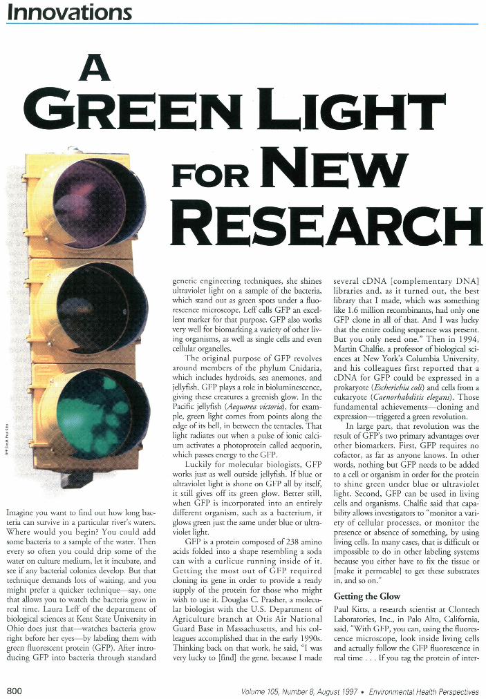
that allows you to watch the bacteria grow inreal time. Laura Leff of the department ofbiological sciences at Kent State University inOhio does just that-watches bacteria growright before her eyes-by labeling them withgreen fluorescent protein (GFP). After intro-ducing GFP into bacteria through standard
genetic engineering techniques, she shinesultraviolet light on a sample of the bacteria,which stand out as green spots under a fluo-rescence microscope. Leff calls GFP an excel-lent marker for that purpose. GFP also worksvery well for biomarking a variety ofother liv-ing organisms, as well as single cells and even
cellular organelles.
The original purpose of GFP -revolvesaround members of the phylum Cnidaria,which indudes hydroids, sea anemones, andjellyfish. GFP plays a role in bioluminescence,giving these creatures a greenish glow. In thePacific jellyfish (Aequorea victoria), for exam-
ple, green light comes from points along theedge of its bell, in between the tentacles. Thatlight radiates out when a pulse of ionic calci-um activates a photoprotein called aequorin,which passes energy to the GFP.
Luckily for molecular biologists, GFPworks just as well outside jellyfish. If blue or
ultraviolet light is shone on GFP all by itself,it still gives off its green glow. Better still,when GFP is incorporated into an entirelydifferent organism, such as a bacterium, itglows green just the same under blue or ultra-violet light.
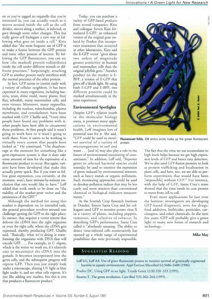
GFP is a protein composed of 238 aminoacids folded into a shape resembling a sodacan with a curlicue running inside of it.Getting the most out of GFP requireddoning its gene in order to provide a readysupply of the protein for those who mightwish to use it. Douglas C. Prasher, a molecu-lar biologist with the U.S. Department ofAgriculture branch at Otis Air NationalGuard Base in Massachusetts, and his col-leagues accomplished that in the early 1990s.Thinking back on that work, he said, "I was
very lucky to [find] the gene, because I made
several cDNA [complementary DNA]libraries and, as it turned out, the bestlibrary that I made, which was somethinglike 1.6 million recombinants, had only one
GFP clone in all of that. And I was luckythat the entire coding sequence was present.But you only need one." Then in 1994,Martin Chalfie, a professor of biological sci-ences at New York's Columbia University,and his colleagues first reported that acDNA for GFP could be expressed in a
prokaryote (Escherichia colt) and cells from a
eukaryote (Caenorhabditis elegans). Thosefundamental achievements-cloning andexpression-triggered a green revolution.
In large part, that revolution was theresult of GFP's two primary advantages over
other biomarkers. First, GFP requires no
cofactor, as far as anyone knows. In otherwords, nothing but GFP needs to be addedto a cell or organism in order for the proteinto shine green under blue or ultravioletlight. Second, GFP can be used in livingcells and organisms. Chalfie said that capa-bility allows investigators to "monitor a vari-ety of cellular processes, or monitor thepresence or absence of something, by usingliving cells. In many cases, that is difficult or
impossible to do in other labeling systemsbecause you either have to fix the tissue or
[make it permeable] to get these substratesin, and so on."
Getting the GlowPaul Kitts, a research scientist at ClontechLaboratories, Inc., in Palo Alto, California,said, "With GFP, you can, using the fluores-cence microscope, look inside living cellsand actually follow the GFP fluorescence inreal time . . . Ifyou tag the protein of inter-
Volume 105, Number 8, August 1997 * Environmental Health Perspectives
-1~
800
Innovations - A Green Light for New Research
est or you've tagged an organelle that you'reinterested in, you can actually watch as itmoves around inside the cell as the celldivides, moves along a surface, is infected, orgoes through some other changes. This hasreally given cell biologists a new way of fol-lowing what goes on inside a cell." Kittsadded that "the most frequent use of GFP isto make a fusion between the GFP proteinand some other protein of interest. By fol-lowing the GFP fluorescence, you can seehow [the marked] protein redistributesinside the cell under different stimuli or dif-ferent processes." Surprisingly, attachingGFP to another protein rarely interferes withthe normal processes of the other protein.
In fact, GFP seems to coexist easily witha variety of cellular neighbors. It has beenexpressed in many organisms, including bac-teria, yeast, slime mold, many plants, fruitflies, zebrafish, many mammalian cells, andeven viruses. Moreover, many organelles,induding the nudeus, mitochondria, plasmamembrane, and cytoskeleton, have beenmarked with GFP. Chalfie said, "Every timepeople have found any problems with it,other people have been able to circumventthose problems. At first people said it wasn'tgoing to work here or it wasn't going towork there. Now, it seems to be working invirtually every system that people havelooked at." He continued, "The disadvan-tage at the moment for something like agene-expression system is that it does takesome amount of time for the expression of afluorescent product to occur. But again, vari-ants have been developed that make thisactually pretty quick. But if you want to fol-low gene expression, you certainly, at themoment, won't have minute-by-minute res-olution that one would like to have." Leffadded that work needs to be done on "thestability of the marker-gene vector and thepersistence of the GFP."
Although the method for using thismarker is dependent on its intended task,the general procedure revolves around onechallenge: getting the GFP to the right place.In essence, that requires a vector system thatdelivers the cDNA into the right organismor even the right cells, where the cDNA getsexpressed, thereby producing GFP. Chalfiesaid, "Basically, what we're doing is trans-forming the organisms with DNA that willencode GFP.... For example, in C. elegans,which is the worm we work on, it's relativelyeasy to microinject the cDNA into thegonads. It becomes incorporated into thegerm cells, and the subsequent progeny willexpress GFP. Then you just simply lookunder a microscope, shining UV light or bluelight under it, and see what cells express. It'sjust like adding any marker, but this is onethat produces a fluorescent product."
Today, you can purchase avariety of GFP-based productsfrom several companies. Kittsand colleague Steven Kain for-mulated E-GFP, an enhanced 'version of the original gene iso-lated by Prasher that incorpo-rates mutations that occurredin other laboratories. Kitts saidthe E-GFP vector "gives abouttwo orders of magnitudegreater sensitivity in humanand mammalian cells than theoriginal jellyfish gene." Anotherproduct on the market is E-BFP, a version of E-GFP thatgives off a bluish light. By usingboth E-GFP and E-BFP, twodifferent proteins could bemarked simultaneously in thesame experiment.
Environmental SpotlightsDespite GFP's relative youthin the molecular biologyarena, it promises many appli-cations in environmentalhealth. Leff imagines lots ofpotential uses for it. She said,"GFP could be used to moni- Fluorescent fator survival of a variety of protein.microorganisms in soil andwater ... [and it] may also play a role in thefuture of environmental monitoring of cont-aminants." In addition, Leff said, "Reportergenes in selected bacterial species couldsomeday be used to monitor the expressionof genes induced by environmental stressors,such as heavy metals or organic pollutants.Such monitoring approaches could be usedto develop pollution indices that may be lesscostly and more sensitive than conventionalchemical or biological indicator measure-ments.
At the Scottish Crop Research Institutein Dundee, Simon Santa Cruz and his col-leagues used GFP to monitor potato virus Xin a variety of plants, including peppers,tomatoes, and relatives of tobacco. Indescribing GFP's performance, Santa Cruzcalled it "absolutely amazing. The ability todetect virus-infected cells noninvasively hasopened up a whole range of experimentalpossibilities that were previously impossible.
Dlds. 238 amino acids make up the green fluorescent
The fact that the virus we use accumulates tohigh levels helps because we get high expres-sion levels of GFP and hence easy detection.We've also used GFP-fusion proteins to lookat protein trafficking and localization inplant cells, and here, too, we are able to per-form experiments that would have been[impossible] without GFP." For instance,with the help of GFP, Santa Cruz's teamshowed that the virus needs its coat proteinto move from cell to cell.
Even more applications lie just overthe horizon; investigators are developingGFP-based diagnostic tests for drugs,food additives, herbicides, pesticides, car-cinogens, and other chemicals. In the nextfew years, GFP will probably give a greenlight to many new forms of research andtechnology.
Mike May
LeffLG, LeffAA. Use of green fluorescent protein to monitor survival of genetically engineeredbacteria in aquatic environments. Appl Environ Microbiol 62:3486-3488 (1996).
Prasher DC. Using GFP to see light. Trends Genet 11(8):320-323 (1995).Stearns T. The green revolution. Curr Biol 5(3):262-264 (1995).
Environmental Health Perspectives * Volume 105, Number 8, August 1997
E
801


![P-ISSN: A review on ethnomedicinal plant Acacia nilotica ...-3 cm long located at the end of branches [1]. ds are 7-15 cm long, green and tomentose (when immature) or greenish black](https://img.pdfslide.us/doc/110x75/5f248ab020c61e2aef47be62/p-issn-a-review-on-ethnomedicinal-plant-acacia-nilotica-3-cm-long-located.jpg)


![A hypothetical reconstruction of [i]Hallucigenia[i] · The cuticula is coloured green, although all no colour is known from Hallucigenia or its close relatives. The greenish colour](https://img.pdfslide.us/doc/110x75/5f80762e8b3b253a9d5958e5/a-hypothetical-reconstruction-of-ihallucigeniai-the-cuticula-is-coloured-green.jpg)