Embed Size (px)
Citation preview

A GDF5 Point Mutation Strikes Twice - Causing BDA1 andSYNS2Elisa Degenkolbe1,2., Jana Konig1,2.¤a, Julia Zimmer1,2¤b, Maria Walther1, Carsten Reißner3,
Joachim Nickel4,5, Frank Ploger6, Jelena Raspopovic7,8, James Sharpe7,8,9, Katarina Dathe10,
Jacqueline T. Hecht11, Stefan Mundlos1,10,12, Sandra C. Doelken10, Petra Seemann1,2,12*
1 Berlin-Brandenburg Center for Regenerative Therapies (BCRT), Charite – Universitatsmedizin Berlin, Berlin, Germany, 2 Berlin-Brandenburg School for Regenerative
Therapies (BSRT), Charite – Universitatsmedizin Berlin, Berlin, Germany, 3 Institute of Anatomy, Dept. Anatomy and Molecular Neurobiology, Universitatsklinikum Munster,
Munster, Germany, 4 Lehrstuhl fur Physiologische Chemie II, Theodor-Boveri-Institut fur Biowissenschaften (Biozentrum) der Universitat Wurzburg, Wurzburg, Germany,
5 Department of Tissue Engineering and Regenerative Medicine, Universitatsklinikum Wurzburg, Wurzburg, Germany, 6 Biopharm GmbH, Heidelberg, Germany, 7 EMBL-
CRG Systems Biology Program, Centre for Genomic Regulation, Barcelona, Spain, 8 Universitat Pompeu Fabra (UPF), Barcelona, Spain, 9 Institucio Catalana de Recerca i
Estudis Avancats (ICREA), Barcelona, Spain, 10 Institut fur Medizinische Genetik und Humangenetik, Charite – Universitatsmedizin Berlin, Berlin, Germany, 11 Department
of Pediatrics, University of Texas Medical School at Houston, Houston, Texas, United States of America, 12 Research Group Development and Disease, Max Planck Institute
for Molecular Genetics, Berlin, Germany
Abstract
Growth and Differentiation Factor 5 (GDF5) is a secreted growth factor that belongs to the Bone Morphogenetic Protein(BMP) family and plays a pivotal role during limb development. GDF5 is a susceptibility gene for osteoarthritis (OA) andmutations in GDF5 are associated with a wide variety of skeletal malformations ranging from complex syndromes such asacromesomelic chondrodysplasias to isolated forms of brachydactylies or multiple synostoses syndrome 2 (SYNS2). Here, wereport on a family with an autosomal dominant inherited combination of SYNS2 and additional brachydactyly type A1(BDA1) caused by a single point mutation in GDF5 (p.W414R). Functional studies, including chondrogenesis assays withprimary mesenchymal cells, luciferase reporter gene assays and Surface Plasmon Resonance analysis, of the GDF5W414R
variant in comparison to other GDF5 mutations associated with isolated BDA1 (p.R399C) or SYNS2 (p.E491K) revealed a dualpathomechanism characterized by a gain- and loss-of-function at the same time. On the one hand insensitivity to the mainGDF5 antagonist NOGGIN (NOG) leads to a GDF5 gain of function and subsequent SYNS2 phenotype. Whereas on the otherhand, a reduced signaling activity, specifically via the BMP receptor type IA (BMPR1A), is likely responsible for the BDA1phenotype. These results demonstrate that one mutation in the overlapping interface of antagonist and receptor bindingsite in GDF5 can lead to a GDF5 variant with pathophysiological relevance for both, BDA1 and SYNS2 development.Consequently, our study assembles another part of the molecular puzzle of how loss and gain of function mutations inGDF5 affect bone development in hands and feet resulting in specific types of brachydactyly and SYNS2. These novelinsights into the biology of GDF5 might also provide further clues on the pathophysiology of OA.
Citation: Degenkolbe E, Konig J, Zimmer J, Walther M, Reißner C, et al. (2013) A GDF5 Point Mutation Strikes Twice - Causing BDA1 and SYNS2. PLoS Genet 9(10):e1003846. doi:10.1371/journal.pgen.1003846
Editor: Gregory S. Barsh, Stanford University School of Medicine, United States of America
Received March 12, 2013; Accepted August 12, 2013; Published October 3, 2013
Copyright: � 2013 Degenkolbe et al. This is an open-access article distributed under the terms of the Creative Commons Attribution License, which permitsunrestricted use, distribution, and reproduction in any medium, provided the original author and source are credited.
Funding: This work was supported by grants from the Deutsche Forschungsgemeinschaft [SE 1778/1-1 to PS and SFB760-A2 to PS and SM] and theBundesministerium fur Bildung und Forschung [Biochance plus3; 0313851A/B to FP and SM]. Contributions were made possible by DFG funding through theBerlin-Brandenburg School for Regenerative Therapies GSC 203 [ED, JK, JZ] and by FCT-Fundacao para a Ciencia e a Tecnologia [JR]. The funders had no role instudy design, data collection and analysis, decision to publish, or preparation of the manuscript.
Competing Interests: Frank Ploger is an employee of the company Biopharm GmbH as indicated in the affiliation. Biopharm GmbH is a 100% privately ownedcompany. All other authors have declared that no competing interests exist.
* E-mail: [email protected]
. These authors contributed equally to this work.
¤a Current address: Roche Diagnostics International AG, Rotkreuz, Switzerland.¤b Current address: Section of Clinical Allergology, Paul-Ehrlich-Institut Federal Institute for Vaccines and Biomedicines, Langen, Germany.
Introduction
Growth and Differentiation Factor 5 (GDF5), which is also
known as Cartilage-Derived Morphogenetic Protein 1 (CDMP1)
belongs to the Transforming Growth Factor Beta superfamily
(TGFB) and the subordinated group of Bone Morphogenetic
Proteins (BMPs) [1]. GDF5 has a fundamental role during limb
development, where it controls the size of the initial cartilag-
inous condensations as well as the process of joint development
[2–4]. As a positive key regulator of early chondrogenesis,
dimeric GDF5 initiates signaling by interacting preferably with
two distinct BMP type I receptors, BMPR1A and BMPR1B,
whereas binding via BMPR1B is favored over BMPR1A [5,6].
Upon receptor phosphorylation, intracellular SMAD transducer
proteins are activated in order to regulate target gene
transcription [7,8]. GDF5 activity is counteracted by BMP
antagonists such as NOGGIN (NOG), which mask the receptor
binding sites of GDF5 by a direct protein-protein interaction,
thereby impeding receptor binding of the ligand and thus
signaling [9,10].
PLOS Genetics | www.plosgenetics.org 1 October 2013 | Volume 9 | Issue 10 | e1003846
Alterations in GDF5 signaling due to specific point mutations
have been associated with various diseases affecting bone and
cartilage development [2,11,12]. Activating mutations in GDF5
lead to a gain of function phenotype, resulting in increased
chondrogenic activity as described for proximal symphalangism
(SYM1, MIM #185800) and the multiple synostoses syndrome 2
(SYNS2; MIM #610017) [13–18]. The SYM1 phenotype is
characterized by ankylosis of the proximal interphalangeal joints
as well as fusion of carpal and tarsal bones. Additional
symphalangism in the elbow and knee joint caused by GDF5
mutations is a hallmark of the SYNS2 phenotype. In contrast to
activating GDF5 mutations, loss of function mutations result in
hypoplastic or absent skeletal elements as described for the
molecular disease family of brachydactylies. Depending on the
affected phalanges, five different types of brachydactylies are
categorized (A–E) including three subgroups (A1–A3) [11]. So far,
mutations in GDF5 have been linked to isolated traits of BDA1
(MIM #112500), BDA2 (MIM #112600) and BDC (MIM
113100) [16,18–21]. Extreme shortening of digits and limbs are
caused by homozygous loss-of-function mutations in GDF5, which
are associated with different types of acromesomelic chondrodys-
plasia (Grebe MIM #200700, Hunter Thompson MIM #201250,
Du Pan MIM#228900) [22].
Here we describe a family carrying a mutation in the mature
domain of GDF5, p.W414R, showing combined clinical features
of BDA1 and SYNS2. In this work we unravel the unique
pathomechanism behind GDF5W414R and thus demonstrate how
one mutation in GDF5 confers a gain- and loss-of-function
phenotype simultaneously.
Results
GDF5W414R results in SYNS2 and BDA1We report on a family of Mexican descent with an autosomal
dominant form of SYNS2 with additional BDA1 (Figure 1A).
Sequencing of the GDF5 gene revealed a c.T1240C mutation
(p.W414R) in three affected individuals from three generations. A
mutation in NOG, a candidate gene for SYNS1, was excluded. The
Author Summary
Mutations can be generally classified in loss- or gain-of-function mutations depending on their specific patho-mechanism. Here we report on a GDF5 mutation, p.W414R,which is associated with brachydactyly type A1 (BDA1) andMultiple Synostoses Syndrome 2 (SYNS2). Interestingly,whereas shortening of phalangeal elements (brachydacty-ly) is thought to be caused by a loss of function, bonyfusions of joints (synostoses) are due to a gain of functionmechanism. Therefore, the question arises as to howp.W414R in GDF5 leads to this combination of phenotypes.In our functional studies, we included two reported GDF5mutations, which are associated with isolated forms ofSYNS2 (GDF5E491K) or BDA1 (GDF5R399C), respectively. Wedemonstrate that an impaired interaction between theextracellular antagonist NOGGIN (NOG) and GDF5 is likelyto cause a joint fusion phenotype such as SYNS2. Incontrast, GDF5 mutations associated with BDA1 ratherexhibit an altered signaling activity through BMPR1A.Consequently, the GDF5W414R mutation negatively affectsboth interactions in parallel, which causes the combinedphenotype of SYNS2 and BDA1.
Figure 1. GDF5W414R is associated with SYNS2 and BDA1. A: Pedigree of a family affected by SYNS2 and BDA1. Filled symbols representaffected family members and plus symbols indicate a confirmed mutation. Arrows identify the probands who underwent X-ray analysis. BRadiographs of hands and feet of individuals I:2 and II:2 displaying phenotypic abnormalities marked as follows: white arrows - proximalsymphalangism of all fingers; arrowheads - distal symphalangism of the 2nd and 5th fingers; black arrowheads -synostoses of the 4th and 5thmetacarpals with the corresponding proximal phalanges; asterisks - carpal and tarsal fusions. Overall, the fused or partially fused middle phalangesappear hypoplastic or rudimentary, consistent with BDA1. For a detailed list of phenotypic abnormalities observed in this family see also Table 1.doi:10.1371/journal.pgen.1003846.g001
GDF5 Point Mutation Causes BDA1 and SYNS2
PLOS Genetics | www.plosgenetics.org 2 October 2013 | Volume 9 | Issue 10 | e1003846

affected individuals are presented with multiple synostoses
including proximal and distal symphalangism, metacarpophalan-
geal synostosis, and synostosis of carpal and tarsal bones as well as
BDA1 with severe hypoplasia and even aplasia of the middle
phalanges (Figure 1B and Table 1). Additional symptoms such as
hearing impairment or short stature were not observed.
GDF5W414R is positioned within the NOG and BMPRIbinding interface
The three mutations of interest (GDF5W414R, GDF5R399C,
GDF5E491K) were highlighted in the GDF5 structure model
(Figure 2A). GDF5W414R is positioned within the long loop of
finger 1, whereas GDF5E491K is located within the second
finger of the GDF5 dimer. GDF5R399C is located at the N-
terminal end, right in front of the b1 sheet of the first finger
[23]. As shown in Figure 2B, all mutated sites in GDF5 are
conserved among different species (human, mouse, chicken).
Based on the crystal structures of the BMP7:NOG,
BMP2:BMPR1A and GDF5:BMPR1B complexes, we predict-
ed residues of GDF5 that are involved in binding to NOG or to
the BMP type I receptors (Figure 2B) [5,24–26]. Both SYNS2
associated variants, GDF5W414R and GDF5E491K, are located
within the NOG interaction site. Contrary, GDF5R399C, which
is linked to an isolated BDA1 phenotype, is positioned outside
of the NOG binding interface. Since all three mutations might
also interfere with BMP type I receptor recruitment, we
analyzed the interactions of the three mutations (GDF5W414R,
GDF5R399C, GDF5E491K) to NOG and to BMPR1A and
BMPR1B.
GDF5W414R is insensitive to inhibition by NOGNOG, the main regulator of GDF5 activity, was initially
identified to be mutated in patients with SYNS1 [27]. As
GDF5W414R is associated with the SYNS2 phenotype and
furthermore located within the critical NOG binding site, we
examined the signaling potency of the GDF5 mutations compared
to wild type GDF5 in the absence and presence of NOG. We
performed in vitro chondrogenesis assays and used the respective
chicken GDF5 constructs to infect chicken limb bud micromass
cultures with and without NOG. Similar expression levels of wild
type and mutant GDF5 were confirmed by Western blot (Figure
S1, Text S1). As a chondrogenic marker, the extracellular matrix
(ECM) produced by the limb bud cells was stained with Alcian
blue (Figure 3).
In the absence of NOG, quantification of Alcian blue revealed a
strong induction of early chondrogenesis for wild type GDF5 and
GDF5W414R as well as for the BDA1 causing variant GDF5R399C
and the SYM1/SYNS2-associated variant GDF5E491K. However,
co-infection of NOG suppressed chondrogenesis effectively in wild
type GDF5 expressing cells, while GDF5W414R infected cells displayed
a clear insensitivity towards NOG. NOG-resistance was also found
for the GDF5E491K variant. In contrast, cartilage formation was
strongly inhibited in micromass cells expressing GDF5R399C.
The reduced sensitivity of GDF5W414R to NOG was also
detected in Biacore measurements. In contrast to the high binding
affinity of wild type GDF5 to NOG (apparent KD: ,2 nM),
GDF5W414R showed a markedly reduced (,12 fold) binding to
NOG (apparent KD: ,25.5 nM) (Table 2).
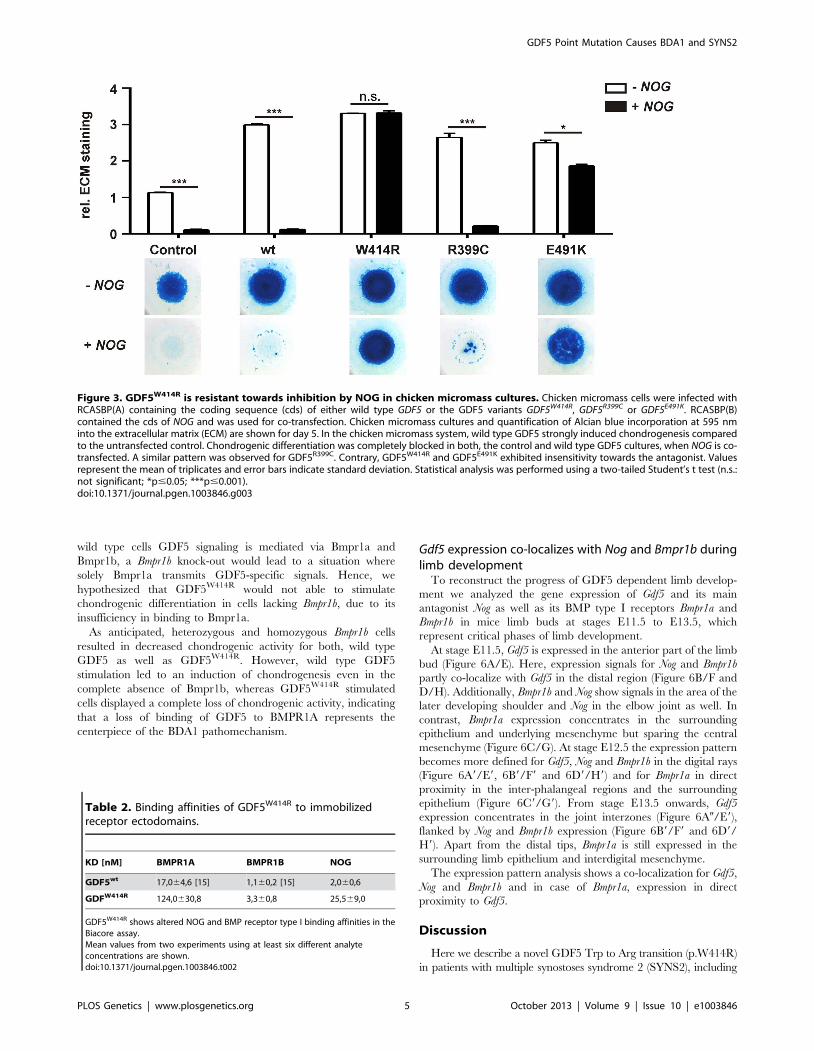
GDF5W414R shows specific loss of BMPR1A signalingAs GDF5W414R is located in the overlapping interface of the
high affinity BMP type I receptors and NOG, we analyzed
subsequently signaling activities of wild type GDF5 and
GDF5W414R after co-expression of either one of the type I
receptors, Bmpr1a or Bmpr1b. Signaling activities were determined
in NIH/3T3 cells using a Smad Binding Element (SBE) luciferase
reporter gene assay (Figure 4A–C).
Overexpression of wild type GDF5 in combination with either
one of the two type I receptors, Bmpr1a and Bmpr1b, resulted in a
strong induction of luciferase activity. As expected from our
Biacore data, wild type GDF5-induced signaling via Bmpr1b was
stronger compared to signaling mediated via Bmpr1a (Figure 4B–
C; Table 2). In case of GDF5W414R, no reporter gene activity was
observed when Bmpr1a was additionally transfected (Figure 4B).
However, co-transfection of Bmpr1b led to a clear induction of the
SBE reporter, even though to a slightly lesser extent compared to
wild type GDF5 (Figure 4C). For the BDA1 associated variant
GDF5R399C, we revealed the same signaling pattern in our
luciferase assay as for GDF5W414R, which leads to the assumption
that the pathomechanism of BDA1 is presumably connected with
an alteration of the GDF5:BMPR1A binding interaction. In
contrast, the SYM1/SYNS2 causing GDF5 variant GDF5E491K
promotes GDF5 signaling via Bmpr1a and Bmpr1b to a similar
extent when compared to wild type GDF5.
Biacore analysis supported the findings from our cell based
assays since GDF5W414R showed a clear deviation from the wild
type GDF5 receptor binding pattern. We could demonstrate a 7-
fold lower affinity of GDF5W414R to BMPR1A (apparent KD:
,124 nM) compared to wild type GDF5 (apparent KD: ,17 nM)
Table 1. Clinical features of the affected family members with mutations in GDF5.
Feature HPO:ID W414R E491K [14] R399C [19]
Proximal symphalangism HP:0100264 + + 2
Distal symphalangism HP:0100263 + + 2
Metacarpophalangeal Synostosis HP:0100325 + 2 2
Synostosis of carpal bones HP:0005048 + + 2
Synostosis of tarsal bones HP:0100330 + + 2
Tarsometatarsal synostosis HP:0100329 2 + 2
Aplasia/Hypoplasia of the middle phalanges of the hand(Brachydactyly Type A1)
HP:0009843 + 2 +
Hypoplastic/short 1st metacarpal HP:0010034 2 2 +
The features are coded using terms from the Human Phenotype Ontology [47]. + present; 2 absent. GDF5 mutations are presented with either features of brachydactyly(GDF5 p.R399C) or features of synostosis (GDF5 p.E491K) or a combination of multiple synostosis with additional brachydactyly (GDF5 p.W414R).doi:10.1371/journal.pgen.1003846.t001
GDF5 Point Mutation Causes BDA1 and SYNS2
PLOS Genetics | www.plosgenetics.org 3 October 2013 | Volume 9 | Issue 10 | e1003846

and only a 3-fold lower affinity for BMPR1B (apparent KD: ,3,3)
compared to wild type GDF5 (apparent KD: ,1,1 nM) (Table 2).
In summary, Biacore analysis and in vitro overexpression studies
indicate a functional link between the phenotypic features of
BDA1 and an impaired BMPR1A signaling of BDA1 associated
GDF5 variants.
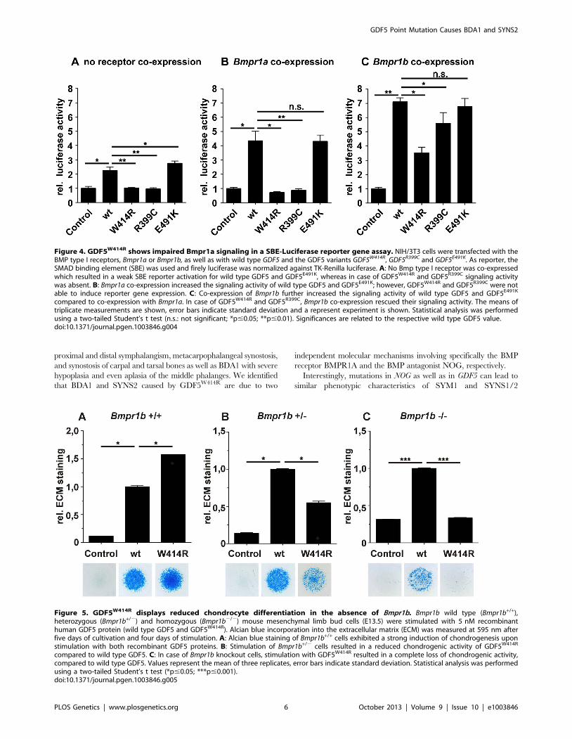
Activity of GDF5W414R is reduced in the absence ofBmpr1b
In order to confirm the previous hypothesis, that GDF5W414R isnot able to transduce signaling via BMPR1A, we conducted an invitro chondrogenesis assay using primary mesenchymal cellsderived from Bmpr1b null mice (Figure 5A–C). Assuming that in
Figure 2. GDF5W414R is positioned within the NOG and BMPR1A/B binding interface of the GDF5 dimer. A: 3D presentation of thehuman GDF5 homodimer (PDB 1waq). The topology of the GDF5 monomer comprises two ß-sheets forming the fingers as well as the four-turn a-helix with the preceding pre-helix loop. The mutations are highlighted in pink (GDF5W414R), violet (GDF5R399C) and orange (GDF5E491K). The image ofthe GDF5 structure was visualized using PyMol (http://www.pymol.org/). B: Protein sequence alignment of human, mouse and chicken GDF5comprising the seven cysteine residues (bold) of the mature domain. Numbering is referred to the pro-protein sequence. Amino acids predicted toform the NOG binding interface are depicted as framed white boxes and based on the BMP7:NOG complex (PDB 1m4u). Residues predicted to beinvolved in BMPR1A binding are shown as grey boxes and refer to the BMP2:BMPR1A structure (PDB 1rew). Black boxes mark amino acids that bind toBMPR1B (PDB 3evs). Arrows indicate the mutated sites for GDF5W414R, GDF5R399C and GDF5E491K. Note that GDF5W414R and GDF5E491K are locatedwithin the NOG binding site. Moreover, all three mutations interfere with the BMP type I receptor (BMPR1A and BMPR1B) binding interface.doi:10.1371/journal.pgen.1003846.g002
GDF5 Point Mutation Causes BDA1 and SYNS2
PLOS Genetics | www.plosgenetics.org 4 October 2013 | Volume 9 | Issue 10 | e1003846
wild type cells GDF5 signaling is mediated via Bmpr1a and
Bmpr1b, a Bmpr1b knock-out would lead to a situation where
solely Bmpr1a transmits GDF5-specific signals. Hence, we
hypothesized that GDF5W414R would not able to stimulate
chondrogenic differentiation in cells lacking Bmpr1b, due to its
insufficiency in binding to Bmpr1a.
As anticipated, heterozygous and homozygous Bmpr1b cells
resulted in decreased chondrogenic activity for both, wild type
GDF5 as well as GDF5W414R. However, wild type GDF5
stimulation led to an induction of chondrogenesis even in the
complete absence of Bmpr1b, whereas GDF5W414R stimulated
cells displayed a complete loss of chondrogenic activity, indicating
that a loss of binding of GDF5 to BMPR1A represents the
centerpiece of the BDA1 pathomechanism.
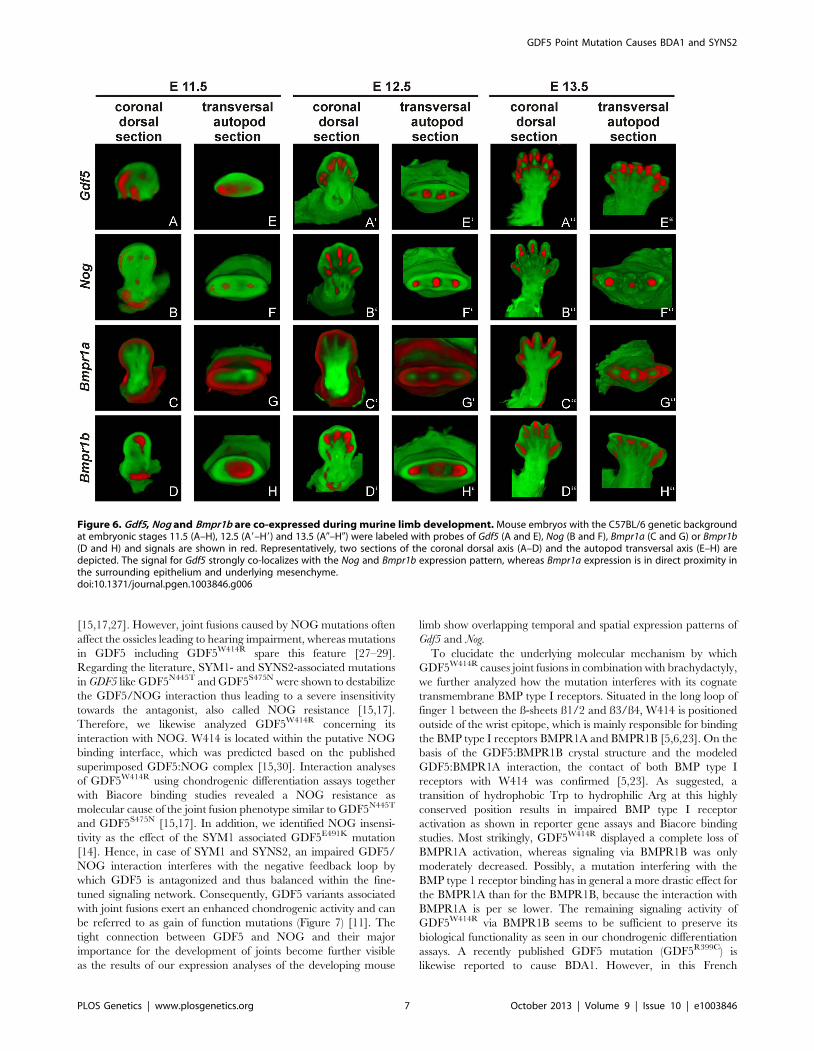
Gdf5 expression co-localizes with Nog and Bmpr1b duringlimb development
To reconstruct the progress of GDF5 dependent limb develop-
ment we analyzed the gene expression of Gdf5 and its main
antagonist Nog as well as its BMP type I receptors Bmpr1a and
Bmpr1b in mice limb buds at stages E11.5 to E13.5, which
represent critical phases of limb development.
At stage E11.5, Gdf5 is expressed in the anterior part of the limb
bud (Figure 6A/E). Here, expression signals for Nog and Bmpr1b
partly co-localize with Gdf5 in the distal region (Figure 6B/F and
D/H). Additionally, Bmpr1b and Nog show signals in the area of the
later developing shoulder and Nog in the elbow joint as well. In
contrast, Bmpr1a expression concentrates in the surrounding
epithelium and underlying mesenchyme but sparing the central
mesenchyme (Figure 6C/G). At stage E12.5 the expression pattern
becomes more defined for Gdf5, Nog and Bmpr1b in the digital rays
(Figure 6A9/E9, 6B9/F9 and 6D9/H9) and for Bmpr1a in direct
proximity in the inter-phalangeal regions and the surrounding
epithelium (Figure 6C9/G9). From stage E13.5 onwards, Gdf5
expression concentrates in the joint interzones (Figure 6A0/E9),
flanked by Nog and Bmpr1b expression (Figure 6B9/F9 and 6D9/
H9). Apart from the distal tips, Bmpr1a is still expressed in the
surrounding limb epithelium and interdigital mesenchyme.
The expression pattern analysis shows a co-localization for Gdf5,
Nog and Bmpr1b and in case of Bmpr1a, expression in direct
proximity to Gdf5.
Discussion
Here we describe a novel GDF5 Trp to Arg transition (p.W414R)
in patients with multiple synostoses syndrome 2 (SYNS2), including
Figure 3. GDF5W414R is resistant towards inhibition by NOG in chicken micromass cultures. Chicken micromass cells were infected withRCASBP(A) containing the coding sequence (cds) of either wild type GDF5 or the GDF5 variants GDF5W414R, GDF5R399C or GDF5E491K. RCASBP(B)contained the cds of NOG and was used for co-transfection. Chicken micromass cultures and quantification of Alcian blue incorporation at 595 nminto the extracellular matrix (ECM) are shown for day 5. In the chicken micromass system, wild type GDF5 strongly induced chondrogenesis comparedto the untransfected control. Chondrogenic differentiation was completely blocked in both, the control and wild type GDF5 cultures, when NOG is co-transfected. A similar pattern was observed for GDF5R399C. Contrary, GDF5W414R and GDF5E491K exhibited insensitivity towards the antagonist. Valuesrepresent the mean of triplicates and error bars indicate standard deviation. Statistical analysis was performed using a two-tailed Student’s t test (n.s.:not significant; *p#0.05; ***p#0.001).doi:10.1371/journal.pgen.1003846.g003
Table 2. Binding affinities of GDF5W414R to immobilizedreceptor ectodomains.
KD [nM] BMPR1A BMPR1B NOG
GDF5wt 17,064,6 [15] 1,160,2 [15] 2,060,6
GDFW414R 124,0630,8 3,360,8 25,569,0
GDF5W414R shows altered NOG and BMP receptor type I binding affinities in theBiacore assay.Mean values from two experiments using at least six different analyteconcentrations are shown.doi:10.1371/journal.pgen.1003846.t002
GDF5 Point Mutation Causes BDA1 and SYNS2
PLOS Genetics | www.plosgenetics.org 5 October 2013 | Volume 9 | Issue 10 | e1003846
proximal and distal symphalangism, metacarpophalangeal synostosis,
and synostosis of carpal and tarsal bones as well as BDA1 with severe
hypoplasia and even aplasia of the middle phalanges. We identified
that BDA1 and SYNS2 caused by GDF5W414R are due to two
independent molecular mechanisms involving specifically the BMP
receptor BMPR1A and the BMP antagonist NOG, respectively.
Interestingly, mutations in NOG as well as in GDF5 can lead to
similar phenotypic characteristics of SYM1 and SYNS1/2
Figure 4. GDF5W414R shows impaired Bmpr1a signaling in a SBE-Luciferase reporter gene assay. NIH/3T3 cells were transfected with theBMP type I receptors, Bmpr1a or Bmpr1b, as well as with wild type GDF5 and the GDF5 variants GDF5W414R, GDF5R399C and GDF5E491K. As reporter, theSMAD binding element (SBE) was used and firely luciferase was normalized against TK-Renilla luciferase. A: No Bmp type I receptor was co-expressedwhich resulted in a weak SBE reporter activation for wild type GDF5 and GDF5E491K, whereas in case of GDF5W414R and GDF5R399C signaling activitywas absent. B: Bmpr1a co-expression increased the signaling activity of wild type GDF5 and GDF5E491K; however, GDF5W414R and GDF5R399C were notable to induce reporter gene expression. C: Co-expression of Bmpr1b further increased the signaling activity of wild type GDF5 and GDF5E491K
compared to co-expression with Bmpr1a. In case of GDF5W414R and GDF5R399C, Bmpr1b co-expression rescued their signaling activity. The means oftriplicate measurements are shown, error bars indicate standard deviation and a represent experiment is shown. Statistical analysis was performedusing a two-tailed Student’s t test (n.s.: not significant; *p#0.05; **p#0.01). Significances are related to the respective wild type GDF5 value.doi:10.1371/journal.pgen.1003846.g004
Figure 5. GDF5W414R displays reduced chondrocyte differentiation in the absence of Bmpr1b. Bmpr1b wild type (Bmpr1b+/+),heterozygous (Bmpr1b+/2) and homozygous (Bmpr1b2/2) mouse mesenchymal limb bud cells (E13.5) were stimulated with 5 nM recombinanthuman GDF5 protein (wild type GDF5 and GDF5W414R). Alcian blue incorporation into the extracellular matrix (ECM) was measured at 595 nm afterfive days of cultivation and four days of stimulation. A: Alcian blue staining of Bmpr1b+/+ cells exhibited a strong induction of chondrogenesis uponstimulation with both recombinant GDF5 proteins. B: Stimulation of Bmpr1b+/2 cells resulted in a reduced chondrogenic activity of GDF5W414R
compared to wild type GDF5. C: In case of Bmpr1b knockout cells, stimulation with GDF5W414R resulted in a complete loss of chondrogenic activity,compared to wild type GDF5. Values represent the mean of three replicates, error bars indicate standard deviation. Statistical analysis was performedusing a two-tailed Student’s t test (*p#0.05; ***p#0.001).doi:10.1371/journal.pgen.1003846.g005
GDF5 Point Mutation Causes BDA1 and SYNS2
PLOS Genetics | www.plosgenetics.org 6 October 2013 | Volume 9 | Issue 10 | e1003846
[15,17,27]. However, joint fusions caused by NOG mutations often
affect the ossicles leading to hearing impairment, whereas mutations
in GDF5 including GDF5W414R spare this feature [27–29].
Regarding the literature, SYM1- and SYNS2-associated mutations
in GDF5 like GDF5N445T and GDF5S475N were shown to destabilize
the GDF5/NOG interaction thus leading to a severe insensitivity
towards the antagonist, also called NOG resistance [15,17].
Therefore, we likewise analyzed GDF5W414R concerning its
interaction with NOG. W414 is located within the putative NOG
binding interface, which was predicted based on the published
superimposed GDF5:NOG complex [15,30]. Interaction analyses
of GDF5W414R using chondrogenic differentiation assays together
with Biacore binding studies revealed a NOG resistance as
molecular cause of the joint fusion phenotype similar to GDF5N445T
and GDF5S475N [15,17]. In addition, we identified NOG insensi-
tivity as the effect of the SYM1 associated GDF5E491K mutation
[14]. Hence, in case of SYM1 and SYNS2, an impaired GDF5/
NOG interaction interferes with the negative feedback loop by
which GDF5 is antagonized and thus balanced within the fine-
tuned signaling network. Consequently, GDF5 variants associated
with joint fusions exert an enhanced chondrogenic activity and can
be referred to as gain of function mutations (Figure 7) [11]. The
tight connection between GDF5 and NOG and their major
importance for the development of joints become further visible
as the results of our expression analyses of the developing mouse
limb show overlapping temporal and spatial expression patterns of
Gdf5 and Nog.
To elucidate the underlying molecular mechanism by which
GDF5W414R causes joint fusions in combination with brachydactyly,
we further analyzed how the mutation interferes with its cognate
transmembrane BMP type I receptors. Situated in the long loop of
finger 1 between the ß-sheets ß1/2 and ß3/ß4, W414 is positioned
outside of the wrist epitope, which is mainly responsible for binding
the BMP type I receptors BMPR1A and BMPR1B [5,6,23]. On the
basis of the GDF5:BMPR1B crystal structure and the modeled
GDF5:BMPR1A interaction, the contact of both BMP type I
receptors with W414 was confirmed [5,23]. As suggested, a
transition of hydrophobic Trp to hydrophilic Arg at this highly
conserved position results in impaired BMP type I receptor
activation as shown in reporter gene assays and Biacore binding
studies. Most strikingly, GDF5W414R displayed a complete loss of
BMPR1A activation, whereas signaling via BMPR1B was only
moderately decreased. Possibly, a mutation interfering with the
BMP type 1 receptor binding has in general a more drastic effect for
the BMPR1A than for the BMPR1B, because the interaction with
BMPR1A is per se lower. The remaining signaling activity of
GDF5W414R via BMPR1B seems to be sufficient to preserve its
biological functionality as seen in our chondrogenic differentiation
assays. A recently published GDF5 mutation (GDF5R399C) is
likewise reported to cause BDA1. However, in this French
Figure 6. Gdf5, Nog and Bmpr1b are co-expressed during murine limb development. Mouse embryos with the C57BL/6 genetic backgroundat embryonic stages 11.5 (A–H), 12.5 (A9–H9) and 13.5 (A0–H0) were labeled with probes of Gdf5 (A and E), Nog (B and F), Bmpr1a (C and G) or Bmpr1b(D and H) and signals are shown in red. Representatively, two sections of the coronal dorsal axis (A–D) and the autopod transversal axis (E–H) aredepicted. The signal for Gdf5 strongly co-localizes with the Nog and Bmpr1b expression pattern, whereas Bmpr1a expression is in direct proximity inthe surrounding epithelium and underlying mesenchyme.doi:10.1371/journal.pgen.1003846.g006
GDF5 Point Mutation Causes BDA1 and SYNS2
PLOS Genetics | www.plosgenetics.org 7 October 2013 | Volume 9 | Issue 10 | e1003846
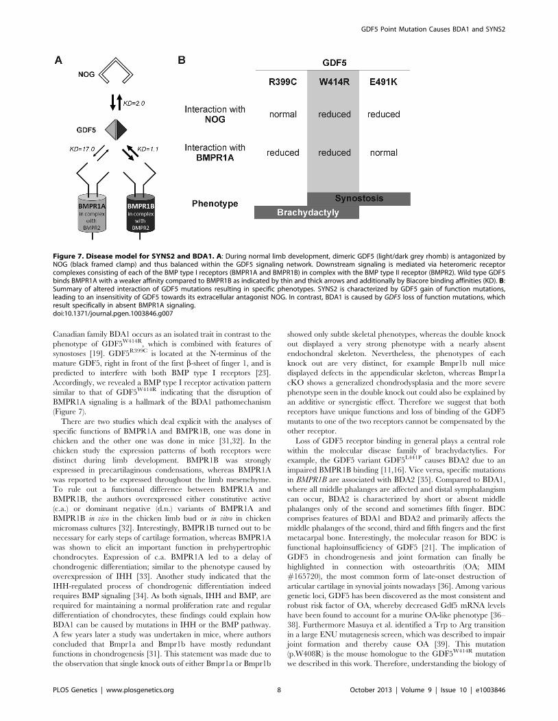
Canadian family BDA1 occurs as an isolated trait in contrast to the
phenotype of GDF5W414R, which is combined with features of
synostoses [19]. GDF5R399C is located at the N-terminus of the
mature GDF5, right in front of the first b-sheet of finger 1, and is
predicted to interfere with both BMP type I receptors [23].
Accordingly, we revealed a BMP type I receptor activation pattern
similar to that of GDF5W414R indicating that the disruption of
BMPR1A signaling is a hallmark of the BDA1 pathomechanism
(Figure 7).
There are two studies which deal explicit with the analyses of
specific functions of BMPR1A and BMPR1B, one was done in
chicken and the other one was done in mice [31,32]. In the
chicken study the expression patterns of both receptors were
distinct during limb development. BMPR1B was strongly
expressed in precartilaginous condensations, whereas BMPR1A
was reported to be expressed throughout the limb mesenchyme.
To rule out a functional difference between BMPR1A and
BMPR1B, the authors overexpressed either constitutive active
(c.a.) or dominant negative (d.n.) variants of BMPR1A and
BMPR1B in vivo in the chicken limb bud or in vitro in chicken
micromass cultures [32]. Interestingly, BMPR1B turned out to be
necessary for early steps of cartilage formation, whereas BMPR1A
was shown to elicit an important function in prehypertrophic
chondrocytes. Expression of c.a. BMPR1A led to a delay of
chondrogenic differentiation; similar to the phenotype caused by
overexpression of IHH [33]. Another study indicated that the
IHH-regulated process of chondrogenic differentiation indeed
requires BMP signaling [34]. As both signals, IHH and BMP, are
required for maintaining a normal proliferation rate and regular
differentiation of chondrocytes, these findings could explain how
BDA1 can be caused by mutations in IHH or the BMP pathway.
A few years later a study was undertaken in mice, where authors
concluded that Bmpr1a and Bmpr1b have mostly redundant
functions in chondrogenesis [31]. This statement was made due to
the observation that single knock outs of either Bmpr1a or Bmpr1b
showed only subtle skeletal phenotypes, whereas the double knock
out displayed a very strong phenotype with a nearly absent
endochondral skeleton. Nevertheless, the phenotypes of each
knock out are very distinct, for example Bmpr1b null mice
displayed defects in the appendicular skeleton, whereas Bmpr1a
cKO shows a generalized chondrodysplasia and the more severe
phenotype seen in the double knock out could also be explained by
an additive or synergistic effect. Therefore we suggest that both
receptors have unique functions and loss of binding of the GDF5
mutants to one of the two receptors cannot be compensated by the
other receptor.
Loss of GDF5 receptor binding in general plays a central role
within the molecular disease family of brachydactylies. For
example, the GDF5 variant GDF5L441P causes BDA2 due to an
impaired BMPR1B binding [11,16]. Vice versa, specific mutations
in BMPR1B are associated with BDA2 [35]. Compared to BDA1,
where all middle phalanges are affected and distal symphalangism
can occur, BDA2 is characterized by short or absent middle
phalanges only of the second and sometimes fifth finger. BDC
comprises features of BDA1 and BDA2 and primarily affects the
middle phalanges of the second, third and fifth fingers and the first
metacarpal bone. Interestingly, the molecular reason for BDC is
functional haploinsufficiency of GDF5 [21]. The implication of
GDF5 in chondrogenesis and joint formation can finally be
highlighted in connection with osteoarthritis (OA; MIM
#165720), the most common form of late-onset destruction of
articular cartilage in synovial joints nowadays [36]. Among various
genetic loci, GDF5 has been discovered as the most consistent and
robust risk factor of OA, whereby decreased Gdf5 mRNA levels
have been found to account for a murine OA-like phenotype [36–
38]. Furthermore Masuya et al. identified a Trp to Arg transition
in a large ENU mutagenesis screen, which was described to impair
joint formation and thereby cause OA [39]. This mutation
(p.W408R) is the mouse homologue to the GDF5W414R mutation
we described in this work. Therefore, understanding the biology of
Figure 7. Disease model for SYNS2 and BDA1. A: During normal limb development, dimeric GDF5 (light/dark grey rhomb) is antagonized byNOG (black framed clamp) and thus balanced within the GDF5 signaling network. Downstream signaling is mediated via heteromeric receptorcomplexes consisting of each of the BMP type I receptors (BMPR1A and BMPR1B) in complex with the BMP type II receptor (BMPR2). Wild type GDF5binds BMPR1A with a weaker affinity compared to BMPR1B as indicated by thin and thick arrows and additionally by Biacore binding affinities (KD). B:Summary of altered interaction of GDF5 mutations resulting in specific phenotypes. SYNS2 is characterized by GDF5 gain of function mutations,leading to an insensitivity of GDF5 towards its extracellular antagonist NOG. In contrast, BDA1 is caused by GDF5 loss of function mutations, whichresult specifically in absent BMPR1A signaling.doi:10.1371/journal.pgen.1003846.g007
GDF5 Point Mutation Causes BDA1 and SYNS2
PLOS Genetics | www.plosgenetics.org 8 October 2013 | Volume 9 | Issue 10 | e1003846

GDF5W414R might also give insights into the pathophysiology of
OA.
In summary, we revealed that GDF5W414R, in contrast to wild
type GDF5, loses the BMPR1A signaling route and at the same
time increases the alternative signaling via BMPR1B in the
presence of NOG. Therefore, the reduced sensitivity of W414R to
Noggin and its reduced interaction with BMPR1A do not actually
‘‘neutralize’’ each other, but lead to a misbalance of BMPR1A and
BMPR1B signaling. Hence, our study assembles another part of
the molecular puzzle how loss and gain of function mutations in
GDF5 affect bone development in hands and feet and result in
specific types of brachydactyly and SYNS2.
Materials and Methods
Clinical investigation and molecular analysisAll clinical investigations have been performed according to
Declaration of Helsinki principles. The study was approved by the
local institutional review board ‘‘Ethikkommission der Charite -
Universitatsmedizin Berlin’’. Informed consent for genetic testing
was obtained from the patient or their legal guardians respectively.
Genomic DNA of affected family members were extracted from
peripheral blood samples by standard methods. The coding
regions of NOG and GDF5 as well as the flanking intronic
sequences were amplified by standard PCR protocols. The primer
sequences and PCR conditions for the molecular testing were
previously described [20,30]. PCR products were analyzed on 2%
agarose gels. Sequencing was done using the ABI Prism BigDye
Terminator Sequencing Kit (Applied Biosystems) with PCR
primers used as sequencing primers. Products were evaluated on
an automated capillary sequencer (Applied Biosystems).
Chicken micromass culturesCloning of the coding sequences of chicken GDF5 and NOG into
RCAS(BP)A or RCAS(BP)B, respectively was previously described
[15]. Mutations (GDF5W414R, GDF5R399C, GDF5E491K) were
introduced into the coding sequence of chicken GDF5 in pSLAX13
by in vitro mutagenesis. Primer sequences are available in the
supplement (Table S1). Production of viral supernatant in DF1 cells
and concentration of viral particles was performed as described
previously [40]. Fertilized chicken eggs were obtained from VALO
BioMedia GmbH (Osterholz-Scharmbeck, Germany) and incubated
at 38uC in a humidified egg incubator for 4.5 days. Micromass
cultures were plated in a drop containing 26105 cells. Infection was
performed with concentrated viral supernatants: RCASBP(A)
containing cDNA encoding chicken wild type GDF5 and the GDF5
mutants GDF5W414R, GDF5R399C, and GDF5E491K with a titer of
16107 plaque forming units (PFU)/ml. RCASBP(B) containing the
cDNA encoding chicken wild type NOG was applied with a titer of
2,56106 PFU/ml. Culture medium containing DMEM-F12 (Bio-
chrom), 10% FBS (Biochrom), 0,2% chicken serum (Sigma), 2 mM
L-Gln (Lonza), 100 U/ml penicillin, and 100 mg/ml streptomycin
(Lonza) was replaced every 2 days. For each condition, three
replicates were performed in parallel. Quantification of Alcian blue
dye was performed at 595 nm after extraction with Guanidin-HCl.
Recombinant proteinsRecombinant human (rh) GDF5 and its variant rhGDF5W414R
were dissolved in 10 mM HCl and provided by Biopharm GmbH.
BIAcore binding assayThe BIA2000 system (Biacore) was used to analyze the binding
affinities of recombinant human GDF5 and its variant
GDF5W414R to immobilized NOG and ectodomains of BMPR1A,
BMPR1B and BMPR2, as previously described [16].
Luciferase activity assayCoding sequences of human GDF5 and mouse Bmpr1a and
Bmpr1b were cloned into pSLAX13. Mutations (GDF5W414R,
GDF5R399C, GDF5E491K) were introduced into the coding
sequence of human GDF5 in pSLAX13 by in vitro mutagenesis.
Primer sequences are available in the supplement (Table S1).
Inserts were subcloned into pCS2+ via ClaI.
Luciferase reporter gene assays were performed using the
murine fibroblast cell line NIH/3T3 (ATCC) which was
maintained in DMEM high glucose (Lonza) with 10% FCS
(Biochrom), 2 mM L-Gln (Lonza), 100 U/ml penicillin, and
100 mg/ml streptomycin (Lonza). Prior to transfection, cells were
seeded in a 96-well plate at a density of 16104 cells per well. BMP
receptors and GDF5 constructs were transfected for 40 hours
together with the Smad Binding Element luciferase construct SBE-
pGL3 [41] and the normalization vector pRLTk (Promega) using
Lipofectamine 2000 (Invitrogen). Luciferase activity was deter-
mined as described previously [42].
Mouse micromass culturesLimb mesenchymal cells were isolated from stage E13.5
embryos resulting from matings of C57BL/6, Bmpr1btm1kml
heterozygous or homozygous knock-out mice on a C57BL/6
background [43]. Mouse embryos were genotyped using primers
for Bmpr1b and neomycin (Table S2), if applicable embryos were
pooled according to their phenotypes. Isolation of mouse
micromass cells was performed as described for chicken micromass
cultures with minor modifications. For mouse micromass cultures
no additional chicken serum was used. After 24 h mouse
micromass cultures were stimulated with 5 nM of recombinant
human wild type GDF5 and GDF5W414R.
Whole mount in situ hybridizationC57BL/6 mouse embryos were harvested at stages E11.5–13.5
and fixed in 4% PFA. Whole mount in situ hybridization was
performed as previously described [44].
DIG-labeled RNA antisense-probes were generated by in
vitro transcription using the coding sequences of mouse Bmpr1a,
Bmpr1b and Nog as a template. The probe for mouse Gdf5 was
previously published [45]. Signal detection was performed with
BMPurple (Roche). 3D imaging of labeled limbs was done by
optical projection tomography (OPT) scans as previously
described [46].
StatisticsStatistical analyses were performed using a two-tailed Student’s
T-test. Results are presented as mean 6 SEM. P values of less than
0.05 were considered significant.
Supporting Information
Figure S1 Wild type and mutant GDF5 transcripts are
expressed at comparable levels in chicken micromass cultures.
Chicken micromass cultures were infected with empty
RCASBP(A) as control and RCASBP(A) containing the cds of
either wild type GDF5 or the GDF5 variants (GDF5W414R,
GDF5R399C, GDF5E491K). After SDS-PAGE under non-reducing
(GDF5) and reducing (ACTIN) conditions and subsequent
Western Blot, GDF5 and ACTIN were detected at comparable
levels using specific antibodies.
(TIF)
GDF5 Point Mutation Causes BDA1 and SYNS2
PLOS Genetics | www.plosgenetics.org 9 October 2013 | Volume 9 | Issue 10 | e1003846
Table S1 Primers used for site-directed mutagenesis. In vitro
mutagenesis of GDF5 mutations (GDF5W414R, GDF5R399C,
GDF5E491K) into the coding sequences of chicken GDF5 and
human GDF5 were carried out by using the following primers.
(DOC)
Table S2 Primers used for mouse genotyping. Genotyping of
Bmpr1b wild type (Bmpr1b+/+), heterozygous (Bmpr1b+/2) and
homozygous (Bmpr1b2/2) mouse embryos for mouse micromass
assays was carried out using the following primers.
(DOC)
Text S1 Materials and Methods for anti-GDF5 Western blot.
(DOC)
Acknowledgments
The authors would like to thank Mareen Schmidt-von Kegler for excellent
technical assistance. pGl3basic-SBE and chicken NOG in RCAS(BP)B were
kindly provided by Peter ten Dijke and Andrea Vortkamp, respectively.
Furthermore, we thank Daniel Graf for providing the genotyping protocol
of BmprIBtm1kml mouse and Lutz Schomburg for critical remarks on the
manuscript.
Author Contributions
Conceived and designed the experiments: PS. Performed the experiments:
ED JK JZ MW JN JR. Analyzed the data: ED JK JZ MW CR JN FP JR JS
KD JTH SM SCD PS. Contributed reagents/materials/analysis tools: CR
JN FP JS JTH SM SCD PS. Wrote the paper: ED PS.
References
1. Chang SC, Hoang B, Thomas JT, Vukicevic S, Luyten FP, et al. (1994)
Cartilage-derived morphogenetic proteins. New members of the transforming
growth factor-beta superfamily predominantly expressed in long bones duringhuman embryonic development. J Biol Chem 269: 28227–28234.
2. Stricker S, Mundlos S (2011) Mechanisms of Digit Formation: HumanMalformation Syndromes Tell the Story. Developmental Dynamics 240: 990–1004.
3. Buxton P, Edwards C, Archer CW, Francis-West P (2001) Growth/differentiation factor-5 (GDF-5) and skeletal development. J Bone Joint Surg
Am 83-A Suppl 1: S23–30.
4. Storm EE, Huynh TV, Copeland NG, Jenkins NA, Kingsley DM, et al. (1994)Limb alterations in brachypodism mice due to mutations in a new member of
the TGF beta-superfamily. Nature 368: 639–643.5. Kotzsch A, Nickel J, Seher A, Sebald W, Muller TD (2009) Crystal structure
analysis reveals a spring-loaded latch as molecular mechanism for GDF-5-type I
receptor specificity. EMBO J 28: 937–947.6. Mueller TD, Nickel J (2012) Promiscuity and specificity in BMP receptor
activation. FEBS Lett 586: 1846–1859.7. Nohe A, Keating E, Knaus P, Petersen NO (2004) Signal transduction of bone
morphogenetic protein receptors. Cell Signal 16: 291–299.8. Schmierer B, Hill CS (2007) TGFbeta-SMAD signal transduction: molecular
specificity and functional flexibility. Nat Rev Mol Cell Biol 8: 970–982.
9. Miyazono K, Kamiya Y, Morikawa M (2010) Bone morphogenetic proteinreceptors and signal transduction. J Biochem 147: 35–51.
10. Bragdon B, Moseychuk O, Saldanha S, King D, Julian J, et al. (2011) Bonemorphogenetic proteins: a critical review. Cell Signal 23: 609–620.
11. Mundlos S (2009) The brachydactylies: a molecular disease family. Clin Genet
76: 123–136.12. Lories RJ, Luyten FP (2005) Bone morphogenetic protein signaling in joint
homeostasis and disease. Cytokine Growth Factor Rev 16: 287–298.13. Dawson K, Seeman P, Sebald E, King L, Edwards M, et al. (2006) GDF5 is a
second locus for multiple-synostosis syndrome. Am J Hum Genet 78: 708–712.14. Wang X, Xiao F, Yang Q, Liang B, Tang Z, et al. (2006) A novel mutation in
GDF5 causes autosomal dominant symphalangism in two Chinese families.
Am J Med Genet A 140A: 1846–1853.15. Seemann P, Brehm A, Konig J, Reissner C, Stricker S, et al. (2009) Mutations in GDF5
reveal a key residue mediating BMP inhibition by NOGGIN. PLoS Genet 5: e1000747.16. Seemann P, Schwappacher R, Kjaer KW, Krakow D, Lehmann K, et al. (2005)
Activating and deactivating mutations in the receptor interaction site of GDF5
cause symphalangism or brachydactyly type A2. J Clin Invest 115: 2373–2381.17. Schwaerzer GK, Hiepen C, Schrewe H, Nickel J, Ploeger F, et al. (2012) New
insights into the molecular mechanism of multiple synostoses syndrome (SYNS):mutation within the GDF5 knuckle epitope causes noggin-resistance. J Bone
Miner Res 27: 429–442.
18. Ploger F, Seemann P, Schmidt-von Kegler M, Lehmann K, Seidel J, et al. (2008)Brachydactyly type A2 associated with a defect in proGDF5 processing. Hum
Mol Genet 17: 1222–1233.19. Byrnes AM, Racacho L, Nikkel SM, Xiao F, MacDonald H, et al. (2010) Mutations in
GDF5 presenting as semidominant brachydactyly A1. Hum Mutat 31: 1155–1162.20. Schwabe GC, Turkmen S, Leschik G, Palanduz S, Stover B, et al. (2004)
Brachydactyly type C caused by a homozygous missense mutation in the
prodomain of CDMP1. Am J Med Genet A 124A: 356–363.21. Everman DB, Bartels CF, Yang Y, Yanamandra N, Goodman FR, et al. (2002)
The mutational spectrum of brachydactyly type C. Am J Med Genet 112: 291–296.22. Thomas JT, Kilpatrick MW, Lin K, Erlacher L, Lembessis P, et al. (1997)
Disruption of human limb morphogenesis by a dominant negative mutation in
CDMP1. Nat Genet 17: 58–64.23. Nickel J, Kotzsch A, Sebald W, Mueller TD (2005) A single residue of GDF-5
defines binding specificity to BMP receptor IB. J Mol Biol 349: 933–947.24. Keller S, Nickel J, Zhang JL, Sebald W, Mueller TD (2004) Molecular
recognition of BMP-2 and BMP receptor IA. Nat Struct Mol Biol 11: 481–488.25. Kirsch T, Sebald W, Dreyer MK (2000) Crystal structure of the BMP-2-BRIA
ectodomain complex. Nat Struct Biol 7: 492–496.
26. Groppe J, Greenwald J, Wiater E, Rodriguez-Leon J, Economides AN, et al.
(2002) Structural basis of BMP signalling inhibition by the cystine knot protein
Noggin. Nature 420: 636–642.27. Gong Y, Krakow D, Marcelino J, Wilkin D, Chitayat D, et al. (1999)
Heterozygous mutations in the gene encoding noggin affect human jointmorphogenesis. Nat Genet 21: 302–304.
28. Brown DJ, Kim TB, Petty EM, Downs CA, Martin DM, et al. (2002) Autosomaldominant stapes ankylosis with broad thumbs and toes, hyperopia, and skeletal
anomalies is caused by heterozygous nonsense and frameshift mutations in
NOG, the gene encoding noggin. Am J Hum Genet 71: 618–624.29. Mangino M, Flex E, Digilio MC, Giannotti A, Dallapiccola B (2002)
Identification of a novel NOG gene mutation (P35S) in an Italian family withsymphalangism. Hum Mutat 19: 308.
30. Lehmann K, Seemann P, Silan F, Goecke TO, Irgang S, et al. (2007) A new
subtype of brachydactyly type B caused by point mutations in the bonemorphogenetic protein antagonist NOGGIN. Am J Hum Genet 81: 388–396.
31. Yoon BS, Ovchinnikov DA, Yoshii I, Mishina Y, Behringer RR, et al. (2005)Bmpr1a and Bmpr1b have overlapping functions and are essential for
chondrogenesis in vivo. Proc Natl Acad Sci U S A 102: 5062–5067.32. Zou H, Wieser R, Massague J, Niswander L (1997) Distinct roles of type I bone
morphogenetic protein receptors in the formation and differentiation of
cartilage. Genes Dev 11: 2191–2203.33. Vortkamp A, Lee K, Lanske B, Segre GV, Kronenberg HM, et al. (1996)
Regulation of rate of cartilage differentiation by Indian hedgehog and PTH-related protein. Science 273: 613–622.
34. Minina E, Wenzel HM, Kreschel C, Karp S, Gaffield W, et al. (2001) BMP and
Ihh/PTHrP signaling interact to coordinate chondrocyte proliferation anddifferentiation. Development 128: 4523–4534.
35. Lehmann K, Seemann P, Stricker S, Sammar M, Meyer B, et al. (2003)Mutations in bone morphogenetic protein receptor 1B cause brachydactyly type
A2. Proc Natl Acad Sci U S A 100: 12277–12282.36. Reynard LN, Loughlin J (2012) Genetics and epigenetics of osteoarthritis.
Maturitas 71: 200–204.
37. Liu J, Cai W, Zhang H, He C, Deng L (2013) Rs143383 in the GrowthDifferentiation Factor 5 (GDF5) Gene Significantly Associated with Osteoar-
thritis (OA)-A Comprehensive Meta-analysis. Int J Med Sci 10: 312–319.38. Daans M, Luyten FP, Lories RJ (2011) GDF5 deficiency in mice is associated
with instability-driven joint damage, gait and subchondral bone changes. Ann
Rheum Dis 70: 208–213.39. Masuya H, Nishida K, Furuichi T, Toki H, Nishimura G, et al. (2007) A novel
dominant-negative mutation in Gdf5 generated by ENU mutagenesis impairsjoint formation and causes osteoarthritis in mice. Hum Mol Genet 16: 2366–2375.
40. Morgan BA, Fekete DM (1996) Manipulating gene expression with replication-
competent retroviruses. Methods Cell Biol 51: 185–218.41. Jonk LJ, Itoh S, Heldin CH, ten Dijke P, Kruijer W (1998) Identification and
functional characterization of a Smad binding element (SBE) in the JunBpromoter that acts as a transforming growth factor-beta, activin, and bone
morphogenetic protein-inducible enhancer. J Biol Chem 273: 21145–21152.42. Hampf M, Gossen M (2006) A protocol for combined Photinus and Renilla
luciferase quantification compatible with protein assays. Anal Biochem 356: 94–99.
43. Yi SE, Daluiski A, Pederson R, Rosen V, Lyons KM (2000) The type I BMP receptorBMPRIB is required for chondrogenesis in the mouse limb. Development 127: 621–630.
44. Pryce BA, Brent AE, Murchison ND, Tabin CJ, Schweitzer R (2007) Generationof transgenic tendon reporters, ScxGFP and ScxAP, using regulatory elements of
the scleraxis gene. Dev Dyn 236: 1677–1682.
45. Sharpe J, Ahlgren U, Perry P, Hill B, Ross A, et al. (2002) Optical projection tomographyas a tool for 3D microscopy and gene expression studies. Science 296: 541–545.
46. Quintana L, Sharpe J (2011) Preparation of mouse embryos for opticalprojection tomography imaging. Cold Spring Harb Protoc 2011: 664–669.
47. Robinson PN, Kohler S, Bauer S, Seelow D, Horn D, et al. (2008) The HumanPhenotype Ontology: a tool for annotating and analyzing human hereditary
disease. Am J Hum Genet 83: 610–615.
GDF5 Point Mutation Causes BDA1 and SYNS2
PLOS Genetics | www.plosgenetics.org 10 October 2013 | Volume 9 | Issue 10 | e1003846