Embed Size (px)
Citation preview

A fully defined and scalable 3D culture system for humanpluripotent stem cell expansion and differentiationYuguo Leia,b,c,d and David V. Schaffera,b,c,d,1
Departments of aBioengineering and bChemical Engineering, cCalifornia Institute for Quantitative Biosciences, and dHelen Wills Neuroscience Institute,University of California, Berkeley, CA 94720
Edited by Linda G. Griffith, Massachusetts Institute of Technology, Cambridge, MA, and accepted by the Editorial Board October 25, 2013 (received for reviewMay 17, 2013)
Human pluripotent stem cells (hPSCs), including human embryonicstem cells and induced pluripotent stem cells, are promising fornumerous biomedical applications, such as cell replacement ther-apies, tissue and whole-organ engineering, and high-throughputpharmacology and toxicology screening. Each of these applica-tions requires large numbers of cells of high quality; however, thescalable expansion and differentiation of hPSCs, especially forclinical utilization, remains a challenge. We report a simple, defined,efficient, scalable, and good manufacturing practice-compatible3D culture system for hPSC expansion and differentiation. Itemploys a thermoresponsive hydrogel that combines easy ma-nipulation and completely defined conditions, free of anyhuman- or animal-derived factors, and entailing only recombi-nant protein factors. Under an optimized protocol, the 3D sys-tem enables long-term, serial expansion of multiple hPSCs lineswith a high expansion rate (∼20-fold per 5-d passage, for a 1072-fold expansion over 280 d), yield (∼2.0 × 10⁷ cells per mL ofhydrogel), and purity (∼95% Oct4+), even with single-cell inoc-ulation, all of which offer considerable advantages relative tocurrent approaches. Moreover, the system enabled 3D directeddifferentiation of hPSCs into multiple lineages, including dopa-minergic neuron progenitors with a yield of ∼8 × 10⁷ dopami-nergic progenitors per mL of hydrogel and ∼80-fold expansionby the end of a 15-d derivation. This versatile system may beuseful at numerous scales, from basic biological investigation toclinical development.
Human pluripotent stem cells (hPSCs), including humanembryonic stem cells (hESCs) (1) and induced pluripotent
stem cells (iPSCs) (2), have the capacities for indefinite in vitroexpansion and differentiation into all cell types within adults (3).They therefore represent highly promising cell sources for nu-merous biomedical applications, such as cell replacement ther-apies (4, 5), tissue and organ engineering (6), and pharmacologyand toxicology screens (7, 8). However, these applications requirelarge numbers of cells of high quality (4, 6–8). For instance, ∼105surviving dopaminergic (DA) neurons, ∼109 cardiomyocytes, or∼109 beta cells are likely required to treat a patient with Par-kinson disease (PD), myocardial infarction (MI), or type I di-abetes, respectively (9). Additionally, far more cells are neededinitially because both in vitro cell culture yields and subsequentin vivo survival of transplanted cells are typically very low. Asexamples of the latter, only ∼6% of transplanted dopaminergicneurons or ∼1% of injected cardiomyocytes reportedly survive inrodent models several months after transplantation (10, 11).Furthermore, there are large patient populations with degener-ative diseases or organ failure (9), including over 1 million peoplewith PD, 1–2.5 million with type I diabetes, and ∼8 million with MIin the United States alone (12). Large numbers of cells are alsonecessary for applications such as tissue engineering, where forexample ∼1010 hepatocytes or cardiomyocytes would be requiredfor an artificial human liver or heart, respectively (6). Additionally,∼1010 cells may be needed to screen a million-compound libraryonce (8), and advances in combinatorial chemistry, noncodingRNAs, and investigations of complex signaling and transcriptional
networks have given rise to large libraries that can be screenedagainst many targets (13). Massive numbers of hPSCs may thereforebe needed to deliver on the biomedical promise of these stem cells.In general, hPSCs require key biological signals from their
substrate, and from one another (14, 15), that promote cellsurvival and rapid proliferation and that culture systems mustthus provide. Current 2D-based cell culture systems—whichsuffer from inherent heterogeneity and limited scalability andreproducibility—are emerging as a bottleneck for producing suf-ficient numbers of high-quality cells for downstream applications(9, 16). An attractive approach for scaling up production is tomove cell culture from 2D to 3D (9, 17), and accordingly several3D suspension systems have been probed for hPSCs production:cell aggregates (18–21), cells on microcarriers (22, 23), and cellsin alginate microencapsulates (24) (SI Appendix, Table S1). Al-though these approaches have some attractive aspects, they alsohighlight significant challenges for 3D hPSC culture (9) (SI Ap-pendix, Table S1) including the following: (i) the use of com-ponents from human or animal tissue (e.g., Matrigel, serum, and/or albumin), which limit reproducibility and/or scalability, poserisks for pathogen and immunogen transfer (18–24), and arethus problematic for good manufacturing practice (GMP) cellproduction (25); (ii) substantial cell agglomeration that can insome cases lead to differentiation and/or death (22, 23); (iii)shear forces in agitated cultures that can compromise cell vi-ability (18–23); (iv) limited cell expansion rates and cell yieldsper volume (18–24); and (v) unclear potential for long-termserial expansion. As an example, in a recent culture of hPSCswithin alginate hydrogel microspheres in mouse embryonicfibroblast conditioned medium, 5% of the encapsulated sin-gle hPSCs remained viable after 7 d, and an ∼10- to 20-foldexpansion to a peak density of 3 × 10⁶ cells per mL occurredafter 20 d (24).
Significance
Human pluripotent stem cells can be cultured in vitro and dif-ferentiated into presumably all cell types of the human body,and they therefore represent highly promising cell sources forbiomedical applications such as cell therapies, tissue engi-neering, and drug discovery. These applications require largenumbers of high-quality cells, and we report an efficient, de-fined, scalable, and good manufacturing practice-compatible3D system for the production of human pluripotent stem cellsand their progeny. The ease of use and flexible scalability ofthis system makes it suitable for numerous applications fromthe laboratory toward the clinic.
Author contributions: Y.L. and D.V.S. designed research; Y.L. performed research; and Y.L.and D.V.S. wrote the paper.
The authors declare no conflict of interest.
This article is a PNAS Direct Submission. L.G.G. is a guest editor invited by the EditorialBoard.1To whom correspondence should be addressed. E-mail: [email protected].
This article contains supporting information online at www.pnas.org/lookup/suppl/doi:10.1073/pnas.1309408110/-/DCSupplemental.
www.pnas.org/cgi/doi/10.1073/pnas.1309408110 PNAS Early Edition | 1 of 10
ENGINEE
RING
PNASPL
US

To address these challenges, we report an alternative systemthat expands and differentiates hPSCs within a thermorever-sible hydrogel, composed of a polymeric solution that is liquidat low temperature but solidifies into an elastic hydrogel whenwarmed. Cells can thus be mixed with the liquid at low tem-perature, suspended and grown in a solid gel at 37 °C, andharvested and passaged by reliquifying the gel at low temper-ature. The resulting hydrogel offers many features that benefithPSC biology and culture, including a 3D environment forrapid cell growth, prevention of large cell aggregate formation,isolation of cells from shear forces, and sufficient porosity fornutrient diffusion. Although thermoreversible materials havebeen used for culturing primary cells in the laboratory (26), large-scale hPSC culture presents numerous additional challenges andconstraints, including apoptosis upon cell dissociation (14) andpassaging; low survival, limited proliferation, and loss of pluri-potency in suboptimal culture conditions; and a strong prefer-ence for defined medium to support downstream biomedicalapplications. We have accordingly investigated these challengesand developed a chemically defined system with no matrix pro-teins that is capable of 1072-fold expansion over 60 passages withstrong maintenance of pluripotency.
ResultsSeveral hydrogel materials—such as alginate (24, 27), agarose(28), and hyaluronic acid (29)—have been previously investi-gated for hPSC expansion or differentiation. However, to datethermoreversible materials have not been studied in hPSC cul-ture, despite the fact that they offer a number of promisingfeatures for GMP-compatible, large-scale culture. In particu-lar, they are synthetic and defined, biocompatible, and enablecell harvest or passaging by simply changing temperature at therange from 4 °C to 37 °C, a mild process for cells. Also, they can bereadily processed (e.g., .into spheres or fibers) for large bioreactors.After initial assessment of several synthesized or commercial avail-able thermoreversible hydrogels, we found single hPSCs couldsurvive and retain pluripotency marker expression in 8–10%(wt/vol) poly(N-isopropylacrylamide)-co-poly(ethylene glycol)(PNIPAAm-PEG) hydrogel (Mebiol Gel) (SI Appendix, Fig. S1).Preliminary investigation showed that cell behavior in this
material was significantly affected by multiple factors, includingthe addition of an inhibitor for the RhoA GTPase signaling ef-fector Rho kinase (ROCK) (30), extent of cell dissociation, theculture medium (3), and initial seeding density. We thereforeconducted a multifactorial analysis to assess the promise of thismaterial relative to other materials or to cell-only suspensions,identify the optimal conditions under which it supports the bi-ology and culture of hPSC, and study the potential interactionsamong numerous parameters. Thirty-six combinations of variousfactors, including seeding with single cells or clusters; use ofROCK inhibitor (RI); cell seeding density; culture medium; andwith or without PNIPAAm-PEG hydrogel scaffold, were tested(Figs. 1 and 2). Specifically, cell dissociation promotes hPSCapoptosis via Rho GTPase signaling (14, 31, 32); however, single-cell seeding enables more reproducible expansion during large-scale hPSC production. hPSCs were thus dissociated into singlecells and either directly encapsulated into the hydrogel (termedsingle-cell seeding) or—based on the poor viability observed forsingle-cell dissociation in 2D (30)—first cultured in suspensionovernight to form small clusters that were subsequently encap-sulated in the gel (termed precluster seeding) (Fig. 1A). Also,addition of RI for the first 24 h has been shown to supportgrowth of single hPSCs in liquid suspension culture (19), and weincluded RI for the whole culture period (4-d RI) to assess itseffects on cell expansion. Additionally, low seeding density (∼1–2.5 × 10⁵ cells/mL) is often used (SI Appendix, Table S1), and wealso included medium and high seeding densities (1.0 × 10⁶ and2.5 × 106 cells/mL, respectively). Furthermore, two media were
used [mTeSR and completely defined Essential 8 (E8)]. More-over, hPSC suspension in static liquid medium was also includedfor comparison with the hydrogel. Finally, iPS–mesenchymalstem cells (MSCs), an iPSC derived from human MSCs (33),were used in this optimization.The study revealed that (i) the hydrogel promoted high ex-
pansion rates and prevented cell agglomeration (Figs. 1B and2A). Culturing single hPSCs in 10% (wt/vol) PNIPAAm-PEGwith E8 in the presence of RI led to the formation of dense,uniform, and small spheroids with ∼8-, 10-, or 4.4-fold expansionover 4 d at low, medium, or high seeding density, respectively.However, adding RI for 1 d was insufficient to support singlecells (Figs. 1B and 2A). When using mTeSR rather than E8, onlymoderate (<1.6-fold) or no expansion was reached with 4-d or1-d RI, respectively, for all of the seeding densities, indicatinga negative interaction between mTeSR and the hydrogel scaffold(Figs. 1B and 2A). (ii) In the hydrogel and E8, preclusteredhPSCs behaved similarly to single hPSCs. However, in thehydrogel and mTeSR with 4-d RI, preclustered hPSCs displayedsignificantly higher expansion (∼4.5-, 4.5-, 2.1-fold for low, me-dium, high density, respectively) than single hPSCs (Figs. 1B and2B), indicating that mTeSR stresses single cells. (iii) For allstatic-suspension cultures in liquid medium without hydrogel,large cell aggregates were found, and only ∼2.8-, 1.6-, or 0.2-foldexpansion was achieved for low, medium, or high seeding density,respectively (Figs. 1B and 2C). Agglomeration and low expansion(<2.0-fold) were also found in a large dynamic suspension culture(a spinner flask) without hydrogel scaffold (SI Appendix, Fig. S2).No significant difference was found between using mTeSR and E8or between with 1-d or 4-d RI for these static-suspension cultures(Figs. 1B and 2C). In summary, a combination of 10% (wt/vol)PNIPAAm-PEG, 4-d RI, defined E8, medium seeding densityvia single-cell or precluster seeding resulted in a high, 10.0-foldcell expansion in a single 4-d passage (Fig. 1 B and C). Final, fine-tuning of conditions revealed that 48 h RI was sufficient to supportthis expansion for precluster seeding, 4-d RI was required forsingle-cell seeding, and 8% (wt/vol) hydrogel performed as well as10% (wt/vol) hydrogel (SI Appendix, Fig. S3).We also assessed whether other, nonthermoreversible mate-
rials systems—including UV cross-linked hyarulonic acid (29),agarose (28), and alginate hydrogel (24)—could support highlevel hPSC expansion. We generated and used these materials asdescribed previously, in conjunction with E8 and RI. For single-seeded iPS-MSCs, moderate expansion (∼2.8-fold) was seen onlyin alginate hydrogel with medium seeding density. A slightlyhigher, ∼5- or 6.5-fold expansion was achieved in agarose oralginate hydrogel for preclustered iPS-MSCs, respectively (SIAppendix, Fig. S4), although these numbers do not approach thelevels of expansion observed with the thermoreversible material.The multifactorial optimization thus resulted in an effective,
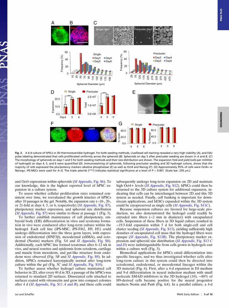
completely defined system for hPSC culture, which was thencharacterized in greater detail. For both single-cell and clusterseeding, live/dead staining revealed very high viability within thehydrogel or spheroids (Fig. 3A). Also, 5-ethynyl-2′-deoxyuridine(EdU) staining showed uniform cell proliferation across sphe-roids, suggesting effective transport of nutrients, oxygen, and/orprotein factors within the hydrogel and spheroids (Fig. 3B).Microscopy also revealed that single-seeded cells expanded andgrew into spheroids with a narrow size distribution (e.g., 40–120μm in diameter at day 5, Fig. 3C), whereas precluster seedingresulted in spheroids with larger sizes and broader distribution(50–300 μm in diameter at day 5, Fig. 3C). For both seedingmethods, 10-, 20-, or 21-fold expansion, with final yields of ∼1,2.0, or 2.1 × 10⁷ cells per mL hydrogel, was achieved for hPSCson day 4, 5, or 6 of the culture, respectively (Fig. 3D). Further-more, immunostaining showed 95% of cells expressed pluri-potency markers octamer-binding transcription factor 4 (Oct4)and Nanog, and alkaline phosphatase expression was both high
2 of 10 | www.pnas.org/cgi/doi/10.1073/pnas.1309408110 Lei and Schaffer

and homogeneous across spheroids (Fig. 3 E–G). An additional13-d culture was conducted to explore the sensitivity of cellulargrowth and pluripotency to spheroid size in this system. Single
iPS-MSCs were cultured in PNIPAAm-PEG hydrogel with E8and RI for 13 d without passaging. An extremely low density (1.0 ×10⁵ cells per mL) was used to provide sufficient space for cell
A Single cells in medium
Small clusters in medium
Small clusters in gel
1d 3 or4d
Singlecells
Single cells in gel
4 or5d
Spheroids in gel
Spheroids in gel
8.0
10.0
12.0B
******
******
******
*** ***
0.0
2.0
4.0
6.0
Fold
Exp
ansi
on
***
***
***
******
***
***
******
*** ***
RI (day): 1, 4 1, 4 1, 4 1, 4 1, 4 1, 4 1, 4 1, 4 1, 4 1, 4 1, 4 1, 4 1, 4 1, 4 1, 4 1, 4 1, 4 1, 4
Material*: s g,s g,c s g,s g,c s g,s g,c s g,s g,c s g,s g,c s g,s g,c
Medium: mTeSR E8
0.25x10⁶ cells/mlDensity:
mTeSR E8
1.0x10⁶ cells/ml
mTeSR E8
2.5x10⁶ cells/ml
*: (s): static suspension; (g,s): in gel, single cell seeding; (g,c): in gel, precluster seeding
1.4E+07C
***
6.0E+06
8.0E+06
1.0E+07
1.2E+07
***
***
***
******
******
***
***
***
***
RI (day): 1, 4 1, 4 1, 4 1, 4 1, 4 1, 4 1, 4 1, 4 1, 4 1, 4 1, 4 1, 4 1, 4 1, 4 1, 4 1, 4 1, 4 1, 4
Material*: s g,s g,c s g,s g,c s g,s g,c s g,s g,c s g,s g,c s g,s g,c
Medium: mTeSR E8 mTeSR E8 mTeSR E8
0.0E+00
2.0E+06
4.0E+06
Fina
l cel
l den
sity
(cel
ls/m
l)
****** ***
******
******
0.25x10⁶ cells/mlDensity: 1.0x10⁶ cells/ml 2.5x10⁶ cells/ml
*: (s): static suspension; (g,s): in gel, single cell seeding; (g,c): in gel, precluster seeding
Fig. 1. A multifactorial experiment was designed to investigate the potential interactions between various system parameters. iPS-MSCs were cultured instatic liquid medium without hydrogel (static suspension) or in 10% (wt/vol) PNIPAAm-PEG hydrogel via single-cell or precluster seeding for 4 d in mTeSR or E8with 1- or 4-d RI at low, medium, or high seeding density (2.5 × 10⁵, 1.0 × 10⁶, or 2.5 × 10⁶ cells/mL, respectively). (A) Schematic illustration of hPSC culture in3D hydrogel. (B and C) Fold of expansion and final cell densities. The triple asterisk (***) indicates statistical significance at a level of P < 0.001.
Lei and Schaffer PNAS Early Edition | 3 of 10
ENGINEE
RING
PNASPL
US

growth during the long 13-d culture. iPS-MSCs grew into sphe-roids with mean diameter of ∼350 μm, with the larger spheroidsreached diameter up to ∼750 μm. Consistent cell growth andpluripotent marker expression were also seen in this size range(SI Appendix, Fig. S5). In summary, the culture system efficientlyexpanded hPSCs with high quality and density.We next assessed the generality of this system for the long-
term expansion of multiple hPSC lines (1, 33), which were con-
tinuously propagated for up to 60 passages or ∼280 d (withpassaging every 4 or 5 d). During each of these passages, iPS-MSCs, iPS-Fib2 iPSCs, and H9 hESCs expanded ∼10- or 20-foldover 4 or 5 d, respectively, and H1 hESCs expanded 7-fold over4 d, indicating some differences in growth rates among the lines(Fig. 4A). However, during this long-term culture with an ac-cumulated expansion up to ∼1072-fold, ∼95% of cells remainedOct4+ (Fig. 4B). Immunostaining again revealed uniform Nanog
1d RI 4d RI
mTeSR
1d RI 4d RI
E8A
2.5x10⁵ cells/ml
1.0x10⁶ cells/ml
In P
NIP
AA
m-P
EG
gel
, sin
gle
cell
seed
ing
2.5x10⁶ cells/ml
2.5x10⁵ cells/ml
PE
G g
el, p
recl
uste
r se
edin
g
B
1.0x10⁶ cells/ml
In P
NIP
AA
m-
2.5x10⁵ cells/ml
Sta
tic s
uspe
nsio
n
C2.5x106 cells/ml
1.0x10⁶ cells/ml
2.5x10⁶ cells/ml
Fig. 2. Phase contrast images showing the cell morphologies for the factorial-designed experiment. iPS-MSCs were cultured in 10% PNIPAAm-PEG hydrogelvia single cell (A) or precluster seeding (B) or in static liquid medium without hydrogel (static suspension) (C) for 4 days in mTeSR or E8 with 1d or 4d RI at low,medium or high seeding density (2.5×10⁵, 1.0×10⁶, or 2.5×10⁶ cells per mL, respectively). (Scale bar: 250 μm.)
4 of 10 | www.pnas.org/cgi/doi/10.1073/pnas.1309408110 Lei and Schaffer
and Oct4 expressions within spheroids (SI Appendix, Fig. S6). Toour knowledge, this is the highest reported level of hPSC ex-pansion in a culture system.To assess whether cellular proliferation rates remained con-
sistent over time, we reevaluated the growth kinetics of hPSCsafter 10 passages in the gel. Notably, the expansion rate (∼10-, 20-,or 21-fold at days 4, 5, or 6, respectively) (SI Appendix, Fig. S7),pluripotency marker expression, and spheroid size distribution(SI Appendix, Fig. S7) were similar to those at passage 1 (Fig. 3).To further establish maintenance of cell pluripotency, em-
bryoid body (EB) differentiation in vitro and teratoma forma-tion in vivo were conducted after long-term culture within thehydrogel. Each cell line (iPS-MSC, iPS-Fib2, H9, H1) couldundergo differentiation into the three germ layers, with expres-sion of endodermal (HNF3β), mesodermal (αSMA), and ecto-dermal (Nestin) markers (Fig. 5A and SI Appendix, Fig. S8).Additionally, each hPSC line formed teratomas after 6–12 wk invivo, and neural rosettes and epidermis from ectoderm, cartilageand muscle from mesoderm, and gut-like structure from endo-derm were observed (Fig. 5B and SI Appendix, Fig. S9). In ad-dition, hPSCs remained karyotypically normal after long-termculture within the gel (Fig. 5C and SI Appendix, Fig. S10).To further assess whether hydrogel culture maintained cell
behavior in 2D, after every 40 d in 3D, a passage of the hPSCs wasreturned to standard 2D surfaces. Dissociated cells attached tosurfaces coated with vitronectin and grew into compact coloniesafter 4 d (SI Appendix, Fig. S11 A and B), and these cells could
subsequently undergo long-term expansion on 2D and maintainhigh Oct4+ levels (SI Appendix, Fig. S12). hPSCs could then bereturned to the 3D culture system for additional expansion, in-dicating that cells can be interchanged between 2D and this 3Dsystem as needed. Finally, cell banking is important for down-stream applications, and hESCs expanded within the 3D systemcould be cryopreserved as single cells (SI Appendix, Fig. S11C).Because suspension cultures are favored for large-scale pro-
duction, we also demonstrated the hydrogel could readily beextruded into fibers (∼2 mm in diameter) with encapsulatedcells. Suspension of these fibers in 3D liquid culture resulted in∼19.5-fold expansion within 5 d for both single-cell and cellcluster seeding (SI Appendix, Fig. S13), yielding sufficiently highdensities of encapsulated cell mass that the hydrogel fibers wereopaque (SI Appendix, Fig. S13B). The pluripotency marker ex-pression and spheroid size distribution (SI Appendix, Fig. S13 Cand D) were indistinguishable from cells grown in hydrogels castwithin a culture well (Fig. 3).Biomedical applications for hPSCs entail differentiation into
specific lineages, and we thus investigated whether cells afterlong-term culture in this system could then be directed intoectodermal, endodermal, or mesodermal lineages within the3D material (Fig. 6). First, after a 4-d expansion in E8 mediumand 9-d differentiation in neural induction medium with smallmolecule SMAD inhibitors in the 3D hydrogel (10), ∼80% ofH9-derived cells became positive for the neural progenitormarkers Nestin and Pax6 (Fig. 6A). In a parallel culture, a 4-d
Single,day1 Single,day5 Precluster,day1 Precluster,day5
C
60Single:
60Precluster:
LiveDeadA
Edu DAPIB
0
20
40
10 30 50 70 90 110
130
150
170
190
Freq
uenc
y (%
)
Spheroid Diameter (µm)
Day3 Day4Day5 Day6
0
20
40
25 75 125
175
225
275
325
375
425
475
Freq
uenc
y (%
)
Spheroid Diameter (µm)
Day3 Day4Day5 Day6
E
Phase AP
Single Precluster
0.0
10.0
20.0
4 5 6
Exp
ansi
on
Day0.E+00
1.E+07
2.E+07
4 5 6
Yiel
d
D***
***
Oct4 Nanog DAPI Merge
F
0.0
40.0
80.0
4 5 6
% O
ct4+
0.0
40.0
80.0
4 5 6%
Nan
og+
Single PreclusterG
Fig. 3. A 6-d culture of hPSCs in 3D thermoreversible hydrogel. For both seeding methods, Live/Dead cell staining revealed a very high viability (A), and EdUpulse labeling demonstrated that cells proliferated uniformly across the spheroid (B). Spheroids on day 5 after precluster seeding are shown in A and B. (C)The morphology of spheroids on days 1 and 5 for both seeding methods and their size distribution are shown. The expansion fold and yield (cells per milliliterof hydrogel) on days 4, 5, and 6 were quantified (D). Immunostaining of spheroids, following precluster seeding and 3D hydrogel culture, shows that themajority of cells expressed the pluripotency markers alkaline phosphatase (E) as well as Oct4 and Nanog (F). (G) Approximately 95% of cells were Oct4+ orNanog+. iPS-MSCs were used for A–G. The triple asterisk (***) indicates statistical significance at a level of P < 0.001. (Scale bar: 250 μm.)
Lei and Schaffer PNAS Early Edition | 5 of 10
ENGINEE
RING
PNASPL
US

expansion in E8 medium and 5-d differentiation in endoderminduction medium (4) in the 3D hydrogel resulted in ∼95% of H9or iPS-MSC–derived cells being positive for the endoderm pro-genitor markers forkhead box protein A2 (FOXA2) and SOX17(Fig. 6B). Finally, a 4-d expansion in E8 medium and 9-d differen-tiation in cardiomyocyte induction medium (34) within the3D hydrogel yielded beating spheroids 2 d after transfer to a fibro-nectin-coated plate (Movie S1).Several clinical trials have demonstrated that fetal ventral
midbrain tissue implants can alleviate motor symptoms in Par-kinson patients under certain circumstances, although this ap-proach is challenged by tissue supply and other considerations(35, 36). In recent important work, Kriks et al. (10) and Kirkebyet al. (37) developed approaches for differentiating hPSCs intoDA progenitors or neurons that could functionally integrate intothe brains of mouse, rat, and nonhuman primate models of PD.We investigated whether the differentiation protocols—whichincluded both defined (Noggin, Shh, FGF8, TGFβ3) and un-defined protein components—could be adapted to 3D cultureunder defined conditions. DA induction of H9 spheroids (40–120μm) within the hydrogel was initiated via dual SMAD inhibition,Shh, and a GSK3β inhibitor to activate Wnt signaling (Fig. 7A).On day 11, 81% of cells within the spheroids (∼100–300 μm)were positive for the ventral midbrain DA progenitor markersFOXA2 and LMX1a (Fig. 7 B–D). These matched levels ob-tained in 2D; however, cells in 3D expanded ∼80-fold over 15 d,resulting in 8 × 10⁷ cells per mL of hydrogel, compared with onlyan approximately ninefold expansion on 2D surface (Fig. 7D).Finally, these DA progenitors could subsequently be cultured on2D surfaces as described (37) for differentiation into mature TH+DA neurons (Fig. 7E).
DiscussionLarge numbers of hPSCs, or their differentiated progeny, areneeded for many biomedical applications (9). For large-scaleexpansion, currently hPSCs from a cell bank are expanded intolarge numbers through progressive proliferation followed by di-rected differentiation into progenitors or mature cells. Efficient,
scalable, and GMP-compliant culture systems are required forsuch processes (9). hPSC culture has made considerable prog-ress, but current systems achieve only moderate expansion, yield,and quality (9) (SI Appendix, Table S1). The most effectivesystem reported to date enabled approximately threefold tofourfold expansion of hESCs per passage in mTeSR mediumcontaining 1% BSA, for a final yield of <1 × 10⁶ cells per mL(18) (SI Appendix, Table S1). We have developed a 3D culturesystem—which combines a synthetic thermoreversible hydrogeland defined medium with an optimized protocol involving single-cell passaging—for simple, defined, scalable, and GMP-complianthESC and hiPSC expansion (Figs. 1–5) and differentiation (Figs.6 and 7) with high yield. Long-term culture (>280 d) with highexpansion rate (20-fold over 5 d, or 6.4 × 10⁷-fold over 1 mo, and1072-fold over 280 d), volumetric cell yield (2 × 10⁷ cells per mLof hydrogel), and quality (∼95% Oct4+) were achieved (Figs. 3and 4). Additionally, the hPSCs were able to differentiate into allof the three germ layers after long-term culture in the 3D system(Fig. 5 and SI Appendix, Figs. S8 and S9). Finally, the 3D system alsosupported directed differentiation into neural progenitors, endo-derm progenitors (Fig. 6), beating cardiomyocytes (Movie S1), ormidbrain dopaminergic progenitors (Fig. 7) by simply replacingthe expansion medium with the differentiation medium. Thisversatile system thus combines many advantageous and impor-tant features.The survival and proliferation of hESCs and hiPSCs—which
both exhibit characteristics typical of epiblast-like pluripotent stemcells (14)—depend on multiple extracellular cues. These includesoluble protein growth factors [e.g., FGF2, TGF-β, and autocrine/paracrine cues (15)] that signal via a number of pathways to sup-port cell survival and pluripotency factors [e.g., Nanog, Oct4, Sox2(38–40)]. In addition, hPSCs engage in cell–matrix interactions(31), and cell–cell contacts via cadherins are particularly importantfor survival and pluripotency maintenance. Disruption of suchcell–cell contacts leads to Abr-dependent activation of RhoA (andinactivation of Rac1), ROCK activation, and downstream acto-myosin hyperactivation and apoptosis (14, 32). Other environ-
H1, Single H9, Single iPS-MSC, Single iPS-Fib2, SingleH1 Precluster H9 Precluster iPS MSC Precluster
A
0
5
10
15
20
25
Fold
Exp
ansi
on
H1, Precluster H9, Precluster iPS-MSC, Precluster
0 5 10 15 20 25 30 35 40 45 50 55 60Passage Number
B
100.0
H1, Single iPS-Fib2, Single H9, Single iPS-MSC, Single
H9, Precluster iPS-MSC, Precluster H1, Precluster
0.0
20.0
40.0
60.0
80.0
5 10 30 60
% O
ct4+
Passage Number
Fig. 4. Long-term, progressive expansion of hPSCs in the 3D hydrogel system. (A) The expansion rates of iPS-MSCs, iPS-Fib2s, H9s, and H1s at eachpassage (4 or 5 d) using single-cell or precluster seeding are shown. Passages with 4 d are indicated (dotted line). (B) Oct4 levels remained high duringthe long-term culture.
6 of 10 | www.pnas.org/cgi/doi/10.1073/pnas.1309408110 Lei and Schaffer

mental stress, such as mechanical forces (9, 41) or apparentlyredox conditions (3), can also adversely affect cell function.Through a combination of rational and empirical investiga-
tion, this culture system based on a thermoresponsive materialapparently addresses a number of these needs. Importantly,multifactorial investigation indicated that the system’s propertiesmay result from a combination of the synthetic PNIPAAm-PEGmatrix, defined medium, and optimized protocol, as any of theseelements alone yielded suboptimal outcomes (Figs. 1 and 2, andSI Appendix, Figs. S1–S5 and S13–S15). The culture mediumprovides key soluble cues (3), and the material may conceivablyconcentrate autocrine/paracrine factors near cells (15). RI ame-liorates dissociation-induced apoptosis that otherwise occurs viacytoskeletal-associated pathways (14, 31, 32), and future inves-
tigation may elucidate whether this mechanically soft (SI Ap-pendix, Fig. S14) thermoresponsive material may more favorablyinterface with these mechanosensitive pathways relative to othermaterials (14). The material also protects cells from shear-inducedstress, yet this readily deformable hydrogel may more favorablyallow single-cell division and subsequent cluster growth relative tochemically cross-linked hyaluronic acid, ionically associated algi-nate, or stiff agarose hydrogels. Finally, E8 unlike mTeSR lacksβ-mercaptoethanol, an apparent stressor of hESCs, particularlyin suboptimal culture environments (SI Appendix, Fig. S15). Acombination of factors thus interacts to provide a permissive 3Dsystem that supports hPSC biology, and future investigations mayelucidate the mechanistic contributions of both biochemical andmechanical features of this microenvironment.
A Nestin/DAPI αSMA/DAPI HNF3β/DAPI
B
neural rosette cartilage gut
C
epidermis muscle
Fig. 5. hPSCs retained pluripotency and normal karyotype after long-term culture in the 3D hydrogel system. (A) EB differentiation in vitro. iPS-MSCs wereexpanded in the 3D hydrogel for 35 passages via single-cell seeding before EB differentiation. Immunostaining for the three germ layers (ectoderm: Nestin;mesoderm: αSMA; and endoderm: HNF3β) are shown. (B) Teratoma formation in vivo. iPS-MSCs expanded in 3D hydrogels for 35 passages via single-cellseeding were injected into mice s.c., and teratomas formed after 6–12 wk. Structures from all three germ layers (arrows) were identified, including ectoderm:neural rosette and epidermis; mesoderm: cartilage and muscle; and endoderm: gut-like-structure. (C) iPS-MSCs cultured in the 3D hydrogel for 35 passages viasingle-cell seeding retained normal karyotypes. (Scale bars: A, 100 μm; B, 50 μm.)
Lei and Schaffer PNAS Early Edition | 7 of 10
ENGINEE
RING
PNASPL
US

The system can also be adapted to multiple scales—from thelaboratory toward the clinic—to support research in cell re-placement therapies, artificial tissues and organs, and/or high-throughput drug screening/toxicity screening with hPSCs. Forinstance, ∼50 mL of hydrogel would be sufficient to produce 109
cells for preclinical animal studies, and a bioreactor with ∼5 L of
hydrogel could yield >1011 cells for clinical studies. The ability tomove a single culture system through multiple scales may aidclinical development.Advances in developmental and stem cell biology have en-
abled the development of approaches to effectively differentiatehPSCs into numerous additional cell types, such as neural crest
DAPI PAX6 Nestin Merge
40.0%
80.0%A
DAPI FOXA2 SOX17 MergeB
100.0
0.0%PAX6 Nestin
H9
H9
iPS-MSC 0.0
20.0
40.0
60.0
80.0
H9 iPS-MSC
% F
OXA
2 &
SO
X17+
Fig. 6. Neural and endodermal induction in the 3D hydrogel. hPSCs that had been cultured in the 3D system for 20–30 passages were used for differen-tiation. (A) After 4 d of expansion and 9 d of dual SMAD inhibition, ∼80% of differentiated H9s were positive for neuroectodermal markers Nestin or PAX6.The spheroids were dissociated into single cells before staining. (B) After 4 d of expansion and 3 d of endodermal induction, ∼95% of differentiated H9s oriPS-MSCs were positive for endodermal progenitor markers FOXA2 and SOX17. (Scale bar: 100 μm.)
-4 11Day : 22A
3D DA Neural Induction
LDN/SB/CHIR/shh
3D Expansion
E8 DMEM/N2/B27
Neural Differentiation on 2D
AA/dbcAMP/BDNF/GDNF
Neurobasal/B27
Day -4 Day 0 Day 11 Day 11, releasedB
DAPI FOXA2 LMX1a MergeC
Day 11
D
020406080
100
2D 3D
Exp
ansi
on
020406080
100
2D 3D
% F
OX
A2
& L
MX
1a+*** E
DAPI/FOXA2/LMX1a
Day 22
DAPI/MAP2/TH
Day 22
Fig. 7. Scalable production of dopaminergic (DA) neuron progenitors in the 3D culture system. (A) Protocol used for generating DA progenitors in 3D. (B)Phase images showing spheroid morphologies during the 15-d production process, as well as spheroids released from the hydrogel on day 11. (C) Immu-nostaining on day 11 for the midbrain DA progenitor makers FOXA2 and LMX1a. (D) The fold of expansion and percentage of FOXA2 and LMX1a double-positive cells on day 11 in the 3D hydrogel system or on the conventional 2D surface. (E) The day 11 DA progenitors produced in the 3D system matured intoTH+ DA neurons after 11 additional days in culture on 2D laminin surfaces. hPSCs cultured in the 3D system for 20–30 passages were used for these dif-ferentiation. The triple asterisk (***) indicates statistical significance at a level of P < 0.001. (Scale bars: B, 250 μm; C and E, 100 μm.)
8 of 10 | www.pnas.org/cgi/doi/10.1073/pnas.1309408110 Lei and Schaffer

cells (42), motor neurons (43), oligodendrocyte progenitors (44,45), pancreatic progenitors (4, 46), hepatocyte-like cells (47),retinal cells (48), and others (49, 50). Several of these processeshave even proceeded to clinical trials (51). Future work canexplore integrating these advances into this defined 3D hydrogelculture system to aid efficient, economical, and reproducible cellproduction for multiple future applications.
MethodsMaintaining hPSCs on 2D Surface. Human ESC lines H1 and H9 were obtainedfromWiCell Research Institute. iPS-MSC (33) (derived from human MSCs) andiPS-Fib2 (33) (derived from human dermal fibroblasts) were gift from GeorgeQ. Daley (Children’s Hospital Boston, Boston). hPSCs were cultured on six-well plate coated with vitronectin (Invitrogen) in E8 medium (Invitrogen). Cellswere passaged every 4 d with 0.5 mM EDTA (Invitrogen). To replate hPSCs on2D surfaces following expansion within the 3D hydrogel, hPSC spheroids weredissociated with Accutase (Life Technologies) at 37 °C for 10 min, and culturedon six-well plate coated with vitronectin in E8 medium (supplied with 10 μMROCK inhibitor, Y-27632, Selleckchem, for the first 24 h).
Expanding hPSCs in 3D Hydrogel. To transfer the culture from 2D to 3D, hPSCson Matrigel or vitronectin-coated tissue culture plates were incubated withAccutase at 37 °C for 5 min and dissociated into single cells. For the single-cellseeding method, dissociated cells were mixed with PNIPAAm-PEG (CosmoBio) solution dissolved in E8 medium at 4 °C and cast on tissue culture plate,then incubated at 37 °C for 15 min to form hydrogels before adding warmE8 medium containing 10 μM ROCK inhibitor. For the precluster seedingmethod, dissociated cells were cultured in suspension overnight in low ad-hesion plates to form small clusters that were subsequently encapsulatedinto the 3D hydrogel as mentioned above.
To passage hPSCs within 3D hydrogel, ice-cold PBS was added to the 3Dculture at day 4 or 5 to dissolve the gel. Spheroids were collected bycentrifuging at 200 × g for 3 min, incubated with Accutase at 37 °C for10 min, and dissociated into single cells for reencapsulation as mentionedabove. The NucleoCounter NC-200 (Chemometec) was used to count cellnumbers. To prepare hydrogel fibers, a 4 °C PNIPAAm-PEG solution con-taining cells was extruded into room temperature E8 medium througha 2-mm-diameter tube. The resulting hydrogel fibers were cultured in sus-pension in E8 medium at 37 °C. Medium was changed daily for all cultures.To measure spheroid sizes, hPSCs were released from the hydrogel, andphase images were taken. The diameters of >2,000 spheroids were quanti-fied with MetaXpress software (Molecular Devices). The frequency ofspheroids within a diameter range was calculated with the Excel HistogramPlug-in. Expanded cells were cryopreserved as single cells in E8 medium with10% (vol/vol) DMSO and 10 μM ROCK inhibitor in liquid N2.
Staining and Imaging. Cells cultured on 2D surfaces werefixedwith 4% (wt/vol)paraformaldehyde (PFA) at room temperature for 15 min, permeabilized with0.25%TritonX-100 for 15min, and blockedwith 5% (vol/vol) goat serum for 1 hbefore incubating with primary antibodies at room temperature for 2 h. Afterextensive washing, secondary antibodies in 2% (wt/vol) BSA were addedand incubated for another 1 h. Cells were washed with PBS for three timesbefore imaging.
To assess the pluripotency marker expression of cells expanded in 3Dhydrogels, hPSCs were dissociated into single cells with Accutase and stained insuspension. Cells were then placed in 96-well plates and analyzed with anImageXpress (Molecular Devices). The percentage of Oct4+ or Nanog+ nucleiwas quantifiedwithMetaXpress software (Molecular Devices). This process wasused to quantify the PAX6+ and Nestin+ cells after neural induction as well.
To stain spheroids, hPSCs were fixed with 4% (wt/vol) PFA at room temper-ature for 30min, and then incubatedwith PBS plus 0.25% Triton X-100 plus 5%(vol/vol) goat serum plus primary antibodies at 4 °C for 48 h. After extensivewashing, secondary antibodies in 2% (wt/vol) BSAwere added and incubated at4 °C for 4 h. Cellswerewashedwith PBS for three times before imaging. Stainingwithout primary antibodies was used as controls for all of the immunostainings.
LIVE/DEAD Cell Viability staining (Invitrogen) was used to assess live anddead cells, the Click-iT EdU Alexa Fluor 594 Imaging Kit (Invitrogen) was usedto label proliferating cells, and an Alkaline Phosphatase Live Stain (Invi-trogen) was used to image alkaline phosphatase.
EB Differentiation. hPSCs were suspended in DMEMplus 20% (vol/vol) FBS plus10 μM β-mercaptoethanol in low adhesion plates for 6 d. The EBs were thentransferred onto plates coated with 0.1% gelatin and cultured in the samemedium for another 6 d, followed by fixation and staining as above.
Teratoma Formation in Vivo. All animal protocols were approved by theAnimal Care and Use Committee of the University of California, Berkeley. Atotal of 3 × 10⁶ hPSCs was suspended in 25 μL of PBS plus 25 μL of Matrigel(BD Biosciences) and injected s.c. at the back of the neck of the SCID Beigemice (Charles River Laboratory), and teratomas were harvested when sizesreached 2 cm. The tissue was then fixed with 4% (wt/vol) PFA for 48 h,dehydrated with 70%, 95%, and 100% (vol/vol) ethanol sequentially, anddefatted with xylene for 2 h before embedding in paraffin. The 10-μm-thick sections were cut and stained with hematoxylin and eosin.
Karyotype. Kyarotyping was performed by Oakland Children’s HospitalCytogenetics Laboratory.
Neural Induction (52). Following expansion, hPSCs were cultured in knockoutserum replacement (KSR) medium with 10 μM SB431542 (Selleckchem) and100 nM LDN193189 (Selleckchem) for 5 d. Starting at day 5, the KSR mediumwas gradually replaced by the N2 medium, and cells were harvested at day 9.KSR medium was as follows: DMEM plus 15% (vol/vol) KSR plus 2 mM glu-tamine plus 10 μM β-mercaptoethanol. N2 medium was as follows: Neuro-basal plus N2 plus B27 (without retinoic acid) plus 2 mM L-glutamine.
Endodermal Induction (4). Following expansion, hPSCs were cultured in RPMI1640 (Sigma) plus 0.2% FBS plus 100 ng/mL Activin-A (Peprotech) plus 20 ng/mL Wnt3a (R&D) for 1 d and in RPMI 1640 plus 0.5% FBS plus 100 ng/mLActivin-A for 2 more days before being fixed and immunostained.
Cardiomyocyte Differentiation (34). Following expansion, hPSCs were culturedin RPMI 1640 plus B27 without insulin between day 0 and 7, and in RPMI 1640plus B27 after. The following small molecules were added: 12 μM CHIR99021(Selleckchem) for days 0–1; 5 μM IWP-2 Inhibitor (Selleckchem) for days 3–5.Spheroids were released on day 9 to fibronectin-coated plate. Beating car-diomyocytes were filmed on day 11.
Differentiating hPSCs into Dopaminergic Neuronal Progenitors. hPSCs werecultured in 50% DMEM/F12 plus 50% (vol/vol) Neurobasal medium (with1:100 N2 and 1:50 B27) for 11 d following expansion (37). The followingproteins and small molecules were added: 10 μM SB431542; 200 ng/mLNoggin (R&D) or (100 nM LDN193189); 0.7 μM CHIR99021 and 200 ng/mLSHH C25II. Cells were harvested at day 11.
Statistical Analysis. Statistical analyses were done using the statistical packageInstat (GraphPad Software). For multiple comparisons, themeans of triplicatesamples were compared using the Tukey multiple-comparisons analysis withthe α level indicated in the figure legend.
Antibodies. Oct4 (Santa Cruz Biotechnology; 1:100), Nanog (Santa Cruz Bio-technology; 1:100), Nestin (Millipore; 1:200), αSMA (Abcam; 1:200), FOXA2/HNF3β (Santa Cruz Biotechnology; 1:200), PAX6 (Covance; 1:200), LMX1a(Millipore; 1:1,000), MAP2 (BD Biosciences; 1:1,000), TH (Pel-Freez; 1:1,000),and SOX17 (R&D; 1:500).
ACKNOWLEDGMENTS. This work was supported by California Instituteof Regenerative Medicine Grant RT2-02022 and a California Institute forRegenerative Medicine Training Grant T1-00007 fellowship (to Y.L.).
1. Thomson JA, et al. (1998) Embryonic stem cell lines derived from human blastocysts.
Science 282(5391):1145–1147.2. Takahashi K, et al. (2007) Induction of pluripotent stem cells from adult human fi-
broblasts by defined factors. Cell 131(5):861–872.3. Chen G, et al. (2011) Chemically defined conditions for human iPSC derivation and
culture. Nat Methods 8(5):424–429.4. Schulz TC, et al. (2012) A scalable system for production of functional pancreatic
progenitors from human embryonic stem cells. PLoS One 7(5):e37004.
5. Lindvall O, Kokaia Z, Martinez-Serrano A (2004) Stem cell therapy for human neu-
rodegenerative disorders-how to make it work. Nat Med 10(Suppl):S42–S50.6. Badylak SF, Taylor D, Uygun K (2011) Whole-organ tissue engineering: Decellulari-
zation and recellularization of three-dimensional matrix scaffolds. Annu Rev Biomed
Eng 13:27–53.7. McNeish J (2004) Embryonic stem cells in drug discovery. Nat Rev Drug Discov 3(1):70–80.8. Desbordes SC, Studer L (2013) Adapting human pluripotent stem cells to high-
throughput and high-content screening. Nat Protoc 8(1):111–130.
Lei and Schaffer PNAS Early Edition | 9 of 10
ENGINEE
RING
PNASPL
US

9. Serra M, Brito C, Correia C, Alves PM (2012) Process engineering of human pluripotentstem cells for clinical application. Trends Biotechnol 30(6):350–359.
10. Kriks S, et al. (2011) Dopamine neurons derived from human ES cells efficiently en-graft in animal models of Parkinson’s disease. Nature 480(7378):547–551.
11. Laflamme MA, Murry CE (2005) Regenerating the heart. Nat Biotechnol 23(7):845–856.
12. Roger VL, et al. (2012) Heart disease and stroke statistics—2012 update: A report fromthe American Heart Association. Circulation 125(1):e2–e220.
13. Zang R, Li D, Tang I, Wang J, Yang S (2012) Cell-based assays in high-throughputscreening for drug discovery. Int J Biotechnol Wellness Ind 1:31–51.
14. Ohgushi M, Sasai Y (2011) Lonely death dance of human pluripotent stem cells:ROCKing between metastable cell states. Trends Cell Biol 21(5):274–282.
15. Peerani R, et al. (2007) Niche-mediated control of human embryonic stem cell self-renewal and differentiation. EMBO J 26(22):4744–4755.
16. Villa-Diaz LG, Ross AM, Lahann J, Krebsbach PH (2013) Concise review: The evolutionof human pluripotent stem cell culture: From feeder cells to synthetic coatings. StemCells 31(1):1–7.
17. McDevitt TC, Palecek SP (2008) Innovation in the culture and derivation of pluripotenthuman stem cells. Curr Opin Biotechnol 19(5):527–533.
18. Chen VC, et al. (2012) Scalable GMP compliant suspension culture system for humanES cells. Stem Cell Res (Amst) 8(3):388–402.
19. Steiner D, et al. (2010) Derivation, propagation and controlled differentiation ofhuman embryonic stem cells in suspension. Nat Biotechnol 28(4):361–364.
20. Amit M, et al. (2011) Dynamic suspension culture for scalable expansion of un-differentiated human pluripotent stem cells. Nat Protoc 6(5):572–579.
21. Zweigerdt R, Olmer R, Singh H, Haverich A, Martin U (2011) Scalable expansion ofhuman pluripotent stem cells in suspension culture. Nat Protoc 6(5):689–700.
22. Nie Y, Bergendahl V, Hei DJ, Jones JMPS, Palecek SP (2009) Scalable culture andcryopreservation of human embryonic stem cells on microcarriers. Biotechnol Prog25(1):20–31.
23. Chen AK, Chen X, Choo AB, Reuveny S, Oh SK (2011) Critical microcarrier propertiesaffecting the expansion of undifferentiated human embryonic stem cells. Stem CellRes (Amst) 7(2):97–111.
24. Serra M, et al. (2011) Microencapsulation technology: A powerful tool for integratingexpansion and cryopreservation of human embryonic stem cells. PLoS One 6(8):e23212.
25. Unger C, Skottman H, Blomberg P, Dilber MS, Hovatta O (2008) Good manufacturingpractice and clinical-grade human embryonic stem cell lines. Hum Mol Genet 17(R1):R48–R53.
26. Medina RJ, Kataoka K, Takaishi M, Miyazaki M, Huh NH (2006) Isolation of epithelialstem cells from dermis by a three-dimensional culture system. J Cell Biochem 98(1):174–184.
27. Chayosumrit M, Tuch B, Sidhu K (2010) Alginate microcapsule for propagation anddirected differentiation of hESCs to definitive endoderm. Biomaterials 31(3):505–514.
28. Stenberg J, et al. (2011) Sustained embryoid body formation and culture in a non-laborious three dimensional culture system for human embryonic stem cells. Cyto-technology 63(3):227–237.
29. Gerecht S, et al. (2007) Hyaluronic acid hydrogel for controlled self-renewal anddifferentiation of human embryonic stem cells. Proc Natl Acad Sci USA 104(27):11298–11303.
30. Watanabe K, et al. (2007) A ROCK inhibitor permits survival of dissociated humanembryonic stem cells. Nat Biotechnol 25(6):681–686.
31. Xu Y, et al. (2010) Revealing a core signaling regulatory mechanism for pluripotentstem cell survival and self-renewal by small molecules. Proc Natl Acad Sci USA 107(18):8129–8134.
32. Ohgushi M, et al. (2010) Molecular pathway and cell state responsible for dissociation-induced apoptosis in human pluripotent stem cells. Cell Stem Cell 7(2):225–239.
33. Park I-H, et al. (2008) Reprogramming of human somatic cells to pluripotency withdefined factors. Nature 451(7175):141–146.
34. Lian X, et al. (2012) Robust cardiomyocyte differentiation from human pluripotentstem cells via temporal modulation of canonical Wnt signaling. Proc Natl Acad Sci USA109(27):E1848–E1857.
35. Lindvall O (2013) Developing dopaminergic cell therapy for Parkinson’s disease—giveup or move forward? Mov Disord 28(3):268–273.
36. Mendez I, et al. (2008) Dopamine neurons implanted into people with Parkinson’sdisease survive without pathology for 14 years. Nat Med 14(5):507–509.
37. Kirkeby A, et al. (2012) Generation of regionally specified neural progenitors andfunctional neurons from human embryonic stem cells under defined conditions. CellRep 1(6):703–714.
38. Xu R-H, et al. (2008) NANOG is a direct target of TGFbeta/activin-mediated SMADsignaling in human ESCs. Cell Stem Cell 3(2):196–206.
39. Vallier L, Alexander M, Pedersen RA (2005) Activin/Nodal and FGF pathways co-operate to maintain pluripotency of human embryonic stem cells. J Cell Sci 118(Pt 19):4495–4509.
40. James D, Levine AJ, Besser D, Hemmati-Brivanlou A (2005) TGFbeta/activin/nodalsignaling is necessary for the maintenance of pluripotency in human embryonic stemcells. Development 132(6):1273–1282.
41. Hsieh MH, Nguyen HT (2005) Molecular mechanism of apoptosis induced by me-chanical forces. Int Rev Cytol 245:45–90.
42. Lee G, Chambers SM, Tomishima MJ, Studer L (2010) Derivation of neural crest cellsfrom human pluripotent stem cells. Nat Protoc 5(4):688–701.
43. Hu B-Y, Zhang S-C (2009) Differentiation of spinal motor neurons from pluripotenthuman stem cells. Nat Protoc 4(9):1295–1304.
44. Keirstead HS, et al. (2005) Human embryonic stem cell-derived oligodendrocyteprogenitor cell transplants remyelinate and restore locomotion after spinal cord in-jury. J Neurosci 25(19):4694–4705.
45. Sharp J, Frame J, Siegenthaler M, Nistor G, Keirstead HS (2010) Human embryonicstem cell-derived oligodendrocyte progenitor cell transplants improve recovery aftercervical spinal cord injury. Stem Cells 28(1):152–163.
46. Kroon E, et al. (2008) Pancreatic endoderm derived from human embryonic stem cellsgenerates glucose-responsive insulin-secreting cells in vivo. Nat Biotechnol 26(4):443–452.
47. Duan Y, et al. (2010) Differentiation and characterization of metabolically function-ing hepatocytes from human embryonic stem cells. Stem Cells 28(4):674–686.
48. Osakada F, Ikeda H, Sasai Y, Takahashi M (2009) Stepwise differentiation of plurip-otent stem cells into retinal cells. Nat Protoc 4(6):811–824.
49. Cheung C, Sinha S (2011) Human embryonic stem cell-derived vascular smooth musclecells in therapeutic neovascularisation. J Mol Cell Cardiol 51(5):651–664.
50. Descamps B, Emanueli C (2012) Vascular differentiation from embryonic stem cells:Novel technologies and therapeutic promises. Vascul Pharmacol 56(5-6):267–279.
51. Trounson A, Thakar RG, Lomax G, Gibbons D (2011) Clinical trials for stem cell ther-apies. BMC Med 9:52.
52. Chambers SM, et al. (2009) Highly efficient neural conversion of human ES and iPScells by dual inhibition of SMAD signaling. Nat Biotechnol 27(3):275–280.
10 of 10 | www.pnas.org/cgi/doi/10.1073/pnas.1309408110 Lei and Schaffer