Embed Size (px)
Citation preview

A fully caninised anti-NGF monoclonal antibodyfor pain relief in dogsGearing et al.
Gearing et al. BMC Veterinary Research 2013, 9:226http://www.biomedcentral.com/1746-6148/9/226

RESEARCH ARTICLE Open Access
A fully caninised anti-NGF monoclonal antibodyfor pain relief in dogsDavid P Gearing1,2*, Elena R Virtue1, Robert P Gearing1 and Alexander C Drew2
Abstract
Background: Monoclonal antibodies are a major class of biological therapies in human medicine but have not yetbeen successfully applied to veterinary species. We have developed a novel approach, PETisation, to rapidly convertantibodies for use in veterinary species. As an example, anti-nerve growth factor (anti-NGF) monoclonal antibodies(mAbs) which are effective in reducing acute and chronic pain in rodents and man are potentially useful for treatingpain in dogs but a fully caninised mAb is required in order to avoid an immune response. The aim of this studywas to determine the optimal properties of a caninised anti-NGF mAb for safe, repeated administration to dogs, todetermine its pharmacokinetic properties and to evaluate its efficacy in a model of inflammatory pain in vivo.
Results: Starting with a rat anti-NGF mAb, we used a novel algorithm based on expressed canine immunoglobulinsequences to design and characterise recombinant caninised anti-NGF mAbs. Construction with only 2 of the 4canine IgG heavy chain isotypes (A and D) resulted in stable antibodies which bound and inhibited NGF withhigh-affinity and potency but did not bind complement C1q or the high-affinity Fc receptor gamma R1 (CD64).One of the mAbs (NV-01) was selected for scale-up manufacture, purification and pre-clinical evaluation. Whenadministered to dogs, NV-01 was well tolerated, had a long serum half-life of 9 days, was not overtly immunogenicfollowing repeated dosing in the dog and reduced signs of lameness in a kaolin model of inflammatory pain.
Conclusions: The combination of stability, high affinity and potency, no effector activity and long half-life, combinedwith safety and activity in the model of inflammatory pain in vivo suggests that further development of the caninisedanti-NGF mAb NV-01 as a therapeutic agent for the treatment of chronic pain in dogs is warranted.
Keywords: Nerve growth factor, Analgesia, Companion animals, Monoclonal antibody, Pharmacokinetics, Chronic pain,Veterinary bio-therapeutic
BackgroundCurrent therapeutic options for pain management indogs are limited to a few classes of drugs includingnon-steroidal anti-inflammatory drugs (NSAID), narcoticsand polysulphated glycosaminoglycans (PSGAG) [1,2].Alternative therapeutic options are desirable, in par-ticular for the management of chronic pain. Recently, anew class of antibody drugs have been developed whichprovide effective analgesia in rodents and man throughinterference of binding of NGF to its cellular receptorson nociceptive neurons.
Whereas during mammalian development, NGF isessential for the survival of sensory and sympatheticneurons [3,4] in the adult it is expressed locally at sitesof injury and inflammation and is a major factor pro-moting pain and hyperalgesia [5,6]. NGF is producedby a variety of inflammatory and immune cells, jointchondrocytes and has also been detected in nerve andneuroma preparations [5–7]. Following binding to itsreceptor trkA on nociceptors, NGF causes immediateand long-termexcitability through activation of ionchannels, the transient receptor potential vanilloidreceptor (TRPV1) and secondary neurotransmittersincluding substance P and brain-derived neurotrophicfactor (BDNF) [5–7]. NGF also causes the sprouting ofnerve endings into the site of inflammation but doesnot appear to play a role in inflammation per se [8].Furthermore, mutations in NGF and its trkA receptor
* Correspondence: [email protected] Biopharma Pty Ltd, Level 39, 385 Bourke St, Melbourne, Victoria3000, Australia2Centre For Innate Immunity & Infectious Diseases, Monash Institute OfMedical Research, Monash University, Clayton, Victoria 3168, Australia
© 2013 Gearing et al.; licensee BioMed Central Ltd. This is an open access article distributed under the terms of the CreativeCommons Attribution License (http://creativecommons.org/licenses/by/2.0), which permits unrestricted use, distribution, andreproduction in any medium, provided the original work is properly cited.
Gearing et al. BMC Veterinary Research 2013, 9:226http://www.biomedcentral.com/1746-6148/9/226

are associated with diminished pain responses [5–7].Neutralising antibodies to NGF are highly effectiveanalgesics in rodent models of inflammatory pain,arthritis pain, cancer pain, and bone fracture pain[5–7]. This encouraging biological activity has resultedin the development of several NGF antagonists for thetreatment of pain in man.The clinical efficacy of anti-human NGF mAbs has
been demonstrated in several human studies includingseveral large-scale phase 3 clinical trials: responses tothe pain associated with osteoarthritis (OA), lower backpain and cystitis have been evaluated [5–7,9–12]. Theanti-NGF antibodies were generally very well tolerated(consistent with a benign profile in 6-month primatestudies [13]), with mild to moderate, transient peripheralsensation changes as the only consequences [7]. A smallnumber (16 of 6,800) of patients with OA that weretreated with anti-NGF mAbs required joint replacementearlier than would be normally expected [14] and thiswas attributed to “rapidly progressing osteoarthritis”. Thecause of this worsening has been debated, although insome patients, the accelerated osteoarthritis was possiblydue to concomitant NSAID use [15].Canine NGF and its receptor are closely homologous
to those of other species. NGF and trkA are expressedin similar tissues in dog and man, appear to be undersimilar control mechanisms, and have similar functions[16–19]. NGF levels are significantly elevated in the synovialfluid of osteoarthritic dogs with chronic lameness [20].As with other mammals, the canine immune system
shares major immunoglobulin types, including IgG (ofwhich there are four isotypes: [21]) and IgG-Fc receptorsincluding the high-affinity FcR CD64 [NCBI reference:NP_001002976.1, XP_003640260.1, XP_536141.3] [22]and the neonatal FcRn [XP_533618.2, XP_003640095.1],which potentiates IgG half-life in vivo [23,24].Based on the conservation of the NGF system between
dog and man, it was at least possible that the rat anti-human NGF MAb may also be reactive with canineNGF. Furthermore, if this reactivity was of high affinitywhich could be retained during the process of conversionto a fully caninised antibody, we postulated that theresulting antibody could well be an effective treatmentfor pain in the dog. We therefore converted a high-affinity,potent rat anti-NGF monoclonal antibody (αD11; [25])to a fully canine form (NV-01) with no loss of bioactivityby a novel process we term "PETisation". Unlike otherapproaches for converting antibody sequences fromone species for use in another, such as CDR grafting,which rely on germline sequences to provide recipientV domain framework structures, the PETisation approachmakes use of sequences from expressed and circulatingIgG. This increases the likelihood that the resultingantibody will be both active and recognized as self by
the recipient species, thereby overcoming immunogenicityconcerns.The design, preparation and in vitro characteristics of
NV-01, together with preliminary studies investigatingits safety and effectiveness are described herein. Collect-ively they show that NV-01 is a potent inhibitor of NGF,is well tolerated and non-immunogenic and showspromise as an analgesic in dogs. These preliminary datasupport our hypothesis that NV-01 might be useful as atreatment for pain in dogs (e.g. treatment of joint painassociated with osteoarthritis, cancer pain and post-surgicalpain) and suggest that its further development as a vet-erinary medicine is warranted.
MethodsSources of NGFA cDNA sequence encoding the amino acid sequence ofcanine pre-pro beta NGF (Figure 1A) with a C-terminalpoly-His tag was synthesized from oligonucleotides, clonedinto pcDNA3.1+ expression vector and transiently trans-fected into HEK293 cells at Geneart AG (Life Tech-nologies, Regensberg, Germany). The supernatant washarvested and purified by Ni-HiTrap chromatography(GE Healthcare, Upsalla, Sweden). Purified mouse NGF(muNGF) was purchased from Biosensis (Thebarton,Australia).
Conversion of αD11 variable domains for use in the dogIn order to reduce the immunogenic potential of ratαD11 [25] in the dog, changes were made to the heavyand light chain variable domain framework sequences byalignment with a matrix of predicted protein sequencesencoded by expressed canine IgG cDNA sequences.Where the αD11 sequence corresponded to the matrix,no changes were made. Where they differed, the mostsimilar amino acid (by charge, size, polarity) in the matrixwas substituted. If no similar amino acid was available,the most abundant canine residue was chosen. Thechanges made are illustrated in Figure 1B and 1C.Twenty-two substitutions were made to the heavychain variable domain, of which 10 were conservativeand 17 substitutions were made to the light chain variabledomain, of which 9 were conservative. By this process,termed PETisation, the αD11 framework sequenceswere completely caninised, with minimal changes madefrom the donor αD11 antibody.
Construction of NV-01 antibody heavy and light chainsThe caninised αD11 heavy chain variable domain sequence(caN) was combined with the αD11 heavy chain signalsequence and the constant domain sequences of eachof the four canine IgG heavy chain isotypes A,B, C andD [21] to form caN-HCA, caN-HCB, caN-HCC andcaN-HCD sequences, respectively. The caninised NV-01
Gearing et al. BMC Veterinary Research 2013, 9:226 Page 2 of 11http://www.biomedcentral.com/1746-6148/9/226

light chain variable domain sequence was combinedwith the αD11 light chain signal sequence and the constantdomain sequence of the canine kappa light chain toform the caN-kLC sequence. The resulting amino acidsequences were converted to codon-optimized nucleotidesequences for expression in CHO cells (Geneart AG,Life Technologies, Regensberg, Germany) and these cDNAwere cloned separately into pcDNA 3.1 + .Co-expression in CHO cells by transient transfection
of caN-kLC light chain cDNA with each of the fourNV-01 heavy chain isotype cDNAs, caN-HCA, caN-HCB,caN-HCC and caN-HCD, produced supernatants con-taining antibodies “caN-HCA1 + kLC1”, “caN-HCB2 +
kLC1”, “caN-HCC1 + kLC1” and “caN-HCD2 + kLC1”,respectively.Further modification of the heavy chain framework
sequence based on alignment to a larger number of caninecDNA sequences resulted in the modified heavy chainsequence “caN2-HCA1” (Figure 1). Co-expression of thissequence with light chain caN-kLC1 formed the antibody“caN2-HCA1-kLC1”, which was assigned the compoundnumber NV-01.
Recombinant antibody preparationsFor small scale, antibodies were transiently expressed inCHO cells (Geneart AG, Life Technologies, Regensberg,
Figure 1 NGF and anti-NGF antibody sequences. A) Alignment of the mature peptide sequence of NGF from human, mouse & dog. Identicalamino acids are indicated by dots and similar amino acids are underlined. B) Variable heavy & C) variable light chain sequences of the anti-NGFantibody αD11 aligned to the caninised antibody NV-01. Identical amino acids are indicated by dots and similar amino acids are underlined.Complementarity-determining regions (CDR) are boxed.
Gearing et al. BMC Veterinary Research 2013, 9:226 Page 3 of 11http://www.biomedcentral.com/1746-6148/9/226

Germany). The type B anti-NGF antibody (caN-HCB2 +kLC1) was purified on protein A from CHO cellsupernatant. The type C anti-NGF antibody (caN-HCC1 + kLC1) was purified on Protein G from CHOcell supernatants. Type A & type D anti-NGF anti-bodies (caN-HCA1 + kLC1 and ca-HCD2 + kLC1) couldnot be purified using Protein A or G and were purifiedusing Protein L.For in vivo experiments, NV-01 antibody was expressed
in CHO cells (Lonza Biologics plc, Cambridge, UK).Stable pooled transfections of CHO cells with cDNAencoding NV-01 heavy & light chains were cultured ina fed batch system for 13 days, before harvesting ofsupernatant containing NV-01. Clarified supernatantwas diluted 1:2 with 50 mM Tris pH 8.0. The proteinwas captured on a HiTrap 5 ml anion exchange Q FFcolumn (GE Healthcare) and impurities removed bywashing the column with 50 mM Tris, 100 mM NaCl,pH 8.0. The protein was eluted with 50 mM Tris,200 mM NaCl, pH 8.0.Anion exchange fractions containing antibody were
concentrated and diluted 1:10 with 50 mM sodiumphosphate, 1 M ammonium sulphate, pH 7.0. The proteinwas captured on a HiTrap hydrophobic interaction PhenylHP column (GE Healthcare) and impurities removed bywashing the column with 50 mM sodium phosphate,1 M ammonium sulphate, pH 7.0 (loading buffer). Theprotein was eluted with a linear gradient from loadingbuffer to 50 mM sodium phosphate, pH 7.0.Material from the hydrophobic interaction step was
further purified by size exclusion chromatography(HiLoad Superdex 200 pg 16/60, GE Healthcare), thenconcentrated and formulated into phosphate bufferedsaline (PBS) pH 7.3 by ultrafiltration (Amicon Ultra-15,molecular weight cut-off 30,000; Millipore, Billerica,USA).NV-01 produced by this method was determined to
be >95% pure and 100% monomeric by size exclusionHPLC. The preparations were free of detectable en-dotoxin (<0.1 EU/mL; EndosafeW-PTS™ Charles RiverLaboratories, Wilmington, USA).
Anti-NGF antibody detection by ELISAELISA plates were coated with 0.1 μg/ml muNGF andblocked with 5% BSA/PBS. muNGF coated wells wereincubated for 1 h at room temperature with recombinantcanine anti-NGF IgG preparations, diluted in PBS/1%BSA. Antibody concentrations ranging from 40 ng/mlto 0.625 ng/ml were used to establish a standard curve.After washing, the plates were incubated with a 1/5000dilution of rabbit anti-canine IgG-HRP (Sigma, St. Louis,USA) in PBS/1% BSA. Plates were washed with PBS 0.05%Tween 20 and developed by the addition of TMB sub-strate (Thermo Scientific, Waltham, USA). Development
was stopped by the addition of 2 M H2SO4 and absorb-ance read at 450 nm and background was subtracted.For the detection of NV-01 in canine plasma samples,
the canine plasma was diluted and used in the ELISAas above. The background for the canine plasma wasdetermined from the O.D. 450 nm of time zero serum.
Complement C1q binding ELISANV-01 cDNA transfected CHO cell supernatants wereconcentrated using Vivaspin 20 concentrators (30 kDacut-off, Vivaproducts, Littleton, USA). The concentrationsof recombinant antibodies in the CHO cell supernatantsand of purified antibodies were determined by titrationin the anti-NGF ELISA, by comparison with standardpreparations of purified B isotype canine anti-NGF anti-body (caN-HCB2 + kLC1) or NV-01.C1q binding was assayed following the method of
Lewis et al. [26]. Plates were coated with 2.5 μg/mlmuNGF and blocked with 5% BSA/PBS. Coated wellswere incubated for 1 h at room temperature withrecombinant canine anti-NGF IgG, diluted in PBS/1%BSA. Antibody concentrations ranged from 10 μg/ml to0.5 μg/ml. The plates were washed and incubated for1 hour at room temperature with human serum or heatinactivated human serum diluted 1/100 in veronal bufferedsaline containing 0.05% Tween-20, 0.1% gelatine and0.5% BSA. After washing, plates were incubated with a1/800 dilution of sheep anti-human C1q-HRP (AbDSerotec, Kidlington, UK) in PBS/1% BSA. After washing,plates were developed by the addition of TMB substrate(Thermo Scientific). Development was stopped by theaddition of 2 M H2SO4 and absorbance read at 450 nm.The O.D. 450 nm obtained using heat-inactivated serumwas used as background and subtracted from the O.D.450 nm obtained using untreated serum.
Soluble CD64 binding assay to caninised antibodiesThe sequence of cDNA encoding canine CD64, the highaffinity Fc receptor gamma R1, was obtained from Ensembl[www.ensembl.org: ENSCAFT00000018253]. The sequencewas modified as previously used to generate a solublehuman CD64 [27]. The trans-membrane domain wasremoved and c-myc and poly-His tags were added tothe C-terminus to allow for detection and purification.The cDNAs were synthesised, cloned into expressionvectors and protein was expressed by transient trans-fection of HEK suspension cells (Geneart AG, LifeTechnologies, Regensberg, Germany). Soluble canine CD64(scaCD64) was purified from the HEK cell supernatantsby Ni-Hi Trap chromatography and size exclusionchromatography (as above).Microtitre plates were coated with 0.1 μg/ml muNGF
and blocked with 5% BSA/PBS/0.05% Tween-20. Coatedwells were incubated for 1 h at room temperature with
Gearing et al. BMC Veterinary Research 2013, 9:226 Page 4 of 11http://www.biomedcentral.com/1746-6148/9/226

recombinant canine anti-NGF IgG, diluted in PBS/1%BSA/0.05% Tween-20. Antibody concentrations rangedfrom 100 ng/ml to 1.6 ng/ml. After washing, plates wereincubated with 1 μg/ml purified scaCD64. Plates werewashed and binding of scaCD64 detected by the additionof anti-c-myc-HRP (Pierce) in PBS/1% BSA/0.05% Tween-20. After washing plates were developed by the additionof TMB substrate (Thermo Scientific). Development wasstopped by the addition of 2 M H2SO4 and absorbanceread at 450 nm. The background (no canine IgG) wassubtracted.
Antibody binding kineticsThe binding affinity of NV-01 to muNGF was analysedby Surface Plasmon Resonance (SPR) using a ProteOnXPR36 SPRi biosensor equipped with a GLM chip(BioRad, Hercules, USA). The chip was conditionedwith 0.5% SDS, 50 mM NaOH and 100 mM HCl. Fol-lowing conditioning, the lanes were activated usingequal parts of EDAC and NHS amine coupling reagents(BioRad). The NGF protein was immobilised to thechip at a concentration of 50 μg/mL in sodium acetatebuffer (pH 4.5). Following immobilisation all threechannels were deactivated using ethanolamine. NV-01was passed across the surface at 500 nM, 250 nM, 125nM, 62.5 nM and 31.25 nM. The binding was displayedas a spectrogram. Controls were subtracted to give specificbinding. A Langmuir curve fit model was then used todetermine the specific affinity.
TF-1 proliferation-inhibition assayTF-1 cells were maintained in RPMI 1640 medium,supplemented with 10% foetal calf serum, 10 mM HEPES,penicillin/streptomycin (10 U/ml and 100 ug/ml finalrespectively) and 2 ng/ml GM-CSF. The TF-1 cellswere centrifuged and resuspended in "starve media" (asabove with no GM-CSF). The flasks were incubated ina humidified 37°C, 5% CO2 incubator for 24 hours. Forthe assay, 1x10⁵ GM-CSF starved cells were used perwell. Half-maximal proliferation of TF-1 cells wasobserved at 1 ng/mL muNGF and dilutions of NV-01(2.5 ng/ml to 0.002 ng/ml) were titrated against thisconcentration of NGF. The plates were incubated for48 hours at 37°C/5% CO2 prior to measuring proliferationCellTiter 96 Aqueous One Solution Cell ProliferationAssay, Promega, Madison, USA). The assay was performedin triplicate. A mouse IgG2a mAb (eBM2a, eBioscience,San Diego, USA) was used as a negative control.
Model of inflammatory painA model of inflammatory pain in the cat, induced byinjection of kaolin into the footpad [28], was adaptedfor use in the dog ([29], following institutional ethicsreview; at Charles River Laboratories (CRL), Ballina,
Ireland). Following kaolin injection into the footpad,the dog becomes lame in that leg within 24 h and thenprogressively recovers over a period of 7–14 days andis then returned to the colony. Following review andapproval by the CRL institutional ethics committee (refAUS001\12-003), the kaolin model was used to assessthe potential for NV-01 delivered by intravenous (i.v.)or sub-cutaneous (s.c.) routes as a pain-relieving drugin the dog. The study was positively controlled using oralmeloxicam, given as a single loading dose (0.2 mg/kg)24 h post-kaolin, followed by six daily maintenancedoses of 0.1 mg/kg. Vehicle (PBS) was administered i.v.as a negative control on day 0. All dogs were preparedfor i.v. administration to maintain blinding during theobservation period. NV-01 was given once as a singlei.v. or s.c. dose of 0.2 mg/kg. This dose was selectedbased on the human anti-NGF antibody tanezumab asused in human clinical trials [11]. To maintain blinding,the administration of the test articles were performedby investigators separate to those who assessed lameness.The investigators involved in the lameness assessmentswere masked to the treatments administered in order toreduce bias due to subjectivity. The investigators involvedin the administration of NV-01, meloxicam or vehiclecontrol were not masked.Thirty-two dogs (17 male and 15 female) were enrolled
in the study and randomly allocated to four treatmentgroups (n = 8 per group). Animals assigned to Group 1served as a negative control group and were treated withPBS administered by i.v. infusion. Animals assigned toGroup 2 were treated with NV-01 administered by i.v.infusion at a dose of 0.2 mg/kg once on Study Day 0.Animals assigned to Group 3 were treated with NV-01administered by s.c. injection at a dose of 0.2 mg/kg onceon Study Day 0. Animals assigned to Group 4 were treatedwith meloxicam, administered daily by oral administrationusing the recommended dose of 0.2 mg/kg on Study Day0 and 0.1 mg/kg daily from Study Day 1 to Study Day 7.The animals underwent experimental induction of
paw inflammation using kaolin (Sigma). The degree oflameness induced by kaolin was scored according to 4levels, where a score of 0 was not lame (full weightbearing), 1 was slightly lame (not fully weight bearingbut walking well), 2 was moderately lame (slightly weightbearing and not walking well) and 3 was severely lame(not weight bearing). All animals that were enrolled inthe study reached the required lameness score (3) 24 hoursafter kaolin administration. Endpoint assessments wereperformed at the following times: prior to kaolin adminis-tration, pre-treatment on Study Day 0; and at the followingtimes after dosing: +0.5 h, +2 h, +4 h, +6 h, +1 d, +2 d, +3d, +4 d, +5 d, +6 d and +7 d. The lameness scores wereun-blinded and average scores from test article treateddogs were compared to the placebo control as previously
Gearing et al. BMC Veterinary Research 2013, 9:226 Page 5 of 11http://www.biomedcentral.com/1746-6148/9/226

described [28,29]. Statistical analysis was performed usinga non-parametric Mann–Whitney test (one-tailed).
ResultsAntibody design and expression in CHO cellsThe αD11 antibody [25] binds with high affinity tomouse and human NGF but binding to canine NGF hadnot been previously described. Given the level of sequencesimilarity between mouse, human and canine NGF proteinsequences (Figure 1A) we reasoned that αD11-basedcaninised antibodies might also bind canine NGF.A small quantity of canine NGF with a C-terminal poly-
His tag was synthesized in HEK-293 cells and purifiedby Ni-HiTrap chromatography. Supernatant of CHOcells expressing caN-HCB2 + kLC1 (see below) boundequally well to canine and mouse NGF by ELISA, whencompared to a commercially available mouse anti-NGFantibody preparation (Figure 2A). The low yield of syntheticcanine NGF precluded its further use and mouse NGFwas used subsequently.Co-expression in CHO cells of caN-kLC light chain
cDNA with each of the four heavy chain isotype cDNAscaN-HCA, caN-HCB, caN-HCC and caN-HCD, pro-duced supernatants containing antibodies “caN-HCA1+ kLC1”, “caN-HCB2 + kLC1”, “caN-HCC1 + kLC1” and
“caN-HCD2 + kLC1”, respectively that bound to muNGFequally well by ELISA (Figure 2B).
Complement C1q and CD64 binding by recombinantcanine anti-NGF antibodiesAnti-NGF antibody supernatants were used in a com-plement C1q binding ELISA (Figure 2C). Wells incu-bated with supernatants containing A and D isotypeheavy chains (caN-HCA + kLC and caN-HCD + kLC)showed no detectable binding of C1q. By contrast,C1q bound to wells incubated with supernatants con-taining B and C isotype heavy chain antibodies (caN-HCB-kLC and caN-HCC-kLC). These results indicatethat antibodies constructed with canine isotype A andD type heavy chains are not likely to activate comple-ment, whereas the B and C type heavy chains are likelyto activate complement.Each of the canine anti-NGF IgG isotypes was
assayed for their ability to bind to the soluble canineCD64 (Figure 2D). Type A and D heavy chains (caN-HCA + kLC and caN-HCD + kLC) showed no binding,while types B and C (caN-HCB-kLC and caN-HCC-kLC) both bound the soluble form of CD64. The typeC isotype had a higher level of binding compared tothe type B isotype.
Figure 2 Functional assays of caninised anti-NGF recombinant antibodies with different heavy chain (HC) isotypes. A) Binding of canineanti-NGF antibody to murine (mu) and canine (ca) NGF. CHO cell supernatant containing canine anti-NGF B type heavy chain was compared witha mouse anti-NGF mAb (SC548) and mock transfected CHO cell supernatant. B) Binding to muNGF determined by ELISA. C) Complement C1qbinding to antigen-captured antibodies detected by ELISA. D) High affinity Fc receptor binding. Soluble canine CD64 binding to antigen-capturedantibodies detected by ELISA.
Gearing et al. BMC Veterinary Research 2013, 9:226 Page 6 of 11http://www.biomedcentral.com/1746-6148/9/226
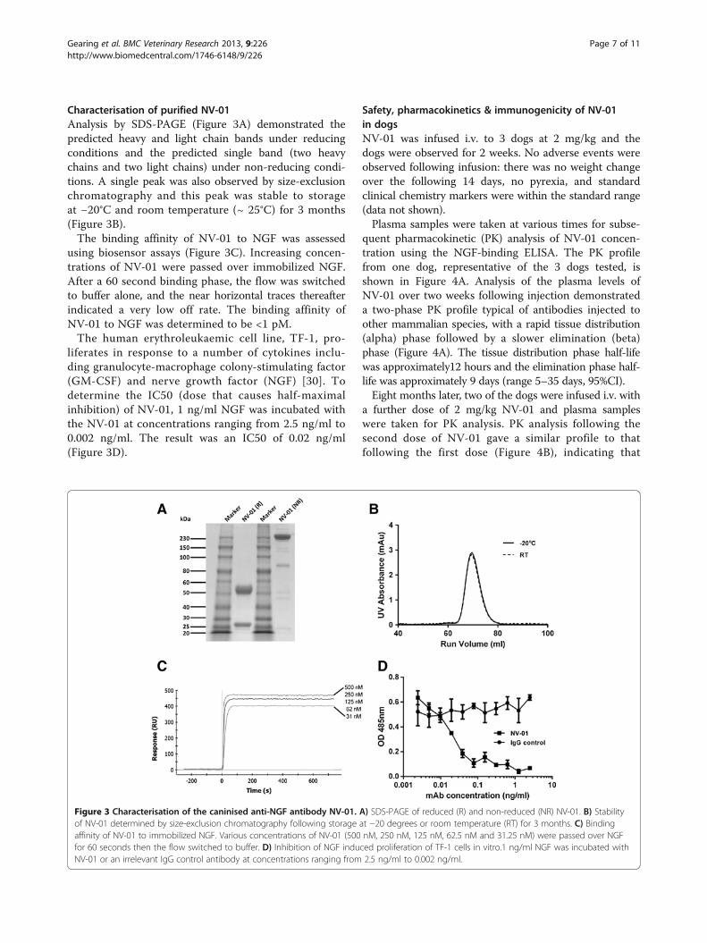
Characterisation of purified NV-01Analysis by SDS-PAGE (Figure 3A) demonstrated thepredicted heavy and light chain bands under reducingconditions and the predicted single band (two heavychains and two light chains) under non-reducing condi-tions. A single peak was also observed by size-exclusionchromatography and this peak was stable to storageat −20°C and room temperature (~ 25°C) for 3 months(Figure 3B).The binding affinity of NV-01 to NGF was assessed
using biosensor assays (Figure 3C). Increasing concen-trations of NV-01 were passed over immobilized NGF.After a 60 second binding phase, the flow was switchedto buffer alone, and the near horizontal traces thereafterindicated a very low off rate. The binding affinity ofNV-01 to NGF was determined to be <1 pM.The human erythroleukaemic cell line, TF-1, pro-
liferates in response to a number of cytokines inclu-ding granulocyte-macrophage colony-stimulating factor(GM-CSF) and nerve growth factor (NGF) [30]. Todetermine the IC50 (dose that causes half-maximalinhibition) of NV-01, 1 ng/ml NGF was incubated withthe NV-01 at concentrations ranging from 2.5 ng/ml to0.002 ng/ml. The result was an IC50 of 0.02 ng/ml(Figure 3D).
Safety, pharmacokinetics & immunogenicity of NV-01in dogsNV-01 was infused i.v. to 3 dogs at 2 mg/kg and thedogs were observed for 2 weeks. No adverse events wereobserved following infusion: there was no weight changeover the following 14 days, no pyrexia, and standardclinical chemistry markers were within the standard range(data not shown).Plasma samples were taken at various times for subse-
quent pharmacokinetic (PK) analysis of NV-01 concen-tration using the NGF-binding ELISA. The PK profilefrom one dog, representative of the 3 dogs tested, isshown in Figure 4A. Analysis of the plasma levels ofNV-01 over two weeks following injection demonstrateda two-phase PK profile typical of antibodies injected toother mammalian species, with a rapid tissue distribution(alpha) phase followed by a slower elimination (beta)phase (Figure 4A). The tissue distribution phase half-lifewas approximately12 hours and the elimination phase half-life was approximately 9 days (range 5–35 days, 95%CI).Eight months later, two of the dogs were infused i.v. with
a further dose of 2 mg/kg NV-01 and plasma sampleswere taken for PK analysis. PK analysis following thesecond dose of NV-01 gave a similar profile to thatfollowing the first dose (Figure 4B), indicating that
Figure 3 Characterisation of the caninised anti-NGF antibody NV-01. A) SDS-PAGE of reduced (R) and non-reduced (NR) NV-01. B) Stabilityof NV-01 determined by size-exclusion chromatography following storage at −20 degrees or room temperature (RT) for 3 months. C) Bindingaffinity of NV-01 to immobilized NGF. Various concentrations of NV-01 (500 nM, 250 nM, 125 nM, 62.5 nM and 31.25 nM) were passed over NGFfor 60 seconds then the flow switched to buffer. D) Inhibition of NGF induced proliferation of TF-1 cells in vitro.1 ng/ml NGF was incubated withNV-01 or an irrelevant IgG control antibody at concentrations ranging from 2.5 ng/ml to 0.002 ng/ml.
Gearing et al. BMC Veterinary Research 2013, 9:226 Page 7 of 11http://www.biomedcentral.com/1746-6148/9/226

NV-01 infusion did not induce an acute neutralisingimmunogenic response.
Comparison of the efficacy of NV-01 and meloxicam in amodel of inflammatory lameness in the dogKaolin injection to the footpad of dogs resulted in aconsistent inflammatory lameness score of 3 (severelylame, not weight bearing) 24 hours later. Four groupsof dogs were then treated with PBS placebo, oralmeloxicam, i.v. NV-01 or s.c. NV-01. Lameness scoresdetermined by assessors blinded to the type of interventionwere determined on a four-point scale. Following 7 daysof observations, the lameness scores were un-blinded,averaged according to intervention type and comparedwith placebo (i.v. PBS infusion). Figure 5A shows lamenessscores for meloxicam-treated or NV-01-treated dogscompared with PBS-treated dogs.Following the initial lameness caused by kaolin injec-
tion, the control PBS-injected dogs remained fully lamefor another day, and then slowly but progressively
improved over the following week (PBS IV, Figure 5A).As anticipated, daily oral meloxicam was effective inreducing lameness compared with i.v. PBS. The effectof daily oral meloxicam in reducing average lamenessscores was matched by the single i.v. dose of NV-01:with meloxicam, significant difference (Mann–Whitneytest: p ≤ 0.10) to placebo was observed at 6 h and onDay 1, 5, 6 and 7; whereas with i.v. NV-01, significancecompared with placebo was seen on Day 1, 3, 6 and 7.
Figure 4 Pharmacokinetic profile of NV-01 in the dog. A) Singledose. NV-01 (2 mg/kg) was infused intravenously and the NV-01serum concentration assayed by ELISA over a period of 14 days.B) Repeat dosing in two dogs. NV-01 (2 mg/kg) was administeredby intravenous infusion twice, with a gap of eight months betweeneach dose. The NV-01 concentration was determined by ELISA inserum samples taken over a period of 7 days after each infusion.
Figure 5 Efficacy of NV-01 in a model of inflammation inducedpain. A) Average lameness scores are shown for each treatmentgroup compared with the average lameness scores for the negativecontrol group: The negative control was a single injection ofphosphate buffered saline by intravenous infusion (IV, black triangles);Meloxicam (positive control) was given daily per os (PO) (grey squares);One 0.2 mg/kg dose of NV-01 was given by intravenous infusion (bluetriangles); or one 0.2 mg/kg dose of NV-01 was given by subcutaneousinjection (SC) (green circles). Points are the mean of eight animals pergroup. Error bars are the standard error of the mean. * p < 0.10; ** p <0.05; *** p < 0.01 (Mann–Whitney test, one-tailed: test article comparedto placebo). Arrows represent administration of the test article on thespecified day. B) NV-01 serum concentration was determined by ELISAover a period of seven days in animals from the study given 0.2 mg/mlof NV-01 by intravenous infusion (blue triangles) or subcutaneousinjection (green circles). Points are the mean NV-01 serumconcentrations of three animals per group. Error bars are thestandard deviation.
Gearing et al. BMC Veterinary Research 2013, 9:226 Page 8 of 11http://www.biomedcentral.com/1746-6148/9/226

(Figure 5A). A single s.c. injection of NV-01 was effectivecompared to placebo later with significance at Day 7.PK analysis of plasma NV-01 levels (Figure 5B) fromthe dogs treated with i.v. or s.c. NV-01 showed adelayed rise in circulating NV-01 following s.c. injectioncompared with i.v. infusion and that the circulatingNV-01 levels were approximately equal 5–7 daysfollowing administration.
DiscussionMethods for the conversion of antibodies from rodent tohuman are well described and are routinely used for theproduction of humanised antibodies for clinical use. Forexample CDR grafting [31,32] makes use of germlineantibody framework sequence as donor sequence forprimary antibody construction. This method often pro-duces antibodies of lower affinity for antigen than thedonor antibody. Affinity restoration or "maturation" isachieved by replacement of donor framework residuesuntil affinity is restored but this often results in antibodiesthat are incompletely converted and so potentiallymore immunogenic. Rather than use germline donorsequences, we devised a simpler but more effectivemethod (termed PETisation) using expressed cDNAsequences of the desired target species, in this case, thedog. Analysis of the greater range of framework sequencepossibilities in canine IgG heavy and light chains enabledsubstitution of fewer (22 heavy chain) residues of thedonor antibody to covert to a fully canine antibodyframework than had we started with germline sequence(35 heavy chain). By making fewer changes, the resultingantibody is structurally most similar to the startingantibody and yet fully caninised. The advantages of thissimple approach are: 1) the minimum changes are requiredto yield an antibody that has considerable similarity tothe starting antibody, 2) the changes made are on thesurface of the antibody structure and so do not affectthe immunoglobulin fold and CDR presentation, and 3)the resulting antibody is completely caninised and socan be expected to be non-immunogenic.The anti-NGF antibody αD11 is well described [7,25]
and was considered a good candidate for conversionfor use in dogs based on its high affinity to mouse andhuman NGF and its activity both in vitro and in vivo[7]. Although sequence differences between murine,human and canine NGF had the potential to affectαD11 binding, they were few and distant to the epitope[33]. Our results with NV-01 indicate that it retainedthe high affinity and activity of the αD11 antibodyin vitro and was expressed at high levels in CHO cellsand was stable following prolonged incubation at roomtemperature.NGF is expressed on nerves and is subject to retro-
grade transport. The safe use of anti-NGF antibodies
demands that the antibody not recruit effector functionsof the immune system, such as complement-dependentcytotoxicity, and so avoid nerve damage. By directanalogy, anti-NGF antibodies for human use have beenconstructed from effector negative isotypes. The effectorfunctions of canine antibodies were not previouslydescribed and so we expressed the caninised antibodieswith each of the four canine isotypes previously described[21]. Two of the isotypes (IgG-heavy chain A and D)did not bind complement C1q (the first component ofthe complement cascade) and so were chosen for furtherdevelopment for their improved safety profile. Interest-ingly, neither antibody isotype bound to the high affinityFcR CD64 (a macrophage receptor involved in the pro-cesses of opsonisation and the respiratory burst), nor toProtein A or Protein G.Anti-NGF mAb therapies are particularly attractive be-
cause of their potential to provide long-lasting analgesiain chronic disease settings, such as osteoarthritis andcancer pain. As a prelude to testing the potential ofneutralising NGF in the management of pain in chroniccanine diseases, a kaolin model of short -term inflammatorylameness was chosen to evaluate the efficacy of NV-01in dogs in vivo. The kaolin model, first validated in catsand then in dogs, has the advantage that the animalsrecover quickly over the period of about 7–14 days andare returned to the colony. However, this ethical advantagealso limits the model since there is not a stable periodof inflammation during which direct comparisons canbe made between drug and placebo [29]. Consequently,any drug benefit must be compared against a steadyimprovement in limb function in the placebo treateddogs. Presently, there are no ethically equivalent models oflong-term inflammation in dogs and so the kaolin modelwas deemed most appropriate for this initial assessmentof the potential pain-relieving properties of NV-01.In the canine kaolin model, oral meloxicam therapy
was effective at reducing signs of lameness in the firsthours after treatment, whereas intravenously infusedNV-01 required longer to become effective presumablydue to slower distribution from the blood to inflamedtissue of the larger antibody molecule (Figure 5). Followingthis distribution phase, the single i.v. dose of NV-01was similarly effective as daily oral meloxicam at reducinglameness. Sub-cutaneously delivered NV-01 was alsoshown to be active in reducing lameness, but somewhatlater than following i.v. delivery due to the slower redistri-bution from skin to blood and then to inflamed tissuefollowing the s.c. route of administration. Nonetheless,these studies confirm that both i.v. and s.c. routes ofadministration of NV-01 have potential to provide painrelief in dogs. As with studies of anti-NGF in the mouse[8], neither rectal temperature, nor paw circumferencewas affected by meloxicam or NV-01 therapies.
Gearing et al. BMC Veterinary Research 2013, 9:226 Page 9 of 11http://www.biomedcentral.com/1746-6148/9/226

Compared with small molecule drugs, antibodies havelong half-lives and are more suited to treatment ofchronic diseases. Pharmacokinetic analyses showedthat following i.v. infusion, NV-01 has an approxi-mately 9-day half-life and has the potential to be adminis-tered repeatedly to dogs without inducing a neutralisingimmune response. The slow rate of decline shown in thepharmacokinetic analysis in Figure 4A suggests that asingle injection may confer an extended period of thera-peutic benefit in the treatment of chronic pain. Thiscombination of safety, long half-life and low immuno-genicity suggests that NV-01 should be further evaluatedas a therapy for chronic pain conditions in dogs, wherea safety profile superior to current therapies together witha long duration between injections would be clinicallydesirable attributes for a novel therapeutic agent.
ConclusionThese preliminary studies lead us to conclude that thenovel fully caninised anti-NGF mAb NV-01 shows consi-derable potential as an analgesic for dogs.
AbbreviationsBSA: Bovine serum albumin; CDR: Complementarity-determining region;CHO: Chinese hamster ovary; EDAC: 1-ethyl-3-(3-dimethylaminopropyl)carbodiimide hydrochloride; ELISA: Enzyme-linked immuno-sorbent assay;FcR: Immunoglobulin Fc receptor; GM-CSF: Granulocyte colony-stimulatingfactor; HEK: Human embryonic kidney; i.v.: Intravenous; mAb: Monoclonalantibody; NGF: Nerve growth factor; NHS: N-hydroxysuccinimide;NSAID: Non-steroidal anti-inflammatory drug; PBS: Phosphate buffered saline;PK: Pharmacokinetic; s.c.: Subcutaneous; scaCD64: Soluble canine CD64;SDS-PAGE: Sodium dodecyl sulphate polyacrylamide gel electrophoresis.
Competing interestsDG, RG and EV are employees and stockholders of Nexvet Biopharma PtyLtd. DG has applied for patents relating to the content of the manuscript.AD is funded by a contract between Nexvet Biopharma and Monash University.Nexvet Biopharma is funding the article processing charges of this manuscript.
Authors’ contributionsDG conceived the PETisation process, antibody design and in vitro assays,designed the in vivo studies and drafted the manuscript. EV carried out thecellular and biochemical assays. RG designed and coordinated the scale-upprocess. AD designed and carried out functional assays, immunoassays andprotein purification. All authors read and approved the final manuscript.
AcknowledgementsThe authors wish to acknowledge the following for their contributions tothis study: Paul Hertzog, Kevin Johnson, Steve Dower, Mark Heffernan, AndyGearing and Tsutomu Mori for technical advice and support; Ralph Webster,Colin Giles and John Cox for critical review of the manuscript; Michael Springand colleagues (Geneart AG, Germany) for gene synthesis, cloning,expression and protein purification services; Kathryn Hjerrild for technicalsupport; Yvette Stallwood, Georg Blaser, Yash Patel and colleagues (Lonza,Cambridge, UK) for protein expression and purification; Edouard Nice andDaniel Layton (MATF, Monash University, Australia) for binding affinitystudies; Angel Lopez, Tim Hercus and Barbara McLure (Centre for CancerResearch, Adelaide) for guidance with the TF1 assay; and Martin Murphy andcolleagues at Charles River Laboratories (Ballina, Ireland) for performing theanimal studies and the kaolin model.
Received: 23 August 2013 Accepted: 7 November 2013Published: 9 November 2013
References1. Wernham BG, Trumpatori B, Hash J, Lipsett J, Davidson G, Wackerow P,
Thomson A, Lascelles BD: Dose reduction of meloxicam in dogs withosteoarthritis-associated pain and impaired mobility. J Vet Intern Med/American College of Veterinary Internal Medicine 2011, 25(6):1298–1305.
2. Papich MG: An update on nonsteroidal anti-inflammatory drugs (NSAIDs)in small animals. Vet Clin N Am Small Anim Pract 2008, 38(6):1243–1266. vi.
3. Levi-Montalcini R: The nerve growth factor 35 years later. Science 1987,237(4819):1154–1162.
4. Levi-Montalcini R, Skaper SD, Dal Toso R, Petrelli L, Leon A: Nerve growthfactor: from neurotrophin to neurokine. Trends Neurosci 1996, 19(11):514–520.
5. Hefti FF, Rosenthal A, Walicke PA, Wyatt S, Vergara G, Shelton DL, DaviesAM: Novel class of pain drugs based on antagonism of NGF.Trends Pharmacol Sci 2006, 27(2):85–91.
6. Mantyh PW, Koltzenburg M, Mendell LM, Tive L, Shelton DL: Antagonism ofnerve growth factor-TrkA signaling and the relief of pain.Anesthesiology 2011, 115(1):189–204.
7. Cattaneo A: Tanezumab, a recombinant humanized mAb against nervegrowth factor for the treatment of acute and chronic pain. Curr Opin MolTher 2010, 12(1):94–106.
8. Ghilardi JR, Freeman KT, Jimenez-Andrade JM, Coughlin KA, Kaczmarska MJ,Castaneda-Corral G, Bloom AP, Kuskowski MA, Mantyh PW: Neuroplasticityof sensory and sympathetic nerve fibers in a mouse model of a painfularthritic joint. Arthritis Rheum 2012, 64(7):2223–2232.
9. Brown MT, Murphy FT, Radin DM, Davignon I, Smith MD, West CR: Tanezumabreduces osteoarthritic hip pain: results of a randomized, double-blind,placebo-controlled phase III trial. Arthritis Rheum 2013, 65(7):1795–1803.
10. Katz N, Borenstein DG, Birbara C, Bramson C, Nemeth MA, Smith MD,Brown MT: Efficacy and safety of tanezumab in the treatment of chroniclow back pain. Pain 2011, 152(10):2248–2258.
11. Lane NE, Schnitzer TJ, Birbara CA, Mokhtarani M, Shelton DL, Smith MD,Brown MT: Tanezumab for the treatment of pain from osteoarthritis ofthe knee. N Engl J Med 2010, 363(16):1521–1531.
12. Nagashima H, Suzuki M, Araki S, Yamabe T, Muto C, Tanezumab I:Preliminary assessment of the safety and efficacy of tanezumab inJapanese patients with moderate to severe osteoarthritis of the knee:a randomized, double-blind, dose-escalation, placebo-controlled study.Osteoarthritis Cartilage/OARS, Osteoarthritis Research Society 2011,19(12):1405–1412.
13. Zorbas M, Hurst S, Shelton D, Evans M, Finco D, Butt M: A multiple-dosetoxicity study of tanezumab in cynomolgus monkeys. Regul ToxicolPharmacol 2011, 59(2):334–342.
14. Garber K: Fate of novel painkiller mAbs hangs in balance. Nat Biotechnol2011, 29(3):173–174.
15. Food and Drug Administration (USA): Summary minutes of the arthritisadvisory committee meeting March 12, 2012. Center for Drug Evaluation andResearch; 2012. http://www.fda.gov/downloads/AdvisoryCommittees/CommitteesMeetingMaterials/Drugs/ArthritisAdvisoryCommittee/UCM307879.pdf. Accessed November 2013.
16. D'Intino G, Vaccari F, Sivilia S, Scagliarini A, Gandini G, Giardino L, Calza L:A molecular study of hippocampus in dogs with convulsion duringcanine distemper virus encephalitis. Brain Res 2006, 1098(1):186–195.
17. Fan TM, Barger AM, Sprandel IT, Fredrickson RL: Investigating TrkAexpression in canine appendicular osteosarcoma. J Vet Intern Med/American College of Veterinary Internal Medicine 2008, 22(5):1181–1188.
18. Ryan VH, German AJ, Wood IS, Hunter L, Morris P, Trayhurn P: NGF geneexpression and secretion by canine adipocytes in primary culture:upregulation by the inflammatory mediators LPS and TNFalpha. HormMetab Res = Hormon- und Stoffwechselforschung = Hormones et metabolisme2008, 40(12):861–868.
19. Woo HM, Bentley E, Campbell SF, Marfurt CF, Murphy CJ: Nerve growthfactor and corneal wound healing in dogs. Exp Eye Res 2005, 80(5):633–642.
20. Isola M, Ferrari V, Miolo A, Stabile F, Bernardini D, Carnier P, Busetto R:Nerve growth factor concentrations in the synovial fluid from healthydogs and dogs with secondary osteoarthritis. Vet Comp Orthop Traumatol2011, 24(4):279–284.
21. Tang L, Sampson C, Dreitz MJ, McCall C: Cloning and characterization ofcDNAs encoding four different canine immunoglobulin gamma chains.Vet Immunol Immunopathol 2001, 80(3–4):259–270.
22. Albanesi M, Daeron M: The interactions of therapeutic antibodies with Fcreceptors. Immunol Lett 2012, 143(1):20–27.
Gearing et al. BMC Veterinary Research 2013, 9:226 Page 10 of 11http://www.biomedcentral.com/1746-6148/9/226

23. Hinton PR, Johlfs MG, Xiong JM, Hanestad K, Ong KC, Bullock C, Keller S,Tang MT, Tso JY, Vasquez M, et al: Engineered human IgG antibodies withlonger serum half-lives in primates. J Biol Chem 2004, 279(8):6213–6216.
24. Mould DR, Sweeney KR: The pharmacokinetics and pharmacodynamics ofmonoclonal antibodies–mechanistic modeling applied to drugdevelopment. Curr Opin Drug Discov Dev 2007, 10(1):84–96.
25. Ruberti F, Bradbury A, Cattaneo A: Cloning and expression of ananti-nerve growth factor (NGF) antibody for studies using theneuroantibody approach. Cell Mol Neurobiol 1993, 13(5):559–568.
26. Lewis MJ, Wagner B, Woof JM: The different effector function capabilitiesof the seven equine IgG subclasses have implications for vaccinestrategies. Cell Mol Immunol 2008, 45(3):818–827.
27. Paetz A, Sack M, Thepen T, Tur MK, Bruell D, Finnern R, Fischer R, Barth S:Recombinant soluble human Fcgamma receptor I with picomolaraffinity for immunoglobulin G. Biochem Biophys Res Commun 2005,338(4):1811–1817.
28. Giraudel JM, Diquelou A, Laroute V, Lees P, Toutain PL: Pharmacokinetic/pharmacodynamic modelling of NSAIDs in a model of reversibleinflammation in the cat. Br J Pharmacol 2005, 146(5):642–653.
29. Jeunesse EC, Bargues IA, Toutain CE, Lacroix MZ, Letellier IM, Giraudel JM,Toutain PL: Paw inflammation model in dogs for preclinicalpharmacokinetic/pharmacodynamic investigations of nonsteroidalanti-inflammatory drugs. J Pharmacol Exp Ther 2011, 338(2):548–558.
30. Kitamura T, Tojo A, Kuwaki T, Chiba S, Miyazono K, Urabe A, Takaku F:Identification and analysis of human erythropoietin receptors on afactor-dependent cell line, TF-1. Blood 1989, 73(2):375–380.
31. Queen C, Schneider WP, Selick HE, Payne PW, Landolfi NF, Duncan JF,Avdalovic NM, Levitt M, Junghans RP, Waldmann TA: A humanizedantibody that binds to the interleukin 2 receptor. Proc Natl Acad Sci USA1989, 86(24):10029–10033.
32. Riechmann L, Clark M, Waldmann H, Winter G: Reshaping humanantibodies for therapy. Nature 1988, 332(6162):323–327.
33. Covaceuszach S, Cassetta A, Konarev PV, Gonfloni S, Rudolph R, Svergun DI,Lamba D, Cattaneo A: Dissecting NGF interactions with TrkA and p75receptors by structural and functional studies of an anti-NGF neutralizingantibody. J Mol Biol 2008, 381(4):881–896.
doi:10.1186/1746-6148-9-226Cite this article as: Gearing et al.: A fully caninised anti-NGF monoclonalantibody for pain relief in dogs. BMC Veterinary Research 2013 9:226.
Submit your next manuscript to BioMed Centraland take full advantage of:
• Convenient online submission
• Thorough peer review
• No space constraints or color figure charges
• Immediate publication on acceptance
• Inclusion in PubMed, CAS, Scopus and Google Scholar
• Research which is freely available for redistribution
Submit your manuscript at www.biomedcentral.com/submit
Gearing et al. BMC Veterinary Research 2013, 9:226 Page 11 of 11http://www.biomedcentral.com/1746-6148/9/226