Embed Size (px)
Citation preview
ARTICLE
A five-residue motif for the design of domainswapping in proteinsNeha Nandwani1, Parag Surana1, Hitendra Negi1,2, Nahren M. Mascarenhas 1,4,
Jayant B. Udgaonkar1,3, Ranabir Das 1 & Shachi Gosavi1
Domain swapping is the process by which identical monomeric proteins exchange structural
elements to generate dimers/oligomers. Although engineered domain swapping is a com-
pelling strategy for protein assembly, its application has been limited due to the lack of simple
and reliable design approaches. Here, we demonstrate that the hydrophobic five-residue
‘cystatin motif’ (QVVAG) from the domain-swapping protein Stefin B, when engineered
into a solvent-exposed, tight surface loop between two β-strands prevents the loop from
folding back upon itself, and drives domain swapping in non-domain-swapping proteins.
High-resolution structural studies demonstrate that engineering the QVVAG stretch inde-
pendently into various surface loops of four structurally distinct non-domain-swapping
proteins enabled the design of different modes of domain swapping in these proteins,
including single, double and open-ended domain swapping. These results suggest that the
introduction of the QVVAG motif can be used as a mutational approach for engineering
domain swapping in diverse β-hairpin proteins.
https://doi.org/10.1038/s41467-019-08295-x OPEN
1 National Centre for Biological Sciences, Tata Institute of Fundamental Research, Bengaluru 560065, India. 2 Sastra University, Thanjavur 613402, India.3 Indian Institute of Science Education and Research, Pune 411008, India. 4Present address: Sacred Heart College, Tirupattur, Tamil Nadu 635601, India.Correspondence and requests for materials should be addressed to J.B.U. (email: [email protected]) or (email: [email protected])or to R.D. (email: [email protected]) or to S.G. (email: [email protected])
NATURE COMMUNICATIONS | (2019) 10:452 | https://doi.org/10.1038/s41467-019-08295-x |www.nature.com/naturecommunications 1
1234
5678
90():,;
Rational design of protein–protein interactions can be usedto build supramolecular assemblies capable of performingboth biological and bio-inspired functions1. The formation
of a dimer is a basic step in building such assemblies2, and diversemethods have been invented to engineer protein homodimers andheterodimers3. However, the required protein manipulation isgenerally difficult because of the presence of a complex array ofcooperative and long-range interactions in proteins. This struc-tural complexity and the marginal stability of proteins necessitatethe optimization of each design approach in a protein-specificmanner. Thus, the number of proteins and protein sites amenableto a given design strategy is typically low.
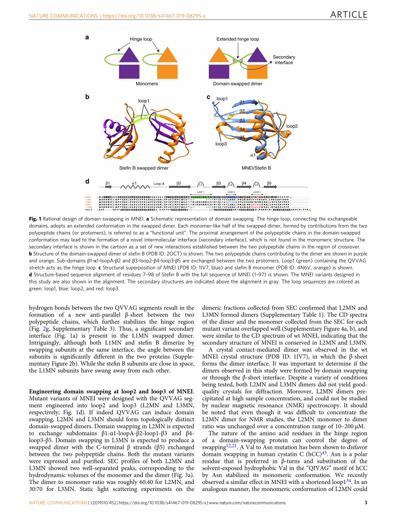
Several proteins dimerize (or oligomerize) naturally throughdomain swapping (sometimes termed 3D domain swapping4). Indomain swapping, two protein molecules exchange “domains” orstructural units connected by a hinge loop, such that inter-molecular interactions replace the intramolecular interactions atthe dimer interface of each monomer4–8 (Fig. 1a). This gives riseto a dimer containing two monomeric units which are almostidentical to the monomeric protein. Domain swapping is well-suited for the construction of oligomeric interfaces for the fol-lowing three reasons. First, the structural diversity of domainswapping proteins reported so far indicates that protein structuredoes not place strong restrictions on the design of domainswapping9,10. Second, domain swapping is likely to generatestable multimers because the same contacts that stabilize theprotein monomer also stabilize the protein dimer. Further, severalproteins domain-swap from the unfolded state11–13 and thus, abarrier larger than the folding free energy barrier, separates themonomer and the domain-swapped dimer. Finally, domainswapping can result in the formation of structurally complexoligomeric assemblies. Proteins can swap domains in an open-ended4 manner, leading to the formation of complex linearassemblies14,15. Proteins can also swap more than one domain7,either separately16,17, or simultaneously18,19 (referred to as doubledomain swapping10), leading to the formation of different proteinassemblies from a single protein10. Moreover, nature uses domainswapping not only as a mechanism for oligomer assembly, butalso to encode for novel functions and for the evolution of novelprotein folds4,20,21.
Thus, engineered domain swapping is likely to be a simple anduniversal strategy for the design of protein oligomers andassemblies. A few non-natural domain-swapped proteins havebeen designed using ad hoc methods11,22–33. Such studies havecollectively led to an understanding of the general principles ofdomain swapping design. Introduction of conformational strainin the monomer, by altering the physico-chemical properties ofthe putative hinge loop, is expected to drive domain swapping.The underlying driving force for oligomerization is the change inthe conformation of the modified and strained hinge loop to anenergetically favorable extended conformation in the domain-swapped structure6,7,12. However, these studies have not led toa specific mutational strategy for the introduction of domainswapping into diverse monomeric proteins. In a previous study,we noticed that placing a bulky, hydrophobic residue at the apexof a solvent-exposed, strained β-turn results in domain swap-ping34. Based on this result, we asked if engineering the largelyhydrophobic pentapeptide motif present in the domain-swappingcystatin (β1-α1-β2-β3-β4-β5 topology) proteins35 into the β-turnsof proteins could be a general strategy for designing domainswapping. In the domain-swapping cystatins, the loop connectingβ2–β3 has a QXVXG consensus motif, which has been implicatedin both protease inhibition and domain swapping35–39 (Fig. 1b).
Here, we engineer the hydrophobic QVVAG hinge loop fromthe domain-swapping cystatin, stefin B, individually into theβ-turn-β motifs of the single chain variant of the sweet protein
monellin40 (MNEI). MNEI is similar structurally to stefin B(Fig. 1c) and other cystatin proteins41, but does not undergodomain swapping. We show that introducing the QVVAG motifinto three different surface loops of MNEI results in domain-swapped dimerization, generating topologically-distinct domain-swapped dimers of MNEI. We then show that engineering theQVVAG stretch simultaneously into two different loops of MNEIcreates two swappable domains in it, resulting in the formation ofa double domain-swapped dimer. Thus, the QVVAG motif maybe used to induce domain swapping in diverse β-hairpin con-taining proteins. Finally, we provide evidence for the generality ofthis design strategy by using the QVVAG motif to engineerdomain swapping in three other proteins, which adopt folds thatare distinct from the monellin/cystatin fold. Together, theseresults indicate that introduction of the QVVAG motif intosurface β-loops of proteins is a simple mutational approach forthe design of domain swapping in diverse β-hairpin proteins.
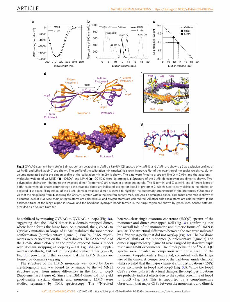
ResultsEngineering domain swapping at loop1 of MNEI. AlthoughMNEI and stefin B are structurally similar (both fold to a β1-α1-β2-β3-β4-β5 topology, Fig. 1c), the functions of the two proteinsare unrelated, and monellin is only 20% identical in sequence tostefin B. Additionally, stefin B domain-swaps42 but MNEI doesnot (Fig. 1c). The major conformational difference between thestefin B dimer and the MNEI monomer occurs in loop1, the loopconnecting the β2 and β3 strands. While a longer loop1 forms aβ-hairpin loop in MNEI, a shorter loop1 containing the QVVAGmotif forms a hinge that connects the two swapped domains inthe stefin B swapped dimer (Fig. 1b, c). A mutant variant ofMNEI (L1MN) having a shorter loop1 carrying the stefin BQVVAG motif in loop1 (Fig. 1d) was designed, expressed andpurified (see Methods and Supplementary Information). Thecircular dichroism (CD) spectrum of L1MN was similar to that ofwild type (wt) MNEI (Fig. 2a), indicating that the secondarystructure of monellin is conserved in L1MN. L1MN was run on asize exclusion chromatography (SEC) column, and was found tobe dimeric (Fig. 2b, c). Neither monomer, nor higher order oli-gomers, were observed over a wide range of protein concentra-tions (10–750 μM). The molar mass of L1MN, determined fromstatic light scattering experiments, was in excellent agreementwith the calculated mass of the dimer (23 kDa and 21 kDa,respectively; Supplementary Table 1). These experiments indicatethat L1MN exists almost completely as a dimer under theexperimental conditions.
L1MN was crystallized and the crystals diffracted to aresolution of 2.5 Å (Supplementary Table 2). The crystal structurerevealed that L1MN forms a symmetrical domain-swappeddimer. The two “subunits” of the dimer exchange “sub-domains”β1-α1-loopA-β2 and β3-loop2-β4-loop3-β5 (Fig. 2d-f, Supple-mentary Figure 1). Individually, both the subunits retain the foldof monellin, and superimpose with wt MNEI with a root meansquare deviation (rmsd) of 1.2 Å (Supplementary Figure 2a).However, the conformation of loop1 is different in L1MN and wtMNEI. In contrast to the β-hairpin conformation observed inMNEI, loop1 exists as an extended β-strand in L1MN (Fig. 2d, f;Supplementary Figure 2a). Modeling a turn into the calculatedelectron densities for loop1 in L1MN, obtained from a compositeomit map calculated by simulated-annealing, resulted in a poor fitand multiple steric clashes.
In the L1MN dimer, each monomeric “subunit” is composed oftwo polypeptide chains. Several van der Waals and hydrophobiccontacts are formed between the QVVAG segments of the twopolypeptide chains in the crossover region (SupplementaryTable 3, Supplementary Figure 3). Moreover, symmetrical
ARTICLE NATURE COMMUNICATIONS | https://doi.org/10.1038/s41467-019-08295-x
2 NATURE COMMUNICATIONS | (2019) 10:452 | https://doi.org/10.1038/s41467-019-08295-x | www.nature.com/naturecommunications
hydrogen bonds between the two QVVAG segments result in theformation of a new anti-parallel β-sheet between the twopolypeptide chains, which further stabilizes the hinge region(Fig. 2g, Supplementary Table 3). Thus, a significant secondaryinterface (Fig. 1a) is present in the L1MN swapped dimer.Intriguingly, although both L1MN and stefin B dimerize byswapping subunits at the same interface, the angle between thesubunits is significantly different in the two proteins (Supple-mentary Figure 2b). While the stefin B subunits are close in space,the L1MN subunits have swung away from each other.
Engineering domain swapping at loop2 and loop3 of MNEI.Mutant variants of MNEI were designed with the QVVAG seg-ment engineered into loop2 and loop3 (L2MN and L3MN,respectively; Fig. 1d). If indeed QVVAG can induce domainswapping, L2MN and L3MN should form topologically distinctdomain-swapped dimers. Domain swapping in L2MN is expectedto exchange subdomains β1-α1-loopA-β2-loop1-β3 and β4-loop3-β5. Domain swapping in L3MN is expected to produce aswapped dimer with the C-terminal β strands (β5) exchangedbetween the two polypeptide chains. Both the mutant variantswere expressed and purified. SEC profiles of both L2MN andL3MN showed two well-separated peaks, corresponding to thehydrodynamic volumes of the monomer and the dimer (Fig. 3a).The dimer to monomer ratio was roughly 60:40 for L2MN, and30:70 for L3MN. Static light scattering experiments on the
dimeric fractions collected from SEC confirmed that L2MN andL3MN formed dimers (Supplementary Table 1). The CD spectraof the dimer and the monomer collected from the SEC for eachmutant variant overlapped well (Supplementary Figure 4a, b), andwere similar to the CD spectrum of wt MNEI, indicating that thesecondary structure of MNEI is conserved in L2MN and L3MN.
A crystal contact-mediated dimer was observed in the wtMNEI crystal structure (PDB ID: 1IV7), in which the β-sheetforms the dimer interface. It was important to determine if thedimers observed in this study were formed by domain swappingor through the β-sheet interface. Despite a variety of conditionsbeing tested, both L2MN and L3MN dimers did not yield good-quality crystals for diffraction. Moreover, L2MN dimers pre-cipitated at high sample concentration, and could not be studiedby nuclear magnetic resonance (NMR) spectroscopy. It shouldbe noted that even though it was difficult to concentrate theL2MN dimer for NMR studies, the L2MN monomer to dimerratio was unchanged over a concentration range of 10–200 μM.
The nature of the amino acid residues in the hinge regionof a domain-swapping protein can control the degree ofswapping12,21. A Val to Asn mutation has been shown to disfavordomain swapping in human cystatin C (hCC)43. Asn is a polarresidue that is preferred in β-turns and substitution of thesolvent-exposed hydrophobic Val in the “QIVAG” motif of hCCby Asn stabilized its monomeric conformation. We recentlyobserved a similar effect in MNEI with a shortened loop134. In ananalogous manner, the monomeric conformation of L2MN could
a
Secondaryinterface
Monomers Domain-swapped dimer
Hinge loop Extended hinge loop
b cloop1 loop1
loop3
loop2
MNEI/Stefin BStefin B swapped dimer
β1
β2
β3
β4
β5
α1
d Loop A
LOOP 1 LOOP 2 LOOP 3
97
98
1
7
MNEIL1MNL2MNL3MNL13MNStefin B
β5β4β3β2β1 α1 L2 L3L1
Fig. 1 Rational design of domain swapping in MNEI. a Schematic representation of domain swapping. The hinge loop, connecting the exchangeabledomains, adopts an extended conformation in the swapped dimer. Each monomer-like half of the swapped dimer, formed by contributions from the twopolypeptide chains (or protomers), is referred to as a “functional unit”. The proximal arrangement of the polypeptide chains in the domain-swappedconformation may lead to the formation of a novel intermolecular interface (secondary interface), which is not found in the monomeric structure. Thesecondary interface is shown in the cartoon as a set of new interactions established between the two polypeptide chains in the region of crossover.b Structure of the domain-swapped dimer of stefin B (PDB ID: 2OCT) is shown. The two polypeptide chains contributing to the dimer are shown in purpleand orange. Sub-domains β1-α1-loopA-β2 and β3-loop2-β4-loop3-β5 are exchanged between the two protomers. Loop1 (green) containing the QVVAGstretch acts as the hinge loop. c Structural superposition of MNEI (PDB ID: 1IV7, blue) and stefin B monomer (PDB ID: 4N6V, orange) is shown.d Structure-based sequence alignment of residues 7–98 of Stefin B with the full sequence of MNEI (1–97) is shown. The MNEI variants designed inthis study are also shown in the alignment. The secondary structures are indicated above the alignment in gray. The loop sequences are colored asgreen: loop1, blue: loop2, and red: loop3
NATURE COMMUNICATIONS | https://doi.org/10.1038/s41467-019-08295-x ARTICLE
NATURE COMMUNICATIONS | (2019) 10:452 | https://doi.org/10.1038/s41467-019-08295-x |www.nature.com/naturecommunications 3
be stabilized by mutating QVVAG to QVNAG in loop2 (Fig. 3a),suggesting that the L2MN dimer is a domain-swapped dimer,where loop2 forms the hinge loop. As a control, the QVVAG toQVNAG mutation in loop1 of L1MN stabilized the monomericconformation (Supplementary Figure 5). Finally, SAXS experi-ments were carried out on the L2MN dimers. The SAXS profile ofthe L2MN dimer closely fit the profile expected from a modelwith domain swapping at loop2 (χ= 1.0, Fig. 3b) (see Supple-mentary Methods), but not to the crystal contact dimer (χ= 2.0,Fig. 3b), providing further evidence that the L2MN dimers areformed by domain swapping.
The structure of the L3MN monomer was solved by X-raycrystallography and was found to be similar to the wt MNEIstructure apart from minor differences in the fold of loop3(Supplementary Figure 6). Since the L3MN dimer did not yieldgood-quality crystals, dimeric and monomeric L3MN werestudied separately by NMR spectroscopy. The 15N-edited
heteronuclear single-quantum coherence (HSQC) spectra of themonomer and dimer overlapped well (Fig. 3c), confirming thatthe overall fold of the monomeric and dimeric forms of L3MN issimilar. The structural differences between the two were indicatedby a few cross-peaks that did not overlap (Fig. 3c). The backbonechemical shifts of the monomer (Supplementary Figure 7) anddimer (Supplementary Figure 8) were assigned by standard tripleresonance NMR experiments. The dimer peaks in the 15N-HSQCspectra were broader in comparison with those seen for themonomer (Supplementary Figure 9a), consistent with the largersize of the dimer. A comparison of the backbone amide chemicalshifts revealed that the major chemical shift perturbations (CSPs)occur exclusively in loop1 and loop3 (Fig. 3d). While the loop3CSPs are due to direct structural changes, the loop1 perturbationsare probably indirect effects due to the spatial proximity of loop1to loop3 (Fig. 1c). This is supported by a complementaryobservation that major CSPs between the monomeric and dimeric
–2000
–4000
MR
E (
mde
g cm
2 dm
ol–1
)
–6000
–8000
–10,000200 210 220 230 240 250
Wavelength (nm)
0
a
C-termProtomer 1
C-termProtomer 2
Protomer 2Protomer 1
N-termProtomer 2
N-termProtomer 1
loop1
loop2
loop3
loop2
VVAG
QV V A G
Q
V50
V50
V49
V49
Q48
Q48
d
Protomer 2Protomer 1
V50
Q48
V49 V50
A51
V49 Q48 I47
e g
f
MNEI
L1MN
1000670,000 Da
158,000 Da17,000 Da 1350 Da
44,000 Da
Calibrant
800
600
400
200
0Abs
orba
nce
at 2
80 n
m (
mA
U)
6 8 10 12
Elution volume (mL)
14 16 18 20
bMNEIL1MN
5.0
4.5
4.0
3.5
3.0
Log
mol
ecul
ar w
eigh
t
Elution volume (mL)
8 10 12 14 16 18 20
c
MNEI
Calibrant
L1MN
Fig. 2 QVVAG segment from stefin B drives domain swapping in L1MN. a Far-UV CD spectra of wt MNEI and L1MN are shown. b Size exclusion profiles ofwt MNEI and L1MN, at pH 7, are shown. The profile of the calibration mix (marker) is shown in gray. c Plot of the logarithm of molecular weight vs. elutionvolume generated using the elution profile of the calibration mix in (b) is shown. The data were fitted to a straight line (r= 0.99), and the apparentmolecular weights of wt MNEI (■ ~10 kDa) and L1MN (● ~20 kDa) were determined. d Structure of the L1MN domain-swapped dimer is shown. Twopolypeptide chains contributing to the swapped dimer (protomers) are shown in orange and purple. The N-termini and C-termini, and different loops ofboth the polypeptide chains contributing to the swapped dimer are indicated, except for loop3 of protomer 2, which is not clearly visible in the orientationdepicted. e A space-filling model of the L1MN domain-swapped dimer is shown to highlight the quaternary arrangement of the protomers. f Zoomed inview of the hinge loop from d, showing the QVVAG stretch within the electron density map. The 2Fo-Fc simulated anneal composite omit map is shown ata contour level of 1.6σ. Side chain nitrogen atoms are colored blue, and oxygen atoms are colored red. All other side chain atoms are colored yellow. g Thebackbone trace of the hinge region is shown, and the backbone hydrogen bonds formed in the hinge region are shown by green lines. Source data areprovided as a Source Data file
ARTICLE NATURE COMMUNICATIONS | https://doi.org/10.1038/s41467-019-08295-x
4 NATURE COMMUNICATIONS | (2019) 10:452 | https://doi.org/10.1038/s41467-019-08295-x | www.nature.com/naturecommunications
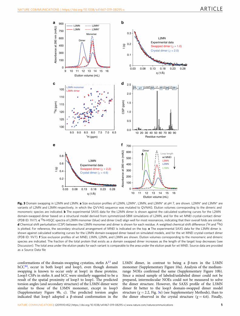
conformations of the domain-swapping cystatins, stefin A13 andhCC44, occur in both loop1 and loop3, even though domainswapping is known to occur only at loop1 in these proteins.Loop3 CSPs in stefin A and hCC were similarly suggested to be aresult of the spatial proximity of loop3 to loop1. The predictedtorsion angles (and secondary structure) of the L3MN dimer weresimilar to those of the L3MN monomer, except in loop3(Supplementary Figure 9b, c). The predicted torsion anglesindicated that loop3 adopted a β-strand conformation in the
L3MN dimer, in contrast to being a β-turn in the L3MNmonomer (Supplementary Figure 10a). Analysis of the medium-range NOEs confirmed the same (Supplementary Figure 10b).Since a mixed sample of labeled/unlabeled dimer could not beprepared, intermolecular NOEs could not be measured to solvethe dimer structure. However, the SAXS profile of the L3MNdimer fit better to the loop3 domain-swapped dimer modelstructure (χ= 2.2, Fig. 3e) (see Supplementary Methods), than tothe dimer observed in the crystal structure (χ= 4.6). Finally,
900
750
600
450
300
150
0
Abs
orba
nce
at 2
80 n
m (
mA
U)
L3MN
L2MN
L3MN*
Monomer
Dimer
aL2MN*
10 12119
Elution volume (mL)
1413 16
105
110
115
12015N
(pp
m)
1H (ppm)
125
130
9.5 9.0 8.5 8.0 7.5 7.0 6.5
L3MN monomerL3MN dimer
c
CS
P (
ppm
)
10 20 30 40 50 60Residue number
70 80 90
d 2.5β1 α1 β2LoopA L1 L2 L3β3 β4 β5
2.0
1.5
1.0
0.5
0.0
0.280.230.180.130.080.03
e
L3MN
Experimental dataSwapped dimer (� = 2.2)Crystal dimer (� = 4.6)
I (q)
q (1/Å)
0.8
0.6
0.4
0.2
0
10 11 12 13 14 15 16
Abs
orba
nce
at 2
80 n
m (
mA
U) Dimer
Loop length
wt MNEIL3MN
f
L2MN
L1MN
Elution volume (mL)
700
600
500
400
300
200
100
0
b
0.3
L2MN
Experimental dataSwapped dimer (� = 1.0)
Crystal dimer (� = 2.0)
0.2
I (q)
q (1/Å)
0.1
00.03 0.08 0.13 0.18 0.23 0.28
15
Fig. 3 Domain swapping in L2MN and L3MN. a Size exclusion profiles of L2MN, L2MN*, L3MN, and L3MN* at pH 7, are shown. L2MN* and L3MN* arevariants of L2MN and L3MN respectively, in which the QVVAG sequence was mutated to QVNAG. Elution volumes corresponding to the dimeric andmonomeric species are indicated. b The experimental SAXS data for the L2MN dimer is shown against the calculated scattering curves for the L2MNdomain-swapped dimer based on a structural model derived from symmetrized-SBM simulations of L2MN, and for the wt MNEI crystal-contact dimer(PDB ID: 1IV7). c 15N-HSQC spectra of L3MN monomer (blue) and dimer (red) align well for most resonances, indicating that their overall folds are similar.d Chemical shift perturbation (CSP) between the L3MN monomer and dimer is shown for each residue. A weighted chemical shift difference (1H and 15N)is plotted. For reference, the secondary structural arrangement of MNEI is indicated on the top. e The experimental SAXS data for the L3MN dimer isshown against calculated scattering curves for the L3MN domain-swapped dimer based on simulated models, and for the wt MNEI crystal-contact dimer(PDB ID: 1IV7). f Size exclusion profiles of wt MNEI, L1MN, L2MN, and L3MN are shown. Elution volumes corresponding to the monomeric and dimericspecies are indicated. The fraction of the total protein that exists as a domain swapped dimer increases as the length of the target loop decreases (seeDiscussion). The total area under the elution peaks for each variant is comparable to the area under the elution peak for wt MNEI. Source data are providedas a Source Data file
NATURE COMMUNICATIONS | https://doi.org/10.1038/s41467-019-08295-x ARTICLE
NATURE COMMUNICATIONS | (2019) 10:452 | https://doi.org/10.1038/s41467-019-08295-x |www.nature.com/naturecommunications 5

similar to L2MN, the QVVAG to QVNAG mutation in loop3 ofL3MN (L3MN*) led to the disappearance of the observed dimer(Fig. 3a). Altogether, the SEC, MALS, NMR and SAXS dataindicate that L3MN forms a dimer by domain swapping, whereinloop3 acts as the hinge loop.
Designing a double domain-swapped variant of MNEI. TheQVVAG segment was next introduced simultaneously into twodifferent loops of MNEI, with the aim of designing two swappabledomains in it. A variant of MNEI containing QVVAG segmentsin the two adjacent loops, loop1 and loop3, was designed(L13MN, Fig. 1d). L13MN was expressed, purified, and appearedfolded from CD measurements (Supplementary Figure 4c). TheSEC profile suggested that L13MN formed dimers (Fig. 4a),whose mass was confirmed by static light scattering experiments(Supplementary Table 1). L13MN was crystallized, and itsstructure was determined by X-ray crystallography to a resolutionof 2.3 Å (Supplementary Table 2). The atomic structure ofL13MN revealed a domain-swapped dimer, which was con-structed by a novel criss-crossed arrangement of the two poly-peptide chains contributing to the dimer (Fig. 4b, c). Both loop1and loop3 acted as hinge loops, and adopted an extended con-formation in the dimer (Fig. 4d, e and Supplementary Figure 11).Modeling a turn into the calculated electron densities for bothloop1 and loop3 in L13MN resulted in a poor fit and multiplesteric clashes. Swapping via loop1 results in an exchange of β1-α1-loopA-β2 and β3-loop2-β4-loop3-β5 sub-domains. Additionalswapping via loop3 splits the β3-loop2-β4-loop3-β5 sub-domain
further into β3-loop2-β4 and β5 sub-domains. This results in areciprocal exchange of more than one sub-domain between thetwo L13MN polypeptide chains, forming a dimer where the β3-loop2-β4 hairpin from one polypeptide chain appears to have“inserted” into the neighboring polypeptide chain. Apart frommultiple contacts within the QVVAG segments of loop1 andloop3, several contacts between these loops stabilize the swappeddimer (Supplementary Figure 12, Supplementary Table 4). Fur-thermore, ten hydrogen bonds were seen to have formed withinthe QVVAG segments at loop1 and loop3 (SupplementaryTable 5). Thus, the introduction of the QVVAG motif in twoadjacent β-hairpin loops in wt MNEI generated a reciprocaldouble domain-swapped dimer with multiple swapped interfaces.
The QVVAG stretch was next introduced into β-turn-β motifsof proteins unrelated to cystatins, to directly demonstrate that theQVVAG stretch can drive domain swapping in proteins that donot fold to the monellin/cystatin fold.
Engineering domain swapping in MK-Ctd. The QVVAG stretchwas next introduced into a β-hairpin loop in the C-terminaldomain of the hyperthermophilic protein MK0293 (MK-Ctd), aprotein domain derived from the protein MK0293 from Metha-nopyrus kandleri AV19 (PDB ID: 3C19, residues 98–178). MK-Ctd was chosen because it is unrelated to the cystatins, folds to adifferent topology45 (β1-β2-β3-α1-α2-β4-β5, Fig. 5a), and itspurification protocol, biochemical properties and folding havebeen characterized in detail in our laboratories. The QVVAGstretch was engineered into loop1 of MK-Ctd, which is a tight
a
Elution volume (mL)
10 11 12 13 14 15 16
Abs
orba
nce
at 2
80 n
m (
mA
U)
0
100
200
300
400
500
600
700 MNEI
L13MN
Dimer
Monomer
b
c
Protomer 1 Protomer 2
loop2
C-termProtomer 1
C-termProtomer 2
N-termProtomer 1
N-termProtomer 2
loop2 loop3
loop1
d loop1Y53
G52
A51 V50 V49 Q48
Y53
G52
A51V50V49Q48
e loop3
G77
A76 V75 V74 Q73
G77A76V75V74Q73
Fig. 4 Crystal structure of L13MN. a Size exclusion profiles of L13MN and wt MNEI at pH 7 are shown. b Ribbon representation of the crystal structure ofL13MN is shown. The two polypeptide chains contributing to the swapped dimer are colored in orange and purple. The three loops, and the N-termini andC-termini of each polypeptide chain are indicated. c A space-filling model of the L13MN domain-swapped dimer is shown to highlight the quaternaryarrangement of the protomers. Zoomed views of the hinge loops along with electron density in loop1 and loop3 are shown in d and e, respectively. Sidechain nitrogen atoms are colored blue, oxygen atoms are colored red, and the rest of the side chain atoms are colored yellow. The 2Fo-Fc simulated annealcomposite omit maps are shown at a contour level of 1.6σ. Source data are provided as a Source Data file
ARTICLE NATURE COMMUNICATIONS | https://doi.org/10.1038/s41467-019-08295-x
6 NATURE COMMUNICATIONS | (2019) 10:452 | https://doi.org/10.1038/s41467-019-08295-x | www.nature.com/naturecommunications

β-turn (3 residues long) connecting two moderately extendedβ-strands, β1 and β2 (Fig. 5a). This mutant variant (L1MK-Ctd)was expressed and purified, and it appeared well-folded from CDmeasurements (Supplementary Figure 13). The SEC profile sug-gested that L1MK-Ctd was entirely dimeric in solution (Fig. 5b),which was confirmed by static light scattering experiments(Supplementary Table 1).
The L1MK-Ctd dimer was analyzed by NMR spectroscopy.The 15N-edited HSQC spectrum of the L1MK-Ctd dimer showedwell-dispersed peaks (Fig. 5c), where several resonance cross-peaks were shifted from those of wt MK-Ctd (Fig. 5c). Thebackbone resonances were assigned for both wt MK-Ctd andL1MK-Ctd by standard triple resonance NMR experiments(Supplementary Figure 14 and 15). Only a single set of resonanceswas observed in the L1MK-Ctd spectra, indicating that L1MK-Ctd is a symmetric dimer. A comparison of the backbone amidechemical shifts of the monomeric wt MK-Ctd and dimericL1MK-Ctd revealed that the major chemical shift perturbations
between the two proteins were localized to three distinct positions(β1-loop1, a few residues within α2, and β4-loop3-β5, Fig. 5d).However, the predicted secondary structure (and torsion angles)of only loop1 residues differed between the two (SupplementaryFigure 16); loop1 adopted a β-strand conformation in the L1MK-Ctd dimer, but a β-turn conformation in the monomeric wt MK-Ctd (Supplementary Figure 16). CSPs at other locations can beattributed to the physical proximity of β4-loop3-β5 to loop1, anddocking of α2 against the β1–β2 hairpin (Fig. 5a). Therefore,similar to L3MN, NMR data along with SEC and light scatteringdata, suggests that L1MK-Ctd forms a dimer by domainswapping, wherein loop1 acts as the hinge loop.
Finally, the solution structure of L1MK-Ctd was determined byNMR. 13C-edited and 15N-edited NOESY-HSQC experimentswere carried out on uniformly 13C, 15N-labeled L1MK-Ctd. TheNOESY cross-peaks in the spectra provided both the intra-protomer connectivities and the inter-protomer connectivities.Exclusive NOE cross-peaks resulting from inter-protomer
a
loop1
loop3
C-term
N-term
loop2
β1 β4
β3β2
β5
α1
α2
c
1H (ppm)
51N
(pp
m)
MK-Ctd
L1MK-Ctd
10 9 8 7
130
125
120
115
110
105
100
e
QVV
AG
Q VV A
G
N-term
loop2
C-termloop3
loop2N-term
Protomer 1 Protomer 2
loop1
b
Elution volume (mL)
8 9 10 11 12 13 14 15
Abs
orba
nce
at 2
80 n
m (
mA
U)
0
100
200
300
400 MK-CtdL1MK-Ctd
L1MK-Ctd*L1MK-Ctd**
MonomerDimer
0
1
2
3
2 12 22 32 42 52 62 72 82C
SP
(pp
m)
Residue number
d loop1 loop2 loop3
f
Protomer 1 Protomer 2
β5β4β3β2β1 α2α1
Fig. 5 Domain swapping in MK-Ctd. a Structure of the C-terminal domain of the hyperthermophilic protein MK0293 (MK-Ctd) (PDB ID: 3C19) is shown.Different secondary structural elements, the N-termini and C-termini, and loop1, loop2, and loop3 of MK-Ctd are indicated. b Size exclusion profiles of MK-Ctd, L1MK-Ctd, L1MK-Ctd*, and L1MK-Ctd**, at pH 8, are shown. L1MK-Ctd* and L1MK-Ctd** are variants of L1MK-Ctd, in which the QVVAG sequence wasmutated to QVNAG and QNNAG, respectively. Elution volumes corresponding to the monomeric and dimeric species are indicated. c An overlay of the15N-HSQC spectra of the monomeric wt MK-Ctd (blue) and the dimeric L1MK-Ctd (red) is shown. d CSP between MK-Ctd and L1MK-Ctd is shown for eachresidue. A weighted chemical shift difference (1H and 15N) is plotted. For reference, the secondary structural arrangement of MK-Ctd is indicated on thetop. e Ribbon representation of the solution dimer of L1MK-Ctd is shown. The two polypeptide chains contributing to the swapped dimer are colored inorange and purple. f A space-filling model of the L1MK-Ctd domain-swapped dimer is shown to highlight the quaternary arrangement of theprotomers. Source data are provided as a Source Data file
NATURE COMMUNICATIONS | https://doi.org/10.1038/s41467-019-08295-x ARTICLE
NATURE COMMUNICATIONS | (2019) 10:452 | https://doi.org/10.1038/s41467-019-08295-x |www.nature.com/naturecommunications 7
connectivities were extracted from filtered NOESY experiments.Briefly, heterolabeled dimeric L1MK-Ctd was prepared byrefolding a 1:1 mixture of unfolded uniformly 15N/13C-labeledand unlabeled L1MK-Ctd proteins (see Supplementary Methods),which is therefore expected to be a mixture of 25% labeled-labeled, 50% labeled-unlabeled, and 25% unlabeled–unlabeleddimers. The SEC profile of the refolded heterolabeled sampleshowed that >90% of the total protein existed as a dimer(Supplementary Figure 17). 92 inter-protomer connectivitiescould be assigned from the various NOESY experiments. About102 dihedral angle restraints (φ and ψ) were determined perprotomer using the 1Hα, 15N, 13Cα, 13Cβ, and 13CO chemicalshifts and the program TALOS+46. Twenty-eight intra-protomerhelix hydrogen bonds were inferred per protomer from thestandard secondary structure of the protein based on NOEpatterns. No hydrogen bond restraints were used for β-strands toavoid bias for swapping. Using the NOE based distance restraints,dihedrals and hydrogen bond restraints, the structure of L1MK-Ctd dimer was calculated in Xplor-NIH47. SupplementaryFigure 18a shows the twenty lowest energy structures, and Fig. 5eshows the lowest energy structure. The structural statistics areprovided in Supplementary Table 6.
The solution structure of L1MK-Ctd dimer revealed a domain-swapped dimer, formed by the exchange of the N-terminalβ1 strand between the two polypeptide chains (Fig. 5e, f), inwhich loop1 is extended into a β-strand conformation, asopposed to a turn in the wt monomeric protein. The individualsubunits in the dimer superimpose well with monomeric wt MK-Ctd (Supplementary Figure 18b). The rmsd of twenty lowestenergy structures within each protomer is 1.1 Å, whereas thermsd of the complete dimer is 2.1 Å (Supplementary Table 6),suggesting that the hinge region between the two protomers isconformationally flexible. When the structures were aligned forone subunit, the other subunit spans an angle of ~24°, whichindicates the range of conformational dynamics between thesubunits (Supplementary Figure 18c). Several van der Waals andhydrophobic contacts are formed between the QVVAG segmentsof the two polypeptide chains in the crossover region (Supple-mentary Figure 18d and Supplementary Table 7). Unlike L1MNand L13MN, the two QVVAG segments at the hinge do not forma complete anti-parallel β-strand. This is probably because thehinge buries the first valine side chain of the QVVAG motif toshield the hydrophobic side chain (Supplementary Figure 18d, e).
Interestingly, the QVVAG to QVNAG mutation in L1MK-Ctddid not result in stabilization of its monomeric conformation(Fig. 5b); similar to L1MK-Ctd, the QVNAG variant, L1MK-Ctd*, was exclusively dimeric in solution. A closer look at theorientation of the side chain groups of the QVVAG residues inthe new anti-parallel β-sheet formed between the two polypeptidechains in the crossover region revealed that the side chain groupof the central valine was solvent exposed (SupplementaryFigure 18d), which might explain why the V →N mutation atthis position did not affect the oligomeric status of L1MK-Ctd.However, the hydrophobic side chain group of the precedingvaline is buried in the secondary interface (SupplementaryFigure 18d, e), and the mutation of the QVVAG motif toQNNAG resulted in complete reversal of the observed domain-swapped dimerization in L1MK-Ctd (Fig. 5b). A similarorientation of the side chains of the two Val residues is seen atthe secondary interface in L1MN (Fig. 2f, g, SupplementaryFigure 3); the hydrophobic side chain of the central Val residue issolvent accessible, while that of the first Val is buried at theinterface. The V to N mutation at the first Val is thereforeexpected to diminish dimerization more effectively due todisruption of the hydrophobic interactions. Therefore, we madea similar QVVAG to QNNAG mutation in L1MN, and the
resulting variant (L1MN**) was also found to be completelymonomeric (Supplementary Figure 5).
Engineering domain swapping in Sso7dv and UBQ. TheQVVAG motif was then introduced into a β-hairpin motif ofSso7d (a small DNA binding protein48,49) and human ubiquitin(UBQ). These proteins were chosen because they adopt folds thatare structurally unrelated to the monellin/cystatin fold, and theirpurification protocols, biochemical properties, and structuraldetails have already been characterized in our laboratories.
Sso7d (SCOPe ID: 54163) has an SH3-like fold50, and folds to aβ1-β2-β3-β4-β5-α1 topology (Fig. 6a). We used a variant ofSso7d, Sso7dv, whose structure was recently determined in ourlaboratories. Sso7dv is derived from a Sso7d scaffold51 where theresidues on the DNA binding interface have been randomized,thereby abolishing the DNA-binding ability of the scaffold(see Supplementary Methods for the scaffold sequence). TheQVVAG motif was introduced into the loop connecting β1–β2(loop1) of Sso7dv (Fig. 6a). This mutant (L1Sso7dv) wasexpressed and purified (see Supplementary Methods). L1Sso7dvappeared folded from CD measurements (Supplementary Fig-ure 19). The SEC profile of L1Sso7dv showed that the protein waspredominantly dimeric (Fig. 6b). To confirm that the observeddimerization was a result of domain swapping, the oligomericstatus of L1Sso7dv* (QVVAG to QVNAG mutation in L1Sso7dv)was assessed. Figure 6b shows that L1Sso7dv* was entirelymonomeric, which indicates that L1Sso7dv dimers are likelydomain-swapped dimers.
UBQ adopts a β-grasp fold, and folds to a β1-β2-α1-β3-β4-310-β5 topology (Fig. 6c). The QVVAG stretch was engineeredindividually into the loops connecting β1-β2 (loop1) or β3-β4(loop4) in UBQ (Fig. 6c). The loop1 variant of UBQ (L1UBQ)was entirely monomeric in solution (Supplementary Figure 20a).Unfolding and refolding of purified monomeric L1UBQ, but notwt UBQ, at high protein concentrations (~ 1 mM; see Supple-mentary Methods) led to the conversion of <5% of the proteininto a dimer (Supplementary Figure 20a), whose CD spectrumoverlapped well with that of wt UBQ (Supplementary Figure 20b),indicating that the dimer possibly forms by domain swapping.However, because the proportion of the observed dimer wasinsignificant, L1UBQ was not characterized further. In contrast toL1UBQ, the loop4 variant of UBQ (L4UBQ) was isolated fromthe insoluble fraction of the cell lysate under denaturingconditions followed by refolding (see Supplementary Methods).The SEC profile of the purified L4UBQ showed multiple peaks(Fig. 6d), indicating that the protein exists as a mixture ofdifferent oligomeric forms. Static light scattering experiments onthe oligomeric fractions collected from SEC confirmed thatL4UBQ formed dimers and higher-order multimers (Supplemen-tary Table 1). Further, ensemble measurements like circulardichroism indicated that the secondary structure of UBQ isconserved in these multimers (Supplementary Figure 21a). Theoligomeric fraction eluting in the void volume (Fig. 6d) wasanalyzed by cryo-electron microscopy (Supplementary Fig-ure 21b), which revealed that the oligomeric fraction is aheterogeneous mixture of linear multimers of varying lengths.The absence of thioflavin T (ThT) binding to the L4UBQoligomeric species ruled out the possibility of an orderedamyloid-like (cross-β sheet) structural arrangement52 in theseoligomers (Supplementary Figure 21c). Finally, the QVVAG toQVNAG mutation was found to significantly stabilize themonomeric conformation of L4UBQ (Fig. 6d). The QVVAG toQNNAG mutation in L4UBQ resulted in complete monomeriza-tion (Fig. 6d), similar to our observations with L1MN and L1MK-Ctd. These results suggest that multimerization of UBQ, observed
ARTICLE NATURE COMMUNICATIONS | https://doi.org/10.1038/s41467-019-08295-x
8 NATURE COMMUNICATIONS | (2019) 10:452 | https://doi.org/10.1038/s41467-019-08295-x | www.nature.com/naturecommunications
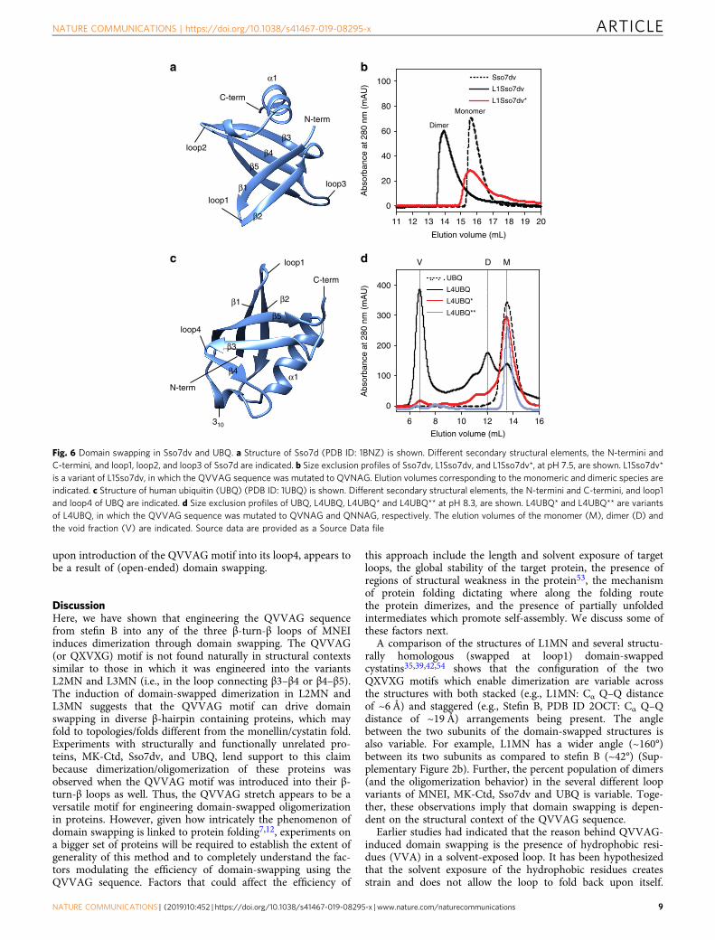
upon introduction of the QVVAG motif into its loop4, appears tobe a result of (open-ended) domain swapping.
DiscussionHere, we have shown that engineering the QVVAG sequencefrom stefin B into any of the three β-turn-β loops of MNEIinduces dimerization through domain swapping. The QVVAG(or QXVXG) motif is not found naturally in structural contextssimilar to those in which it was engineered into the variantsL2MN and L3MN (i.e., in the loop connecting β3–β4 or β4–β5).The induction of domain-swapped dimerization in L2MN andL3MN suggests that the QVVAG motif can drive domainswapping in diverse β-hairpin containing proteins, which mayfold to topologies/folds different from the monellin/cystatin fold.Experiments with structurally and functionally unrelated pro-teins, MK-Ctd, Sso7dv, and UBQ, lend support to this claimbecause dimerization/oligomerization of these proteins wasobserved when the QVVAG motif was introduced into their β-turn-β loops as well. Thus, the QVVAG stretch appears to be aversatile motif for engineering domain-swapped oligomerizationin proteins. However, given how intricately the phenomenon ofdomain swapping is linked to protein folding7,12, experiments ona bigger set of proteins will be required to establish the extent ofgenerality of this method and to completely understand the fac-tors modulating the efficiency of domain-swapping using theQVVAG sequence. Factors that could affect the efficiency of
this approach include the length and solvent exposure of targetloops, the global stability of the target protein, the presence ofregions of structural weakness in the protein53, the mechanismof protein folding dictating where along the folding routethe protein dimerizes, and the presence of partially unfoldedintermediates which promote self-assembly. We discuss some ofthese factors next.
A comparison of the structures of L1MN and several structu-rally homologous (swapped at loop1) domain-swappedcystatins35,39,42,54 shows that the configuration of the twoQXVXG motifs which enable dimerization are variable acrossthe structures with both stacked (e.g., L1MN: Cα Q–Q distanceof ~6 Å) and staggered (e.g., Stefin B, PDB ID 2OCT: Cα Q–Qdistance of ~19 Å) arrangements being present. The anglebetween the two subunits of the domain-swapped structures isalso variable. For example, L1MN has a wider angle (~160°)between its two subunits as compared to stefin B (~42°) (Sup-plementary Figure 2b). Further, the percent population of dimers(and the oligomerization behavior) in the several different loopvariants of MNEI, MK-Ctd, Sso7dv and UBQ is variable. Toge-ther, these observations imply that domain swapping is depen-dent on the structural context of the QVVAG sequence.
Earlier studies had indicated that the reason behind QVVAG-induced domain swapping is the presence of hydrophobic resi-dues (VVA) in a solvent-exposed loop. It has been hypothesizedthat the solvent exposure of the hydrophobic residues createsstrain and does not allow the loop to fold back upon itself.
a b
Elution volume (mL)
11 12 13 14 15 16 17 18 19 20
0
20
40
60
80
100 Sso7dv
L1Sso7dv
L1Sso7dv*
Monomer
Dimer
Abs
orba
nce
at 2
80 n
m (
mA
U)
c d
Elution volume (mL)
6 8 10 12 14 16
Abs
orba
nce
at 2
80 n
m (
mA
U)
0
100
200
300
400UBQ
L4UBQ
L4UBQ*
L4UBQ**
MDVloop1
loop4
C-term
N-term
β1
β4
β3
β2
β5
α1
310
loop1
loop3
C-term
N-term
loop2
β1
β4
β3
β2
β5
α1
Fig. 6 Domain swapping in Sso7dv and UBQ. a Structure of Sso7d (PDB ID: 1BNZ) is shown. Different secondary structural elements, the N-termini andC-termini, and loop1, loop2, and loop3 of Sso7d are indicated. b Size exclusion profiles of Sso7dv, L1Sso7dv, and L1Sso7dv*, at pH 7.5, are shown. L1Sso7dv*is a variant of L1Sso7dv, in which the QVVAG sequence was mutated to QVNAG. Elution volumes corresponding to the monomeric and dimeric species areindicated. c Structure of human ubiquitin (UBQ) (PDB ID: 1UBQ) is shown. Different secondary structural elements, the N-termini and C-termini, and loop1and loop4 of UBQ are indicated. d Size exclusion profiles of UBQ, L4UBQ, L4UBQ* and L4UBQ** at pH 8.3, are shown. L4UBQ* and L4UBQ** are variantsof L4UBQ, in which the QVVAG sequence was mutated to QVNAG and QNNAG, respectively. The elution volumes of the monomer (M), dimer (D) andthe void fraction (V) are indicated. Source data are provided as a Source Data file
NATURE COMMUNICATIONS | https://doi.org/10.1038/s41467-019-08295-x ARTICLE
NATURE COMMUNICATIONS | (2019) 10:452 | https://doi.org/10.1038/s41467-019-08295-x |www.nature.com/naturecommunications 9

Support for this hypothesis comes from the fact that the central Vof the QVVAG motif in the monomeric conformation of Stefin B(Fig. 1c) is strained and lies in a disallowed region of theRamachandran plot35. One way to stabilize such solvent-exposedloop hydrophobic residues is domain-swappeddimerization35,36,54. The stacked arrangement of the QVVAGstretches observed in the L1MN and L13MN crystal structuresand L1MK-Ctd NMR structure reduces the solvent-exposure ofthese hydrophobic residues, but is also likely to contribute tostability due to the formation of a new anti-parallel β-sheetbetween the two polypeptide chains in the crossover region(secondary interface). However it is difficult to delineate therelative contribution of these two factors in driving domainswapping because Val is both hydrophobic and has a high pro-pensity to form β-sheets due to its β-branched side chain.
Domain-swapping which occurs due to the hydrophobicity ofthe QVVAG motif and its surface exposure can be modulated inthree ways. The first, the most direct approach, is through thereduction in hydrophobicity of the exposed residues. For exam-ple, a mutation of the apex V to the polar N in the solventexposed QIVAG motif of human cystatin C (hCC)43 reduces thedestabilization due to solvent exposure, shifts the N to a “gen-erously allowed” region of the Ramachandran plot and stabilizesthe monomer. A similar monomer stabilization is seen in theQVVAG to QVNAG or QVVAG to QNNAG mutants in ourexperiments (Figs. 3a, 5b, 6b, 6d, Supplementary Figure 5).
The second approach to monomer stabilization is through areduction in the solvent-exposure of the hydrophobic QVVAGloop. The monomeric conformation of L1UBQ was not destabi-lized enough to induce domain swapping in it (SupplementaryFigure 19a) possibly because the QVVAG stretch was introducedinto a loop with low solvent-exposure due to the presence of ahydrophobic network around it. The central valine in theQVVAG stretch replaces Leu-8 of UBQ, which is a part of thefunctionally important ubiquitin surface hydrophobic patchcomprised of Leu-8, Ile-44, and Val-7055–57.
The third method for monomer stabilization is a change in thelength of the QVVAG containing hinge loop. Hinge loop length isknown to manipulate the strain in proteins6,7,11,25,58. Longerloops disfavor domain swapping due to increased conformationalplasticity and very few domain-swapping proteins are known tohave hinge loops longer than 5–6 residues59. For MNEI, weobserved an inverse correlation between the length of the loopinto which the QVVAG motif was introduced (e.g., loop1 inL1MN (1 residue long) < loop2 in L2MN (2 residues long) <loop3 in L3MN (6 residues long); calculated using60) and theextent of observed dimerization in the resultant variant (dimerpopulation: L1MN > L2MN > L3MN, Fig. 3f). Together, ourresults indicate that the insertion of the QVVAG stretch is mostlikely to succeed in promoting domain-swapping induced mul-timerization in proteins which have tight and solvent-exposedpolar β-turns.
A high wt protein stability could overcome the effects ofdestabilization due to the solvent exposure of the QVVAG motifand also lead to monomer stabilization. For instance, the designedthermostable (Tm ~101 °C) peptide display scaffold Adhiron61
(PDB ID: 4N6T), derived from a photocystatin62 consensussequence, is monomeric despite the presence of the QVVAGmotif. Nevertheless, a correlation between protein stability andefficiency of dimerization was not observed for the four proteinsstudied here (stability of the four proteins was in the range 3–9kcal mol-1, measured at 25 °C, pH 7, Supplementary Figure 22). Itis also possible that the introduction of the QVVAG motif intotight β-turns alters the packing interactions between the β-strandsof the target β-hairpin motif, selectively destabilizing the mono-meric conformation thereby promoting domain swapping. We
compared the backbone H-bond distances between the intra-molecularly formed β-sheets, β2–β3 in wt MNEI and β1–β2 in wtMK-Ctd, with the H-bond distances in the intermolecularlyformed β-sheets in the respective domain swapped dimers(L1MN and L1MK-Ctd), and found them to be similar (Sup-plementary Table 8). This indicates that domain-swapping in atleast L1MN and L1MK-Ctd is not due to altered β-sheet pairing.
The versatility of the QVVAG motif is highlighted by the factthat loop engineering using this motif enabled the design ofseveral different modes of domain swapping in the differentmonomeric proteins used in this study, including single domainswapping, double domain swapping and open-ended domainswapping. L13MN is the first instance of a designed doubledomain-swapped protein. The parallel arrangement of the twosubunits in the L13MN dimer results in the stacking of fourQVVAG stretches, generating a small hydrophobic patch at theirinterface. The stacked arrangement creates a significant secondaryinterface. Consequently, the structure of the L13MN swappeddimer is more integrated than the structure of the L1MN dimer.The number of contacts (including van der Waals contacts andhydrogen bonds) observed at the secondary interface in L1MN is15 (Supplementary Table 3), whereas the same in L13MN is 66(Supplementary Tables 4 and 5), indicating significantly higherintegration in L13MN. Linking the two protomers of L13MNcovalently via a flexible linker could create a single polypeptidethat folds to a structure that is the same as that of the L13MNdimer. Thus, domain swapping can be used to design novel foldswith minimal computational intervention by reshaping existingfolds and exploiting evolutionarily optimized interactions andinterfaces.
The induction of multimerization by engineered open-endeddomain swapping has been achieved earlier for a fewproteins11,23,31,63, however, the rational design of open-endeddomain swapping is more difficult as compared to the design ofreciprocal domain swapping in proteins. We next discuss thepotential reasons for the observation of multimerization inL4UBQ and not the other mutants. It is possible that the struc-tural malleability in the region following loop4 (which includesthe short β-strand, β4, and a short 310 helix; Fig. 6c) in UBQpromotes multimerization due to the increased kinetic accessi-bility of such conformations12. Higher-order domain-swappedoligomers have been observed earlier upon introduction ofgreater flexibility in the hinge loop, by increasing the length oraltering the amino acid composition, in a few proteins11,31,32,63.Further, a comparison of the structures of the dimeric N-terminalfragment of human UBQ64 (PDB ID: 1GJZ) and the domain-swapped dimer of a ubiquitin-like plant protein, ATG1265 (PDBID: 1WZ3), where loop4 exists in an extended conformation,suggests that it is plausible that there are two distinct modes ofdomain swapping possible for L4UBQ, a phenomenon whichcould also increase the probability of generating higher-orderoligomers. Structural characterization of the L4UBQ multimersshould aid in further understanding the factors that contribute tothe observed distinct domain-swapping behavior of L4UBQ andthe rational design of higher-order oligomerization.
In summary, the design of dimerization (or oligomerization)through domain swapping is advantageous because it requires themutation of only a few amino-acid residues12,66. The similarity ofthe swapped structure with the monomer is expected to reducethe inadvertent loss of protein function that can occur whenmany amino acid residues are mutated to design a new protein-protein interaction interface. The lack of a clear strategy forinducing domain swapping has hindered its use in the design ofprotein oligomerization and assembly. Here, we provide a pos-sible strategy, the insertion of the QVVAG stretch into tight andsolvent-exposed β-turns of proteins. The design of domain
ARTICLE NATURE COMMUNICATIONS | https://doi.org/10.1038/s41467-019-08295-x
10 NATURE COMMUNICATIONS | (2019) 10:452 | https://doi.org/10.1038/s41467-019-08295-x | www.nature.com/naturecommunications
swapping using the QVVAG stretch does not require the use ofcomputational tools, and given the minimal perturbation to asequence that this strategy presents, it could be easily integratedwith existing methods for the design of protein assemblies and toyield a rich complexity of protein nanostructures.
MethodsDesign of the loop variants. Loop variants of MNEI, MK-Ctd, Sso7dv, and UBQwith the QVVAG motif engineered into different loops were constructed by site-directed mutagenesis (SDM), using a single mutagenic (forward) primer67. InL1MN, residues 51–56 (EGFREI) were deleted (MNEIΔ6Asn)34 followed bymutation of the flanking YENK stretch to QVVA (48–51) to convert residuesnumber 48–52 to QVVAG. L1MN is 91 residues long. In L2MN, residues 66–70were mutated from YASDK to QVVAG. In L3MN, residues 79–83 were mutatedfrom DYKTR to QVVAG. Both L2MN and L3MN are 97 amino acid residues long.In L13MN, residues 73–77 (which correspond to residues 79–83 in wt MNEI) inL1MN were mutated from DYKTR to QVVAG. L13MN is 91 residues long. InL1MK-Ctd, residues 13–17 were mutated from YGERE to QVVAG. In L1Sso7dv,residues 7–12 were mutated from KYKGEE to QVVAG. In L1UBQ, residues 6–9were mutated from KTLT to QVVA to convert residues number 6–10 to QVVAG.In L4UBQ, residues 44–48 were mutated from IFAGK to QVVAG. Primers forSDM were obtained from BioServe, India. The purification methods for MNEI,MK-Ctd, Sso7dv, and UBQ loop variants are described in detail in the Supple-mentary Methods.
Buffers and reagents. All the chemicals used for the study were of high puritygrade, and were procured from Sigma. Experiments with MNEI and its loopvariants were carried out using 50 mM phosphate buffer, containing 250 μM EDTAand 1mM DTT, at pH 7. Experiments with MK-Ctd and its loop variants werecarried out using 20 mM Tris-HCl buffer, containing 500 mM GdnHCl, at pH 8.Experiments with Sso7dv and its loop variants were carried out using 50 mMphosphate buffer, containing 200 mM NaCl, at pH 7.5. Experiments with UBQ andits loop variants were carried out using 20 mM Tris-HCl buffer, containing 150mM NaCl, at pH 8.3.
Size exclusion chromatography. The oligomeric status of different proteins wasdetermined using a Superdex 75 10/300 GL size exclusion column (which canresolve proteins in the molecular weight range 3–70 kDa) on an ÄKTA FPLC. Thecolumn was run at 0.5 ml min-1, and protein elution was monitored at 280 nm. Theapparent molecular weight of different proteins was estimated from a calibrationcurve generated using a Bio-Rad gel filtration standard. For all the protein variants,a representative SEC profile is reported. Proteins were lyophilized immediatelyafter purification and stored at −20 °C. These proteins were later dissolved inappropriate buffers and their oligomeric status was analyzed by SEC. Each variantwas expressed and purified at least three times to verify the reproducibility of theobserved trends.
Circular dichroism measurements. The far-UV circular dichroism (CD) spectraof MNEI, MK-Ctd, Sso7dv, and UBQ, and their different loop variants, wereacquired on a Jasco J-815 spectropolarimeter, using a protein concentration of10–20 μM in a 1 mm path length quartz cuvette, with a bandwidth of 1 nm, a scanspeed of 50 nmmin−1, and a digital integration time of 1 s. Fifteen scans wereaveraged for each sample.
Protein crystallization. 2 μl of 2–4 mgml−1 of protein (L1MN, L13MN, andL3MN monomer) was mixed with 2 μl of reservoir solution, and set for crystal-lization using the hanging drop vapor diffusion method at 4 °C. The reservoirsolution contained 8–12% (wt/vol) PEG 8000 and 50 mM sodium phosphate at pH6.4–6.8. Crystals of L1MN dimer and L3MN monomer grew to their maximum sizein 4–5 days. Crystals for L13MN appeared only after a week. Crystals were cryo-protected by soaking them in a solution containing 15% (wt/vol) PEG 8000 and50 mM sodium phosphate pH 6.4–6.8, supplemented with glycerol that wasincreased in steps of 5% from 0 to 30% (vol/vol). At each incremental step, crystalswere dehydrated at 4 °C for 6–12 h. Crystals were then flash frozen in liquid N2.
X-ray diffraction data collection and structure determination. Data collectionfor L1MN crystals was carried out at 100 K at the Proxima-1 beamline of the SoleilSynchrotron France, on a PILATUS 6M detector using a beam of wavelength0.97857 Å68. Data collection for L13MN crystals was carried out at 100 K at theID30A-1/MASSIF-1 beamline in ESRF Synchrotron France, on a PILATUS3 2Mdetector using a beam of wavelength 0.966 Å69. Data collection for L3MNmonomer was carried out on a Rigaku FR-X machine (1.5418 Å wavelength) at100 K. Data were indexed and integrated using the XDS software70. Scaling andmerging of diffraction intensities were carried out using the POINTLESS andAIMLESS software in the CCP4 package71. The structures of the different MNEIvariants were solved by molecular replacement using wt MNEI (PDB ID: 1IV7) as asearch model, and the program MOLREP72. The model was refined iteratively
using the program PHENIX73, and manually rebuilt using the program COOT74.Water molecules were modeled using COOT. The model was partitioned intomultiple groups, identified from the TLSMD server75, and was subjected to TLS(Translation/Libration/Screw) vibrational motion refinement in PHENIX. Simu-lated Annealing Composite Omit 2Fo-Fc electron density map was generated usingPHENIX. Cartesian dynamics with starting temperature of 5000 K was used forsimulated annealing. The map was calculated over the entire unit cell and about 5%of the structure was omitted at a time. The map was then used to manually rebuildand correct the structure model of any discrepancy. The Fo-Fc map was generatedsimilarly. All the structures had good geometries, with 93–95% residues in thefavored region and 0–0.5% residues in the outlier regions of the RamachandranPlot. Clashscores, calculated by Molprobity76, were <6 for all structures. Diffractiondata and refinement statistics are summarized in Supplementary Table 2.
NMR experiments and structure determination. The monomeric and dimericfractions of 13C, 15N-labeled L3MN were separated using size exclusion chroma-tography. Each fraction was concentrated to ~400 μM in 20 mM phosphate bufferat pH 7.2, containing 0.03% sodium azide. For the backbone assignment of theL3MN dimer, NMR spectra were recorded at 298 K on an 800MHz Bruker AvanceIII HD spectrometer, equipped with a cryo-probe head. All NMR samples con-tained 10% D2O (vol/vol). Standard HN(CO)CACB, HNCACB, HNCO, HNCAand HN(CO)CA NMR triple resonance 3D experiments were used for backboneassignments. All NMR data were processed using NMRPipe77, and analyzed by theSparky software78. Following peak picking of the backbone experimental data inSparky, the data were assigned by the PINE NMR-server79 and then verified,corrected and completed manually. The backbone assignments are 97% complete.The TALOS+ software46 was used to predict the ϕ, ψ torsion angles from theassigned 1Hα, 15N, 13Cα, 13Cβ, and 13CO chemical shifts, for both the monomerand the dimer. The backbone resonance assignment of 13C, 15N-labeled mono-meric wt MK-Ctd, and the 13C, 15N-labeled L1MK-Ctd dimer were carried out bystandard triple resonance 3D experiments, as mentioned above. The torsion anglesand secondary structure were calculated as described above. The side chain reso-nances of wt MK-Ctd were assigned by triple resonance 13C-edited HSQC, (H)CC(CO)NH and H(CCCO)NH experiments. Some side chain resonances of L1MK-Ctd dimer were assigned by comparing peaks between 13C-edited HSQC of themutant dimer and the wt MK-Ctd. The rest of the side chain resonances wereassigned by using HAHB(CO)NH and HCCH-TOCSY experiments. 13C-editedand 15N-edited NOESY-HSQC experiments were carried out on uniformly 13C,15N-labeled L1MK-Ctd. A 12C/14N,13C/15N heterolabeled dimeric sample wasprepared by unfolding the unlabeled (12C/14N) and uniformly 13C/15N labeleddimeric proteins separately, by incubating them in 7M GdnHCl (in 20 mM Tris-HCl, pH 8) for 3 h at a concentration of ~0.7 mM. Following this, equal volumes ofthe two proteins were mixed and left at room temperature for 15 min. Finally,refolding was initiated by reducing the denaturant concentration to ~0.5 M by 15-fold dilution in the refolding buffer (20 mM Tris-HCl, pH 8), which decreased thetotal protein concentration to ~50 μM. The mixture was incubated overnight forequilibration. The SEC profile of the refolded mixture prepared in this mannershowed that >90% of the total protein existed as a dimer (Supplementary Fig-ure 17), which was collected and analyzed by NMR spectroscopy. The 15N-editedHSQC and 13C-edited HSQC compared well between the standard sample andrefolded heterolabeled sample, indicating that the refolding protocol did not dis-turb the fold of the protein. 13C/15N-F1-filtered, 13C-F3-edited-NOESY-HSQC and13C/15N-F1-filtered, 15N-F3-edited-NOESY-HSQC data were collected with theheterolabeled sample to obtain intermolecular restraints between the two proto-mers. About 800 NOE based distance restraints and 102 dihedral restraints wereobtained per protomer. Given L1MK-Ctd is a symmetric dimer with an exclusiveset of peaks in the HSQC, the NOE and dihedral restraints were applied to both theprotomers during the structure calculation in Xplor-NIH47. At the refinementstage, 28 helix hydrogen bonds were added based on the NOE patterns. Twohundred structures were calculated in Xplor-NIH47 by simulated annealing. Thetwenty lowest energy structures have been deposited in the PDB server (PDB ID:6IWJ).
SAXS data collection and analysis. SAXS measurements were carried out using aBIOSAXS-1000 small-angle X-ray scattering Kratky camera system, installed on aRigaku microfocus X-ray generator (1.5418 Å wavelength). Purified L2MN andL3MN dimer fractions at 2–3 different concentrations were subjected to X-rays for30 min each at 25 °C. Buffer scattering, recorded under identical conditions, wassubtracted from the scattering of the protein sample. The scattering curve was fittedto structural models using the software FoXS80. Data were analyzed using thePrimus and Gnom software in the ATSAS suite (EMBL Hamburg). The radius ofgyration (Rg) was averaged for all concentrations to obtain the mean value. The Rgvalues were found to be 26.5 ± 2.2 Å and 27.4 ± 2.8 Å (n= 3, mean ± s.e.m.) for theL2MN and L3MN dimers, respectively.
Electron microscopy. Three microliter of the L4UBQ void fraction at 1 mgml−1
was applied to a glow discharged Quantifoil 1.2/1.3 holey carbon grids. Grids werefrozen in liquid ethane with a Vitrobot (FEI) at 100% humidity and 18 °C for 3 s.The grids were transferred to Krios Autogrids and images were acquired on a Titan
NATURE COMMUNICATIONS | https://doi.org/10.1038/s41467-019-08295-x ARTICLE
NATURE COMMUNICATIONS | (2019) 10:452 | https://doi.org/10.1038/s41467-019-08295-x |www.nature.com/naturecommunications 11

Krios with a Falcon 3 detector at a nominal magnification of 47,000× (calibratedmagnification—78651) resulting in 1.78 Å per pixel. The total exposure was 3 s,and dose was ~70 e- per Å2.
Reporting summary. Further information on experimental design is available inthe Nature Research Reporting Summary linked to this article.
Data availabilityThe coordinates and structure factors for L1MN, L13MN, and L3MN monomerhave been deposited in the Protein Data Bank (PDB), under the accession codes5YCU, 5YCW, and 5YCT, respectively. The coordinates of the NMR models of theL1MK-Ctd dimer have been deposited in the PDB, under the accession code 6IWJ.NMR data for the L3MN monomer, L3MN dimer and L1MK-Ctd dimer aredeposited in the BMRB under the accession codes 27248, and 27247 and 36222,respectively. The source data underlying Figs. 2a–c, 3a, b, d–f, 4a, 5b, d, 6b, d, andSupplementary Figures 4, 5, 9, 13, 16, 17, 19, 20, 21a, 21c and 22 are provided asa Source Data file, available as a Supplementary Information file. A reportingsummary for this Article is available as a Supplementary Information file. Allunique materials are available on reasonable request from the correspondingauthors.
Received: 5 November 2017 Accepted: 28 December 2018
References1. Cortajarena, A. L. & Grove, T. Protein-based engineered nanostructures. Adv.
Exp. Med. Biol. 940, 1–5 (2016).2. Ahnert, S. E., Marsh, J. A., Hernández, H., Robinson, C. V. & Teichmann, S. A.
Principles of assembly reveal a periodic table of protein complexes. Science350, aaa2245 (2015).
3. Luo, Q., Hou, C., Bai, Y., Wang, R. & Liu, J. Protein assembly: versatileapproaches to construct highly ordered nanostructures. Chem. Rev. 116,13571–13632 (2016).
4. Bennett, M. J., Schlunegger, M. P. & Eisenberg, D. 3D domain swapping: amechanism for oligomer assembly. Protein Sci. 4, 2455–2468 (1995).
5. Bennett, M. J., Choe, S. & Eisenberg, D. Domain swapping: entanglingalliances between proteins. Proc. Natl Acad. Sci. 91, 3127–3131 (1994).
6. Rousseau, F., Schymkowitz, J. & Itzhaki, L. S. Protein Dimerization andOligomerization in Biology 137–152 (Springer, New York, 2012).
7. Gronenborn, A. M. Protein acrobatics in pairs—dimerization via domainswapping. Curr. Opin. Struct. Biol. 19, 39–49 (2009).
8. Mascarenhas, N. M. & Gosavi, S. Understanding protein domain-swappingusing structure-based models of protein folding. Prog. Biophys. Mol. Biol. 128,113–120 (2016).
9. Liu, Y. & Eisenberg, D. 3D domain swapping: as domains continue to swap.Protein Sci. 11, 1285–1299 (2002).
10. Bennett, M. J., Sawaya, M. R. & Eisenberg, D. Deposition diseases and 3Ddomain swapping. Structure 14, 811–824 (2006).
11. Rousseau, F., Schymkowitz, J. W. H., Wilkinson, H. R. & Itzhaki, L. S. Three-dimensional domain swapping in p13suc1 occurs in the unfolded state and iscontrolled by conserved proline residues. Proc. Natl Acad. Sci. 98, 5596–5601(2001).
12. Rousseau, F., Schymkowitz, J. W. H. & Itzhaki, L. S. The unfolding story ofthree-dimensional domain swapping. Structure 11, 243–251 (2003).
13. Jerala, R. & Žerovnik, E. Accessing the global minimum conformation of stefinA dimer by annealing under partially denaturing conditions. J. Mol. Biol. 291,1079–1089 (1999).
14. Mu, X.-Q. & Bullitt, E. Structure and assembly of P-pili: a protruding hingeregion used for assembly of a bacterial adhesion filament. Proc. Natl Acad. Sci.103, 9861–9866 (2006).
15. Baker, M. A. B. et al. Domain-swap polymerization drives the self-assembly ofthe bacterial flagellar motor. Nat. Struct. Mol. Biol. 23, 197 (2016).
16. Liu, Y., Hart, P. J., Schlunegger, M. P. & Eisenberg, D. The crystal structure ofa 3D domain-swapped dimer of RNase A at a 2.1-Å resolution. Proc. NatlAcad. Sci. 95, 3437–3442 (1998).
17. Liu, Y., Gotte, G., Libonati, M. & Eisenberg, D. A domain-swapped RNase Adimer with implications for amyloid formation. Nat. Struct. Mol. Biol. 8,211–214 (2001).
18. Liu, Y., Gotte, G., Libonati, M. & Eisenberg, D. Structures of the two 3Ddomain‐swapped RNase A trimers. Protein Sci. 11, 371–380 (2002).
19. Lawson, C. L., Benoff, B., Berger, T., Berman, H. M. & Carey, J. E. coli trprepressor forms a domain-swapped array in aqueous alcohol. Structure 12,1099–1108 (2004).
20. Hadjithomas, M. & Moudrianakis, E. N. Experimental evidence for the role ofdomain swapping in the evolution of the histone fold. Proc. Natl Acad. Sci.108, 13462–13467 (2011).
21. Liu, L. & Gronenborn, A. M. Comprehensive Biophysics. p. 148–169 (Elsevier:New York, 2011).
22. Green, S. M., Gittis, A. G., Meeker, A. K. & Lattman, E. E. One-step evolutionof a dimer from a monomeric protein. Nat. Struct. Mol. Biol. 2, 746–751(1995).
23. Ogihara, N. L. et al. Design of three-dimensional domain-swapped dimersand fibrous oligomers. Proc. Natl Acad. Sci. 98, 1404–1409 (2001).
24. Kuhlman, B., O’Neill, J. W., Kim, D. E., Zhang, K. Y. J. & Baker, D.Conversion of monomeric protein L to an obligate dimer by computationalprotein design. Proc. Natl Acad. Sci. 98, 10687–10691 (2001).
25. Ha, J.-H. et al. Engineering domain-swapped binding interfaces by mutuallyexclusive folding. J. Mol. Biol. 416, 495–502 (2012).
26. Lin, Y. et al. Rational design of heterodimeric protein using domain swappingfor myoglobin. Angew. Chem. Int. Ed. 54, 511–515 (2015).
27. Reis, J. M., Burns, D. C. & Woolley, G. A. Optical control of protein–proteininteractions via blue light-induced domain swapping. Biochemistry 53,5008–5016 (2014).
28. Karchin, J. M., Ha, J.-H., Namitz, K. E., Cosgrove, M. S. & Loh, S. N. Smallmolecule-induced domain swapping as a mechanism for controlling proteinfunction and assembly. Sci. Rep. 7, 44388 (2017).
29. Ha, J.-H., Karchin, J. M., Walker-Kopp, N., Castañeda, C. A. & Loh, S. N.Engineered domain swapping as an on/off switch for protein function.Chem. Biol. 22, 1384–1393 (2015).
30. Murray, A. J., Head, J. G., Barker, J. J. & Brady, R. L. Engineering anintertwined form of CD2 for stability and assembly. Nat. Struct. Mol. Biol. 5,778–782 (1998).
31. Stott, K., Blackburn, J. M., Butler, P. J. & Perutz, M. Incorporation ofglutamine repeats makes protein oligomerize: implications forneurodegenerative diseases. Proc. Natl Acad. Sci. 92, 6509–6513 (1995).
32. Chen, Y. W., Stott, K. & Perutz, M. F. Crystal structure of a dimericchymotrypsin inhibitor 2 mutant containing an inserted glutamine repeat.Proc. Natl Acad. Sci. 96, 1257–1261 (1999).
33. Pica, A. et al. Three-dimensional domain swapping and supramolecularprotein assembly: insights from the X-ray structure of a dimeric swappedvariant of human pancreatic RNase. Acta Crystallogr. Sect. D 69, 2116–2123(2013).
34. Nandwani, N., Surana, P., Udgaonkar, J. B., Das, R. & Gosavi, S. Amino‐acidcomposition after loop deletion drives domain swapping. Protein Sci. 26,1994–2002 (2017).
35. Staniforth, R. A. et al. Three‐dimensional domain swapping in the folded andmolten‐globule states of cystatins, an amyloid‐forming structural superfamily.EMBO J. 20, 4774–4781 (2001).
36. Stubbs, M. T. et al. The refined 2.4 A X-ray crystal structure of recombinanthuman stefin B in complex with the cysteine proteinase papain: a novel typeof proteinase inhibitor interaction. EMBO J. 9, 1939 (1990).
37. Martin, J. R. et al. The three-dimensional solution structure of human stefin A.J. Mol. Biol. 246, 331–343 (1995).
38. Kolodziejczyk, R. et al. Crystal structure of human cystatin C stabilized againstamyloid formation. Febs J. 277, 1726–1737 (2010).
39. Valadares, N. F. et al. X‐ray crystallography and NMR studies of domain‐swapped canecystatin‐1. Febs J. 280, 1028–1038 (2013).
40. Tancredi, T., Iijima, H., Saviano, G., Amodeo, P. & Temussi, P. A. Structuraldetermination of the active site of a sweet protein A 1H NMR investigation ofpMNEI. FEBS Lett. 310, 27–30 (1992).
41. Murzin, A. G. Sweet-tasting protein monellin is related to the cystatin familyof thiol proteinase inhibitors. J. Mol. Biol. 230, 689–694 (1993).
42. Kokalj, S. J. et al. Essential role of proline isomerization in stefin B tetramerformation. J. Mol. Biol. 366, 1569–1579 (2007).
43. Orlikowska, M., Jankowska, E., Kołodziejczyk, R., Jaskólski, M. &Szymańska, A. Hinge-loop mutation can be used to control 3D domainswapping and amyloidogenesis of human cystatin C. J. Struct. Biol. 173,406–413 (2011).
44. Ekiel, I. et al. NMR structural studies of human cystatin C dimers andmonomers. J. Mol. Biol. 271, 266–277 (1997).
45. Patskovsky, Y. et al. Crystal structure of protein MK0293 from Methanopyruskandleri AV19. https://doi.org/10.2210/PDB3C19/PDB
46. Shen, Y., Delaglio, F., Cornilescu, G. & Bax, A. TALOS+: a hybrid method forpredicting protein backbone torsion angles from NMR chemical shifts. J.Biomol. NMR 44, 213–223 (2009).
47. Schwieters, C. D., Kuszewski, J. J. & Clore, G. M. Using Xplor–NIH for NMRmolecular structure determination. Prog. Nucl. Magn. Reson. Spectrosc. 48,47–62 (2006).
48. Gao, Y.-G. et al. The crystal structure of the hyperthermophile chromosomalprotein Sso7d bound to DNA. Nat. Struct. Mol. Biol. 5, 782 (1998).
ARTICLE NATURE COMMUNICATIONS | https://doi.org/10.1038/s41467-019-08295-x
12 NATURE COMMUNICATIONS | (2019) 10:452 | https://doi.org/10.1038/s41467-019-08295-x | www.nature.com/naturecommunications
49. Choli, T., Henning, P., Wittmann-Liebold, B. & Reinhardt, R. Isolation,characterization and microsequence analysis of a small basic methylatedDNA-binding protein from the Archaebacterium, Sulfolobus solfataricus.Biochim. Biophys. Acta 950, 193–203 (1988).
50. Fox, N. K., Brenner, S. E. & Chandonia, J.-M. SCOPe: structural classificationof proteins—extended, integrating SCOP and ASTRAL data and classificationof new structures. Nucleic Acids Res. 42, D304–D309 (2013).
51. Gera, N., Hussain, M., Wright, R. C. & Rao, B. M. Highly stable bindingproteins derived from the hyperthermophilic Sso7d scaffold. J. Mol. Biol. 409,601–616 (2011).
52. Levine, H. Thioflavine T interaction with synthetic Alzheimer’s disease β‐amyloid peptides: Detection of amyloid aggregation in solution. Protein Sci. 2,404–410 (1993).
53. Dehouck, Y., Biot, C., Gilis, D., Kwasigroch, J. M. & Rooman, M. Sequence-structure signals of 3D domain swapping in proteins. J. Mol. Biol. 330,1215–1225 (2003).
54. Janowski, R. et al. Human cystatin C, an amyloidogenic protein, dimerizesthrough three-dimensional domain swapping. Nat. Struct. Mol. Biol. 8,316–320 (2001).
55. Lee, I. & Schindelin, H. Structural insights into E1-catalyzed ubiquitinactivation and transfer to conjugating enzymes. Cell 134, 268–278 (2008).
56. Olsen, S. K. & Lima, C. D. Structure of a ubiquitin E1-E2 complex: insights toE1-E2 thioester transfer. Mol. Cell 49, 884–896 (2013).
57. Schäfer, A., Kuhn, M. & Schindelin, H. Structure of the ubiquitin-activatingenzyme loaded with two ubiquitin molecules. Acta Crystallogr. Sect. D 70,1311–1320 (2014).
58. Raag, R. & Whitlow, M. Single-chain Fvs. FASEB J. 9, 73–80 (1995).59. Shingate, P. & Sowdhamini, R. Analysis of domain-swapped oligomers reveals
local sequence preferences and structural imprints at the linker regions andswapped interfaces. PLoS One 7, e39305 (2012).
60. Kabsch, W. & Sander, C. Dictionary of protein secondary structure: patternrecognition of hydrogen‐bonded and geometrical features. Biopolymers 22,2577–2637 (1983).
61. Tiede, C. et al. Adhiron: a stable and versatile peptide display scaffoldfor molecular recognition applications. Protein Eng. Des. Sel. 27, 145–155(2014).
62. Kondo, H., Abe, K., Emori, Y. & Arai, S. Gene organization of oryzacystatin-II,a new cystatin superfamily member of plant origin, is closely related to that oforyzacystatin-I but different from those of animal cystatins. FEBS Lett. 278,87–90 (1991).
63. Ren, C. et al. Oligomerization enhancement and two domain swapping modedetection for thermostable cytochrome c 552 via the elongation of the majorhinge loop. Mol. Biosyst. 11, 3218–3221 (2015).
64. Bolton, D., Evans, P. A., Stott, K. & Broadhurst, R. W. Structure and propertiesof a dimeric N-terminal fragment of human ubiquitin. J. Mol. Biol. 314,773–787 (2001).
65. Suzuki, N. N., Yoshimoto, K., Fujioka, Y., Ohsumi, Y. & Inagaki, F. The crystalstructure of plant ATG12 and its biological implication in autophagy.Autophagy 1, 119–126 (2005).
66. O’Neill, J. W., Kim, D. E., Johnsen, K., Baker, D. & Zhang, K. Y. J. Single-sitemutations induce 3D domain swapping in the B1 domain of protein L fromPeptostreptococcus magnus. Structure 9, 1017–1027 (2001).
67. Shenoy, A. R. & Visweswariah, S. S. Site-directed mutagenesis using a singlemutagenic oligonucleotide and DpnI digestion of template DNA. Anal.Biochem. 319, 335–336 (2003).
68. Ascone, I. et al. Proxima 1, a new beamline on the third generation SR sourceSOLEIL combining PX and single‐crystal BioXAS. AIP Conf. Proc. 882,872–874 (2007). AIP.
69. Bowler, M. W. et al. MASSIF-1: a beamline dedicated to the fully automaticcharacterization and data collection from crystals of biologicalmacromolecules. J. Synchrotron. Radiat. 22, 1540–1547 (2015).
70. Kabsch, W. Xds. Acta Crystallogr. Sect. D 66, 125–132 (2010).71. Winn, M. D. et al. Overview of the CCP4 suite and current developments.
Acta Crystallogr. Sect. D 67, 235–242 (2011).72. Vagin, A. & Teplyakov, A. MOLREP: an automated program for molecular
replacement. J. Appl. Crystallogr. 30, 1022–1025 (1997).73. Adams, P. D. et al. PHENIX: a comprehensive Python-based system for
macromolecular structure solution. Acta Crystallogr. Sect. D 66, 213–221(2010).
74. Emsley, P. & Cowtan, K. Coot: model-building tools for molecular graphics.Acta Crystallogr. Sect. D 60, 2126–2132 (2004).
75. Painter, J. & Merritt, E. A. TLSMD web server for the generation of multi-group TLS models. J. Appl. Crystallogr. 39, 109–111 (2006).
76. Chen, V. B. et al. MolProbity: all-atom structure validation formacromolecular crystallography. Acta Crystallogr. Sect. D 66, 12–21 (2010).
77. Delaglio, F. et al. NMRPipe: a multidimensional spectral processing systembased on UNIX pipes. J. Biomol. NMR 6, 277–293 (1995).
78. Kneller, D. G. & Kuntz, I. D. Journal of Cellular Biochemistry 254(Wiley, New York, NY, 1993).
79. Bahrami, A., Assadi, A. H., Markley, J. L. & Eghbalnia, H. R. Probabilisticinteraction network of evidence algorithm and its application to completelabeling of peak lists from protein NMR spectroscopy. PLoS Comput. Biol. 5,e1000307 (2009).
80. Schneidman-Duhovny, D., Hammel, M., Tainer, J. A. & Sali, A. AccurateSAXS profile computation and its assessment by contrast variationexperiments. Biophys. J. 105, 962–974 (2013).
AcknowledgementsWe thank Harish Kumar for help with MALS measurements, Brajabandhu Pradhan forgenerating the plasmid construct for 6xHis-tagged wt MK-Ctd, Hitesh Rafalia for gen-erously providing the 13C, 15N-labeled wt MK-Ctd protein, Rupam Bhattacharjee forhelp with generating the plasmid constructs for L1UBQ and L4UBQ, and PraneethReddy for help with L2MN* purification. We thank S. Ramaswamy and Vinod Nayak forassistance with X-ray diffraction data collection at the synchrotron. X-ray and NMR datafor L3MN was collected in the X-ray and NMR facilities at NCBS. We thank Vinoth-kumar Kutti Ragunath for the cryo-EM characterization of L4UBQ. This work wasfunded by the Tata Institute of Fundamental Research and the Department of Scienceand Technology, Government of India. S.G. is supported by a grant from the Govern-ment of India-SERB (EMR/2016/003885), and by a grant from the Simons Foundation tothe Center for the Study of Living Machines at NCBS, Bangalore. R.D. is a recipient of aDBT-Ramalingaswamy fellowship (BT/HRD/23/02/2006). J.B.U. is a recipient of a JCBose National Fellowship from the Government of India. P.S. is supported by a ResearchAssociate Fellowship from the Department of Biotechnology, India.
Author contributionsS.G. developed the concept and supervised the project. N.N., P.S., and H.N. performedthe experiments. N.M.M. carried out the MD simulations. J.B.U. supervised the proteinpurification and characterization experiments. R.D. supervised the protein purification,crystallization, X-ray diffraction, SAXS and NMR experiments. All authors contributedto the design of experiments, discussions and data analysis. N.N., R.D., and S.G. draftedthe manuscript, with inputs from J.B.U., P.S. and H.N. All authors approved themanuscript.
Additional informationSupplementary Information accompanies this paper at https://doi.org/10.1038/s41467-019-08295-x.
Competing interests: The authors declare no competing interests.
Reprints and permission information is available online at http://npg.nature.com/reprintsandpermissions/
Journal peer review information: Nature Communications thanks Karla-Luise Herpoldtand the other anonymous reviewers for their contribution to the peer review of this work.
Publisher’s note: Springer Nature remains neutral with regard to jurisdictional claims inpublished maps and institutional affiliations.
Open Access This article is licensed under a Creative CommonsAttribution 4.0 International License, which permits use, sharing,
adaptation, distribution and reproduction in any medium or format, as long as you giveappropriate credit to the original author(s) and the source, provide a link to the CreativeCommons license, and indicate if changes were made. The images or other third partymaterial in this article are included in the article’s Creative Commons license, unlessindicated otherwise in a credit line to the material. If material is not included in thearticle’s Creative Commons license and your intended use is not permitted by statutoryregulation or exceeds the permitted use, you will need to obtain permission directly fromthe copyright holder. To view a copy of this license, visit http://creativecommons.org/licenses/by/4.0/.
© The Author(s) 2019
NATURE COMMUNICATIONS | https://doi.org/10.1038/s41467-019-08295-x ARTICLE
NATURE COMMUNICATIONS | (2019) 10:452 | https://doi.org/10.1038/s41467-019-08295-x |www.nature.com/naturecommunications 13