Embed Size (px)
Citation preview
A CREB3–ARF4 signalling pathway mediates theresponse to Golgi stress and susceptibility to pathogens
The MIT Faculty has made this article openly available. Please share how this access benefits you. Your story matters.
Citation Reiling, Jan H., Andrew J. Olive, Sumana Sanyal, Jan E. Carette,Thijn R. Brummelkamp, Hidde L. Ploegh, Michael N. Starnbach, andDavid M. Sabatini. “A CREB3–ARF4 Signalling Pathway Mediates theResponse to Golgi Stress and Susceptibility to Pathogens.” NatureCell Biology 15, no. 12 (November 3, 2013): 1473–1485.
As Published http://dx.doi.org/10.1038/ncb2865
Publisher Nature Publishing Group
Version Author's final manuscript
Citable link http://hdl.handle.net/1721.1/96686
Terms of Use Article is made available in accordance with the publisher'spolicy and may be subject to US copyright law. Please refer to thepublisher's site for terms of use.
A Luman/CREB3–ADP-ribosylation factor 4 (ARF4) signalingpathway mediates the response to Golgi stress andsusceptibility to pathogens
Jan H. Reiling1,2,3,6,9, Andrew J. Olive5, Sumana Sanyal1,2, Jan E. Carette1,7, Thijn R.Brummelkamp1,8, Hidde L. Ploegh1,2, Michael N. Starnbach5, and David M. Sabatini1,2,3,4,10
1Whitehead Institute for Biomedical Research, Nine Cambridge Center, Cambridge, MA 02142,USA2Massachusetts Institute of Technology (MIT), Department of Biology, Cambridge, MA 02142,USA3Koch Institute for Integrative Cancer Research, 77 Massachusetts Avenue, Cambridge, MA02139, USA4Howard Hughes Medical Institute, Department of Biology, Massachusetts Institute ofTechnology, Cambridge, MA 02139, USA5Department of Microbiology and Immunobiology, Harvard Medical School, Boston, MA 02115,USA
SUMMARYTreatment of cells with Brefeldin A (BFA) blocks secretory vesicle transport and causes a collapseof the Golgi apparatus. To gain more insight into the cellular mechanisms mediating BFA toxicity,we conducted a genome-wide haploid genetic screen that led to the identification of the small Gprotein ADP-ribosylation factor 4 (ARF4). ARF4 depletion preserves viability, Golgi integrity andcargo trafficking in the presence of BFA, and these effects depend on the guanine nucleotideexchange factor GBF1 and other ARF isoforms including ARF1 and ARF5. ARF4 knockdowncells show increased resistance to several human pathogens including Chlamydia trachomatis andShigella flexneri. Furthermore, ARF4 expression is induced when cells are exposed to severalGolgi-disturbing agents and requires the CREB3/Luman transcription factor whosedownregulation mimics ARF4 loss. Thus, we have uncovered a CREB3–ARF4 signaling cascadethat may be part of a Golgi stress response set in motion by stimuli compromising Golgi capacity.
INTRODUCTIONThe processing, sorting and transport of proteins and lipids in the Golgi to their destinedintracellular site of action is of fundamental importance to the maintenance and functions of
9Correspondence: [email protected]. 10Correspondence: [email protected] address: BioMed X GmbH, Im Neuenheimer Feld 583, 69120 Heidelberg, Germany7Current address: Department of Microbiology and Immunology, Stanford University School of Medicine, Stanford, CA 94305, USA8Current address: Department of Biochemistry, Netherlands Cancer Institute, Plesmanlaan 121 1066 CX, Amsterdam, TheNetherlands.
AUTHOR CONTRIBUTIONSJ.H.R. designed and carried out the majority of the experiments, analyzed data, and wrote the manuscript with input and contributionsfrom all other co-authors. D.M.S. supervised the project, analyzed data and edited the manuscript. A.J.O. performed all Chlamydiaand Shigella infection assays, edited the manuscript and was supervised by M.N.S. S.S. carried out the pulse-chase labeling andinfluenza A virus experiments and was supervised by H.L.P. J.E.C and T.R.B assisted with haploid genetic screening.
NIH Public AccessAuthor ManuscriptNat Cell Biol. Author manuscript; available in PMC 2014 March 26.
Published in final edited form as:Nat Cell Biol. 2013 December ; 15(12): 1473–1485. doi:10.1038/ncb2865.
NIH
-PA Author Manuscript
NIH
-PA Author Manuscript
NIH
-PA Author Manuscript
subcellular compartments. The Golgi fulfills several other functions beyond regulation ofvesicular transport of cargo such as serving as an important signaling platform wheredifferent stimuli converge and by bringing multiple factors together1–3. Moreover, correctGolgi positioning is essential for several directed cell movements including wound healing,secretion or maintenance of cell polarity4,5.
Brefeldin A (BFA) was first described as a molecule with antiviral, antibiotic, and antifungalactivity and is used as pharmacological tool to interrogate vesicular transport processes6.BFA also showed anticancer effects against several human tumor cell lines and wasexplored as lead chemotherapeutic compound7–10. BFA treatment of cells inhibits ER-Golgitransport, impedes protein secretion, and disperses the Golgi.
The ADP-ribosylation factors (ARFs) are evolutionarily conserved and ubiquitouslyexpressed guanosine triphosphatases (GTPases), which belong to the Ras superfamily ofsmall G proteins. They fulfill critical roles in the secretory pathway for transport of cargobetween ER and Golgi or between different Golgi cisternae and in the endocytic system byrecruiting coat proteins, activating lipid-modifying enzymes, and by regulating thecytoskeleton dynamics11,12. The effects of BFA are largely ascribed to the inhibition of ARFsmall G protein-guanine nucleotide exchange factor (GEF) complexes. BFA acts asuncompetitive inhibitor by binding in a hydrophobic cavity formed between ARF-GDP anda subset of GEFs thereby trapping the interaction partners in an unproductive conformation,which prevents exchange of GDP for GTP13. The BFA-sensitive group of large GEFsincludes GBF1, BIG1 and BIG2 that exert their functions at distinct sites throughout theGolgi complex.
Although there is substantial degree of sequence homology between different ARFs, thefamily of five small ARF G proteins found in humans is further divided into three classesbased on amino acid similarity: class I is composed of ARF1 and ARF3, ARF4 and ARF5belong to class II, and class III contains a sole member, ARF6. A further distinctive featureis their subcellular localization. With the exception of ARF6, the most distantly related ARFthat is present at the plasma membrane and on endosomes14,15, the other ARFs mainlylocalize at the ER-Golgi interface, with ARF1, 4 and 5 found predominantly at the earlyGolgi and ERGIC, and ARF3 at the trans-Golgi and trans-Golgi network (TGN). ARFs alsodiffer in their abundance, e.g. ARF1 and ARF3 are expressed 3–10-fold higher thanARF4-616. The high sequence conservation between the class I and II ARFs (>80%), theiroverlapping biochemical activities upon overexpression, and their similar expression patternhas hampered progress to unambiguously assign defined roles to the individual ARFs.
Here, we conducted a large scale insertional mutagenesis screen in a near-haploid humanchronic myeloid leukemia cell line to identify genes essential for BFA-induced apoptosisand thereby uncovered C5orf44/TRAPPC13, and, unexpectedly, ARF4. Despite thepreviously reported redundancy between different ARF members, our study uncovers a rolefor ARF4 in response to BFA not shared with other ARF isoforms. We functionallydissected the mechanism of BFA resistance in ARF4-depleted cells and demonstrate thatthis occurs through upregulation of other ARF members such as ARF1 and ARF5, and alsorequires GBF1. Furthermore, we uncovered a signaling module involving the CREB3/Luman transcription factor and ARF4 whose expressions correlate with Golgi integrity andmight be part of a Golgi stress response. The physiological importance of these results isindicated by additional findings revealing that ARF4 is critical for pathogenicity ofChlamydia trachomatis and Shigella flexneri, which infect millions each year.
Reiling et al. Page 2
Nat Cell Biol. Author manuscript; available in PMC 2014 March 26.
NIH
-PA Author Manuscript
NIH
-PA Author Manuscript
NIH
-PA Author Manuscript
RESULTSTo identify genes involved in BFA-induced toxicity, we employed a gene trap (GT)-basedinsertional mutagenesis approach using the near-haploid KBM7 cell line17,18. Aheterogeneous population of approximately 100 million cells containing random knockoutmutations was assessed for their ability to resist cell death induced by chronic (3 weeks)BFA treatment. BFA-resistant GT cells were pooled at the end of the experiment, their DNAisolated, and the individual insertions mapped to the genome using deep sequencing19. Twogenes were enriched for GT insertions and associated with highly significant p-values (Fig.1a): 37 GTs mapped to ARF4 (p<8.39×10−110) and 12 to C5orf44/TRAPPC13(p<7.86×10−29). For the remainder of this article we will focus on ARF4, and findingsrelated to C5orf44/TRAPPC13 will be described elsewhere.
Using several lentivirally-transduced shRNA hairpins targeting ARF4, resistance to BFAwas recapitulated in multiple cell lines including A549, HeLa, HT29, U251, PANC1, PC3,DU145, 786-0, MCF7 and MDA-MB231, thereby revealing an essential, conserved role forARF4 in mediating BFA susceptibility (Fig. 1b and data not shown; for knockdownvalidation, see also Figs. 3b, 3c and Supplementary Fig. S1a). Loss of ARF4 did notsignificantly alter proliferation or cell cycle phases relative to control cells (data not shown).To elucidate whether loss of ARF4 protects against other Golgi-disrupting agents, cells weretreated for several days with Golgicide A (GCA) or Exo1. Similar to BFA treatment, ARF4KD cells were largely protected from undergoing apoptosis upon GCA or Exo1 exposure incomparison to control cells (Fig. 1c). ARF4-depleted cells were, however, not resistant toother ER stress inducers including Tunicamycin, Thapsigargin, or A23187 pointing to aspecific function of ARF4 in the secretory pathway (Supplementary Fig. S1b).
The ARFs may act pairwise or in a sequential manner to reinforce or diversify secretorytransport processes, because earlier RNA interference (RNAi) studies revealed that onlycombinatorial KD of different ARF isoforms caused aberrations in the secretorypathway20–22. The finding that ARF4 loss alone was sufficient to render cells BFA-resistantwas therefore not anticipated and hinted at a discrete role of ARF4 not shared with otherARFs. Our screen recovered GT integrations only in ARF4 but not in any other ARF locus.To rule out the possibility that all ARFs but ARF4 were false negatives either due toessentiality or to potential limitations associated with our screening approach such asinsertion site preferences of the retroviral GT vector, we depleted several cell linesindividually of each ARF family member by transduction with lentiviral hairpins.Reassuringly, when we knocked down ARF5, the other class II ARF, in HeLa or PANC1cells, no BFA-resistance was observed. Instead, these cells were more sensitive to BFA thancontrols (Supplementary Figs. S1c and S1d). Lentivirus-mediated depletion of ARF1 usingmultiple independent hairpins caused lethality in A549, HeLa, MCF7, PC3, HEK293T andPANC1 cells indicating that ARF1 function is essential (data not shown). We thereforerepeated the infection with a several-fold lower virus titer to generate cells with reducedARF1 expression yet compatible with survival. Reminiscent of ARF5 loss of function cells,ARF1-depleted cells were hypersensitive to BFA treatment (Fig. 4b, “vector control” panel).Therefore, loss of ARF4 protects against whereas loss of ARF1 or ARF5 sensitize to BFAsuggesting a unique function for ARF4 in mediating BFA susceptibility.
Given the protection of ARF4 KD cells from the cell-lethal effects of lower BFAconcentrations, Golgi morphology was assessed next by immunofluorescence (IF). Noobvious difference was detected between control and ARF4 KD cells under untreatedconditions when stained for Giantin, GM130, or GBF1 (Fig. 2a and Supplementary Fig.S2a). BFA treatment of cells infected with control hairpins promoted a diffuse appearance ofthe Golgi markers throughout the cytoplasm indicative of Golgi disassembly. Strikingly,
Reiling et al. Page 3
Nat Cell Biol. Author manuscript; available in PMC 2014 March 26.
NIH
-PA Author Manuscript
NIH
-PA Author Manuscript
NIH
-PA Author Manuscript
most cells depleted of ARF4 displayed a normal Golgi morphology after BFA applicationsimilar to Golgi staining pattern in untreated conditions (Fig. 2a and Supplementary Fig.S2b). In agreement with these results, general protein secretion was not inhibited by BFA incells lacking ARF4 compared with control cells (Fig. 2b). Moreover, Hemagglutinin (HA)glycan maturation of cells infected with influenza A virus or class I MHC receptortrafficking was blocked in BFA-treated control cells but not in ARF4 KD cells (Fig. 2c andSupplementary Fig. S2c). Thus, the integrity and functionality of the Golgi and secretorypathway following treatment with low BFA concentrations are preserved in ARF4-depletedcells. Under acute short-term treatment with high BFA concentrations, however, GBF1staining appeared dispersed in ARF4 KD cells similar to controls indicative of a disruptedGolgi (Supplementary Fig. S2d).
Compensatory ARF upregulation in cells with downregulated ARF4 functionExpression of activated forms of ARF1 or Rab1 and increased GBF1 or BIG2 expressionwere described to protect against Golgi disintegration upon BFA application23–27. However,no obvious alterations in expression levels of GBF1, BIG1 and BIG2, several Golgi-matrixproteins (GM130, GRASP65, Giantin, Golgin-84), or Rab1b were detected under basalconditions in ARF4 KD protein lysates (Figs. 3a and 3b, and data not shown). The ARF1monoclonal 1D9 antibody is a pan-ARF antibody recognizing ARF1, ARF3, ARF5, ARF6,and to lesser extent ARF4. Using this antibody, increased expression was observed inmultiple cancer cell lines including U251, PC3, DU145, HT29, 786-0, MDA-MB231,PANC1, and HeLa cells (Figs. 3a–c, and data not shown). We also tested a panel ofadditional ARF antibodies that discriminate ARF isoforms. Enhanced ARF1, ARF3, andARF5 expression but not ARF6 was detected upon ARF4 loss (Figs. 3a and 3b). To assesswhether increased ARF levels translated into activity changes, an ARF-pull-down assay wasemployed28. In agreement with the observed pan-ARF expression changes, ARF activityincreased 1.5–2 fold in untreated or BFA-treated HeLa and PC3 ARF4 KD cells comparedwith control cells (Figs. 3a and 3b).
Based on these findings, we surmised that increased ARF1, ARF3 and ARF5 expressionmight play causative roles in ARF4-depleted cells levels endowing them with the capacity towithstand BFA toxicity. Accordingly, lentivirus-mediated stable overexpression of eitherARF1 or ARF5 alone but not ARF4 was sufficient to enhance cellular survival in thepresence of BFA (Fig. 4a). We next asked whether blocking ARF1, ARF3, or ARF5function in cells without ARF4 might reverse BFA resistance. To test this we re-infectedstable ARF4 KD or control cells with lentiviral ARF1, ARF3, or ARF5 hairpin vectorscarrying different antibiotic markers to select for double-KD (DKD) cells. ARF1 ARF4DKD and control cells were then grown in the presence or absence of BFA for several days,and their survival ratio was calculated. Strikingly, while ARF4 KD cells reinfected withshLUC or shRFP hairpins were BFA-resistant, ARF1 ARF4 DKD cells displayed up to 60%lower survival comparable to cells that had been successively infected with two innocuoushairpins (i.e. vector control + shLUC or shRFP) (Fig. 4b). We also found that ARF4 ARF5DKD cells had a lower survival ratio compared with ARF4 KD cells re-infected with controlhairpins (Supplementary Fig. S3a). Unlike ARF1 ARF4 or ARF4 ARF5 co-depletion,survival of BFA-exposed ARF3 ARF4 DKD cells did not differ from ARF4 KD cellsinfected with control hairpins suggesting that BFA protection against BFA occurs at theearly Golgi but not at the TGN where ARF3 localizes29 (Supplementary Fig. S3b). GBF1acts as a GEF for several ARF isoforms including ARF1, ARF4, and ARF5 and is the rate-limiting factor to control their activity at the ERGIC and early Golgi30. Downregulation ofGBF1 therefore inhibits ARF activity, and GBF1 depletion should be functionallyequivalent to ARF1 and ARF5 loss. Cells infected with both a control and a GBF1 hairpinshowed a similar survival ratio as cells infected with control hairpins upon BFA treatment,
Reiling et al. Page 4
Nat Cell Biol. Author manuscript; available in PMC 2014 March 26.
NIH
-PA Author Manuscript
NIH
-PA Author Manuscript
NIH
-PA Author Manuscript
unlike ARF4 LUC DKD cells that were largely resistant to BFA. Strikingly, ARF4 GBF1DKD A549 and HeLa cells became BFA-sensitive and displayed the same survival as singleGBF1 KD cells (Fig. 4c and data not shown). In summary, resistance of ARF4-depletedcells is abrogated upon ARF1, ARF5, or GBF1, but not ARF3 co-depletion, demonstratingthat BFA resistance following ARF4 loss depends on compensatory accumulation of otherARF isoforms or GBF1. When DKD protein lysates were analyzed for ARF4 expression, wefound that loss of ARF1 or GBF1 entailed the re-emergence of ARF4 expression both invector control and ARF4-haripin infected cells indicative of a coordinate regulation of theirexpression (Figs. 4b and 4c, see also Supplementary Fig. S7).
Golgi stress causes ARF4 induction mediated through CREB3/LumanDuring this study we made the observation that most of the analyzed cancer cell linesupregulated ARF4 levels upon BFA treatment (Fig. 5a). In addition, treatment with GCA,Exo1 or Monensin also enhanced ARF4 expression independent of general ER stressinduction since GRP78 or CHOP levels were unchanged in Exo1 treated cells, and becauseTunicamycin or Thapsigargin had minimal effects on ARF4 levels (Figs. 5b and 5c).Interestingly, Thapsigargin treatment was reported to induce Golgi fragmentation, whichmight explain the slightly increased ARF4 levels31. To test if ARF4 upregulation could beprevented by antagonizing BFA-induced Golgi dispersal, cells were simultaneously treatedwith BFA and H-89 or Forskolin, respectively. H-89 is a non-selective, cell permeableprotein kinase inhibitor that sustains Golgi morphology and ARF1-GTP levels in thepresence of BFA, and Forskolin acts through a BFA-detoxification mechanism andaccordingly renders cells less susceptible to BFA2,32,33. Indeed, H-89 or Forskolin mitigatedARF4 upregulation in a dose-dependent manner when co-treated with BFA, which wasparalleled by UPR marker expression (Figs. 5d and 5e). ARF4 levels therefore correlate withGolgi integrity with low ARF4 expression under basal conditions but increased levelsfollowing certain stress conditions impinging on Golgi homeostasis.
Quantitative real-time PCR analysis revealed that ARF4 is transcriptionally induced uponBFA treatment (Fig. 6a). Although all other ARFs trended towards increased expressionfollowing BFA exposure, only ARF1 reached significance, however, to a lower magnitudethan ARF4. In addition to the ARFs, transcript levels of the three GEFs BIG1, BIG2, andGBF1 became upregulated in the presence of BFA (Supplementary Fig. S4a). As shownearlier for GBF1, we also observed a concomitant increase in protein levels following BFAtreatment (Fig. 5a). ARF4 increase was prevented by co-treatment of Cycloheximide andBFA (Supplementary Fig. S4b). Together, this indicates that ARF4 expression is controlledvia new mRNA and protein production.
It was reported that phorbol 12-myristate 13-acetate (PMA) treatment induces ARF4expression, in a manner dependent on sLZIP, a CREB3 (also called Luman or LZIP)isoform lacking the transmembrane domain34. CREB3 is a basic leucine zipper (bLZIP) ER-bound transcription factor that is proteolytically cleaved following activation in the Golgiapparatus by site-1 protease (S1P) and site-2 protease (S2P), then enters the nucleus where itbinds as a homodimer to canonical cAMP-response elements (CRE) to enhance transcriptionof its targets35. How ARF4 levels are regulated upon BFA treatment is unknown. To test ifBFA-induced ARF4 expression requires CREB3, several cell lines were infected withCREB3 hairpins to deplete CREB3. Upon BFA treatment CREB3 KD A549 cells displayedsubstantially reduced ARF4 levels paralleled by GRP78 and CHOP downregulationcompared to control cells (Fig. 6b). Similar results were obtained in PC3, PANC1, or MDA-MB231 cells demonstrating that CREB3 is a conserved mediator of the cellular response toBFA (Fig. 6c). Further strengthening this link, we found that CREB3 downregulationpromoted ARF1 induction (Supplementary Figs. S4d and S6f). Hence, CREB3 KD mirrors
Reiling et al. Page 5
Nat Cell Biol. Author manuscript; available in PMC 2014 March 26.
NIH
-PA Author Manuscript
NIH
-PA Author Manuscript
NIH
-PA Author Manuscript
loss of ARF4 function. Akin to BFA treatment, CREB3 is also required for ARF4 inductionin response to GCA, Exo1 and Monensin (Supplementary Fig. S4d). These treatments alsoactivated and redistributed ER-localized CREB3 into the nucleus (Supplementary Fig. S5a).Moreover, proteolytic activation of CREB3 and ARF4 upregulation is dependent on S1P,because S1P inhibitors (AEBSF and PF-429242) lower CREB3 stabilization and ARF4expression in response to BFA, GCA, Exo1 and Monensin (Supplementary Figs. S5b andS5c). Western blot analysis also revealed that in the presence of BFA CREB3 levels increasebeyond the weakly detectable levels observed under basal conditions presumably due toinherent instability36,37 (Figs. 6b and 6c). To assess whether loss of CREB3 alters cellularBFA susceptibility, we cultured the KD cells in the absence or presence of the drug. Similarto ARF4-depletion, CREB3 KD cells showed enhanced resistance to BFA relative to controlcells (Fig. 6d). Next, we monitored the effects of CREB3 overexpression on ARF4 levelsand BFA sensitivity by establishing stable cell lines overexpressing epitope-tagged CREB3.Despite only modest CREB3 overexpression levels, ARF4 induction was readily discernablewhile other ARF isoforms were slightly but reproducibly downregulated (Fig. 6e andSupplementary Fig. S4c). CREB3 overexpression sensitized cells to undergo apoptosis inresponse to BFA correlating with higher ARF4 levels and concomitant decrease in theamount of other ARFs (Fig. 6f).
We wondered whether the increased BFA sensitivity of ARF1 or GBF1 KD cells andreemergence of ARF4 expression in these cells (Fig. 4) might be associated with alteredCREB3 localization. To this end A549 GBF1 or ARF1 KD and control cells weretransfected with a Flag-CREB3 overexpression vector and CREB3 localization tested byimmunostaining. Similar to BFA treatment, ARF1 KD or GBF1 KD resulted in CREB3accumulation in the nucleus presumably caused by partial Golgi fragmentation providing apossible explanation for enhanced ARF4 expression (Supplementary Fig. S7).
Loss of ARF4 protects against propagation of several pathogensWhile BFA chemically induces Golgi dispersal, two intracellular bacterial pathogens,Chlamydia trachomatis and Shigella flexneri, naturally subvert host trafficking pathways toacquire lipids and disrupt cytokine secretion by inducing Golgi fragmentation38. Wehypothesized infection-mediated Golgi disruption was similar to BFA treatment, andtherefore ARF4 KD cells may prevent efficient pathogen growth. When we tested the effectsof Chlamydia infection on ARF4, no appreciable upregulation was detected in control cells.A slight boost in ARF4 expression was detected in ARF4 KD cells (Supplementary Fig.S6a). We wondered whether the life cycle of either Chlamydia or Shigella might be affectedby ARF4 loss since its depletion protected against pharmacological compound-inducedGolgi disruption. We infected control and ARF4 KD HeLa cells with Chlamydia or Shigella,and growth was assessed at various time points after infection. For both pathogens nosignificant replication level differences between control and ARF4-deprived cells weredetected early, indicating that invasion of ARF4-depleted cells is not disrupted. However,we noted defects in persistence/growth for both pathogens late during infection. 48 hourspost-infection Chlamydia replication was reduced by about 40–60% with two independenthairpins against ARF4 using a quantitative PCR assay (Fig. 7a). Because Chlamydia mustcomplete a biphasic lifecycle to invade new host cells, we examined the production ofinfectious progeny in control or ARF4-deprived cells and found depletion inhibited theproduction of inclusion-forming units (IFU) (Fig. 7b). We noted that 8 hours followingShigella infection there was a two-fold reduction in intracellular bacteria in ARF4 KD cells,a phenotype that became even stronger after 20 hours when intracellular persistence wasreduced about 70–100-fold (Fig. 7d). Both Shigella and Chlamydia directly target ARF1-dependent pathways during infection39. We also examined the growth of the pathogenSalmonella enterica serovar typhimurium, that does not fragment the Golgi during
Reiling et al. Page 6
Nat Cell Biol. Author manuscript; available in PMC 2014 March 26.
NIH
-PA Author Manuscript
NIH
-PA Author Manuscript
NIH
-PA Author Manuscript
intracellular replication. However, no survival defect was observed in ARF4-depleted cellscompared to control cells (data not shown). This shows that restriction of both Chlamydiaand Shigella, two pathogens with distinct intracellular lifestyles, may be due to defects inGolgi fragmentation and not pleiotropic effects of ARF4 depletion.
We further examined the mechanisms driving resistance to Chlamydia infection following areduction in ARF4 levels. One requirement for Chlamydia propagation is sphingomyelin,which is synthesized from ceramide produced in the ER, and actively acquired by thebacteria40. Blocking Golgi fragmentation in infected cells via chemical inhibition preventsefficient uptake of ceramide into the inclusion38. We assessed the consequences of addingfluorescent NBD-C6-ceramide to control and ARF4 hairpin-infected HeLa cells. The resultsshow a significant reduction of intracellular ceramide accumulation upon loss of ARF4 (Fig.7b, right lower panel) raising the possibility that sphingolipid transfer to the inclusion maybe compromised. Using IF microscopy we assessed Golgi morphology of Chlamydia-infected cells. GM130 staining presented more discernable and pronounced in ARF4 KDcells with a more compact and dense appearance, whereas shRFP cells displayed a thinnerstructure and more dispersed Golgi elements (ministacks) (Supplementary Fig. S6b).Complementary to the ARF4 loss of function phenotype, ARF4 but not ARF1 or ARF5overexpression enhanced Chlamydia replication (Fig. 7c).
Previous work showed that loss of ARF1 or GBF1 enhances Chlamydia infection suggestingthat enhanced ARF activity limits replication41,42. We hypothesized that the compensatoryincrease in ARF1 and/or ARF5 may underlie the resistance of ARF4 KD cells to Chlamydiagrowth, similar to BFA resistance. To test this we established the corresponding DKD HeLacells and examined whether increased resistance to Chlamydia is affected in cells co-depleted of ARF4 and other ARF isoforms or GBF1. Since we were not able to establishARF1 ARF4 DKD HeLa cells due to lethality, our analysis was restricted to the remainingARF4 DKD combinations. In line with our earlier results, the absence of both GBF1 andARF4 enhanced the production of infectious Chlamydia compared with control ARF4 DKDcells (shARF4 shGFP and shARF4 shRFP). Combinatorial loss of both ARF4 and ARF5 ledto a slight recovery of bacterial growth, but not to the levels of GBF1, suggesting animportant role of ARF1 in bacterial growth. ARF3 depletion was without effect(Supplementary Figs. S6c and S6d). These data show that bacterial restriction in ARF4 KDcells is driven by the compensatory upregulation of other ARFs similar to BFA treatment.
Since CREB3 regulates ARF4 expression and sensitivity to BFA (Fig. 6), we also assessedits effects on the survival of Chlamydia and Shigella. HeLa CREB3 KD cells were subjectedto the same pathogen infection assays as described above. We found that these cells wereseveral-fold more resistant to both pathogens relative to controls (Supplementary Fig. S6e).Interestingly, under basal conditions we noted higher expression of ARF1 in CREB3 KDcells, consistent with the upregulation and pathogen restriction seen in ARF4 KD cells(Supplementary Fig. S6f). Thus, these data suggest that a CREB3-ARF4 signaling pathwaymediates the survival response to a number of pharmacological Golgi-perturbing agents andcertain Golgi-fragmenting pathogens.
DiscussionWe have identified ARF4, one of the five human members of the ARF family of small Gproteins, in a loss of function viability screen in human cells for genes providing resistanceto BFA. The ARF family members are regarded as providing overlapping functions suchthat depletion of any single ARF does not cause discernable secretory trafficking pathwayalterations. Volpicelli-Daley et al. (2005) used siRNA treatment of HeLa cells to depleteARFs individually or in pairwise combinations, but no effects on the levels of other ARF
Reiling et al. Page 7
Nat Cell Biol. Author manuscript; available in PMC 2014 March 26.
NIH
-PA Author Manuscript
NIH
-PA Author Manuscript
NIH
-PA Author Manuscript
isoforms were reported. We, however, found increased expression of other ARFs uponlentiviral-mediated downregulation of ARF4. A possible explanation could be differences inthe ARF KD levels achieved in each respective study. Here, we have shown that ARF4 KDis sufficient to make cells resistant to BFA, GCA or Exo1, which is the opposite phenotypecompared with ARF1- or ARF5-depleted cells. Cells lacking ARF4 preserve Golgi integrityand function even in the presence of BFA, and we found upregulated ARF1, ARF3 andARF5 levels accompanied by increased pan-ARF activity in these cells. Furthermore wediscovered that BFA exposure increased the levels of GBF1, BIG and BIG2. Altogether,these findings suggest that the combination of enhanced ARF and ARF-GEF levels inARF4-depleted cells enables them to cope with toxic BFA concentrations allowing tosustain secretory pathway activity and hence survival in the presence of the drug. Inagreement with this model, downregulation of ARF1, ARF5, and GBF1, but not ARF3, inARF4 loss of function cells restored BFA sensitivity.
In addition to conferring resistance to several Golgi-disassembling agents, we found thatARF4 and CREB3 depletion protected against intracellular growth of two pathogenicbacteria, C. trachomatis and S. flexneri that cause Golgi fragmentation upon infection. It islikely that intracellular trafficking pathways important for bacterial nutrient supply or toescape host defense mechanisms are impaired in these cells38,43. For Chlamydia, transportof sphingolipids or cholesterol into the inclusion, which depends on Golgi fragmentation,could be inhibited upon loss of ARF438. Chlamydia and Shigella infect millions every yearyet no effective vaccine is available44,45. In light of the increased occurrence of antibioticresistant Chlamydia and Shigella strains and the fact that cells without CREB3 or ARF4restrict bacterial growth, inhibitors that target CREB3/ARF4 functions may become a newtreatment option to combat a subset of intracellular infections. Altogether, these resultssuggest that inhibition of ARF4 or CREB3 could prove beneficial in several disease settingsto restrict the pathogenicity of intracellular pathogens.
A further unanticipated finding of this study was that incubation of cells with BFA inducedARF4 expression through CREB3. CREB3 belongs to a family of bZIP ER membrane-bound transcription factors that also includes OASIS (CREB3L1), BBF2H7 (CREB3L2),CREBH (CREB3L3) and CREB4 (CREB3L4/AIbZIP/Tisp40). This family is also related toATF6α/β, one of the three major proximal ER stress transducers, and furthermore shares thesame mechanism of activation. The functions of CREB3 are ill defined, and whereas severalother of the five CREB3-like transcription factors are activated in response to ER stress andcontribute to diversification of the UPR in a cell-type specific manner, there is some debatewhether CREB3 is induced under ER stress conditions46–51. ARF1 or GBF1 depletion byRNAi caused ARF4 levels to rise indicating that their levels are coordinately regulated(Figs. 4b and 4d). GBF1 depletion causes relocation of S2P from the Golgi to the ER52, andwe demonstrate that inhibition of S1P alleviates CREB3 and ARF4 induction elicited byBFA treatment (Supplementary Figs. S5b and S5c). Furthermore we found that exogenousCREB3 expression in GBF1 KD and ARF1 KD cells leads to its nuclear relocation(Supplementary Fig. S7). Presumably full length CREB3 is cleaved by S1P and S2P uponGBF1 or ARF1 KD thereby mimicking BFA treatment.
We extended the findings made with BFA to three other compounds compromising theGolgi – GCA, Exo1 and Monensin – which all increased ARF4 expression suggesting thatits levels might parallel Golgi integrity or Golgi stress induction. Treatment with thesechemicals leads to proteolytic CREB3 activation through S1P and S2P followed by itsnuclear accumulation (Supplementary Fig. S5a). What could be the specific Golgi stressstimulus leading to ARF4 induction? GCA has a mode of action comparable to BFA byinhibiting ARF1-GBF1 function and potentially other ARFs causing COPI vesicle coatdissociation from Golgi membranes53,54. The Golgi-fragmenting effects of Exo1 might be
Reiling et al. Page 8
Nat Cell Biol. Author manuscript; available in PMC 2014 March 26.
NIH
-PA Author Manuscript
NIH
-PA Author Manuscript
NIH
-PA Author Manuscript
promoted by GTP hydrolysis on all Golgi-localized ARF members thereby mimickingreduced ARF activity55. The cationic ionophore Monensin promotes H+/Na+ exchange,which leads to alkalization of the Golgi lumen causing osmotic swelling mainly in late Golgiand post-Golgi/endosomal compartments and a block in anterograde and retrogradetransport56–58. Increased ARF4 expression therefore might be related to loss of ARF1 fromGolgi membranes and/or decreased ARF1 activity leading to defects in Golgi integrity.
Another question is what the purpose of ARF4 upregulation during Golgi stress might be.Whether enhanced ARF4 expression as a result of Golgi stress treatments represents acellular attempt to boost Golgi capacity and thereby to regain sufficient secretory pathwayfunction in the presence of BFA, or conversely whether ARF4 induction initiates a so faruncharacterized apoptotic program to eliminate cells with irreparable Golgi damage, is notknown. It could also be that increased ARF4 levels negatively regulate the activities ofGolgi-protective ARFs such as ARF1. Notwithstanding the precise signaling mechanismsdownstream of ARF4, we speculate that the Golgi apparatus might actively sense certainstress stimuli and relay a signal to the nucleus to induce factors critical for the re-establishment of Golgi homeostasis or to trigger pro-apoptotic events if the stress isinsurmountable. The proposed signaling pathway could therefore be viewed as Golgi stressresponse comparable to the unfolded protein response that is initiated at the ER59,60. HowGolgi stress might be sensed, and what the stress signals are, remains to be furtherinvestigated.
Golgi fragmentation is a pathological feature of several neurodegenerative diseasesincluding amyotrophic lateral sclerosis (ALS), Alzheimer, Parkinson, spinocerebellar ataxiatype 2 (SCA2)31,61–63 and is also observed in patients with Niemann-Pick type C (NPC)disease64. In addition, several bacteria and viruses induce Golgi disassembly65. It istherefore possible that these pathologies are associated with Golgi stress induction.Elucidating the components acting in this proposed Golgi stress signaling network mightthus be valuable in identifying novel treatment options for neuronal disorders, cancer, orpathogen-induced disease states.
METHODSGeneral methods
A detailed description of common methods and protocols including extraction of DNA anddetermination of GT insertion sites, proliferation assays, immunofluoresecence (IF), andlentivirus production has been described previously17,19. Briefly, cell viability assays wereperformed either using a Z2 coulter counter apparatus (Beckman Coulter) for direct cellnumber counts (cells with diameters between 10 and 30 μm were counted), or using theCTG assay (Promega). IF pictures were generally acquired with an epifluorescencemicroscope (Zeiss Axiovert 200 M, 63× magnification) equipped with a Zeiss AxioCamcamera. To acquire the images shown in Supplementary Fig. S6b a Zeiss LSM 710 NLOLaser Scanning confocal microscope was used. For Western blotting proteins were resolvedon 4–12% NuPAGE Novex gradient Bis_Tris gels (Invitrogen) and transferred onto PVDFmembranes (Immobilon). IgG-HRP–coupled secondary antibodies (Santa CruzBiotechnology) for immunoblotting were used at 1:5000 in 5% milk/PBS-T for 1 hourincubation at room temperature. Proteins bands were visualized with Western Lightningchemiluminescence (ECL)/plus-ECL reagent (Perkin-Elmer).
Cell cultureAll cell lines described with the exception of KBM7 cells were grown in standardDulbecco’s medium (DMEM) with 10% heat-inactivated fetal serum (IFS) in the presence
Reiling et al. Page 9
Nat Cell Biol. Author manuscript; available in PMC 2014 March 26.
NIH
-PA Author Manuscript
NIH
-PA Author Manuscript
NIH
-PA Author Manuscript
of 250 units/mL penicillin and 250 μg/mL streptomycin. KBM7 cells were grown inIscove’s modified Dulbecco’s medium (IMDM) supplemented with antibiotics.
Densitometric analysesQuantification of Western blots presented in Fig. 3b to determine normalized ARF-GTPlevels was done using ImageJ analysis software (densitometry measurement tool). In Fig.3b, pan-ARF bands were normalized to PAR-4 protein lysate levels, which remainunchanged by BFA.
Bacterial strains and mediaShigella flexneri serovar 2a WT strain 2457T, Salmonella enterica serovar typimuriumstrain SL1344, and Chlamydia trachomatis serovar L2 434/Bu were used in this study andhave been previously described66–68.
Chlamydia quantitative PCR and Infectious forming unit assaysTo assess the levels of C. trachomatis present, we used a previously established qPCRmethod69. Briefly, we isolated nucleic acid from infected cells using the DNeasy kit(Qiagen). Chlamydia 16S DNA was quantified by qPCR on an ABI Prism 7000 sequencedetection system using primer pairs and dual-labeled probes. Using standard curves fromknown amounts of Chlamydia the levels of 16S DNA per well was calculated. For IFUexperiments control cells or ARF4 KD cells were seeded at a density of 5×10⁴ into 24-wellplates in triplicate. Cells were infected the following day with 5×10⁴ IFU of C. trachomatisfor 48 hours. Cells were subsequently lysed and diluted 1:1000 or 1:10000 and re-seededinto 24- or 96-well plates containing 104 wild type HeLa cells. Twenty-four hours followinginfection, cells were fixed with ice-cold methanol for 30 minutes. Cells were washed twotimes with PBS containing 0.1% Tween. Cells were then stained using DAPI and anantibody to C. trachomatis MOMP. Cells were quantified by fluorescence microscopy andthe mean number of IFU produced by control cells and ARF4 KD cells for each well wascalculated.
Gentamicin protection assay5×104 control or ARF4 KD cells were seeded in 24-wells plates in triplicate. Cells wereinfected with S. flexneri or S. typhimurium by centrifuging exponential phase bacteriadiluted in PBS onto semi-confluent monolayers of cells at an MOI of 1:1 at 700×g for 10minutes. The cells were subsequently incubated for 20 minutes at 37°C and 5% CO2,washed 3 times with PBS, and replaced with media containing gentamicin (25 μg/mL) to killextracellular bacteria. To assess intracellular bacterial number, the cells were then incubatedfor indicated amounts of time in media containing gentamicin, washed 3 times with PBS,and lysed in 0.1% sodium deoxycholate/PBS. Cell lysates were then plated on tryptic soyagar (TSA) or LB plates, and CFU were counted after overnight incubation at 37°C.
NBD C6-ceramide pulse labelingControl or shARF4 cells were infected with C.trachomatis at an MOI of 5 for 20 hours. Themedia was aspirated and replaced with PBS containing NBD-Ceramide (Invitrogen) andplaced at 37° C for 1 hour. Cells were then washed with PBS and replaced with fresh mediato allow back exchange of fluorescent ceramide. Five hours following pulse, the media wasremoved and replaced with PBS. Plates were then read using SpectraMax M2 plate readerwith an excitation/emission of 466/536 nm.
Reiling et al. Page 10
Nat Cell Biol. Author manuscript; available in PMC 2014 March 26.
NIH
-PA Author Manuscript
NIH
-PA Author Manuscript
NIH
-PA Author Manuscript
Virus infections and pulse chase experimentsHeLa cells were grown in 10 cm culture dishes and infected with wild type WSN influenzaA at an MOI of 0.5 for 4 hours in DMEM supplemented with BSA and 1 μg/mL TPCK-treated trypsin. Cells were harvested and resuspended in methionine- and cysteine-freeDMEM and starved for 45 mins at 37°C followed by a 10 min pulse labeling with[35S]cysteine/methionine (Perkin Elmer) at 0.77mCi/mL and a chase with DMEMcontaining cold cysteine/methionine for 0, 30 and 60 min. For all experiments, 1 μg/mLtrypsin was added to the chase media. At indicated time points during chase, cell pelletswere collected, lysed in buffer containing NP-40 followed by immunoprecipitation with fluanti-HA serum.
Virus fusion with the plasma membrane at low pHInfluenza A in DMEM containing BSA and 20 mM MES (pH 5.5) was allowed to bind toHeLa cells (at an MOI of 0.5) for 1 hour at room temperature. The binding medium wasremoved and the cells were incubated for 4 hours at 37°C in DMEM containing BSA and 1μg/mL of TPCK-treated trypsin. Following infection cells were harvested and processed forpulse-chase as described above.
Trafficking of Class I MHCHeLa cells infected with control or ARF4 lentiviral hairpins were grown in 10 cm dishes.Cells were either vehicle treated or treated with 50 ng/mL Brefeldin A for 2 hours at 37°C.Cells (−/+BFA) were harvested and resuspended in methionine- and cysteine-free DMEMand starved for 45 minutes at 37°C followed by a 15 min biosynthetic labeling with[35S]cysteine/methionine (Perkin Elmer) at 0.77mCi/ml and chase for 0, 30 and 60 minuteseither in the absence or presence of 50 ng/mL BFA. At indicated time points, cells werelysed in buffer containing NP-40 and subjected to immunoprecipitation using mousemonoclonal W6/32 anti-HC antibodies, resolved by SDS-PAGE and visualized byautoradiography.
Assay for total protein secretionHeLa cells infected with a shLUC control or ARF4 hairpins were grown in 10 cm dishes andbiosynthetically labeled with [35S]cysteine/methionine (Perkin Elmer) at 0.77mCi/ml for 4hours in DMEM supplemented with inactivated calf serum. Cells were either vehicle(ethanol) treated or treated with 50 ng/mL Brefeldin A for 2 hours during the course ofmetabolic labeling. Following BFA treatment, labeling media was replaced with fresh mediawith or without 50 ng/mL BFA. At indicated time points, media was collected, total proteinprecipitated using trichloro acetic acid (TCA) and subjected to SDS-PAGE to resolve totalsecreted proteins.
Quantitative (Q) real-time PCRCells were grown in 6 cm dishes, and mRNA was isolated using the RNeasy Plus Mini kit(Qiagen). 1 μg total RNA was used for the reverse transcription (RT) reaction using OligodT primers and Superscript III (Invitrogen Life Sciences). cDNA was diluted 1:15 after RTfor subsequent use for Q real-time PCR. SYBR Green PCR master mix (AppliedBiosystems) was used and reaction volume was 10 μL per q real-time PCR reaction (384well plate run on an ABI 7900 machine). Three biological replicates were analyzed pergenotype and condition, and two technical replicates were run per biological replicate forcalculating the mean Ct values.
Reiling et al. Page 11
Nat Cell Biol. Author manuscript; available in PMC 2014 March 26.
NIH
-PA Author Manuscript
NIH
-PA Author Manuscript
NIH
-PA Author Manuscript
VHS-GAT pull-down assayARF activity was measured using an ARF pull-down assay essentially as described earlier70.GST-VHS-GAT (VHS-GAT from GGA3) was bacterially expressed, and the cell pelletspun down at 4000 rpm−1. The cell pellet was lysed by sonication in 20 mL lysis buffer (40mM Hepes pH 7.4, 150 mM NaCl, 2 mM EDTA, 1 μM DTT and protease inhibitor cocktail(Roche) added). The bacterial cell lysate was then centrifuged for 30 minutes at 20,000rpm−1, and Glutathione Agarose beads (Thermo Scientific) that had been washed two timeswith PBS and once with bacterial lysis buffer were added to the bacterial supernatant for 1hour to bind GST-VHS-GAT to the beads. Between 750 μg and 1.5 mg total human cellprotein extracts (PC3 or HeLa lysed in 1% NP40 lysis buffer) were combined with 60 μLGlutathione Agarose-GST-VHS-GAT slurry and rotated for 2 hours at 4° C for ARF pull-down. Immunoprecipitates were washed three times in 1% NP40 lysis buffer before elutionin 50–60 μL 2X sample buffer and boiling for 3 minutes. Eluates were resolved on a 4–12%gradient Bis-Tris gel and probed with anti-ARF1(1D9) antibody (pan-ARF).
Antibodies and reagentsPrimary antibodies: mouse anti-FLAG M2 (1:500, F1804/clone M2, Sigma-Aldrich), mouseanti-Myc (1:500, #2276/clone 9B11, Cell Signaling Technologies), mouse anti-GBF1(1:1000, #612116, BD Transduction Laboratories), rabbit anti-Giantin (1:3000, ab24586,Abcam), rabbit anti-CREB3 (1:500, ab42454, Abcam), mouse anti-ARF1 1(1D9)/pan-ARF(1:500, NB300-505/clone 1D9, Novus Biologicals), mouse anti-ARF1 (1:500, sc-53168/clone 1A9/5, Santa Cruz), mouse anti-ARF3 (1:500, sc-135841/clone 41, Santa CruzBiotechnology), rabbit anti-ARF4 (1:500, 11673-1-AP, Proteintech), mouse anti-ARF5(1:500, H00000381-M01/clone 1B4, Abnova), rabbit anti-ARF6 (1:500, #3546, CellSignaling Technologies), rabbit anti-Rab1b (1:1000, #17824-1-AP, Proteintech), rabbit anti-GRP78 (1:1000, sc-13968, Santa Cruz Biotechnology), mouse anti-CHOP (1:1000, #2895/clone L63F7, Cell Signaling Technologies), rabbit anti-Golgin-84/GOLGA5 (1:1000,GTX104255, GeneTex), goat anti-GM130 (1:500, sc-16268, Santa Cruz Biotechnology),mouse anti-Ctr. HSP60 (1:500, sc-57840/clone A57-B9, Santa Cruz Biotechnology), rabbitanti-GRASP65/GORASP1 (1:1000, GTX112112, GeneTex), mouse anti-p84 (1:1000,GTX70220/clone 5E10, GeneTex), rabbit anti-GSK3β (1:1000, #9315/clone 27C10, CellSignaling Technologies), rabbit anti-BIG1 (1:1000, A300-998A, Bethyl Laboratories),rabbit anti-BIG2 (1:1000, A301-004A, Bethyl Laboratories), mouse anti-C. trachomatis-MOMP (1:5 Fluorescein-conjugated; #30702, Biorad). Representative Western blots wererepeated at least twice.
Compounds were obtained from following companies: Brefeldin A (Sigma-Aldrich),Golgicide A (Santa Cruz Biotechnology), Exo1 (Santa Cruz Biotechnology), H-89 (SantaCruz Biotechnology), Forskolin (Santa Cruz Biotechnology), Monensin (Enzo LifeSciences), AEBSF (VWR), PF 429242 (AdooQ BioScience).
Cloning of CREB3 and ARFsCREB3 was amplified from a HeLa cDNA pool using the primers LZIP/SalI and LZIP/NotI,sequence-verified and cloned into pLJM60 yielding N-terminally Flag-tagged CREB3.EGFPmyc was PCR-amplified using the primers oJR401 and oJR402myc/EcoRI and avector containing EGFP sequence as DNA source. For ARF1myc cloning, we used primersoJR447/AgeI and oJR448myc/EcoRI in combination with ARF1-HA cDNA (Addgeneplasmid 10830, principal investigator: Thomas Roberts). ARF4myc cDNA was PCR-amplified from an A549 cDNA pool using primers oJR403/AgeI and oJR405myc/XbaI.ARF5myc cDNA construct was derived from two successive PCR steps: for the first PCR,ARF5 was amplified out of an A549 cDNA pool with primers oJR439/SalI and oJR440/NotI, for the second PCR primers ARF5/AgeI and ARF5myc/EcoRI were used. All PCR
Reiling et al. Page 12
Nat Cell Biol. Author manuscript; available in PMC 2014 March 26.
NIH
-PA Author Manuscript
NIH
-PA Author Manuscript
NIH
-PA Author Manuscript
products were TOPO TA-subcloned and sequence-verified before digestion of the cDNAsand ligation into lentiviral expression vectors.
Transient transfection assays70,000 GBF1 KD and ARF1 KD or control A549 cells were seeded into wells of 12 wellcell culture plates and transfected with 100 ng Flag-CREB3 cDNA expression vector 24hours post cell seeding using X-tremeGene 9 transfection reagent (Roche). After additional24 hours, cells were left untreated or treated with 20 ng/mL BFA for 24 hours beforefixation in 4% paraformaldehyde, permeabilization in 0.05% Triton-X in PBS and furtherprocessing for immunofluorescent staining.
RNAi reagents and primer sequences for KD studiesLentiviral hairpins were obtained from the RNAi Consortium (TRC), and the TRC clone IDsfor each individual shRNA are as follows:
shLUC: TRCN0000072246
shRFP: TRCN0000072203
shGFP: TRCN0000072186
shARF1#1: TRCN0000039874
shARF1#2: TRCN0000039876
shARF1#3: TRCN0000039875
shARF3#1: TRCN0000047677
shARF3#2: TRCN0000047675
shARF3#3: TRCN0000047676
shARF3#4: TRCN0000047674
shARF4#1: TRCN0000047941
shARF4#2: TRCN0000047938
shARF5#1: TRCN0000047992
shARF5#2: TRCN0000047991
shARF5#3: TRCN0000047988
shGBF1#1: TRCN0000154341
shGBF1#3: TRCN0000155659
shCREB3#1: TRCN0000329948
shCREB3#2: TRCN0000329950
shCREB3#3: TRCN0000329949
Primer sequences for Q real-time PCR and cDNA cloning36B4_forward: CAGCAAGTGGGAAGGTGTAATCC
36B4_reverse: CCCATTCTATCATCAACGGGTACAA
ARF1_P2_forward: CCATTCCCACCATAGGCTTCA
ARF1_P2_reverse: TGTTCTTGTACTCCACGGTTTC
Reiling et al. Page 13
Nat Cell Biol. Author manuscript; available in PMC 2014 March 26.
NIH
-PA Author Manuscript
NIH
-PA Author Manuscript
NIH
-PA Author Manuscript
ARF3_P1_forward: ATGGGCAATATCTTTGGAAACCT
ARF3_P1_reverse: TGAACCCAATGGTAGGGATGG
ARF4_P1_forward: CCCTCTTCTCCCGACTATTTGG
ARF4_P1_reverse: GCACAAGTGGCTTGAACATACC
ARF5_P3_forward: GAGCGGGTCCAAGAATCTGC
ARF5_P3_reverse: CCAGTCCAGACCATCGTACAG
ARF6_P2_forward: CCGGCAAGACAACAATCCTGT
ARF6_P2_reverse: CACATCCCATACGTTGAACTTGA
GBF1_forward_P1: TTGGGGCCATCAAACGAAATG
GBF1_reverse_P1: CCAGTGGTGTATGGGTGCTC
BIG1_forward_P1: TTGCGAGGTGGCGTTAGAG
BIG1_reverse_P1: GGTGCTTGATCCAGCTTTTGC
BIG2_forward_P1: GGCGCTCGATGAAATTAAAGC
BIG2_reverse_P1: GGACTTGGACTGGCAAGCTA
LZIP/SalI: GTCGACTATGGAGCTGGAATTGGATGC
LZIP/NotI: GCGGCCGCCTAGCCTGAGTATCTGTCCT
oJR401: CGCTACCGGTCGCCACCATGG
oJR402myc/EcoRI:GAATTCTCACAGATCCTCTTCTGAGATGAGTTTTTGTTCCACATTGATCCTAGCAGAAGC
oJR403/AgeI: ACCGGTATGGGCCTCACTATCTCCTCC
oJR405myc/XbaI:TCTAGATCACAGATCCTCTTCTGAGATGAGTTTTTGTTCACG
ARF5/AgeI: ACCGGTCATGGGCCTCACCGTGTCCG
ARF5myc/EcoRI:GAATTCTCACAGATCCTCTTCTGAGATGAGTTTTTGTTCGCGCTTTGACAGCTCGTGG
oJR439/SalI: GTCGACATGGGCCTCACCGTGTCCG
oJR440/NotI: GCGGCCGCTTAGCGCTTTGACAGCTCGTGG
oJR447/AgeI: ACCGGTATGGGGAACATCTTCGCCAACC
oJR448myc/EcoRI:GAATTCTCACAGATCCTCTTCTGAGATGAGTTTTTGTTCCTTCTGGCTGATTGG
Supplementary MaterialRefer to Web version on PubMed Central for supplementary material.
Reiling et al. Page 14
Nat Cell Biol. Author manuscript; available in PMC 2014 March 26.
NIH
-PA Author Manuscript
NIH
-PA Author Manuscript
NIH
-PA Author Manuscript
AcknowledgmentsWe thank Julie G. Donaldson for providing the pGEX-VHS-GAT construct, Robert A. Weinberg for the Calu-1 cellline and Dohoon Kim for help with confocal microscopy. This work was supported by a grants from the USNational Institutes of Health (NIH; CA103866) and the David H. Koch Institute for Integrative Cancer Research toD.M.S. D.M.S is an investigator if the Howard Hughes Medical Institute.
References1. Chiu VK, et al. Ras signalling on the endoplasmic reticulum and the Golgi. Nat Cell Biol. 2002;
4:343–350. [PubMed: 11988737]
2. Altan-Bonnet N, Phair RD, Polishchuk RS, Weigert R, Lippincott-Schwartz J. A role for Arf1 inmitotic Golgi disassembly, chromosome segregation, and cytokinesis. Proc Natl Acad Sci U S A.2003; 100:13314–13319. [PubMed: 14585930]
3. Sutterlin C, Hsu P, Mallabiabarrena A, Malhotra V. Fragmentation and dispersal of thepericentriolar Golgi complex is required for entry into mitosis in mammalian cells. Cell. 2002;109:359–369. [PubMed: 12015985]
4. Yadav S, Puri S, Linstedt AD. A primary role for Golgi positioning in directed secretion, cellpolarity, and wound healing. Mol Biol Cell. 2009; 20:1728–1736. [PubMed: 19158377]
5. Kupfer A, Louvard D, Singer SJ. Polarization of the Golgi apparatus and the microtubule-organizingcenter in cultured fibroblasts at the edge of an experimental wound. Proc Natl Acad Sci U S A.1982; 79:2603–2607. [PubMed: 7045867]
6. Singleton VL, Bohonos N, Ullstrup AJ. Decumbin, a new compound from a species of Penicillium.Nature. 1958; 181:1072–1073. [PubMed: 13541371]
7. Shao RG, Shimizu T, Pommier Y. Brefeldin A is a potent inducer of apoptosis in human cancercells independently of p53. Experimental cell research. 1996; 227:190–196. [PubMed: 8831555]
8. Nojiri H, Manya H, Isono H, Yamana H, Nojima S. Induction of terminal differentiation andapoptosis in human colonic carcinoma cells by brefeldin A, a drug affecting gangliosidebiosynthesis. FEBS letters. 1999; 453:140–144. [PubMed: 10403391]
9. Sausville EA, et al. Antiproliferative effect in vitro and antitumor activity in vivo of brefeldin A.The cancer journal from Scientific American. 1996; 2:52–58. [PubMed: 9166499]
10. Anadu NO, Davisson VJ, Cushman M. Synthesis and anticancer activity of brefeldin A esterderivatives. Journal of medicinal chemistry. 2006; 49:3897–3905. [PubMed: 16789745]
11. Donaldson JG, Jackson CL. ARF family G proteins and their regulators: roles in membranetransport, development and disease. Nature reviews. Molecular cell biology. 2011; 12:362–375.
12. D’Souza-Schorey C, Chavrier P. ARF proteins: roles in membrane traffic and beyond. Naturereviews. Molecular cell biology. 2006; 7:347–358.
13. Peyroche A, et al. Brefeldin A acts to stabilize an abortive ARF-GDP-Sec7 domain proteincomplex: involvement of specific residues of the Sec7 domain. Molecular cell. 1999; 3:275–285.[PubMed: 10198630]
14. D’Souza-Schorey C, Li G, Colombo MI, Stahl PD. A regulatory role for ARF6 in receptor-mediated endocytosis. Science. 1995; 267:1175–1178. [PubMed: 7855600]
15. Peters PJ, et al. Overexpression of wild-type and mutant ARF1 and ARF6: distinct perturbations ofnonoverlapping membrane compartments. J Cell Biol. 1995; 128:1003–1017. [PubMed: 7896867]
16. Cavenagh MM, et al. Intracellular distribution of Arf proteins in mammalian cells. Arf6 is uniquelylocalized to the plasma membrane. J Biol Chem. 1996; 271:21767–21774. [PubMed: 8702973]
17. Reiling JH, et al. A haploid genetic screen identifies the major facilitator domain containing 2A(MFSD2A) transporter as a key mediator in the response to tunicamycin. Proc Natl Acad Sci U SA. 2011; 108:11756–11765. [PubMed: 21677192]
18. Carette JE, et al. Haploid genetic screens in human cells identify host factors used by pathogens.Science. 2009; 326:1231–1235. [PubMed: 19965467]
19. Carette JE, et al. Global gene disruption in human cells to assign genes to phenotypes by deepsequencing. Nature biotechnology. 2011; 29:542–546.
Reiling et al. Page 15
Nat Cell Biol. Author manuscript; available in PMC 2014 March 26.
NIH
-PA Author Manuscript
NIH
-PA Author Manuscript
NIH
-PA Author Manuscript
20. Volpicelli-Daley LA, Li Y, Zhang CJ, Kahn RA. Isoform-selective effects of the depletion ofADP-ribosylation factors 1–5 on membrane traffic. Mol Biol Cell. 2005; 16:4495–4508. [PubMed:16030262]
21. Nakai W, et al. ARF1 and ARF4 regulate recycling endosomal morphology and retrogradetransport from endosomes to the Golgi apparatus. Mol Biol Cell. 2013
22. Kondo Y, et al. ARF1 and ARF3 are required for the integrity of recycling endosomes and therecycling pathway. Cell Struct Funct. 2012; 37:141–154. [PubMed: 22971977]
23. Claude A, et al. GBF1: A novel Golgi-associated BFA-resistant guanine nucleotide exchangefactor that displays specificity for ADP-ribosylation factor 5. J Cell Biol. 1999; 146:71–84.[PubMed: 10402461]
24. Ooi CE, Dell’Angelica EC, Bonifacino JS. ADP-Ribosylation factor 1 (ARF1) regulatesrecruitment of the AP-3 adaptor complex to membranes. J Cell Biol. 1998; 142:391–402.[PubMed: 9679139]
25. Shinotsuka C, Yoshida Y, Kawamoto K, Takatsu H, Nakayama K. Overexpression of an ADP-ribosylation factor-guanine nucleotide exchange factor, BIG2, uncouples brefeldin A-inducedadaptor protein-1 coat dissociation and membrane tubulation. J Biol Chem. 2002; 277:9468–9473.[PubMed: 11777925]
26. Alvarez C, Garcia-Mata R, Brandon E, Sztul E. COPI recruitment is modulated by a Rab1b-dependent mechanism. Mol Biol Cell. 2003; 14:2116–2127. [PubMed: 12802079]
27. Teal SB, Hsu VW, Peters PJ, Klausner RD, Donaldson JG. An activating mutation in ARF1stabilizes coatomer binding to Golgi membranes. J Biol Chem. 1994; 269:3135–3138. [PubMed:8106346]
28. Santy LC, Casanova JE. Activation of ARF6 by ARNO stimulates epithelial cell migration throughdownstream activation of both Rac1 and phospholipase D. J Cell Biol. 2001; 154:599–610.[PubMed: 11481345]
29. Manolea F, et al. Arf3 is activated uniquely at the trans-Golgi network by brefeldin A-inhibitedguanine nucleotide exchange factors. Mol Biol Cell. 2010; 21:1836–1849. [PubMed: 20357002]
30. Bui QT, Golinelli-Cohen MP, Jackson CL. Large Arf1 guanine nucleotide exchange factors:evolution, domain structure, and roles in membrane trafficking and human disease. Moleculargenetics and genomics : MGG. 2009; 282:329–350. [PubMed: 19669794]
31. Nakagomi S, et al. A Golgi fragmentation pathway in neurodegeneration. Neurobiol Dis. 2008;29:221–231. [PubMed: 17964175]
32. Lee TH, Linstedt AD. Potential role for protein kinases in regulation of bidirectional endoplasmicreticulum-to-Golgi transport revealed by protein kinase inhibitor H89. Mol Biol Cell. 2000;11:2577–2590. [PubMed: 10930455]
33. Nickel W, Helms JB, Kneusel RE, Wieland FT. Forskolin stimulates detoxification of brefeldin A.J Biol Chem. 1996; 271:15870–15873. [PubMed: 8663452]
34. Jang SY, Jang SW, Ko J. Regulation of ADP-ribosylation factor 4 expression by small leucinezipper protein and involvement in breast cancer cell migration. Cancer letters. 2012; 314:185–197.[PubMed: 22004728]
35. Asada R, Kanemoto S, Kondo S, Saito A, Imaizumi K. The signalling from endoplasmicreticulum-resident bZIP transcription factors involved in diverse cellular physiology. Journal ofbiochemistry. 2011; 149:507–518. [PubMed: 21454302]
36. Kondo S, et al. Activation of OASIS family, ER stress transducers, is dependent on itsstabilization. Cell death and differentiation. 2012
37. Denboer LM, et al. JAB1/CSN5 inhibits the activity of Luman/CREB3 by promoting itsdegradation. Biochim Biophys Acta. 2013; 1829:921–929. [PubMed: 23583719]
38. Heuer D, et al. Chlamydia causes fragmentation of the Golgi compartment to ensure reproduction.Nature. 2009; 457:731–735. [PubMed: 19060882]
39. Burnaevskiy N, et al. Proteolytic elimination of N-myristoyl modifications by the Shigellavirulence factor IpaJ. Nature. 2013; 496:106–109. [PubMed: 23535599]
40. Hackstadt T, Scidmore MA, Rockey DD. Lipid metabolism in Chlamydia trachomatis-infectedcells: directed trafficking of Golgi-derived sphingolipids to the chlamydial inclusion. Proc NatlAcad Sci U S A. 1995; 92:4877–4881. [PubMed: 7761416]
Reiling et al. Page 16
Nat Cell Biol. Author manuscript; available in PMC 2014 March 26.
NIH
-PA Author Manuscript
NIH
-PA Author Manuscript
NIH
-PA Author Manuscript
41. Elwell CA, et al. Chlamydia trachomatis co-opts GBF1 and CERT to acquire host sphingomyelinfor distinct roles during intracellular development. PLoS pathogens. 2011; 7:e1002198. [PubMed:21909260]
42. Gurumurthy RK, et al. A loss-of-function screen reveals Ras- and Raf-independent MEK-ERKsignaling during Chlamydia trachomatis infection. Science signaling. 2010; 3:ra21. [PubMed:20234004]
43. Dong N, et al. Structurally distinct bacterial TBC-like GAPs link Arf GTPase to Rab1 inactivationto counteract host defenses. Cell. 2012; 150:1029–1041. [PubMed: 22939626]
44. Brunham RC, Rey-Ladino J. Immunology of Chlamydia infection: implications for a Chlamydiatrachomatis vaccine. Nature reviews. Immunology. 2005; 5:149–161.
45. Kotloff KL, et al. Global burden of Shigella infections: implications for vaccine development andimplementation of control strategies. Bull World Health Organ. 1999; 77:651–666. [PubMed:10516787]
46. Raggo C, et al. Luman, the cellular counterpart of herpes simplex virus VP16, is processed byregulated intramembrane proteolysis. Mol Cell Biol. 2002; 22:5639–5649. [PubMed: 12138176]
47. DenBoer LM, et al. Luman is capable of binding and activating transcription from the unfoldedprotein response element. Biochemical and biophysical research communications. 2005; 331:113–119. [PubMed: 15845366]
48. Liang G, et al. Luman/CREB3 induces transcription of the endoplasmic reticulum (ER) stressresponse protein Herp through an ER stress response element. Mol Cell Biol. 2006; 26:7999–8010.[PubMed: 16940180]
49. Chen X, Shen J, Prywes R. The luminal domain of ATF6 senses endoplasmic reticulum (ER) stressand causes translocation of ATF6 from the ER to the Golgi. J Biol Chem. 2002; 277:13045–13052. [PubMed: 11821395]
50. Nadanaka S, Okada T, Yoshida H, Mori K. Role of disulfide bridges formed in the luminal domainof ATF6 in sensing endoplasmic reticulum stress. Mol Cell Biol. 2007; 27:1027–1043. [PubMed:17101776]
51. Zhang K, et al. Endoplasmic reticulum stress activates cleavage of CREBH to induce a systemicinflammatory response. Cell. 2006; 124:587–599. [PubMed: 16469704]
52. Citterio C, et al. Unfolded protein response and cell death after depletion of brefeldin A-inhibitedguanine nucleotide-exchange protein GBF1. Proc Natl Acad Sci U S A. 2008; 105:2877–2882.[PubMed: 18287014]
53. Saenz JB, et al. Golgicide A reveals essential roles for GBF1 in Golgi assembly and function.Nature chemical biology. 2009; 5:157–165.
54. Chun J, Shapovalova Z, Dejgaard SY, Presley JF, Melancon P. Characterization of class I and IIADP-ribosylation factors (Arfs) in live cells: GDP-bound class II Arfs associate with the ER-Golgiintermediate compartment independently of GBF1. Mol Biol Cell. 2008; 19:3488–3500. [PubMed:18524849]
55. Feng Y, et al. Exo1: a new chemical inhibitor of the exocytic pathway. Proc Natl Acad Sci U S A.2003; 100:6469–6474. [PubMed: 12738886]
56. Barzilay E, Ben-Califa N, Hirschberg K, Neumann D. Uncoupling of brefeldin a-mediatedcoatomer protein complex-I dissociation from Golgi redistribution. Traffic. 2005; 6:794–802.[PubMed: 16101682]
57. Dinter A, Berger EG. Golgi-disturbing agents. Histochemistry and cell biology. 1998; 109:571–590. [PubMed: 9681636]
58. Zhang GF, Driouich A, Staehelin LA. Effect of monensin on plant Golgi: reexamination of themonensin-induced changes in cisternal architecture and functional activities of the Golgi apparatusof sycamore suspension-cultured cells. Journal of cell science. 1993; 104 ( Pt 3):819–831.[PubMed: 8314876]
59. Hicks SW, Machamer CE. Golgi structure in stress sensing and apoptosis. Biochim Biophys Acta.2005; 1744:406–414. [PubMed: 15979510]
60. Oku M, et al. Novel cis-acting element GASE regulates transcriptional induction by the Golgistress response. Cell Struct Funct. 2011; 36:1–12. [PubMed: 21150128]
Reiling et al. Page 17
Nat Cell Biol. Author manuscript; available in PMC 2014 March 26.
NIH
-PA Author Manuscript
NIH
-PA Author Manuscript
NIH
-PA Author Manuscript
61. Huynh DP, Yang HT, Vakharia H, Nguyen D, Pulst SM. Expansion of the polyQ repeat in ataxin-2alters its Golgi localization, disrupts the Golgi complex and causes cell death. Human moleculargenetics. 2003; 12:1485–1496. [PubMed: 12812977]
62. Winslow AR, et al. alpha-Synuclein impairs macroautophagy: implications for Parkinson’sdisease. J Cell Biol. 2010; 190:1023–1037. [PubMed: 20855506]
63. Cooper AA, et al. Alpha-synuclein blocks ER-Golgi traffic and Rab1 rescues neuron loss inParkinson’s models. Science. 2006; 313:324–328. [PubMed: 16794039]
64. Walkley SU, Suzuki K. Consequences of NPC1 and NPC2 loss of function in mammalian neurons.Biochim Biophys Acta. 2004; 1685:48–62. [PubMed: 15465426]
65. Gonatas NK, Stieber A, Gonatas JO. Fragmentation of the Golgi apparatus in neurodegenerativediseases and cell death. Journal of the neurological sciences. 2006; 246:21–30. [PubMed:16545397]
66. Jehl SP, Nogueira CV, Zhang X, Starnbach MN. IFNgamma inhibits the cytosolic replication ofShigella flexneri via the cytoplasmic RNA sensor RIG-I. PLoS pathogens. 2012; 8:e1002809.[PubMed: 22912573]
67. Gondek DC, Olive AJ, Stary G, Starnbach MN. CD4+ T cells are necessary and sufficient toconfer protection against Chlamydia trachomatis infection in the murine upper genital tract. JImmunol. 2012; 189:2441–2449. [PubMed: 22855710]
68. van der Velden AW, Dougherty JT, Starnbach MN. Down-modulation of TCR expression bySalmonella enterica serovar Typhimurium. J Immunol. 2008; 180:5569–5574. [PubMed:18390741]
69. Coers J, et al. Compensatory T cell responses in IRG-deficient mice prevent sustained Chlamydiatrachomatis infections. PLoS pathogens. 2011; 7:e1001346. [PubMed: 21731484]
70. Cohen, LA.; Donaldson, JG. Analysis of Arf GTP-binding protein function in cells. In: Bonifacino,Juan S., et al., editors. Current protocols in cell biology. Vol. Chapter 3. 2010. p. 11-17.
Reiling et al. Page 18
Nat Cell Biol. Author manuscript; available in PMC 2014 March 26.
NIH
-PA Author Manuscript
NIH
-PA Author Manuscript
NIH
-PA Author Manuscript
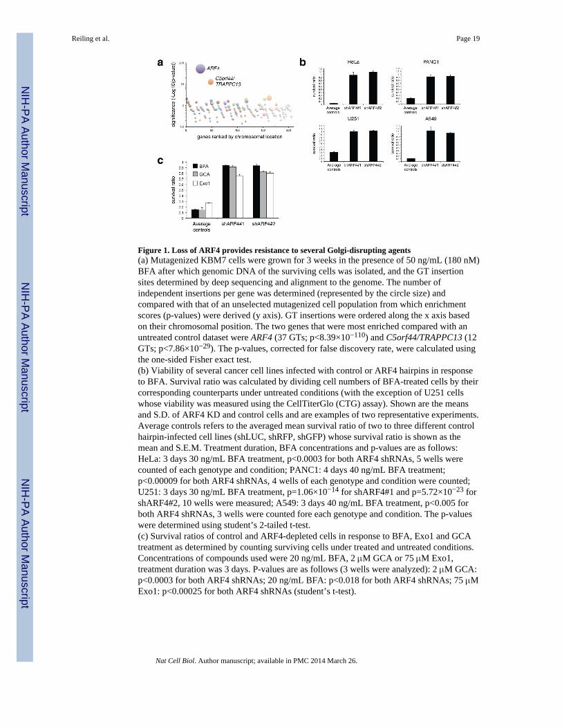
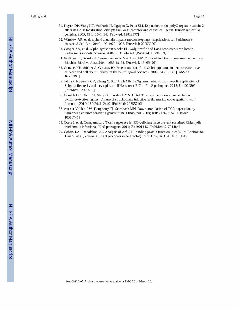
Figure 1. Loss of ARF4 provides resistance to several Golgi-disrupting agents(a) Mutagenized KBM7 cells were grown for 3 weeks in the presence of 50 ng/mL (180 nM)BFA after which genomic DNA of the surviving cells was isolated, and the GT insertionsites determined by deep sequencing and alignment to the genome. The number ofindependent insertions per gene was determined (represented by the circle size) andcompared with that of an unselected mutagenized cell population from which enrichmentscores (p-values) were derived (y axis). GT insertions were ordered along the x axis basedon their chromosomal position. The two genes that were most enriched compared with anuntreated control dataset were ARF4 (37 GTs; p<8.39×10−110) and C5orf44/TRAPPC13 (12GTs; p<7.86×10⁻²⁹). The p-values, corrected for false discovery rate, were calculated usingthe one-sided Fisher exact test.(b) Viability of several cancer cell lines infected with control or ARF4 hairpins in responseto BFA. Survival ratio was calculated by dividing cell numbers of BFA-treated cells by theircorresponding counterparts under untreated conditions (with the exception of U251 cellswhose viability was measured using the CellTiterGlo (CTG) assay). Shown are the meansand S.D. of ARF4 KD and control cells and are examples of two representative experiments.Average controls refers to the averaged mean survival ratio of two to three different controlhairpin-infected cell lines (shLUC, shRFP, shGFP) whose survival ratio is shown as themean and S.E.M. Treatment duration, BFA concentrations and p-values are as follows:HeLa: 3 days 30 ng/mL BFA treatment, p<0.0003 for both ARF4 shRNAs, 5 wells werecounted of each genotype and condition; PANC1: 4 days 40 ng/mL BFA treatment;p<0.00009 for both ARF4 shRNAs, 4 wells of each genotype and condition were counted;U251: 3 days 30 ng/mL BFA treatment, p=1.06×10⁻¹⁴ for shARF4#1 and p=5.72×10⁻²³ forshARF4#2, 10 wells were measured; A549: 3 days 40 ng/mL BFA treatment, p<0.005 forboth ARF4 shRNAs, 3 wells were counted fore each genotype and condition. The p-valueswere determined using student’s 2-tailed t-test.(c) Survival ratios of control and ARF4-depleted cells in response to BFA, Exo1 and GCAtreatment as determined by counting surviving cells under treated and untreated conditions.Concentrations of compounds used were 20 ng/mL BFA, 2 μM GCA or 75 μM Exo1,treatment duration was 3 days. P-values are as follows (3 wells were analyzed): 2 μM GCA:p<0.0003 for both ARF4 shRNAs; 20 ng/mL BFA: p<0.018 for both ARF4 shRNAs; 75 μMExo1: p<0.00025 for both ARF4 shRNAs (student’s t-test).
Reiling et al. Page 19
Nat Cell Biol. Author manuscript; available in PMC 2014 March 26.
NIH
-PA Author Manuscript
NIH
-PA Author Manuscript
NIH
-PA Author Manuscript
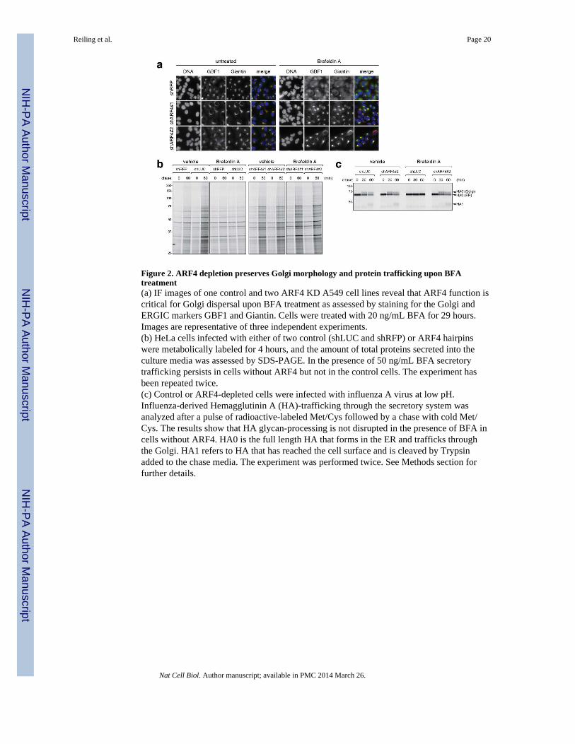
Figure 2. ARF4 depletion preserves Golgi morphology and protein trafficking upon BFAtreatment(a) IF images of one control and two ARF4 KD A549 cell lines reveal that ARF4 function iscritical for Golgi dispersal upon BFA treatment as assessed by staining for the Golgi andERGIC markers GBF1 and Giantin. Cells were treated with 20 ng/mL BFA for 29 hours.Images are representative of three independent experiments.(b) HeLa cells infected with either of two control (shLUC and shRFP) or ARF4 hairpinswere metabolically labeled for 4 hours, and the amount of total proteins secreted into theculture media was assessed by SDS-PAGE. In the presence of 50 ng/mL BFA secretorytrafficking persists in cells without ARF4 but not in the control cells. The experiment hasbeen repeated twice.(c) Control or ARF4-depleted cells were infected with influenza A virus at low pH.Influenza-derived Hemagglutinin A (HA)-trafficking through the secretory system wasanalyzed after a pulse of radioactive-labeled Met/Cys followed by a chase with cold Met/Cys. The results show that HA glycan-processing is not disrupted in the presence of BFA incells without ARF4. HA0 is the full length HA that forms in the ER and trafficks throughthe Golgi. HA1 refers to HA that has reached the cell surface and is cleaved by Trypsinadded to the chase media. The experiment was performed twice. See Methods section forfurther details.
Reiling et al. Page 20
Nat Cell Biol. Author manuscript; available in PMC 2014 March 26.
NIH
-PA Author Manuscript
NIH
-PA Author Manuscript
NIH
-PA Author Manuscript
Figure 3. Compensatory upregulation of other ARF family members in ARF4 knockdown cells(a–c) Several cancer cell lines were transduced with lentiviral control hairpins or shRNAsagainst ARF4, and cell lysates were probed with the indicated antibodies.(a, b) Stable hairpin-control or ARF4 KD PC3 (a) or HeLa (b) cells were analyzed for totalARF-GTP levels in the absence or presence of 20 ng/mL BFA (24 hours treatment) using aGST-VHS-GAT pull-down assay. In this assay, increased ARF binding to VHS-GAT, atruncated GGA3 form and ARF-substrate that is bound only when ARFs are GTP-loaded,serves as proxy for ARF activity. A representative example of two independent experimentsshowing a quantification of ARF-GTP levels in HeLa cells is shown (note that we omittedthe shRFP control in the bar graph for this particular analysis. AU= arbitrary units). Thecorresponding protein lysates for the samples used in (a) and (b) were tested with indicatedantibodies.(c) Two additional examples representing two independent experiments (DU145 and U251cells) showing upregulated (pan-) ARF levels in ARF4-depleted cells.
Reiling et al. Page 21
Nat Cell Biol. Author manuscript; available in PMC 2014 March 26.
NIH
-PA Author Manuscript
NIH
-PA Author Manuscript
NIH
-PA Author Manuscript
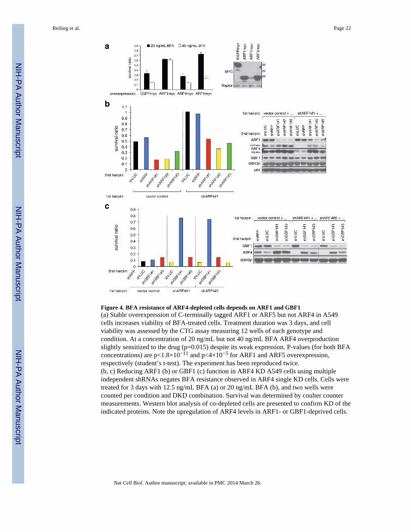
Figure 4. BFA resistance of ARF4-depleted cells depends on ARF1 and GBF1(a) Stable overexpression of C-terminally tagged ARF1 or ARF5 but not ARF4 in A549cells increases viability of BFA-treated cells. Treatment duration was 3 days, and cellviability was assessed by the CTG assay measuring 12 wells of each genotype andcondition. At a concentration of 20 ng/mL but not 40 ng/mL BFA ARF4 overproductionslightly sensitized to the drug (p=0.015) despite its weak expression. P-values (for both BFAconcentrations) are p<1.8×10⁻¹¹ and p<4×10⁻⁵ for ARF1 and ARF5 overexpression,respectively (student’s t-test). The experiment has been reproduced twice.(b, c) Reducing ARF1 (b) or GBF1 (c) function in ARF4 KD A549 cells using multipleindependent shRNAs negates BFA resistance observed in ARF4 single KD cells. Cells weretreated for 3 days with 12.5 ng/mL BFA (a) or 20 ng/mL BFA (b), and two wells werecounted per condition and DKD combination. Survival was determined by coulter countermeasurements. Western blot analysis of co-depleted cells are presented to confirm KD of theindicated proteins. Note the upregulation of ARF4 levels in ARF1- or GBF1-deprived cells.
Reiling et al. Page 22
Nat Cell Biol. Author manuscript; available in PMC 2014 March 26.
NIH
-PA Author Manuscript
NIH
-PA Author Manuscript
NIH
-PA Author Manuscript
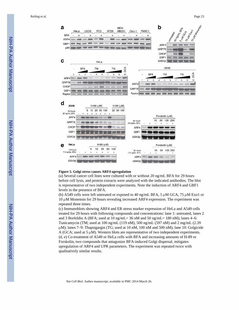
Figure 5. Golgi stress causes ARF4 upregulation(a) Several cancer cell lines were cultured with or without 20 ng/mL BFA for 29 hoursbefore cell lysis, and protein extracts were analyzed with the indicated antibodies. The blotis representative of two independent experiments. Note the induction of ARF4 and GBF1levels in the presence of BFA.(b) A549 cells were left untreated or exposed to 40 ng/mL BFA, 5 μM GCA, 75 μM Exo1 or10 μM Monensin for 29 hours revealing increased ARF4 expression. The experiment wasrepeated three times.(c) Immunoblots showing ARF4 and ER stress marker expression of HeLa and A549 cellstreated for 29 hours with following compounds and concentrations: lane 1: untreated, lanes 2and 3 Brefeldin A (BFA; used at 10 ng/mL= 36 nM and 50 ng/mL= 180 nM); lanes 4–6:Tunicamycin (TM; used at 100 ng/mL (119 nM), 500 ng/mL (597 nM) and 2 mg/mL (2.39μM); lanes 7–9: Thapsigargin (TG; used at 10 nM, 100 nM and 500 nM); lane 10: GolgicideA (GCA; used at 5 μM). Western blots are representative of two independent experiments.(d, e) Co-treatment of A549 or HeLa cells with BFA and increasing amounts of H-89 orForskolin, two compounds that antagonize BFA-induced Golgi dispersal, mitigatesupregulation of ARF4 and UPR parameters. The experiment was repeated twice withqualitatively similar results.
Reiling et al. Page 23
Nat Cell Biol. Author manuscript; available in PMC 2014 March 26.
NIH
-PA Author Manuscript
NIH
-PA Author Manuscript
NIH
-PA Author Manuscript
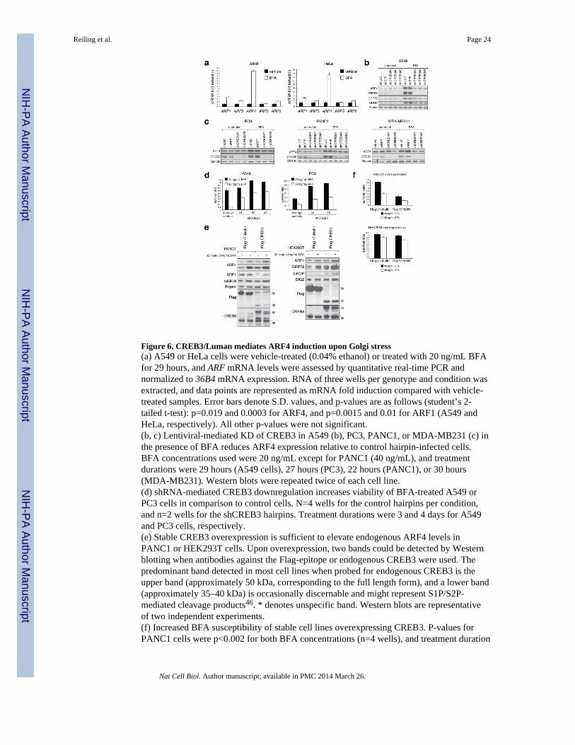
Figure 6. CREB3/Luman mediates ARF4 induction upon Golgi stress(a) A549 or HeLa cells were vehicle-treated (0.04% ethanol) or treated with 20 ng/mL BFAfor 29 hours, and ARF mRNA levels were assessed by quantitative real-time PCR andnormalized to 36B4 mRNA expression. RNA of three wells per genotype and condition wasextracted, and data points are represented as mRNA fold induction compared with vehicle-treated samples. Error bars denote S.D. values, and p-values are as follows (student’s 2-tailed t-test): p=0.019 and 0.0003 for ARF4, and p=0.0015 and 0.01 for ARF1 (A549 andHeLa, respectively). All other p-values were not significant.(b, c) Lentiviral-mediated KD of CREB3 in A549 (b), PC3, PANC1, or MDA-MB231 (c) inthe presence of BFA reduces ARF4 expression relative to control hairpin-infected cells.BFA concentrations used were 20 ng/mL except for PANC1 (40 ng/mL), and treatmentdurations were 29 hours (A549 cells), 27 hours (PC3), 22 hours (PANC1), or 30 hours(MDA-MB231). Western blots were repeated twice of each cell line.(d) shRNA-mediated CREB3 downregulation increases viability of BFA-treated A549 orPC3 cells in comparison to control cells. N=4 wells for the control hairpins per condition,and n=2 wells for the shCREB3 hairpins. Treatment durations were 3 and 4 days for A549and PC3 cells, respectively.(e) Stable CREB3 overexpression is sufficient to elevate endogenous ARF4 levels inPANC1 or HEK293T cells. Upon overexpression, two bands could be detected by Westernblotting when antibodies against the Flag-epitope or endogenous CREB3 were used. Thepredominant band detected in most cell lines when probed for endogenous CREB3 is theupper band (approximately 50 kDa, corresponding to the full length form), and a lower band(approximately 35–40 kDa) is occasionally discernable and might represent S1P/S2P-mediated cleavage products46. * denotes unspecific band. Western blots are representativeof two independent experiments.(f) Increased BFA susceptibility of stable cell lines overexpressing CREB3. P-values forPANC1 cells were p<0.002 for both BFA concentrations (n=4 wells), and treatment duration
Reiling et al. Page 24
Nat Cell Biol. Author manuscript; available in PMC 2014 March 26.
NIH
-PA Author Manuscript
NIH
-PA Author Manuscript
NIH
-PA Author Manuscript
was 4 days. For HEK239T cells (n=4 wells) p=0.045 and p=0.001 using 30 ng/mL and 40ng/mL BFA, respectively (student’s 2-tailed t-test). * denotes unspecific band.
Reiling et al. Page 25
Nat Cell Biol. Author manuscript; available in PMC 2014 March 26.
NIH
-PA Author Manuscript
NIH
-PA Author Manuscript
NIH
-PA Author Manuscript
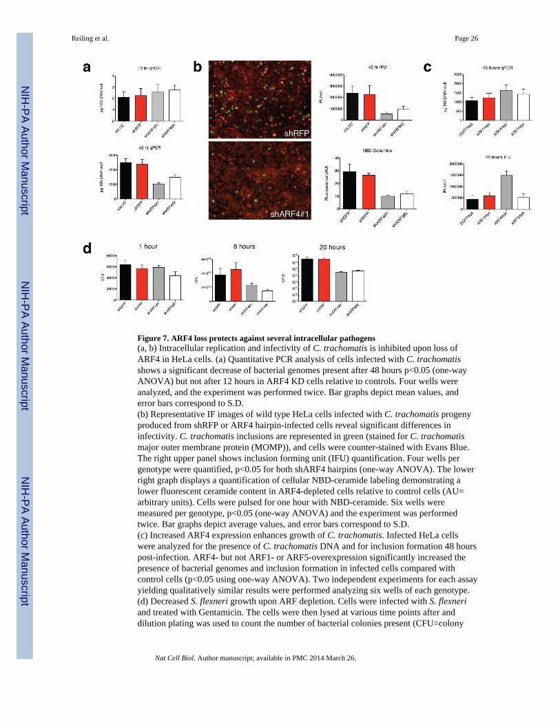
Figure 7. ARF4 loss protects against several intracellular pathogens(a, b) Intracellular replication and infectivity of C. trachomatis is inhibited upon loss ofARF4 in HeLa cells. (a) Quantitative PCR analysis of cells infected with C. trachomatisshows a significant decrease of bacterial genomes present after 48 hours p<0.05 (one-wayANOVA) but not after 12 hours in ARF4 KD cells relative to controls. Four wells wereanalyzed, and the experiment was performed twice. Bar graphs depict mean values, anderror bars correspond to S.D.(b) Representative IF images of wild type HeLa cells infected with C. trachomatis progenyproduced from shRFP or ARF4 hairpin-infected cells reveal significant differences ininfectivity. C. trachomatis inclusions are represented in green (stained for C. trachomatismajor outer membrane protein (MOMP)), and cells were counter-stained with Evans Blue.The right upper panel shows inclusion forming unit (IFU) quantification. Four wells pergenotype were quantified, p<0.05 for both shARF4 hairpins (one-way ANOVA). The lowerright graph displays a quantification of cellular NBD-ceramide labeling demonstrating alower fluorescent ceramide content in ARF4-depleted cells relative to control cells (AU=arbitrary units). Cells were pulsed for one hour with NBD-ceramide. Six wells weremeasured per genotype, p<0.05 (one-way ANOVA) and the experiment was performedtwice. Bar graphs depict average values, and error bars correspond to S.D.(c) Increased ARF4 expression enhances growth of C. trachomatis. Infected HeLa cellswere analyzed for the presence of C. trachomatis DNA and for inclusion formation 48 hourspost-infection. ARF4- but not ARF1- or ARF5-overexpression significantly increased thepresence of bacterial genomes and inclusion formation in infected cells compared withcontrol cells (p<0.05 using one-way ANOVA). Two independent experiments for each assayyielding qualitatively similar results were performed analyzing six wells of each genotype.(d) Decreased S. flexneri growth upon ARF depletion. Cells were infected with S. flexneriand treated with Gentamicin. The cells were then lysed at various time points after anddilution plating was used to count the number of bacterial colonies present (CFU=colony
Reiling et al. Page 26
Nat Cell Biol. Author manuscript; available in PMC 2014 March 26.
NIH
-PA Author Manuscript
NIH
-PA Author Manuscript
NIH
-PA Author Manuscript
forming units). ARF4-depleted cells show less CFUs compared with control hairpin-infectedcells (p<0.05 for ARF4 KD hairpins at 8 hours and 20 hours after infection (one-wayANOVA). Results are representative of two independent experiments measuring each timethree plates per genotype.
Reiling et al. Page 27
Nat Cell Biol. Author manuscript; available in PMC 2014 March 26.
NIH
-PA Author Manuscript
NIH
-PA Author Manuscript
NIH
-PA Author Manuscript