Embed Size (px)
Citation preview

University of Tennessee at Chattanooga University of Tennessee at Chattanooga
UTC Scholar UTC Scholar
Honors Theses Student Research, Creative Works, and Publications
5-2020
A contribution to the characterization of the diversity of A contribution to the characterization of the diversity of
ectomycorrhizal fungi associated with American chestnut at the ectomycorrhizal fungi associated with American chestnut at the
UTC Fortwood Street nursery UTC Fortwood Street nursery
Colton Jones University of Tennessee at Chattanooga, [email protected]
Follow this and additional works at: https://scholar.utc.edu/honors-theses
Part of the Plant Biology Commons
Recommended Citation Recommended Citation Jones, Colton, "A contribution to the characterization of the diversity of ectomycorrhizal fungi associated with American chestnut at the UTC Fortwood Street nursery" (2020). Honors Theses.
This Theses is brought to you for free and open access by the Student Research, Creative Works, and Publications at UTC Scholar. It has been accepted for inclusion in Honors Theses by an authorized administrator of UTC Scholar. For more information, please contact [email protected].

A contribution to the characterization of the diversity of ectomycorrhizal fungi associated
with American chestnut at the UTC Fortwood Street nursery
Colton Jones
Departmental Honors Thesis
The University of Tennessee at Chattanooga
Department of Biology, Geology, and Environmental Sciences
March 2020
Dr. Hill Craddock
Professor
Biology, Geology, and Environ. Sci.
Thesis director
Dr. Jose Barbosa
Associate Professor
Biology, Geology, and Environ. Sci.
Department Examiner
Dr. Margaret Kovach
Professor
Biology, Geology, and Environ. Sci.
Department Examiner

1
Table of Contents
I. Abstract……………………………………………………………………………….2
II. Introduction
Endo- vs Ectomycorrhizas…………………………………………………..…….4-5
Evolution of the mycorrhiza………………………………………………..……..5-6
Ectomycorrhizal Morphology………………………………………………..…......7
Plant Symbionts. ……………………………………………………………....….…8
Fungal Symbionts……………………………………………………………..……..8
Benefits of Ectomycorrhizal Symbiosis……………………………………..……8-9
American chestnut Background……………………………………..…………….10
Project Outline……………………………………………………………..……11-12
III. Methods
Sampling……………………………………………………………………...….12-13
DNA Extraction and Precipitation………………………………………..……….13
DNA Amplification………………………………………………………..………..13
PCR Product Purification………………………………………………..……..14-15
DNA Sequencing………………………………………………………..…………..15
IV. Results……………………………………………………………………………15-18
V. Discussion………………………………………………………………..………18-21
VI. References………………………………………………………………………..22-27

2
I. Abstract
Ectomycorrhizas play several essential roles in the biosphere and have immeasurable
implications on the ecosystems in which they exist. Much has been discovered about the
relationships between ectomycorrhizal fungi and the trees with which they associate, but there is
still much to learn. Due to the nature of ectomycorrhizal morphology, DNA analysis is
frequently required in order to accurately identify the fungal partner. Some ectomycorrhizal
fungi produce above-ground fruiting bodies that presumably contain the same DNA sequences as
the fungi encapsulating corresponding plant root tips below the soil; these fruiting bodies have
been frequently observed growing in nursery containers at the UTC Fortwood Street nursery. We
hypothesized that DNA extracted from fruiting bodies found in these containers would match the
DNA of fungi enveloping the tree’s root tips. Additionally, we hypothesized that the variety of
sequences produced by ectomycorrhizal root tips may display a greater diversity of mycorrhizal
fungi than is represented by hypergeous fruiting bodies alone. In the course of this thesis,
genomic DNA was extracted from mycorrhizal American chestnut root tips and fruiting bodies
found in nursery containers at the Fortwood Street nursery; the DNA underwent PCR and was
purified prior to sequencing and BLAST alignment. However, due to complications in the
preparation of DNA for sequencing and the finite timeframe provided, the results of this project
are limited. An ITS sequence from one fruiting body was successfully amplified and sequenced;
the identity of this sporocarp was determined to be the obligate ectomycorrhizal fungus
Hebeloma hiemale s.l.

3
II. Introduction
A mycorrhiza is a symbiotic relationship between a plant root and a mycorrhizal fungus.
The term, coming from the Greek words mykos and rhizon literally means fungus root (Carlile et
al., 1994). There are at least seven types of mycorrhizas described in scientific literature, each
forming unique types of symbioses (Brundrett, 2002). Some of these relationships have existed
for hundreds of millions of years; however, their significance has only recently been established
and is still not fully understood (Strullu-Derrien et al., 2018). In the case of a mutualistic
mycorrhiza, a soilborne fungus becomes physically and chemically associated with a plant’s
roots; the fungus radiates into the soil, increasing the surface area of the roots and transferring
essential nutrients (e.g., phosphorus, nitrogen) back to the plant. In exchange, the plant transfers
photosynthates to the fungus; the fungus uses the carbohydrates acquired as its primary source of
energy (Carlile et al., 1994). The interactions between mycorrhizal plant roots and fungi involve
complex cascades of gene expression and molecular mechanisms that are currently being
researched worldwide (Hilbert & Martin, 1988). Benefits provided by mutualistic mycorrhizal
symbioses are still being characterized not only in terms of the advantages provided to the host
plant by the mycobiont, but also in potential ways mycorrhizas can be used in broader
environmental restoration efforts (Blaudez et al., 2000). Distinguishing the species of
mycorrhizal fungi colonizing particular plants is the first step to further research concerning the
implications of mycorrhizal relationships. However, accurate identification of mycorrhizal fungi
proves to be more difficult than identification of the host plant; for this reason, the modern
characterization of mycorrhizal fungi often takes a molecular approach (Norris et al., 1994). The
diversity of mycorrhizal fungi colonizing the American chestnut is of particular interest at the
4
University of Tennessee at Chattanooga, as the campus houses a breeding program that works
towards re-establishing the vulnerable species in the wild.
ENDO- VS ECTOMYCORRHIZAS
Endomycorrhizas, of which there are several types, make up the vast majority of
mycorrhizal relationships in the biosphere; these types of mycorrhizas occur in more than 80% of
vascular plants (Brundrett & Tedersoo, 2018). Implied by their name, endomycorrhizas are those
that penetrate host root cells and form characteristic intracellular structures used to facilitate the
chemical transactions between fungi and their hosts. Ectomycorrhizal (ECM) fungi do not
penetrate host cell walls, but are capable of facilitating nutrient exchange through unique
extracellular structures. Although only ~2% of vascular plants form ectomycorrhizas, this still
accounts for over 6,000 species of plants colonized by 20,000-25,000 species of ECM fungi
(Brundrett & Tedersoo, 2018; Christenhusz & Byng, 2016; Tedersoo et al., 2009).
The majority of ECM fungi can penetrate root cells as the roots begin to senesce or if
equilibrium between the symbionts is disrupted (Smith & Read, 2017). However, it is rare for an
ectomycorrhiza-forming fungus to penetrate host root cells; these types of relationships are only
known to occur in Pinus and Larix spp. and are referred to as ectendomycorrhizas (Yu et al.,
2001)
Though uncommon, a few tree genera and some shrubs and ferns are susceptible to
colonization by both vesicular-arbuscular mycorrhizas (VAM) and ectomycorrhizas. Chilvers et
al. observed an initial colonization of root tips by VAM with relatively high inoculum potential
and rapid root colonization, followed by secondary infections by ECM fungi. The
ectomycorrhizal fungi outcompeted the VAM later in terms of colonization of lateral roots and
hyphal spread, and, while the existing VAM did not inhibit subsequent infection by

5
ectomycorrhizal fungi, the sheath of the latter formed a physical barrier to colonization by other
VAM (Chilvers et al., 1987).
EVOLUTION OF THE MYCORRHIZA
Endomycorrhizas were the first real mycorrhizas to evolve following the colonization of
land by early plants (Strullu-Derrien et al., 2018). In fact, it has been hypothesized that symbiotic
relationships between aquatic plants and fungi made the transition to a terrestrial lifestyle
possible (Pirozynski & Malloch, 1975). The first direct evidence concerning the evolution of
mycorrhizas was found in the 407 million-yr-old Rhynie chert, in which paramycorrhizas
(structures resembling mycorrhizas) were visible. These included the presence of members of the
Mucoromycotina and Glomeromycotina in fossils of minute rootless stem vascular plants. By
315-300 million years ago, arbuscular mycorrhizas, or true mycorrhizas, had begun colonizing
plant roots; Glomeromycetous arbuscules have been observed in lycopod root fossils from this
era (Strullu-Derrien et al., 2018).
Figure A.
An illustrated cross section of root
tip in which ectomycorrhizal and
ectendomycorrhizal morphologies
are compared with the
morphologies of three types of
endomycorrhizas (Weiss et al.,
2016)

6
The Princeton chert of Canada contains the oldest known fossil of an ectomycorrhiza.
This 48.7 million-yr-old chert shows evidence of Pinus roots with the essential features of an
ectomycorrhiza: the fungal sheath and a Hartig net extending intercellularly to the endodermis. It
is thought that ectomycorrhizal symbioses evolved independently more than 18 times in
Angiosperms and ~78-82 times in fungi over at least 48Ma, indicating a relationship between the
effects of the symbiosis and biological fitness (Strullu-Derrien et al., 2018).
Analyses of 12 ECM fungal genomes, some belonging to ectomycorrhizal fungi known
to colonize C. dentata, suggest that some ECM fungi are the result of the convergent evolution of
soil saprotrophs and of brown- and white-rot fungi. Many mycorrhizal fungi have exhibited
decreases in genes encoding plant cell wall-degrading enzymes relative to their primarily
saprotrophic ancestors; several genes encoding enzymes involved in the decomposition of
various plant tissues and the cleavage of sucrose have been lost from ECM fungal genomes as
well. Genes involved in the breakdown of soil particles, which may aid in the acquisition of
organic nitrogen and phosphorus, have been preserved in these fungi. Consequently,
ectomycorrhizal fungi have become more dependent on their hosts’ carbohydrate reserves and
more efficient in acquiring organic nutrients over time (Kohler et al., 2015; Strullu-Derrien et al.,
2018).
Mycorrhizal symbioses are thought to have greatly impacted the evolution of both
terrestrial plants and fungi. The benefits of these relationships have likely broadened the
diversity and ranges of plant and fungal species due to their contributions in the expansion of
available niches (Duplessis et al., 2001).
7
ECTOMYCORRHIZAL MORPHOLOGY
An ectomycorrhiza consists of three essential structures: the Hartig net, the mantle, and a
network of hyphae radiating from the mantle into the surrounding soil. These structures function
in the collection and transfer of water and nutrients between fungi and plants. The Hartig net
serves as the extracellular junction of nutrient exchange between symbionts.
The formation of an ectomycorrhiza begins with stimulation of fungal growth by plant
metabolites in the soil. As hyphae radiate outward and come into contact with roots, they
envelop the root tips with denser hyphal networks. ECM fungi cultured on agar media have been
observed growing significantly faster along root tips than across the media itself. Once hyphal
envelopes have formed, specialized hyphae penetrate dead root cap cells and colonize
intercellular spaces within the root tip, forming the Hartig net. The wedge-like hyphal tips
expand in the plant tissue, separating plant cells and filling the newly-created spaces; hyphae
may extend to the endodermis or be confined to the outermost layers of the root tip. These
hyphae envelop individual cortical cells but do not disrupt intercellular plant communication, as
plasmodesmata are not damaged in the process. Secretion of fibrillary polymers at the fungus-
plant interface facilitates the adhesion of fungal and plant cell walls (Carlile et al., 1994).
Subsequently, the true mantle is established as specialized hyphae form layer after layer of tissue
enclosing the root tip. A hyphal network radiates outward from the mantle and spreads
throughout the soil (Nylund & Unestam, 1981; Kendrick, 2017)
Colonization of a root tip by an ECM fungus slows rates of cell division at the root tip
and root hairs and leads to radial elongation of cortical cells; this results in a relatively stumpy
appearance of the colonized root (Carlile et al., 1994). However, if a root cap penetrates the
mantle, the root may be subject to colonization by other ECM fungi (Kendrick, 2017).
8
PLANT SYMBIONTS
Although a relatively small percentage of seed plant species are ectomycorrhizal, these
plants occupy a disproportionately large land area and dominate in the production of timber.
Thus, ECM plants play a significant role in the ecosystems of boreal, temperate, and tropical
forests, as well as in the infrastructures and economies of countries worldwide (Smith & Read,
2017; Alexander & Hogberg, 1987).
FUNGAL SYMBIONTS
Ectomycorrhizal fungi belong to the phyla Basidiomycota, Ascomycota, and
Zygomycota. The most diverse orders include Agaricales, Cantharellales, Helotiales, Boletales,
and Pezizales (Tedersoo et al., 2010).
BENEFITS OF ECTOMYCORRHIZAL SYMBIOSIS
The carbohydrates produced from photosynthesis, primarily hexose sugars, are delivered
to the fungus where they are converted into sugar alcohols, e.g., mannitol, erythritol. This creates
a hexose gradient that continually translocates sugars towards the fungus via passive diffusion.
Fungi are able to excise the sugars from root tips by selectively increasing the permeability of
plant cell membranes (Carlile et al., 1994).
In addition to increasing water and nutrient uptake in plants, ectomycorrhizal associations
may confer resistance to a myriad of biotic and abiotic stresses. Some ectomycorrhizal fungi are
capable of the uptake and compartmentalization of the heavy metal cadmium from soil; this
reduces uptake by plant roots (Blaudez et al., 2000) and could also provide an effective
mechanism for the phytoextraction of heavy metals from contaminated soils in land reclamation
efforts (Sell et al., 2005). Ectomycorrhizal inoculation has also been noted for its role in

9
increasing plant tolerance to stresses inflicted by hyper-saline soils (Bandou et al., 2006) and
drought conditions. (Wang & Gui-jie, 2013)
The benefits associated with ectomycorrhizal symbioses are not limited to the symbionts
directly involved in the relationship; the presence of the ectomycorrhizal fungus Pisolithus
tinctorius in soil may play a significant role in mitigating the effects of acid rain on below-
ground microbial communities. (Maltz et al., 2019) In addition to decreasing biodiversity, acidic
conditions can also increase the solubility of metals e.g., aluminum; dissolved aluminum can
react with phosphorus in the soil, forming AlPO4 and decreasing phosphorus availability in the
soil. P. tinctorius can extract P from AlPO4, allowing its host plant to benefit from the otherwise
inaccessible nutrient (Cumming & Weinstein, 1990)
Underground hyphal connections formed by VAM fungi can serve as a means of plant-
to-plant signaling and can enhance plant resistance to herbivory. Hyphae of a VAM fungus may
connect multiple plants within a community; when attacked by aphids, a plant may send
chemical messages through this junction which stimulates the production of chemicals that repel
aphids and attract aphid parasitoids. Thus, plants not yet under attack can launch a preemptive
defense to minimize predation (Babikova et al., 2013). The potential for communication
between plants via ectomycorrhizal connections is supported by modern evidence, but this is an
area that requires further research (Wagner et al., 2015).
VAM fungi are also thought to enhance plant resistance to pathogens in the soil. In the
formation of an endomycorrhiza, the plant’s immune defenses are modified, leading to a
moderate systemic activation of the plant’s immune system. The stimulation of primary immune
responses may aid in the plant’s defense against parasitic bacteria, fungi, plants, and nematodes
(Jung et al., 2012). This topic also requires further research with respect to ectomycorrhizas.

10
AMERICAN CHESTNUT BACKGROUND
American chestnut populations have declined by approximately 90% in the past 115
years. This can be attributed primarily to Cryphonectria parasitica, the pathogenic fungus that
causes chestnut blight. This necrotrophic parasite likely spread to American chestnuts from
infected Japanese chestnut trees, which were first imported to North America as nursery stock in
1876. Japanese and Chinese chestnut trees have developed resistance to chestnut blight through
natural selection. Contrarily, American chestnut trees had not been previously introduced to
Cryphonectria parasitica and therefore carried no resistance to the pathogen. Phytophthora root
rot, caused by the soil-borne oomycete Phytophthora cinnamomi, is another introduced disease
that has contributed to declines in numbers and ranges of American chestnut. P. cinnamomi may
be responsible for the permanent retraction of C. dentata from the southern portion of its native
range and is now spreading to northern regions as temperatures warm (Dalgleish et al., 2015).
Although approximately four billion American chestnut trees have been afflicted by
chestnut blight thus far, the future outlook is hopeful. Scientists are currently working towards
breeding blight-resistant American chestnuts by crossing susceptible American chestnut trees
with naturally resistant Chinese chestnut trees. The resulting hybrids are backcrossed with
American chestnut, producing trees that are essentially American but possess genes for blight
resistance. Many scientists breeding for blight resistance are now selectively breeding for
Phytophthora resistance as well (Rellou, 2002). The American Chestnut Foundation (TACF) was
founded in 1983 in an effort to combat the demise of the American chestnut. Since its
foundation, TACF has worked towards breeding disease tolerant chestnut trees in order to renew
decimated native populations (Steiner et al., 2017).
11
PROJECT OUTLINE
Dr. Hill Craddock runs an American chestnut breeding program on the University of
Tennessee at Chattanooga campus. Coordinating with TACF, Dr. Craddock breeds pathogen-
resistant chestnut trees in an effort to restore the fungus-ravaged species to its former glory. The
chestnuts are initially planted in 2-gallon containers containing well fertilized potting medium
and grown for one year in the nursery before outplanting into experimental orchards and forest
plots within C. dentata’s natural range. Dr. Craddock has long noticed sporocarps from various
fungal species fruiting in the nursery containers. The fruiting bodies can be identified with
reasonable confidence and as most of these fungi are obligate symbionts, they are likely products
of mycorrhizal relationships within the pot. However, identification of these fruiting bodies may
not provide a real estimate of ectomycorrhizal biodiversity within the nursery; not all
ectomycorrhizal fungi produce hypergeous fruiting bodies and those that are produced may be
ephemeral. Another option in assessing ectomycorrhizal diversity is through visual identification
of ectomycorrhizal fungi based on mantle and hypha morphologies. This would likely be an
arduous, if not impossible, task; many species of ectomycorrhizal fungi are indistinguishable
morphologically, cannot be cultured in vitro, or vary in morphology depending on sexual state.
Consequently, identification of ectomycorrhizal fungi typically requires genetic analysis (Norris
et al., 1994). In this project, the internal transcribed spacer (ITS) region was used for fungal
species identification.
The ITS region of fungal DNA has been referred to as the universal DNA barcode for the
identification of fungi. Fungal ITS regions are relatively short (420-825 base pairs) (Manter &
Vivanco, 2007) and are repeated up to thousands of times in the fungal genome. Additionally,
the sequences are highly variable between closely related organisms due to their relatively fast

12
evolution (Raja et al., 2017). High rates of amplification via PCR can be partially attributed to
well-conserved flanking sequences, which allow for one set of primers to be nearly universal
(Baldwin et al., 1995). The primers ITS1F and ITS4 are particularly effective in priming the
amplification of the ITS regions of ECM fungi, e.g., Basidiomycetes, Ascomycetes, and
Zygomycetes (Manter & Vivanco, 2007).
Although still concerning ECM fungi identification, the scope of this project has changed
significantly over the past year. The initial proposal for this thesis was ambitious and involved
multiple sampling locations in a several-hundred-mile radius and the extraction of DNA from
mycorrhizal root tips and fruiting bodies at each location in an effort to distinguish the
mycorrhizal diversity of American chestnut in different growing conditions. However, due to
time constraints and unforeseen difficulties in the extraction and amplification of mycorrhizal
DNA, the scope of this research was narrowed. This paper serves as a contribution to the
characterization of the mycorrhizal diversity of the American chestnut at the University of
Tennessee at Chattanooga Fortwood Street nursery.
III. Methods
(Modified from Palmer et al., 2008; Sahu et al., 2012)
Sampling:
Mycorrhizal fruiting bodies were collected from pots containing C. dentata saplings
located at a container nursery on the UTC campus. The fruiting bodies were dried at 26.7˚C for
approximately 24 hours and, once dry, were brushed gently with a small paintbrush to remove
adhering soil particles. Each sample was packaged independently in a ziplock bag and frozen at -
80˚C. Mycorrhizal root tips were collected from pots bearing fruiting bodies, isolated and

13
brushed gently to remove soil particles, and frozen at -80˚C. The root tips and fruiting bodies
underwent the same treatment after freezing.
DNA extraction and precipitation:
Approximately 70mg of dried fruiting body or 100mg of root tips were taken directly
from the -80˚C freezer and immediately ground to a powder with a -80˚C mortar and pestle. The
powdered tissue was transferred to a 2.5mL microcentrifuge tube and 500µL of cell lysis buffer
(CLB) was added. CLB consisted of 1.4M NaCl, 0.1M Tris-HCl 20mM
ethylendiaminetetraacetic acid, and 2% hexadecyltrimethylammonium bromide. Each tube was
agitated for 20 seconds and then heated at 65˚C for 1 hour. The samples were then centrifuged
for 5 minutes at 21,130 xg and the supernatants were transferred to clean tubes. This was
followed by addition of one volume of -20˚C isopropanol and placement in a -80˚C freezer for
10 minutes. Samples were centrifuged for 15 minutes at 21,130 xg and the supernatants were
discarded. Pellets were washed three times with 100µL of 70% ethanol and dried in a DNA
concentrator before resuspension in 25µL of RNAse-free water.
DNA amplification:
Polymerase chain reactions were carried out using the fungal ITS primers ITS1F and
ITS4, diluted to 10µM with deionized RNAse-free water. Ratios of PCR reagents were as
follows: 0.5µL DNA : 25µL LongAmp master mix : 1µL forward primer : 1µL reverse primer:
22.5µL water. The thermocycler was programmed with the following specifications: initial
denaturing at 94°C for 2 min; 30 cycles of denaturing at 94°C for 40 s, annealing at 53°C for 40
s, and extension at 72°C for 5 min.
The PCR product was then used as template for four additional PCR reactions using the
same primer/buffer ratios.

14
PCR product purification:
All PCR products derived from a particular DNA solution were mixed together and
initially purified using phenol:chloroform:isoamyl alcohol. One volume of
phenol:chloroform:isoamyl alcohol was added to the PCR product and shaken by hand for 20
seconds. This was centrifuged at room temperature at 16,000 xg for 5 minutes. The top layer was
then extracted and placed in a clean microcentrifuge tube. One half volume of ammonium
acetate (5M) and 2.5 volumes of 100% ethanol were added to the sample and the tube was placed
at -80˚C for 1 hour. The solution was centrifuged at 21,130 xg for 15 minutes and the
supernatant was discarded. 150µL of 70% ethanol was added and the sample was centrifuged at
16,000 xg for 2 minutes; the supernatant was discarded and this step was repeated at 21,130 xg
for 1 minute. The pellet was dried in a Centrivap DNA concentrator and resuspended in 300µL
of TE (10 mM Tris-HCl, 1mM EDTA, pH 8).
The entirety of the resuspended DNA was electrophoresed in a 1% agarose gel.
Prominent bands ~750bp were excised from the gel and the DNA within was purified with a
Thermo Scientific GeneJET Gel Extraction Kit. Gel slices were weighed and placed in individual
microcentrifuge tubes; one volume (volume: weight) of binding buffer was added to each. Tubes
were incubated at 60˚C for 10 minutes and then vortexed for 5 seconds. All melted gel solutions
from a given sample were transferred to a single GeneJET purification column and centrifuged
for 1 minute at 14,000 xg; the flow-through was discarded. An additional 100µL of binding
buffer was added to the column, the sample was centrifuged for 1 minute at 14,000 xg, and the
flow-through discarded. This was followed by the addition of 700µL of wash buffer,
centrifugation for 1 minute at 14,000 xg, and discarding of flow-through. An additional
centrifugation of the sample was undertaken to remove residual ethanol. The column was placed

15
into a clean microcentrifuge tube and 30µL of elution buffer was added to the center of the
purification column membrane. This was centrifuged for 1 minute at 14,000 xg. The DNA
solution was subsequently concentrated to a volume of approximately 15µL.
DNA sequencing:
Sequencing was performed by Psomagen, Inc. via Sanger sequencing, or the chain-
termination method. The primers ITS1F and ITS4 were used to sequence both strands of the PCR
product for confirmation. Two tubes were sent for each ITS sequence extracted. Each tube
contained 5µL of the purified PCR product and 5µL of the forward or reverse primer (diluted to
5µM). The resulting sequences can be seen in Figure 3. and Figure 4.
IV. Results
Over the course of this project, genomic DNA was successfully extracted from nine fruiting
bodies and nine mycorrhizal root tip samples (Figure 1.). Each sample of genomic DNA was
prepared for PCR and submitted to thermocycling as described above. Of these 18 samples, one
ITS sequence was successfully amplified. Overall, seven different methods of genomic DNA
isolation were tested for their quality and reliability with respect to downstream PCR
amplification. The initial protocol involved freezing ECM root tips in CLB and using a plastic
pestle to grind the sample in a plastic microcentrifuge tube. This resulted in a thick brownish
solution. Genomic DNA could be observed in agarose immediately following extraction, but not
following PCR. A second method of cell-lysing, bead beating, was used on following samples and
produced genomic DNA that was not successfully amplified via PCR. PCR of another DNA
sample purified with phenol:chloroform:isoamyl alcohol was also unsuccessful. Additional
extractions utilized a mechanical tissue homogenizer; using this method to grind the cells also
produced a viscous brown solution. This approach resulted in degraded DNA fragments.

16
Additionally, a yeast DNA extraction kit (Masterpure MPY80200) was used along with the
mechanical cell grinding approach; this also resulted in degraded DNA. The most successful
method for the lysing of fungal cell walls involved grinding a frozen sample using a -80˚C mortar
and pestle. Disruption of cell walls was most effective in the absence of CLB; lysing each sample
without adding reagents produced powdered tissue which was kept relatively cool due to the
chilled mortar and pestle. Using this method for cell lysis with the remainder of the original
protocol for DNA isolation, an ITS sequence of a single fruiting body was successfully amplified
(Figure 2.).
Figure 1. Genomic DNA
representing fourteen of eighteen
extractions
Top row left to right: 2log ladder,
fruiting body, fruiting body, root
tips, fruiting body, root tips,
fruiting body
Bottom row left to right: 2log
ladder, root tips, fruiting body,
root tips, root tips, root tips, root
tips
Figure 2.
Left column: 2log DNA ladder
Right column: ITS sequence of
fruiting body amplified with the
primers ITS1F and ITS4 and sent for
sequencing

17
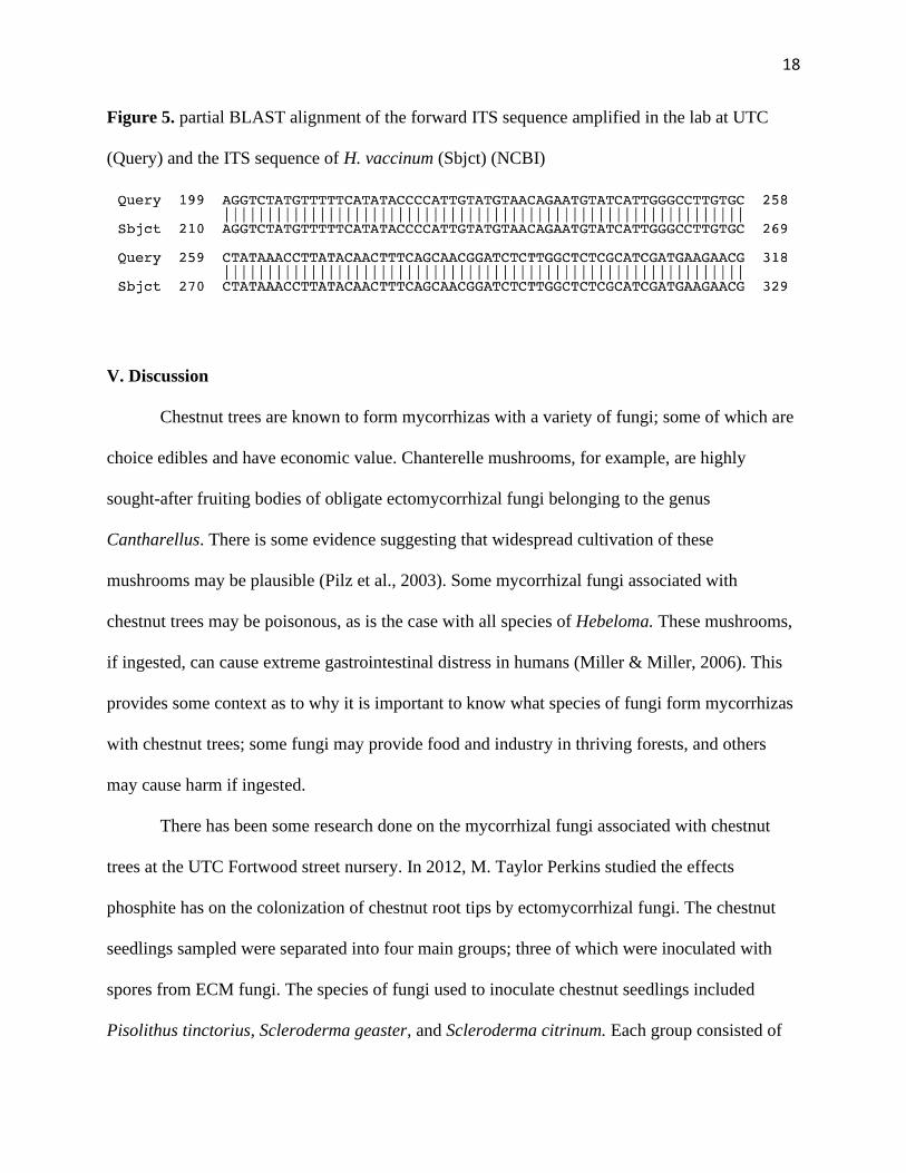
BLAST was used to compare the resulting sequence to those in the NCBI database. Three
species of Hebeloma were found to be equally likely in terms of alignment. The ITS sequences
of H. vaccinum,, H. cavipes, and H. helodes were all 97.23% identical to the sample ITS
sequence (Figure 5.). Each alignment had an E value of 0.0, indicating that one would expect
zero sequences in a database of this size to match the sample sequence by chance alone.
Figure 3. Forward ITS sequence: primed by ITS1F
Figure 4. Reverse ITS sequence: primed by ITS4
18
Figure 5. partial BLAST alignment of the forward ITS sequence amplified in the lab at UTC
(Query) and the ITS sequence of H. vaccinum (Sbjct) (NCBI)
V. Discussion
Chestnut trees are known to form mycorrhizas with a variety of fungi; some of which are
choice edibles and have economic value. Chanterelle mushrooms, for example, are highly
sought-after fruiting bodies of obligate ectomycorrhizal fungi belonging to the genus
Cantharellus. There is some evidence suggesting that widespread cultivation of these
mushrooms may be plausible (Pilz et al., 2003). Some mycorrhizal fungi associated with
chestnut trees may be poisonous, as is the case with all species of Hebeloma. These mushrooms,
if ingested, can cause extreme gastrointestinal distress in humans (Miller & Miller, 2006). This
provides some context as to why it is important to know what species of fungi form mycorrhizas
with chestnut trees; some fungi may provide food and industry in thriving forests, and others
may cause harm if ingested.
There has been some research done on the mycorrhizal fungi associated with chestnut
trees at the UTC Fortwood street nursery. In 2012, M. Taylor Perkins studied the effects
phosphite has on the colonization of chestnut root tips by ectomycorrhizal fungi. The chestnut
seedlings sampled were separated into four main groups; three of which were inoculated with
spores from ECM fungi. The species of fungi used to inoculate chestnut seedlings included
Pisolithus tinctorius, Scleroderma geaster, and Scleroderma citrinum. Each group consisted of
19
two subgroups; one received phosphite treatment and the other did not. The plants inoculated
with P. tinctorius and S. citrinum and those that were not inoculated formed significantly fewer
mycorrhizas when treated with phosphite (Perkins, 2012).
In 2014, J. Miles Jorgensen tested the effects that inoculating chestnut seedlings with P.
tinctorius and administering different levels of phosphite have on seedling survival. Inoculation
with P. tinctorius was associated with a significant increase in seedling survival, regardless of
phosphite concentration. Lower phosphite levels were associated with significant increases
seedling growth but not with significant changes in mycorrhiza formation, while higher
phosphite levels were associated with decreased seedling growth, survival, and mycorrhiza
formation (Jorgensen, 2014). The experiments carried out by Perkins and Jorgensen provided
valuable information with respect to chestnut breeding, but there was no confirmation that the
fungi used to inoculate the seedlings was the same species that formed the mycorrhizas observed.
Despite the successful extraction of genomic DNA from mycorrhizal fungi found at the
UTC Fortwood Street nursery, the final results of this thesis were limited by failures in
consistently amplifying target DNA sequences via PCR. One possible cause of a failed
polymerase chain reaction is contamination of the DNA solution with secondary metabolites.
Certain polysaccharides have been shown to hinder the efficacy of eukaryotic DNA polymerases
and increase the viscosity of the solutions in which DNA molecules are dissolved (Shioda &
Murakami-Murofushi, 1987). Additionally, the oxidation of polyphenols (due to exposure to
atmospheric oxygen during cell lysis) can lead to their binding to DNA molecules; this can cause
the DNA to appear brownish in color (Sahu et al., 2012). Phenolic compounds present in plant
tissues play a role in resistance to pests and pathogens, and they can also inhibit amplification
reactions (Barakat et al., 2009). Fragmentation of DNA samples from tissues homogenized via

20
bead-beating was likely due in part to overbeating. The root tips were never fully homogenized;
this may be attributed to their submersion in CLB, which warmed the root tips and decreased
their rigidity. Similarly, the DNA extracted from mechanically-ground root tips was likely
fragmented for the same reason; failed attempts to fully homogenize samples led to the shearing
of DNA from already lysed cells. Grinding the samples with a -80˚C mortar and pestle was likely
more successful due to the sustained low temperature of the sample. Colder temperatures
decrease the rates of oxidation reactions, therefore minimizing the opportunities for these
reactions to occur. Utilization of liquid nitrogen in the cell-lysing stage may significantly
increase the success rate of the protocol outlined above. Due to the obstacles outlined above,
impending DHON deadlines, and expected graduation at the end of the Spring 2020 semester,
the results of this thesis were limited. One fungal fruiting body was identified via DNA
sequencing and BLAST alignment. Due to most members of Hebeloma being obligate
symbionts, it is reasonable to suspect the sporocarp sampled to be an ectomycorrhizal fruiting
body (Hacskaylo & Bruchet, 1972).
The ITS sequence of H. vaccinum is reported to be distinct from all known species of
Hebeloma except for H. cavipes; these species must be distinguished by their ecology and
morphology. H. vaccinum is primarily found in arctic habitats and dunes; it is usually associated
with trees belonging to the genera Populus and Salix. H. cavipes is said to be capable of
associating with most trees; it is typically found with broadleaf trees but has also been observed
with some conifers. This fungus can live in most soil types and has been found in the continental
United States (Eberhardt et al., 2015). However, it is not known to occur in Tennessee. H.
helodes has been used as a synonym to H. cavipes in some literature, and both species fall under
the more general species Hebeloma hiemale s.l. (Grilli, 2007). The mushroom in question was a

21
little brown mushroom (LBM). These kinds of mushrooms are notoriously difficult to identify;
for this reason, consumption of LBMs is generally not advised. Furthermore, the taxonomy of
these mushrooms is frequently debated as species overlap and are often described independently
by multiple researchers. It is not plausible to assign definite, lasting taxonomic labels to many of
these organisms, i.e., the taxonomy is confused. The taxonomic intricacies/ debate of these fungi
are beyond the scope of this research; therefore, identification as Hebeloma hiemale s.l. is
adequate for this report.

22
VI. References
Alexander, I. J., & Hogberg, P. (1986). Ectomycorrhizas Of Tropical Angiospermous Trees. New
Phytologist, 102(4), 541–549. doi: 10.1111/j.1469-8137.1986.tb00830.x
Babikova, Z., Gilbert, L., Bruce, T. J. A., Birkett, M., Caulfield, J. C., Woodcock, C., . . .
Johnson, D. (2013). Underground signals carried through common mycelial networks
warn neighbouring plants of aphid attack. Ecology Letters, 16(7), 835-843. Retrieved
from https://proxy.lib.utc.edu/login?url=https://search-proquest-
com.proxy.lib.utc.edu/docview/1366720299?accountid=14767
Baldwin, B. G., Sanderson, M. J., Porter, J. M., Wojciechowski, M. F., Campbell, C. S., &
Donoghue, M. J. (1995). The its Region of Nuclear Ribosomal DNA: A Valuable Source
of Evidence on Angiosperm Phylogeny. Annals of the Missouri Botanical Garden, 82(2),
247. doi: 10.2307/2399880
Bandou, E., Lebailly, F., Muller, F., Dulormne, M., Toribio, A., Chabrol, J., … Bâ, A. M.
(2006). The ectomycorrhizal fungus Scleroderma bermudense alleviates salt stress in
seagrape (Coccoloba uvifera L.) seedlings. Mycorrhiza, 16(8), 559–565. doi:
10.1007/s00572-006-0073-6
Barakat, A., Diloreto, D. S., Zhang, Y., Smith, C., Baier, K., Powell, W. A., … Carlson, J. E.
(2009). Comparison of the transcriptomes of American chestnut (Castanea dentata) and
Chinese chestnut (Castanea mollissima) in response to the chestnut blight infection. BMC
Plant Biology, 9(1), 51. doi: 10.1186/1471-2229-9-51
Blaudez, D., Botton, B., & Chalot, M. (2000). Cadmium uptake and subcellular
compartmentation in the ectomycorrhizal fungus Paxillus involutus. Microbiology,
146(5), 1109–1117. doi: 10.1099/00221287-146-5-1109
23
Brundrett, M. C. (2002). Coevolution of roots and mycorrhizas of land plants. New Phytologist,
154(2), 275–304. doi: 10.1046/j.1469-8137.2002.00397.x
Brundrett, M. C., & Tedersoo, L. (2018). Evolutionary history of mycorrhizal symbioses and
global host plant diversity. New Phytologist, 220(4), 1108–1115. doi: 10.1111/nph.14976
Carlile, M. J., Watkinson, S. C., & Gooday, G. W. (1994). The fungi. London: Academic Press.
Chilvers, G. A., Lapeyrie, F. F., & Horan, D. P. (1987). Ectomycorrhizal Vs Endomycorrhizal
Fungi Within The Same Root System. New Phytologist, 107(2), 441–448. doi:
10.1111/j.1469-8137.1987.tb00195.x
Christenhusz, M. J., & Byng, J. W. (2016). The number of known plants species in the world and
its annual increase. Phytotaxa, 261(3), 201. doi: 10.11646/phytotaxa.261.3.1
Cumming, J. R., & Weinstein, L. H. (1990). Utilization of AlPO4 as a phosphorus source by
ectomycorrhizal Pinus rigida Mill. seedlings. New Phytologist, 116(1), 99–106. doi:
10.1111/j.1469-8137.1990.tb00514.x
Dalgleish, H., Nelson, C., Scrivani, J., & Jacobs, D. (2015). Consequences of Shifts in
Abundance and Distribution of American Chestnut for Restoration of a Foundation
Forest Tree. Forests, 7(12), 4. doi: 10.3390/f7010004
Eberhardt, U., Beker, H. J., Vesterholt, J., & Schütz, N. (2016). The taxonomy of the European
species of Hebeloma section Denudata subsections Hiemalia , Echinospora subsect. nov.
and Clepsydroida subsect. nov. and five new species. Fungal Biology, 120(1), 72–103.
doi: 10.1016/j.funbio.2015.09.014
Grilli, E. (2007). Type studies in Hebeloma. Unravelling a taxonomic-nomenclatural problem:
what is Hebeloma leucosarx?. Micologia e vegetazione mediterranea. 22. 133-176.
24
Hacskaylo, E., & Bruchet, G. (1972). Hebelomas as Mycorrhizal Fungi. Bulletin of the Torrey
Botanical Club, 99(1), 17. doi: 10.2307/2484235
Hilbert, J. L., & Martin, F. (1988). Regulation of gene expression in ectomycorrhizas. I. Protein
changes and the presence of ectomycorrhiza-specific polypeptides in the Pisolithus-
Eucalyptus symbiosis. New Phytologist, 110(3), 339–346. doi: 10.1111/j.1469-
8137.1988.tb00270.x
Jorgensen, J.M. (2014). The Effects of Different Concentrations of Phosphite on Ectomycorrhiza
Formation by Pisolithus tinctorius in Castanea dentata. University of Tennessee at
Chattanooga, Biological and Environmental Sciences.
Jung, S. C., Martinez-medina, A., Lopez-raez, J., & Pozo, M. J. (2012). Mycorrhiza-induced
resistance and priming of plant defenses. Journal of Chemical Ecology, 38(6), 651-64.
doi:http://dx.doi.org.proxy.lib.utc.edu/10.1007/s10886-012-0134-.6
Kendrick, B. (2017). The fifth kingdom: an introduction to mycology. Hackett Publishing
Company, Inc.
Kohler, A., Kuo, A., Nagy, L. G., Morin, E., Barry, K. W., Buscot, F., … Martin, F. (2015).
Convergent losses of decay mechanisms and rapid turnover of symbiosis genes in
mycorrhizal mutualists. Nature Genetics, 47(4), 410–415. doi: 10.1038/ng.3223
Maltz, M. R., Chen, Z., Cao, J., Arogyaswamy, K., Shulman, H., & Aronson, E. L. (2019).
Inoculation with Pisolithus tinctorius may ameliorate acid rain impacts on soil microbial
communities associated with Pinus massoniana seedlings. Fungal Ecology, 40, 50–61.
doi: 10.1016/j.funeco.2018.11.011
Manter, D. K., and Vivanco, J. M. (2007). Use of the ITS primers, ITS1F and ITS4, to
characterize fungal abundance and diversity in mixed-template samples by qPCR and

25
length heterogeneity analysis. Journal of Microbiological Methods, 71(1), 7-14.
doi:10.1016/j.mimet.2007.06.016
Martin, F., Duplessis, S., Ditengou, F., Lagrange, H., Voiblet, C., & Lapeyrie, F. (2001).
Developmental cross talking in the ectomycorrhizal symbiosis: signals and
communication genes. New Phytologist, 151(1), 145–154. doi: 10.1046/j.1469-
8137.2001.00169.x
Miller, O. K., & Miller, H. (2006). North American mushrooms: a field guide to edible and
inedible fungi. Guilford, CT: Falcon Guide.
National Center for Biotechnology Information (NCBI). Bethesda (MD): National Library of
Medicine (US), National Center for Biotechnology Information; [1988] – [cited 2020
March 21]. Available from: https://www.ncbi.nlm.nih.gov/
Norris, J. R., Read, D. J. and Varma, A. K (1994). Techniques for mycorrhizal research.
Academic Press, London
Nylund, J. & Unestam, T. (1981). The process of mycorrhiza formation in Norway spruce in
vitro. Structure and Physiology of Ectomycorrhizae, Vol.(91), 63-79.
Perkins, M.T. (2012). The Effect of Phosphite on Mycorrhiza Formation in American Chestnut
(Castanea dentata). University of Tennessee at Chattanooga, Biological and
Environmental Sciences.
Pilz, D., Norvell, L., Danell, E., Molina, R. (2003). Ecology and management of commercially
harvested chanterelle mushrooms. Portland, OR: U.S. Dept. of Agriculture, Forest
Service, Pacific Northwest Research Station.
Pirozynski, K., & Malloch, D. (1975). The origin of land plants: A matter of mycotrophism.
Biosystems, 6(3), 153–164. doi: Raja, H. A., Miller, A. N., Pearce, C. J., & Oberlies, N.

26
H. (2017). Fungal Identification Using Molecular Tools: A Primer for the Natural
Products Research Community. Journal of natural products, 80(3), 756–770.
https://doi.org/10.1021/acs.jnatprod.6b01085 10.1016/0303-2647(75)90023-4
Rellou, J. (2002, March 15). Chestnut Blight Fungus (Cryphonectria parasitica). Retrieved
February 9, 2020, from http://www.columbia.edu/itc/cerc/danoff-
burg/invasion_bio/inv_spp_summ/Cryphonectria_parasitica.htm
Sahu, S. K., Thangaraj, M., & Kathiresan, K. (2012). DNA Extraction Protocol for Plants with
High Levels of Secondary Metabolites and Polysaccharides without Using Liquid
Nitrogen and Phenol. ISRN Molecular Biology, 2012, 1–6. doi: 10.5402/2012/205049
Sell, J., Kayser, A., Schulin, R., & Brunner, I. (2005). Contribution of Ectomycorrhizal Fungi to
Cadmium Uptake of Poplars and Willows from a Heavily Polluted Soil. Plant and Soil,
277(1-2), 245–253. doi: 10.1007/s11104-005-7084-5
Shioda, M., & Murakami-Murofushi, K. (1987). Selective inhibition of DNA polymerase α by a
polysaccharide purified from slime of Physarumpolycephalum. Biochemical and
Biophysical Research Communications, 146(1), 61–66. doi: 10.1016/0006-
291x(87)90690-5
Smith, S. E., & Read, D. J. (2017). Mycorrhizal symbiosis. Amsterdam: Elsevier, Academic
Press.
Steiner, K.C., Westbrook, J.W., Hebard, F.V., Georgi, L.L., Powell, W.A., and Fitzsimmons,
S.F. (2017). Rescue of American chestnut with extraspecific genes following its
destruction by a naturalized pathogen. New Forests 48: 317-336.
https://doi.org/10.1007/s11056-016-9561-5

27
Strullu-Derrien, C., Selosse, M.-A., Kenrick, P., & Martin, F. M. (2018). The origin and
evolution of mycorrhizal symbioses: from palaeomycology to phylogenomics. New
Phytologist, 220(4), 1012–1030. doi: 10.1111/nph.15076
Tedersoo, L., May, T. W., & Smith, M. E. (2010). Ectomycorrhizal lifestyle in fungi: global
diversity, distribution, and evolution of phylogenetic lineages. Mycorrhiza, 20(4), 217–
263. doi: 10.1007/s00572-009-0274-x
Wang, B. & Qiu, YL. Mycorrhiza (2006) 16: 299. https://doi.org/10.1007/s00572-005-0033-6
Wang, Y. & Gui-jie, D. (2013). Influence of Ectomycorrhiza on nutrient absorption of Pinus
massoniana seedlings under water stress. Forest Research, Vol. 26(2), 227-233.
Wagner, K., Linde, J., Krause, K., Gube, M., Koestler, T., Sammer, D., . . . Kothe, E. (2015).
0RW1S34RfeSDcfkexd09rT2Tricholoma vaccinum1RW1S34RfeSDcfkexd09rT2 host
communication during ectomycorrhiza formation. FEMS Microbiology Ecology, 91(11)
doi:http://dx.doi.org.proxy.lib.utc.edu/10.1093/femsec/fiv120
Weiss, M., Waller, F., Zuccaro, A., & Selosse, M. (2016). Sebacinales - one thousand and one
interactions with land plants. New Phytologist (Online), 211(1), 20-40.
doi:http://dx.doi.org.proxy.lib.utc.edu/10.1111/nph.13977
Yu, T.E., Egger, K.N. & Peterson, L.R. Ectendomycorrhizal associations – characteristics and
functions. Mycorrhiza 11, 167–177 (2001). https://doi.org/10.1007/s005720100110