Embed Size (px)
Citation preview
The Journal of Clinical Investigation | December 2003 | Volume 112 | Number 11 1707
IntroductionB cell development is achieved with sequential andcombined effects of participating molecules. B-line-age commitment from common lymphoid progeni-tors is initiated with a transcription factor, PAX5,which presumably suppresses differentiation towardother lineages (1–3). EBF and E2A are two other B lin-eage–specific transcription factors that work at anearly developmental stage (4, 5). At a later develop-mental stage, the pre-B receptor complex and itsrelated molecules SYK, BLNK, and BTK are indis-pensable for B cell development (6–11). Interactionswith intrinsic factors within the cells have not yet
been fully revealed. Humoral factors derived from thehematopoietic microenvironment are also significantfor B cell development during early and late develop-mental stages (12): stromal cell–derived factor-1 isindispensable in both humans and mice (13, 14),while IL-7 is essential in mice (15) but not in humans(16, 17). In addition, bone marrow stromal cells playsignificant roles via contact with B-precursor cells aswell as secretion of humoral factors such as thecytokines and chemokines mentioned above. Howev-er, the majority of participating molecules residingon stromal cells still remain undetermined.
Agammaglobulinemia is a congenital syndromecharacterized by a defect of B cells with preserved Tcell function. This syndrome seems to be caused byabnormalities of molecules involved in B cell devel-opment and/or proliferation. The molecules respon-sible for the syndrome are therefore presumed to beintrinsic factors acting within or on B-precursor cells,or factors residing in the microenvironment that sup-port B cell development. Thus, their delineation willcontribute to a further understanding of the B cellontogeny. The most common cause of agammaglob-ulinemia is a defect of the BTK gene (18, 19). Muta-tions of the BTK gene are found in approximately80% of patients with agammaglobulinemia (20).Recently, defects of BLNK and some subunits of the
Received for publication May 15, 2003, and accepted in revised formOctober 7, 2003.
Address correspondence to: Junichi Hara, Department ofDevelopmental Medicine (Pediatrics), Osaka University GraduateSchool of Medicine, 2-2, Yamadaoka, Suita, Osaka 565-0871,Japan. Phone: 81-6-6879-3932; Fax: 81-6-6879-3939; E-mail: [email protected] of interest: The authors have declared that no conflict ofinterest exists.Nonstandard abbreviations used: leucine-rich repeat–containing8 (LRRC8); bacterial artificial chromosome (BAC); P1-derivedartificial chromosome (PAC); polyclonal antibody (poAb);leucine-rich repeat (LRR); mouse stem cell virus (MSCV); yellowfluorescent protein (YFP); T cell activation leucine repeat–richprotein (TA-LRRP); derivative of chromosome 20 (der20).
A congenital mutation of the novel gene LRRC8 causes agammaglobulinemia in humans
Akihisa Sawada,1,2 Yoshihiro Takihara,2 Ji Yoo Kim,1,2 Yoshiko Matsuda-Hashii,1
Sadao Tokimasa,1 Hiroyuki Fujisaki,1 Keiko Kubota,1 Hiroko Endo,2
Takashi Onodera,3 Hideaki Ohta,1 Keiichi Ozono,1 and Junichi Hara1
1Department of Developmental Medicine (Pediatrics), Osaka University Graduate School of Medicine, Osaka, Japan2Department of Developmental Biology and Medicine, Osaka Medical Center for Cancer and Cardiovascular Diseases,Osaka, Japan
3Department of Pediatrics, Suita Municipal Hospital, Osaka, Japan
A girl with congenital agammaglobulinemia and minor facial anomalies lacked B cells in periph-eral blood: karyotypic analysis of white blood cells showed balanced translocation,t(9;20)(q33.2;q12). In the current study, we isolated a novel gene, leucine-rich repeat–containing 8(LRRC8), at the translocation site on chromosome 9. It has four transmembrane helixes with oneisolated and eight sequentially located leucine-rich repeats (LRRs) and constitutes a new proteinfamily. It is expressed on T cells as well as on B-lineage cells. Translocation truncates the LRRC8gene, resulting in deletion of the eighth, ninth, and half of the seventh LRR domains located closeto the C-terminal. The truncated form of the LRRC8 gene is transcribed with sequences from thenoncoding region adjacent to the truncated seventh LRR. Protein products derived from the trun-cated gene are coexpressed on white blood cells with the intact LRRC8 protein from the untranslo-cated allele. Transplantation experiments with murine bone marrow cells that were forced toexpress the truncated LRRC8 show that expression of the truncated protein inhibited B cell devel-opment. These results indicate that LRRC8 is responsible for the B cell deficiency in this patientand is required for B cell development.
J. Clin. Invest. 112:1707–1713 (2003). doi:10.1172/JCI200318937.
See the related Commentary beginning on page 1636.
1708 The Journal of Clinical Investigation | December 2003 | Volume 112 | Number 11
pre-B receptor complex, µ heavy chain, λ5/14.1, andCD79a, have been reported in patients with agam-maglobulinemia (6–9, 11). However, such abnormal-ities are displayed in a minority of cases, and patho-genesis remains unresolved in the remainder. Thus,other unknown molecules essential for developmentand/or proliferation of B cells probably exist (21).
A girl with agammaglobulinemia and minor facialanomalies lacked B cells in peripheral blood. Kary-otypic analysis of her white blood cells showed a bal-anced chromosomal translocation, t(9;20)(q33.2;q12).Expression of BTK in her white blood cells was unaf-fected, and all other previously mentioned moleculesknown to be responsible for agammaglobulinemiawere not located on the translocation site. In the cur-rent study, we isolated a novel gene, leucine-richrepeat–containing 8 (LRRC8), and its truncated formthat resulted from the chromosomal translocation.Transfer of the truncated gene into murinehematopoietic stem cells inhibited B cell develop-ment. Our data indicate that LRRC8 plays a signifi-cant role in B cell development, thereby adding anovel candidate to the group of molecules essentialfor B cell development.
MethodsPatient. A 17-year-old girl with congenital agamma-globulinemia lacked B lymphocytes in peripheralblood and showed epicanthic folds, mild hyper-telorism, high-arched palate, and lowered ears; nofamily member exhibited immunodeficiency. Theabsolute count of her peripheral lymphocytes was3,300 per microliter, and the proportions of CD20-,CD2-, CD4-, and CD8-positive lymphocytes were0.6%, 97.0%, 56.0%, and 34.7%, respectively. G-band-ed chromosomal analysis of her leukocytes showed46, XX, t(9;20)(q33.2;q12), de novo, i.e., her parentshad no chromosomal abnormality. Informed consentwas obtained from the patient and her parents.
Isolation of the LRRC8 gene. For FISH analysis, wechose bacterial artificial chromosome (BAC) and P1-derived artificial chromosome (PAC) clones on thelong arm of chromosome 20 based on the Contigmap from the Sanger Centre and the physical map ofGeneMap’99 from the National Center for Biotech-nology Information, both purchased from BACPACResources (Oakland, California, USA). FISH analysiswas performed in a commercial clinical laboratory.The ET1033 probe was a PCR product amplified withprimers ET1033s and ET1033a. Inverse PCR involvedgenomic DNA digestion with BamHI and BglII withself-ligation using T4 DNA ligase, followed by PCRamplification with an inverse pair of primers onchromosome 20 to uncover the mutation of der20(22). For cycle sequencing, templates were labeledwith Terminator Ready Reaction Mix, and cDNAsequences were read with an ABI PRISM 310 GeneticAnalyzer (Applied Biosystems Applera Corp., FosterCity, Utah, USA). mRNAs were isolated from periph-
eral mononuclear cells obtained from the patient andhealthy donors. The primers for RT-PCR wereNG1044 on exon 1 and PH221 on exon 2 for wild-type, and NG459 on exon 1 and TWIN2 on chromo-some 20 for mutant.
Detection of the LRRC8 protein. Rabbit polyclonalantibodies (poAbs) against amino acids 342–589 ofhuman LRRC8 were made by a commercial laborato-ry and used as primary antibodies for Western blot orflow cytometry analysis. The poAb’s also reacted withthe murine ortholog. We carried out Western blotanalysis using an HRP-conjugated donkey anti-rab-bit antibody as a secondary antibody, and ECL West-ern Blotting Detection Kit (Amersham PharmaciaBiotech, Piscataway, New Jersey, USA). For flowcytometry analysis (FACSCalibur; Becton Dickinsonand Co., Franklin Lakes, New Jersey, USA), cells werepreincubated with goat serum and then incubatedwith the poAb’s against LRRC8. After several washes,stained cells were reacted with a phycoerythrin-con-jugated goat anti-rabbit Ig antibody (Sigma-Aldrich,St. Louis, Missouri, USA). Lineage-specific mAb’s(Becton Dickinson and Co.) used in this study wereas follows: murine granulocytes, Gr-1; monocytes,Mac-1; T cells, CD3; and human and murine B-line-age cells, CD19 and B220, respectively.
Preparation of the retroviral vectors MIY and MutY. MIYis a mock vector, and MutY is a vector producing atruncated form of the LRRC8 protein. The truncatedform of the LRRC8 gene was synthesized by PCRamplification using an NF9 and AIN2 primer pair.Ecotropic Phoenix packaging cells (23) were trans-fected with MIY or MutY using a calcium phosphatecoprecipitation method (24). After 24-hour culture,medium was replaced with DMEM supplementedwith 20% FBS, and removed supernatant containingvirus was cleansed by passage through a 0.20-µmsyringe filter and frozen at –80°C until use. This pro-cedure was repeated two more times every 24 hours.
Gene transfer and bone marrow transplantation. Threemilligrams of 5-fluorouracil was intravenously inject-ed into 8-week-old C57BL/6 mice. Three days later,bone marrow cells were collected from the femursand tibiae and cultured on a 35-mm RetroNectinDish (Takara Bio Inc., Shiga, Japan) with 3 ml ofDMEM containing 20% FBS, 100 ng/ml stem cell fac-tor, 100 ng/ml thrombopoietin, and 100 ng/ml Flt3-L(Genzyme Techne, Minneapolis, Minnesota, USA;Cosmo Bio Co., Tokyo, Japan) for 24 hours as pres-timulation (25). The medium was then replaced byretrovirus-containing supernatant with addition ofthe same FBS and cytokine concentrations as thoseused in the prestimulation medium and was changedevery day for 3 days. Then the cells on the 35-mmRetroNectin dish were harvested with cell dissocia-tion buffer (GIBCO BRL; Invitrogen Corp., Carlsbad,California, USA) and injected into 8-week-old syn-geneic recipients preconditioned with a single 9.0-Gyirradiation dose. For analysis of hematopoietic recon-
The Journal of Clinical Investigation | December 2003 | Volume 112 | Number 11 1709
stitution by flow cytometry, peripheral blood wasobtained by retro-orbital sinus puncture, and bonemarrow cells were obtained from femurs and tibiae 3months after transplantation.
Primers. The following primers were used: ET1033s:5′-GCACAGGTCTGTTATTTACCAGGTG-3′; ET1033a: 5′-CTGCCCCTTCTAGCCATTCTTTC-3′; NG1044: 5′-CAA-GAAGTACTCGTTTGAGTCGATCCGTGAG-3′; PH221: 5′-GTGTTGAACAGGTCCTCCTCCACCACCAAG-3′; NG459:5 ′-GGAGCACTTTGTGTCTATCCTGCTGAAGTG-3 ′ ;TWIN2: 5′-TCAGCCTCCCAAGTAGCTGGGATTATAG-3′;NF9: 5′-GGTTGAACCATGATTCCGGTGACAGAGC-3′;AIN2: 5′-GAACAGAGCTTCTATCCTGGATGGTTCTG-3′
URLs. The following URLs were used: Contig map ofthe Sanger Centre, http://www.sanger.ac.uk/; Gen-eMap’99, http://www.ncbi.nlm.nih.gov:80/genemap/;GenBank, http://www.ncbi.nlm.nih.gov/Genbank/;TMHMM, http://www.cbs.dtu.dk/services/TMHMM/;Pfam, http://www.sanger.ac.uk/Software/Pfam/search.
shtml/; the Human Genome Organisation, http://www.gene.ucl.ac.uk/hugo/.
GenBank accession numbers. The following genes arementioned in this paper: LRRC8, AY143166; dJ890O15,AL049540; RP11-98H23, AL136108; FLJ10337,
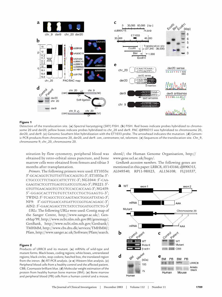
Figure 1Detection of the translocation site. (a) Spectral karyotyping (SKY) FISH. (b) FISH. Red boxes indicate probes hybridized to chromo-some 20 and der20; yellow boxes indicate probes hybridized to chr_20 and der9. PAC dJ890O15 was hybridized to chromosome 20,der20, and der9. (c) Genomic Southern blot hybridization with the ET1033 probe. The arrowhead indicates the mutation. (d) Genom-ic PCR products from chromosome 20, der20, and der9. cen, centromere; tel, telomere. (e) Sequences of the translocation site. Chr_9,chromosome 9; chr_20, chromosome 20.
Figure 2Products of LRRC8 and its mutant. (a) mRNAs of wild-type andmutant forms. Black boxes, coding regions; white boxes, untranslatedregions; black circles, stop codons; hatched box, the translated regionfrom the intron. (b) RT-PCR analysis. (c–e) Western blot analysis. (c)Peripheral blood cells from a healthy control and the affected patient.CBB, Coomassie brilliant blue. (d) Molecular weight estimation of theprotein from healthy human bone marrow (BM). (e) Bone marrowand peripheral blood (PB) cells from a human control and a mouse.
1710 The Journal of Clinical Investigation | December 2003 | Volume 112 | Number 11
AB037858; FLJ20996, AK024649; murine LRRC8,XM_140864; AD158, NP_115646; T cell activationleucine repeat–rich protein (TA-LRRP), NP_056165;LRRC5, NP_060573; FLJ23420, NP_079337.
ResultsIsolation of LRRC8 and its mutant form. Translocationbetween the long arms of chromosomes 9 and 20 wasdetermined by spectral karyotyping (SKY) FISHanalysis (Figure 1a). To identify the translocation siteof the derivative of chromosome 20 (der20), we pre-pared BAC and PAC clones located on the long armof chromosome 20. FISH analysis showed that thePAC clone dJ890O15 stepped over the translocationsite (Figure 1b). According to Southern blothybridization analysis with the ET1033 probe derivedfrom dJ890O15, the translocation site was estimatedto exist between the 37,345th and 38,000th bp ofdJ890O15 (Figure 1c). We then analyzed the patient’sDNA by inverse PCR and obtained PCR productscontaining the translocation site on der20 (Figure1d). A cDNA fragment containing the translocationsite on der9 was also obtained by PCR. Sequencinganalysis of these products showed that the translo-cation site existed between the 122,214th and122,215th bp of the BAC clone RP11-98H23 on chro-mosome 9, and between the 37,632nd and 37,633rdbp of the dJ890O15 on chromosome 20. A single
nucleotide insertion (T) was observed at the translo-cation site on der20 (Figure 1e).
Although no genes located at the translocation site onchromosome 9 were found, exons or coding sequencesfor two full-length mRNAs, termed FLJ10337 andFLJ20996, and several spliced mRNAs were split andlocated over the translocation site, and computer analy-sis using both GENESCAN and HMMGENE predictedthe existence of a hypothetical protein. There were nosequences for mRNA around the translocation site (∼20kbp) on chromosome 20. Sequences for the wild andmutant genes were obtained with 3′ rapid amplificationof cDNA ends from peripheral white blood cells of thepatient. Their schemata are shown in Figure 2a. Tran-scription from the mutant gene as well as the wild-typegene in the patient’s cells was confirmed by RT-PCR(Figure 2b). Wild and mutant forms of the LRRC8 pro-tein were also detected in the patient’s cells by Westernblot analysis (Figure 2c).
Characteristics of LRRC8. The length of the codingregion of this gene was predicted to be 2,433 bp, andthe protein consisted of 810 amino acids (∼94.2 kDa).The result of Western blot analysis with poAb’s againstthis protein was compatible with the predicted molec-ular weight (Figure 2d). It had two exons; exons 1 and2 encoded 719 and 91 amino acids, respectively. Theprotein was produced at a higher level in bone marrowcells than in peripheral blood cells (Figure 2e).
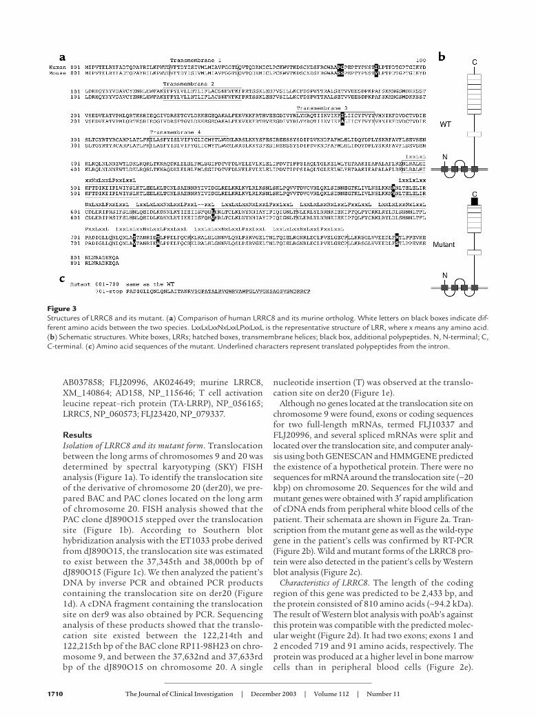
Figure 3Structures of LRRC8 and its mutant. (a) Comparison of human LRRC8 and its murine ortholog. White letters on black boxes indicate dif-ferent amino acids between the two species. LxxLxLxxNxLxxLPxxLxxL is the representative structure of LRR, where x means any amino acid.(b) Schematic structures. White boxes, LRRs; hatched boxes, transmembrane helices; black box, additional polypeptides. N, N-terminal; C,C-terminal. (c) Amino acid sequences of the mutant. Underlined characters represent translated polypeptides from the intron.
The Journal of Clinical Investigation | December 2003 | Volume 112 | Number 11 1711
Similarly, murine bone marrow cells contained greateramounts of the protein than peripheral blood cells.
The gene was highly conserved between humans andmice: 93% of nucleotide sequences and 99% of aminoacid sequences were identical (Figure 3a). The struc-ture of the protein predicted by TMHMM and Pfamcomputer-assisted analysis is shown in Figure 3b. Itconsisted of four transmembrane helixes located closeto the N-terminal and nine leucine-rich repeats (LRRs)(26) close to the C-terminal. Both terminal ends wereoutside the cell, and the second to ninth LRRs werelocated consecutively. This gene was named LRRC8 bythe nomenclature committee, the Human GenomeOrganisation. The truncated gene encoded 754 aminoacids, including 719 derived from exon 1 and 35 fromthe intron (Figure 3c).
Both wild and mutant LRRC8 proteins wereexpressed on the patient’s cells. There was no muta-tion in the coding sequences on the unaffected allele.
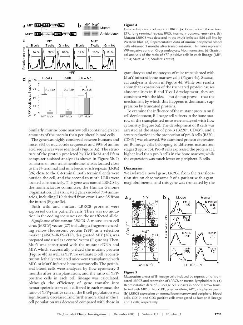
Significance of the mutant LRRC8. A mouse stem cellvirus (MSCV) vector (27) including a fragment encod-ing yellow fluorescent protein (YFP) as a selectionmarker (MSCV-IRES-YFP), designated MIY (28), wasprepared and used as a control vector (Figure 4a). Then,MutY was constructed with the mutant cDNA andMIY, which successfully yielded the mutant protein(Figure 4b) as well as YFP. To evaluate B cell reconsti-tution, lethally irradiated mice were transplanted withMIY- or MutY-infected bone marrow cells. The periph-eral blood cells were analyzed by flow cytometry 3months after transplantation, and the ratio of YFP-positive cells in each cell lineage was calculated.Although the efficiency of gene transfer intohematopoietic stem cells differed in each mouse, theratio of YFP-positive cells in the B cell population wassignificantly decreased, and furthermore, that in the Tcell population was decreased compared with those in
granulocytes and monocytes of mice transplanted withMutY-infected bone marrow cells (Figure 4c). Statisti-cal analysis is shown in Figure 4d. While our resultsshow that expression of the truncated protein causesabnormalities in B and T cell development, they areconsistent with the idea — but do not prove — that themechanism by which this happens is dominant sup-pression by truncated proteins.
To examine the influence of the mutant protein on Bcell development, B-lineage cell subsets in the bone mar-row of the transplanted mice were analyzed with flowcytometry (Figure 5a). The development of B cells wasarrested at the stage of pro-B (B220+, CD43+), and asevere reduction in the proportion of pre-B cells (B220+,CD43–) was observed. We examined protein expressionon B-lineage cells belonging to different maturationstages (Figure 5b). Pro-B cells expressed the protein at ahigher level than pre-B cells in the bone marrow, whilethe expression was much lower on peripheral B cells.
DiscussionWe isolated a novel gene, LRRC8, from the transloca-tion site on chromosome 9 of a patient with agam-maglobulinemia, and this gene was truncated by the
Figure 4Enforced expression of mutant LRRC8. (a) Constructs of the vectors.LTR, long terminal repeat; IRES, internal ribosomal entry site. (b)Mutant LRRC8 was detected in the MutY-infected E86 cell line byWestern blot. (c) Representative data of murine peripheral bloodcells obtained 3 months after transplantation. Thin lines representYFP-negative control. Gr, granulocytes; Mo, monocytes. (d) Statisti-cal analysis of the ratio of YFP-positive cells in each lineage (MIY, n = 4; MutY, n = 3; Student’s t test).
Figure 5Maturation arrest of B-lineage cells induced by expression of trun-cated LRRC8 and expression of LRRC8 on normal lymphoid cells. (a)Representative data of B-lineage cell subsets in bone marrow trans-fected with MIY or MutY. PE, phycoerythrin; APC, allophycocyanin.(b) LRRC8 expression on normal bone marrow and peripheral bloodcells. CD19- and CD3-positive cells were gated as human B-lineageand T cells, respectively.
1712 The Journal of Clinical Investigation | December 2003 | Volume 112 | Number 11
translocation. The patient's white blood cellsexpressed both wild-type and truncated LRRC8 pro-teins. LRRC8 is highly conserved between humansand mice: 93% and 99% of nucleotide and amino acidsequences were identical, respectively. Transplanta-tion of bone marrow cells that were forced to expresstruncated LRRC8 to lethally irradiated mice induceda severe reduction of the B cell number in peripheralblood. Our results indicate that the LRRC8 deficit isresponsible for the B cell deficiency in this patient andthat normal LRRC8 is required for B cell development.
We showed that development of B-lineage cellsexpressing the truncated form of LRRC8 is arrestedat the stage of pro-B, and the proportion of pre-Bcells was severely reduced in the bone marrow. Sincethe highest expression of LRRC8 protein was seen onpro-B cells among subsets of B-lineage cells, this pro-tein seems to be a key molecule for growth and/ordifferentiation in pro-B and pre-B cells.
The LRRs are functional domains that mediate adhe-sion to other molecules (26). The LRRs of LRRC8 arelocated on the outside of cells. Various molecules withsuch a structure are known to act as cell surface recep-tors, including RP105 (LPS receptor), CD42 (vWFreceptor), TrkA (high-affinity nerve growth factorreceptor), and follicle-stimulating hormone receptor(26, 29). It is possible that LRRC8 has a novel specificligand that induces B cell development. LRRC8 lacksdomains that can transduce signals to the cytosol ornucleus. RP105, a Toll-like receptor on B cells, has anadapter protein named MD-1 instead of a signal-trans-ducing domain (30). Similarly, LRRC8 might also haveadapter molecules for signal transduction.
The intact LRRC8 gene on the unaffected allele of thepatient was transcribed, and intact LRRC8 protein wasexpressed. Nevertheless, B cells were totally absent inthe peripheral blood. This suggests that the truncatedform has not a quantitative effect but a dominant-sup-pressor effect on B cell development. Autoimmunelymphoproliferative syndrome is ascribed to a domi-nant-negative mutant of Fas (31). Fas is a homotrimerof Fas subunits. When one of the three subunits isreplaced with a mutant, the Fas complex loses its func-tion. Therefore, only one-eighth of the receptors cantransduce death signal into cells in patients with theheterozygous mutation. Dominant-negative mutantsare known in several receptors that compose homo-oligomers: dynamin-1, growth hormone receptor, andinsulin receptor (32–34). Although there are no knownproteins containing LRR structures that form homo-oligomers, LRRC8 might form such a complex.
In addition to the deficiency of B cells, a decrease inthe proportion of T cells among cells transfectedwith the mutant LRRC8 was also observed. Althoughboth human and murine T cells express LRRC8 (datanot shown), the number of T cells was not decreasedin our patient. The difference between the functionof LRRC8 in mice and its function in humans shouldbe clarified in a further study.
Minor facial anomalies as well as agammaglobu-linemia were observed in the patient. Using RT-PCR,we have determined that LRRC8 is expressed in brain,heart, lung, liver, and kidney (unpublished observa-tions). Since this gene is expressed in a ubiquitousfashion beyond lymphoid and hematopoietic systems,the protein might therefore play a role in morpho-genesis. A further analysis of LRRC8 may contributeto our understanding of its roles in development.
There are no known proteins homologous toLRRC8, but there are at least two hypothetical pro-teins, AD158 and T cell activation leucine repeat–richprotein (TA-LRRP). There are also two predictableproteins, LRRC5 and FLJ23420, which may be frag-ments of other LRRC8 homologs. LRRC8, AD158,and TA-LRRP conserve 59% of their amino acidsequences and share a unique structure: four trans-membrane helixes and eight sequentially locatedLRRs. TA-LRRP was found by a microarray assay ofinduced genes in activated T cells. These putativeproteins, sharing a unique structure, constitute anovel protein family that has not, to our knowledge,been reported so far (unpublished observations).Although their functions remain to be elucidated, itseems plausible that they play essential and funda-mental roles in morphogenesis.
AcknowledgmentsWe thank N. Sakai and H. Kurahashi for discussions,K.R. Humphries, R.G. Hawley, G.P. Nolan, and A.Bank for MSCV vectors and virus-producing celllines, T. Okuda for assistance, and N. Ozawa and R.Hasegawa for secretarial help.
1. Urbanek, P., Wang, Z.Q., Fetka, I., Wagner, E.F., and Busslinger, M.1994. Complete block of early B cell differentiation and altered pat-terning of the posterior midbrain in mice lacking Pax5/BSAP. Cell.79:901–912.
2. Nutt, S.L., Heavey, B., Rolink, A.G., and Busslinger, M. 1999. Com-mitment to the B-lymphoid lineage depends on the transcription fac-tor Pax5. Nature. 401:556–562.
3. Schaniel, C., Gottar, M., Roosnek, E., Melchers, F., and Rolink, A.G.2002. Extensive in vivo self-renewal, long-term reconstitution capac-ity, and hematopoietic multipotency of Pax5-deficient precursor B-cell clones. Blood. 99:2760–2766.
4. Lin, H., and Grosschedl, R. 1995. Failure of B-cell differentiation inmice lacking the transcription factor EBF. Nature. 376:263–267.
5. Zhuang, Y., Soriano, P., and Weintraub, H. 1994. The helix-loop-helixgene E2A is required for B cell formation. Cell. 79:875–884.
6. Yel, L., et al. 1996. Mutations in the mu heavy-chain gene in patientswith agammaglobulinemia. N. Engl. J. Med. 335:1486–1493.
7. Minegishi, Y., et al. 1998. Mutations in the human lambda5/14.1 generesult in B cell deficiency and agammaglobulinemia. J. Exp. Med.187:71–77.
8. Minegishi, Y., et al. 1999. Mutations in Igalpha (CD79a) result in acomplete block in B-cell development. J. Clin. Invest. 104:1115–1121.
9. Wang, Y., et al. 2002. Novel Igalpha (CD79a) gene mutation in a Turk-ish patient with B cell-deficient agammaglobulinemia. Am. J. Med.Genet. 108:333–336.
10. Cheng, A.M., et al. 1995. Syk tyrosine kinase required for mouse via-bility and B-cell development. Nature. 378:303–306.
11. Minegishi, Y., et al. 1999. An essential role for BLNK in human B celldevelopment. Science. 286:1954–1957.
12. Egawa, T., et al. 2001. The earliest stages of B cell development requirea chemokine stromal cell-derived factor/pre-B cell growth-stimulat-ing factor. Immunity. 15:323–334.
13. Nagasawa, T., et al. 1996. Defects of B-cell lymphopoiesis and bone-mar-row myelopoiesis in mice lacking the CXC chemokine PBSF/SDF-1.Nature. 382:635–638.
The Journal of Clinical Investigation | December 2003 | Volume 112 | Number 11 1713
14. Ma, Q., et al. 1998. Impaired B-lymphopoiesis, myelopoiesis, andderailed cerebellar neuron migration in CXCR4- and SDF-1-deficientmice. Proc. Natl. Acad. Sci. U. S. A. 95:9448–9453.
15. von Freeden-Jeffry, U., et al. 1995. Lymphopenia in interleukin (IL)-7gene-deleted mice identifies IL-7 as a nonredundant cytokine. J. Exp.Med. 181:1519–1526.
16. Prieyl, J.A., and LeBien, T.W. 1996. Interleukin 7 independent develop-ment of human B cells. Proc. Natl. Acad. Sci. U. S. A. 93:10348–10353.
17. Puel, A., Ziegler, S.F., Buckley, R.H., and Leonard, W.J. 1998. DefectiveIL7R expression in T(-)B(+)NK(+) severe combined immunodeficiency.Nat. Genet. 20:394–397.
18. Tsukada, S., et al. 1993. Deficient expression of a B cell cytoplasmic tyro-sine kinase in human X-linked agammaglobulinemia. Cell. 72:279–290.
19. Vetrie, D., et al. 1993. The gene involved in X-linked agammaglobuline-mia is a member of the src family of protein-tyrosine kinases. Nature.361:226–233.
20. Conley, M.E., Mathias, D., Treadaway, J., Minegishi, Y., and Rohrer, J.1998. Mutations in btk in patients with presumed X-linked agamma-globulinemia. Am. J. Hum. Genet. 62:1034–1043.
21. Meffre, E., et al. 1996. A human non-XLA immunodeficiency diseasecharacterized by blockage of B cell development at an early proB cellstage. J. Clin. Invest. 98:1519–1526.
22. Willis, T.G., et al. 1997. Rapid molecular cloning of rearrangements ofthe IGHJ locus using long-distance inverse polymerase chain reaction.Blood. 90:2456–2464.
23. Kinsella, T.M., and Nolan, G.P. 1996. Episomal vectors rapidly and sta-bly produce high-titer recombinant retrovirus. Hum. Gene Ther.7:1405–1413.
24. Wigler, M., et al. 1977. Transfer of purified herpes virus thymidine kinasegene to cultured mouse cells. Cell. 11:223–232.
25. Cavazzana-Calvo, M., et al. 2000. Gene therapy of human severe com-bined immunodeficiency (SCID)-X1 disease. Science. 288:669–672.
26. Kobe, B., and Deisenhofer, J. 1994. The leucine-rich repeat: a versatilebinding motif. Trends Biochem. Sci. 19:415–421.
27. Hawley, R.G., Fong, A.Z., Burns, B.F., and Hawley, T.S. 1992. Trans-plantable myeloproliferative disease induced in mice by an interleukin6 retrovirus. J. Exp. Med. 176:1149–1163.
28. Antonchuk, J., Sauvageau, G., and Humphries, R.K. 2001. HOXB4 over-expression mediates very rapid stem cell regeneration and competitivehematopoietic repopulation. Exp. Hematol. 29:1125–1134.
29. Miyake, K., Yamashita, Y., Ogata, M., Sudo, T., and Kimoto, M. 1995.RP105, a novel B cell surface molecule implicated in B cell activation, isa member of the leucine-rich repeat protein family. J. Immunol.154:3333–3340.
30. Nagai, Y., et al. 2002. Requirement for MD-1 in cell surface expression ofRP105/CD180 and B-cell responsiveness to lipopolysaccharide. Blood.99:1699–1705.
31. Fisher, G.H., et al. 1995. Dominant interfering Fas gene mutationsimpair apoptosis in a human autoimmune lymphoproliferative syn-drome. Cell. 81:935–946.
32. Lee, A., Frank, D.W., Marks, M.S., and Lemmon, M.A. 1999. Dominant-negative inhibition of receptor-mediated endocytosis by a dynamin-1mutant with a defective pleckstrin homology domain. Curr. Biol.9:261–264.
33. Ayling, R.M., et al. 1997. A dominant-negative mutation of the growthhormone receptor causes familial short stature. Nat. Genet. 16:13–14.
34. Moritz, W., Froesch, E.R., and Boni-Schnetzler, M. 1994. Functionalproperties of a heterozygous mutation (Arg1174 → Gln) in the tyrosinekinase domain of the insulin receptor from a type A insulin resistantpatient. FEBS Lett. 351:276–280.