Embed Size (px)
Citation preview
APPLIED AND ENVIRONMENTAL MICROBIOLOGY, May 2007, p. 2963–2975 Vol. 73, No. 90099-2240/07/$08.00�0 doi:10.1128/AEM.02623-06
Flagellin Diversity in Clostridium botulinum Groups I and II: a NewStrategy for Strain Identification�
Catherine J. Paul,1,2 Susan M. Twine,2 Kevin J. Tam,1 James A. Mullen,2 John F. Kelly,2John W. Austin,1* and Susan M. Logan2
Bureau of Microbial Hazards, HFPB, Health Canada, Sir Frederick G. Banting Research Centre, PL2204A2, Ottawa, Ontario,Canada K1A 0L2,1 and Institute for Biological Sciences, National Research Council, 100 Sussex Dr.,
Ottawa, Ontario, Canada K1A 0R62
Received 9 November 2006/Accepted 26 February 2007
Strains of Clostridium botulinum are traditionally identified by botulinum neurotoxin type; however, identi-fication of an additional target for typing would improve differentiation. Isolation of flagellar filaments andanalysis by sodium dodecyl sulfate-polyacrylamide gel electrophoresis (SDS-PAGE) showed that C. botulinumproduced multiple flagellin proteins. Nano-liquid chromatography-tandem mass spectrometry (nLC-MS/MS)analysis of in-gel tryptic digests identified peptides in all flagellin bands that matched two homologous tandemflagellin genes identified in the C. botulinum Hall A genome. Designated flaA1 and flaA2, these open readingframes encode the major structural flagellins of C. botulinum. Colony PCR and sequencing of flaA1/A2 variableregions classified 80 environmental and clinical strains into group I or group II and clustered isolates into 12flagellar types. Flagellar type was distinct from neurotoxin type, and epidemiologically related isolates clus-tered together. Sequencing a larger PCR product, obtained during amplification of flaA1/A2 from type E strainBennett identified a second flagellin gene, flaB. LC-MS analysis confirmed that flaB encoded a large typeE-specific flagellin protein, and the predicted molecular mass for FlaB matched that observed by SDS-PAGE.In contrast, the molecular mass of FlaA was 2 to 12 kDa larger than the mass predicted by the flaA1/A2sequence of a given strain, suggesting that FlaA is posttranslationally modified. While identification of FlaB,and the observation by SDS-PAGE of different masses of the FlaA proteins, showed the flagellin proteins of C.botulinum to be diverse, the presence of the flaA1/A2 gene in all strains examined facilitates single locussequence typing of C. botulinum using the flagellin variable region.
Clostridium botulinum is a gram-positive, spore-forming an-aerobic bacterium that produces a potent neurotoxin. Botuli-num neurotoxin (BoNT) causes botulism, a neuroparalytic dis-ease which occurs after ingestion of food contaminated withBoNT or by other means of toxin exposure. C. botulinum canalso colonize wounds and, rarely, the adult gastrointestinaltract as well as the infant gastrointestinal tract, where growthof the bacterium and subsequent production of BoNT resultsin wound botulism, adult colonization botulism, or infant bot-ulism, respectively (4).
Strains of C. botulinum are divided into four distinct groupsbased on physiological characteristics. Only groups I and IIcontain strains that have been associated with human illness.Group I strains are proteolytic with an optimal growth tem-perature of 37°C, while group II strains are nonproteolytic andgrow optimally at 30°C. Strains of C. botulinum are differenti-ated into serotypes A through G based on seroneutralization ofBoNT. Group I strains can produce serotype A, B, or F BoNT,and group II strains can produce serotype B, E, or F BoNT.Group III strains produce either serotype C or D and areassociated with botulism in animals, and group IV containsonly strains producing BoNT serotype G (8, 28). In addition tocategorizing strains based on physiological characteristics or
BoNT serotype, other methods have been used for epidemio-logical and molecular typing of C. botulinum. These methodsinclude the following: randomly amplified polymorphic DNAanalysis; repetitive element sequence-based PCR; fatty acidanalysis; amplified rRNA gene restriction analysis; pulsed-fieldgel electrophoresis (PFGE); and most recently, amplified frag-ment length polymorphism and gene sequencing (16S, neuro-toxin) (18, 26, 33).
The flagella of gram-negative bacteria are the most widelycharacterized of the bacterial motility structures (5, 30). Ingeneral, flagella are composed of over 20 distinct structuralproteins that assemble to form the flagellar basal body, hook,and filament, with the filament comprising around 20,000 sub-units of the flagellin protein. Due to commonalities in foldingand filament assembly, all bacterial flagellin proteins can bedivided into three distinct subdomains: the N and C termini,which are conserved within a given species, and a middle vari-able region (31, 41). The flagellin monomers are arrangedsurrounding the central channel of the filament with the N andC termini of each protein buried in the interior nearest thefilament channel. The flagellin variable domain protrudes out-wards and, stacked together with other flagellin monomers,forms the external surface of the filament (30). The surface-exposed variable domain of the flagellin is antigenically diverseand forms the basis of a wide range of typing methods exploit-ing diversity at the DNA sequence level or structural differ-ences at the protein level for strain identification (12, 49).Flagella are often involved in pathogenesis, with roles in mo-
* Corresponding author. Mailing address: Bureau of Microbial Haz-ards, Health Canada, 251 Sir Frederick Banting Driveway, Tunney’sPasture PL2204A2, Ottawa, Ontario, Canada K1A 0L2. Phone: (613)957-0902. Fax: (613) 941-0280. E-mail: [email protected].
� Published ahead of print on 9 March 2007.
2963
on January 4, 2019 by guesthttp://aem
.asm.org/
Dow
nloaded from
tility adhesion and, in some cases, the secretion of virulencefactors (7, 19).
While the genetics, regulation, assembly, and physical struc-ture of the gram-negative bacterial flagellum have been exten-sively investigated, less is known about the flagella of gram-positive bacteria, and in particular clostridial flagella. Severalstudies document preliminary investigations of flagella in thepathogens Clostridium chauvoei, Clostridium haemolyticum,Clostridium novyi, and Clostridium septicum as well as Clostrid-ium tyrobutyricum, a clostridial species of relevance to thecheese industry (3, 22, 23, 39). The flagella of the humanpathogen Clostridium difficile divides the species into 10 sero-groups, has a role in the adherence of C. difficile to the mucouslayer of the intestine, and is considered a potential vaccinecandidate (36, 43, 45, 46). There are, however, no publishedinvestigations addressing the flagella of C. botulinum, a bacte-rium that has largely been studied only in the context of theneurotoxin.
This study examines flagellins from a range of isolates fromdifferent geographical, clinical, and environmental origins. Weexplored the diversity of flagellin structural proteins and genesequences to determine the potential for using flagellin genevariable region sequences to improve genotyping of group Iand group II C. botulinum strains.
MATERIALS AND METHODS
Bacterial strains and growth conditions. The origin, source, and date ofisolation for each of the C. botulinum strains used in this study are described inTable 1.
C. botulinum strains were grown for flagellar isolation in SPGY broth con-taining 5% (wt/vol) special peptone (Oxoid Inc., Basingstoke, United Kingdom),0.5% (wt/vol) peptone (Difco, Tucker, GA), 2% (wt/vol) yeast extract (Difco),0.4% (wt/vol) glucose (Difco) and 0.1% (wt/vol) sodium thioglycolate (SigmaAldrich, St. Louis, MO) adjusted to a pH of 7.2 using HCl. For colony PCR,bacteria were grown on McClung-Toabe 1.5% agar (Difco) with 5% egg yolk and0.5% yeast extract (MTEYE). Cells were grown for 24 to 48 h in an atmosphereof 10% H2, 10% CO2, and 80% N2 at either 35°C (group I strains) or roomtemperature (group II strains). Cells were occasionally grown in 3.5-liter anaer-obe jars (Oxoid Inc.) using AnaeroGen (Oxoid Inc.) for atmosphere production,at either 35°C or room temperature. C. botulinum strains were stored andarchived at the Botulism Reference Service for Canada at �86°C on Microbankbeads (Pro-Lab Diagnostics, Richmond Hill, Canada).
Escherichia coli strain DH10B (Invitrogen, Carlsbad, CA), grown in LB broth(Difco) at 37°C with shaking, was used for cloning manipulations.
Electron microscopy. Copper grids (Electron Microscopy Sciences, FortWashington, PA) were covered with Formvar film and coated with carbon. Forsample preparation, grids were floated on a drop of bacterial cells or purifiedflagella in 0.1 M Tris-HCl, pH 7.0, for 5 min and negatively stained with 1%(wt/vol) ammonium molybdate. Images were taken with a Zeiss EM902 trans-mission electron microscope operated at an accelerating voltage of 80 kV andrecorded on Kodak electron image film SO-163 (Kodak, Rochester, NY).
Isolation and preliminary analysis of flagellar proteins. Cultures for flagellarisolation were grown overnight at the required temperature in SPGY broth. Cellswere harvested and resuspended in 1/20 of the original volume, in 0.1 M Tris-HCl, pH 7.0. Flagella were sheared from the cell surface by 10 repetitions in a50-ml tissue homogenizer. Flagella were isolated from the homogenate by re-moving whole cells by two low-speed centrifugations (5,000 � g; 15 min) beforea high-speed centrifugation for 1 h at 130,000 � g (70 Ti rotor; Beckman CoulterCanada Inc., Mississauga, Ontario, Canada). Pellets containing flagella werewashed once in ultrapure water (Gibco, Grand Island, NY), followed by a secondcentrifugation at 130,000 � g for 1 h and resuspension in ultrapure water.Flagella preparations were analyzed by standard sodium dodecyl sulfate-poly-acrylamide gel electrophoresis (SDS-PAGE) on 12.5% acrylamide gels and stainedwith standard Coomassie blue, as described previously (38). Molecular weights ofprotein bands were determined using Quantity One software (Bio-Rad, Hercu-les, CA) with comparison to molecular mass standards. Protein bands for trypticdigest and mass spectrometry were excised from 12.5% acrylamide gels following
SDS-PAGE and staining with Bio-Safe Coomassie (Bio-Rad). A ProQ Emerald300 glycoprotein detection kit (Invitrogen, Carlsbad, CA) or a Dig glycan dif-ferentiation kit (Roche Diagnostics, Indianapolis, IN) was used following themanufacturer’s instructions to examine posttranslational modification of flagellinproteins.
Mass spectrometry analysis of flagellin proteins. Protein bands excised fromBio-Safe Coomassie blue (Bio-Rad)-stained SDS-PAGE gels were cut into1-mm cubes. In microcentrifuge tubes, gel pieces were covered with 100 mMammonium bicarbonate and 30% (vol/vol) acetonitrile for 5 min and washedthree times with water or until the blue coloration disappeared. The predictedflagellin protein did not contain cysteine residues; therefore, samples werenot subjected to reduction/alkylation. Gel pieces were treated with acetoni-trile and then rehydrated with a trypsin-containing solution as describedpreviously (47). Peptides were extracted from gel pieces by sonication for 20min., then analyzed by nano-liquid chromatography–tandem mass spectrom-etry (nLC-MS/MS) using a ‘CapLC’ capillary chromatography system (Wa-ters, Milford, MA) coupled to a ‘QTOF Ultima’ hybrid quadrupole time-of-flight mass spectrometer (Waters, Milford, MA) (47). MS/MS spectraobtained were searched against the Hall A genome sequence (http://www.sanger.ac.uk/Projects/C_botulinum) using MASCOT 2.0.1 (Matrix Science,United Kingdom). The following parameters for mass spectral identificationwere used: peptide tolerance of 1.5 Da, MS/MS tolerance of 0.8 Da, andpossible 1 missed cleavage site. Peptide identifications were accepted if theymet all of the following criteria: MASCOT peptide score �20, rank � 1, andmass accuracy �100 ppm. MASCOT scores evaluate the match between anMS/MS profile of a tryptic peptide and a protein sequence database andindicate the probability that the observed match is a random event. In addi-tion, all MS/MS spectra were manually assessed for data quality and high-confidence identification, requiring a clear series of high mass y-ions andcorrect charge state assignment for all fragments.
Cloning, colony PCR, and sequencing of flagellin genes. Flagellin genes wereamplified from chromosomal DNA preparations or from single colonies resus-pended in 50 �l Tris-EDTA buffer. Chromosomal DNA was isolated as de-scribed previously with slight modifications (21). Primers cbotflaF (5�-CGCGGGGATCCATGATAATTAATCACAATTTAAATG-3�) and cbotflaR (5�-CGCGGGGATCCCTTAATAATTGAAGAACTCCTTGTG-3�) were designed tocorrespond to the N and C termini of a Hall A flagellin gene (C. botulinumgenome sequence from the C. botulinum Sequencing Group at the WellcomeTrust Sanger Institute [ftp://ftp.sanger.ac.uk/pub/pathogens/cb]) and to includeBamHI sites. PCR products were amplified using Pwo (Roche Diagnostics) orHigh Fidelity Triple Master (Eppendorf, Hamburg, Germany) polymerase, fol-lowing the manufacturer’s instructions, in an Applied Biosystems Gene AmpPCR system 9700 thermocycler (cycling parameters: 3 min at 94°C; 30 cycles of30 s at 94°C, 30 s at 55/50°C, and 1 min at 72°C; and 2 min at 72°C). For colonyPCR, 5 �l of bacterial cell/Tris-EDTA suspension was used instead of chromo-somal DNA with a step of 10 min at 94°C added at the beginning of the PCR forcell lysis. Annealing temperatures were lowered from an initial temperature of55°C until the product of the expected size could be visualized by standardagarose gel electrophoresis. All strains produced flaA1/A2 amplicons at 50°C.PCR amplicons were purified either by excision of the desired product frompreparative 1.0% agarose gels and Qiaquick gel extraction (QIAGEN, Hilden,Germany) or by using a Qiaquick nucleotide removal kit (QIAGEN). Initially,PCR products were ligated into an EcoRV site in vector pCR2.1 (Invitrogen),screened for insert following miniprep (QIAGEN) with BamHI digestion, andsequenced using M13F and M13R primers to vector sequence. Colony PCRproducts were purified using a Qiaquick nucleotide removal kit (QIAGEN) fordirect amplicon sequencing using a Big Dye Terminator 3.1 kit (Applied Bio-systems, Foster City, CA), cbotflaF and cbotflaR as sequencing primers, and anABI3130 genetic analyzer (Applied Biosystems). All restriction enzymes wereobtained from New England Biolabs (Ipswich, MA).
Analysis of DNA and protein sequences. Candidate Hall A flagellin genes wereidentified by tBlastN against the Hall A genome sequence (www.sanger.ac.uk/cgi-bin/blast/submitblast/c_botulinum) using flagellin protein sequences from C.tyrobutyricum, C. difficile, C. chauvoei, and Bacillus subtilis (GenBank accessionnumbers CAB44444.1, AAD46086.1, BAB13814.1, and NP_391416.1, respec-tively). Hall A flagellin open reading frames (ORFs) were identified by start andstop codons flanking DNA sequence identified by tBlastN as candidate flagellingenes and by comparison to the ORF assignments in homologous Clostridiumtetani flagellin sequences (GenBank accession numbers AAO36250.1, AAO36226.1,and AAO36215.1). DNA sequences were compiled using Pregap and Gap4 fromthe Staden package of software for cloned flagellins (42) or Sequencher 4.6software for colony PCR products (Gene Codes Corporation, Ann Arbor, MI).
2964 PAUL ET AL. APPL. ENVIRON. MICROBIOL.
on January 4, 2019 by guesthttp://aem
.asm.org/
Dow
nloaded from
TABLE 1. C. botulinum strains used in this study
Strain Origin Locationj Yr ofisolation Source or referencea
Group IType A
Hall A/ATCC 3502 11/IFRb
17A 196362A 1/NCAc
A6 R. Woods/T. Midura16037 Canned tomato United States 1974 Leatherhead/IFRCK2-A Feces Canada 1974 BRSd
F9604-A Feces Calgary, AB, Canada* 1996 BRSF9801-A Feces Laval, QC, Canada* 1998 BRSFE9909ACS Alberta Feces Loughheed, AB, Canada* 1999 BRSFE0101AJO Feces Desbiens, QC, Canada 2001 BRSGA0101AJO Gastric liquid Desbiens, QC, Canada 2001 BRSPC0101AJO Pork St-Fulgence, QC, Canada 2001 BRSNG0107ASA Gastric liquid Sanikiluaq, NN, Canada 2001 BRSMUL0109ASA Mullet fish Gulf of Kuwait, Kuwait 2001 BRSFE0205A1AK Feces Calgary, AB, Canada* 2002 BRSFE0207AMB Feces Meaford, ON, Canada 2002 BRSFE0303A1YO Feces Toronto, ON, Canada* 2003 BRSSO300A1 Soil Kuujuaraapiik, QC, Canada 2003 BRSINWB2202A2 Seal intestine Kangiqsualujjuaq, QC, Canada 2004 BRS
Type ABNCTC 2916 Canned corn Colorado 1929 M. Wictome/IFRFE9504ACG Feces Baie Comeau, QC, Canada* 1995 BRS
Type BIB1-B Feces Peterborough, ON, Canada* 1979 BRSMRB Mushrooms Montreal, QC, Canada 1973 BRS13983B Asparagus Unknown 1962 2/NCA427-2-76 Honey Modesto, CA 1976 D. Arvant426B Honey Modesto, CA 1976 R. Wood/T. Midura1366-1-77 Honey San Francisco, CA* 1977 T. Midura1344-1-77 Feces San Francisco, CA* 1977 T. Midura920A276 Feces Modesto, CA* 1976 T. Midura368B Feces Modesto, CA* 1976 T. MiduraBL 143 Fish United Kingdom 1986 IFRFE9508BRB Feces Sherbrooke, QC, Canada 1995 BRSFE9508BPD Feces Sherbrooke, QC, Canada 1995 BRSPA9508B Pate Sherbrooke, QC, Canada 1995 BRSFE9904BMT Feces Toronto, ON, Canada 1999 BRSGA0108BEC Gastric liquid Riviere-du-Loup, QC, Canada 2001 BRSFE0507BLP Feces Ottawa, ON, Canada* 2005 BRSEN0509BLP Dust Ottawa, ON, Canada* 2005 BRS
Type FLangeland Liver paste Langeland, Denmark 1960 V. Moller/Catherwoode
H461297F Honey Wisconsin 1998 BRS
Group IIType B
2B Marine sediment Pacific coast, United States 1960s M. W. Eklund/H. Solomonf
17B Marine sediment Pacific coast, United States 1960s M. W. Eklund/H. SolomonDB-2 Marine sediment Pacific coast, United States 1968 M. W. Eklund/H. SolomonPrevot 59 IFRKAP-B-3 Kapchunka California 1981 H. SolomonKAP-B-8 Kapchunka California 1981 H. SolomonII60-15B Feces British Columbia, Canada 1982 BRS
Type EEruss Sturgeon Russia 1935 24/NRg
Gordon Clinical Kuujjuaq, QC, Canada 1975 L. Gauvreauh
Bennett Gastric liquid Happy Valley, NL, Canada 1976 BRSFE9507EEA Feces Kangiqsualujjuaq, QC, Canada 1995 BRSMI9507E Misiraq Kangiqsualujjuaq, QC, Canada 1995 BRSF9508EMA Feces Kuujjuaq, QC, Canada 1995 BRSVI9508E Seal meat Kuujjuaq, QC, Canada 1995 BRSFE9508EPB Feces Tasiujaq, QC, Canada 1995 BRSS9510E Seal meat Kuujjuaq, QC, Canada 1995 BRSGA9709EHS Gastric liquid Kangiqsualujjuaq, QC, Canada 1998 BRS
Continued on following page
VOL. 73, 2007 FLAGELLIN DIVERSITY IN C. BOTULINUM 2965
on January 4, 2019 by guesthttp://aem
.asm.org/
Dow
nloaded from

Flagellin gene sequences were obtained from this study with the exception ofHall A CBO0242, CBO2666, CBO2695, and flaA1/2 sequences, which wereextracted from the Hall A genome sequence (C. botulinum genome sequencefrom the C. botulinum Sequencing Group at the Wellcome Trust SangerInstitute ftp://ftp.sanger.ac.uk/pub/pathogens/cb). Flagellin protein se-quences used for dendrogram construction were translated from gene se-quences obtained in this study, the Hall A genome sequence (see above), orGenBank (see figure legends for accession numbers). In silico translation offlaA and flaB was performed using ExPASy proteomic tools (http://ca.expasy.org/tools/). To avoid analyzing multiple flagellin variants from a populationsurvey, a representative single flagellin protein sequence was selected for asingle species. Multiple flagellins present in one contiguous genome or flagel-lins that originated from only one species were also included. Dendrogramswere generated using Bionumerics 4.5 (Applied Maths, Kortrijk, Belgium).The dendrogram comparing flagellin gene sequences used the unweighted-pair group method with arithmetic averages (UPGMA) with default settingsand the neighbor-joining method incorporating the Kimura 2P parameter.The dendrogram comparing flagellin proteins was generated using UPGMAfollowed by the neighbor-joining method, but no correction was applied. Toaccommodate alignment of flagellins from multiple species and differing inmolecular mass by as much as 25 kDa, the maximum number of gaps allowedwas increased from default to 98 and the gap penalty was reduced to 50%. Alldendrograms were bootstrapped using 2,000 simulations.
Variable and hypervariable regions in flagellin sequences were assigned byinspection of sequence alignments. Assignment of flaB as a flagellin gene wasdetermined by comparison of the predicted amino acid sequence to the NCBIdatabase using BlastP and tBlastX.
Nucleotide sequence accession numbers. All full and partial sequences of C.botulinum flagellin genes were deposited in GenBank with the following acces-sions numbers: flaB, DQ658239; and flaA, DQ844946 to DQ845031.
RESULTS
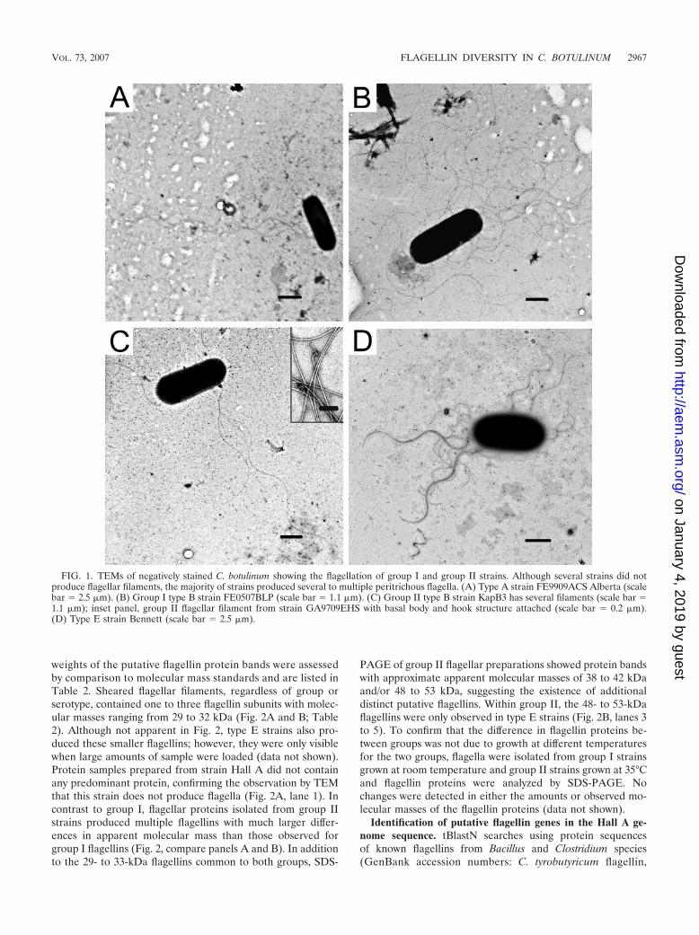
TEM shows variation in the degree of flagellation among C.botulinum strains. Fifteen strains of C. botulinum were exam-ined for flagellation by transmission electron microscopy(TEM). C. botulinum strain Hall A was not flagellated (datanot shown); however, almost all other group I strains producedmultiple peritrichous flagella when grown in SPGY broth (Fig.1A and B). Group I type F strain Langeland produced only oneor two filaments per cell. All group II strains examined pro-duced multiple filaments (Fig. 1C and D). The flagella fromgroup II type F strain 610F appeared short and fragile, withsome cells observed not producing filaments (data not shown).
To resolve the protein components making up the filaments,flagella were sheared from the surface of C. botulinum cells.Examination by TEM of initial flagellar preparations fromselect group I and group II strains showed that the shearingtechnique isolated large amounts of long intact flagellar fila-ments. Several filaments demonstrated the characteristic sin-gle-ring structure of a gram-positive flagellar basal body (Fig.1C, inset).
Group I and group II strains produce both common anddistinct flagellins. Flagellar filaments sheared from the surfaceof C. botulinum strains were analyzed by SDS-PAGE, and theresultant protein profiles for group I strains (Fig. 2A) differedsignificantly from those of group II (Fig. 2B). Molecular
TABLE 1—Continued
Strain Origin Locationj Yr ofisolation Source or referencea
GA9709ENS Gastric liquid Kangiqsualujjuaq, QC, Canada 1998 BRSGA9709EJA Gastric liquid Kangiqsualujjuaq, QC, Canada 1998 BRSFE9709ELB Feces Kangiqsualujjuaq, QC, Canada 1998 BRSFE9709EBB Feces Kangiqsualujjuaq, QC, Canada 1998 BRSFE9909ERG Feces Inuvik, NU, Canada 1999 BRSFE0005EJT Feces Inuvik, NU, Canada 2000 BRSMU0005EJT Muktuk Inuvik, NU, Canada 2000 BRSSW280E Saltwater Koksoak River, QC, Canada 2001 BRSSO309E2 Shoreline soil Hudson Bay, QC, Canada 2001 BRSSO325E1 Shoreline soil Ungava Bay, QC, Canada 2001 BRSSO326E1 Shoreline soil Ungava Bay, QC, Canada 2001 BRSSO329E1 Shoreline soil Ungava Bay, QC, Canada 2001 BRSSP455456E2 Coastal rock Ungava Bay, QC, Canada 2001 BRSSOKR-23E1 Marine sediment Koksoak River, QC, Canada 2002 BRSSOKR-29E1 Shoreline soil Ungava Bay, QC, Canada 2002 BRSSOKR-37E2 Freshwater sediment Koksoak River, QC, Canada 2002 BRSHNB0804E Honey New Brunswick, Canada 2004 BRS
Type F70F Marine sediment California coast 1960s M. W. Eklund/H. Solomon190F Marine sediment California coast 1960s M. W. Eklund/H. Solomon202F Marine sediment California coast 1965 M. W. Eklund/H. Solomon205F Marine sediment California coast 1960s M. W. Eklund/H. Solomon610F Salmon Columbia River, OR 1966 Craig/H. Solomon19501F Marine sediment Oregon coast 1960s M. W. Eklund/J. S. Crowtheri
a First name is the initial source and the second is the donor source.b M. Peck, Institute for Food Research, Norwich, United Kingdom.c National Canners Association.d Botulism Reference Service for Canada, Health Canada, Ottawa, Ontario, Canada.e Canadian Food Inspection Agency.f H. Solomon, U.S. Food and Drug Administration, Washington, DC.g NR, no records of the donor source found.h L. Gauvreau, Laboratoire de microbiologie, Centre hospitalier de l’Universite Laval, Ste-Foy, QC, Canada.i J. S. Crowther, Unilever Research, Sharnbrook, Bedford, England.j An asterisk indicates a strain associated with a case of infant botulism.
2966 PAUL ET AL. APPL. ENVIRON. MICROBIOL.
on January 4, 2019 by guesthttp://aem
.asm.org/
Dow
nloaded from
weights of the putative flagellin protein bands were assessedby comparison to molecular mass standards and are listed inTable 2. Sheared flagellar filaments, regardless of group orserotype, contained one to three flagellin subunits with molec-ular masses ranging from 29 to 32 kDa (Fig. 2A and B; Table2). Although not apparent in Fig. 2, type E strains also pro-duced these smaller flagellins; however, they were only visiblewhen large amounts of sample were loaded (data not shown).Protein samples prepared from strain Hall A did not containany predominant protein, confirming the observation by TEMthat this strain does not produce flagella (Fig. 2A, lane 1). Incontrast to group I, flagellar proteins isolated from group IIstrains produced multiple flagellins with much larger differ-ences in apparent molecular mass than those observed forgroup I flagellins (Fig. 2, compare panels A and B). In additionto the 29- to 33-kDa flagellins common to both groups, SDS-
PAGE of group II flagellar preparations showed protein bandswith approximate apparent molecular masses of 38 to 42 kDaand/or 48 to 53 kDa, suggesting the existence of additionaldistinct putative flagellins. Within group II, the 48- to 53-kDaflagellins were only observed in type E strains (Fig. 2B, lanes 3to 5). To confirm that the difference in flagellin proteins be-tween groups was not due to growth at different temperaturesfor the two groups, flagella were isolated from group I strainsgrown at room temperature and group II strains grown at 35°Cand flagellin proteins were analyzed by SDS-PAGE. Nochanges were detected in either the amounts or observed mo-lecular masses of the flagellin proteins (data not shown).
Identification of putative flagellin genes in the Hall A ge-nome sequence. tBlastN searches using protein sequencesof known flagellins from Bacillus and Clostridium species(GenBank accession numbers: C. tyrobutyricum flagellin,
FIG. 1. TEMs of negatively stained C. botulinum showing the flagellation of group I and group II strains. Although several strains did notproduce flagellar filaments, the majority of strains produced several to multiple peritrichous flagella. (A) Type A strain FE9909ACS Alberta (scalebar � 2.5 �m). (B) Group I type B strain FE0507BLP (scale bar � 1.1 �m). (C) Group II type B strain KapB3 has several filaments (scale bar �1.1 �m); inset panel, group II flagellar filament from strain GA9709EHS with basal body and hook structure attached (scale bar � 0.2 �m).(D) Type E strain Bennett (scale bar � 2.5 �m).
VOL. 73, 2007 FLAGELLIN DIVERSITY IN C. BOTULINUM 2967
on January 4, 2019 by guesthttp://aem
.asm.org/
Dow
nloaded from

CAB44444.1; C. difficile, AAD46086.1; C. chauvoei, BAB13814.1;and B. subtilis, BAB58984.1) identified five ORFs within theHall A genome sequence as putative flagellin genes (CBO0242,CBO2666, CBO2695, CBO2730, and CBO2731; Fig. 3A). Fourof the five putative Hall A flagellins had homologs within the C.tetani E88 genome sequence, with the genetic organization of thefour ORFs also conserved. CBO0242, CBO2666, CBO2695,CBO2730, and CBO2731 all encoded putative flagellin pro-teins with predicted molecular masses of 29.8 to 31.2 kDa.Compared via BlastP to the NCBI GenBank database, all fiveORFs matched numerous bacterial flagellins and containedprotein subdomains that corresponded to conserved flagellin Nand C termini domains (Fig. 3; also data not shown). ORFsCBO2730 and CBO2731, located in tandem orientation in theHall A genome (base pairs 2882498 to 2883325 and 2883539 to2884366, respectively), were 99% identical and were similar tothe structural subunit of the well-characterized B. subtilis flagel-lin, Hag. CBO0242, CBO2666, and CBO2695 were all homolo-gous to other known flagellins but represented unique andindividual putative Hall A flagellin genes (Fig. 3B).
The alternative RNA polymerase sigma factors D, in gram-positive bacteria, or F, in gram-negative bacteria, transcribeflagellin structural genes and recognize highly conserved �35or �10 promoter elements (16). We were unable to identifyconserved promoter signatures for D, 54, or 70 upstream ofany of the putative flagellin ORFs (13, 16, 32). One explana-tion for a lack of conserved promoter elements is the low GCcontent of the C. botulinum upstream sequences (22%); how-
ever, sequences similar to low GC flagellar promoter regionsalso could not be identified upstream of any of the C. botuli-num flagellins (14, 35). The only consensus regulatory se-quence identified was the ribosome binding site (CAGGGAGGAA) of the B. subtilis flagellin structural gene, hag(32). This motif was identified upstream of ORFs CBO2730and CBO2731.
Characterization of the flagellin structural proteins, FlaA1and FlaA2. To determine if any of the five Hall A flagellinORFs were present as flagellar filament structural subunits,peptide sequences from putative group I and group II flagellinspreviously identified by SDS-PAGE were obtained by nLC-MS/MS (Table 2 and Fig. 4). Peptide MS/MS spectra frommost protein bands matched the predicted amino acid se-quences for ORFs CBO2730 (predicted molecular mass,29.816 kDa) and CBO2731 (predicted molecular mass, 29.845kDa) (Fig. 4A). We have designated CBO2731 as flaA1 andCBO2730 as flaA2, as our data indicate that these homologousgenes encode the major structural proteins of the C. botulinumflagellar filament. No peptides matched any of three additionalputative flagellins identified in this study or the putative flagel-lin identified by Dineen et al. (10), indicating that these flagel-lins are either not present in the purified filaments or arepresent as minor components. A 30-kDa band in group II type
TABLE 2. Identification of flagellin proteins by tryptic digestand nLC-MS/MS analysis
StrainMol mass (kDa) MASCOT score
(no. of peptides)Predicted Observed
Group IType A
FE9909ACS Alberta 29.4 30.3 450 (8)17A 30 28.0 308 (6)
Type BMRB 29.5 32.1 253 (5)FE0507BLP 29.6 28.9 213 (3)
30.7 174 (2)32.7 206 (3)
Type ABFE9504ACG 29.7 28.6 363 (6)
30.5 265 (5)Type F
Langeland 29.4 29.3 133 (3)
Group IIType B
17B 29.1 32.6 132 (2)38.3 79 (1)43.6 86 (1)
Kapchunka B3 29.1 33.0 81 (1)Type E
Bennett 28.9 30.2a 81 (1)31.8 188 (3)53.0 84 (1)
GA9709EHS 28.9 30.2a 437 (7)31.5 176 (3)47.7 102 (1)
Type F610F 29.3 33.2 82 (1)
38.8 70 (1)41.9 79 (1)
a Bands were excised from overloaded SDS-PAGE gels and are not visible inFig. 2.
FIG. 2. SDS-PAGE profiles of flagellar proteins. (A) Group Iflagellins. Lane 1, Hall A; lane 2, FE9504ACG; lane 3, 17A; lane 4,FE9909ACS Alberta; lane 5, FE0303A1YO; lane 6, MUL0109ASA;lane 7, PC0101AJO; lane 8, MRB; lane 9, FE0507BLP; lane 10,PA9508B; lane 11, 920A276; lane 12, FE9904BMT; lane 13,FE9508BPD; and lane 14, Langeland. (B) Group II flagellins. Lane 1,17B; lane 2, KapB3; lane 3, ERuss; lane 4, Bennett; lane 5,GA9709EHS; and lane 6, 610F. Molecular masses in kilodaltons areindicated by arrows. Serotypes of strains are indicated by a letter beloweach lane. All predominant bands were identified as flagellin by massspectrometry, with the exception of the 30-kDa molecular mass band inpanel B, lanes 1, 2, and 6 (see Table 2 and the text). The minor bandat 38 kDa in lanes 1, 2, and 6 was also identified as flagellin.
2968 PAUL ET AL. APPL. ENVIRON. MICROBIOL.
on January 4, 2019 by guesthttp://aem
.asm.org/
Dow
nloaded from

B and F strains and a 50-kDa band in a group II type B strain(Fig. 2, lane 2) contained peptide sequences matching subunitsof an electron-transferring flavoprotein (Clostridium beijer-inckii), an NAD-dependent beta-hydroxybutyryl coenzyme A(CoA) dehydrogenase (Clostridium saccharobutylicum), 3-hy-droxybutyryl-CoA dehydrogenase (C. tetani E88, locus tagCTC02423), and related proteins. An enzyme complex com-prising propionyl-CoA dehydrogenase and electron-transfer-ring flavoprotein has been characterized in Clostridium propi-onicum, where it comprises an estimated one-third of thesoluble protein in a cell extract, suggesting that in this study itmay be a contaminant of the flagellar preparations (17). A30-kDa band was also observed in type E strains, and this banddid contain peptides matching FlaA1/A2. This flagellin pro-tein, however, is only a minor component of type E flagellar
filaments, as it was observed only when SDS-PAGE gels wereoverloaded fivefold and is not visible in Fig. 2B (lanes 3 to 5).
Identification of the flaVR. While MS/MS of tryptic peptidesidentified the expressed flagellin proteins as FlaA1/A2, themajority of flagellins observed by SDS-PAGE showed appar-ent molecular weights that differed from, and in some caseswere significantly larger than, those predicted for Hall AFlaA1/A2. The flaA1/A2 genes were amplified from each strainfor which the flagellin proteins had been examined by MS/MS,to determine the predicted molecular weight for eachFlaA1/A2 protein (Table 2). By using primers cbotflaF andcbotflaR, designed to the conserved regions of Hall AflaA1/A2, an amplicon corresponding to the approximate ex-pected size of 827 bp was obtained from each strain. Cloningand sequencing of each product gave an ORF that when trans-
FIG. 3. Identification of putative flagellin genes in the Hall A genome sequence. (A) Arrows indicate the ORF identified as a putative flagellinwith numbers indicating the location in base pairs of the ORF in the Hall A genome DNA sequence. (B) Comparison of the protein sequencesof all Hall A putative flagellins and flagellin homologs in C. tetani and B. subtilis. Of the putative Hall A flagellins, CBO2730 and CBO2731 aremost similar in protein sequence to the known structural subunit Hag of B. subtilis flagellin. The scale above the dendrogram is proportional tothe similarity between sequences. Bootstrap values from 2,000 simulations are included adjacent to each branch.
VOL. 73, 2007 FLAGELLIN DIVERSITY IN C. BOTULINUM 2969
on January 4, 2019 by guesthttp://aem
.asm.org/
Dow
nloaded from
lated was identified as flaA1/A2 by extensive sequence identityto the conserved regions of Hall A flaA1/A2, as expected.Alignment of DNA sequences identified a flagellin variableregion (flaVR) corresponding to base pairs 277 to 672 of theHall A flaA1/A2 (data not shown). Sequencing of independentE. coli clones originating from the same PCR amplicon in somecases allowed the resolution of the separate flaA1 and flaA2allelic ORFs. Alignment of flaA1 and flaA2 sequence pairsfrom FE9504ACG, GA9709EHS, and the Hall A genomeshowed that, while the majority of the flaVR sequence wasconserved between the flaA1 and flaA2 of a given strain, singlebase pairs and, in two cases, small clusters of base pairs werenot conserved. Base pairs differing between flaA alleles weredesignated as a distinct allelic sequence (DAS). In some cases,the location of the DAS differed between group I and group IIflaA1 and flaA2 pairs: the two small clusters of DAS werelocated at base pairs 420 to 438 and base pair 486 in group Istrains and base pairs 541 to 553 and base pair 569 in group IIstrains (all base pair locations reference Hall A flaA1).
Group I and group II strains posttranslationally modifyFlaA1/A2. The predicted molecular masses for FlaA1/A2 forseveral strains were determined from flaA1/A2 sequences forthe corresponding strain (Table 2). Comparison of the ob-served molecular masses of FlaA1/A2 proteins, following SDS-PAGE, to that predicted from the translated flaA1/A2 se-quences showed that most FlaA1/A2 proteins were larger thanexpected. Several species of clostridia are known to posttrans-lationally modify flagellin proteins. Several bacterial species,including Campylobacter jejuni, Pseudomonas aeruginosa, andListeria monocytogenes, glycosylate their flagellin proteins (27).The putative modifications could not be immediately identifiedas carbohydrates, however, as both a periodate oxidation-based assay to detect glycan modification and screening with apanel of digoxigenin-labeled lectins failed to react with any ofthe C. botulinum flagellins (data not shown).
Identification of flaB, a novel type E flagellin. During theamplification of flaA1/A2 from type E strains, a second ampli-con of 1.3 kb was detected in addition to the expected PCRproduct for flaA1/A2. Cloning and sequencing of the 1.3-kbPCR product from type E strain Bennett identified a putativeflagellin ORF with similar conserved regions to that of Hall AflaA1/A2 but with a substantially larger and distinct variableregion, comprising 1,000 bp in contrast to the roughly 400-bpregion assigned for the flaA genes. nLC-MS/MS analysis oftryptic digests of a 53-kDa band present in Bennett flagellarpreparations matched the peptide sequences predicted fromthis larger flagellin gene (Fig. 4B). Due to the presence ofmultiple flagellins as well as the similarity in the N- and C-terminal protein sequence to that of the smaller FlaA1/A2, wehave designated the larger flagellin gene as flaB (GenBankaccession number DQ658239). FlaB differs from FlaA in thatit does not appear to be posttranslationally modified: transla-tion of flaB gave a predicted molecular mass of 52.151 kDa,closely correlating to the 53-kDa observed molecular mass ofthe large flagellin from strain Bennett (Fig. 2B, lane 4). As alarge flagellin protein was only observed in flagellar filamentsisolated from type E C. botulinum, and a large flaB ORF didnot exist in the Hall A genome sequence, FlaB appears to bea type E-specific flagellin.
BlastP analysis suggested that FlaB is one of several largeclostridial flagellins, with 46% identity to FliC of C. chauvoei,38% identity to FliC of C. haemolyticum, and 37% identity toFliC of C. septicum. Dendrogram comparison of FlaB to otherknown and putative Clostridium flagellin sequences reflectedthis sequence conservation, clustering FlaB with the flagellinfrom C. chauvoei and C. septicum while the FlaA1/A2 se-quences from type E strain Bennett clustered with the Hall AFlaA1/A2 proteins and a flagellin from C. tetani (Fig. 5). TheFlaA1/A2 proteins clustered together with the well-character-ized B. subtilis Hag flagellin subunit. FlaB clustered in a second
FIG. 4. nLC-MS/MS analysis matched peptides from the tryptic digests of the expressed flagellin proteins to FlaA1 (Hall A) or FlaB (Bennett).(A) Protein sequence for Hall A FlaA1/CBO2731. Peptides in bold and underlined were identified following MS/MS of various flagellin proteinsfrom different strains of group I and group II C. botulinum. At least one peptide matching the Hall A FlaA1 sequence was identified in all flagellinproteins examined. Amino acids 142 and 145 are indicated in bold with italics: these residues differ between FlaA1 and FlaA2 protein sequences(E1423 K142 and K1453D145, respectively). (B) FlaB from type E strain Bennett. Peptides identified by MS/MS of FlaB are in bold and underlined.While some peptides (i.e., AGDDAAGLAISEK) were present in both FlaA1/A2 and FlaB, a number of peptides matched only the sequencepredicted for FlaB. For MASCOT scores and the number of peptides from each putative flagellin protein matching Hall A FlaA1 or FlaB (whenapplicable), see Table 2.
2970 PAUL ET AL. APPL. ENVIRON. MICROBIOL.
on January 4, 2019 by guesthttp://aem
.asm.org/
Dow
nloaded from
FIG. 5. Dendrogram analysis of protein sequences for known flagellins from different members of the genus Clostridium. Protein sequences forC. botulinum FlaA1/A2 from strain Hall A and strain Bennett and FlaB from strain Bennett were compared by generating a dendrogram usingthe UPGMA and nearest-neighbor-joining methods in Bionumerics 4.5 with a large maximum gap size of 98 to accommodate flagellins with largedifferences in molecular mass. Flagellins indicated with an asterisk have been documented in the literature as expressed proteins. In the case ofORF pairs, where it was not specified which was expressed, both ORFs were given an asterisk. Gray shaded boxes show subclusters containing C.botulinum flagellins. C. botulinum Hall A FlaA1 and FlaA2 sequences were obtained from the Hall A genome project at www.sanger.ac.uk/Projects/C_botulinum. For all other flagellin sequences, GenBank accession numbers are as follows: C. botulinum Bennett FlaA1 (DQ845000), FlaA2(DQ845001), and FlaB (DQ658239.1); C. acetobutylicum ATCC 824 (genome strain) FlaC (AAC16553.1), CAC1634 (AAK79601.1), CAC1555(AAK79522.1), CAC2211 (AAK80168.1), and CAC2167 (AAK80125.1); C. beijerinckii NCIMB 8052 CbeiDRAFT4550 (ZP_00907552.1) andCbeiDRAFT4535 (ZP_00907537.1); C. chauvoei FliC (BAB13814.1) and FliB (C) (BAB87728.1); C. difficile FliC (AAF09167.1); C. haemolyticumFliA (H) (BAB87738.1); C. novyi FliA (B) (BAB87737.1), FliB (A) (BAB87735.1), and FliA (A) (BAB87734.1); C. septicum FliA(S) (BAB87729.1), FliC (S) (BAB87732.1), and FliB (S) (BAB87730.1); C. tetani E88 (genome strain) CTC01679 (AAO36215.1), CTC01691(AAO36226.1), and CTC01715 (AAO36250.1); C. thermocellum CtheDRAFT 2324 (ZP_00504695.1) and CtheDRAFT 2323 (ZP_00504694.1); C.tyrobutyricum Fla (CAB44444.1); and B. subtilis 168 genome strain Hag (NP_391416.1). The scale above the dendrogram is proportional to thesimilarities between sequences. Bootstrap values from 2,000 simulations are included adjacent to each branch.
VOL. 73, 2007 FLAGELLIN DIVERSITY IN C. BOTULINUM 2971
on January 4, 2019 by guesthttp://aem
.asm.org/
Dow
nloaded from
2972 PAUL ET AL. APPL. ENVIRON. MICROBIOL.
on January 4, 2019 by guesthttp://aem
.asm.org/
Dow
nloaded from
major branch that divided into two separate clusters, with onecontaining FlaB and its homologs while the other containedthe putative Hall A flagellins, CBO0242, CBO2666, andCBO2695. Interestingly, in addition to C. botulinum, C. novyi,Clostridium thermocellum, C. septicum, and C. chauvoei alsocontained multiple copies of similar flagellins.
While the flaA1/A2 and flaB genes account for the 29- to33-kDa and 48- to 53-kDa flagellin proteins, respectively, nogene was identified that would encode a 38- to 42-kDa flagellinprotein. Peptides from several of the 38- to 42-kDa flagellinproteins matched the conserved N terminus of Hall A FlaA1/A2, suggesting that they may be FlaA1/A2 proteins with ex-tensive posttranslational modification.
Sequencing of the flaA1/A2 flagellin variable region frommultiple strains of C. botulinum. The use of the flaVR se-quence to replace traditional flagellar H antigen serotyping foridentification of strains by single locus sequence typing hasbeen effective in typing a variety of motile gram-negative bac-teria (49). Identification of the flaA1/A2 flaVR in both groupsI and II suggested that a PCR-based single locus sequencetyping method for C. botulinum would facilitate strain typingthat would not be required to accommodate the physiologicaldifferences between the two groups.
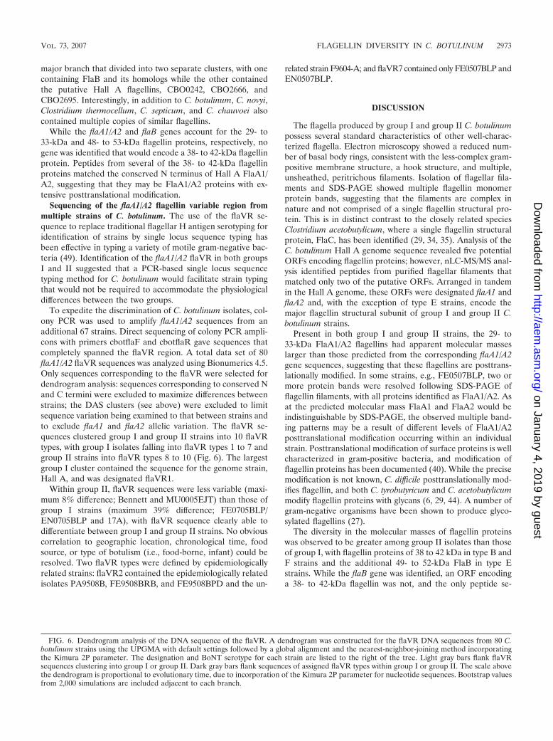
To expedite the discrimination of C. botulinum isolates, col-ony PCR was used to amplify flaA1/A2 sequences from anadditional 67 strains. Direct sequencing of colony PCR ampli-cons with primers cbotflaF and cbotflaR gave sequences thatcompletely spanned the flaVR region. A total data set of 80flaA1/A2 flaVR sequences was analyzed using Bionumerics 4.5.Only sequences corresponding to the flaVR were selected fordendrogram analysis: sequences corresponding to conserved Nand C termini were excluded to maximize differences betweenstrains; the DAS clusters (see above) were excluded to limitsequence variation being examined to that between strains andto exclude flaA1 and flaA2 allelic variation. The flaVR se-quences clustered group I and group II strains into 10 flaVRtypes, with group I isolates falling into flaVR types 1 to 7 andgroup II strains into flaVR types 8 to 10 (Fig. 6). The largestgroup I cluster contained the sequence for the genome strain,Hall A, and was designated flaVR1.
Within group II, flaVR sequences were less variable (maxi-mum 8% difference; Bennett and MU0005EJT) than those ofgroup I strains (maximum 39% difference; FE0705BLP/EN0705BLP and 17A), with flaVR sequence clearly able todifferentiate between group I and group II strains. No obviouscorrelation to geographic location, chronological time, foodsource, or type of botulism (i.e., food-borne, infant) could beresolved. Two flaVR types were defined by epidemiologicallyrelated strains: flaVR2 contained the epidemiologically relatedisolates PA9508B, FE9508BRB, and FE9508BPD and the un-
related strain F9604-A; and flaVR7 contained only FE0507BLP andEN0507BLP.
DISCUSSION
The flagella produced by group I and group II C. botulinumpossess several standard characteristics of other well-charac-terized flagella. Electron microscopy showed a reduced num-ber of basal body rings, consistent with the less-complex gram-positive membrane structure, a hook structure, and multiple,unsheathed, peritrichous filaments. Isolation of flagellar fila-ments and SDS-PAGE showed multiple flagellin monomerprotein bands, suggesting that the filaments are complex innature and not comprised of a single flagellin structural pro-tein. This is in distinct contrast to the closely related speciesClostridium acetobutylicum, where a single flagellin structuralprotein, FlaC, has been identified (29, 34, 35). Analysis of theC. botulinum Hall A genome sequence revealed five potentialORFs encoding flagellin proteins; however, nLC-MS/MS anal-ysis identified peptides from purified flagellar filaments thatmatched only two of the putative ORFs. Arranged in tandemin the Hall A genome, these ORFs were designated flaA1 andflaA2 and, with the exception of type E strains, encode themajor flagellin structural subunit of group I and group II C.botulinum strains.
Present in both group I and group II strains, the 29- to33-kDa FlaA1/A2 flagellins had apparent molecular masseslarger than those predicted from the corresponding flaA1/A2gene sequences, suggesting that these flagellins are posttrans-lationally modified. In some strains, e.g., FE0507BLP, two ormore protein bands were resolved following SDS-PAGE offlagellin filaments, with all proteins identified as FlaA1/A2. Asat the predicted molecular mass FlaA1 and FlaA2 would beindistinguishable by SDS-PAGE, the observed multiple band-ing patterns may be a result of different levels of FlaA1/A2posttranslational modification occurring within an individualstrain. Posttranslational modification of surface proteins is wellcharacterized in gram-positive bacteria, and modification offlagellin proteins has been documented (40). While the precisemodification is not known, C. difficile posttranslationally mod-ifies flagellin, and both C. tyrobutyricum and C. acetobutylicummodify flagellin proteins with glycans (6, 29, 44). A number ofgram-negative organisms have been shown to produce glyco-sylated flagellins (27).
The diversity in the molecular masses of flagellin proteinswas observed to be greater among group II isolates than thoseof group I, with flagellin proteins of 38 to 42 kDa in type B andF strains and the additional 49- to 52-kDa FlaB in type Estrains. While the flaB gene was identified, an ORF encodinga 38- to 42-kDa flagellin was not, and the only peptide se-
FIG. 6. Dendrogram analysis of the DNA sequence of the flaVR. A dendrogram was constructed for the flaVR DNA sequences from 80 C.botulinum strains using the UPGMA with default settings followed by a global alignment and the nearest-neighbor-joining method incorporatingthe Kimura 2P parameter. The designation and BoNT serotype for each strain are listed to the right of the tree. Light gray bars flank flaVRsequences clustering into group I or group II. Dark gray bars flank sequences of assigned flaVR types within group I or group II. The scale abovethe dendrogram is proportional to evolutionary time, due to incorporation of the Kimura 2P parameter for nucleotide sequences. Bootstrap valuesfrom 2,000 simulations are included adjacent to each branch.
VOL. 73, 2007 FLAGELLIN DIVERSITY IN C. BOTULINUM 2973
on January 4, 2019 by guesthttp://aem
.asm.org/
Dow
nloaded from

quences obtained from the 38- to 42-kDa flagellins matchedFlaA1/A2. A distinct genetic locus, that is not represented inthe Hall A genome and cannot be amplified with the flaA1/A2primers, could be present in group II strains and contain se-quence encoding conserved peptides common to both thisORF and the FlaA1/A2 proteins. Conversely, the approxi-mately 12-kDa difference in mass could be a unique and largerform of posttranslational modification of the FlaA1/A2 pro-teins, distinct from that of the 28- to 32-kDa FlaA1/A2. C.difficile and C. acetobutylicum flagellins are significantly largerthan their predicted molecular weights, with posttranslationalmodifications of approximately 8 kDa and 12.5 kDa, respec-tively (29, 44).
Within group II, type E strains were distinct in that theirflagellar filaments contained primarily the large and apparentlyunmodified flagellin, FlaB. A recent study identified large (65-kDa) and small (33-kDa) flagellins that comprised the thickand thin filaments, respectively, of Bradyrhizobium japonicumflagella (20). TEM micrographs of several type E strains didnot consistently show morphologically distinct filaments, sug-gesting that FlaA1/A2 and FlaB may be found in the samefilament. Type E strains possess other unique characteristics,including the mechanism of regulation of the type E BoNT (9).
The majority of existing tools for genetic characterization ofC. botulinum are able to discriminate to the strain level forgroup II but are limited for group I strains (26). Typing strainsby sequencing of the neurotoxin gene still clusters large num-bers of strains together, with maximum differences betweensubtypes not exceeding 8% for group I strains (18). Sequencingof type A neurotoxin genes showed that 90% of serotype Astrains are of subtype A1, with divisions in this subclusterrequiring resolution of a single base pair change in approxi-mately 3.8 kb of gene sequence (18). 16S sequences withingroup I are also highly identical (8). The maximum diversity ingroup I flaVR sequence was 39% over a region of 400 bp,providing a level of strain-to-strain discrimination not previ-ously possible within this group. This large region of diversityin the flaVR nucleotide sequence is advantageous for estab-lishing epidemiological relationships as the likelihood that twostrains will have 100% identical flaVR sequences is low, singlebase pair errors related to a DNA polymerase error duringamplification or gene sequencing will not have a significantimpact, and lastly, all sequence required for typing can beobtained in a single sequencing reaction. It must, however, beestablished to what overall extent flaA varies among C. botu-linum strains, as the ultimate usefulness of this typing will beinfluenced by the total amount of variation at the flaA locus.For example, as strains FE0507BLP and EN0507BLP are theonly two strains that possess flagellin genes in a deep-branch-ing cluster, they are likely related. However, with many strainsin the flaA type I cluster (HallA, 62A, and FE0303IYO),strains within this flaVR group are obviously not all from thesame epidemiological source. PCR-based detection and typingbased on the nucleotide sequence of the flagellin gene hasallowed differentiation between closely related strains for C.jejuni, Borrelia spp., P. aeruginosa, and members of the Burk-holderia cepacia complex (48, 49).
In group I, flaVR typing placed strains of serotypes A, B,and in some cases F in the same cluster. This is not surprisinggiven that horizontal gene transfer of BoNT and associated
genes has likely scattered these genes into various geneticbackgrounds, including other clostridia species (10, 26, 37).With the mobility and extensive sequence conservation of theBoNT genes, inferring phylogeny based on the C. botulinumflagellin gene sequence could be preferable, with nucleotidechanges in the variable region representing a passage of timeover which the mutations have been accumulated. As tandemflagellin genes are subject to recombination, the stability of theflaA sequence would need to be established, although in con-trast to the flagellins in Campylobacter, conservation of DNAsequence between flaA1 and flaA2 suggests that recombinationmight not be noticeable (15). Since identification of strains byeither flagellin or BoNT sequencing appears uncorrelated, se-quencing of BoNT genes and flaA1/A2 would rapidly differen-tiate isolates of the same BoNT serotype. This approach wouldbe analogous to Escherichia coli O and H antigen typing.
Within group II, flaVR sequences were less diverse thanthose within group I, with three group II flaVR types looselyassociated with BoNT serotype, and flaVR sequence differingonly by a maximum of 8% at the nucleotide level. PFGEanalysis of the group II strains used in this study resolvedsignificant genetic differences while still clustering group IIserotype B and F strains separately (25), showing that forgroup II strains, other methods may be preferred for phyloge-netic analysis. Sequencing of the flaVR region did clearly sep-arate group I strains from those of group II, suggesting thatrapid methods such as restriction fragment length polymor-phism, based on group-specific sequences, could first identifythe group of a strain and thus determine the most suitabledownstream method (flaVR sequencing, PFGE) for genotypicanalysis.
While gene sequence analysis based on BoNT requires mul-tiple PCR conditions or previous knowledge of a strain’s sero-type, the flaA gene, by virtue of DNA sequence conservedacross groups I and II, can be amplified in a single PCR froman uncharacterized strain of C. botulinum. The ability of theflaA1/A2 primers to detect group III and group IV C. botuli-num strains and other clostridia species is currently being in-vestigated. Analysis of flagellin proteins has shown that thisdiversity is further amplified at the protein level, where eachstrain produces flagellins in different amounts and with differ-ent apparent molecular masses. It thus appears that while theflagellin genes can be used to differentiate among strains of C.botulinum, the potential to further discriminate among isolatesbased on posttranslational modification of flagellin proteinsremains to be investigated.
ACKNOWLEDGMENTS
We thank Greg Sanders for assistance with electron microscopy andNadia Mykytczuk for preliminary flagellin peptide identification.
This work was supported by project CRTI-02-0091TA of the De-fense Research and Development Canada CBRN Research and Tech-nology Initiative (J.W.A. and S.M.L.).
REFERENCES
1. Andersen, A. A. 1951. A rapid plate method of counting spores of Clostridiumbotulinum. J. Bacteriol. 62:425–432.
2. Anellis, A., and R. B. Koch. 1962. Comparative resistance of strains ofClostridium botulinum to gamma rays. Appl. Microbiol. 10:326–330.
3. Arnold, F., L. Bedouet, P. Batina, G. Robreau, F. Talbot, P. Lecher, and R.Malcoste. 1998. Biochemical and immunological analyses of the flagellin ofClostridium tyrobutyricum ATCC 25755. Microbiol. Immunol. 42:23–31.
2974 PAUL ET AL. APPL. ENVIRON. MICROBIOL.
on January 4, 2019 by guesthttp://aem
.asm.org/
Dow
nloaded from

4. Austin, J. W. 2001. Clostridium botulinum, p. 329–349. In M. P. Doyle, R. L.Beuchat, and T. J. Montville (ed.), Food microbiology: fundamentals andfrontiers. ASM Press, Washington, DC.
5. Bardy, S. L., S. Y. M. Ng, and K. F. Jarrell. 2003. Prokaryotic motilitystructures. Microbiology 149:295–304.
6. Bedouet, L., F. Arnold, G. Robreau, P. Batina, F. Talbot, and A. Binet. 1998.Evidence for an heterogeneous glycosylation of the Clostridium tyrobutyricumATCC 25755 flagellin. Microbios 94:183–192.
7. Blocker, A., K. Komoriya, and S.-I. Aizawa. 2003. Type III secretion systemsand bacterial flagella: insights into their function from structural similarities.Proc. Natl. Acad. Sci. USA 100:3027–3030.
8. Collins, M. D., and A. K. East. 1998. Phylogeny and taxonomy of the food-borne pathogen Clostridium botulinum and its neurotoxins. J Appl. Micro-biol. 84:5–17.
9. Couesnon, A., S. Raffestin, and M. R. Popoff. 2006. Expression of botulinumneurotoxins A and E, and associated non-toxin genes, during the transitionphase and stability at high temperature: analysis by quantitative reversetranscription-PCR. Microbiology 152:759–770.
10. Dineen, S. S., M. Bradshaw, and E. A. Johnson. 2003. Neurotoxin geneclusters in Clostridium botulinum type A strains: sequence comparison andevolutionary implications. Curr. Microbiol. 46:345–352.
11. Duff, J. T., G. G. Wright, J. Klerer, D. E. Morre, and R. H. Bibler. 1957.Studies on immunity to toxins of Clostridium botulinum. J. Bacteriol. 73:42–47.
12. Fields, P. I., K. Blom, H. J. Hughes, L. O. Helsel, P. Feng, and B. Swami-nathan. 1997. Molecular characterization of the gene encoding H antigen inEscherichia coli and development of a PCR-restriction fragment length poly-morphism test for identification of E. coli O157:H7 and O157:NM. J. Clin.Microbiol. 35:1066–1070.
13. Fredrick, K., T. Caramori, Y. Chen, A. Galizzi, and J. D. Helmann. 1995.Promoter architecture in the flagellar regulon of Bacillus subtilis: high-levelexpression of flagellin by the sigmaD RNA polymerase requires an upstreampromoter element. Proc. Natl. Acad. Sci. USA 92:2582–2586.
14. Ge, Y., I. G. Old, I. Saint Girons, and N. W. Charon. 1997. Molecularcharacterization of a large Borrelia burgdorferi motility operon which is ini-tiated by a consensus 70 promoter. J. Bacteriol. 179:2289–2299.
15. Harrington, C. S., F. M. Thomson-Carter, and P. E. Carter. 1997. Evidencefor recombination in the flagellin locus of Campylobacter jejuni: implicationsfor the flagellin gene typing scheme. J. Clin. Microbiol. 35:2386–2392.
16. Helmann, J. D. 1991. Alternative sigma factors and the regulation of flagellargene expression. Mol. Microbiol. 5:2875–2882.
17. Hetzel, M., M. Brock, T. Selmer, A. J. Pierik, B. T. Golding, and W. Buckel.2003. Acryloyl-CoA reductase from Clostridium propionicum. An enzymecomplex of propionyl-CoA dehydrogenase and electron-transferring flavo-protein. Eur. J. Biochem. 270:902–910.
18. Hill, K. K., T. J. Smith, C. H. Helma, L. O. Ticknor, B. T. Foley, R. T.Svensson, J. L. Brown, E. A. Johnson, L. A. Smith, R. T. Okinaka, P. J.Jackson, and J. D. Marks. 2007. Genetic diversity among botulinum neuro-toxin-producing clostridial strains. J. Bacteriol. 189:818–832.
19. Josenhans, C., and S. Suerbaum. 2002. The role of motility as a virulencefactor in bacteria. Int. J. Med. Microbiol. 291:605–614.
20. Kanbe, M., J. Yagasaki, S. Zehner, M. Gottfert, and S. Aizawa. 2007. Char-acterization of two sets of subpolar flagella in Bradyrhizobium japonicum. J.Bacteriol. 189:1083–1089.
21. Keto-Timonen, R., M. Nevas, and H. Korkeala. 2005. Efficient DNA finger-printing of Clostridium botulinum types A, B, E, and F by amplified fragmentlength polymorphism analysis. Appl. Environ. Microbiol. 71:1148–1154.
22. Kojima, A., K. Amimoto, T. Ohgitani, and Y. Tamura. 1999. Characteriza-tion of flagellin from Clostridium chauvoei. Vet. Microbiol. 67:231–237.
23. Kojima, A., I. Uchida, T. Sekizaki, Y. Sasaki, Y. Ogikubo, M. Kijima, and Y.Tamura. 2000. Cloning and expression of a gene encoding the flagellin ofClostridium chauvoei. Vet. Microbiol. 76:359–372.
24. Kushnir, E. D., T. M. Breen, and S. S. Paikina. 1937. Sources of infection ofsturgeons (red fish) with Bacillus botulinum. Zh. Mikrobiol. Epidemiol. Im-munobiol. 19:80–85.
25. Leclair, D., F. Pagotto, J. M. Farber, B. Cadieux, and J. W. Austin. 2006.Comparison of DNA fingerprinting methods for use in investigation of typeE botulism outbreaks in the Canadian Arctic. J. Clin. Microbiol. 44:1635–1644.
26. Lindstrom, M., and H. Korkeala. 2006. Laboratory diagnostics of botulism.Clin. Microbiol. Rev. 19:298–314.
27. Logan, S. M. 2006. Flagellar glycosylation—a new component of the motilityrepertoire? Microbiology 152:1249–1262.
28. Lund, B. M., and M. W. Peck. 2000. Clostridium botulinum, p. 1057–1109. InB. M. Lund, T. C. Baird-Parker, and G. W. Gould (ed.), The microbiologicalsafety and quality of food. Aspen Publishers Inc., Gaithersburg, MD.
29. Lyristis, M., Z. L. Boynton, D. Petersen, Z. Kan, G. N. Bennett, and F. B.Rudolph. 2000. Cloning, sequencing, and characterization of the gene en-coding flagellin, flaC, and the post-translational modification of flagellin,FlaC, from Clostridium acetobutylicum ATCC824. Anaerobe 6:69–79.
30. Macnab, R. M. 2003. How bacteria assemble flagella. Annu. Rev. Microbiol.57:77–100.
31. Macnab, R. M. 2004. Type III flagellar protein export and flagellar assembly:protein export/secretion in bacteria. Biochim. Biophys. Acta 1694:207–217.
32. Mirel, D. B., and M. J. Chamberlin. 1989. The Bacillus subtilis flagellin gene(hag) is transcribed by the 28 form of RNA polymerase. J. Bacteriol. 171:3095–3101.
33. Nevas, M., M. Lindstrom, S. Hielm, K. J. Bjorkroth, M. W. Peck, and H.Korkeala. 2005. Diversity of proteolytic Clostridium botulinum strains, de-termined by a pulsed-field gel electrophoresis approach. Appl. Environ Mi-crobiol. 71:1311–1317.
34. Paredes, C. J., K. V. Alsaker, and E. T. Papoutsakis. 2005. A comparativegenomic view of clostridial sporulation and physiology. Nat. Rev. Microbiol.3:969–978.
35. Paredes, C. J., I. Rigoutsos, and E. T. Papoutsakis. 2004. Transcriptionalorganization of the Clostridium acetobutylicum genome. Nucleic Acids Res.32:1973–1981.
36. Pituch, H., P. Obuch-Woszczatynski, N. van den Braak, A. van Belkum, M.Kujawa, M. Luczak, and F. Meisel-Mikolajczyk. 2002. Variable flagellaexpression among clonal toxin A�/B� Clostridium difficile strains with highlyhomogeneous flagellin genes. Clin. Microbiol. Infect. 8:187–188.
37. Pourshaban, M., G. Franciosa, L. Fenicia, and P. Aureli. 2002. Taxonomicidentity of type E botulinum toxin-producing Clostridium butyricum strainsby sequencing of a short 16S rDNA region. FEMS Microbiol. Lett. 214:119–125.
38. Sambrook, J., E. F. Fritsch, and T. Maniatis. 1989. Molecular cloning: alaboratory manual. Cold Spring Harbor Laboratory Press, Cold SpringHarbor, NY.
39. Sasaki, Y., A. Kojima, H. Aoki, Y. Ogikubo, N. Takikawa, and Y. Tamura.2002. Phylogenetic analysis of PCR detection of Clostridium chauvoei, Clos-tridium haemolyticum, Clostridium novyi types A and B, and Clostridiumsepticum based on the flagellin gene. Vet. Microbiol. 86:257–267.
40. Schaffer, C., and P. Messner. 2004. Surface-layer glycoproteins: an examplefor the diversity of bacterial glycosylation with promising impacts on nano-biotechnology. Glycobiology 14:31R–42R.
41. Schoenhals, G., and C. Whitfield. 1993. Comparative analysis of flagellinsequences from Escherichia coli strains possessing serologically distinctflagellar filaments with a shared complex surface pattern. J. Bacteriol. 175:5395–5402.
42. Staden, R., K. F. Beal, and J. K. Bonfield. 2000. The Staden package, 1998.Methods Mol. Biol. 132:115–130.
43. Tasteyre, A., M. C. Barc, A. Collignon, H. Boureau, and T. Karjalainen.2001. Role of FliC and FliD flagellar proteins of Clostridium difficile inadherence and gut colonization. Infect. Immun. 69:7937–7940.
44. Tasteyre, A., M. C. Barc, T. Karjalainen, P. Dodson, S. Hyde, P. Bourlioux,and P. Borriello. 2000. A Clostridium difficile gene encoding flagellin. Mi-crobiology 146:957–966.
45. Tasteyre, A., T. Karjalainen, V. Avesani, M. Delmee, A. Collignon, P. Bour-lioux, and M. C. Barc. 2000. Phenotypic and genotypic diversity of theflagellin gene (fliC) among Clostridium difficile isolates from different sero-groups. J. Clin. Microbiol. 38:3179–3186.
46. Tasteyre, A., T. Karjalainen, V. Avesani, M. Delmee, A. Collignon, P. Bour-lioux, and M. C. Barc. 2001. Molecular characterization of fliD gene encod-ing flagellar cap and its expression among Clostridium difficile isolates fromdifferent serogroups. J. Clin. Microbiol. 39:1178–1183.
47. Twine, S. M., N. C. S. Mykytczuk, M. Petit, T.-L. Tremblay, P. Lanthier,J. W. Conlan, and J. F. Kelly. 2005. Francisella tularensis proteome: lowlevels of ASB-14 facilitate the visualization of membrane proteins in totalprotein extracts. J. Proteome Res. 4:1848–1854.
48. Winstanley, C. 2003. Improved flagellin genotyping in the Burkholderia cepaciacomplex. FEMS Microbiol. Lett. 229:9–14.
49. Winstanley, C., and J. A. Morgan. 1997. The bacterial flagellin gene as abiomarker for detection, population genetics and epidemiological analysis.Microbiology 143:3071–3084.
VOL. 73, 2007 FLAGELLIN DIVERSITY IN C. BOTULINUM 2975
on January 4, 2019 by guesthttp://aem
.asm.org/
Dow
nloaded from