Embed Size (px)
Citation preview
A Comprehensive Toolkit of Plant Cell WallGlycan-Directed Monoclonal Antibodies1[W][OA]
Sivakumar Pattathil, Utku Avci, David Baldwin, Alton G. Swennes, Janelle A. McGill, Zoe Popper2,Tracey Bootten3, Anathea Albert4, Ruth H. Davis, Chakravarthy Chennareddy, Ruihua Dong,Beth O’Shea5, Ray Rossi6, Christine Leoff, Glenn Freshour7, Rajesh Narra8, Malcolm O’Neil,William S. York, and Michael G. Hahn*
Complex Carbohydrate Research Center (S.P., U.A., D.B., A.G.S., J.A.M., Z.P., T.B., A.A., C.L., G.F., R.N., M.O.,W.S.Y., M.G.H.), Monoclonal Antibody Facility, College of Veterinary Medicine (R.H.D., C.C., R.D., B.O., R.R.),Department of Biochemistry and Molecular Biology (W.S.Y.), and Department of Plant Biology (M.G.H.),University of Georgia, Athens, Georgia 30602
A collection of 130 new plant cell wall glycan-directed monoclonal antibodies (mAbs) was generated with the aim offacilitating in-depth analysis of cell wall glycans. An enzyme-linked immunosorbent assay-based screen against a diversepanel of 54 plant polysaccharides was used to characterize the binding patterns of these new mAbs, together with 50 otherpreviously generated mAbs, against plant cell wall glycans. Hierarchical clustering analysis was used to group these mAbsbased on the polysaccharide recognition patterns observed. The mAb groupings in the resulting cladogram were furtherverified by immunolocalization studies in Arabidopsis (Arabidopsis thaliana) stems. The mAbs could be resolved into 19 cladesof antibodies that recognize distinct epitopes present on all major classes of plant cell wall glycans, including arabinogalactans(both protein- and polysaccharide-linked), pectins (homogalacturonan, rhamnogalacturonan I), xyloglucans, xylans, mannans,and glucans. In most cases, multiple subclades of antibodies were observed to bind to each glycan class, suggesting that themAbs in these subgroups recognize distinct epitopes present on the cell wall glycans. The epitopes recognized by many of themAbs in the toolkit, particularly those recognizing arabinose- and/or galactose-containing structures, are present on more thanone glycan class, consistent with the known structural diversity and complexity of plant cell wall glycans. Thus, these cell wallglycan-directed mAbs should be viewed and utilized as epitope-specific, rather than polymer-specific, probes. The currentworld-wide toolkit of approximately 180 glycan-directed antibodies from various laboratories provides a large and diverse setof probes for studies of plant cell wall structure, function, dynamics, and biosynthesis.
Cell walls play important roles in the structure, phys-iology, growth, and development of plants (Carpitaand Gibeaut, 1993). Plant cell wall materials are alsoimportant sources of human and animal nutrition,natural textile fibers, paper and wood products, andraw materials for biofuel production (Somerville,2007). Many genes thought to be responsible for plantwall biosynthesis and modification have been identi-fied (Burton et al., 2005; Lerouxel et al., 2006; Mohnenet al., 2008), and 15% of the Arabidopsis (Arabidopsisthaliana) genome is likely devoted to these functions(Carpita et al., 2001). However, phenotypic analysis inplants carrying cell wall-related mutations has provenparticularly difficult. First, cell wall-related genes areoften expressed differentially and at low levelsbetween cells of different tissues (Sarria et al., 2001).Also, plants have compensatory mechanisms to main-tain wall function in the absence of a particular gene(Somerville et al., 2004). Thus, novel tools and ap-proaches are needed to characterize wall structuresand the genes responsible for their synthesis andmodification.
Monoclonal antibodies (mAbs) developed againstcell wall polymers have emerged as an important toolfor the study of plant cell wall structure and function
1 This work was supported by the National Science FoundationPlant Genome Program (grant no. DBI–0421683).
2 Present address: Botany and Plant Sciences, School of NaturalSciences, National University of Ireland-Galway, Galway, Ireland.
3 Present address: Industrial Research Limited, P.O. Box 31-310,Lower Hutt 5010, New Zealand.
4 Present address: National Institute of Water and AtmosphericResearch Limited, Gate 10 Silverdale Road, Hillcrest, Hamilton 3216,New Zealand.
5 Present address: Department of Foods and Nutrition, DawsonHall, University of Georgia, Athens, GA 30602.
6 Present address: Abeome Corporation, Georgia BiobusinessCenter, College Station Road, University of Georgia, Athens, GA30602.
7 Present address: 907 14th Avenue E, Seattle, WA 98112.8 Present address: E*Trade Financial, 4005Windward Plaza Drive,
Alpharetta, GA 30005.* Corresponding author; e-mail [email protected] author responsible for distribution of materials integral to the
findings presented in this article in accordance with the policydescribed in the Instructions for Authors (www.plantphysiol.org) is:Michael G. Hahn ([email protected]).
[W] The online version of this article contains Web-only data.[OA] Open Access articles can be viewed online without a sub-
scription.www.plantphysiol.org/cgi/doi/10.1104/pp.109.151985
514 Plant Physiology�, June 2010, Vol. 153, pp. 514–525, www.plantphysiol.org � 2010 American Society of Plant Biologists
Dow
nloaded from https://academ
ic.oup.com/plphys/article/153/2/514/6109425 by guest on 16 February 2022
(Knox, 2008). Previous studies have utilized mAbs thatbind epitopes present on rhamnogalacturonan I (RG-I;Freshour et al., 1996; Jones et al., 1997; Willats et al.,1998; McCartney et al., 2000; Clausen et al., 2004;Altaner et al., 2007), homogalacturonan (Willats et al.,2001; Clausen et al., 2003), xylogalacturonan (Willatset al., 2004), xylans and arabinoxylans (McCartneyet al., 2005), xyloglucan (Freshour et al., 1996, 2003;Marcus et al., 2008), arabinogalactan(protein) (Pennellet al., 1991; Puhlmann et al., 1994; Dolan et al., 1995;Smallwood et al., 1996), and extensins (Smallwoodet al., 1995) to localize these epitopes in plant cells andtissues. In addition, mAbs have been used to charac-terize plants carrying mutations in genes thought to beassociated with cell wall biosynthesis and metabolism(Orfila et al., 2001; Seifert, 2004; Persson et al., 2007;Cavalier et al., 2008; Zabotina et al., 2008). Despite theirutility, the available set of mAbs against carbohydratestructures is relatively small given the structural com-plexity of wall polymers (Ridley et al., 2001; O’Neilland York, 2003), and knowledge of their epitopespecificity is limited. Thus, additional mAbs specificto diverse epitope structures and methods for rapidepitope characterization are needed (Somerville et al.,2004).Here, we report the generation of 130 new mAbs
that bind to diverse epitopes present on a broadspectrum of plant cell wall glycans. In addition, ap-proximately 50 previously reported or generatedmAbs were included in the ELISA-based screensused to group the antibodies according to their bind-ing patterns against a diverse panel of 54 polysaccha-rides. The resulting ELISA data were analyzed byhierarchical clustering to illustrate the relationshipsbetween the available mAbs. Nineteen groups ofmAbs were identified from the clustering analysis.Some initial information regarding possible epitopesrecognized by some of these antibodies could beinferred from the clustering analysis.
RESULTS
Immobilization of Plant Polysaccharides for ELISA
Eight commercially available 96-well plates weretested to determine their suitability for plant cell wallpolysaccharide-based ELISAs. These plates weretested simultaneously using a standard ELISA proto-col and 12 mAb/polysaccharide pairs: CCRC-M1/sycamore xyloglucan, CCRC-M2/gum karaya, CCRC-M7/sycamore pectic polysaccharides, CCRC-M10/mustard seed mucilage, CCRC-M14/ArabidopsisRG-I, CCRC-M16/soybean RG-I, CCRC-M30/Arabi-dopsis seed mucilage, CCRC-M58/tamarind xylo-glucan, PN16.4B4/gum arabic, JIM5/citrus pectin,JIM13/gum ghatti, and LM10/4-O-methylglucuro-noxylan. Of the ELISA plates tested, Costar 3598gave the highest mean signal across all of the mAb/polysaccharide pairs tested, showing a mean optical
density (OD) of 0.551 6 0.150 (Supplemental Fig. S1).Other plate types, including Immulon 1B (0.340 60.131), Immulon 4HB (0.4426 0.173), Immulon 2HB (aplate that we had used previously [Puhlmann et al.,1994]; 0.422 6 0.161), Nunc 269620 (0.425 6 0.150),Nunc 439454 (0.454 6 0.154), Costar 2507 (0.335 60.141), and Costar 3590 (0.435 6 0.118), gave lowermean signals. Costar 3598 plates also displayed thehighest minimum signal (minimum OD = 0.316) forthe mAb/polysaccharide pairs tested. Immulon 1B(0.101), Immulon 4HB (0.213), Immulon 2HB (0.195),Nunc 269620 (0.179), Nunc 439454 (0.265), Costar 2507(0.185), and Costar 3590 (0.250) had lower thresholdOD values. Based on these results, Costar 3598 plateswere used to carry out all subsequent ELISAs.
Data Correlation Analysis Emphasizes Reproducibilityof ELISAs
ELISA analyses were used to test mAb-bindingspecificities against a diverse panel of plant polysac-charide preparations. The reproducibility of the ELISAdata pattern for each antibody was examined bygenerating a correlation heat map (Supplemental Ma-terials and Methods S1). This correlation heat mapanalysis was done using the data obtained from sixreplicate experiments involving ELISA screening of 41antibodies against a panel of diverse polysaccharides.In the correlation heat map, the value (color) of eachsquare corresponds to the correlation of the ELISAresponse vector for one mAb in one experiment to theELISA response vector for each mAb in another ex-periment or group of experiments. Perfect reproduc-ibility corresponds to a heat map with all diagonalelements equal to 1.0 (brightest yellow) and perfectsymmetry about the diagonal. (This would be theresult if a data set were compared with itself, as shownin Fig. 1A). A second heat map, shown in Figure 1B,depicts the correlation of a randomly selected ELISApanel test replicate with the average of six replicates.In this case, each diagonal heat map element correlatesthe response pattern of a specific mAb in the selectedexperiment with the average ELISA response patternfor that mAb. Almost all of the correlation coefficientswere greater than 0.98 (Supplemental Table S3). Eachoff-diagonal heat map element in Figure 1B shows thecorrelation of the ELISA response pattern of a mAb inthe selected experiment to the mean ELISA responsepattern of a different mAb. The presence of very fewdeviations from symmetry about the diagonal in thecorrelation heat map indicates that the ELISAs arehighly reproducible. The reproducibility of ELISAs isalso emphasized by the significant resemblance of theexperimental correlation heat map (Fig. 1B) to theautocorrelation heat map (Fig. 1A).
The correlation heat maps (Fig. 1) also group anti-bodies that show similar binding patterns to the panelof polysaccharides. These groups (clades) of anti-bodies are highlighted by the coloring of the correla-tion heat map (from black to bright yellow; Fig. 1). For
Plant Cell Wall Glycan-Directed Monoclonal Antibody Toolkit
Plant Physiol. Vol. 153, 2010 515
Dow
nloaded from https://academ
ic.oup.com/plphys/article/153/2/514/6109425 by guest on 16 February 2022
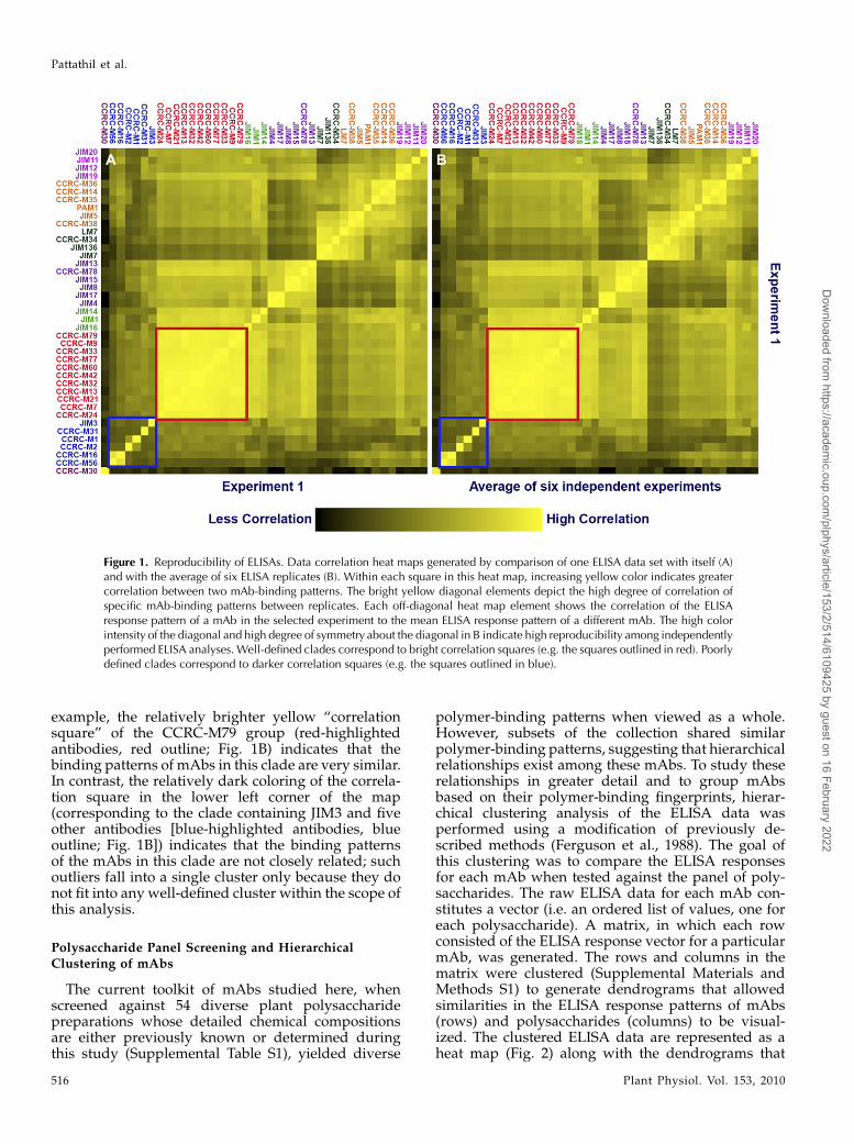
example, the relatively brighter yellow “correlationsquare” of the CCRC-M79 group (red-highlightedantibodies, red outline; Fig. 1B) indicates that thebinding patterns of mAbs in this clade are very similar.In contrast, the relatively dark coloring of the correla-tion square in the lower left corner of the map(corresponding to the clade containing JIM3 and fiveother antibodies [blue-highlighted antibodies, blueoutline; Fig. 1B]) indicates that the binding patternsof the mAbs in this clade are not closely related; suchoutliers fall into a single cluster only because they donot fit into any well-defined cluster within the scope ofthis analysis.
Polysaccharide Panel Screening and HierarchicalClustering of mAbs
The current toolkit of mAbs studied here, whenscreened against 54 diverse plant polysaccharidepreparations whose detailed chemical compositionsare either previously known or determined duringthis study (Supplemental Table S1), yielded diverse
polymer-binding patterns when viewed as a whole.However, subsets of the collection shared similarpolymer-binding patterns, suggesting that hierarchicalrelationships exist among these mAbs. To study theserelationships in greater detail and to group mAbsbased on their polymer-binding fingerprints, hierar-chical clustering analysis of the ELISA data wasperformed using a modification of previously de-scribed methods (Ferguson et al., 1988). The goal ofthis clustering was to compare the ELISA responsesfor each mAb when tested against the panel of poly-saccharides. The raw ELISA data for each mAb con-stitutes a vector (i.e. an ordered list of values, one foreach polysaccharide). A matrix, in which each rowconsisted of the ELISA response vector for a particularmAb, was generated. The rows and columns in thematrix were clustered (Supplemental Materials andMethods S1) to generate dendrograms that allowedsimilarities in the ELISA response patterns of mAbs(rows) and polysaccharides (columns) to be visual-ized. The clustered ELISA data are represented as aheat map (Fig. 2) along with the dendrograms that
Figure 1. Reproducibility of ELISAs. Data correlation heat maps generated by comparison of one ELISA data set with itself (A)and with the average of six ELISA replicates (B). Within each square in this heat map, increasing yellow color indicates greatercorrelation between two mAb-binding patterns. The bright yellow diagonal elements depict the high degree of correlation ofspecific mAb-binding patterns between replicates. Each off-diagonal heat map element shows the correlation of the ELISAresponse pattern of a mAb in the selected experiment to the mean ELISA response pattern of a different mAb. The high colorintensity of the diagonal and high degree of symmetry about the diagonal in B indicate high reproducibility among independentlyperformed ELISA analyses. Well-defined clades correspond to bright correlation squares (e.g. the squares outlined in red). Poorlydefined clades correspond to darker correlation squares (e.g. the squares outlined in blue).
Pattathil et al.
516 Plant Physiol. Vol. 153, 2010
Dow
nloaded from https://academ
ic.oup.com/plphys/article/153/2/514/6109425 by guest on 16 February 2022
were used to order the data. The color of each cell inthe heat map represents the ELISA response of aparticular mAb when tested against a particular poly-saccharide.Dendrograms generated by our initial clustering
experiments, performed essentially as described pre-viously (Ferguson et al., 1988), were often in disagree-ment with groupings obtained by manual comparisonof the ELISA responses. We showed that this initialapproach, which is based on using the Pearson corre-
lation coefficient as the distance metric for clustering,can lead to dendrograms that imply close associationsbetween dissimilar patterns. Therefore, we used adifferent approach in which the inverse cosine of thedot product of each pair of ELISA response vectorswas used as the distance metric for clustering. This issimilar to the use of Pearson correlation coefficients inthat it builds dendrograms using response patternsrather than absolute responses. When applied to ourELISA data, this new approach produced dendro-
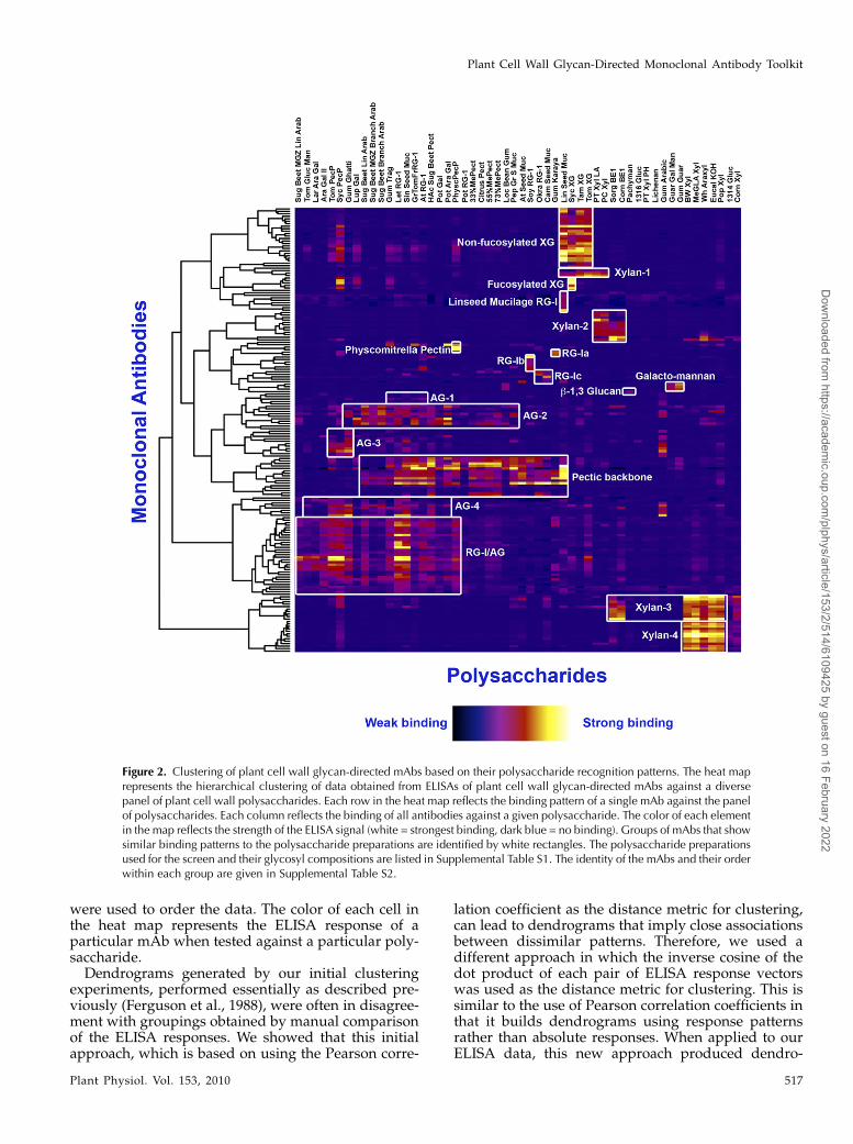
Figure 2. Clustering of plant cell wall glycan-directed mAbs based on their polysaccharide recognition patterns. The heat maprepresents the hierarchical clustering of data obtained from ELISAs of plant cell wall glycan-directed mAbs against a diversepanel of plant cell wall polysaccharides. Each row in the heat map reflects the binding pattern of a single mAb against the panelof polysaccharides. Each column reflects the binding of all antibodies against a given polysaccharide. The color of each elementin the map reflects the strength of the ELISA signal (white = strongest binding, dark blue = no binding). Groups of mAbs that showsimilar binding patterns to the polysaccharide preparations are identified by white rectangles. The polysaccharide preparationsused for the screen and their glycosyl compositions are listed in Supplemental Table S1. The identity of the mAbs and their orderwithin each group are given in Supplemental Table S2.
Plant Cell Wall Glycan-Directed Monoclonal Antibody Toolkit
Plant Physiol. Vol. 153, 2010 517
Dow
nloaded from https://academ
ic.oup.com/plphys/article/153/2/514/6109425 by guest on 16 February 2022
grams that were more consistent with the mAb group-ings obtained by manual comparison of the ELISAresponses.
Encouraged by these results, we used the R lan-guage (R Development Core Team, 2006) to develop asoftware application that uses the alternative ap-proach. Given the ELISA response data for a collectionof mAbs against the panel of polysaccharides, thesoftware provides dendrograms for the mAbs andthe polysaccharides and a heat map ordered using thedendrograms. The software also allows the subtrees ofa selected vertex in either dendrogram to be reversed,which does not formally or materially alter the den-drogram but can provide images that are easier tointerpret. This software also can produce a heat mapthat illustrates the correlation of one data set to an-other, providing a rapid method of assessing repro-ducibility between data sets and identifying those datapoints and ELISA response patterns that differ signif-icantly between the two data sets. We are making thissoftware freely available for use by others (http://glycomics.ccrc.uga.edu/cluster/).
The hierarchical clustering analysis grouped themAbs into well-resolved clades that are characterizedby commonalities in polymer recognition (Fig. 2).Based on this clustering analysis, we identified 19groups of mAbs that recognize a range of glycostruc-tures covering most major cell wall polysaccharides(outlined in white boxes in Fig. 2). Some examplesinclude a nonfucosylated xyloglucan-directed clade ofmAbs, a fucosylated xyloglucan-directed clade, thepectic backbone-directed clade, the RG-I/AG clade,four distinct xylan-directed clades of mAbs (Xylan-1to -4), and several arabinogalactan-directed clades(AG-1 to -4). The mAbs that are grouped within eachclade are identified in Supplemental Table S2. Thus,the clustering analysis yielded important informationidentifying polysaccharide preparations rich in epi-topes recognized by these new mAbs that can be usedto focus future, more detailed epitope characterizationstudies.
Very few of the clades of antibodies showed polymer-specific binding patterns. Those mAbs that showpolymer-specific binding patterns include a set ofmAbs that bind only to linseed mucilage (LinseedMucilage RG-I clade), two sets of xylan-directed mAbs(Xylan-2 and -4), a set of mAbs that bind only togalactomannans, the b-1,3-glucan-directed antibody(LAMP), and a set of antibodies that selectively rec-ognize a pectic polysaccharide preparation from Phys-comitrella patens. The majority of the antibodies in thetoolkit show less specificity with respect to the poly-saccharide preparations that they recognize, reflectinga broader distribution of the epitopes recognized bythese mAbs among plant cell wall glycans and/orcovalent linkages between different glycans.
The collection of mAbs screened against the poly-saccharide panel included those whose generation andpartial characterization had been reported previously(e.g. CCRC-M1 to -M12, PN and MH series, JIM series,
MAC series, LM series, PAM1, AX1, LAMP). Thesepreviously generated mAbs were broadly distributedamong the antibody clades that emerged from thehierarchical clustering analyses of the entire mAbcollection. In the case of several JIM and MAC seriesantibodies, the current clustering analysis led to newgroupings of these antibodies relative to groupingsthat had emerged from previous analyses (Yates andKnox, 1994; Moller et al., 2008). Thus, the mAbs in theformer “HRGP” group are now divided among twoclades that we have called AG-1 and AG-2. The mAbsin the AG-1 clade (with JIM11, JIM20, JIM93, JIM94,and MAC204) bind to gum tragacanth and to lettuceand green tomato RG-I preparations. The mAbs in theAG-2 clade (with JIM12, JIM14, JIM19, MAC207, LM5,and LM6) bind to linear and branched arabinans andRG-I preparations from diverse plants but do not bindto larch arabinogalactan. The mAbs in the former“AGP” group are distributed among three distinctclades of mAbs: RG-I/AG, AG-3, and AG-4. The mAbsin the RG-I/AG clade (which includes JIM1, JIM16,JIM131, and JIM132) bind to RG-I preparations from abroad range of plants but do not bind to gum arabic.The mAbs in the AG-3 clade (which includes JIM4,JIM17, JIM8, and JIM15) bind strongly to gum ghattiand gum arabic and also to pectic polysaccharidepreparations from tomato and sycamore maple. ThemAbs in the AG-4 clade (with JIM13 and JIM133) bindto RG-I preparations from a broad range of plants andalso bind to gum arabic.
Immunolabeling
Immunolabeling studies were carried out on Arabi-dopsis inflorescence stems (Figs. 3–5) to obtain in-dependent verification of the clades or subcladesresulting from the hierarchical clustering of ELISAdata. These studies were done using three sets ofmAbs that resulted from the hierarchical clusteringanalyses (Fig. 2; Supplemental Table S2), two distinctsets of xylan-directed mAbs (Xylan-3 and -4), andanother set of mAbs directed against the arabinoga-lactan side chains of RG-I (RG-I/AG).
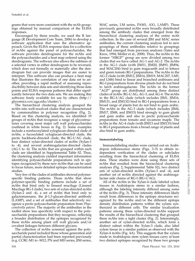
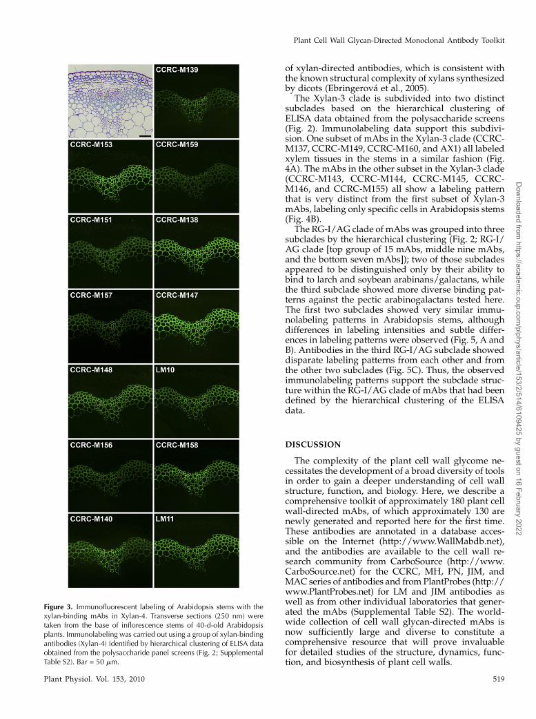
All of the mAbs in the Xylan-4 clade labeled xylemtissues in Arabidopsis stems in a similar fashion,although the labeling intensity differed among someof the mAbs (Fig. 3). These intensity differences likelyresult from differences in the epitope structures rec-ognized by the mAbs and/or the different epitopedensity distribution patterns within the xylans syn-thesized in different cells. The similar localizationpatterns among these xylan-directed mAbs supportthe results of the hierarchical clustering that groupedthese mAbs into a tight cluster (Fig. 2). Interestingly,another set of xylan-directed mAbs, which form adistinct subset within the Xylan-3 clade, also labelxylem tissue in a similar pattern as observed with theXylan-4 mAbs (Fig. 4A). This suggests that the xylansmade in Arabidopsis stem xylem tissues carry at leasttwo distinct epitopes recognized by these two groups
Pattathil et al.
518 Plant Physiol. Vol. 153, 2010
Dow
nloaded from https://academ
ic.oup.com/plphys/article/153/2/514/6109425 by guest on 16 February 2022
of xylan-directed antibodies, which is consistent withthe known structural complexity of xylans synthesizedby dicots (Ebringerova et al., 2005).
The Xylan-3 clade is subdivided into two distinctsubclades based on the hierarchical clustering ofELISA data obtained from the polysaccharide screens(Fig. 2). Immunolabeling data support this subdivi-sion. One subset of mAbs in the Xylan-3 clade (CCRC-M137, CCRC-M149, CCRC-M160, and AX1) all labeledxylem tissues in the stems in a similar fashion (Fig.4A). The mAbs in the other subset in the Xylan-3 clade(CCRC-M143, CCRC-M144, CCRC-M145, CCRC-M146, and CCRC-M155) all show a labeling patternthat is very distinct from the first subset of Xylan-3mAbs, labeling only specific cells in Arabidopsis stems(Fig. 4B).
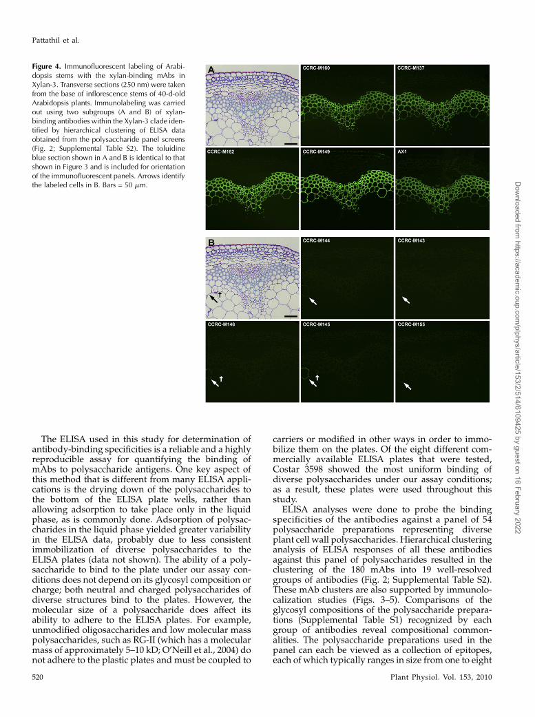
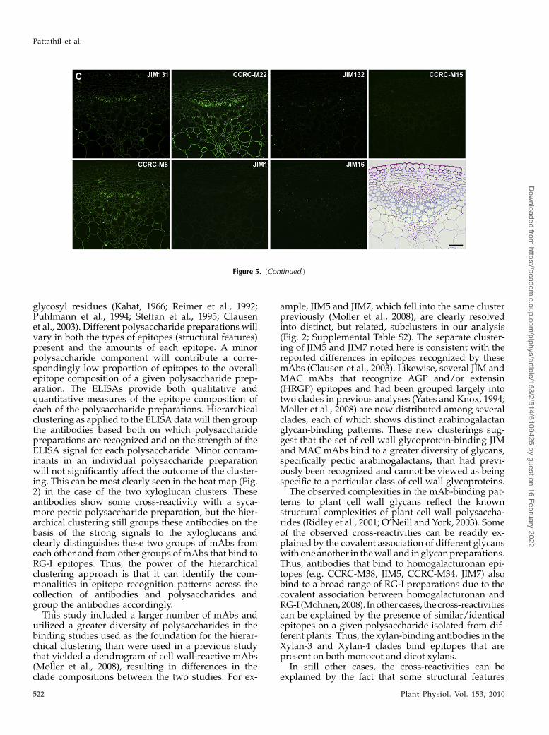
The RG-I/AG clade of mAbs was grouped into threesubclades by the hierarchical clustering (Fig. 2; RG-I/AG clade [top group of 15 mAbs, middle nine mAbs,and the bottom seven mAbs]); two of those subcladesappeared to be distinguished only by their ability tobind to larch and soybean arabinans/galactans, whilethe third subclade showed more diverse binding pat-terns against the pectic arabinogalactans tested here.The first two subclades showed very similar immu-nolabeling patterns in Arabidopsis stems, althoughdifferences in labeling intensities and subtle differ-ences in labeling patterns were observed (Fig. 5, A andB). Antibodies in the third RG-I/AG subclade showeddisparate labeling patterns from each other and fromthe other two subclades (Fig. 5C). Thus, the observedimmunolabeling patterns support the subclade struc-ture within the RG-I/AG clade of mAbs that had beendefined by the hierarchical clustering of the ELISAdata.
DISCUSSION
The complexity of the plant cell wall glycome ne-cessitates the development of a broad diversity of toolsin order to gain a deeper understanding of cell wallstructure, function, and biology. Here, we describe acomprehensive toolkit of approximately 180 plant cellwall-directed mAbs, of which approximately 130 arenewly generated and reported here for the first time.These antibodies are annotated in a database acces-sible on the Internet (http://www.WallMabdb.net),and the antibodies are available to the cell wall re-search community from CarboSource (http://www.CarboSource.net) for the CCRC, MH, PN, JIM, andMAC series of antibodies and fromPlantProbes (http://www.PlantProbes.net) for LM and JIM antibodies aswell as from other individual laboratories that gener-ated the mAbs (Supplemental Table S2). The world-wide collection of cell wall glycan-directed mAbs isnow sufficiently large and diverse to constitute acomprehensive resource that will prove invaluablefor detailed studies of the structure, dynamics, func-tion, and biosynthesis of plant cell walls.
Figure 3. Immunofluorescent labeling of Arabidopsis stems with thexylan-binding mAbs in Xylan-4. Transverse sections (250 nm) weretaken from the base of inflorescence stems of 40-d-old Arabidopsisplants. Immunolabeling was carried out using a group of xylan-bindingantibodies (Xylan-4) identified by hierarchical clustering of ELISA dataobtained from the polysaccharide panel screens (Fig. 2; SupplementalTable S2). Bar = 50 mm.
Plant Cell Wall Glycan-Directed Monoclonal Antibody Toolkit
Plant Physiol. Vol. 153, 2010 519
Dow
nloaded from https://academ
ic.oup.com/plphys/article/153/2/514/6109425 by guest on 16 February 2022
The ELISA used in this study for determination ofantibody-binding specificities is a reliable and a highlyreproducible assay for quantifying the binding ofmAbs to polysaccharide antigens. One key aspect ofthis method that is different from many ELISA appli-cations is the drying down of the polysaccharides tothe bottom of the ELISA plate wells, rather thanallowing adsorption to take place only in the liquidphase, as is commonly done. Adsorption of polysac-charides in the liquid phase yielded greater variabilityin the ELISA data, probably due to less consistentimmobilization of diverse polysaccharides to theELISA plates (data not shown). The ability of a poly-saccharide to bind to the plate under our assay con-ditions does not depend on its glycosyl composition orcharge; both neutral and charged polysaccharides ofdiverse structures bind to the plates. However, themolecular size of a polysaccharide does affect itsability to adhere to the ELISA plates. For example,unmodified oligosaccharides and low molecular masspolysaccharides, such as RG-II (which has a molecularmass of approximately 5–10 kD; O’Neill et al., 2004) donot adhere to the plastic plates and must be coupled to
carriers or modified in other ways in order to immo-bilize them on the plates. Of the eight different com-mercially available ELISA plates that were tested,Costar 3598 showed the most uniform binding ofdiverse polysaccharides under our assay conditions;as a result, these plates were used throughout thisstudy.
ELISA analyses were done to probe the bindingspecificities of the antibodies against a panel of 54polysaccharide preparations representing diverseplant cell wall polysaccharides. Hierarchical clusteringanalysis of ELISA responses of all these antibodiesagainst this panel of polysaccharides resulted in theclustering of the 180 mAbs into 19 well-resolvedgroups of antibodies (Fig. 2; Supplemental Table S2).These mAb clusters are also supported by immunolo-calization studies (Figs. 3–5). Comparisons of theglycosyl compositions of the polysaccharide prepara-tions (Supplemental Table S1) recognized by eachgroup of antibodies reveal compositional common-alities. The polysaccharide preparations used in thepanel can each be viewed as a collection of epitopes,each of which typically ranges in size from one to eight
Figure 4. Immunofluorescent labeling of Arabi-dopsis stems with the xylan-binding mAbs inXylan-3. Transverse sections (250 nm) were takenfrom the base of inflorescence stems of 40-d-oldArabidopsis plants. Immunolabeling was carriedout using two subgroups (A and B) of xylan-binding antibodies within the Xylan-3 clade iden-tified by hierarchical clustering of ELISA dataobtained from the polysaccharide panel screens(Fig. 2; Supplemental Table S2). The toluidineblue section shown in A and B is identical to thatshown in Figure 3 and is included for orientationof the immunofluorescent panels. Arrows identifythe labeled cells in B. Bars = 50 mm.
Pattathil et al.
520 Plant Physiol. Vol. 153, 2010
Dow
nloaded from https://academ
ic.oup.com/plphys/article/153/2/514/6109425 by guest on 16 February 2022
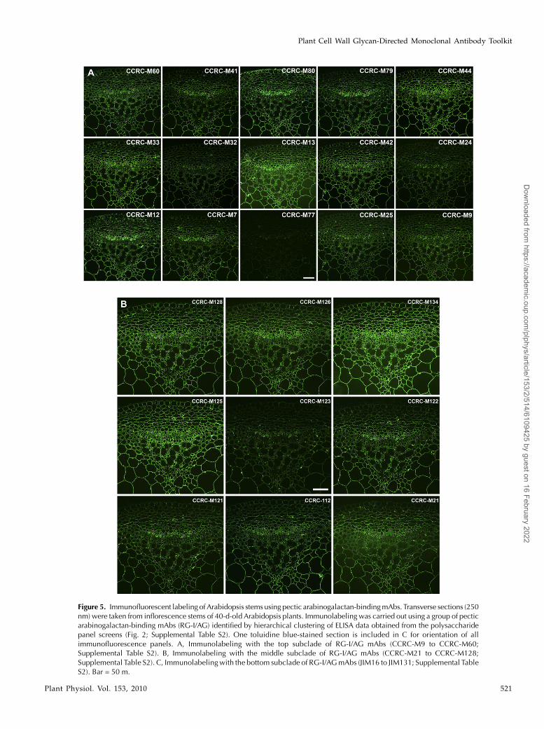
Figure 5. Immunofluorescent labeling of Arabidopsis stems using pectic arabinogalactan-bindingmAbs. Transverse sections (250nm) were taken from inflorescence stems of 40-d-old Arabidopsis plants. Immunolabeling was carried out using a group of pecticarabinogalactan-binding mAbs (RG-I/AG) identified by hierarchical clustering of ELISA data obtained from the polysaccharidepanel screens (Fig. 2; Supplemental Table S2). One toluidine blue-stained section is included in C for orientation of allimmunofluorescence panels. A, Immunolabeling with the top subclade of RG-I/AG mAbs (CCRC-M9 to CCRC-M60;Supplemental Table S2). B, Immunolabeling with the middle subclade of RG-I/AG mAbs (CCRC-M21 to CCRC-M128;Supplemental Table S2). C, Immunolabelingwith the bottom subclade of RG-I/AGmAbs (JIM16 to JIM131; Supplemental TableS2). Bar = 50 m.
Plant Cell Wall Glycan-Directed Monoclonal Antibody Toolkit
Plant Physiol. Vol. 153, 2010 521
Dow
nloaded from https://academ
ic.oup.com/plphys/article/153/2/514/6109425 by guest on 16 February 2022
glycosyl residues (Kabat, 1966; Reimer et al., 1992;Puhlmann et al., 1994; Steffan et al., 1995; Clausenet al., 2003). Different polysaccharide preparations willvary in both the types of epitopes (structural features)present and the amounts of each epitope. A minorpolysaccharide component will contribute a corre-spondingly low proportion of epitopes to the overallepitope composition of a given polysaccharide prep-aration. The ELISAs provide both qualitative andquantitative measures of the epitope composition ofeach of the polysaccharide preparations. Hierarchicalclustering as applied to the ELISA data will then groupthe antibodies based both on which polysaccharidepreparations are recognized and on the strength of theELISA signal for each polysaccharide. Minor contam-inants in an individual polysaccharide preparationwill not significantly affect the outcome of the cluster-ing. This can be most clearly seen in the heat map (Fig.2) in the case of the two xyloglucan clusters. Theseantibodies show some cross-reactivity with a syca-more pectic polysaccharide preparation, but the hier-archical clustering still groups these antibodies on thebasis of the strong signals to the xyloglucans andclearly distinguishes these two groups of mAbs fromeach other and from other groups of mAbs that bind toRG-I epitopes. Thus, the power of the hierarchicalclustering approach is that it can identify the com-monalities in epitope recognition patterns across thecollection of antibodies and polysaccharides andgroup the antibodies accordingly.
This study included a larger number of mAbs andutilized a greater diversity of polysaccharides in thebinding studies used as the foundation for the hierar-chical clustering than were used in a previous studythat yielded a dendrogram of cell wall-reactive mAbs(Moller et al., 2008), resulting in differences in theclade compositions between the two studies. For ex-
ample, JIM5 and JIM7, which fell into the same clusterpreviously (Moller et al., 2008), are clearly resolvedinto distinct, but related, subclusters in our analysis(Fig. 2; Supplemental Table S2). The separate cluster-ing of JIM5 and JIM7 noted here is consistent with thereported differences in epitopes recognized by thesemAbs (Clausen et al., 2003). Likewise, several JIM andMAC mAbs that recognize AGP and/or extensin(HRGP) epitopes and had been grouped largely intotwo clades in previous analyses (Yates and Knox, 1994;Moller et al., 2008) are now distributed among severalclades, each of which shows distinct arabinogalactanglycan-binding patterns. These new clusterings sug-gest that the set of cell wall glycoprotein-binding JIMand MAC mAbs bind to a greater diversity of glycans,specifically pectic arabinogalactans, than had previ-ously been recognized and cannot be viewed as beingspecific to a particular class of cell wall glycoproteins.
The observed complexities in the mAb-binding pat-terns to plant cell wall glycans reflect the knownstructural complexities of plant cell wall polysaccha-rides (Ridley et al., 2001; O’Neill and York, 2003). Someof the observed cross-reactivities can be readily ex-plained by the covalent association of different glycanswithoneanother in thewall and inglycanpreparations.Thus, antibodies that bind to homogalacturonan epi-topes (e.g. CCRC-M38, JIM5, CCRC-M34, JIM7) alsobind to a broad range of RG-I preparations due to thecovalent association between homogalacturonan andRG-I (Mohnen, 2008). Inothercases, thecross-reactivitiescan be explained by the presence of similar/identicalepitopes on a given polysaccharide isolated from dif-ferent plants. Thus, the xylan-binding antibodies in theXylan-3 and Xylan-4 clades bind epitopes that arepresent on both monocot and dicot xylans.
In still other cases, the cross-reactivities can beexplained by the fact that some structural features
Figure 5. (Continued.)
Pattathil et al.
522 Plant Physiol. Vol. 153, 2010
Dow
nloaded from https://academ
ic.oup.com/plphys/article/153/2/514/6109425 by guest on 16 February 2022
(epitopes) are present onmultiple glycans that occur inplant cell walls. This is particularly the case withantibodies that bind to epitopes containing arabinosyland/or galactosyl residues, which are present in mul-tiple structural contexts within diverse plant cell wallglycans. For example, mAbs that bind to arabinoga-lactan side chains of RG-I frequently, but not always,also bind to free and/or protein-linked arabinogalac-tans, which contain similar structural features (Ridleyet al., 2001; Seifert and Roberts, 2007). The data pre-sented here emphasize that glycan-directed antibodiesshould be utilized as epitope-directed reagents andfrequently are not polymer selective.Some observed cross-reactivities are not as readily
explained based on current knowledge of cell wallglycan structures. For example, mAbs that bind tofucosylated xyloglucans (e.g. CCRC-M1) also bindstrongly to sycamore RG-I (but not to other RG-Isincluded in this study). This cross-reactivity is not dueto contamination of the sycamore RG-I preparationwith xyloglucan, since treatment of sycamore RG-Iwith a xyloglucan-specific endoglucanase did not af-fect binding of CCRC-M1 (data not shown). The cross-reactivity of the mAbs in the Xylan-1 clade withxyloglucans included in this study is also not readilyexplained. The epitope(s) recognized by the Xylan-1mAbs appears not to be present on all xyloglucans, asthese mAbs do not label any cells in Arabidopsistissues (data not shown). Interestingly, carbohydrate-binding modules that recognize xylan have beenreported to also bind to xyloglucans (Boraston et al.,2001; Gunnarsson et al., 2006). Xyloglucan and xylanare not known to be covalently linked or to sharecommon structural features (except that both have ab-1,4-linked backbone composed of pyranosyl resi-dues in which all exocyclic oxygens are equatorial).Resolution of these cross-reactivities must await de-tailed characterizations of the epitopes recognized bythese mAbs.Most of the mAb clades identified through hierar-
chical clustering are divided further into subclades.Some of these subdivisions are informative with re-spect to possible epitopes recognized by newly gener-ated mAbs due to tight clustering with previouslycharacterized mAbs. For example, several new mAbs(CCRC-M39, CCRC-M84, CCRC-M102, CCRC-M106)cluster with CCRC-M1, suggesting that the newlygenerated mAbs bind to the same or similar fucosy-lated xyloglucan epitope recognized by CCRC-M1(Puhlmann et al., 1994). Other newly reported mAbscluster in distinct subclades with previously charac-terized mAbs directed against homogalacturonans(Fig. 2; Supplemental Table S2). CCRC-M34, CCRC-M130, and JIM136 cluster closely in a subclade withJIM7 and LM7, mAbs that bind to densely methyl-esterified homogalacturonan epitopes (Clausen et al.,2003), suggesting that these three mAbs bind tomethylated homogalacturonan epitopes. In contrast,CCRC-M38, CCRC-M131, and CCRC-M132 clustertightly in a subclade with JIM5, a mAb that binds to
a homogalacturonan epitope having a low density ofmethyl esterification (Clausen et al., 2003), suggestingthat these three mAbs bind to a largely or completelydeesterified homogalacturonan epitope. Lastly, abouta dozen newly generated mAbs cluster tightly in asubclade of the RG-I/AG clade (Fig. 2; SupplementalTable S2) that includes CCRC-M7, suggesting thatthese mAbs recognize a b-1,6-galactan epitope similaror identical to that recognized by CCRC-M7 (Steffanet al., 1995). Verification of these tentative epitopeassignments awaits more detailed studies, which arecurrently under way in our laboratory.
MATERIALS AND METHODS
Polysaccharides
Polysaccharides from various plant sources were obtained from commer-
cial sources (Megazyme, Sigma, and Sunkist) and various laboratories at the
University of Georgia’s Complex Carbohydrate Research Center and else-
where. Detailed information about these polysaccharides, such as glycan
class, preparation, source, and sugar composition, are given in Supplemental
Table S1. Stock solutions were prepared by dissolving the polysaccharides at
1 mg mL21 in deionized water and were stored at 220�C.
mAbs
mAbs were obtained as hybridoma cell culture supernatants either from
laboratory stocks (CCRC series, MH series, PN series, JIM series, MAC series;
available from CarboSource [http://www.carbosource/net]) or from Plant
Probes (LM series, PAM1 [http://www.plantprobes.net/]) unless otherwise
indicated. A detailed list of all mAbs included in this study showing the
immunogens used to develop them, their isotype, and the cell wall polysac-
charide class they primarily recognize is provided in Supplemental Table S2.
ELISA
Flat-bottom 96-well plates tested for use in the ELISA were Immulon 1B,
Immulon 2HB, Immulon 4HB, Nunc 269620, and Nunc 439454 (Thermo Fisher
Scientific) and Costar 2507, Costar 3590, and Costar 3598 (Corning Life
Sciences). Initial experiments were carried out with several polysaccharides
over a broad concentration range (1 ng well21 to 10 mg well21) in order to
determine the maximum loading of polysaccharides onto the plates. These
studies showed that an amount of 0.5 mg well21 saturates the ELISAwells with
a given polysaccharide antigen (data not shown). Polysaccharides were
applied (50 mL of 10 mg mL21 in deionized water per well) to 96-well plates
and were dried to the well surfaces by evaporation overnight at 37�C. Controlwells were coated with deionized water. The plates were blocked with 200 mL
of 1% (w/v) instant nonfat dry milk (Carnation) in Tris-buffered saline (50 mM
Tris-HCl, pH 7.6, containing 100 mM sodium chloride) for 1 h. All subsequent
aspiration and wash steps were performed using an ELx405 microplate
washer (Bio-Tek Instruments). Blocking agent was removed by aspiration, and
50 mL of undiluted hybridoma supernatant were added to each well and
incubated for 1 h at room temperature. Supernatant was removed and wells
were washed three times with 300 mL of 0.1% (w/v) instant nonfat dry milk in
Tris-buffered saline (wash buffer). Peroxidase-conjugated goat anti-mouse
IgG or goat anti-rat IgG antibodies (Sigma-Aldrich), depending on the
primary antibody used, was diluted 1:5,000 in wash buffer, and 50 mL were
added to each well and incubated for 1 h. Note that the secondary antibodies
used in this study are generated against whole immunoglobin molecules and
thus bind to several isotypes of primary antibodies, including IgGs, IgMs, and
IgAs, according to the manufacturers. Wells were then washed five times with
300 mL of wash buffer. 3,3#,5,5#-Tetramethylbenzidine substrate solution
(Vector Laboratories) was freshly prepared according to the manufacturer’s
instructions, and 50 mL were added to each well. After 20 min, the reaction
was stopped by adding 50 mL of 0.5 N sulfuric acid to each well. The OD of
each well was read as the difference in A450 and A655 using a model 680
microplate reader (Bio-Rad). The reading from each test well was subtracted
Plant Cell Wall Glycan-Directed Monoclonal Antibody Toolkit
Plant Physiol. Vol. 153, 2010 523
Dow
nloaded from https://academ
ic.oup.com/plphys/article/153/2/514/6109425 by guest on 16 February 2022
from that of a control well on the same plate that contained the same primary
and secondary antibodies but no immobilized polysaccharide.
Polysaccharide Panel Screening
Polysaccharide panel screening of mAbs was carried out by ELISA against
54 plant polysaccharides (Supplemental Table S1) immobilized to Costar 3598
96-well plates. A single preparation of each polysaccharide was used for all
experiments reported here.
Hierarchical Clustering
Hierarchical clustering of the ELISA results for binding of each mAb to
each polysaccharide in the panel was carried out using previously described
methods (Ferguson et al., 1988) with modifications (Supplemental Materials
and Methods S1). The R language for statistical computing was used for these
analyses (R Development Core Team, 2006).
Plant Culture Conditions
Seeds of Arabidopsis (Arabidopsis thaliana ecotype Columbia) were surface
sterilized by immersion in 70% (v/v) aqueous ethanol for 2 min followed by a
2-min immersion in 1% sodium hypochlorite solution (20% [v/v] Clorox
containing 0.02% [v/v] Triton X-100). Seeds were then rinsed three times with
sterile distilled water. Sterilized seeds were germinated and grown in sterile
petri dishes on 1% (w/v) agar with Murashige and Skoog basal salt medium
(Sigma-Aldrich) supplemented with 1% (w/v) Suc, pH 6.9. The petri dishes
were oriented vertically, maintained at 23�C, and received 12 h of fluorescent
illumination (100 mE m22 s21) daily. Two-week-old seedlings grown on the
plates were then transferred to 4-inch pots containing compost with vermic-
ulite and perlite. These plants were grown in a growth chamber at 20�Cwith a
16-h-light/8-h-dark cycle.
Tissue Fixation
Forty-day-old Arabidopsis inflorescence stems were fixed for 2.5 h in 1.6%
(w/v) paraformaldehyde and 0.2% (w/v) glutaraldehyde in 25 mM sodium
phosphate buffer, pH 7.1. Tissue was then washed with buffer twice for 15
min, washed with water twice for 15 min, and dehydrated through a graded
ethanol series (35%, 50%, 75%, 95%, 100%, 100%, and 100% [v/v] ethanol) for
30 min each step. The dehydrated tissue wasmoved to 4�C and then gradually
infiltrated with cold LR White embedding resin (Ted Pella) using 33% (v/v)
and 66% (v/v) resin in 100% ethanol for 24 h each, followed by 100% resin for
24 h three times. The infiltrated tissue was transferred to gelatin capsules
containing 100% resin for embedding, and resin was polymerized by exposing
the capsules to 365-nm UV light at 4�C for 48 h.
Immunolabeling
Semithin sections (250 nm) were cut with a Leica EM UC6 ultramicrotome
(Leica Microsystems) and mounted on glass slides (colorfrost/plus; Fisher
Scientific). Immunolabeling was performed at room temperature by applying
and removing a series of 10-mL droplets of the appropriate reagents to the
sections as follows. Sections were blocked with 3% (w/v) nonfat dry milk in
KPBS (0.01 M potassium phosphate, pH 7.1, containing 0.5 M NaCl) for 45 min
and then were washed with KPBS for 5 min. Undiluted hybridoma superna-
tant of the mAb under study was applied and incubated for 120 to 150 min.
Sections were then washed with KPBS three times for 5 min each, and goat
anti-mouse IgG or goat anti-rat IgG conjugated to Alexa-fluor 488 (Invitrogen)
diluted 1:100 in KPBS was applied and incubated for 90 to 120 min. Sections
were then washed with KPBS for 5 min and distilled water for 5 min. Prior to
applying a coverslip, Citifluor antifade mounting medium AF1 (Electron
Microscopy Sciences) was applied.
Light Microscopy
Light microscopy was carried out on an Eclipse 80i microscope (Nikon)
equipped with epifluorescence optics. Images were captured with a Nikon
DS-Ri1 camera head using NIS-Elements Basic Research software, and images
were assembled without further processing using Adobe Photoshop (Adobe
Systems).
Supplemental Data
The following materials are available in the online version of this article.
Supplemental Figure S1. Suitability of 96-well plates for polysaccharide
ELISAs.
Supplemental Table S1. Plant polysaccharide preparations used in this
study.
Supplemental Table S2. mAbs included in this study.
Supplemental Table S3. Coefficients correlating the ELISA responses
obtained in one experiment with the averages of ELISA responses from
six experiments.
Supplemental Materials and Methods S1. Hierarchical clustering of
mAbs.
ACKNOWLEDGMENTS
We thank the following scientists for their generous contributions of
polysaccharide preparations included in the screening panel of polysaccha-
rides. Members of the Complex Carbohydrate Research Center, University of
Georgia, are thanked for their contribution of the following polysaccharide
preparations: Chenghua Deng for green tomato fruit RG-I and lettuce RG-I
and Christian Heiss for tamarind xyloglucan. We also thank Mark Davis of
the National Renewable Energy Laboratory (Golden, Colorado) for the corn
xylan and Ian Sims of Industrial Research Limited (Lower Hutt, New
Zealand) for the samples of Phormium xylans. We thank Dr. Henk Schols
(Laboratory of Food Chemistry, Wageningen, The Netherlands) for providing
several different xylan and arabinogalactan preparations and okra RG-I.
Lastly, we acknowledge the generosity of Keith Roberts and Nick Brewin
(John Innes Institute, Norwich, UK) in providing us with the JIM and MAC
hybridoma lines and Dr. Fabienne Guillon (INRA, Nantes, France) for
supplying us with the AX1 mAb.
Received December 11, 2009; acceptedMarch 30, 2010; published April 2, 2010.
LITERATURE CITED
Altaner C, Hapca AI, Knox JP, Jarvis MC (2007) Detection of b-1-4-galactan
in compression wood of Sitka spruce [Picea sitchensis (Bong.) Carriere]
by immunofluorescence. Holzforschung 61: 311–316
Boraston AB, Creagh AL, Alam MM, Kormos JM, Tomme P, Haynes CA,
Warren RAJ, Kilburn DG (2001) Binding specificity and thermody-
namics of a family 9 carbohydrate-binding module from Thermotoga
maritima xylanase 10A. Biochemistry 40: 6240–6247
Burton RA, Farrokhi N, Bacic A, Fincher GB (2005) Plant cell wall
polysaccharide biosynthesis: real progress in the identification of par-
ticipating genes. Planta 221: 309–312
Carpita N, Tierney M, Campbell M (2001) Molecular biology of the plant
cell wall: searching for the genes that define structure, architecture and
dynamics. Plant Mol Biol 47: 1–5
Carpita NC, Gibeaut DM (1993) Structural models of primary cell walls in
flowering plants: consistency of molecular structure with the physical
properties of the walls during growth. Plant J 3: 1–30
Cavalier DM, Lerouxel O, Neumetzler L, Yamauchi K, Reinecke A,
Freshour G, Zabotina OA, Hahn MG, Burgert I, Pauly M, et al (2008)
Disrupting two Arabidopsis thaliana xylosyltransferase genes results in
plants deficient in xyloglucan, a major primary cell wall component.
Plant Cell 20: 1519–1537
Clausen MH, Ralet MC, Willats WGT, McCartney L, Marcus SE, Thibault
JF, Knox JP (2004) A monoclonal antibody to feruloylated-(1/4)-b-D-
galactan. Planta 219: 1036–1041
Clausen MH, Willats WGT, Knox JP (2003) Synthetic methyl hexagalac-
turonate hapten inhibitors of anti-homogalacturonan monoclonal anti-
bodies LM7, JIM5 and JIM7. Carbohydr Res 338: 1797–1800
Dolan L, Linstead P, Roberts K (1995) An AGP epitope distinguishes a
Pattathil et al.
524 Plant Physiol. Vol. 153, 2010
Dow
nloaded from https://academ
ic.oup.com/plphys/article/153/2/514/6109425 by guest on 16 February 2022
central metaxylem initial from other vascular initials in the Arabidopsis
root. Protoplasma 189: 149–155
Ebringerova A, Hromadkova Z, Heinze T (2005) Hemicellulose. Adv
Polym Sci 186: 1–67
Ferguson MW, Wycoff KL, Ayers AR (1988) Use of cluster analysis with
monoclonal antibodies for taxonomic differentiation of phytopatho-
genic fungi and for screening and clustering antibodies. Curr Microbiol
17: 127–132
Freshour G, Bonin CP, Reiter WD, Albersheim P, Darvill AG, Hahn MG
(2003) Distribution of fucose-containing xyloglucans in cell walls of the
mur1 mutant of Arabidopsis thaliana. Plant Physiol 131: 1602–1612
Freshour G, Clay RP, Fuller MS, Albersheim P, Darvill AG, Hahn MG
(1996) Developmental and tissue-specific structural alterations of the
cell-wall polysaccharides of Arabidopsis thaliana roots. Plant Physiol 110:
1413–1429
Gunnarsson LC, Zhou Q, Montanier C, Karlsson EN, Brumer H, Ohlin M
(2006) Engineered xyloglucan specificity in a carbohydrate-binding
module. Glycobiology 16: 1171–1180
Jones L, Seymour GB, Knox JP (1997) Localization of pectic galactan in
tomato cell walls using a monoclonal antibody specific to (1/4)-b-D-
galactan. Plant Physiol 113: 1405–1412
Kabat EA (1966) The nature of an antigenic determinant. J Immunol
97: 1–11
Knox JP (2008) Revealing the structural and functional diversity of plant
cell walls. Curr Opin Plant Biol 11: 308–313
Lerouxel O, Cavalier DM, Liepman AH, Keegstra K (2006) Biosynthesis of
plant cell wall polysaccharides: a complex process. Curr Opin Plant Biol
9: 621–630
Marcus SE, Verhertbruggen Y, Herve C, Ordaz-Ortiz JJ, Farkas V,
Pedersen HL, Willats WGT, Knox JP (2008) Pectic homogalacturonan
masks abundant sets of xyloglucan epitopes in plant cell walls. BMC
Plant Biol 8: 60–71
McCartney L, Marcus SE, Knox JP (2005) Monoclonal antibodies to plant
cell wall xylans and arabinoxylans. J Histochem Cytochem 53: 543–546
McCartney L, Ormerod AP, Gidley MJ, Knox JP (2000) Temporal and
spatial regulation of pectic (1/4)-b-D-galactan in cell walls of devel-
oping pea cotyledons: implications for mechanical properties. Plant J 22:
105–113
Mohnen D (2008) Pectin structure and biosynthesis. Curr Opin Plant Biol
11: 266–277
Mohnen D, Bar-Peled M, Somerville C (2008) Cell wall polysaccharide
synthesis. In DE Himmel, ed, Biomass Recalcitrance: Deconstructing the
Plant Cell Wall for Bioenergy. Blackwell Publishing, Oxford, pp 94–187
Moller I, Marcus SE, Haeger A, Verhertbruggen Y, Verhoef R, Schols H,
Ulvskov P, Mikkelsen JD, Knox JP, Willats W (2008) High-throughput
screening of monoclonal antibodies against plant cell wall glycans by
hierarchical clustering of their carbohydrate microarray binding pro-
files. Glycoconj J 25: 37–48
O’Neill MA, Ishii T, Albersheim P, Darvill AG (2004) Rhamnogalactur-
onan II: structure and function of a borate cross-linked cell wall pectic
polysaccharide. Annu Rev Plant Biol 55: 109–139
O’Neill MA, YorkWS (2003) The composition and structure of primary cell
walls. In JKC Rose, ed, The Plant Cell Wall. Blackwell Publishers,
Oxford, pp 1–54
Orfila C, Seymour GB, Willats WGT, Huxham IM, Jarvis MC, Dover CJ,
Thompson AJ, Knox JP (2001) Altered middle lamella homogalactur-
onan and disrupted deposition of (1/5)-a-L-arabinan in the pericarp of
Cnr, a ripening mutant of tomato. Plant Physiol 126: 210–221
Pennell RI, Janniche L, Kjellbom P, Scofield GN, Peart JM, Roberts K
(1991) Developmental regulation of a plasma membrane arabinogalac-
tan protein epitope in oilseed rape flowers. Plant Cell 3: 1317–1326
Persson S, Caffall KH, Freshour G, Hilley MT, Bauer S, Poindexter P,
Hahn MG, Mohnen D, Somerville C (2007) The Arabidopsis irregular
xylem8 mutant is deficient in glucuronoxylan and homogalacturonan,
which are essential for secondary cell wall integrity. Plant Cell 19:
237–255
Puhlmann J, Bucheli E, SwainMJ, Dunning N, Albersheim P, Darvill AG,
HahnMG (1994) Generation of monoclonal antibodies against plant cell
wall polysaccharides. I. Characterization of a monoclonal antibody to a
terminal a-(1/2)-linked fucosyl-containing epitope. Plant Physiol 104:
699–710
R Development Core Team (2006) R: A Language and Environment for
Statistical Computing. R Foundation for Statistical Computing (http://
www.R-project.org)
Reimer KB, Gidney MAJ, Bundle DR, Pinto BM (1992) Immunochemical
characterization of polyclonal and monoclonal Streptococcus group A
antibodies by chemically defined glycoconjugates and synthetic oligo-
saccharides. Carbohydr Res 232: 131–142
Ridley BL, O’Neill MA, Mohnen D (2001) Pectins: structure, biosynthesis,
and oligogalacturonide-related signaling. Phytochemistry 57: 929–967
Sarria R, Wagner TA, O’Neill MA, Faik A, Wilkerson CG, Keegstra K,
Raikhel NV (2001) Characterization of a family of Arabidopsis genes
related to xyloglucan fucosyltransferase1. Plant Physiol 127: 1595–1606
Seifert GJ (2004) Nucleotide sugar interconversions and cell wall biosyn-
thesis: how to bring the inside to the outside. Curr Opin Plant Biol 7:
277–284
Seifert GJ, Roberts K (2007) The biology of arabinogalactan proteins. Annu
Rev Plant Biol 58: 137–161
Smallwood M, Martin H, Knox JP (1995) An epitope of rice threonine- and
hydroxyproline-rich glycoprotein is common to cell wall and hydro-
phobic plasma-membrane glycoproteins. Planta 196: 510–522
Smallwood M, Yates EA, Willats WGT, Martin H, Knox JP (1996) Immu-
nochemical comparison of membrane-associated and secreted arabino-
galactan-proteins in rice and carrot. Planta 198: 452–459
Somerville C (2007) Biofuels. Curr Biol 17: R115–R119
Somerville C, Bauer S, Brininstool G, Facette M, Hamann T, Milne J,
Osborne E, Paredez A, Persson S, Raab T, et al (2004) Toward a systems
approach to understanding plant cell walls. Science 306: 2206–2211
Steffan W, Kovac P, Albersheim P, Darvill AG, Hahn MG (1995) Charac-
terization of a monoclonal antibody that recognizes an arabinosylated
(1/6)-b-D-galactan epitope in plant complex carbohydrates. Carbo-
hydr Res 275: 295–307
Willats WGT, Marcus SE, Knox JP (1998) Generation of a monoclonal
antibody specific to (1/5)-a-L-arabinan. Carbohydr Res 308: 149–152
Willats WGT, McCartney L, Steele-King CG, Marcus SE, Mort A, Huisman
M, Van Alebeek GJ, Schols HA, Voragen AGJ, Le Goff A, et al (2004) A
xylogalacturonan epitope is specifically associated with plant cell detach-
ment. Planta 218: 673–681
Willats WGT, Orfila C, Limberg G, Buchholt HC, Van Alebeek GJWM,
Voragen AGJ, Marcus SE, Christensen TMIE, Mikkelsen JD, Murray
BS, et al (2001) Modulation of the degree and pattern of methyl-
esterification of pectic homogalacturonan in plant cell walls: implica-
tions for pectin methyl esterase action, matrix properties, and cell
adhesion. J Biol Chem 276: 19404–19413
Yates EA, Knox JP (1994) Investigations into the occurrence of plant cell
surface epitopes in exudate gums. Carbohydr Polym 24: 281–286
Zabotina OA, van de Ven WTG, Freshour G, Drakakaki G, Cavalier D,
Mouille G, Hahn MG, Keegstra K, Raikhel NV (2008) Arabidopsis
XXT5 gene encodes a putative a-1,6-xylosyltransferase that is involved
in xyloglucan biosynthesis. Plant J 56: 101–115
Plant Cell Wall Glycan-Directed Monoclonal Antibody Toolkit
Plant Physiol. Vol. 153, 2010 525
Dow
nloaded from https://academ
ic.oup.com/plphys/article/153/2/514/6109425 by guest on 16 February 2022