Embed Size (px)
Citation preview

Research ArticleA Compound of Chinese Herbs Protects against Alcoholic LiverFibrosis in Rats via the TGF-𝛽1/Smad Signaling Pathway
Xiaomeng Li 1,2, Yunjie Liu1,2, Wuyang Yue1,2, Yuefeng Tan1,2, HeWang1,2,Lishi Zhang 1,2, and Jinyao Chen 1,2
1West China School of Public Health and West China fourth Hospital, Sichuan University, Chengdu, China2Food Safety Monitoring and Risk Assessment Key Laboratory of Sichuan Province, Chengdu, China
Correspondence should be addressed to Lishi Zhang; lishizhang [email protected] and Jinyao Chen; [email protected]
Received 8 January 2019; Revised 11 March 2019; Accepted 31 March 2019; Published 21 April 2019
Academic Editor: Yoshiji Ohta
Copyright © 2019 Xiaomeng Li et al. This is an open access article distributed under the Creative Commons Attribution License,which permits unrestricted use, distribution, and reproduction in any medium, provided the original work is properly cited.
Alcoholic liver fibrosis (ALF) has become a major public health concern owing to its health impacts and the lack of effectivetreatment strategies for the disease. In this study, we investigated the effect of a compound composed of Chinese herbs Puerarialobata (Willd.), Salvia miltiorrhiza, Schisandra chinensis, and Silybummarianum on ALF. An ALFmodel was established. Rats werefedwithmodified Lieber–Decarli alcohol liquid diet and injectedwith trace CCl
4at late stage.The ratswere then treatedwith several
doses of the compound. Biochemical and fibrosis-relevant parameters were measured from the sera obtained from the rats. Livertissues were obtained for hematoxylin and eosin andMasson’s trichrome staining. Matrix metalloproteinase-13 and tissue inhibitorofmetalloproteinase-1were determinedby immunohistochemistry assays.ThemRNAandprotein expression levels of transforminggrowth factor-𝛽1 (TGF-𝛽1), Smad2, Smad3, and Smad7 on the livers were also measured by quantitative polymerase chain reactionand Western blot. Results showed that the compound treatment alleviated pathological lesions in the liver, decreased the serumlevels of hyaluronan, laminin, and hydroxyproline, and diminished the expression of hepatic tissue inhibitor of metalloproteinase-1. Compound treatment also increased hepatic matrix metalloproteinase-13 expression and inhibited the TGF-𝛽1/Smad signalingpathway. In conclusion, the compound has a protective effect against ALF in rats, and an underlying mechanism is involved in theTGF-𝛽1/Smad signaling pathway.
1. Introduction
Alcoholic liver disease (ALD) is an important public healthproblem with wide pathologic spectrum, including steato-sis, steatohepatitis, fibrosis, and cirrhosis [1]. These patho-logic manifestations can exist simultaneously, successively,or independently [2]. Alcoholic liver fibrosis (ALF) is areversible turning point and progresses into cirrhosis andeven hepatocellular carcinoma if unsuccessfully treated. Inrecent decades, great efforts have been made to exploreprospect therapeutic medicines for ALF, but effective strate-gies of treatment are still not available. Meanwhile, Chinesetraditional herbal medicines have been demonstrated to bepromising in preventing and even reversing theALDprogress(ALF included) [3].
Chinese traditional herbs Pueraria lobata (Willd.), Salviamiltiorrhiza, Schisandra chinensis, and Silybum marianum
can ameliorate chemical-induced liver fibrosis [4–7] andalcohol-induced acute and chronic lesions in the liver [8–11]. However, the effects of these herbs on ALF are rarelyreported. In traditional Chinese medical theory, herbs tend tobe prescribed concomitantly to enhance efficacy and reducethe side effects [12, 13]. The combined protective effects ofthese herbs on ALF and potential mechanisms have not beeninvestigated yet.
Liver fibrosis is characterized as an excessive accumu-lation of extracellular matrix (ECM) generated by activatedhepatic stellate cells (HSCs) [14]. HSC activation, a key eventin liver fibrosis, is a complicated process comprising twomajor stages: initiation and perpetuation [15].The transform-ing growth factor-𝛽 (TGF-𝛽) is a vitalmolecule that promotesthe stimulation of collagen I and other ECMs producedby HSCs [16]. Signals downstream of TGF-𝛽 are known asSmads proteins.
HindawiEvidence-Based Complementary and Alternative MedicineVolume 2019, Article ID 9121347, 11 pageshttps://doi.org/10.1155/2019/9121347

2 Evidence-Based Complementary and Alternative Medicine
0 56 91
Days
121
Lieber-Decarli alcohol liquid diet
0.4 ml/kg.bw/d, once a week
Positive medicine, i.g.
Distilled water, i.g.
Model control group
Positive control group 3 g/kg.bw/d
PSSS compound, i.g.
Low, medium, high dose groups 0.333, 0.667, 1 g/kg.bw/d
Non-alcoholic liquid diet
Normal saline, i.p.
Blank control group
Distilled water, i.g.
25%CCF4, i.p.
Figure 1: Experimental protocol onmodel establishment and test substance administration. Notes: i.p., intraperitoneally; i.g., intragastrically.
In this study, we established an ALF rat model to inves-tigate the possible protective effect of the compound andits underlying mechanism in order to provide evidence forthe application of Chinese traditional herb therapies in ALFprevention and treatment.
2. Materials and Methods
2.1. Chemicals. The compound used in this study was pro-vided by the By-Health Co., Ltd. (Zhuhai, China). Thecompound is amixture of extracts from four kinds of Chineseherbs, namely, P. lobata (Willd.) (40%), S. miltiorrhiza (25%),S. chinensis (20%), and S. marianum (15%) (abbreviated asPSSS compound). Ethanol, CCl4, and olive oil, all analyticalgrade, were purchased from Chengdu Kelong ChemicalReagent Factory (Chengdu, China).
2.2. Animals and Treatment. Male Sprague–Dawley rats,Specific Pathogen Free, weighing approximately 150±20 g,were obtained from Sichuan Academy of Chinese MedicineSciences (Certificate No. SCYK 2013-011) (Chengdu, China).All animals were acclimatized to the laboratory environmentfor 1 week before the experimental procedures and housedunder constant temperature, appropriate humidity, and a12 h light/dark cycle. Animal treatment protocol followedthe guidance of the Ethical Committee for Research onLaboratory Animals of Sichuan University.
Sixty-three rats were randomly divided into two groups:the blank control group (n=9) and ALF model group (n=54).ALF model animals received modified Lieber–Decarli alco-hol liquid diet (energy source: 30% from alcohol, 35% fromfat, 18% from protein, and 17% from carbohydrate; TrophicAnimal Feed High-Tech, Nantong, China), plus 25% CCl
4
(intraperitoneally; 0.4 mL/kg⋅bw/day, dissolved in olive oil)once a week from the ninth week of Lieber–Decarli dietfeeding for five times. The blank control group intraperi-toneally received nonalcoholic liquid diet (energy source:35% from fat, 18% from protein, and 47% from carbohydrate)and equivalent volume of normal saline. Rats were all keptin single cage, and the feeding amount of all animals wasadjusted to deliver equivalent calories.
After successful establishment of the ALF model, 44 ratsin the ALF model group (10 rats were sacrificed to determineestablishment of the ALFmodel) were randomly divided intofive groups. The model control group was composed of ALFrats gavaged with distilled water; the positive control groupwas composed of ALF rats that received liver fibrosis treat-ment medicine (Fufangbiejiaruanganpian; 3 g/kg⋅bw/day)approved by the China Food and Drug Administration; thelow, medium, and high dose groups were composed of ALFrats that received 0.333, 0.667, and 1 g/kg⋅bw/day of PSSScompound, respectively; and the blank control group wascomposed of rats that received distilledwater alone (Figure 1).All groups were treated by oral gavage once a day.

Evidence-Based Complementary and Alternative Medicine 3
Table 1: qPCR primer sequences used in this study.
Genes Forward 5’-3’ Reverse 5’-3’TGF-𝛽1 CTTCAATACGTCAGACATTCGGG GTAACGCCAGGAATTGTTGCTASmad-2 ATGTCGTCCATCTTGCCATTC AACCGTCCTGTTTTCTTTAGCTTSmad-3 CATTCCATTCCCGAGAACACTAA GCTGTGGTTCATCTGGTGGTSmad-7 GACAGCTCAATTCGGACAACA CAGTGTGGCGGACTTGATGA𝛽-actin CATCCGTAAAGACCTCTATGCCAAC ATGGAGCCACCGATCCACA
After treatment for 30 days, all rats were anesthetizedwith 7% chloral hydrate and then sacrificed. The liver,kidney, spleen, testis, and thymus samples were obtainedand weighed. A fraction of each serum sample was usedfor the measurement of biochemical and fibrosis-relevantparameters, and the rest was kept frozen at −80∘C untilassayed. One piece of liver was fixed in paraformaldehyde(4%) solution for histological analysis, and the other wasfrozen at −80∘C until used.
2.3. Measurement of Index of Organs
Index of organs = (Absolute organs weightBody weight
) × 100. (1)
2.4. Histological Analysis. The same portion of right lobeof the liver was fixed in paraformaldehyde (4%) solution.Paraffin-embedded 5 𝜇m-liver sections were stained withhematoxylin and eosin (H&E) for histological evaluationand Masson’s trichrome for fibrosis assessing. Images werecaptured with an Olympus BX 53 light microscope (Tokyo,Japan) and an Olympus DP73 CCD (Tokyo, Japan) at 200×magnification. Steatosis, hepatitis, and fibrosis were scoredaccording toChineseGuidelines formanagement of alcoholicliver disease: an updated and revised edition [17].
2.5. Biochemical Parameters in the Serum. Theserum levels ofalanine aminotransferase (ALT), aspartate aminotransferase(AST), alkaline phosphatase (ALP), 𝛾-glutamyl transpepti-dase (GGT), and triglyceride (TG) were measured by anAu 400 automatic biochemical analyzer (Olympus, Tokyo,Japan).
2.6. Determinations of Hyaluronan (HA), Laminin (LN), andHydroxyproline (Hyp) in the Serum. HA, LN, and Hyp levelsin the sera were assayed with commercially available enzyme-linked immunosorbent assay kits (Cusabio Biotech, Wuhan,China) following the manufacturer’s instructions.
2.7. Immunohistochemistry Assays of Matrix Metallopro-teinase-13 (MMP-13) and Tissue Inhibitors of Metallo-proteinase-1 (TIMP-1) in the Liver. Immunohistochemistryassays were performed using rabbit anti-MMP-13 (dilution1:200, Proteintech group, Wuhan, China) and anti-TIMP-1 (dilution 1:50, Proteintech group, Wuhan, China).
Quantitative analysis was conducted with ImageJ software(version 1.8.0 112, National Institutes of Health, USA).
2.8. mRNA Expression Levels of TGF-𝛽1, Smad2, Smad3, andSmad7 in the Liver. The mRNA expression levels of TGF-𝛽1, Smad2, Smad3, and Smad7 were determined by real-time quantitative polymerase chain reaction (qPCR) analysis.Total RNA was extracted from frozen liver tissues by usinga Trizol reagent (Tiangen Biotech, Beijing, China) and thenreversely transcribed with a cDNA synthesis kit (TiangenBiotech, Beijing, China) according to the manufacturer’sinstructions. Primer sequences were synthesized by TsingkeBiotech Co., Ltd. (Chengdu, China), and are listed in Table 1.qPCR was conducted using a Fast qPCR Mix kit (TsingkeBiotech, Chengdu, China) at 95∘C for 1 min, followed by40 cycles of 95∘C for 10 s and 60∘C for 10 s with BIO-RAD CFX96 real-time system. 𝛽-Actin was used as theendogenous reference gene, and target gene expression levelswere analyzed by 2−ΔΔCt method.
2.9. Protein Expression Levels of TGF-𝛽1, Smad2, Smad3, andSmad7 in the Liver. Western blotwas conducted to determinethe protein expression levels of TGF-𝛽1, Smad2, Smad3, andSmad7. Total protein was obtained from frozen liver tissues(100 mg) by homogenization with cold 1 ml lysis buffer.Then, the concentrations were measured by a bicinchoninicacid protein assay kit (Beyotime Biotech, Shanghai, China).Proteins were separated by 10% sodium dodecyl sulfate-polyacrylamide gel electrophoresis gel and transferred ontopolyvinylidene fluoride (PVDF) membranes (Millipore,USA). PVDF membranes were blocked in 5% bovine serumalbumin dissolved in tris-buffered saline containing Tween-20 (TBST) for 2 h and then incubated in primary rabbit anti-rat TGF-𝛽1 (monoclonal, 1:1000, Abcam, USA), rabbit anti-rat p-Smad2, rabbit anti-rat Smad2, rabbit anti-rat p-Smad3,rabbit anti-rat Smad3 (all monoclonal, 1:1000, CST, USA),rabbit anti-rat Smad7 (polyclonal, 1:1000, Proteintech group,Wuhan, China), and rabbit anti-rat GADPH (polyclonal,1:10000, Proteintech group, Wuhan, China) overnight at 4∘C.After washing by TBST, the membranes were incubatedwith HRP-conjugated affinipure goat anti-rabbit secondaryantibodies (1:1000, Proteintech group, Wuhan, China) for1 h. Subsequently, proteins were visualized with enhancedchemiluminescence solution (Millipore, USA) and BIO-RADChemiDoc XRS+. The relative expression of proteins wasanalyzed using ImageJ software (version 1.8.0 112, NationalInstitutes of Health, USA).
4 Evidence-Based Complementary and Alternative Medicine
Table 2: Effects on body weight and index of organs.
Groups Primary body weight (g) Terminal body weight (g) Index of liver Index ofspleen
Index ofkidneys Index of testis Index of thymus
Blank 201.14±9.29 414.71±42.49 2.07±0.13 0.15±0.02 0.60±0.06 0.88±0.10 0.06±0.02Model 201.00±11.70 418.56±29.29 2.12±0.11 0.14±0.02 0.61±0.05 0.89±0.13 0.04±0.02Positive 201.44±11.39 425.80±24.06 2.10±0.12 0.16±0.03 0.63±0.06 0.90±0.04 0.05±0.01Low 201.36±14.14 419.98±10.43 1.98±0.10 0.14±0.04 0.62±0.02 0.89±0.06 0.05±0.01Medium 198.46±6.91 415.20±33.50 2.12±0.12 0.15±0.02 0.66±0.32 0.86±0.10 0.04±0.01High 209.11±14.99 430.96±29.42 2.15±0.11 0.13±0.02 0.62±0.04 0.84±0.09 0.05±0.02Notes: Data are shown as mean±S.E. (n=8 or 9). Terminal body weight means the weight of rats fasted before blood collection.
(a) (b) (c) (d)
(e) (f)
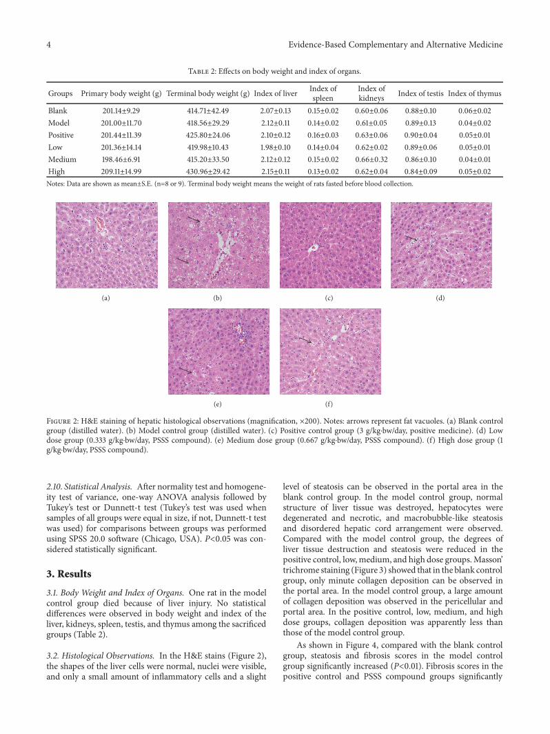
Figure 2: H&E staining of hepatic histological observations (magnification, ×200). Notes: arrows represent fat vacuoles. (a) Blank controlgroup (distilled water). (b) Model control group (distilled water). (c) Positive control group (3 g/kg⋅bw/day, positive medicine). (d) Lowdose group (0.333 g/kg⋅bw/day, PSSS compound). (e) Medium dose group (0.667 g/kg⋅bw/day, PSSS compound). (f) High dose group (1g/kg⋅bw/day, PSSS compound).
2.10. Statistical Analysis. After normality test and homogene-ity test of variance, one-way ANOVA analysis followed byTukey’s test or Dunnett-t test (Tukey’s test was used whensamples of all groups were equal in size, if not, Dunnett-t testwas used) for comparisons between groups was performedusing SPSS 20.0 software (Chicago, USA). P<0.05 was con-sidered statistically significant.
3. Results
3.1. Body Weight and Index of Organs. One rat in the modelcontrol group died because of liver injury. No statisticaldifferences were observed in body weight and index of theliver, kidneys, spleen, testis, and thymus among the sacrificedgroups (Table 2).
3.2. Histological Observations. In the H&E stains (Figure 2),the shapes of the liver cells were normal, nuclei were visible,and only a small amount of inflammatory cells and a slight
level of steatosis can be observed in the portal area in theblank control group. In the model control group, normalstructure of liver tissue was destroyed, hepatocytes weredegenerated and necrotic, and macrobubble-like steatosisand disordered hepatic cord arrangement were observed.Compared with the model control group, the degrees ofliver tissue destruction and steatosis were reduced in thepositive control, low,medium, and high dose groups.Masson’trichrome staining (Figure 3) showed that in the blank controlgroup, only minute collagen deposition can be observed inthe portal area. In the model control group, a large amountof collagen deposition was observed in the pericellular andportal area. In the positive control, low, medium, and highdose groups, collagen deposition was apparently less thanthose of the model control group.
As shown in Figure 4, compared with the blank controlgroup, steatosis and fibrosis scores in the model controlgroup significantly increased (P<0.01). Fibrosis scores in thepositive control and PSSS compound groups significantly

Evidence-Based Complementary and Alternative Medicine 5
(a) (b) (c) (d)
(e) (f)
Figure 3:Masson’ trichrome staining of hepatic histological observations (magnification,×200). Notes: arrows represent collagen deposition.(a) Blank control group (distilled water). (b) Model control group (distilled water). (c) Positive control group (3 g/kg⋅bw/day, positivemedicine). (d) Low dose group (0.333 g/kg⋅bw/day, PSSS compound). (e) Medium dose group (0.667 g/kg bw/day, PSSS compound). (f)High dose group (1 g/kg bw/day, PSSS compound).
Stea
tosis
scor
es
0
1
2
3
4∗∗
Hep
atiti
s sco
res
0.0
0.5
1.0
1.5
2.0
Fibr
osis
scor
es
0
1
2
3
4∗∗
## ##
###
Blank
Model
Positive Low
Medium
HighBlan
kModel
Positive Low
Medium
High Blank
Model
Positive Low
Medium
High
Figure 4: Scores of hepatic histological observations. Notes: data are shown as mean±SE (n=8 or 9). One-way ANOVA analysis followed byDunnett-t test was conducted to calculate statistical significance. ∗∗P<0.01 versus the blank control group; #𝑃 <0.05 and ##𝑃 <0.01 versus themodel control group.
decreased compared with the model control group (P<0.05or P<0.01). The histological results suggest that the ALFrat model was established successfully, and PSSS compoundplays a protective role on ALF.
3.3. Serum Levels of Biochemical Parameters. No statisticallysignificant differences of serum AST, ALP, and GGT levelsbetween groups were observed. Serum ALT levels in themodel control group were significantly elevated comparedwith the blank control group, whereas the ALT levels inthe positive control group were significantly lower comparedwith that in the model control group (P<0.01). Serum TGlevels in the model control, positive control, and PSSScompound groups were all significantly lower than those inthe blank control group (P<0.01) (Figure 5).3.4. Serum Levels of HA, LN, and Hyp. As shown in Figure 6,in the model control group, the serum levels of HA, LN, and
Hyp, all vital indicators of liver fibrosis, were significantlyincreased compared with those in the blank control group(P<0.01). Compared with themodel control group, the serumlevels of HA were significantly decreased in the positivecontrol and PSSS compound groups (P<0.01). The serumlevels of LN were significantly decreased in the positivecontrol, low, and medium dose groups (P<0.05 or P<0.01).The serum levels of Hyp were significantly decreased inthe medium and high dose groups (P<0.05 or P<0.01).These results also demonstrate antifibrotic effect of the PSSScompound.
3.5. Expression Levels of Hepatic MMP-13 and TIMP-1. Asignificant reduction of hepatic MMP-13 and increase ofhepatic TIMP-1 were observed in the model control groupcompared with those in the blank control group. Moreover,the changes were significantly inhibited by PSSS compoundadministration (P<0.01) (Figures 7, 8, and 9), suggesting

6 Evidence-Based Complementary and Alternative Medicine
ALT
(U/L
)
0
20
40
60
80
100
##
AST
(U/L
)
0
100
200
300
ALP
(U/L
)
0
50
100
150G
GT(
U/L
)
0
1
2
3
4
TG(m
mol
/l)
0.0
0.2
0.4
0.6
0.8
1.0
∗∗ ∗∗ ∗∗ ∗∗
Blank
Model
Positive Low
Medium
HighBlan
kModel
Positive Low
Medium
HighBlan
kModel
Positive Low
Medium
High
Blank
Model
Positive Low
Medium
HighBlan
kModel
Positive Low
Medium
High
∗∗
Figure 5: Serum levels of ALT, AST, ALP, GGT, and TG. Notes: data are shown as mean±SE (n=8 or 9). One-way ANOVA analysis followedby Dunnett-t test was conducted to calculate statistical significance. ∗∗P<0.01 versus the blank control group; ##𝑃 <0.01 versus the modelcontrol group. ALT, alanine aminotransferase; AST, aspartate aminotransferase; ALP, alkaline phosphatase; GGT, 𝛾-glutamyl transpeptidase;TG, triglyceride.
HA
(ng/
ml)
0
1
3
2
∗∗
## ####
## LN (p
g/m
l)
0
200
400
600## # ##
∗∗H
yp (p
g/m
l)
0
50
100
150
200∗∗
###
Blank
Model
Positive Low
Medium
HighBlan
kModel
Positive Low
Medium
High Blank
Model
Positive Low
Medium
High
Figure 6: Serum levels of HA, LN, and Hyp. Notes: data are shown as mean±SE (n=6). One-way ANOVA analysis followed by Tukey’s testwas conducted to calculate statistical significance. ∗∗P<0.01 versus the blank control group; #𝑃 <0.05 and ##𝑃 <0.01 versus the model controlgroup. HA, hyaluronan; LN, laminin; Hyp, hydroxyproline.
protective effect of the compound involves MMP-13 andTIMP-1.
3.6. mRNA and Protein Expression Levels of Hepatic TGF-𝛽1/Smad Signaling Pathway. As shown in Figures 10, 11, and12, in the model control group, relative mRNAand/or proteinexpression levels of hepatic TGF-𝛽1, (phospho-)Smad2, and(phospho-)Smad3 were significantly higher, and the Smad7level was significantly lower than that of the blank controlgroup (P<0.05 or P<0.01). Compared with the model con-trol group, the protein expression levels of TGF-𝛽1 weresignificantly lower in the positive control and the mediumand high dose groups (P<0.05 or P<0.01). Both mRNA andprotein expression levels of (phospho-)Smad2 and (phospho-)Smad3 were significantly lower in the positive control andPSSS compound groups (P<0.05 or P<0.01). The protein
expression levels of Smad7 were significantly high in thePSSS compound groups (P<0.01). These results suggest thatALF alleviation of PSSS compound involves TGF-𝛽1/Smadsignaling pathway.
4. Discussion
ALF is an important health problem, causing tremendousdisease burden and medical expenses. Thus, research onits pathogenesis and potential therapeutic agents are ofgreat concern in medical science. The establishment of anALF animal model is necessary to explore latent treatmentremedy, and an ideal ALF animal model should have similarpathological characteristics to human ALF. The models ofLieber–Decarli alcohol liquid diet feeding ad libitum havebeen widely used [18], whereas applying alcohol alone is not

Evidence-Based Complementary and Alternative Medicine 7
(a) (b) (c)
(d) (e) (f)
Figure 7: Expression levels of hepatic MMP-13 (magnification, ×400). Notes: arrows represent the positive expression of MMP-13. (a) Blankcontrol group (distilled water). (b) Model control group (distilled water). (c) Positive control group (3 g/kg⋅bw/day, positive medicine). (d)Low dose group (0.333 g/kg⋅bw/day, PSSS compound). (e) Medium dose group (0.667 g/kg⋅bw/day, PSSS compound). (f) High dose group (1g/kg⋅bw/day, PSSS compound). MMP-13, matrix metalloproteinase-13.
(a) (b) (c)
(d) (e) (f)
Figure 8: Expression levels of hepatic TIMP-1 (magnification, ×400). Notes: arrows represent the positive expression of TIMP-1. (a) Blankcontrol group (distilled water). (b) Model control group (distilled water). (c) Positive control group (3 g/kg⋅bw/day, positive medicine). (d)Low dose group (0.333 g/kg⋅bw/day, PSSS compound). (e) Medium dose group (0.667 g/kg⋅bw/day, PSSS compound). (f) High dose group (1g/kg⋅bw/day, PSSS compound). TIMP-1, tissue inhibitor of metalloproteinase-1.

8 Evidence-Based Complementary and Alternative Medicine
MM
P-13
area
(%)
0
5
10
15
20
25
##
####
##
TIM
P-1
area
(%)
0
5
10
15
##
####
##∗∗
∗∗
Blank
Model
Positive Low
Medium
HighBlan
kModel
Positive Low
Medium
High
Figure 9: Quantitative analysis of the expression levels of hepatic MMP-13 and TIMP-1. Notes: data are shown as mean±SE (n=5). One-wayANOVA analysis followed by Tukey’s test was conducted to calculate the statistical significance. ∗∗P<0.01 versus the blank control group;##𝑃 <0.01 versus the model control group. MMP-13, matrix metalloproteinase-13; TIMP-1, tissue inhibitor of metalloproteinase-1.
TGF-
1/
-act
in m
RNA
0
2
4
6∗∗
Smad
2/
-act
in m
RNA
0
5
10
15∗∗
#### ##
#
Smad
3/
-act
in m
RNA
0
10
20
30
40
## ## ##
##
Smad
7/
-act
in m
RNA
0
10
20
30
40
50
##
Blank
Model
Positive Low
Medium
HighBlan
kModel
Positive Low
Medium
High
Blank
Model
Positive Low
Medium
HighBlank
Model
Positive Low
Medium
High
∗∗
∗∗
Figure 10: mRNA relative expression levels of hepatic TGF-𝛽1, Smad2, Smad3, and Smad7. Notes: data are shown as mean±SE (n=3). One-way ANOVA analysis followed by Tukey’s test was conducted to calculate statistical significance. ∗∗P<0.01 versus the blank control group;#𝑃 <0.05 and ##𝑃 <0.01 versus the model control group. TGF-𝛽1, transforming growth factor-𝛽1.
enough to induce liver fibrosis in animals under laboratoryenvironment [19]. Therefore, in this study, the ALF animalmodels were developed by a secondary agent (CCl
4) accom-panied with chronic alcohol treatment, called a “two-hit”model [20].
In traditional Lieber–Decarli diet, alcohol supplies calo-ries of as high as 36%. However, rats’ natural aversion toalcohol may affect the intake of this diet and cause nutritionaldeficiencies [21]. In this study, we successfully developedan ALF rat model by 13-week feeding of Lieber–Decarlialcohol liquid diet with reducing alcohol calories (30%), plustrace CCl
4 injection in the subsequent 5 weeks. Pathologicalchanges similar to human ALF, which includes steatosis,hepatocellular damage (ballooning), and a variable degree
of pericellular and lobular fibrosis typically manifestedin ALF [22], were replicated in our rat model (Figures2 and 3).
In recent decades, a series of studies reported that extractsor a compound from herb exhibited protective effects onALD [23–25], ALF included [20, 26], certainly. P. lobata(Willd.), S. miltiorrhiza, S. chinensis, and S. marianum haveall been reported to have potential capability in alleviatingALD [25, 27–29] and chemical-induced liver fibrosis [6, 30–32]. However, to the best of our knowledge, no report on theeffect onALFof the combination of the four herbs is available.Interestingly, combination of traditional Chinese herbs hasbeen practiced for thousands of years and can strengthen theefficacies. Therefore, in this study, four Chinese herbs were

Evidence-Based Complementary and Alternative Medicine 9
TGF-1
p-Smad2
Smad2
p-Smad3
Smad3
Smad7
GADPH
A B C D E F
Figure 11: Protein expression levels of hepatic TGF-𝛽1, p-Smad2, Smad2, p-Smad3, Smad3, and Smad7. Notes: A: blank control group(distilled water); B: model control group (distilled water); C: positive control group (3 g/kg⋅bw/day, positive medicine); D: low dose group(0.333 g/kg⋅bw/day, PSSS compound); E: medium dose group (0.667 g/kg⋅bw/day, PSSS compound); F: High dose group (1 g/kg⋅bw/day, PSSScompound). TGF-𝛽1, transforming growth factor-𝛽1.
TGF-
1/
GA
DPH
pro
tein
0.0
0.2
0.4
0.6
0.8
1.0
##
##
p-Sm
ad2/
Smad
2 pr
otei
n
0.0
0.1
0.2
0.3
0.4
∗∗
####
## ##
p-Sm
ad3/
Smad
3 pr
otei
n
0.0
0.2
0.4
0.6
0.8
∗∗
##
## ##
Smad
7/G
AD
PH p
rote
in
0.0
0.2
0.4
0.6
0.8
1.0
∗∗
####
##
Blank
Model
Positive Low
Medium
High
Blank
Model
Positive Low
Medium
High
Blank
Model
Positive Low
Medium
High
Blank
Model
Positive Low
Medium
High
##
Figure 12: Semiquantitative analysis of the relative expression levels of hepatic TGF-𝛽1, p-Smad2, p-Smad3, and Smad7. Data are shown asmean±SE (n=3). One-way ANOVA analysis followed by Tukey’s test was conducted to calculate statistical significance. ∗∗P<0.01 versus theblank control group; #𝑃 <0.05 and ##𝑃 <0.01 versus the model control group. TGF-𝛽1, transforming growth factor-𝛽1.
administered collectively, and the compound significantlydecreased the liver fibrosis scores, indicators of liver fibrosis,i.e., serum levels of HA, LN, and Hyp, indicating beneficialrole on ALF of this compound.
TGs are mainly synthesized, secreted, and catabolized inthe liver [33]. As a result, TG synthesis would be disrupted
when liver function is impaired, and serum levels of TGcan be reduced. Our study found a significant decline in theserum levels of TG in the model control group, the positivecontrol group, and the PSSS compound groups, probably dueto damaged rat liver function or not fully recovered liverfunction in these groups.

10 Evidence-Based Complementary and Alternative Medicine
ECMs are degraded by MMPs regulated by theirinhibitors, i.e., TIMPs [34]. MMP-13 and TIMP-1 play acrucial role in modulation of liver fibrosis in rodents [35].In the present study, a significantly decreased expressionof hepatic TIMP-1 and an increased expression of hepaticMMP-13 in the PSSS compound groups demonstrate that theantifibrotic effect relates to regulation of ECMs via MMP-13and TIMP-1.
TGF-𝛽1 plays an essential role in fibrotic diseases, whichcan directly or indirectly bind to TGF-𝛽 type II receptors,and then phosphorylate and activate TGF-𝛽 type I receptors[36]. Activated type I TGF-𝛽 receptors phosphorylate Smad2and Smad3, which bind to Smad4 and form a complex [37].The complex then translocates from the cytoplasm into thenucleus and interacts with other transcription factors [38].In addition, Smad6 or Smad7 is an inhibitive factor in thephosphorylation of Smad2 and Smad3 [39]. In our study,the compound treatment significantly decreased relativemRNA and/or protein expression levels of hepatic TGF-𝛽1, (phospho-)Smad2, and (phospho-)Smad3 but increasedthe Smad7 expression. The changes of TGF-𝛽1 and Smadsare consistent with previous studies about the molecularmechanism of liver fibrosis [39–41], suggesting that the pro-tective effect of the compound is via TGF-𝛽1/Smad signalingpathway.
It is still not fully clear about which categories of bioactivecomponents could effectively reverse alcoholic liver fibrosis.Puerarin, salvianolic acid A, schisandrin B, and silymarin,major bioactive components of P. lobata (Willd.), S. mil-tiorrhiza, S. chinensis, and S. marianum, respectively, havebeen reported to can alleviate CCl4-induced liver fibrosisor chronic alcoholic liver injuries [6, 7, 9, 31]. Furthermore,compound used in our study is rich in these bioactivecomponents. Thus, it is reasonable to presume that thesebioactive components may be the functional componentsfor ALF. Nevertheless, identifying and maybe extractingbioactive components using the instrumental analysis, fromthese Chinese herbs, i.e., P. lobata (Willd.), S. miltiorrhiza,S. chinensis, and S. marianum, are surely a direction of ourfuture study.
5. Conclusion
This study demonstrates that a compound of four Chineseherbs, namely, P. lobata (Willd.), S. miltiorrhiza, S. chinensis,and S. marianum, can be a novel therapeutic agent on pre-venting and reversing ALF, and the underlying mechanismof the protective effects may involve TGF-𝛽1/Smad signalingpathway.
Data Availability
All the data related to this article were available from thecorresponding author upon reasonable request.
Conflicts of Interest
All authors declare that there are no conflicts of interest.
Acknowledgments
This study is supported by Nutrition Research Fund of By-Health (TY0161104).Thanks are due to the research platformprovided by Public Health and Medicine Experiment Teach-ing Center of West China School of Public Health, SichuanUniversity.
References
[1] F. Magdaleno, C. C. Blajszczak, and N. Nieto, “Key eventsparticipating in the pathogenesis of alcoholic liver disease,”Biomolecules, vol. 7, no. 1, p. e9, 2017.
[2] A. Louvet and P. Mathurin, “Alcoholic liver disease: mech-anisms of injury and targeted treatment,” Nature ReviewsGastroenterology & Hepatology, vol. 12, no. 4, pp. 231–242, 2015.
[3] R.-B. Ding, K. Tian, L.-L. Huang et al., “Herbal medicines forthe prevention of alcoholic liver disease: a review,” Journal ofEthnopharmacology, vol. 144, no. 3, pp. 457–465, 2012.
[4] L. Abenavoli, A. A. Izzo, N. Milic, C. Cicala, A. Santini, and R.Capasso, “Milk thistle (Silybummarianum): a concise overviewon its chemistry, pharmacological, and nutraceutical uses inliver diseases,” Phytotherapy Research, vol. 32, no. 11, pp. 2202–2213, 2018.
[5] Y. Peng, T. Yang, K. Huang, L. Shen, Y. Tao, and C. Liu,“Salvia miltiorrhiza ameliorates liver fibrosis by activatinghepatic natural killer cells in vivo and in vitro,” Frontiers inPharmacology, vol. 9, no. 762, article 762, 2018.
[6] Q. Chen, H. Zhang, Y. Cao et al., “Schisandrin B attenuatesCCl4-induced liver fibrosis in rats by regulation of Nrf2-ARE and TGF-beta/Smad signaling pathways,” Drug Design,Development and�erapy, vol. 11, pp. 2179–2191, 2017.
[7] C. Guo, L. Xu, Q. He, T. Liang, X. Duan, and R. Li, “Anti-fibroticeffects of puerarin on CCl4-induced hepatic fibrosis in ratspossibly through the regulation of PPAR-gamma expressionand inhibition of PI3K/Akt pathway,” Food and ChemicalToxicology, vol. 56, pp. 436–442, 2013.
[8] R. Yuan, X. Tao, S. Liang et al., “Protective effect of acidicpolysaccharide from Schisandra chinensis on acute ethanol-induced liver injury through reducing CYP2E1-dependentoxidative stress,” Biomedicine & Pharmacotherapy, vol. 99, pp.537–542, 2018.
[9] X. Shi, Y. Zhao, C. Ding et al., “Salvianolic acid A alleviateschronic ethanol-induced liver injury via promotion of beta-catenin nuclear accumulation by restoring SIRT1 in rats,”Toxicology and Applied Pharmacology, vol. 350, pp. 21–31, 2018.
[10] X. Chen, R. Li, T. Liang, K. Zhang, Y. Gao, and L. Xu, “Puerarinimprovesmetabolic function leading to hepatoprotective effectsin chronic alcohol-induced liver injury in rats,” Phytomedicine,vol. 20, no. 10, pp. 849–852, 2013.
[11] S. K. Das and D. M. Vasudevan, “Protective effects of silymarin,a milk thistle (Silybium marianum) derivative on ethanol-induced oxidative stress in liver,” Indian Journal of Biochemistryand Biophysics, vol. 43, no. 5, pp. 306–311, 2006.
[12] L. Shi, X. Tang, X. Dang et al., “Investigating herb-herbinteractions: the potential attenuated toxicity mechanism of thecombined use of Glycyrrhizae radix et rhizoma (Gancao) andSophorae flavescentis radix (Kushen),” Journal of Ethnopharma-cology, vol. 165, pp. 243–250, 2015.
[13] S. Wang, Y. Hu, W. Tan et al., “Compatibility art of traditionalChinese medicine: from the perspective of herb pairs,” Journalof Ethnopharmacology, vol. 143, no. 2, pp. 412–423, 2012.

Evidence-Based Complementary and Alternative Medicine 11
[14] F. J. Cubero, R. Urtasun, and N. Nieto, “Alcohol and liverfibrosis,” Seminars in Liver Disease, vol. 29, no. 2, pp. 211–221,2009.
[15] S. L. Friedman, “Hepatic stellate cells: protean, multifunctional,and enigmatic cells of the liver,” Physiological Reviews, vol. 88,no. 1, pp. 125–172, 2008.
[16] S. CrespoYanguas, B. Cogliati, J.Willebrords et al., “Experimen-tal models of liver fibrosis,” Archives of Toxicology, vol. 90, no. 5,pp. 1025–1048, 2016.
[17] Y.M. Li, J. G. Fan, B. Y.Wang et al., “Guidelines formanagementof nonalcoholic fatty liver disease: an updated and revisededition,” Zhonghua Gan Zang Bing Za Zhi (in Chinese), vol. 18,no. 3, pp. 163–166, 2010.
[18] Q. Wang, X. Dai, W. Yang et al., “Caffeine protects againstalcohol-induced liver fibrosis by dampening the cAMP/PKA/CREB pathway in rat hepatic stellate cells,” InternationalImmunopharmacology, vol. 25, no. 2, pp. 340–352, 2015.
[19] G. E. Arteel, “Animalmodels of alcoholic liver disease,”DigestiveDiseases, vol. 28, no. 6, pp. 729–736, 2010.
[20] I. Bingul, C. Basaran-Kucukgergin, A. F. Aydin et al., “Betainetreatment decreased oxidative stress, inflammation, and stellatecell activation in rats with alcoholic liver fibrosis,” Environmen-tal Toxicology and Pharmacology, vol. 45, pp. 170–178, 2016.
[21] S. Mathews, M. Xu, H. Wang, A. Bertola, and B. Gao, “Animalsmodels of gastrointestinal and liver diseases. animal modelsof alcohol-induced liver disease: pathophysiology, translationalrelevance, and challenges,” American Journal of Physiology-Gastrointestinal and Liver Physiology, vol. 306, no. 10, pp. G819–G823, 2014.
[22] R. Bataller and B. Gao, “Liver fibrosis in alcoholic liver disease,”Seminars in Liver Disease, vol. 35, no. 2, pp. 146–156, 2015.
[23] S.-J. Yoon, E.-J. Koh, C.-S. Kim et al., “Agrimonia eupatoriaprotects against chronic ethanol-induced liver injury in rats,”Food and Chemical Toxicology, vol. 50, no. 7, pp. 2335–2341,2012.
[24] S.-J. Kim and S.-M. Lee, “Effect of baicalin on toll-like receptor4-mediated ischemia/reperfusion inflammatory responses inalcoholic fatty liver condition,” Toxicology and Applied Pharma-cology, vol. 258, no. 1, pp. 43–50, 2012.
[25] N. Zhang, Y. Hu, C. Ding et al., “Salvianolic acid B protectsagainst chronic alcoholic liver injury via SIRT1-mediated inhi-bition of CRP and ChREBP in rats,” Toxicology Letters, vol. 267,pp. 1–10, 2017.
[26] M. Liu, Y. Xu, X. Han et al., “Dioscin alleviates alcoholicliver fibrosis by attenuating hepatic stellate cell activationvia the TLR4/MyD88/NF-kappaB signaling pathway,” ScientificReports, vol. 5, article 18038, 2015.
[27] R. Li, T. Liang, Q. He et al., “Puerarin, isolated from Kudzu root(Willd.), attenuates hepatocellular cytotoxicity and regulatesthe GSK-3beta/NF-kappaB pathway for exerting the hepato-protection against chronic alcohol-induced liver injury in rats,”International Immunopharmacology, vol. 17, no. 1, pp. 71–78,2013.
[28] B. Li, D. Li, Y. Wang et al., “Schisantherin A alleviated alcohol-induced liver injury by the regulation of alcohol metabolismand NF-kB pathway,” Journal of Experimental Animal Science,vol. 67, no. 4, pp. 451–461, 2018.
[29] S. K. Das and S. Mukherjee, “Biochemical and immunologicalbasis of silymarin effect, a milk thistle (Silybum marianum)against ethanol-induced oxidative damage,” Toxicology Mecha-nisms and Methods, vol. 22, no. 5, pp. 409–413, 2012.
[30] S. Wang, X.-L. Shi, M. Feng et al., “Puerarin protects againstCCl4-induced liver fibrosis in mice: Possible role of PARP-1 inhibition,” International Immunopharmacology, vol. 38, pp.238–245, 2016.
[31] S. Clichici, D. Olteanu, A. Filip, A.-L. Nagy, A. Oros, and P. A.Mircea, “Beneficial effects of silymarin after the discontinuationof CCl4-induced liver fibrosis,” Journal of Medicinal Food, vol.19, no. 8, pp. 789–797, 2016.
[32] D. R. Parajuli, Y.-Z. Zhao, H. Jin et al., “Anti-fibrotic effectof PF2401-SF, a standardized fraction of Salvia miltiorrhiza,in thioacetamide-induced experimental rats liver fibrosis,”Archives of Pharmacal Research, vol. 38, no. 4, pp. 549–555, 2015.
[33] A. P. Jensen-Urstad and C. F. Semenkovich, “Fatty acid synthaseand liver triglyceride metabolism: housekeeper or messenger?”Biochimica et Biophysica Acta, vol. 1821, no. 5, pp. 747–753, 2012.
[34] E. Roeb, “Matrix metalloproteinases and liver fibrosis (transla-tional aspects),”Matrix Biology, vol. 68-69, pp. 463–473, 2018.
[35] S. Hemmann, J. Graf, M. Roderfeld, and E. Roeb, “Expressionof MMPs and TIMPs in liver fibrosis - a systematic reviewwith special emphasis on anti-fibrotic strategies,” Journal ofHepatology, vol. 46, no. 5, pp. 955–975, 2007.
[36] G. C. Blobe, W. P. Schiemann, and H. F. Lodish, “Role oftransforming growth factor beta in human disease,” �e NewEngland Journal of Medicine, vol. 342, no. 18, pp. 1350–1358,2000.
[37] R. Derynck and Y. E. Zhang, “Smad-dependent and Smad-independent pathways in TGF-beta family signalling,” Nature,vol. 425, no. 6958, pp. 577–584, 2003.
[38] J. H. Yang, S. C. Kim, K. M. Kim et al., “Isorhamnetin atten-uates liver fibrosis by inhibiting TGF-beta/Smad signaling andrelieving oxidative stress,” European Journal of Pharmacology,vol. 783, pp. 92–102, 2016.
[39] A. A. Ganai and M. Husain, “Genistein attenuates D-GalNinduced liver fibrosis/chronic liver damage in rats by blockingthe TGF-beta/Smad signaling pathways,” Chemico-BiologicalInteractions, vol. 261, pp. 80–85, 2017.
[40] L.-X. Tang, R.-H. He, G. Yang et al., “Asiatic acid inhibits liverfibrosis by blocking TGF-beta/smad signaling in vivo and invitro,” PLoS ONE, vol. 7, no. 2, Article ID e31350, 2012.
[41] T. Wu, J.-M. Chen, T.-G. Xiao et al., “Qinggan Huoxue Recipesuppresses epithelial-to-mesenchymal transition in alcoholicliver fibrosis through TGF-beta1/Smad signaling pathway,”World Journal ofGastroenterology, vol. 22, no. 19, pp. 4695–4706,2016.

Stem Cells International
Hindawiwww.hindawi.com Volume 2018
Hindawiwww.hindawi.com Volume 2018
MEDIATORSINFLAMMATION
of
EndocrinologyInternational Journal of
Hindawiwww.hindawi.com Volume 2018
Hindawiwww.hindawi.com Volume 2018
Disease Markers
Hindawiwww.hindawi.com Volume 2018
BioMed Research International
OncologyJournal of
Hindawiwww.hindawi.com Volume 2013
Hindawiwww.hindawi.com Volume 2018
Oxidative Medicine and Cellular Longevity
Hindawiwww.hindawi.com Volume 2018
PPAR Research
Hindawi Publishing Corporation http://www.hindawi.com Volume 2013Hindawiwww.hindawi.com
The Scientific World Journal
Volume 2018
Immunology ResearchHindawiwww.hindawi.com Volume 2018
Journal of
ObesityJournal of
Hindawiwww.hindawi.com Volume 2018
Hindawiwww.hindawi.com Volume 2018
Computational and Mathematical Methods in Medicine
Hindawiwww.hindawi.com Volume 2018
Behavioural Neurology
OphthalmologyJournal of
Hindawiwww.hindawi.com Volume 2018
Diabetes ResearchJournal of
Hindawiwww.hindawi.com Volume 2018
Hindawiwww.hindawi.com Volume 2018
Research and TreatmentAIDS
Hindawiwww.hindawi.com Volume 2018
Gastroenterology Research and Practice
Hindawiwww.hindawi.com Volume 2018
Parkinson’s Disease
Evidence-Based Complementary andAlternative Medicine
Volume 2018Hindawiwww.hindawi.com
Submit your manuscripts atwww.hindawi.com