Embed Size (px)
DESCRIPTION
Wawannnnnn
Citation preview
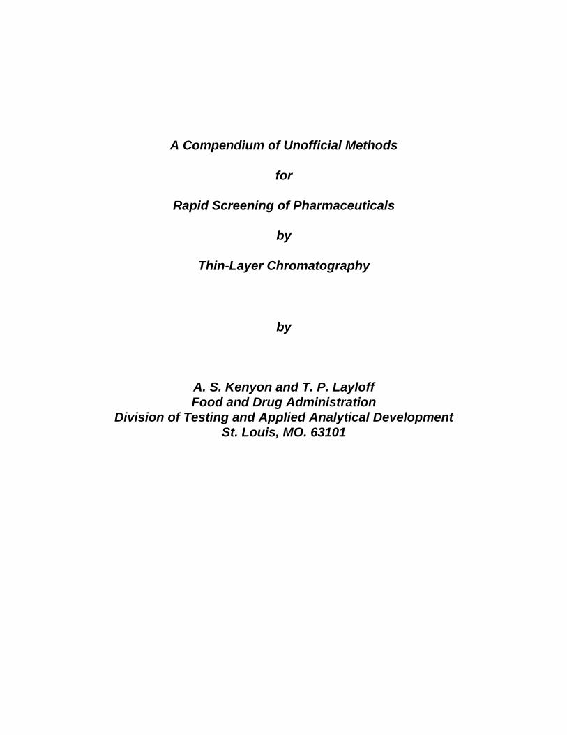
A Compendium of Unofficial Methods
for
Rapid Screening of Pharmaceuticals
by
Thin-Layer Chromatography
by
A. S. Kenyon and T. P. LayloffFood and Drug Administration
Division of Testing and Applied Analytical DevelopmentSt. Louis, MO. 63101

Table of ContentsTitle PageIntroduction 4Rapid Screening of Drugs by Thin-Layer Chromatography 4Detector Solutions 9
References 10 Acknowledgements 10 Acetylsalicylic Acid 11Allopurinol 13Aminophylline 15Amoxicillin 17Ampicillin 19Atropine 21Betamethasone 23Carbamazepine 25Cephalexin 27Cephradine 29Chloramphenicol 31Chloroquine phosphate 33Chlorpheniramine maleate 35Ciprofloxacine Hcl 37Cloxicillin 39Dexamethasone 41Dexamethasone sodium phosphate 43Diazepam 45Digoxin 47Diphenhydramine Hcl 49Ergotamine tartrate 51Erythromycin ethylsuccinate 53Erythromycin estolate 55Erythromycin & erythromycin stearate 57Estradiol cypionate 59Ethambutol 61Furosemide 65Gentamycin sulfate 67Hydrochlorothiazide 69Hydrocortisone 71Ibuprofen 73Imipramine Hcl 75
Title PageIndomethacin 77Isoniazid 79Kanamycin Sulfate 84Ketoconazole 86Mebendazole 88Medroxyprogesterone acetate 90Medroxyprogesterone acetate injection 92Methyldopa 94Metronidazole 96Neomycin sulfate 98Nitrofurantoin 100Norgestrel 102Nystatin 104Paracetamol 106Penicillin/procaine 108Phenobarbital 110Phenytoin 112Praziquantel 116Prednisolone 118Prednisone 120Promethazine 122Pyrimetamine 124Quinine sulfate 126Rifampicin 128Salbutamol (albuterol) 132Streptomycin sulfate 134Sulfamethazine 136Sulfamethoxazole 138Testosterone cypionate 140Tetracycline 142Theophylline 145Triamcinolone 147Triamterene 149Trifluoperazine Hcl 151Trimethoprim 153Vitamin A (retinol) 155Warfarin 157Appendix 1 - Diethylene Glycol and Glycerin 159
Appendix 2 - Improved TLC Procedures 177
Rapid Screening of Pharmaceuticals by Thin-Layer Chromatography
IntroductionThin-layer chromatography provides a quick, economical, and reliable method
for rapid screening of pharmaceuticals. The screening method can be used after littletraining, and in areas outside the laboratory. This compendium of drug analyticalmethods has been developed for rapid screening of drugs in such places as ports ofentry, pharmacies, distribution centers, or areas lacking resources for other methodsof analysis. The technique reduces the need for other analytical methods which aremore costly and time consuming, and which require highly trained operators. Themethods are based on a portable system using a plastic bag for development andare easy to use in field-type operations. None of the methods described are officialin any compendium.
In working with any chemical, safety and disposal must be considered beforeperforming an analysis. All chemicals are toxic, and should be handled accordingly.The analyst should not breathe or inhale vapors or dust from any of these chemicals,including the dust from the finely divided silica on the TLC plates. Plastic or rubbergloves should be used whenever contact with these chemicals is possible. In thesemethods, an effort has been made to reduce the risk of toxicity of the solvents byusing small quantities to reduce exposure and by eliminating toxic chlorinatedsolvents. The toxicity of the chemicals used in these methods is similar to that ofsolvents used in applying paint. All analyses should be performed in areas withadequate ventilation. The rules of disposal for your local area should be followed.Chemicals used in TLC operations are flammable, and must be kept away fromflames or ignition sources. Because iodine stains skin and clothing, protectiveclothing and rubber gloves should be worn when handling it.
This compendium describes the procedures for the analysis of the listed drugsin which rapid TLC is used as a screening method. These methods were developedin our laboratory and have not been collaboratively tested. If problems areencountered with any of these test methods, please notify us by FAX or by mailmarked to the attention of the Director, Division of Drug Analysis, Food and DrugAdministration. FAX number: 314-539-2113. Address: 1114 Market St, Room 1002, St. Louis, MO63101, USA.
Rapid Screening of Drugs by Thin-Layer ChromatographyThe TLC method described here is semiquantitative. The method gives a
good estimate of whether the drug is the same as that listed on the label, and if thecontent is the correct amount as specified. It is not intended to replace any officialcompendium method. The drugs selected for this compendium were taken from thelist of essential drugs developed by the World Health Organization, essential druglists from several countries in Africa, and reports of actual field use in developingcountries. The methods do not cover the complete list of essential drugs, butrepresent many different classes of drugs. The drugs were selected on a prioritybasis, i.e., those needed for life-threatening diseases were given the highest priority.
The TLC method was designed to rapidly screen drugs by using apolyethylene bag as the chamber. Two reference concentrations representing theupper and lower concentration for the dosage limits (85% and 115 or 120%) arespotted on a plate along with the sample solution representing 100%. The samplesolution is spotted between the reference solutions. The spots are examined visuallyeither by ultraviolet light or by iodine staining. The drug is considered to be withinspecifications if the intensity of the sample spots lies between the intensity of the tworeference solution spots. The sample should be further tested by an official methodif the intensity of the sample spot lies near the lower limit. The screening methodeliminates the need for further analysis of those drugs which show concentrationswithin the specification range.
Many drugs that are not listed in the compendium will need to be analyzed.Methods can be developed for a drug not listed in this compendium by followingthese simple steps:
1. Determine a suitable solvent system by studying the molecular structure ofthe drug; consult some reference book such as the Merck Index for a suitablesolvent. Choose the solvent with the lowest polarity when more than one solvent ispossible.
2. Prepare a solution of the standard drug at a concentration approximatelyequal to 1 mg/mL. Spot this solution on a TLC plate.
3. Prepare a developer solution mixture to have a middle range of polarity,such as equal volumes of toluene and methanol. If the drug is acidic, add a smallamount of glacial acetic acid; if the drug is basic, add concentrated ammoniumhydroxide. To select a developer mixture, dip the spotted plate into a beakercontaining the developer, cover the beaker to prevent evaporation, and observe themovement of the spots.
4. Reduce the polarity of the developer if the spots follow the solvent front;increase the polarity of the developer if the spots do not move or move less than 1cm.
5. After determining the solvent mixture for suitable separation, adjust theconcentrations to show differences in spot intensities.
6. Add the new method to the compendium for later use.

3
The methods described in this compendium have been based on iodinestaining as a satisfactory means of detection. In many cases the concentrationsspecified may be too high for suitable detection by ultraviolet light because of highabsorbance at the 254 nm UV wavelength. The concentrations of the solutions aretoo high when no difference in intensity between solutions of different concentrationscan be detected. In this case, the concentration should be reduced by diluting theprepared solutions of sample and reference. The concentrations specified for thesample and standards were determined experimentally to give suitable detection.Many drugs are supplied in dosages other than those listed in the compendium. Thefinal concentration of the sample should be kept the same as the listed concentrationwhen other dosages are used and may be prepared either by using larger volumesof solvent or by diluting a concentrated solution. Diluting the concentrated solutionwill use less solvent. The drug may also be supplied in different dosage forms, suchas liquids. Drugs in liquid form are handled on a volume basis (mg/mL) and arediluted if necessary.
The availability of reference standards and their cost is a matter of concernto all who analyze drugs. These procedures have been written for standardssupplied as primary or secondary standards. Primary standards are costly, butsecondary standards can be used successfully. Secondary standards may beobtained from a previously analyzed sample or from reputable chemical suppliers.When either primary or secondary standards are used, the standards must beweighed on an analytical balance capable of weighing to 0.1 mg, and a large enoughquantity must be weighed to minimize the error. The error can be further reduced byusing a semi-micro balance (one that weighs to the 5th place or 0.01 mg). Anotherpossibility for a reference material is a tablet containing a fixed quantity of the drug;it can be used by simply dissolving a reference tablet in the specified volume ofsolvent to produce the high and low concentrations needed for reference. Noweighing is required when reference tablets are available, but at present, theavailability of reference tablets is limited. Therefore weighing is usually necessary.A study is underway to develop reference tablets with the correct content to preparethe high and low reference solutions. It is noted that the procedures for bothconditions are given for three of the drugs described here. These three drugs wereused to establish the feasibility of the reference tablet concept.
The TLC procedures described are based on the use of a portable kit whichis supplied with plastic bags, holders, and all the accessories required to perform theanalysis. Volumes used in the compendium methods are those suitable for a flatplastic bag 8 cm wide. The kits have been supplied with plastic bags 10 cm widewhich require 30 mL of the developer; therefore all volumes of the developer mixturesmust be adjusted by increasing each volume by 50%. The flat 8 cm plastic tubingcan be obtained in rolls (066 gage), and bags can be fabricated from the 8 cm tubingby using a bag sealer. It is recommended that a roll of flat plastic tubing and bagsealer be purchased to ensure an adequate supply over an extended period, and toreduce the cost of developer solvents.
TLC plates are available with many different coatings and supports. Themethods developed in this compendium are based on plastic-backed silica platescontaining a fluorescent material. Merck plastic-backed plates designated as 60F254 have been found most satisfactory. TLC plates made by other manufacturersare also acceptable if they have the same specifications. Coated glass plates aresuitable, but will increase the cost. A plate 5 X 10 cm is required for the apparatus.Cutting glass plates from larger plates is not recommended. Aluminum-backed plateshave also been satisfactory when used with developers that are not too stronglyacidic or basic. TLC plates without the fluorescent materials cannot be used forultraviolet detection; the detection must be done by other means. If both kinds ofplates are used, they must be kept separate to avoid mistakes. Plain silica-coatedplates are more easily damaged. The 60 F254 plastic- backed plates give the bestall-around performance.
The bag for iodine staining can be made as follows: Cut the development bagapproximately 12 cm above the seal. Cut a slit in one side of the bag approximately9 cm above the seal. Place some protective covering on a vertical surface to protectthe surface from stain. The protective covering can be cardboard, plastic film, or anyother type of material which can be discarded. Tape the bag (top of the bag abovethe slit) to a vertical surface on top of the protective covering. Tape the bottom ofthe bag to the vertical surface. Tape a small, flat, rigid object to the bag at the sealpoint of the bag so that the rigid object can act as a hinge to displace the iodinesolution upwards.
The TLC analysis is based on the use of one dosage unit to prepare theneeded concentration. The complete ground tablet must be placed into the vessel;it can be added by performing the grinding in a small plastic bag and then adding thebag and contents to the vessel. A bag approximately 3 X 5 cm is adequate. Bagsof this size can be prepared from the flat 8 cm plastic tubing by sealing the bottomof the bag and then making two parallel vertical seals to make two small bags.These two bags will be approximately 3 X 5 cm each. Drugs in capsule form do notneed to be ground.
The developers described do not include chloroform or other halogencompounds because of their toxicity. To eliminate chloroform, mixtures of solventsare required to achieve a polarity similar to that of chloroform as calculated from aseries of polarities. The compositions of the developer solutions were selected sothat they could be used safely where little or no laboratory facility exists. Otherdevelopers may be used for screening purposes to obtain different separations.Chloroform may be used by trained operators in well-equipped laboratories withproper hoods. Chloroform should not be used in open areas or by untrainedpersonnel. Other developers produce different heights of the spots and differenttimes for the solvent front to reach the migration limit. Spot positions should be keptbetween R values of 0.2 and 0.8.f
When any drug is shown not to meet specifications, the analysis should berepeated to verify the result. The drug should be submitted for analysis by an
approved method when the result shows a marginal content near the 85% level.Most analyses will show a drug to be near the midpoint between the upper and lowerreference solutions, thus eliminating the need for further analysis.
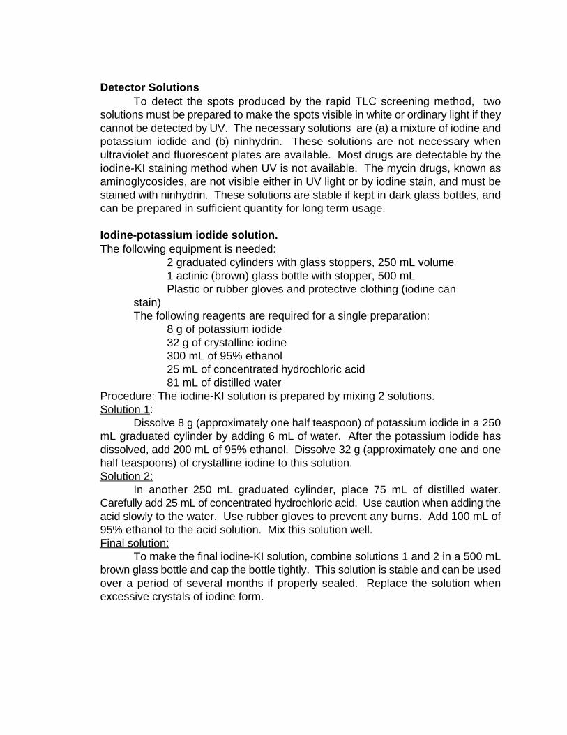
Detector SolutionsTo detect the spots produced by the rapid TLC screening method, two
solutions must be prepared to make the spots visible in white or ordinary light if theycannot be detected by UV. The necessary solutions are (a) a mixture of iodine andpotassium iodide and (b) ninhydrin. These solutions are not necessary whenultraviolet and fluorescent plates are available. Most drugs are detectable by theiodine-KI staining method when UV is not available. The mycin drugs, known asaminoglycosides, are not visible either in UV light or by iodine stain, and must bestained with ninhydrin. These solutions are stable if kept in dark glass bottles, andcan be prepared in sufficient quantity for long term usage.
Iodine-potassium iodide solution.The following equipment is needed:
2 graduated cylinders with glass stoppers, 250 mL volume1 actinic (brown) glass bottle with stopper, 500 mLPlastic or rubber gloves and protective clothing (iodine can
stain)The following reagents are required for a single preparation:
8 g of potassium iodide32 g of crystalline iodine300 mL of 95% ethanol25 mL of concentrated hydrochloric acid81 mL of distilled water
Procedure: The iodine-KI solution is prepared by mixing 2 solutions.Solution 1:
Dissolve 8 g (approximately one half teaspoon) of potassium iodide in a 250mL graduated cylinder by adding 6 mL of water. After the potassium iodide hasdissolved, add 200 mL of 95% ethanol. Dissolve 32 g (approximately one and onehalf teaspoons) of crystalline iodine to this solution. Solution 2:
In another 250 mL graduated cylinder, place 75 mL of distilled water.Carefully add 25 mL of concentrated hydrochloric acid. Use caution when adding theacid slowly to the water. Use rubber gloves to prevent any burns. Add 100 mL of95% ethanol to the acid solution. Mix this solution well.Final solution:
To make the final iodine-KI solution, combine solutions 1 and 2 in a 500 mLbrown glass bottle and cap the bottle tightly. This solution is stable and can be usedover a period of several months if properly sealed. Replace the solution whenexcessive crystals of iodine form.
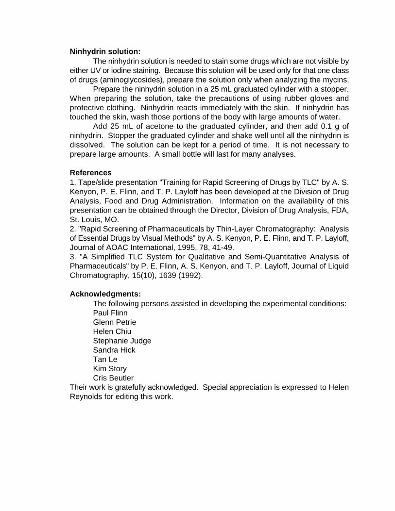
Ninhydrin solution: The ninhydrin solution is needed to stain some drugs which are not visible by
either UV or iodine staining. Because this solution will be used only for that one classof drugs (aminoglycosides), prepare the solution only when analyzing the mycins.
Prepare the ninhydrin solution in a 25 mL graduated cylinder with a stopper.When preparing the solution, take the precautions of using rubber gloves andprotective clothing. Ninhydrin reacts immediately with the skin. If ninhydrin hastouched the skin, wash those portions of the body with large amounts of water.
Add 25 mL of acetone to the graduated cylinder, and then add 0.1 g ofninhydrin. Stopper the graduated cylinder and shake well until all the ninhydrin isdissolved. The solution can be kept for a period of time. It is not necessary toprepare large amounts. A small bottle will last for many analyses.
References1. Tape/slide presentation "Training for Rapid Screening of Drugs by TLC" by A. S.Kenyon, P. E. Flinn, and T. P. Layloff has been developed at the Division of DrugAnalysis, Food and Drug Administration. Information on the availability of thispresentation can be obtained through the Director, Division of Drug Analysis, FDA,St. Louis, MO.2. "Rapid Screening of Pharmaceuticals by Thin-Layer Chromatography: Analysisof Essential Drugs by Visual Methods" by A. S. Kenyon, P. E. Flinn, and T. P. Layloff,Journal of AOAC International, 1995, 78, 41-49.3. "A Simplified TLC System for Qualitative and Semi-Quantitative Analysis ofPharmaceuticals" by P. E. Flinn, A. S. Kenyon, and T. P. Layloff, Journal of LiquidChromatography, 15(10), 1639 (1992). Acknowledgments:
The following persons assisted in developing the experimental conditions: Paul Flinn
Glenn PetrieHelen ChiuStephanie JudgeSandra HickTan LeKim StoryCris Beutler
Their work is gratefully acknowledged. Special appreciation is expressed to HelenReynolds for editing this work.

Acetylsalicylic acid300 mg tablet
Structure
Molecular formula and mass: C H O - 180.159 8 4
Category: AnalgesicSample:
Dissolve 1 tablet in 49 mL of methanol and 1 mL of glacial acetic acid.Concentration of the solution = 300 mg/50 mL = 6 mg/mL. The required concentrationof the sample solution for analysis is 2 mg/mL. Dilute 1 mL of the 6 mg/mL solutionto 3 mL by adding 2 mL of methanol. This solution will represent 100% sample.Standards:High standard:
The high limit is 115%; therefore the concentration of the high standard = (2mg/mL) X 1.15 = 2.30 mg/mL. Weigh approximately 10 mg of the standard. If youweighed 8 mg of standard, dissolve it in: 8 mg X 2.30 mg/mL) = 18.4 mL ofmethanol. This makes the high standard solution concentration equal to 2.30 mg/mL.Low standard:
The low limit is 85%; therefore the concentration of the low standard = (2mg/mL) X 0.85 = 1.70 mg/mL. Dilute 1 mL of high standard solution to 1.35 mL byadding 0.35 mL of methanol (2.30/1.70 = 1.35).Spotting:
Spot on the TLC plate as follows:Left spot low standard (85%)Center spot 100% sampleRight spot high standard (115%)
Development:Mix 17 mL of toluene, 13 mL of ethyl acetate, and 1 mL of acetic acid. Add
approximately 20 mL of this mixture to the TLC development bag. Develop until the

solvent front reaches within 1 cm of the top of the TLC plate.Detection:UV:
Dry the plate and observe under UV light (254 nm).Iodine stain:
Dip the plate in the iodine-KI solution in the detection bag. Allow the plate todry and observe the size and intensity of the spots.

Allopurinol100 mg tablet
Structure:
Molecular Formula and Mass C H N O - 136.115 4 4
Category : UricosuricSample:
Grind 1 tablet and dissolve in 100 mL of 0.1 N NaOH. The concentration ofthe solution = (100 mg/100 mL) = 1 mg/mL. The required concentration of thesample solution representing 100% is 1 mg/mL.Standards:High standard:
The high limit is 115%; therefore the concentration of the high standard = (1mg/mL) X 1.15 = 1.15 mg/mL. Weigh approximately 12 mg of standard. If youweighed 12.3 mg of standard, dissolve it in: 12.3 mg/1.15 mg/mL = 10.7 mL of 0.1N NaOH.Low standard:
The low limit is 85%; therefore the concentration of the high standard = (1mg/mL) X 0.85 mg/mL. Dilute 1 mL of the 1.15 mg/mL solution to 1.35 mL by adding0.35 mL to 1 mL of the high standard (1.15/0.85 = 1.35).Spotting:
Spot on the TLC plate as follows:Left spot low standard (85%)Center spot 100% sampleRight spot high standard (115%)
Development:Mix together 17 mL of 95% ethanol, 8 mL of toluene, and 1.5 mL of
concentrated ammonium hydroxide. Add approximately 20 mL of this mixture to theTLC development bag. Develop until the solvent front reaches to within 1 cm of the
top of the TLC plate.Detection:UV:
Dry the plate and observe under UV light (254 nm). Observe the intensity andthe size of the spots.Iodine stain:
The concentration of the solutions at 1 mg/mL is too low to be detectable byiodine staining. If you do not have a UV source, the concentration of the solutionsmust be increased to 5 mg/mL by dissolving 1 tablet in 20 mL of 0.1 N NaOH. Dipthe plate in the iodine solution in the detection bag. Allow the plate to dry, andobserve the intensity and size of the spots.

Aminophylline100 mg capsule
Structure:
MolecularFormula & Mass: (C H N O ) .C H (NH ) .2H O - 456.46 7 8 4 2 2 2 4 2 2 2
Category: Diuretic, cardiac stimulant Sample:
Aminophylline contains two components, theophylline as the dihydrate andethylene diamine. This means that the content of theophylline is only 79 mg as theanhydrous structure (100 mg X 360.46/456.46 = 79mg). The molecular weightcorrection is necessary because the standard is in the anhydrous form. The declareddrug content of 100 mg per tablet is for the combined mixture. Grind 1 tablet anddissolve in 50 mL of distilled water. Shake at least 1 min. Concentration of thesolution = 79 mg/50 mL = 1.58 mg/mL. The required concentration of the samplesolution representing 100% is 0.50 mg/mL. Add 2.16 mL of distilled water to 1 mLof the 1.58 mg/mL solution to make the sample solution equal to 0.50 mg/mL. Standards:High standard:
The high limit is 115%; therefore the concentration of high standard = (0.50mg/mL) X 1.15 = 0.575 mg/mL. Weigh approximately 5 mg of standard(anhydrous).If you weighed 4.6 mg of standard, dissolve it in: (4.6 mg)/(0.575 mg/mL) = 8.0 mLof distilled water. This makes the high standard solution concentration equal to 0.575mg/mL. Low standard:
The low limit is 85%; therefore the concentration of low standard = (0.50mg/mL) X 0.85 = 0.425 mg/mL. Dilute 1 mL of high standard to 1.35 mL by adding0.35 mL of distilled water (0.575/0.425 = 1.35).
Spotting:
Spot on the TLC plate as follows: Left spot low standard (85%) Center spot 100% sample Right spot high standard (115%)
Development:Mix 14 mL of acetone and 7 mL of toluene. Add this mixture to the TLC
development bag. Develop until the solvent front reaches within 1 cm of the top ofthe TLC plate. Detection:UV:
Dry the plate and observe under UV light. The maximum is at 270 nm but ashort wavelength UV light (254 nm) will work. Observe the size and intensity of thespots. Iodine stain:
Dip the plate in the iodine-KI solution in the detection bag. Allow the plate todry and observe the size and intensity of the spots.

Amoxicillin250 mg capsule
Structure:
Molecular Formula & Mass: C H N O S - 365.40 (anhydrous) 16 19 3 5
419.45 (trihydrate) Category: AntibacterialSample:
Dissolve the contents of 1 capsule in 10 mL of 0.1 N HCl. When completelydissolved, add 40 mL of acetone. The required concentration of the sample solutionrepresenting 100% is 5.0 mg/mL. Standards:High standard:
The high limit is 120%; therefore the concentration of the high standard = (5.0mg/mL) X 1.20 = 6 mg/mL. Weigh approximately 25 mg of standard. If you weighed24 mg of standard, dissolve it in: (24 mg X 0.871 mg)/6 mg/mL = 3.48 mL of 4:1acetone:0.1 N HCl. Note: The ratio of the molecular weight of the anhydrous form to the trihydrate is:365.4/419.45 = 0.871. This makes the high standard solution concentration equal to6.00 mg/mL.Low standard:
The low limit is 85%; therefore the concentration of the low standard = (5mg/mL X 0.85) = 4.25 mg/mL. Dilute 1 mL of high standard to 1.41 mL by adding0.41 mL of 4:1 acetone:0.1 N HCl (6.00/4.25 = 1.41).Spotting:
Spot on the TLC plate as followsLeft spot low standard (85%)Center spot 100% sampleRight spot high standard (120%)

Development:Mix 26 mL of acetone, 4 mL of water, 4 mL of toluene, and 1 mL of glacial
acetic acid. Add approximately 20 mL of this mixture to the TLC development bag.Develop until the solvent front reaches within 1 cm of the top of the TLC plate.Detection:UV:
Dry the plate and observe under UV light (254 nm). Observe the size andintensity of the spots.Iodine stain:
Dip the plate into the iodine-KI solution in the detection bag. Allow the plateto dry and observe the size and intensity of the spots.
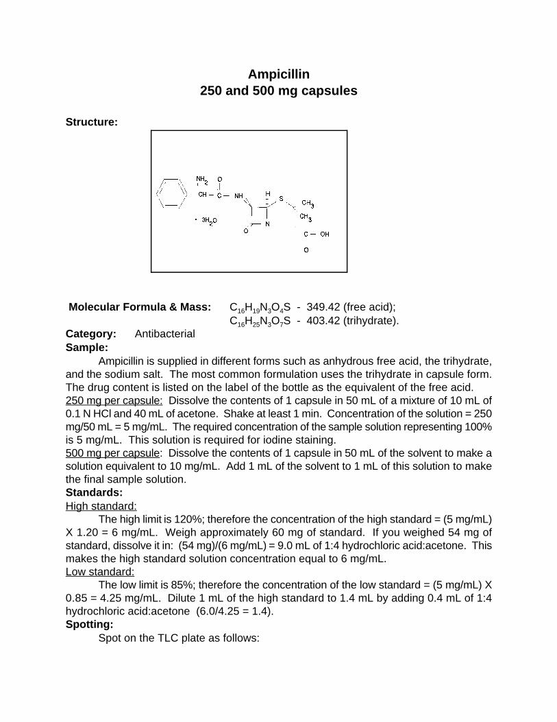
Ampicillin250 and 500 mg capsules
Structure: Molecular Formula & Mass: C H N O S - 349.42 (free acid); 16 19 3 4
C H N O S - 403.42 (trihydrate). 16 25 3 7
Category: Antibacterial Sample:
Ampicillin is supplied in different forms such as anhydrous free acid, the trihydrate,and the sodium salt. The most common formulation uses the trihydrate in capsule form.The drug content is listed on the label of the bottle as the equivalent of the free acid. 250 mg per capsule: Dissolve the contents of 1 capsule in 50 mL of a mixture of 10 mL of0.1 N HCl and 40 mL of acetone. Shake at least 1 min. Concentration of the solution = 250mg/50 mL = 5 mg/mL. The required concentration of the sample solution representing 100%is 5 mg/mL. This solution is required for iodine staining.500 mg per capsule: Dissolve the contents of 1 capsule in 50 mL of the solvent to make asolution equivalent to 10 mg/mL. Add 1 mL of the solvent to 1 mL of this solution to makethe final sample solution. Standards:High standard:
The high limit is 120%; therefore the concentration of the high standard = (5 mg/mL)X 1.20 = 6 mg/mL. Weigh approximately 60 mg of standard. If you weighed 54 mg ofstandard, dissolve it in: (54 mg)/(6 mg/mL) = 9.0 mL of 1:4 hydrochloric acid:acetone. Thismakes the high standard solution concentration equal to 6 mg/mL. Low standard:
The low limit is 85%; therefore the concentration of the low standard = (5 mg/mL) X0.85 = 4.25 mg/mL. Dilute 1 mL of the high standard to 1.4 mL by adding 0.4 mL of 1:4hydrochloric acid:acetone (6.0/4.25 = 1.4). Spotting:
Spot on the TLC plate as follows:
Left spot low standard (85%) Center spot 100% sample Right spot high standard (120%)
Development:Mix 26 mL of acetone, 4 mL of water, 4 mL of toluene, and 1 mL of glacial acetic
acid. Pour approximately 20 mL of this mixture into the TLC development bag. Develop untilthe solvent front reaches within 1 cm from the top of the TLC plate. Detection: UV:
Dry the plate and observe under UV light (254 nm). Observe the size and intensityof the spots. Iodine stain:
Dip the plate into the iodine-KI solution in the detction bag. Allow the plate to dry andobserve the size and intensity of the spots.

Atropine
Structure
Molecular Formula & Mass: C H NO - 289.38 17 23 3
Category: Anticholinergic Sample:
Standard:High standard:
The high limit is 115%; therefore the concentration of high standard is (10 mg/mL) X1.15 = 11.5 mg/mL. Weigh approximately 47 mg of standard. If you weighed 47.2.mg ofstandard, dissolve it in: (47.2 mg)/(11.5 mg/mL) = 4.1 mL of anhydrous ethanol.
Low standard:The low limit is 85%; therefore the concentration of low standard = (10 mg/mL) X
0.85 = 8.5 mg/mL. Dilute 1 mL of high standard to 1.35 mL by adding 0.35 mL of anhydrousethanol (1.15/0.85 = 1.35).Spotting:
Spot on TLC plate as follow:Left spot low standard (115%)Center spot 100% sampleRight spot high standard (85%)
Development:Mix 22 mL of methanol and 0.25 mL of concentrated ammonium hydroxide. Add this
mixture to the TLC development bag. Develop until the solvent front reaches within 1 cm ofthe top of the TLC plate.Detection:UV:
Dry the plate and observe under UV light. The maximum is at 260 nm but a short

wavelength light (254 nm) will work. Observe the size and intensity of the spots.Iodine stain:
Dip the plate into the iodine-KI solution in the detection bag. Allow the plate to dryand observe the size and intensity of the spots.Note:
The UV absorption is weak but can be distinguished.

Betamethasone
4 mg tablet
Structure:
Molecular Formula & Mass: C H FO - 392.45 22 29 5
Category: Glucocorticoid Sample:
Grind 1 tablet and dissolve in 10 mL of 95% ethanol. Shake at least 1 min.Concentration of the solution = 4 mg/10 mL = 0.4 mg/mL. The required concentration of thesample solution representing 100% is 0.4 mg/mL.Standards:High standard:
The high limit is 115%; therefore the concentration of the high standard = (0.40mg/mL) X 1.15 = 0.46 mg/mL. Weigh approximately 4 mg of standard. If you weighed 3.75mg of standard, dissolve it in: (3.75 mg)/(0.46 mg/mL) = 8.05 mL of ethanol. This makes theconcentration of the high standard solution equal to 0.46 mg/mL. Low standard:
The low limit is 85%; therefore the concentration of the low standard = (0.40 mg/mL)X 0.85 = 0.34 mg/mL. Dilute 1 mL of high standard to 1.35 mL by adding 0.35 mL ofethanol (0.46/0.34 = 1.35). Spotting:
Spot on the TLC plate as follows: Left spot low standard (85%) Center spot 100% sample Right spot high standard (115%)
Development:Mix 14 mL of toluene and 7 mL of acetone. Add this mixture to the TLC development
bag. Develop until the solvent front reaches within 1 cm of the top of the TLC plate.

Detection:UV:
Dry the plate and observe under UV light. The maximum is at 240 nm but a shortwavelength UV light (254 nm) will work. Observe the size and intensity of the spots.Iodine stain:
The 0.40 mg/mL concentration is too low to allow for quantitation by iodine. Theconcentration needs to be 4 mg/mL. This can be accomplished by using 10 tablets ratherthan 1 tablet. Dip the plate in the iodine-KI solution in the detection bag. Allow the plate todry and observe the size and intensity of the spots.

Carbamazepine100 mg tablet
Structure
Molecular Formula & Mass: C H N O - 236.2615 12 2
Category: AnalgesicSample:Grind 1 tablet and dissolve in 50 mL of anhydrous ethanol. Concentration of the solution =100 mg/50 mL = 2 mg/mL. Dilute 1 mL of the 2 mg/mL solution with 1 mL of ethanol tomake a final solution equal to 1 mg/mL.
Standard:High standard:
The high limit is 115%; therefore the concentration of high standard = (1 mg/mL) X1.15 = 1.15 mg/mL. Weigh approximately 5 mg of standard. If you weighed 4.98 mg ofstandard, dissolve it in: (4.98 mg)/(1.15 mg/mL) = 4.3 mL of anhydrous ethanol.Low standard:
The low limit is 85%; therefore the concentration of low standard = (1 mg/mL) X 0.85= 0.85 mg/mL. Dilute 1 mL of high standard to 1.35 mL by adding 0.35 mL of anhydrousethanol (1.15/0.85 = 1.35).Spotting:
Spot on the TLC plate as follows:Left spot low standard (85%)Center spot 100% sample Right spot high standard (115%)
Development:Mix 22 mL of methanol and 0.25 mL of concentrated ammonium hydroxide. Add this
mixture to the TLC development bag. Develop until the solvent front reaches within 1 cm ofthe top of the TLC plate.Detection:

UV:Dry the plate and observe under UV light. The maximum is at 235 nm but a short
wavelength light (254 nm) will work. Observe the size and intensity of the spots.Iodine stain:
Dip the plate in iodine-KI solution in the detection bag. Allow the plate to dry andobserve the size and intensity of the spots.

Cephalexin250 mg capsule
Structure
Molecular Formula & Mass: C H N O S - 347.40 16 17 3 4
Category: Antibacterial Sample:
Dissolve the contents of 1 capsule in 50 mL of methanol. Concentration of the solution= 250 mg/50 mL = 5 mg/mL. The required concentration of sample solution representing100% is 5 mg/mL.Standard:High standard:
The high limit is 115% therefore the concentration of high standard = (5 mg/mL) X1.15 = 6 mg/mL. Weigh approximately 21 mg of standard. If you weighed 21.3 mg ofstandard, dissolve it in: (21.3 mg)/(6 mg/mL) = 3.55 mL of methanol.Low standard:
The low limit is 85%; therefore the concentration of low standard = (5 mg/mL) X 0.85= 4.25 mg/mL. Dilute 1 mL of high standard to 1.35 mL by adding 0.35 mL of methanol(1.15/0.85 = 1.35).Spotting:
Spot on the TLC plate as follows:Left spot low standard (85%)Center spot 100% sample Right spot high standard (115%)
Development:Mix 12.5 mL of ethyl acetate, 5 mL of acetone, 5 mL of glacial acetic acid, and 2.5
mL of distilled water. Add this mixture to the TLC development bag. Develop until the

solvent front reaches within 1 cm of the top of the TLC plate.Detection:UV:
Dry the plate and observe under UV light. The maximum is at 250 nm but a shortwavelength light (254 nm) will work. Observe the size and intensity of the spots.Iodine stain:
Dip the plate in the iodine-KI solution in the detection bag. Allow the plate to dry andobserve the size and intensity of the spots.Note:
The spots are not well defined in UV light; however, they are clearly distinguished inthe iodine solution.
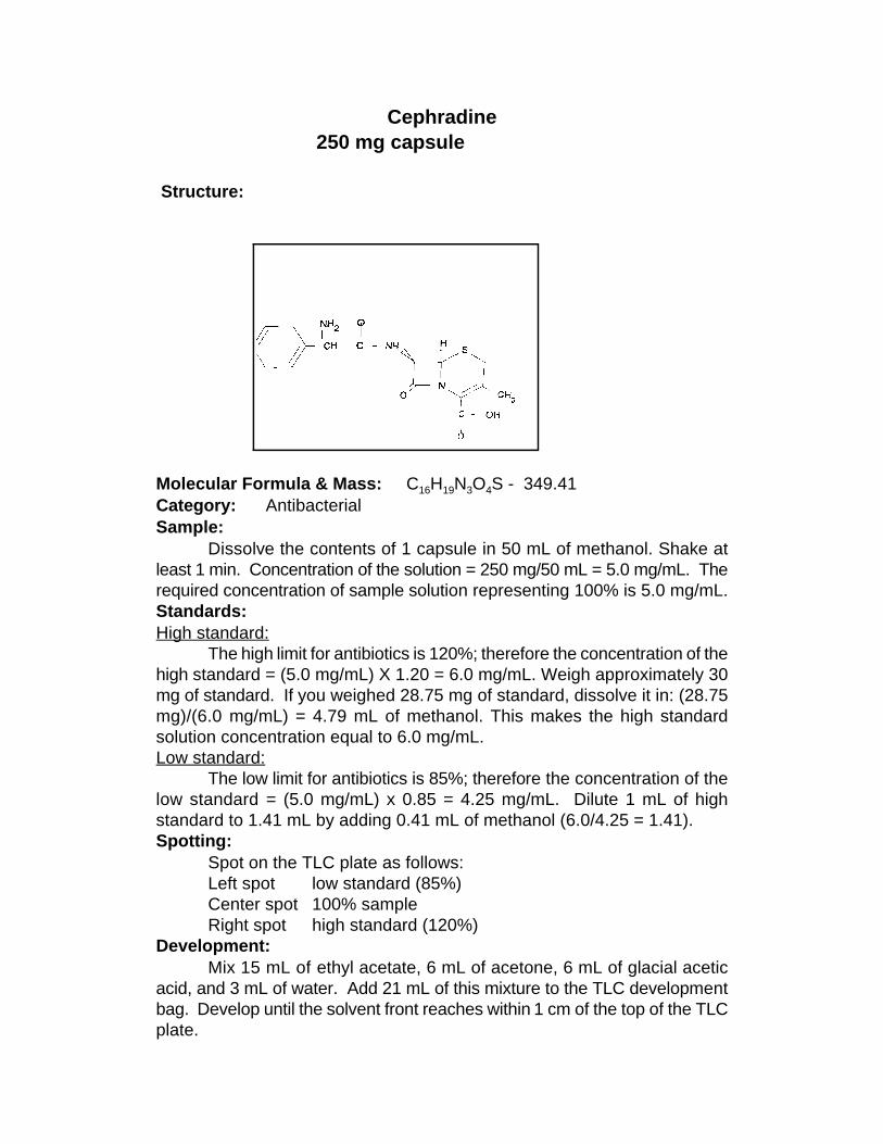
Cephradine250 mg capsule
Structure: Molecular Formula & Mass: C H N O S - 349.41 16 19 3 4
Category: Antibacterial Sample:
Dissolve the contents of 1 capsule in 50 mL of methanol. Shake atleast 1 min. Concentration of the solution = 250 mg/50 mL = 5.0 mg/mL. Therequired concentration of sample solution representing 100% is 5.0 mg/mL.Standards:High standard:
The high limit for antibiotics is 120%; therefore the concentration of thehigh standard = (5.0 mg/mL) X 1.20 = 6.0 mg/mL. Weigh approximately 30mg of standard. If you weighed 28.75 mg of standard, dissolve it in: (28.75mg)/(6.0 mg/mL) = 4.79 mL of methanol. This makes the high standardsolution concentration equal to 6.0 mg/mL. Low standard:
The low limit for antibiotics is 85%; therefore the concentration of thelow standard = (5.0 mg/mL) x 0.85 = 4.25 mg/mL. Dilute 1 mL of highstandard to 1.41 mL by adding 0.41 mL of methanol (6.0/4.25 = 1.41). Spotting:
Spot on the TLC plate as follows: Left spot low standard (85%) Center spot 100% sample Right spot high standard (120%)
Development:Mix 15 mL of ethyl acetate, 6 mL of acetone, 6 mL of glacial acetic
acid, and 3 mL of water. Add 21 mL of this mixture to the TLC developmentbag. Develop until the solvent front reaches within 1 cm of the top of the TLCplate.

Detection:UV:
Dry the plate and observe under UV light. The maximum is at 275 nmbut a short wavelength UV light (254 nm) will work. Observe the size andintensity of the spots. Iodine stain:
Dip the plate into the iodine solution in the detection bag. Allow theplate to dry and observe the size and intensity of the spots. Note:
This procedure should work with any of the cephalosporins that havesimilar molecular masses and functional groups. Standards of cephradine,cephalexin, and cefuroxime were processed by this procedure, and R valuesf
were as follows: Cephradine = 0.26 Cephalexin = 0.26 Cefuroxime = 0.47
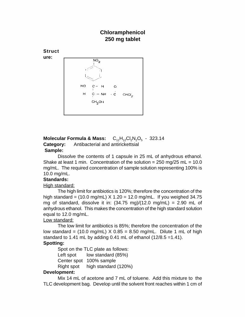
Chloramphenicol250 mg tablet
Structure: Molecular Formula & Mass: C H Cl N O - 323.14 11 12 2 2 5
Category: Antibacterial and antirickettsial Sample:
Dissolve the contents of 1 capsule in 25 mL of anhydrous ethanol.Shake at least 1 min. Concentration of the solution = 250 mg/25 mL = 10.0mg/mL. The required concentration of sample solution representing 100% is10.0 mg/mL. Standards:High standard:
The high limit for antibiotics is 120%; therefore the concentration of thehigh standard = (10.0 mg/mL) X 1.20 = 12.0 mg/mL. If you weighed 34.75mg of standard, dissolve it in: (34.75 mg)/(12.0 mg/mL) = 2.90 mL ofanhydrous ethanol. This makes the concentration of the high standard solutionequal to 12.0 mg/mL. Low standard:
The low limit for antibiotics is 85%; therefore the concentration of thelow standard = (10.0 mg/mL) X 0.85 = 8.50 mg/mL. Dilute 1 mL of highstandard to 1.41 mL by adding 0.41 mL of ethanol (12/8.5 =1.41). Spotting:
Spot on the TLC plate as follows: Left spot low standard (85%) Center spot 100% sample Right spot high standard (120%)
Development:Mix 14 mL of acetone and 7 mL of toluene. Add this mixture to the
TLC development bag. Develop until the solvent front reaches within 1 cm of
the top of the TLC plate. Detection: UV:
The 10.0 mg/mL concentration is too high to allow for quantitation.Therefore, dilute the sample and standard solutions 1:10 by adding 9 mL ofanhydrous ethanol to 1 mL of the sample and 9 mL of anhydrous ethanol to1 mL of each of the standards. Dry the plate and observe under UV light.The maximum is at 275 nm but a short wavelength UV light (254 nm) will work.Observe the size and intensity of the spots. Iodine stain:
Dip the plate in the iodine-KI solution in the detection bag. Allow theplate to dry and observe the size and intensity of the spots.

Chloroquine diphosphate 250 mg tablet
Structure:
Molecular Formula & Mass: C H ClN .2H PO - 515.86 18 26 3 3 4
Category: Antimalarial Sample:
Grind 1 tablet and dissolve in 50 mL of distilled water. Shake at least 1 min.Concentration of the solution = 250 mg/50 mL = 5.0 mg/mL. The required concentration ofthe sample solution representing 100% is 0.50 mg/mL. Add 9 mL of distilled water to makea sample solution equal to 0.5 mg/mL.Standards:High standard:
The high limit is 115%; therefore the concentration of the high standard = (0.50mg/mL X 1.15 = 0.575 mg/mL.When a reference tablet is available:
The reference tablet is specified to contain 2.3 mg of active drug. However, thecontent may vary. The concentration is shown on the label. If the tablet contains 2.19 mgof the drug, dissolve it in: 2.19 mg/0.575 mg/mL = 3.8 mL of distilled water.When no reference tablet is available:
Weigh approximately 5 mg of standard. If you weighed 4.7 mg of standard, dissolveit in: (4.7 mg)/(0.575 mg/mL) = 8.17 mL of distilled water. This makes the high standardsolution concentration equal to 0.575 mg/mL.Low standard:
The low limit is 85%; therefore the concentration of the low standard = (0.50 mg/mL)X 0.85 = 0.425 mg/mL. Dilute 1 mL of high standard to 1.35 mL by adding 0.35 mL ofdistilled water (0.575/0.425 = 1.35).Spotting:
Spot on the TLC plate as follows:Left spot low standard (85%)Center spot 100% sampleRight spot high standard (115%)
Development:
Mix 21 mL of anhydrous ethanol, 3 mL of distilled water, and 1 mL of concentratedammonium hydroxide. Add this mixture to the TLC development bag. Develop until thesolvent front reaches to within 1 cm of the top of the TLC plate.Detection:UV:
Dry the plate and observe under UV light. The maximum is at 333 nm but a shortwavelength UV light (254 nm) will work. Observe the size and intensity of the spots.Iodine stain:
Dip the plate in the iodine-KI solution in the detection bag. Allow the plate to dry andobserve the size and intensity of the spots.

Chlorpheniramine maleate2 mg tablet
Structure
Molecular formula & mass: C H ClN O 390.88 20 23 2 4
Category: Antihistiminic Sample:
Grind 4 tablets and dissolve in 8 mL of anhydrous ethanol. The requiredconcentration of the sample solution representing 100% is 1 mg/mL. Because of the smallvolume required to make the proper concentration, multiple tablets are used so thatsufficient volume of solvent is available for sampling. Standard:High standard:
The high limit is 115%; therefore the concentration of the high standard = (1 mg/mL)X 1.15 = 1.15 mg/mL. Weigh approximately 9 mg of standard. If you weighed 8.9 mg ofstandard, dissolve it in: (8.9 mg)/(1.15 mg/mL) = 7.74 mL of anhydrous ethanol.Low standard:
The low limit is 85%; therefore the concentration of the low standard = (1 mg/mL) X0.85 = 0.85 mg/mL. Dilute 1 mL of high standard to 1.35 mL by adding 0.35 mL of ethanol(1.15/0.85 = 1.35).Spotting:
Spot on the TLC plate as follow:Left spot low standard (85%)Center spot 100% sample Right spot high standard (115%)
Development:Mix 22 mL of methanol and 1 mL of glacial acetic acid. Add this mixture to the TLC
development bag. Develop until the solvent front reaches within 1 cm of the top of the TLCplate.Detection:UV:
Dry the plate and observe under UV light. The maximum is at 265 nm but a shortwavelength light (254 nm) will work. Observe the size and intensity of the spots.Iodine stain:
Dip the plate in the iodine-KI solution in the detection bag. Allow the plate to dry andobserve the size and intensity of the spots.
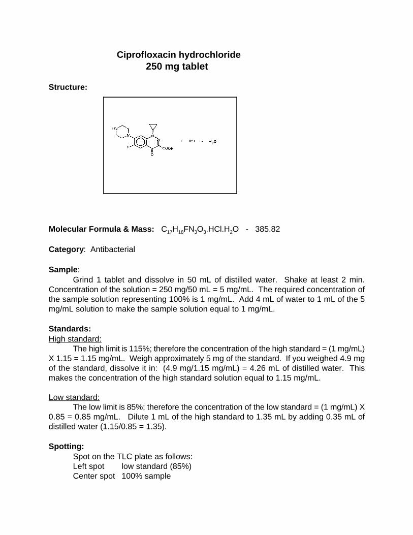
Ciprofloxacin hydrochloride250 mg tablet
Structure:
Molecular Formula & Mass: C H FN O .HCl.H O - 385.8217 18 3 3 2
Category: Antibacterial
Sample:Grind 1 tablet and dissolve in 50 mL of distilled water. Shake at least 2 min.
Concentration of the solution = 250 mg/50 mL = 5 mg/mL. The required concentration ofthe sample solution representing 100% is 1 mg/mL. Add 4 mL of water to 1 mL of the 5mg/mL solution to make the sample solution equal to 1 mg/mL.
Standards:High standard:
The high limit is 115%; therefore the concentration of the high standard = (1 mg/mL)X 1.15 = 1.15 mg/mL. Weigh approximately 5 mg of the standard. If you weighed 4.9 mgof the standard, dissolve it in: (4.9 mg/1.15 mg/mL) = 4.26 mL of distilled water. Thismakes the concentration of the high standard solution equal to 1.15 mg/mL.
Low standard:The low limit is 85%; therefore the concentration of the low standard = (1 mg/mL) X
0.85 = 0.85 mg/mL. Dilute 1 mL of the high standard to 1.35 mL by adding 0.35 mL ofdistilled water (1.15/0.85 = 1.35).
Spotting:Spot on the TLC plate as follows:Left spot low standard (85%)Center spot 100% sample

Right spot high standard (115%)
Development:Mix 2.5 mL of toluene, 5 mL of acetone, 10 mL of methanol, and 5 mL of
concentrated ammonium hydroxide. Add this solution to the TLC development bag. Developthe plate until the solvent front reaches within 1 cm of the top of the TLC plate.
Detection:UV:
Dry the plate and observe under UV light (254 nm). Observe the size and intensityof the spots.
Iodine stain:Dip the plate in the iodine-KI solution in the detection bag. Allow the plate to dry and
observe the size and intensity of the spots.
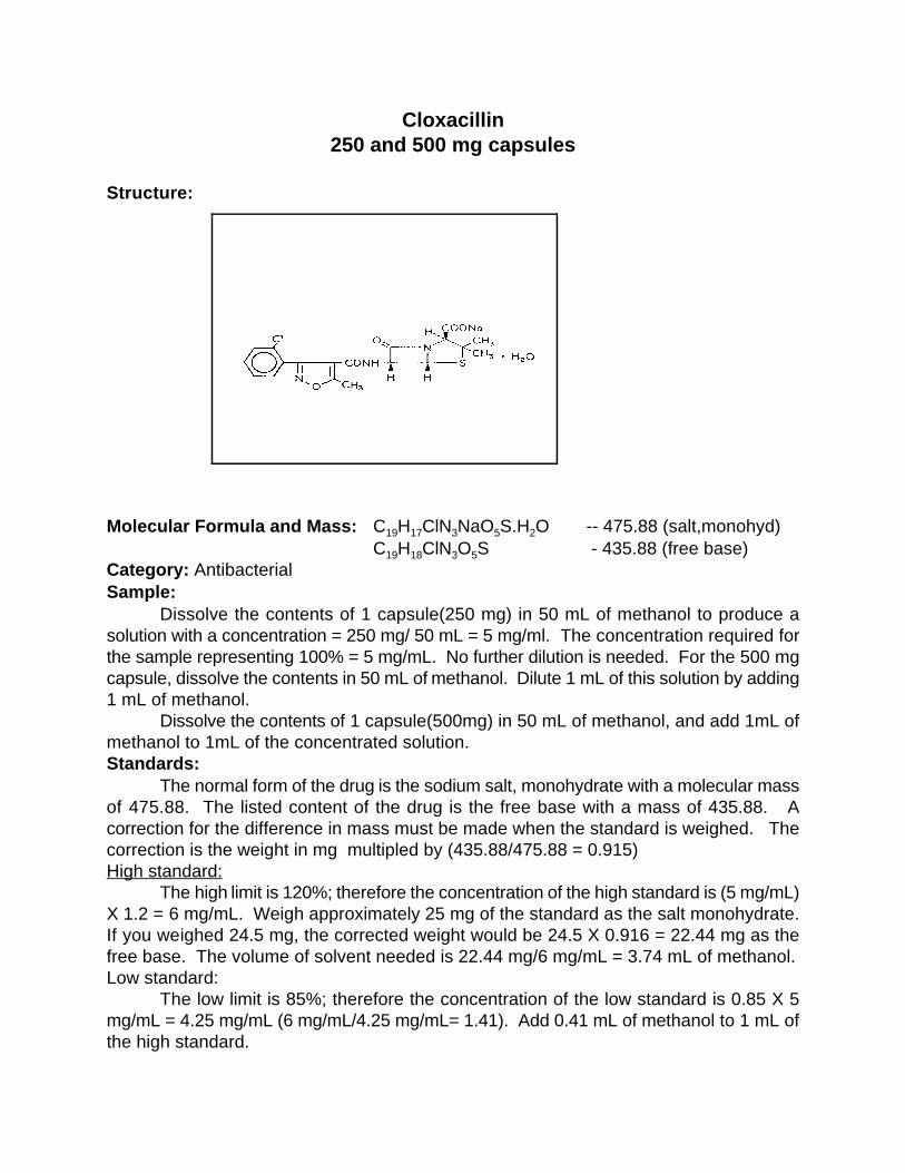
Cloxacillin250 and 500 mg capsules
Structure:
Molecular Formula and Mass: C H ClN NaO S.H O -- 475.88 (salt,monohyd)19 17 3 5 2
C H ClN O S - 435.88 (free base)19 18 3 5
Category: AntibacterialSample:
Dissolve the contents of 1 capsule(250 mg) in 50 mL of methanol to produce asolution with a concentration = 250 mg/ 50 mL = 5 mg/ml. The concentration required forthe sample representing 100% = 5 mg/mL. No further dilution is needed. For the 500 mgcapsule, dissolve the contents in 50 mL of methanol. Dilute 1 mL of this solution by adding1 mL of methanol.
Dissolve the contents of 1 capsule(500mg) in 50 mL of methanol, and add 1mL ofmethanol to 1mL of the concentrated solution.Standards:
The normal form of the drug is the sodium salt, monohydrate with a molecular massof 475.88. The listed content of the drug is the free base with a mass of 435.88. Acorrection for the difference in mass must be made when the standard is weighed. Thecorrection is the weight in mg multipled by (435.88/475.88 = 0.915) High standard:
The high limit is 120%; therefore the concentration of the high standard is (5 mg/mL)X 1.2 = 6 mg/mL. Weigh approximately 25 mg of the standard as the salt monohydrate.If you weighed 24.5 mg, the corrected weight would be 24.5 X 0.916 = 22.44 mg as thefree base. The volume of solvent needed is 22.44 mg/6 mg/mL = 3.74 mL of methanol. Low standard:
The low limit is 85%; therefore the concentration of the low standard is 0.85 X 5mg/mL = 4.25 mg/mL (6 mg/mL/4.25 mg/mL= 1.41). Add 0.41 mL of methanol to 1 mL ofthe high standard.

Spotting:Spot on the TLC plate as follows:Left spot low standard (85%)Center spot sample (100%)Right spot high standard (120%)
Developer:Mix 26 mL of acetone, 4 mL of water, 4 mL of toluene, and 1 mL of glacial acetic
acid. Add approximately 20 mL of the developer mixture to the TLC developing bag.Develop until the solvent front reaches to within 1 cm from the top of the TLC plate.Detection:UV:
Dry the plate and observe under UV light at 254nm. Observe the size and intensityof the spots.Iodine stain:
Dip the plate into the iodine-KI solution. Allow the plate to dry until the stained spotsbecome clearly visible. Observe the intensities of the spots.
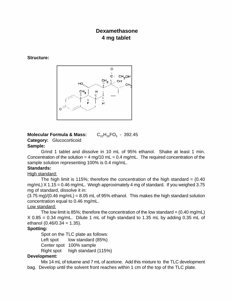
Dexamethasone4 mg tablet
Structure: Molecular Formula & Mass: C H FO - 392.45 22 29 5
Category: Glucocorticoid Sample:
Grind 1 tablet and dissolve in 10 mL of 95% ethanol. Shake at least 1 min.Concentration of the solution = 4 mg/10 mL = 0.4 mg/mL. The required concentration of thesample solution representing 100% is 0.4 mg/mL. Standards:High standard:
The high limit is 115%; therefore the concentration of the high standard = (0.40mg/mL) X 1.15 = 0.46 mg/mL. Weigh approximately 4 mg of standard. If you weighed 3.75mg of standard, dissolve it in: (3.75 mg)/(0.46 mg/mL) = 8.05 mL of 95% ethanol. This makes the high standard solutionconcentration equal to 0.46 mg/mL. Low standard:
The low limit is 85%; therefore the concentration of the low standard = (0.40 mg/mL)X 0.85 = 0.34 mg/mL. Dilute 1 mL of high standard to 1.35 mL by adding 0.35 mL ofethanol (0.46/0.34 = 1.35). Spotting:
Spot on the TLC plate as follows: Left spot low standard (85%) Center spot 100% sample Right spot high standard (115%)
Development:Mix 14 mL of toluene and 7 mL of acetone. Add this mixture to the TLC development
bag. Develop until the solvent front reaches within 1 cm of the top of the TLC plate.

Detection:UV:
Dry the plate and observe under UV light. The maximum is at 240 nm but a shortwavelength UV light (254 nm) will work. Observe the size and intensity of the spots. Iodine stain:
The 0.40 mg/mL concentration is too low to allow for quantitation by iodine. Theconcentration needs to be 4 mg/mL. This can be accomplished by using 10 tablets ratherthan 1 tablet. Dip the plate in the iodine-KI solution in the detection bag. Allow the plate todry and observe the size and intensity of the spots.
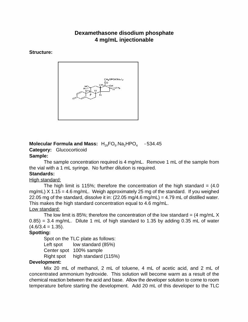
Dexamethasone disodium phosphate4 mg/mL injectionable
Structure:
Molecular Formula and Mass: H FO .Na HPO - 534.45 29 5 2 4
Category: GlucocorticoidSample:
The sample concentration required is 4 mg/mL. Remove 1 mL of the sample fromthe vial with a 1 mL syringe. No further dilution is required.Standards:High standard:
The high limit is 115%; therefore the concentration of the high standard = (4.0mg/mL) X 1.15 = 4.6 mg/mL. Weigh approximately 25 mg of the standard. If you weighed22.05 mg of the standard, dissolve it in: (22.05 mg/4.6 mg/mL) = 4.79 mL of distilled water.This makes the high standard concentration equal to 4.6 mg/mL.Low standard:
The low limit is 85%; therefore the concentration of the low standard = (4 mg/mL X0.85) = 3.4 mg/mL. Dilute 1 mL of high standard to 1.35 by adding 0.35 mL of water(4.6/3.4 = 1.35).Spotting:
Spot on the TLC plate as follows: Left spot low standard (85%) Center spot 100% sample Right spot high standard (115%)
Development: Mix 20 mL of methanol, 2 mL of toluene, 4 mL of acetic acid, and 2 mL ofconcentrated ammonium hydroxide. This solution will become warm as a result of thechemical reaction between the acid and base. Allow the developer solution to come to roomtemperature before starting the development. Add 20 mL of this developer to the TLC
development bag. Develop until the solvent front reaches 1 cm from the top of the plate.Detection:UV:
Dry the plate and examine the spots under UV light (254 nm). Observe the size andintensity of the spots.Iodine stain:
Dip the plate into the iodine-KI solution. Allow the iodine to disappear, and observethe spots as soon as they become clear.

Diazepam
2 mg tablet Structure:
Molecular Formula and Mass: C H ClN O - 284.76 (the above structure shows16 13 2
deuterium atoms as D. In the normal structure the D is replaced by H atoms) Category: Minor tranquilizer Sample:
Grind 1 tablet and dissolve in 2 mL of 95% ethanol. Shake at least 1 min.Concentration of the solution = 2 mg/ 2 mL = 1 mg/mL. The required concentration of thesample solution representing 100% is 1 mg/mL.Standards:High standard:
The high limit is 115%; therefore the concentration of the high standard = (1 mg/mL)X 1.15 = 1.15 mg/mL. Weigh approximately 5 mg of standard. If you weighed 4.75 mg ofstandard, dissolve it in: (4.75 mg)/(1.15 mg/mL) = 4.1 mL of 95% ethanol. This makes thehigh standard solution concentration equal to 1.15 mg/mL. Low standard:
The low limit is 85%; therefore the concentration of the low standard = (1 mg/mL) X0.85 = 0.85 mg/mL. Dilute 1 mL of high standard to 1.35 mL by adding 0.35 mL of ethanol(1.15/0.85 = 1.35). Spotting:
Spot on the TLC plate as follows: Left spot low standard (85%) Center spot 100% sample Right spot high standard (115%)
Development:Mix 33 mL of methanol and 0.5 mL of concentrated ammonium hydroxide. Add 20
mL of this mixture to the TLC development bag. Develop until the solvent front reaches to

within 1 cm of the top of the TLC plate.
Detection:UV:
Dry the plate and observe under UV light. The maximum is at 240 nm but a shortwavelength UV light (254 nm) will work. Observe the size and intensity of the spots. Iodine stain:
Dip the plate in the iodine-KI solution in the detection bag. Allow the plate to dry andobserve the size and intensity of the spots.
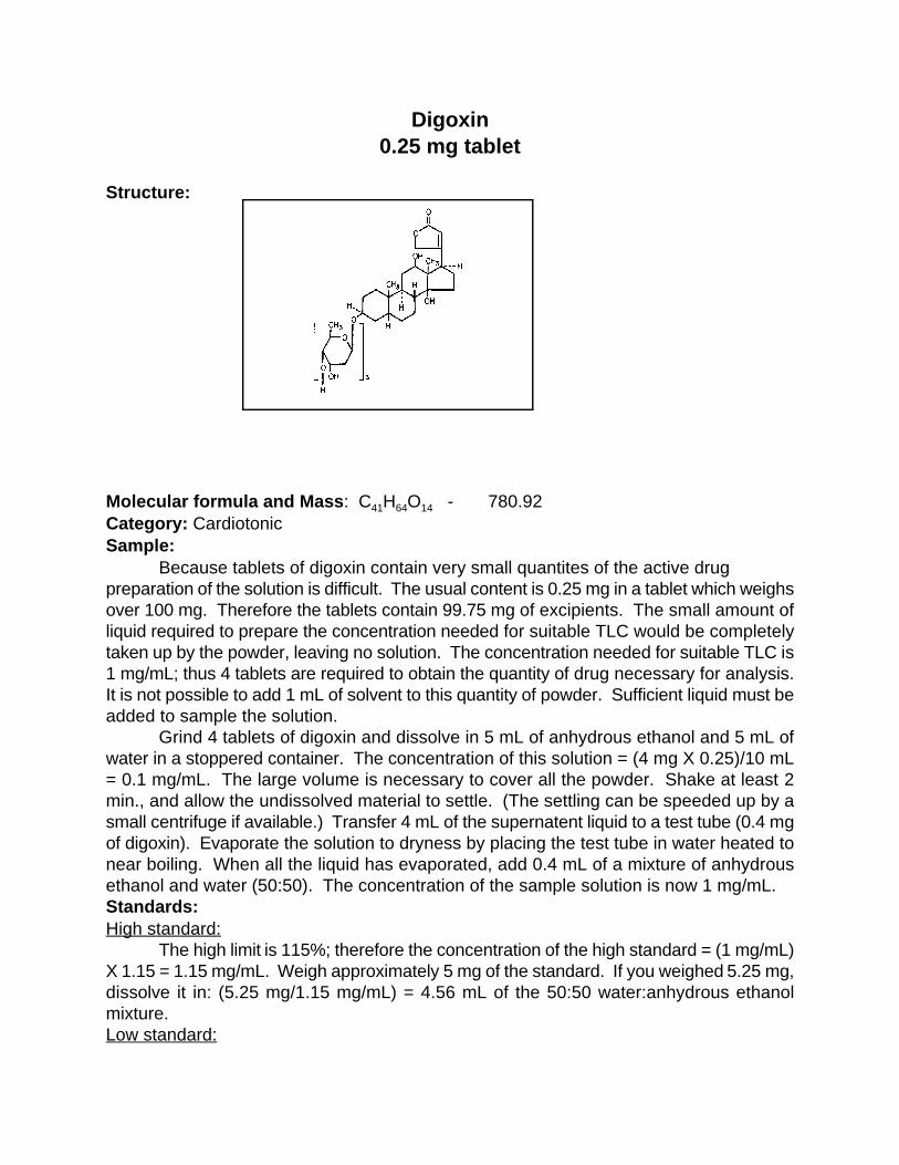
Digoxin0.25 mg tablet
Structure:
Molecular formula and Mass: C H O - 780.9241 64 14
Category: CardiotonicSample:
Because tablets of digoxin contain very small quantites of the active drugpreparation of the solution is difficult. The usual content is 0.25 mg in a tablet which weighsover 100 mg. Therefore the tablets contain 99.75 mg of excipients. The small amount ofliquid required to prepare the concentration needed for suitable TLC would be completelytaken up by the powder, leaving no solution. The concentration needed for suitable TLC is1 mg/mL; thus 4 tablets are required to obtain the quantity of drug necessary for analysis.It is not possible to add 1 mL of solvent to this quantity of powder. Sufficient liquid must beadded to sample the solution.
Grind 4 tablets of digoxin and dissolve in 5 mL of anhydrous ethanol and 5 mL ofwater in a stoppered container. The concentration of this solution = (4 mg X 0.25)/10 mL= 0.1 mg/mL. The large volume is necessary to cover all the powder. Shake at least 2min., and allow the undissolved material to settle. (The settling can be speeded up by asmall centrifuge if available.) Transfer 4 mL of the supernatent liquid to a test tube (0.4 mgof digoxin). Evaporate the solution to dryness by placing the test tube in water heated tonear boiling. When all the liquid has evaporated, add 0.4 mL of a mixture of anhydrousethanol and water (50:50). The concentration of the sample solution is now 1 mg/mL.Standards:High standard:
The high limit is 115%; therefore the concentration of the high standard = (1 mg/mL)X 1.15 = 1.15 mg/mL. Weigh approximately 5 mg of the standard. If you weighed 5.25 mg,dissolve it in: (5.25 mg/1.15 mg/mL) = 4.56 mL of the 50:50 water:anhydrous ethanolmixture.Low standard:
The low limit is 85%; therefore the concentration of the low standard = (1 mg/ml) X0.85 = 0.85 mg/mL. Dilute 1 mL of the high standard to 1.35 mL by adding 0.35 mL of the50:50 water:anhydrous ethanol mixture.Spotting:
Spot on the TLC plate as follows:Left spot low standard (85%)Center spot 100% sampleRight spot high standard (115%)
Developer:Mix 42 mL of ethyl acetate, 5 mL of methanol, and 2.5 mL of concentrated
ammonium hydroxide. Add 20 mL of this mixture to the TLC development bag. Develop untilthe solvent front reaches within 1 cm of the top of the plate.Detection:UV:
Dry the plate and observe under UV light (254 nm). Observe the size and intensityof the spots.Iodine stain:
Dip the plate into the iodine-KI solution. Allow the iodine to fade until the spotsbecome visible. Observe the size and intensity of the spots.
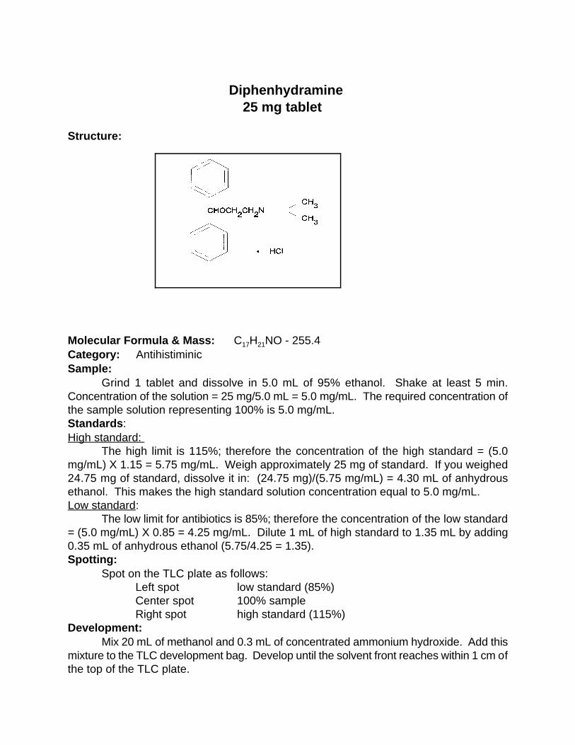
Diphenhydramine
25 mg tablet Structure: Molecular Formula & Mass: C H NO - 255.4 17 21
Category: Antihistiminic Sample:
Grind 1 tablet and dissolve in 5.0 mL of 95% ethanol. Shake at least 5 min.Concentration of the solution = 25 mg/5.0 mL = 5.0 mg/mL. The required concentration ofthe sample solution representing 100% is 5.0 mg/mL. Standards: High standard:
The high limit is 115%; therefore the concentration of the high standard = (5.0mg/mL) X 1.15 = 5.75 mg/mL. Weigh approximately 25 mg of standard. If you weighed24.75 mg of standard, dissolve it in: (24.75 mg)/(5.75 mg/mL) = 4.30 mL of anhydrousethanol. This makes the high standard solution concentration equal to 5.0 mg/mL. Low standard:
The low limit for antibiotics is 85%; therefore the concentration of the low standard= (5.0 mg/mL) X 0.85 = 4.25 mg/mL. Dilute 1 mL of high standard to 1.35 mL by adding0.35 mL of anhydrous ethanol (5.75/4.25 = 1.35). Spotting:
Spot on the TLC plate as follows: Left spot low standard (85%) Center spot 100% sample Right spot high standard (115%)
Development:Mix 20 mL of methanol and 0.3 mL of concentrated ammonium hydroxide. Add this
mixture to the TLC development bag. Develop until the solvent front reaches within 1 cm ofthe top of the TLC plate.

Detection :UV.
Dry the plate and observe under ultraviolet light at 254 nm. Observe the intensitiesand the sizes of the spots.Iodine stain.
Dip the plate into the iodine solution. Allow the iodine to fade until the spots becomevisible. Observe the intensity and sizes of the spots.

Ergotamine tartrate2 mg tablet
Structure:
Molecular Formula and Mass : C H N O - 131270 76 10 16
Category: VasoconstrictorSample:
Grind 4 tablets and dissolve in 8 mL of 95% ethanol. The drug content in each tabletis low. Enough liquid is needed to obtain a solution of the correct concentration. Shake atleast 2 min. and allow the insolubles to settle. Withdraw approximately 1 mL of thesupernatant liquid for the sample. The final concentration of the sample solution is 1 mg/mL.Standards:High standard:
The high limit is 115%; therefore the concentration of the high standard = (1 mg/mL)X 1.15 = 1.15 mg/mL. Weigh approximately 5 mg of the standard. If you weighed 4.8 mgof the standard, dissolve it in: (4.8 mg/1.15 mg/mL) = 4.17 mL of 95% ethanol.Low standard:
The low limit is 85%; therefore the concentration of the low standard = (1 mg/mL) X0.85 = 0.85 mg/mL. Dilute 1 mL of high standard to 1.35 mL by adding 0.35 mL of 95%ethanol (1.15/0.85 = 1.35).Spotting:
Spot on the TLC plate as follows:Left spot low standard (85%)Center spot 100% sampleRight spot high standard (115%)
Development:Mix 24 mL of methanol and 0.4 mL of concentrated ammonium hydroxide. Add this
solution to the TLC development bag. Develop until the solvent front reaches within 1 cmof the top of the plate.Detection:
UV: Dry the plate and observe under UV light (254 nm). Observe the size and intensity
of the spots. Iodine stain:
Dip the plate into the iodine-KI solution, and observe the size and intensity of thespots after they become visible.
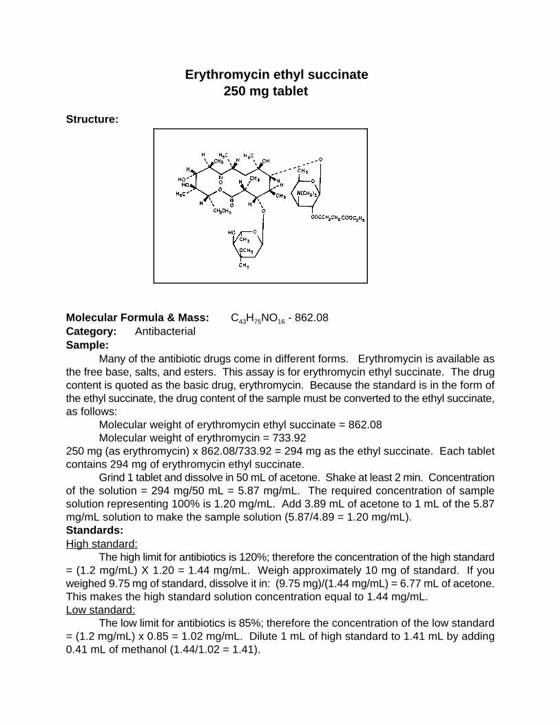
Erythromycin ethyl succinate 250 mg tablet
Structure:
Molecular Formula & Mass: C H NO - 862.08 43 75 16
Category: Antibacterial Sample:
Many of the antibiotic drugs come in different forms. Erythromycin is available asthe free base, salts, and esters. This assay is for erythromycin ethyl succinate. The drugcontent is quoted as the basic drug, erythromycin. Because the standard is in the form ofthe ethyl succinate, the drug content of the sample must be converted to the ethyl succinate,as follows: Molecular weight of erythromycin ethyl succinate = 862.08
Molecular weight of erythromycin = 733.92 250 mg (as erythromycin) x 862.08/733.92 = 294 mg as the ethyl succinate. Each tabletcontains 294 mg of erythromycin ethyl succinate. Grind 1 tablet and dissolve in 50 mL of acetone. Shake at least 2 min. Concentrationof the solution = 294 mg/50 mL = 5.87 mg/mL. The required concentration of samplesolution representing 100% is 1.20 mg/mL. Add 3.89 mL of acetone to 1 mL of the 5.87mg/mL solution to make the sample solution (5.87/4.89 = 1.20 mg/mL). Standards:High standard:
The high limit for antibiotics is 120%; therefore the concentration of the high standard= (1.2 mg/mL) X 1.20 = 1.44 mg/mL. Weigh approximately 10 mg of standard. If youweighed 9.75 mg of standard, dissolve it in: (9.75 mg)/(1.44 mg/mL) = 6.77 mL of acetone.This makes the high standard solution concentration equal to 1.44 mg/mL. Low standard:
The low limit for antibiotics is 85%; therefore the concentration of the low standard= (1.2 mg/mL) x 0.85 = 1.02 mg/mL. Dilute 1 mL of high standard to 1.41 mL by adding0.41 mL of methanol (1.44/1.02 = 1.41).

Spotting:Spot on the TLC plate as follows: Left spot low standard (85%) Center spot 100% sample Right spot high standard (120%)
Development:Mix 16 mL of acetone and 8 mL of toluene. Add this mixture to the TLC development
bag. Develop until the solvent front reaches within 1 cm of the top of the TLC plate.Detection:UV:
The spots are not visible in the UV. Iodine stain:
Dip the plate in the iodine-KI solution in the detection bag. Allow the plate to dry andobserve the size and intensity of the spots. To make the spots last longer, allow the plateto dry for 30 min. after it has been dipped in the iodine-KI solution. Heat the plate at 100ECfor 5 min. The spots turn gray-green and are stable for 24 hr. Notes: The ethyl succinate ester is very easily hydrolyzed in the presence of water ormethanol. Therefore the sample and standard must be prepared in acetone. The free baseof erythromycin is visible as a spot near the spotting line (R = 0.04). If the content off
erythromycin free base is to be determined, the sample must be dissolved in acetone andthe TLC plate developed in the developing solvent for erythromycin free base (seeerythromycin and erythromycin stearate) to prevent the hydrolysis of the ester to the freebase during sample preparation.
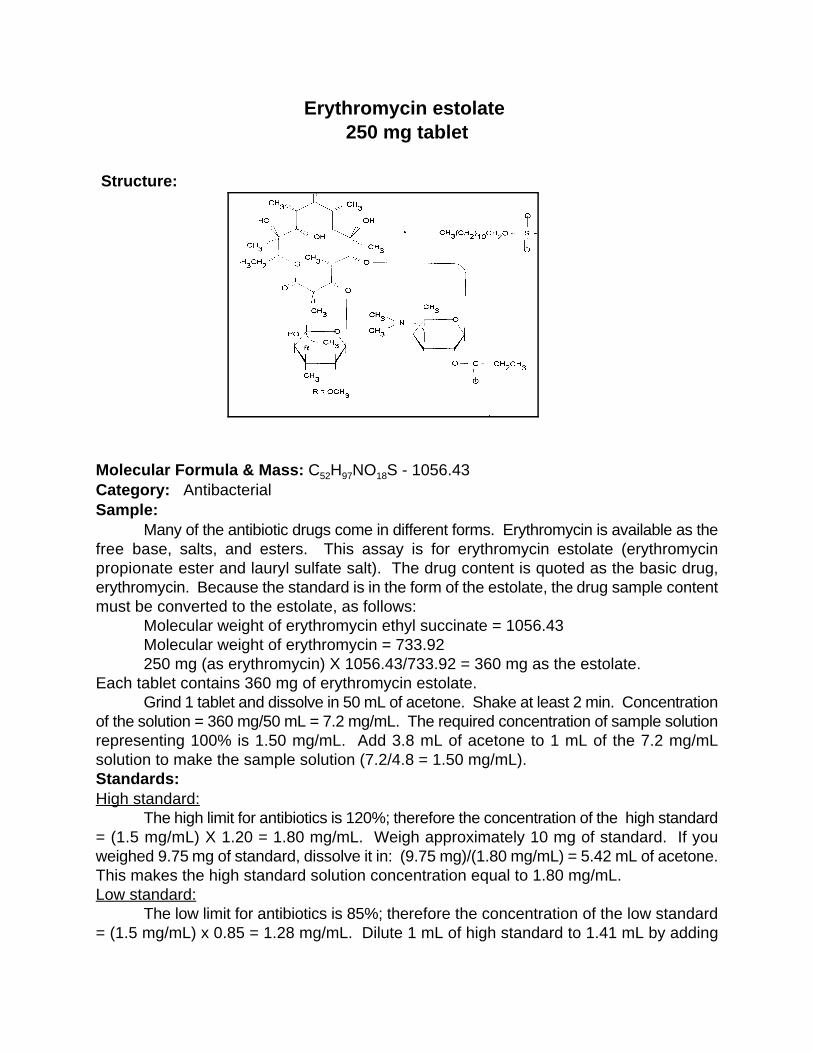
Erythromycin estolate 250 mg tablet
Structure:
Molecular Formula & Mass: C H NO S - 1056.43 52 97 18
Category: Antibacterial Sample:
Many of the antibiotic drugs come in different forms. Erythromycin is available as thefree base, salts, and esters. This assay is for erythromycin estolate (erythromycinpropionate ester and lauryl sulfate salt). The drug content is quoted as the basic drug,erythromycin. Because the standard is in the form of the estolate, the drug sample contentmust be converted to the estolate, as follows: Molecular weight of erythromycin ethyl succinate = 1056.43
Molecular weight of erythromycin = 733.92 250 mg (as erythromycin) X 1056.43/733.92 = 360 mg as the estolate.
Each tablet contains 360 mg of erythromycin estolate. Grind 1 tablet and dissolve in 50 mL of acetone. Shake at least 2 min. Concentration
of the solution = 360 mg/50 mL = 7.2 mg/mL. The required concentration of sample solutionrepresenting 100% is 1.50 mg/mL. Add 3.8 mL of acetone to 1 mL of the 7.2 mg/mLsolution to make the sample solution (7.2/4.8 = 1.50 mg/mL). Standards:High standard:
The high limit for antibiotics is 120%; therefore the concentration of the high standard= (1.5 mg/mL) X 1.20 = 1.80 mg/mL. Weigh approximately 10 mg of standard. If youweighed 9.75 mg of standard, dissolve it in: (9.75 mg)/(1.80 mg/mL) = 5.42 mL of acetone.This makes the high standard solution concentration equal to 1.80 mg/mL. Low standard:
The low limit for antibiotics is 85%; therefore the concentration of the low standard= (1.5 mg/mL) x 0.85 = 1.28 mg/mL. Dilute 1 mL of high standard to 1.41 mL by adding

0.41 mL of methanol (1.80/1.28 = 1.41). Spotting:
Spot on the TLC plate as follows: Left spot low standard (85%) Center spot 100% sample Right spot high standard (120%)
Development:Mix 16 mL of acetone and 8 mL of toluene. Add this mixture to the TLC development
bag. Develop until the solvent front reaches within 1 cm of the top of the TLC plate. Detection: UV:
The spots are not visible in the UV.Iodine stain:
Dip the plate in the iodine-KI solution in the detection bag. Allow the plate to dry andobserve the size and intensity of the spots. To make the spots last longer, allow the plateto dry for 30 min. after it has been dipped in the iodine solution. Heat the plate at 100EC for5 min. The spots turn gray-green and are stable for 24 hr. Notes:
The estolate is the lauryl sulfate salt of erythromycin propionate. The erythromycinfree base is visible if present as a spot near the spotting line (R = 0.04). If it is to bef
quantitatively determined, see the directions for erythromycin free base and erythromycinstearate.
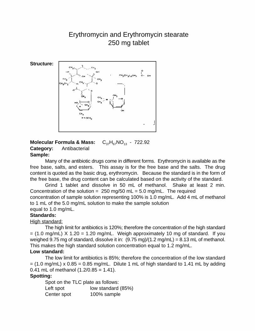
Erythromycin and Erythromycin stearate 250 mg tablet
Structure:
Molecular Formula & Mass: C H NO - 722.92 37 67 13
Category: Antibacterial Sample:
Many of the antibiotic drugs come in different forms. Erythromycin is available as thefree base, salts, and esters. This assay is for the free base and the salts. The drugcontent is quoted as the basic drug, erythromycin. Because the standard is in the form ofthe free base, the drug content can be calculated based on the activity of the standard. Grind 1 tablet and dissolve in 50 mL of methanol. Shake at least 2 min.Concentration of the solution = 250 mg/50 mL = 5.0 mg/mL. The required concentration of sample solution representing 100% is 1.0 mg/mL. Add 4 mL of methanolto 1 mL of the 5.0 mg/mL solution to make the sample solution equal to 1.0 mg/mL. Standards:High standard:
The high limit for antibiotics is 120%; therefore the concentration of the high standard= (1.0 mg/mL) X 1.20 = 1.20 mg/mL. Weigh approximately 10 mg of standard. If youweighed 9.75 mg of standard, dissolve it in: (9.75 mg)/(1.2 mg/mL) = 8.13 mL of methanol.This makes the high standard solution concentration equal to 1.2 mg/mL. Low standard:
The low limit for antibiotics is 85%; therefore the concentration of the low standard= (1.0 mg/mL) x 0.85 = 0.85 mg/mL. Dilute 1 mL of high standard to 1.41 mL by adding0.41 mL of methanol (1.2/0.85 = 1.41). Spotting:
Spot on the TLC plate as follows: Left spot low standard (85%) Center spot 100% sample

Right spot high standard (120%) Development:
Mix 21 mL of methanol, 2 mL of toluene, and 2 mL of acetone. Add this mixture tothe TLC development bag. Develop until the solvent front reaches within 1 cm of the top ofthe TLC plate. Detection:UV:
The spots are not visible in the UV. Iodine stain:
Dip the plate in the iodine-KI solution in the detection bag. Allow the plate to dry andobserve the size and intensity of the spots. To make the spots last longer, allow the plateto dry for 30 min. after it has been dipped in the iodine solution. Heat the plate at 100EC for5 min. The spots turn gray-green and are stable for 24 hr. Notes:
This procedure works for the free base and stearate form. It should also work withthe lactobionate and gluconate forms. This solvent system can be used to separate the freebase from the ethyl succinate and propionate (estolate) esters. The ethyl succinatehydrolyzes in methanol quickly (less than 24 hr) to form the free base and erythromycin.Thus, the esters should be quantitated by using the appropriate procedure (seeerythromycin estolate and erythromycin ethyl succinate).
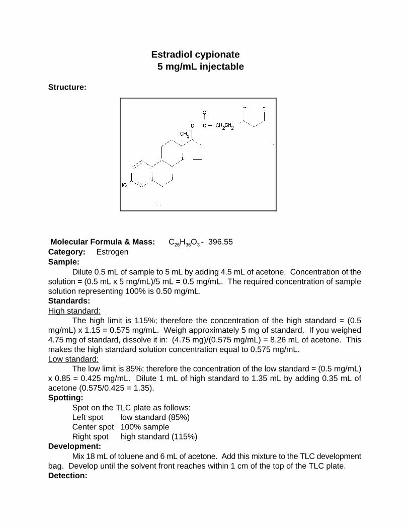
Estradiol cypionate 5 mg/mL injectable
Structure: Molecular Formula & Mass: C H O - 396.55 26 36 3
Category: Estrogen Sample:
Dilute 0.5 mL of sample to 5 mL by adding 4.5 mL of acetone. Concentration of thesolution = (0.5 mL x 5 mg/mL)/5 mL = 0.5 mg/mL. The required concentration of samplesolution representing 100% is 0.50 mg/mL. Standards:High standard:
The high limit is 115%; therefore the concentration of the high standard = (0.5mg/mL) x 1.15 = 0.575 mg/mL. Weigh approximately 5 mg of standard. If you weighed4.75 mg of standard, dissolve it in: (4.75 mg)/(0.575 mg/mL) = 8.26 mL of acetone. Thismakes the high standard solution concentration equal to 0.575 mg/mL.Low standard:
The low limit is 85%; therefore the concentration of the low standard = (0.5 mg/mL)x 0.85 = 0.425 mg/mL. Dilute 1 mL of high standard to 1.35 mL by adding 0.35 mL ofacetone (0.575/0.425 = 1.35). Spotting:
Spot on the TLC plate as follows: Left spot low standard (85%) Center spot 100% sample Right spot high standard (115%)
Development:Mix 18 mL of toluene and 6 mL of acetone. Add this mixture to the TLC development
bag. Develop until the solvent front reaches within 1 cm of the top of the TLC plate.Detection:

UV:Dry the plate and observe under UV light. The maximum is at 240 nm but a short
wavelength UV light (254 nm) will work. Observe the size and intensity of the spots. Iodine stain:
The 0.50 mg/mL concentration is too low to allow for quantitation by iodine. Theconcentration needs to be 5.0 mg/mL. This can be accomplished by using the liquid sampleneat. Do not dilute the sample. Dip the plate into the iodine-KI solution in the detection bag.Allow the plate to dry and observe the size and intensity of the spots.

60
Ethambutol HCl100 and 400 mg tablets
Revised 8/18/1997METHOD# 1
Structure
Molecular Formula & Mass: C H N O - 204.3110 24 2 2
Category: AntibacterialSample:100 mg tablet:
Grind 1 tablet and dissolve in 10 mL of methanol which makes a solution having aconcentration of 10 mg/mL. Concentration of the required solution = 2 mg/mL to representa 100% solution. Take 1 mL of the 10 mg/mL solution and add 4 mL of methanol to makethe required concentration of 2mg/mL.400 mg tablet:
Grind 1 tablet and dissolve in 25 mL of methanol. Concentration of the solution =16mg/mL. Add 7 mL of methanol to 1 mL of the 16 mg/mL solution to make a finalconcentration equal to 2 mg/mL.Standard:High standard:
The high limit is 115%; therefore the concentration of high standard = (2 mg/mL X1.15) = 2.3 mg/mL. Weigh approximately 21 mg of standard. If you weighed 21.7 mg ofstandard, dissolve it in: (21.7 mg)/(2.3 mg/mL) = 9.4 mL of methanol.Low standard:
The low limit is 85%; therefore the concentration of low standard = (2 mg/mL) X 0.85= 1.7 mg/mL. Dilute 1 mL of high standard to 1.35 mL by adding 0.35 mL of methanol=1.35).Spotting:
61
Spot on the TLC plate as follow:Left spot low standard (85%)Center spot 100% sampleRight spot high standard (115%)
Development:Mix 25 mL of methanol and 0.38 mL of concentrated ammonium hydroxide. Add 24
mL of this mixture to the TLC development bag. Develop until the solvent front reacheswithin 1 cm of the top of the TLC plate.Detection:UV:
The spots are not visible under UV.Iodine stain:
Dip the plate in the iodine-KI solution in the detection bag. Allow the plate to dry andobserve the size and intensity of the spots.

62
METHOD#2 Revised March 16,1999
Ethambutol HCl100 and 400 mg tablets
Structure
Molecular Formula & Mass: C H Cl N O - 27710 26 2 2 2
Category: Antibacterial (tuberculostatic)
Preparation of the sample solution: Analytical balance available:
Prepare the sample solution by weighing an aliquot of the drug. Follow the proceduredescribed in the previous section. Determine the weight of the drug and add solvent to produce aconcentration of 2mg/mL. Measure the volume accurately using a combination of pipetts plus a 1 mLgraduated tuberculin syringe for the fractional volumes. Pipetts are available in 1mL increments upto 10 mL. For example: You weighed 23 mg of the drug, then you would add 11.5mL of solvent toprepare a solution with a concentration of 2mg/mL. (Use a 10 and 1mL pipetts and the 0.5 mL ismeasured by a 1mL graduated syringe). NOTE: The above procedure applies to all of the TB drugs using the aliquot method.
Analytical balance not available.The tablets listed below are representative of different drug content. Adjust the volumes
accordingly for different composition. Always use volumes in full mL so that fractional volumes isnot required. Most drugs have their contents in multiples of 5 which give a whole number for theconcentration. Dissolve the sample in volumes which do give you a whole number for theconcentrated solution. An aliquot of the concentrated solution is diluted to obtain the properconcentration for the TLC.

63
100 mg tablet: Grind 1 tablet and dissolve in 10 mL of methanol which makes a solution having a
concentration of 10 mg/mL. Concentration of the required solution = 2 mg/mL to represent a 100%solution. Take 1 mL of the 10 mg/mL solution and add 4 mL of methanol to make the requiredconcentration of 2mg/mL.400 mg tablet:
Grind 1 tablet and dissolve in 25 mL of methanol. Concentration of the solution =16 mg/mL.Add 7 mL of methanol to 1 mL of the 16 mg/mL solution to make a final concentration equal to 2mg/mL.
Preparation of standard or reference solutions:Reference solutions are prepared depending on the availability of reference compounds.
Reference tablets are available. The tablets contain a predetermined weight of the drug which whendissolved in 5 mL of the solvent will produce a solution concentration representing 115% of thesample solution. No weighing is required. Weighing is required when the reference compound is notavailable in tablet form. The reference solutions then must be prepared using either primary orsecondary standards.Preparation of the high standard when no reference tablets are available:
The high concentration limit is 115%; therefore the concentration of high standard = (2 mg/mLX 1.15) = 2.3 mg/mL. Weigh approximately 21 mg of standard. If you weighed 21.7 mg of standard,dissolve it in: (21.7 mg)/(2.3 mg/mL) = 9.4 mL of methanol.Low standard:
The low limit is 85%; the concentration of low standard = (2 mg/mL) X 0.85 = 1.7 mg/mL.Dilute 1 mL of high standard to 1.35 mL by adding 0.35 mL of methanol= 1.35). This low referencesolution is always prepared by the same procedure regardless of the reference source.Spotting:
Spot on the TLC plate as follows:Sample each of the solutions with a 3µL capillary pipette and spot. Left spot low standard (85%)Center spot 100% sampleRight spot high standard (115%)
Development:Mix 25 mL of methanol and 0.38 mL of concentrated ammonium hydroxide. Add 24 mL this
mixture to the TLC development bag. Develop until the solvent front reaches within 1 cm of the topof the TLC plate.Detection:UV:
The spots are not visible under UV.Iodine stain:
Dip the plate in the iodine-KI solution in the detection bag. Allow the plate to dry and observethe size and intensity of the spots.

64
Furosemide 20, 40 and 80 mg tablets
Structure:
Molecular Formula and Mass: C H ClN O S - 330.77 12 11 2 5
Category: DiureticSample:
The sample solution representing 100% requires a concentration of 2.5 mg/mL.20 mg tablet: Grind 1 tablet and dissolve in 8 mL of methanol.40 mg tablet: Grind 1 tablet and dissolve in 16 mL of methanol.80 mg tablet: Grind 1 tablet and dissolve in 32 mL of methanol.
Shake for at least 2 min.Standards:High standard: The high limit is 115%; therefore the concentration of the high standard = (2.5mg/mL) X 1.15 = 2.875 mg/mL. Weigh approximately 15 mg of standard. If you weighed13.2 mg of standard, dissolve it in: (13.2 mg)/(2.875 mg/mL) = 4.59 mL of methanol. Thismakes the high standard solution concentration equal to 2.875 mg/mL. Low standard: The low limit is 85%; therefore the concentration of the low standard = (2.5 mg/mL)X 0.85 = 2.125 mg/mL. Dilute 1 mL of high standard to 1.35 mL by adding 0.35 mL ofmethanol (2.875/2.125 = 1.35). Spotting:
Spot on the TLC plate as follows: Left spot low standard (85%) Center spot 100% sample Right spot high standard (115%)
Development:

65
Mix 5 mL of toluene, 5 mL of acetone, 20 mL of methanol, and 1.5 mL ofconcentrated ammonium hydroxide. Pour 24 mL of the mixture into a TLC development bag.Develop until the solvent front reaches within 1 cm of the top of the TLC plate. Detection:UV: Dry the plate and observe under UV light. The maximum is at 275 nm but a shortwavelength UV light (254 nm) works. Observe the size and intensity of the spots. Iodine stain:
Dip the plate in the iodine-KI solution in the detection bag. Allow the plate to dry andobserve the size and intensity of the spots.

66
Gentamycin40 mg/mL injectable
Structure:
Molecular Formula & Mass: C1;C H N O .H SO4 - 575.6821 43 5 7 2
C1a;C H N O .H SO4 - 547.6319 39 5 7 2
C2;C H N O .H SO - 561.66 20 41 5 7 2 4
Category: AntibacterialGentamicin is one of the aminoglycoside antibiotics and is composed of more than
one component. Injectable gentamicin sulfate is a mixture of three components: gentamicinC1, C1a, and C2 sulfates. The drug content is quoted as the basic drug, gentamicin.Because the standard is in the form of the sulfate, the potency is quoted as units ofgentamicin activity. Thus the standards must be corrected for activity. Sample:
Dilute 1 mL of the injectable solution to 8 mL by adding 7 mL of distilled water.Shake at least 1 min. Concentration of the solution = 5 mg/mL. Standard:High standard:
The high limit for antibiotics is 120%; therefore the concentration of the high standard= (5.0 mg/mL) X 1.20 = 6.0 mg/mL. Weigh approximately 35 mg of standard. If youweighed 34.9 mg of standard with an activity of 648, dissolve it in: (34.9 mg)(0.648)/(6.00 mg/mL) = 3.77 mL of distilled water. This makes the high standardsolution concentration equal to 6.0 mg/mL. Low standard:
The low limit is 85%; therefore the concentration of the low standard = (5.0 mg/mL)X 0.85 = 4.25 mg/mL. Dilute 1 mL of high standard to 1.41 mL by adding 0.41 mL of water(6.0/4.25 = 1.41).Spotting:
Mix 20 mL of methanol, 4 mL of water, and 20 mL of concentrated ammonium

67
hydroxide. Add 22 mL to the TLC development bag and develop a blank TLC plate until thesolvent front reaches 1 cm from the top of the plate. Remove the plate and allow it to dryuntil the odor of ammonia can no longer be detected. Remove the solvent from thedeveloping bag.
Spot on the plate as follows: Left spot low standard (85%) Center spot 100% sample Right spot high standard (120%)
Development: Place 24 mL of the methanol-water-concentrated ammonium hydroxide mixture in the
TLC development bag. Develop the plate until the solvent front reaches 1 cm from the topof the plate. Allow the plate to dry until the odor of ammonia cannot be detected. Detection:UV:
The spots are not visible in the UV. Iodine stain:
Dip the plate in the iodine-KI solution in the detection bag. Allow the spots to dry anddetermine their size and intensity.Visible:
To make the spots visible in white light, dip the plate in a ninhydrin solution. Dissolve100 mg of ninhydrin, 2,2-dihydroxy-1,3-indanedione (Sigma Chemical Co. No. N4876), in 25mL of acetone. Place 7 mL of this solution in the detection bag instead of the iodine-KIsolution. The TLC plate should be dry and completely free of any ammonia (it may benecessary to allow the plate to sit overnight). Dip the TLC plate in the ninhydrin solution.Allow the plate to dry for 60 min. A blue-violet color develops with gentamicin. Observe thesize and intensity of the spots. If the TLC plate is not completely free of ammonia, thebackground color of the plate will change from pale pink to dark rose in about 1 hr. Notes:
The use of ninhydrin is recommended because the iodine colors do not last very long.The color may become unreadable in 5 min. and yet may be readable for as long as 20 min.The ninhydrin color can be developed in less time if an oven or a hot plate is available. Afterthe TLC plate air-dries for 30 min., the ammonia on the TLC plate can be removed byheating the plate at 100EC for 10 min. Dip the TLC plate in the ninhydrin solution and allowit to air-dry for 30 min. (The color usually develops in 30-60 min.) Heat the TLC plate for2 min. at 100EC to fully develop the violet color. The TLC plate is pre-developed to removethe possibility of streaking or solvent front effects.
68
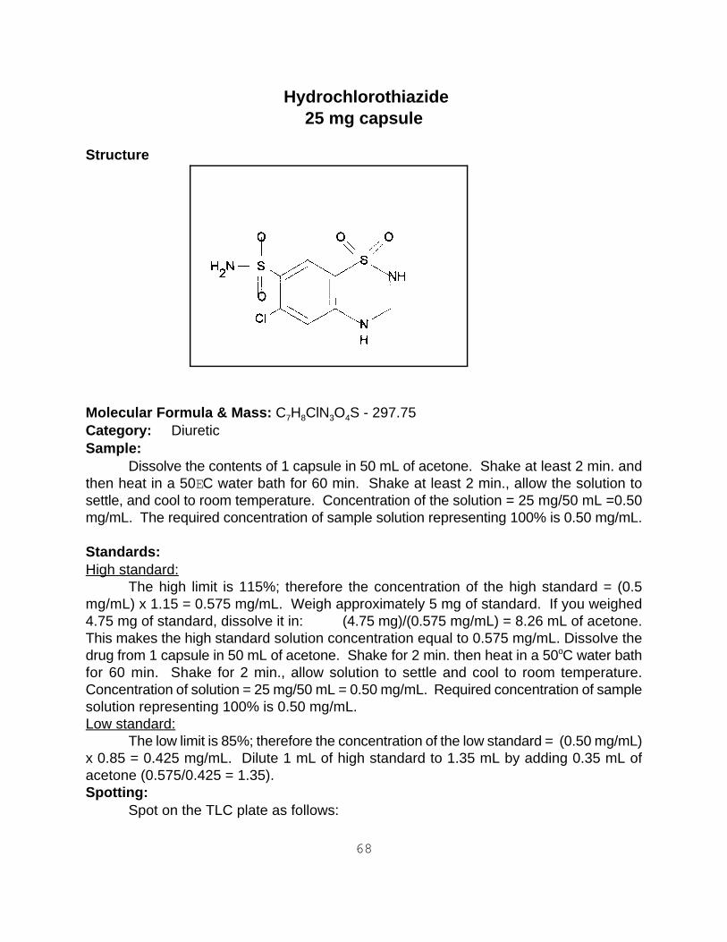
Hydrochlorothiazide25 mg capsule
Structure
Molecular Formula & Mass: C H ClN O S - 297.75 7 8 3 4
Category: Diuretic Sample:
Dissolve the contents of 1 capsule in 50 mL of acetone. Shake at least 2 min. andthen heat in a 50EC water bath for 60 min. Shake at least 2 min., allow the solution tosettle, and cool to room temperature. Concentration of the solution = 25 mg/50 mL =0.50mg/mL. The required concentration of sample solution representing 100% is 0.50 mg/mL. Standards: High standard:
The high limit is 115%; therefore the concentration of the high standard = (0.5mg/mL) x 1.15 = 0.575 mg/mL. Weigh approximately 5 mg of standard. If you weighed4.75 mg of standard, dissolve it in: (4.75 mg)/(0.575 mg/mL) = 8.26 mL of acetone.This makes the high standard solution concentration equal to 0.575 mg/mL. Dissolve thedrug from 1 capsule in 50 mL of acetone. Shake for 2 min. then heat in a 50 C water batho
for 60 min. Shake for 2 min., allow solution to settle and cool to room temperature.Concentration of solution = 25 mg/50 mL = 0.50 mg/mL. Required concentration of samplesolution representing 100% is 0.50 mg/mL. Low standard:
The low limit is 85%; therefore the concentration of the low standard = (0.50 mg/mL)x 0.85 = 0.425 mg/mL. Dilute 1 mL of high standard to 1.35 mL by adding 0.35 mL ofacetone (0.575/0.425 = 1.35). Spotting:
Spot on the TLC plate as follows:
69
Left spot low standard (85%) Center spot 100% sample Right spot high standard (115%)
Development: Add 24 mL of ethyl acetate to the TLC development bag. Develop until the solventfront reaches within 1 cm of the top of the TLC plate. Detection:UV:
Dry the plate and observe under UV light. The maximum is at 240 nm but a shortwavelength UV light (254 nm) will work. Observe the size and intensity of the spots. Iodine stain:
The 0.50 mg/mL concentration is too low to allow for quantitation by iodine. Theconcentration must be 10.0 mg/mL. This can be accomplished by using 2.5 mL of acetoneto dissolve 1 tablet. Dip the plate in the iodine-KI solution in the detection bag. Allow theplate to dry and observe the size and intensity of the spots. The spots take a long time todevelop, and the plates can be read 24 hr later.
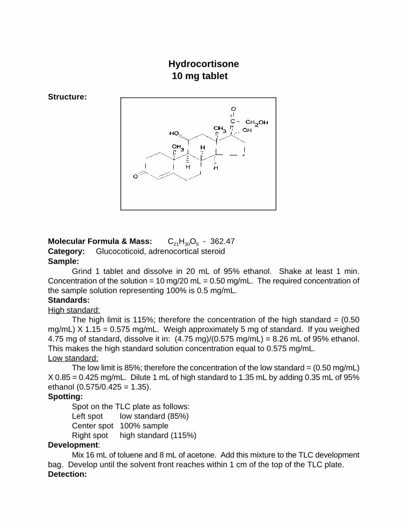
Hydrocortisone10 mg tablet
Structure: Molecular Formula & Mass: C H O - 362.47 21 30 5
Category: Glucocoticoid, adrenocortical steroid Sample:
Grind 1 tablet and dissolve in 20 mL of 95% ethanol. Shake at least 1 min.Concentration of the solution = 10 mg/20 mL = 0.50 mg/mL. The required concentration ofthe sample solution representing 100% is 0.5 mg/mL. Standards:High standard:
The high limit is 115%; therefore the concentration of the high standard = (0.50mg/mL) X 1.15 = 0.575 mg/mL. Weigh approximately 5 mg of standard. If you weighed4.75 mg of standard, dissolve it in: (4.75 mg)/(0.575 mg/mL) = 8.26 mL of 95% ethanol.This makes the high standard solution concentration equal to 0.575 mg/mL. Low standard:
The low limit is 85%; therefore the concentration of the low standard = (0.50 mg/mL)X 0.85 = 0.425 mg/mL. Dilute 1 mL of high standard to 1.35 mL by adding 0.35 mL of 95%ethanol (0.575/0.425 = 1.35). Spotting:
Spot on the TLC plate as follows: Left spot low standard (85%) Center spot 100% sample Right spot high standard (115%)
Development: Mix 16 mL of toluene and 8 mL of acetone. Add this mixture to the TLC development
bag. Develop until the solvent front reaches within 1 cm of the top of the TLC plate.Detection:

UV:Dry the plate and observe under UV light. The maximum is at 240 nm but a short
wavelength UV light (254 nm) will work. Observe the size and intensity of the spots. Iodine stain:
The 0.50 mg/mL concentration is too low to allow for quantitation by iodine. Theconcentration needs to be 5 mg/mL. This can be accomplished by using 10 mL of 95%ethanol rather than 20 mL. Dip the plate in the iodine-KI solution in the detection bag. Allowthe plate to dry and observe the size and intensity of the spots.

Ibuprofen 300mgtablet
Structure:
Molecular formula and weight: C H O - 206.2713 18 2
Category: AnalgesicSample: Grind 1 tablet of the drug, and dissolve in 50 mL of a mixture containing 1 mL of aceticacid and 49 mL of methanol ( prepare a volume of the methanol/acid mixture greater than50 mL before making the solution of the drug). This produces a solution having aconcentration equal to 6 mg/mL. The concentration required for the analysis is 2 mg/mL.Take 1 mL of the 6 mg/mL solution and dilute to 3 mL by adding 2 mL of the aceticacid/methanol mixture to make the final concentration. The drug is also supplied as a 400 mg tablet. In this case, grind and dissolve 1 tabletin 50 mL of the solvent mixture to make a concentration equal to 8 mg/mL. Take 1 mL ofthe 8 mg/mL solution and add 3 mL of the solvent mixture to make the final solution equalto 2 mg/mL.Standards:High standard: The high standard solution concentration requires a solution equivalent to 115% of thesample concentration (1.15 X 2 mg/mL = 2.3 mg/mL). Weigh approximately 10 mg of the standard. Divide the weight of the standard by the high standard concentration to obtain the volume of solvent needed. For example, 9.4 mgwas weighed, then the volume needed would be 9.4mg/2.3 = 4.09 mL of the acid/methanolmixture.Low standard: The low standard concentration is equivalent to 85% of the sample concentration. The concentration is 0.85 X 2 mg/mL= 1.7 mg/mL. This

concentration is prepared by taking 1 mL of the 2.3 mg/mL solution and adding 0.35 mL ofthe solvent mixture.Spotting:
Spot the TLC plate as follows: Left spot low standard(85%) Center spot 100% sample Right spot high standard(115%) Development: Prepare the developer by mixing 17 mL of toluene, 13 mL of ethyl acetate, and 1 mL of acetic acid. Add 24 mL of this solution to the TLC development bag. Develop until the solvent front reaches to within 1 cm from the top of the TLC plate. Detection:UV: Dry the plate and observe under UV at 254 nm. Observe the sizes and intensities of the spots.Iodine stain: Dip the plate into the iodine-KI solution. Remove and allow the iodine to sublime, and observe the spots as soon as they become distinct. Note the sizes and intensities.
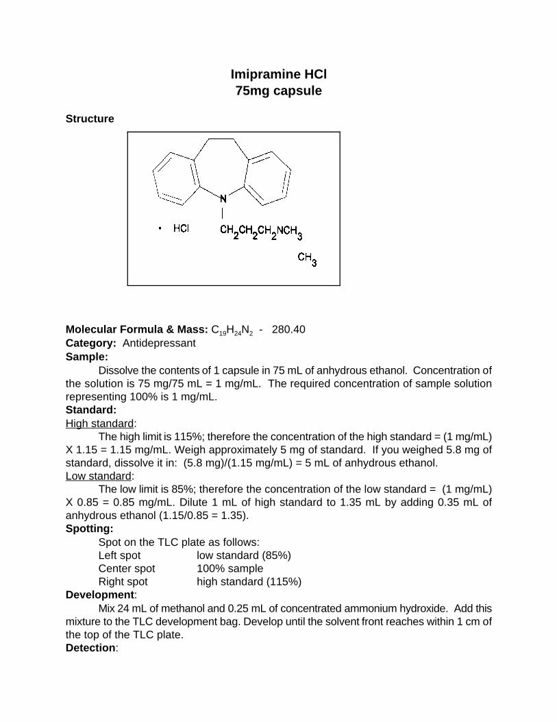
Imipramine HCl75mg capsule
Structure
Molecular Formula & Mass: C H N - 280.40 19 24 2
Category: AntidepressantSample:
Dissolve the contents of 1 capsule in 75 mL of anhydrous ethanol. Concentration ofthe solution is 75 mg/75 mL = 1 mg/mL. The required concentration of sample solutionrepresenting 100% is 1 mg/mL.Standard:High standard:
The high limit is 115%; therefore the concentration of the high standard = (1 mg/mL)X 1.15 = 1.15 mg/mL. Weigh approximately 5 mg of standard. If you weighed 5.8 mg ofstandard, dissolve it in: (5.8 mg)/(1.15 mg/mL) = 5 mL of anhydrous ethanol.Low standard:
The low limit is 85%; therefore the concentration of the low standard = (1 mg/mL)X 0.85 = 0.85 mg/mL. Dilute 1 mL of high standard to 1.35 mL by adding 0.35 mL ofanhydrous ethanol (1.15/0.85 = 1.35).Spotting:
Spot on the TLC plate as follows:Left spot low standard (85%)Center spot 100% sample Right spot high standard (115%)
Development:Mix 24 mL of methanol and 0.25 mL of concentrated ammonium hydroxide. Add this
mixture to the TLC development bag. Develop until the solvent front reaches within 1 cm ofthe top of the TLC plate.Detection:

UV:Dry the plate and observe under UV light. The maximum is at 270 nm but a short
wavelength light (254 nm) will work. Observe the size and intensity of the spots.Iodine stain:
Dip the plate in the iodine-KI solution in the detection bag. Allow the plate to dry andobserve the size and intensity of the spots.

Indomethacin 25 mg capsule
Sample:
Molecular Formula & Mass: C H ClNO - 357.8119 16 4
Category: Anti-inflamatory, antipyretic,analgesic Sample:
Dissolve the contents of 1 capsule in 5 mL of methanol. Shake at least 1 min.Concentration of the solution = 25 mg/5 mL = 5.00 mg/mL. The required concentration ofthe sample solution representing 100% is 5.00 mg/mL. Standards:High standard:
The high limit is 115%; therefore the concentration of the high standard = (5.00mg/mL) x 1.15 = 5.75 mg/mL. Weigh approximately 25 mg of standard. If you weighed 25mg of standard, dissolve it in: (25.0 mg)/(5.75 mg/mL) = 4.35 mL of methanol. This makesthe high standard solution concentration equal to 5.75 mg/mL. Low standard:
The low limit is 85%; therefore the concentration of the low standard = (5.00 mg/mL)x 0.85 = 4.25 mg/mL. Dilute 1 mL of high standard to 1.35 mL by adding 0.35 mL ofmethanol (5.75/4.25 = 1.35). Spotting:
Spot on the TLC plate as follows: Left spot low standard (85%) Center spot 100% sample Right spot high standard (115%)
Development: Mix 25 mL of toluene, 12 mL of acetone, and 1 mL of glacial acetic acid. Add about
24 mL of this mixture to a TLC development bag. Develop until the solvent front reaches

within 1 cm of the top of the TLC plate. Detection:UV:
Allow the plate to dry and observe under UV light (254 nm). Observe the size andintensity of the spots. Iodine stain:
Dip the plate into the iodine-KI solution in the detection bag. Observe the size andintensity of the spots as the plate dries.

METHOD # 1 Isoniazid
300 and 100 mg tabletsRevised procedure(Aug.12,1997) Structure:
Molecular Formula & Mass: C H N O - 137.156 7 3
Category: Antibacterial (tuberculostatic)Sample:300 mg tablet
Crush 1 tablet and dissolve in 50 mL of methanol. Concentration of the solution = 300mg/50 mL = 6 mg/mL. The required concentration of the sample solution representing 100%= 0.5 mg/mL. Take 1 mL of the 6 mg/mL solution and add 11 mL of methanol to make 12mL of solution which makes a final concentration of 0.5mg/mL.100 mg tablet
Crush 1 tablet and dissolve in 25 mL of methanol. Concentration of the solution = 100mg/25 mL = 4 mg/mL. The concentration of the sample solution representing 100% =0.5mg/mL. Take 1 mL of the 4mg/mL solution and add 7 mL of methanol to make 8 mL ofsolution with a concentration of 0.5mg/mL.Standards:High standard:
The high limit is 115%; therefore the concentration of the high standard = (0.5mg/mL) X 1.15 = 0.575 mg/mL. Weigh approximately 4-5 mg of standard. If you weighed4.2 mg of standard, dissolve it in: (4.2mg)/(0.575 mg/mL) = 7.3 mL of methanol. Thismakes the high standard solution concentration equal to 0.575 mg/mL.Low standard:
The low limit is 85%; therefore the concentration of the low standard = (0.5 mg/mL)X 0.85 = 0.425 mg/mL. Dilute 1 mL of high standard to 1.35 mL by adding 0.35 mL ofmethanol (0.575/0.425 = 1.35).Spotting:

Spot on the TLC plate as follows:Left spot low standard (85%) Center spot 100% sample Right spot high standard (115%)
Developer:Mix 25 mL of methanol and 0.5 mL of concentrated ammonium hydroxide. Add 24
mL of this mixture to the TLC development bag. Develop until the solvent front reacheswithin 1 cm of the top of the plate. Detection:UV:
Dry the plate and observe under UV light (254 nm). Observe the size and intensityof the spots.Iodine stain:
Dip the plate in the iodine-KI solution in the detection bag. Allow the plate to dry andobserve the size and intensity of the spot.
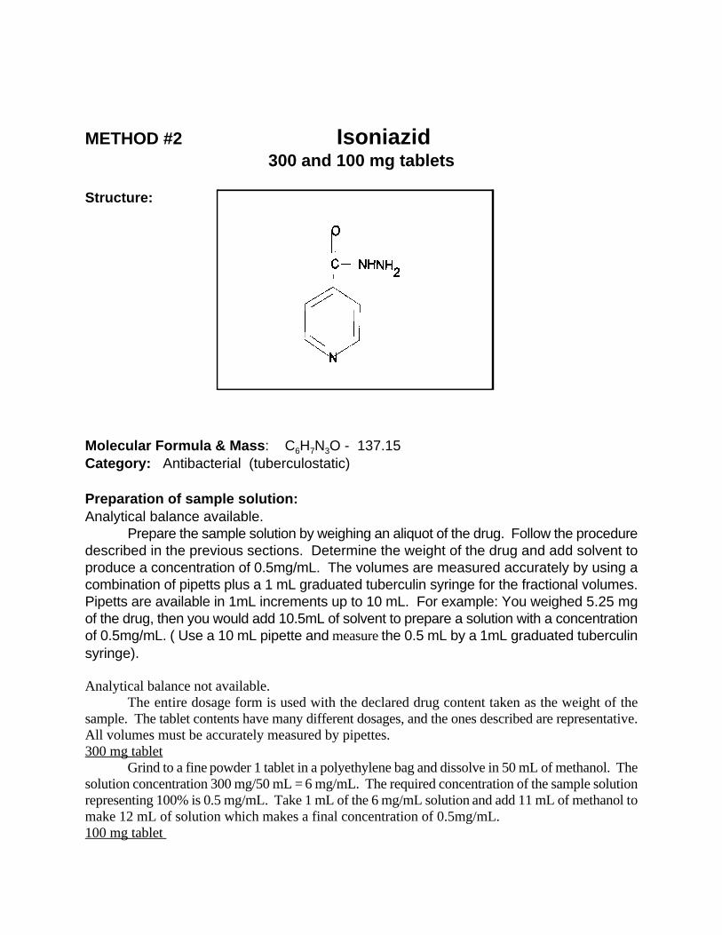
METHOD #2 Isoniazid 300 and 100 mg tablets
Structure:
Molecular Formula & Mass: C H N O - 137.156 7 3
Category: Antibacterial (tuberculostatic)
Preparation of sample solution:Analytical balance available.
Prepare the sample solution by weighing an aliquot of the drug. Follow the proceduredescribed in the previous sections. Determine the weight of the drug and add solvent toproduce a concentration of 0.5mg/mL. The volumes are measured accurately by using acombination of pipetts plus a 1 mL graduated tuberculin syringe for the fractional volumes.Pipetts are available in 1mL increments up to 10 mL. For example: You weighed 5.25 mgof the drug, then you would add 10.5mL of solvent to prepare a solution with a concentrationof 0.5mg/mL. ( Use a 10 mL pipette and measure the 0.5 mL by a 1mL graduated tuberculinsyringe). Analytical balance not available.
The entire dosage form is used with the declared drug content taken as the weight of thesample. The tablet contents have many different dosages, and the ones described are representative.All volumes must be accurately measured by pipettes.300 mg tablet
Grind to a fine powder 1 tablet in a polyethylene bag and dissolve in 50 mL of methanol. Thesolution concentration 300 mg/50 mL = 6 mg/mL. The required concentration of the sample solutionrepresenting 100% is 0.5 mg/mL. Take 1 mL of the 6 mg/mL solution and add 11 mL of methanol tomake 12 mL of solution which makes a final concentration of 0.5mg/mL.100 mg tablet

Grind to a fine powder 1 tablet and dissolve in 25 mL of methanol. The concentration of thesolution is 100 mg/25 mL = 4 mg/mL. The required concentration of the sample solution representing100% is 0.5 mg/mL. Take 1 mL of the 4 mg/mL solution and add 7 mL of methanol to make 8 mL ofsolution with a concentration of 0.5mg/mL.
Preparation of standards solutions:Reference solutions are prepared depending on the availability of reference compounds. The
reference materials may be either in the form of reference tablets or powders of primary/secondarystandards. Reference tablets may be available containing a predetermined weight of the drug whichwhen dissolved in 5 mL of the solvent produces a solution concentration representing 115% of thesample solution. No weighing is required. Weighing is required when the reference compound is not available in tablet form. The referencesolutions must be prepared using either primary or secondary standards.High standard solution:
1. Reference tablet available.The reference tablet contains 2.88 mg of isoniazid which when dissolved in 5 mL of methanolproduces a solution having a concentration of 2.88mg/5 mL equal to 0.576 mg/mL which is 115% ofthe sample concentration. Drop one reference tablet into a vessel and add 5 mL of the solvent. Noweighing is needed.
2. Reference material in the powder form (primary or secondary).A reference solution having a concentration of 0.576 mg/mL is required. Weigh approximately 4-5mg of powdered standard. For example: you weighed 4.2 mg of standard, dissolve it in:(4.2mg)/(0.575 mg/mL) = 7.3 mL of methanol. Measure the volumes by pipettes and a 1 mL graduatedtuberculin syringe. This makes the high standard solution concentration equal to 0.575 mg/mL equalto 115% .Low standard:
The low limit is 85%; therefore the concentration of the low standard = (0.5 mg/mL) X 0.85= 0.425 mg/mL. Dilute 1 mL of high standard to 1.35 mL by adding 0.35 mL of methanol (0.575/0.425= 1.35). The low standard is always prepared by taking 1 mL of the high and adding 0.35 mL of thesolvent when the high solution represents 115% of the sample.Spotting:
Spot on the TLC plate as follows:Sample each of the solutions with a 3µL capillary pipette and spot.Left spot low standard (85%) Center spot sample (100%)Right spot high standard (115%)
Developer:Mix 13 mL of methanol, 17 mL of acetone and 1 mL of concentrated ammonium hydroxide.
Add 24 mL of this mixture to the TLC development bag. Develop until the solvent front reaches within1 cm of the top of the plate. Detection:UV:
Dry the plate and observe under UV light (254 nm). Observe the size and intensity of the spotsor stain with iodine when no UV available..Iodine stain:
Dip the plate in the iodine-KI solution in the detection bag. Allow the plate to dry and observe

the size and intensity of the spot.

Kanamycin sulfate 75 mg/2 mL injectable
Structure:
Molecular Formula & Mass: A & C; C H N O - 386.4318 36 4 11
B; C H N O - 385.4518 37 5 11
Category: Antimicrobial Kanamycin is one of the aminoglycoside antibiotics and is composed of more than
one component. Injectable kanamycin sulfate is a mixture of three components: kanamycinA, B, and C sulfates. The drug content is quoted as the basic drug, kanamycin. Thestandard is in the form of the sulfate and the potency is quoted as units of kanamycinactivity. Thus the standards must be corrected for activity. Sample:
Dilute 0.5 mL of the injectable solution to 15 mL by adding 14.5 mL of distilled water.Shake at least 1 min. Concentration of the solution = (0.5 mL x 75 mg)/(2 mL)/15 mL = 1.25mg/mL. The required concentration of sample solution representing 100% is 1.25 mg/mL.Standards:High standard:
The high limit for antibiotics is 120%; therefore the concentration of the high standard= (1.25 mg/mL) X 1.20 = 1.50 mg/mL. Weigh approximately 20 mg of standard. If youweighed 19.9 mg of standard with an activity of 648, dissolve it in: (19.9 mg)(0.648)/(1.50mg/mL) = 8.60 mL of water. This makes the high standard solution concentration equal to1.50 mg/mL. Low standard:
The low limit for antibiotics is 85%; therefore the concentration of the low standard= (1.25 mg/mL) x 0.85 = 1.06 mg/mL. Dilute 1 mL of high standard to 1.41 mL by adding0.41 mL of water (1.50/1.06 =1.41). Spotting:
Spot on the TLC plate as follows:

Left spot low standard (85%) Center spot 100% sample Right spot high standard (120%)
Development:Mix 20 mL of methanol, 4 mL of water, and 20 mL of concentrated ammonium
hydroxide. Add 24 mL of this mixture to the TLC development bag. Develop a blank TLCplate until the solvent front reaches 1 cm from the top of the plate. Remove the plate andallow it to dry until the odor of ammonia can no longer be detected. Remove the solventfrom the developing bag.Detection:UV:
The spots are not visible in the UV. Iodine stain:
Dip the plate in the iodine-KI solution in the detection bag. Allow the spots to dry anddetermine their size and intensity.Visible:
To make the spots visible in white light, dip the plate in a ninhydrin solution preparedby dissolving 100 mg of ninhydrin, 2,2-dihydroxy-1,3-indanedione (Sigma Chemical Co. No.N4876), in 25 mL of acetone. Place 7 mL of this solution in the detection bag instead of theiodine developing solution. The TLC plate should be dry and completely free of anyammonia (the plate may have to sit overnight). Dip the TLC plate in the ninhydrin solution.Allow the plate to dry for 60 min. A blue/violet color develops with kanamycin. Observethe size and intensity of the spots. If the TLC plate is not completely free of ammonia, thebackground color of the TLC plate changes from pale pink to dark rose in about 1 hr. Notes:
The use of ninhydrin is recommended because the iodine intensities are variable anddo not last very long. The ninhydrin color can be developed in less time if an oven or a hotplate is available. After the TLC plate air-dries for 30 min., the ammonia on the TLC platecan be removed by heating the plate at 100EC for 10 min. Dip the TLC plate in the ninhydrinsolution and allow it to air-dry for 30 min. (The color usually develops in 30-60 min.) Heatthe TLC plate for 2 min. at 100EC to fully develop the violet color. The TLC plate ispre-developed to remove the possibility of streaking or solvent front effects.
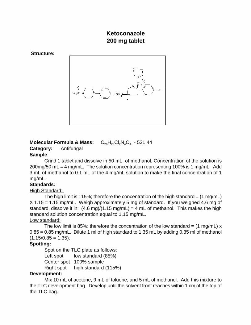
Ketoconazole200 mg tablet
Structure:
Molecular Formula & Mass: C H Cl N O - 531.44 26 28 2 4 4
Category: Antifungal Sample: Grind 1 tablet and dissolve in 50 mL of methanol. Concentration of the solution is200mg/50 mL = 4 mg/mL. The solution concentration representing 100% is 1 mg/mL. Add3 mL of methanol to 0 1 mL of the 4 mg/mL solution to make the final concentration of 1mg/mL. Standards: High Standard:
The high limit is 115%; therefore the concentration of the high standard = (1 mg/mL)X 1.15 = 1.15 mg/mL. Weigh approximately 5 mg of standard. If you weighed 4.6 mg ofstandard, dissolve it in: (4.6 mg)/(1.15 mg/mL) = 4 mL of methanol. This makes the highstandard solution concentration equal to 1.15 mg/mL. Low standard:
The low limit is 85%; therefore the concentration of the low standard = (1 mg/mL) x0.85 = 0.85 mg/mL. Dilute 1 ml of high standard to 1.35 mL by adding 0.35 ml of methanol(1.15/0.85 = 1.35). Spotting:
Spot on the TLC plate as follows: Left spot low standard (85%) Center spot 100% sample Right spot high standard (115%)
Development:Mix 10 mL of acetone, 9 mL of toluene, and 5 mL of methanol. Add this mixture to
the TLC development bag. Develop until the solvent front reaches within 1 cm of the top ofthe TLC bag.

Detection:UV: Dry the plate and observe under UV light. The maximum is at 225 nm but a shortwavelength UV light (254 nm) will work. Observe the size and intensity of the spots. Iodine stain:
Dip the plate in the iodine-KI solution in the detection bag. Allow the plates to dry andobserve the size and intensity of the spots.

Mebendazole 100 mg tablet
Structure:
Molecular Formula & Mass: C H N O - 295.30 16 13 3 3
Category: Anthelmintic Sample:
Mebendazole is difficult to dissolve and requires the use of formic acid or acetic acid.Prepare a 1:1 solvent mixture of toluene and acetic acid by adding 30 mL of toluene to 30mL of glacial acetic acid. Grind 1 tablet and suspend in 50 mL of the toluene-glacial aceticacid. Shake at least 2 min.; then heat in a 50EC water bath for 30 min. Shake at least 2min., allow the solution to settle, and cool to room temperature. Concentration of thesolution = 100 mg/50 mL = 2.0 mg/mL. The required concentration of sample solutionrepresenting 100% is 0.50 mg/mL. Add 3 mL of the toluene-glacial acetic acid to 1 mL ofthe 2.0 mg/mL solution to make the sample solution equal to 0.50 mg/mL. Standard:High standard:
The high limit is 115%; therefore the concentration of the high standard = (0.50mg/mL) X 1.15 = 0.575 mg/mL. Weigh approximately 5 mg of standard. If you weighed 4.5mg of standard, dissolve it in: (4.5 mg)/(0.575 mg/mL) = 7.83 mL of the toluene-glacialacetic acid. This makes the high standard solution concentration equal to 0.575 mg/mL. Low standard:
The low limit is 85%; therefore the concentration of the low standard = (0.50 mg/mL)X 0.85 = 0.425 mg/mL. Dilute 1 mL of high standard to 1.35 mL by adding 0.35 mL of thetoluene-glacial acetic acid (0.575/0.425 = 1.35). Spotting:
Spot on the TLC plate as follows: Left spot low standard (85%)
Center spot 100% sample Right spot high standard (115%)
Development:Mix 18 mL of acetone and 6 mL of toluene. Add this mixture to the TLC development
bag. Develop until the solvent front reaches within 1 cm of the top of the TLC plate.Detection:UV:
Dry the plate and observe under UV light. The maximum is at 245 nm but a shortwavelength UV light (254 nm) will work. Observe the size and intensity of the spots. Iodine stain:
Dip the plate into the iodine-KI solution in the detection bag. Allow the plate to dryand observe the size and intensity of the spots.

Medroxyprogesterone acetate 5 mg/mL inject.
Structure:
Molecular Formula & Mass: C H O - 396.52 24 34 4
Category: Progesterone; with estradiol as oral contraceptive Sample:
Dilute 1.0 mL to 10 mL by adding 9 mL of ethanol. Shake for 1 min. Concentrationof solution = (5 mg/mL x 1 ml)/10 mL = 0.50 mg/mL. The required concentration of samplesolution representing 100% is 0.50 mg/mL. Standards: High standard:
Since the sample concentration(100%) is 0.50 mg/mL and the high limit is 115%, theconcentration of the high standard(115%) = 0.50 mg/mL X 1.15 = 0.575 mg/mL. Weighapproximately 5 mg of standard. If you weighed 4.75 mg of standard, dissolve it in: (4.75mg)/(0.575 mg/mL) = 8.26 mL of ethanol. This makes the high standard solution concentration equal to 0.575 mg/mL. Low standard:
Since the sample concentration (100%) is 0.50 mg/mL and the low limit is 85%, theconcentration of the low standard (85%) = 0.50 mg/mL X 0.85 = 0.425 mg/mL. Dilute 1 mLof high standard to 1.35 mL by adding 0.35 mL of ethanol (0.575/0.425 = 1.35). Spotting:
Spot on TLC plate as follows: Left spot low standard (85%) Center spot 100% sample Right spot high standard (115%)
Development:
Mix 16 mL of toluene and 8 mL of acetone. Add this mixture to the plastic TLC bag.Develop until the solvent front reaches to within 1 cm of the top of the TLC plate. Detection: UV:
Dry the plate and observe under UV light. The maximum is at 241nm but a shortwavelength UV(254 nm) light will work. Observe the size and intensity of the spots. Iodine stain
Dip the plate into the iodine-KI solution in the detection bag. Allow the plate to dryand observe the spot's size and intensity. This concentration is too low to allow quantitationby iodine. The neat solution of steroid can be used to spot a 5 mg/mL solution.

Medroxyprogesterone Acetate injection 100 mg/mL
Structure:
Molecular Formula & Mass: C H O - 38624 34 4
Category: Birth ControlSample:
Dilute 1 ml of Medroxyprogesterone Acetate injection solution in 19 mL of methanol,making a concentration of 5 mg/mL. Concentration of sample solution for Iodine visualizationis 5 mg/mL and represents 100%. For UV visualization, a concentration of 0.5 mg/mL ismade by diluting 1 ml of the 5 mg/ml solution with 9 mL of Methanol. Standards: High standard:
Since the sample concentration (100%) is 0.50 mg/mL and the high limit is 115%, theconcentration of the high standard = 0.50 mg/mL X 1.15 = 0.575 mg/mL. Weighapproximately 5 mg of standard. If you weighed 4.6 mg of standard, dissolve it in: 4.6 mg/ 0.575 mg/mL= 8.0 mL of methanol.This makes the high standard solution concentrationequal to 0.575 mg/mL. Low standard:
Since the sample concentration (100%) is 0.50 mg/mL and the low limit is 85%, theconcentration of the low standard = 0.50 mg/mL X 0.85 = 0.425 mg/mL. Dilute 1 mL of thehigh standard to 1.35 mL by adding 0.35 mL of methanol, (0.575/0.425 = 1.35).Spotting:
Spot on the TLC plate as follows: Left spot low standard (85%)Center spot 100% sample
Right spot high standard (115%) Developer: Mix 18 mL of toluene and 6 mL of acetone. Add mixture to the plastic TLC bag.
Develop until solvent front reaches 1 cm from the top of the plate.Detection:UV:
Observe spots under UV light for sample concentration = 5 mg/ml.Iodine stain
Spots are well visualized in white light, if the sample concentration = 0.5 mg/ml. Dipthe plate in the iodine-KI solution in the detection bag. Allow the plate to dry and observethe spot's size and intensity.

Methyldopa250 mg tablet
Structure:
Molecular Formula and Mass: C H NO 211.2110 13 4
Category:AntihypertensiveSample:Methyldopa must be dissolved in an acid media. Prepare the acid solution by mixing 98mL of distilled water and 2 mL of concentrated hydrochloric acid. Use care whenhandling the strong acid because it is corrosive and will burn skin. Use rubber gloves andeye protection when handling the acid. The volume is required to prepare solutionsGrind 1 tablet and dissolve in 50 mL of the acid solution. This volume of solventproduces a concentration of 5 mg/mL. The sample solution representing 100% is 5mg/mL. No further dilution is necessary.The drug also comes in 125 mg tablets. It is necessary to either use 2 tablets dissolvedin 50 mL of the solvent or use 25 mL of the solvent to obtain the needed concentration of5 mg/mL.Standards:High standard:The high limit is 115%; therefore the concentration of the standard is(5 mg/mL) X 1.15= 5.75 mg/mL. Weigh approximately 15 mg of the standard and add a volume of theacid solution to prepare the needed concentration. If you weighed 14.7 mg of thestandard , then the volume of solvent needed would be (14.7 mg)/5.75 mg/mL = 2.55 mLof the acid solvent.Low standard:The low limit is equal to 85%; therefore the concentration of the low standard = (5mg/mL) X 0.85. Add 0.35 mL of the acid solvent to 1 mL of the high concentrationstandard.l Spotting:Spot the solutions as follows:
Left spotlow standard (85%)Center spot sample (100%)Right spothigh standard (115%)Developer:Mix 25 mL of methanol and 0.4 mL of ammonium hydroxide. Add 24 mL of this mixtureto the TLC development bag. Develop the spots until the solvent front reaches within 1cm from the top of the TLC plate.Detection:UV:Allow the plates to dry and observe under UV light at 254 nm. Observe the size and theintensity of the spots.Iodine stain: Dip the plate into the solution of iodine-KI. Allow the plate to dry and observe theintensity of the spots as soon as they become clearly visible.
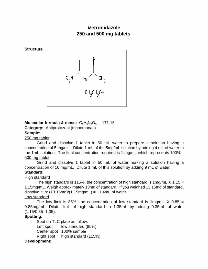
Metronidazole250 and 500 mg tablets
Structure
Molecular formula & mass: C H N O - 171.166 9 3 3
Category: Antiprotozoal (trichomonas)Sample:250 mg tablet:
Grind and dissolve 1 tablet in 50 mL water to prepare a solution having aconcentration of 5 mg/mL. Dilute 1 mL of the 5mg/mL solution by adding 4 mL of water tothe 1mL solution. The final concentration required is 1 mg/mL which represents 100%.500 mg tablet:
Grind and dissolve 1 tablet in 50 mL of water making a solution having aconcentration of 10 mg/mL. Dilute 1 mL of this solution by adding 9 mL of water.Standard:High standard
The high standard is 115%, the concentration of high standard is 1mg/mL X 1.15 =1.15mg/mL. Weigh approximately 13mg of standard. If you weighed 13.15mg of standard,dissolve it in: (13.15mg)/(1.15mg/mL) = 11.4mL of water.Low standard
The low limit is 85%, the concentration of low standard is 1mg/mL X 0.85 =0.85mg/mL. Dilute 1mL of high standard to 1.35mL by adding 0.35mL of water(1.15/0.85=1.35).Spotting:
Spot on TLC plate as follow:Left spot low standard (85%)Center spot 100% sample Right spot high standard (115%)
Development:

Mix 25mL methanol and 0.25mL ammonium hydroxide. Add 24 mL of this mixture tothe plastic TLC bag. Develop until the solvent front reaches to within 1cm of the top of theTLC plate.Detection:UV;
Dry the plate and observe under UV light. The maximum is at 305nm but at 254nmwill work. Observe the size and intensity of the spots.Iodine stain:
Dip plate in the iodine solution in the detection bag. Allow plate to dry and observespot size and intensity.
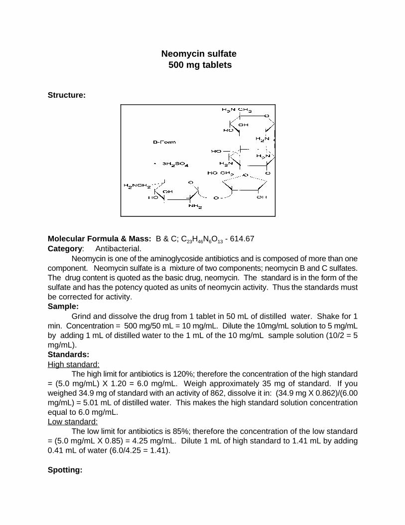
Neomycin sulfate 500 mg tablets
Structure:
Molecular Formula & Mass: B & C; C H N O - 614.67 23 46 6 13
Category: Antibacterial. Neomycin is one of the aminoglycoside antibiotics and is composed of more than one
component. Neomycin sulfate is a mixture of two components; neomycin B and C sulfates.The drug content is quoted as the basic drug, neomycin. The standard is in the form of thesulfate and has the potency quoted as units of neomycin activity. Thus the standards mustbe corrected for activity. Sample:
Grind and dissolve the drug from 1 tablet in 50 mL of distilled water. Shake for 1min. Concentration = 500 mg/50 mL = 10 mg/mL. Dilute the 10mg/mL solution to 5 mg/mLby adding 1 mL of distilled water to the 1 mL of the 10 mg/mL sample solution (10/2 = 5mg/mL). Standards: High standard:
The high limit for antibiotics is 120%; therefore the concentration of the high standard= (5.0 mg/mL) X 1.20 = 6.0 mg/mL. Weigh approximately 35 mg of standard. If youweighed 34.9 mg of standard with an activity of 862, dissolve it in: (34.9 mg X 0.862)/(6.00mg/mL) = 5.01 mL of distilled water. This makes the high standard solution concentrationequal to 6.0 mg/mL. Low standard:
The low limit for antibiotics is 85%; therefore the concentration of the low standard= (5.0 mg/mL X 0.85) = 4.25 mg/mL. Dilute 1 mL of high standard to 1.41 mL by adding0.41 mL of water (6.0/4.25 = 1.41).
Spotting:

Spot on the TLC plate as follows: Left spot low standard (85%)Center spot 100% sample Right spot high standard (120%)
Development: Mix 15 mL of methanol, 15 mL of acetone, 7.5 mL of distilled water, and 7.5 mL of
concentrated ammonium hydroxide. Add 24 mL of this mixture to the TLC developmentbag. Develop a blank TLC plate until the solvent front reaches 1 cm from the top of theplate. Remove the plate and allow it to dry until the odor of ammonia can no longer bedetected. Remove the solvent from the developing bag. Prepare another mixture of thesame solvent, and add 22 mL of the mixture to the TLC development bag. Develop theplate until the solvent front reaches 1 cm from the top of the plate. Allow the plate to dryuntil the odor of ammonia cannot be detected. Detection:UV:
The spots are not visible in the UV. Iodine stain:
Dip the plate in the iodine-KI solution in the detection bag. Allow the spots to dry anddetermine their size and intensity.Visible:
To make the spots visible in white light, dip the plate in a ninhydrin solution, preparedby dissolving 100 mg of ninhydrin, 2,2-dihydroxy-1,3-indanedione (Sigma Chemical Co. No.N4876), in 25 mL of acetone. Place 7 mL of this solution in the detection bag instead of theiodine-KI developing solution. The TLC plate should be dry and completely free of anyammonia (this may require allowing the plate to sit overnight). Dip the TLC plate in theninhydrin solution. Allow the plate to dry for at least 60 min. A blue-violet color developswith neomycin. Observe the size and intensity of the spots. If the TLC plate is notcompletely free of ammonia, the background color of the plate will change from pale pinkto dark rose in about 1 hr. Notes:
The use of ninhydrin is recommended because the iodine intensities are variable anddo not last very long. The ninhydrin color can be developed in less time if an oven or a hotplate is available. After the TLC plate air-dries for 30 min., the ammonia on the TLC platecan be removed by heating the plate at 100EC for 10 min. Dip the TLC plate in the ninhydrinsolution and allow it to air dry at least 30 min. (The color usually develops in 30-60 min.)Heat the TLC plate at least 2 min. at 100EC to fully develop the violet color. The TLC plateis pre-developed to remove the possibility of streaking or solvent front effects.
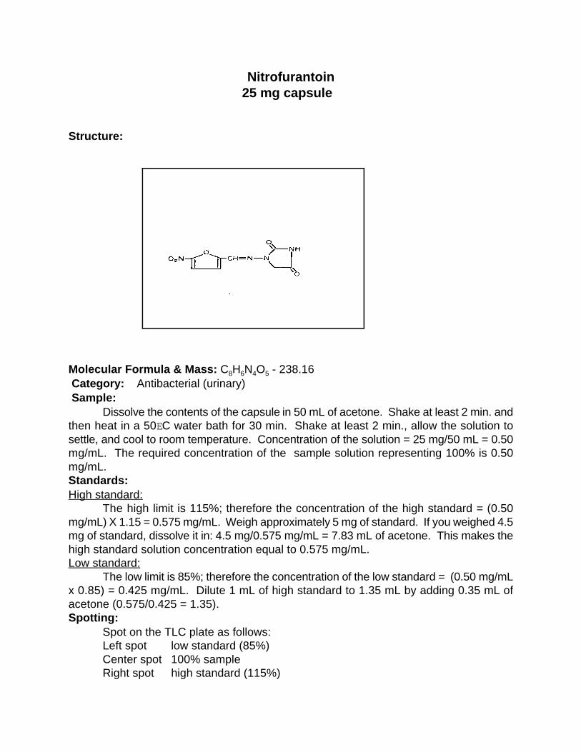
Nitrofurantoin 25 mg capsule
Structure:
Molecular Formula & Mass: C H N O - 238.16 8 6 4 5
Category: Antibacterial (urinary) Sample:
Dissolve the contents of the capsule in 50 mL of acetone. Shake at least 2 min. andthen heat in a 50EC water bath for 30 min. Shake at least 2 min., allow the solution tosettle, and cool to room temperature. Concentration of the solution = 25 mg/50 mL = 0.50mg/mL. The required concentration of the sample solution representing 100% is 0.50mg/mL. Standards:High standard:
The high limit is 115%; therefore the concentration of the high standard = (0.50mg/mL) X 1.15 = 0.575 mg/mL. Weigh approximately 5 mg of standard. If you weighed 4.5mg of standard, dissolve it in: 4.5 mg/0.575 mg/mL = 7.83 mL of acetone. This makes thehigh standard solution concentration equal to 0.575 mg/mL. Low standard:
The low limit is 85%; therefore the concentration of the low standard = (0.50 mg/mLx 0.85) = 0.425 mg/mL. Dilute 1 mL of high standard to 1.35 mL by adding 0.35 mL ofacetone (0.575/0.425 = 1.35). Spotting:
Spot on the TLC plate as follows: Left spot low standard (85%) Center spot 100% sample Right spot high standard (115%)
Development:Mix 16 mL of acetone and 8 mL of toluene. Add this mixture to the TLC development
bag. Develop until the solvent front reaches within 1 cm of the top of the TLC plate.Detection:UV:
Dry the plate and observe under UV light. The maximum is at 360 nm but a shortwavelength UV light (254 nm) will work. Observe the size and intensity of the spots. Iodine stain:
Dip the plate into the iodine-KI solution in the detection bag. Allow the plate to dryand observe the size and intensity of the spots.

Norgestrel 0.5 mg tablet
Structure:
Molecular Formula & Mass: C H O - 312.44 21 28 2
Category: Oral progesterone; with estradiol as oral contraceptive Sample:
Grind 10 tablets and dissolve in 10 mL of anhydrous ethanol. Shake at least 5 min.Concentration of the solution = (10 x 0.5 mg)/10 mL = 0.50 mg/mL. The requiredconcentration of the sample solution representing 100% is 0.50 mg/mL. Standards:High standard:
The high limit is 115%; therefore the concentration of the high standard = (0.50mg/mL) X 1.15 = 0.575 mg/mL. Weigh approximately 5 mg of standard. If you weighed4.75 mg of standard, dissolve it in: (4.75 mg)/(0.575 mg/mL) = 8.26 mL of anhydrousethanol. This makes the high standard solution concentration equal to 0.575 mg/mL. Low standard:
The low limit is 85%; therefore the concentration of the low standard = (0.50 mg/mL)X 0.85 = 0.425 mg/mL. Dilute 1 mL of high standard to 1.35 mL by adding 0.35 mL ofanhydrous ethanol (0.575/0.425 = 1.35). Spotting:
Spot on the TLC plate as follows: Left spot low standard (85%) Center spot 100% sample Right spot high standard (115%)
Development:Mix 16 mL of toluene and 8 mL of acetone. Add this mixture to the TLC development
bag. Develop until the solvent front reaches within 1 cm of the top of the TLC plate.

Detection:UV:
Dry the plate and observe under UV light. The maximum is at 241 nm but a shortwavelength UV light (254 nm) will work. Observe the size and intensity of the spots. Iodine stain:
This concentration is too low to allow quantitation by iodine-KI solution. A higherconcentration of steroid can be prepared by using 1 mL of solvent and filtering the solutionthrough glass wool in a small funnel. However, the ethanol tends to evaporate during thisprocess and give high results.
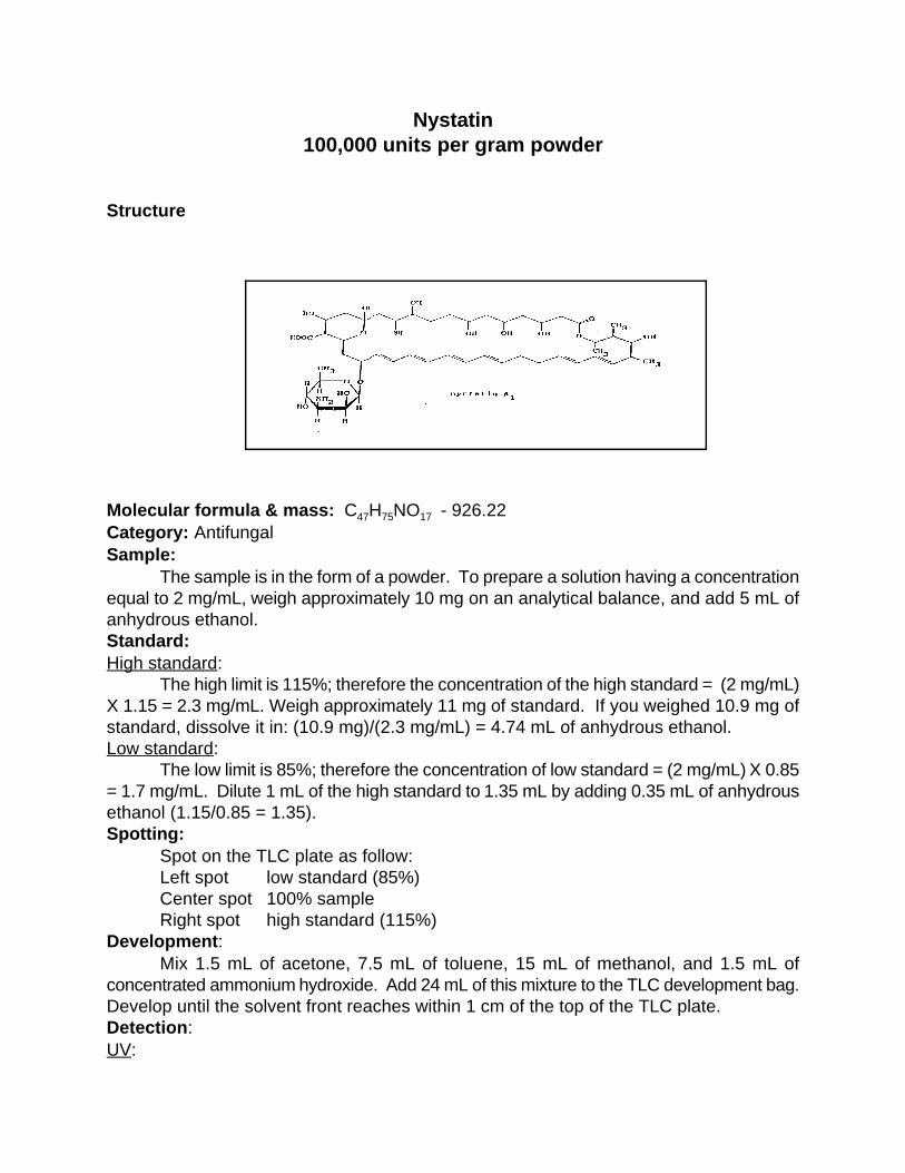
Nystatin100,000 units per gram powder
Structure
Molecular formula & mass: C H NO - 926.2247 75 17
Category: Antifungal Sample:
The sample is in the form of a powder. To prepare a solution having a concentrationequal to 2 mg/mL, weigh approximately 10 mg on an analytical balance, and add 5 mL ofanhydrous ethanol.Standard:High standard:
The high limit is 115%; therefore the concentration of the high standard = (2 mg/mL)X 1.15 = 2.3 mg/mL. Weigh approximately 11 mg of standard. If you weighed 10.9 mg ofstandard, dissolve it in: (10.9 mg)/(2.3 mg/mL) = 4.74 mL of anhydrous ethanol.Low standard:
The low limit is 85%; therefore the concentration of low standard = (2 mg/mL) X 0.85= 1.7 mg/mL. Dilute 1 mL of the high standard to 1.35 mL by adding 0.35 mL of anhydrousethanol (1.15/0.85 = 1.35).Spotting:
Spot on the TLC plate as follow:Left spot low standard (85%)Center spot 100% sampleRight spot high standard (115%)
Development:Mix 1.5 mL of acetone, 7.5 mL of toluene, 15 mL of methanol, and 1.5 mL of
concentrated ammonium hydroxide. Add 24 mL of this mixture to the TLC development bag.Develop until the solvent front reaches within 1 cm of the top of the TLC plate.Detection:UV:

Dry the plate and observe under UV light. The maximum is at 300 nm but a shortwavelength light (254 nm) will work. Observe the size and intensity of the spots.Iodine stain:
Dip the plate into the iodine-KI solution in the detection bag. Allow the plate to dryand observe the size and intensity of the spots.

Paracetamol(Acetaminophen)
325 mg tablet Structure:
Molecular Formula & Mass: C H O N - 151.168 9 2
Category: Analgesic, antipyretic Sample:
Grind 1 tablet and dissolve in 50 mL of 95% ethanol. Shake at least 1 min.Concentration of the solution = 325 mg/50 mL = 6.5 mg/mL. The required concentrationof the sample solution representing 100% is 5 mg/mL. To prepare the 5 mg/mL solution,dilute 1 mL of the 6.5 mg/mL solution with 0.3 mL of 95% ethanol. This solution is requiredfor iodine staining. Paracetamol may be supplied in different contents. In this case, add 50mL of the solvent to the ground tablet to make a master solution, and determine theconcentration. Dilute an aliquot of the master solution to prepare a solution having aconcentration equal to 5 mg/mL. Reference solutions: (a) When reference drug is available in tablet form. High reference solution: The high limit for the reference is equal to 115%; therefore the concentration ofthe high reference solution = (5 mg/mL) X 1.15 = 5.75 mg/mL. Add 95% ethanol to 1 tabletof the reference drug. To determine the volume of alcohol needed, divide the drug contentof the reference tablet in mg by 5.75 mg/mL. If the reference tablet contains 46 mg: (46mg/5.75 mg/mL) = 8 mL of 95% ethanol.Low reference solution:
The low limit of the reference is 85%; therefore the concentration of the lowreference solution = (5 mg/mL) X 0.85 = 4.25 mg/mL. Dilute 1 mL of the high referencesolution to 1.35 mL by adding 0.35 mL of 95% ethanol (5.75/4.25 = 1.35). (b) When no reference tablets are available. When reference tablets are not available, a reference material must be weighed. The
reference material may be either a primary or a secondary standard. Weigh approximately25 mg of the standard on an analytical balance. Divide the weight (mg) by 5.75 mg/mL toobtain the volume needed to make the proper concentration for the high solution. The low concentration standard solution is prepared by diluting the high concentrationreference as above.Spotting:
Spot on the TLC plate as follows:Left spot low standard (85%)Center spot 100% sample Right spot high standard (115%)
Development:Mix 24 mL of ethyl acetate, 3 mL of methanol, and 1 ml of concentrated
ammonium hydroxide. Add 24m of this mixture to the TLC development bag. Developuntil the solvent front reaches within 1 cm of the top of the TLC sheet. Detection: Dry the TLC sheet until no odor can be detected. Dip the sheet into the iodine-KIsolution. Allow the iodine to evaporate, and observe the difference between theintensities of the spots. Note:
Follow this procedure for detection by iodine staining. Many drugs can bedetected by UV light, and the concentrations may be different from the concentrationneeded for the iodine. Paracetamol can be detected by UV at a concentration of 1mg/mL. To prepare the sample and reference solutions, dilute the above solutions byadding 4 mL of the solvent to 1 mL of the concentrated solutions.
Detection by UV is the simplest method and should be used whenever a UVsource is available.
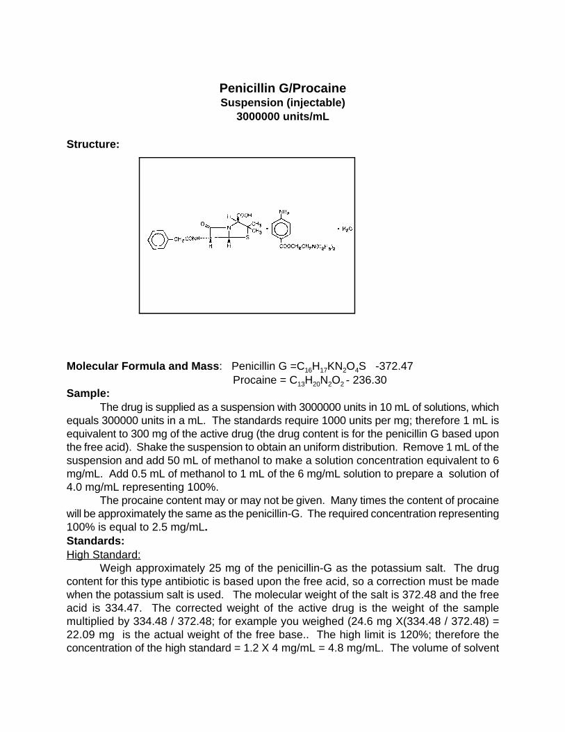
Penicillin G/ProcaineSuspension (injectable)
3000000 units/mL
Structure:
Molecular Formula and Mass: Penicillin G =C H KN O S -372.47 16 17 2 4
Procaine = C H N O - 236.3013 20 2 2
Sample:The drug is supplied as a suspension with 3000000 units in 10 mL of solutions, which
equals 300000 units in a mL. The standards require 1000 units per mg; therefore 1 mL isequivalent to 300 mg of the active drug (the drug content is for the penicillin G based uponthe free acid). Shake the suspension to obtain an uniform distribution. Remove 1 mL of thesuspension and add 50 mL of methanol to make a solution concentration equivalent to 6mg/mL. Add 0.5 mL of methanol to 1 mL of the 6 mg/mL solution to prepare a solution of4.0 mg/mL representing 100%.
The procaine content may or may not be given. Many times the content of procainewill be approximately the same as the penicillin-G. The required concentration representing100% is equal to 2.5 mg/mL.Standards: High Standard:
Weigh approximately 25 mg of the penicillin-G as the potassium salt. The drugcontent for this type antibiotic is based upon the free acid, so a correction must be madewhen the potassium salt is used. The molecular weight of the salt is 372.48 and the freeacid is 334.47. The corrected weight of the active drug is the weight of the samplemultiplied by 334.48 / 372.48; for example you weighed (24.6 mg X(334.48 / 372.48) =22.09 mg is the actual weight of the free base.. The high limit is 120%; therefore theconcentration of the high standard = 1.2 X 4 mg/mL = 4.8 mg/mL. The volume of solvent
needed will equal weight of the drug divided by the high concentration which in this case is22.09 mg / (4.8 mg/mL) = 4.6 mL of methanol.Low standard:
The low limit is 85%; therefore the concetration of the low standard = 0.85 X 4.0mg/mL = 3.4 mg/mL. Add 0.41 mL of methanol to 1 mL of the high concentrationstandard (4.8 mg/mL).
Spotting:Spot the solutions as follows:Left spot low standard(85%)Center spot sample(100%)Right spot high standard(120%)
Development:Mix 26 mL acetone, 4 mL toluene, 4 mL water, and 1 mL ammonium hydroxide.
Pour 24 mL of this solution into the TLC development bag. Develop until the solvent frontreaches to within 1 cm from the top of the TLC plate.Detection:UV:
Dry the plate and observe under UV light at 254nm. Since this drug formulation has2 compounds (penicilling-G and procaine), there will be 2 positions on the plate. The spotsin the lower position are penicilline-G and the upper spots are procaine. Observe the sizeand the intensity of the spots. Concentrations may require adjustment for analysis of bothdrugs at the same time.Iodine stain:
Dip the plate into the iodine-KI solution. Allow plate to dry and observe the spots'size and intensity.

Phenobarbital15 mg tablet
Structure:
Molecular Formula & Mass: C H N O - 232.23 12 12 2 3
Category: Anticonvulsant, hypnotic, sedative Sample:
The sample contains 15 mg of the active drug, and a final solution concentration of10 mg/mL is required to represent 100%. Grind 4 tablets and dissolve in 6 mL ofanhydrous ethanol. Because of the high concentration needed for suitable visualization, itis necessary to use multiple tablets and a volume of liquid large enough to obtain a suitablesample. Shake at least 2-3 min.Reference Materials:High standard:
The high limit is 115%; therefore the concentration of the high standard = (10 mg/mL)X 1.15 = 11.5 mg/mL. Weigh approximately 50 mg of standard. If you weighed 46 mg ofstandard, dissolve it in: (46 mg)/(11.5 mg/mL) = 4 mL of anhydrous ethanol. This makesthe high standard solution concentration equal to 11.5 mg/mL. Low standard:
The low limit is 85%; therefore the concentration of the low standard = (10 mg/mL)X 0.85 = 8.5 mg/mL. Dilute 1 mL of high standard to 1.35 mL by adding 0.35 mL ofanhydrous ethanol (11.5/8.5 = 1.35). Spotting:
Spot on the TLC plate as follows: Left spot low standard (85%) Center spot 100% sample Right spot high standard (115%)
Development: Mix 12 mL of acetone and 12 mL of toluene. Add this mixture to the TLC
development bag. Develop until the solvent front reaches within 1 cm of the top of the TLCplate. Detection:

UV:Dry the plate and observe under UV light. The maximum is at 250 nm but a short
wavelength UV light (254 nm) will work. Observe the size and intensity of the spots. Iodine stain:
Dip the plate into the iodine-KI solution in the detection bag. Allow the plate to dryand observe the size and intensity of the spots.
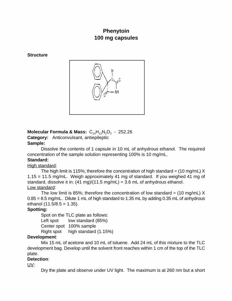
Phenytoin100 mg capsules
Structure
Molecular Formula & Mass: C H N O - 252.2615 12 2 2
Category: Anticonvulsant, antiepilepticSample:
Dissolve the contents of 1 capsule in 10 mL of anhydrous ethanol. The requiredconcentration of the sample solution representing 100% is 10 mg/mL.Standard:High standard:
The high limit is 115%; therefore the concentration of high standard = (10 mg/mL) X1.15 = 11.5 mg/mL. Weigh approximately 41 mg of standard. If you weighed 41 mg ofstandard, dissolve it in: (41 mg)/(11.5 mg/mL) = 3.6 mL of anhydrous ethanol.Low standard:
The low limit is 85%; therefore the concentration of low standard = (10 mg/mL) X0.85 = 8.5 mg/mL. Dilute 1 mL of high standard to 1.35 mL by adding 0.35 mL of anhydrousethanol (11.5/8.5 = 1.35).Spotting:
Spot on the TLC plate as follows:Left spot low standard (85%)Center spot 100% sample Right spot high standard (1.15%)
Development:Mix 15 mL of acetone and 10 mL of toluene. Add 24 mL of this mixture to the TLC
development bag. Develop until the solvent front reaches within 1 cm of the top of the TLCplate.Detection:UV:
Dry the plate and observe under UV light. The maximum is at 260 nm but a short

wavelength light (254 nm) will work. Observe the size and intensity of the spots.Iodine stain:
Dip the plate in the iodine-KI solution in the detection bag. Allow the plate to dry andobserve the size and intensity of the spots.

Pyrazinamide 400 mg tablet
Structure:
Molecular formula & mass: C H ON --123.115 5 3
Category: Antibacterial ( tuberculostatic)
Preparation of the sample solution:Analytical balance available.
Prepare the sample solution by weighing an aliquot of the drug. Follow the proceduredescribed in the previous sections. Determine the weight of the drug and add the volume ofsolvent to produce a concentration such of 1mg/mL. The volumes must be measured accurately using a combination of pipetts plus a 1 mL graduated tuberculin syringe for the fractional volumes. Pipetts are available in 1mL increments up to 10 mL. For example: You weighed 10.5 mg of thedrug, then you would add 10.5mL of solvent to prepare a solution with a concentration of 1.0mg/mL. ( use a 10 mL and a 1mL pipetts and the 0.5 mL is measured by a 1mL graduated syringe). Analytical balance not available.
Grind 1 tablet to a fine powder in a small polyethylene bag and insert the bag and contentsinto a suitable vessel and add 50 mL of methanol. Shake vigorously for at least 1 minute todissolve the powder. The concentration of this solution is = 400/50= 8 mg/mL. The requiredconcentration for the sample solution representing 100 % solution is 1 mg/mL. The concentratedsolution must be further diluted. Take 1 mL of the 8 mg/mL solution and add 7 mL of methanolwhich will prepare the concentration of 1 mg/mL. The volume of solvent needed must be adjustedto produce the proper concentration.
Preparation of standard solutions:Reference solutions are prepared depending on the availability of reference compounds.
The reference materials may be either in the form of reference tablets or powders ofprimary/secondary standards. Reference tablets may be available containing a predeterminedweight of the drug which when dissolved in 5 mL of the solvent produces a solution concentration

representing 115% of the sample solution. No weighing is required. Weighing is required when the reference compound is not available in tablet form. The referencesolutions then must be prepared using either primary or secondary standards.
Reference solutions are prepared depending on the availability of reference compounds. Reference tablets are available in many cases. These tablets contain a predetermined weight ofthe drug which when dissolved in 5 mL of the solvent will produce the high reference solutionconcentration representing 115% of the sample solution. No weighing is required.Weighing is required when the reference compound is not available in tablet form. The referencesolutions then must be prepared using either primary or secondary standards.Preparation of the High Standard: 1. Reference tablet available.The reference tablet for pyrazinamide contains 5.75 mg. Add one reference tablet to a vessel andadd 5 mL of methanol to prepare a solution having a concentration of 5.75mg/5mL equal to 1.15mg/mL. This represents 115% of the sample concentration.
2. Reference material available as a powder. Weigh approximately 10 mg of the standard. For example you weighed 9.7 mg, then the volume ofsolvent added would be 9.7 mg/1.15 mg/mL = 8.43 mL of methanol. This will make a finalconcentration of 1.15 mg/mL which will represent the 115% solution.Preparation of the low standard:
Dilute 1 mL of the high standard to 1.35 mL by adding 0.35 mL of methanol.(1.15/0.85) =1.35 which is the high concentration divided by the low concentration).Spotting:
Spot on the TLC plate as follows:Sample each of the solutions with a 3µL pipette and spot.Left spot low standard (85%)Center spot Sample (100%)Right spot high standard (115%)
Development:Mix 13 mL of methanol, 17 mL of acetone and 1 mL of concentrated ammonium hydroxide.
Add 24 mL of this mixture to the TLC development bag. Develop until the solvent front reacheswithin 1 cm of the top of the plate. Detection:UV:
Dry the plate and observe under UV light (254 nm). Observe the size and intensity of thespots or stain with iodine when no UV available.Iodine stain:
Dip the plate in the iodine-KI solution in the detection bag. Allow the plate to dry and
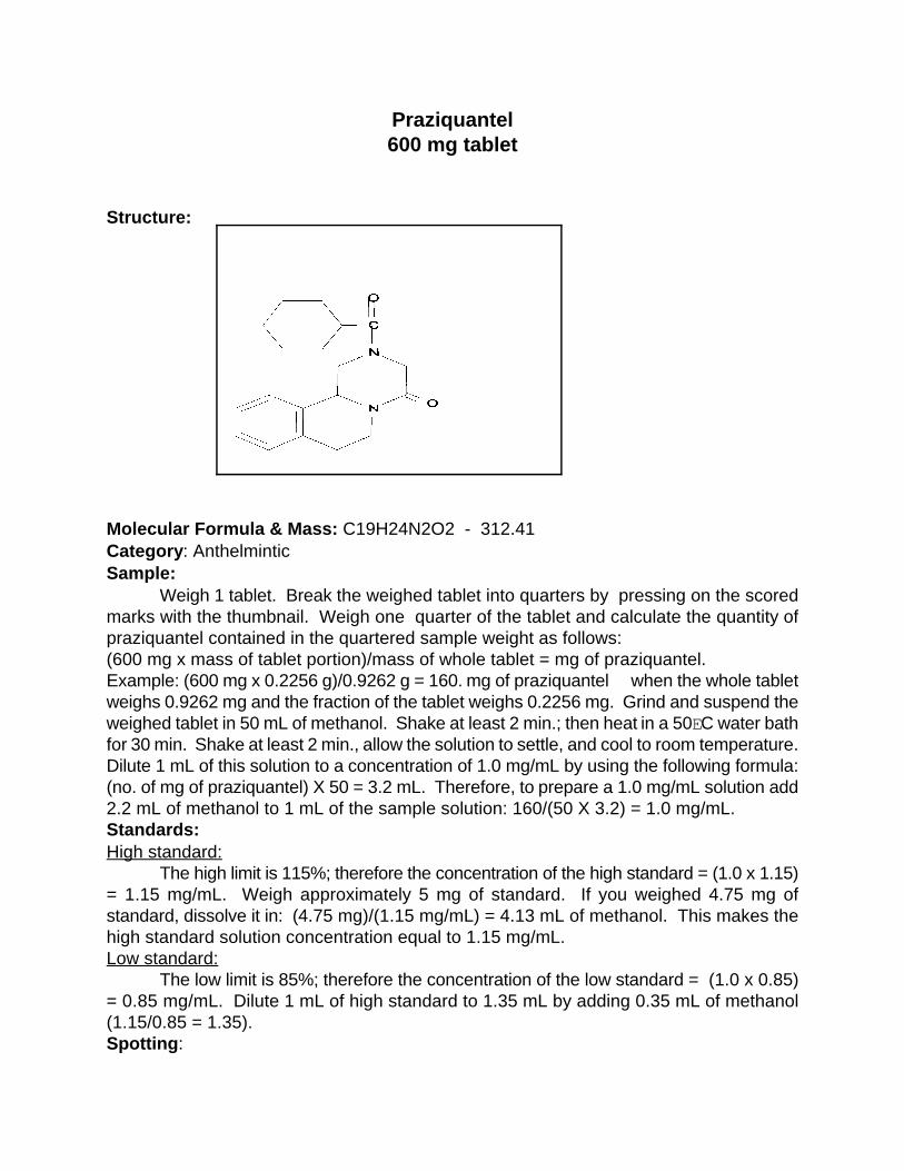
Praziquantel600 mg tablet
Structure:
Molecular Formula & Mass: C19H24N2O2 - 312.41 Category: Anthelmintic Sample:
Weigh 1 tablet. Break the weighed tablet into quarters by pressing on the scoredmarks with the thumbnail. Weigh one quarter of the tablet and calculate the quantity ofpraziquantel contained in the quartered sample weight as follows: (600 mg x mass of tablet portion)/mass of whole tablet = mg of praziquantel. Example: (600 mg x 0.2256 g)/0.9262 g = 160. mg of praziquantel when the whole tabletweighs 0.9262 mg and the fraction of the tablet weighs 0.2256 mg. Grind and suspend theweighed tablet in 50 mL of methanol. Shake at least 2 min.; then heat in a 50EC water bathfor 30 min. Shake at least 2 min., allow the solution to settle, and cool to room temperature.Dilute 1 mL of this solution to a concentration of 1.0 mg/mL by using the following formula:(no. of mg of praziquantel) X 50 = 3.2 mL. Therefore, to prepare a 1.0 mg/mL solution add2.2 mL of methanol to 1 mL of the sample solution: 160/(50 X 3.2) = 1.0 mg/mL.Standards:High standard:
The high limit is 115%; therefore the concentration of the high standard = (1.0 x 1.15)= 1.15 mg/mL. Weigh approximately 5 mg of standard. If you weighed 4.75 mg ofstandard, dissolve it in: (4.75 mg)/(1.15 mg/mL) = 4.13 mL of methanol. This makes thehigh standard solution concentration equal to 1.15 mg/mL.Low standard:
The low limit is 85%; therefore the concentration of the low standard = (1.0 x 0.85)= 0.85 mg/mL. Dilute 1 mL of high standard to 1.35 mL by adding 0.35 mL of methanol(1.15/0.85 = 1.35).Spotting:
Spot on the TLC plate as follows:Left spot low standard (85%)Center spot 100% sampleRight spot high standard (115%)
Development:Mix 16 mL of acetone and 8 mL of toluene. Add this mixture to the TLC development
bag. Develop until the solvent front reaches within 1 cm of the top of the TLC plate. Detection:UV:
The UV maximum is at 205-210 nm; thus a short wavelength UV light (254 nm) willnot detect praziquantel. Iodine stain: Dip the plate in the iodine-KI solution in the detection bag. Allow the plate to dry andobserve the size and intensity of the spots.
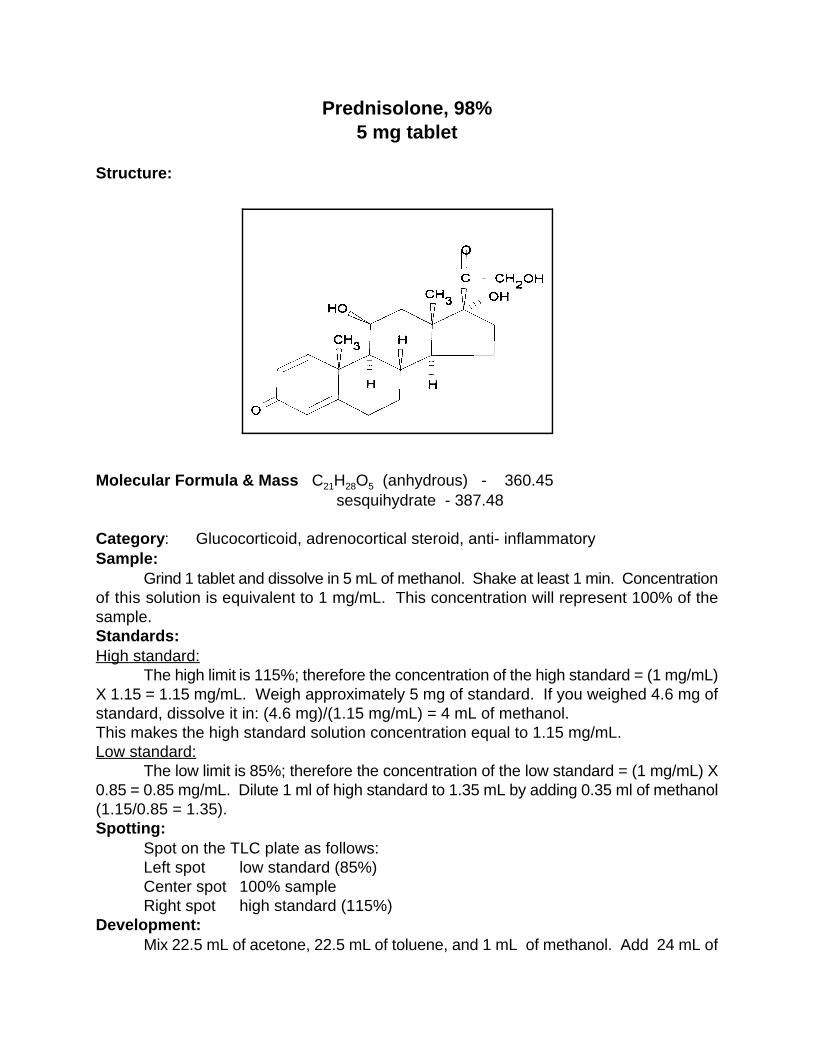
Prednisolone, 98%5 mg tablet
Structure:
Molecular Formula & Mass C H O (anhydrous) - 360.45 21 28 5
sesquihydrate - 387.48
Category: Glucocorticoid, adrenocortical steroid, anti- inflammatorySample:
Grind 1 tablet and dissolve in 5 mL of methanol. Shake at least 1 min. Concentrationof this solution is equivalent to 1 mg/mL. This concentration will represent 100% of thesample.Standards:High standard:
The high limit is 115%; therefore the concentration of the high standard = (1 mg/mL)X 1.15 = 1.15 mg/mL. Weigh approximately 5 mg of standard. If you weighed 4.6 mg ofstandard, dissolve it in: (4.6 mg)/(1.15 mg/mL) = 4 mL of methanol.This makes the high standard solution concentration equal to 1.15 mg/mL.Low standard:
The low limit is 85%; therefore the concentration of the low standard = (1 mg/mL) X0.85 = 0.85 mg/mL. Dilute 1 ml of high standard to 1.35 mL by adding 0.35 ml of methanol(1.15/0.85 = 1.35).Spotting:
Spot on the TLC plate as follows:Left spot low standard (85%)Center spot 100% sampleRight spot high standard (115%)
Development: Mix 22.5 mL of acetone, 22.5 mL of toluene, and 1 mL of methanol. Add 24 mL of
this mixture to the TLC development bag. Develop until the solvent front reaches within 1cm of the top of the TLC bag.Detection:UV:
Dry the plate and observe under UV light. The maximum is at 250 nm but a shortwavelength UV light (254 nm) will work. Observe the size and intensity of the spots.Iodine stain:
Dip the plate into the iodine-KI solution in the detection bag. Allow the plates to dryand observe the size and intensity of the spots.

Prednisone 10 mg tablet
Structure:
Molecular Formula & Mass: C H O - 358.4421 26 5
Category: Glucocorticoid, adrenocortical steroidSample:
Grind 1 tablet and dissolve in 20 mL of 95% ethanol. Shake at least 1 min.Concentration of the solution = 10 mg/20 mL = 0.50 mg/mL. The required concentration ofthe sample solution representing 100% is 0.5 mg/mL. Standards:High standard:
The high limit is 115%; therefore the concentration of the high standard = (0.50mg/mL) X 1.15 = 0.575 mg/mL. Weigh approximately 5 mg of standard. If you weighed4.75 mg of standard, dissolve it in: 4.75 mg/0.575 mg/mL = 8.26 mL of anhydrous ethanol.This makes the high standard solution concentration equal to 0.575 mg/mL.Low standard:
The low limit is 85%; therefore the concentration of the low standard = (0.50 mg/mL)X 0.85 = 0.425 mg/mL. Dilute 1 mL of high standard to 1.35 mL by adding 0.35 mL ofethanol (0.575/0.425 = 1.35).Spotting:
Spot on the TLC plate as follows:Left spot low standard (85%)Center spot 100% sampleRight spot high standard (115%)
Development:Mix 16 mL of toluene and 8 mL of acetone. Add this mixture to the TLC development
bag. Develop until the solvent front reaches within 1 cm of the top of the TLC plate. Detection:UV:
Dry the plate and observe under UV light. The maximum is at 240 nm but a shortwavelength UV light (254 nm) will work. Observe the size and intensity of the spots.Iodine stain:
The 0.50 mg/mL concentration is too low to allow for quantitation by iodine. Theconcentration needs to be 5 mg/mL. This can be accomplished by using 10 mL ofanhydrous ethanol rather than 20 mL. Dip the plate in the iodine-KI solution in the detectionbag. Allow the plate to dry and observe the size and intensity of the spots.

Promethazine25 mg tablet
Structure:
Molecular Formula & Mass: C H N S - 284.4117 20 2
Category: AntihistaminicSample:
Grind1 tablet and dissolve in 25 mL of anhydrous ethanol. Shake at least 1 min.Concentration of the solution = 25 mg/25 mL = 1.0 mg/mL. The required concentration ofsample solution representing 100% is 0.25 mg/mL. Dilute 1 mL of the 1mg/mL samplesolution to 4 mL by adding 3 mL of ethanol to obtain a 0.25 mg/mL sample solution(1/4 =0.25 mg/mL).Standards:High standard:
The high limit is 115%; therefore the concentration of the high standard = (0.25mg/mL) x 1.15 = 0.288 mg/mL. Weigh approximately 5 mg of standard. If you weighed4.75 mg of standard, dissolve it in: (4.75 mg) /( 0.288 mg/mL) = 16.5 mL of anhydrousethanol. This makes the high standard solution concentration equal to 0.288 mg/mL.Low standard:
The low limit is 85%; therefore the concentration of the low standard = (0.25 mg/mL)x 0.85 = 0.21 mg/mL. Dilute 1 mL of high standard to 1.35 mL by adding 0.35 mL ofethanol (0.288/0.21 = 1.35).Spotting:
Spot on TLC plate as follows:Left spot low standard (85%)Center spot 100% sampleRight spot high standard (115%)
Development:Mix 20 mL of acetone and 4 mL of water. Add this mixture to the TLC development
bag. Develop until the solvent front reaches to within 1 cm of the top of the TLC plate. Detection:UV:
Dry the plate and observe under UV light. The maximum is at 250 nm but a shortwavelength UV light(254 nm) will work. Observe the size and intensity of the spots.Iodine stain
The 0.25 mg/mL concentration is too low to allow for quantitation by iodine. Theconcentration needs to be 1 mg/mL. This can be accomplished by using the undilutedsample solution. Dip the plate in the iodine-KI solution in the detection bag. Allow the plateto dry and observe the size and intensity of the spots.

Pyrimethamine25 mg tablet
Structure:
Molecular Formula & Mass: C H ClN - 248.7112 13 4
Category: AntimalarialSample:
Grind 1 tablet and dissolve in 12.5 mL of 95% ethanol. Concentration of the solution= 2 mg/mL. This concentration represents 100% of the sample.Standards:High standard:
The high limit is 115%; therefore the concentration of the high standard = (2 mg/mL)X 1.15 = 2.3 mg/mL. Weigh approximately 25 mg of standard. If you weighed 23 mg ofstandard, dissolve it in: (23 mg/2.3 mg/mL) = 10 mL of 95% ethanol. This makes the highstandard solution concentration equal to 2.3 mg/mL. Low standard:
The low limit is 85%; therefore the concentration of the low standard = (2 mg/mL) X0.85 = 1.70 mg/mL. Dilute 1 mL of high standard to 1.35 mL by adding 0.35 mL of 95%ethanol (2.3/1.70 = 1.35).Spotting:
Spot on the TLC plate as follows:Left spot low standard (85%)Center spot 100% sampleRight spot high standard (115%)
Development:Mix 11 mL of acetone, 11 mL of toluene, and 2.5 mL of methanol. Add this mixture
to the TLC development bag. Develop until the solvent front reaches within 1 cm of the topof the TLC plate.Detection:UV:

Dry the plate and observe under UV light. The maximum is at 280 nm but a shortwavelength UV light (254) will work. Observe the size and intensity of the spots.Iodine stain:
Dip the plate into the iodine-KI solution in the detection bag. Allow the plate to dryand observe the size and intensity of the spots.
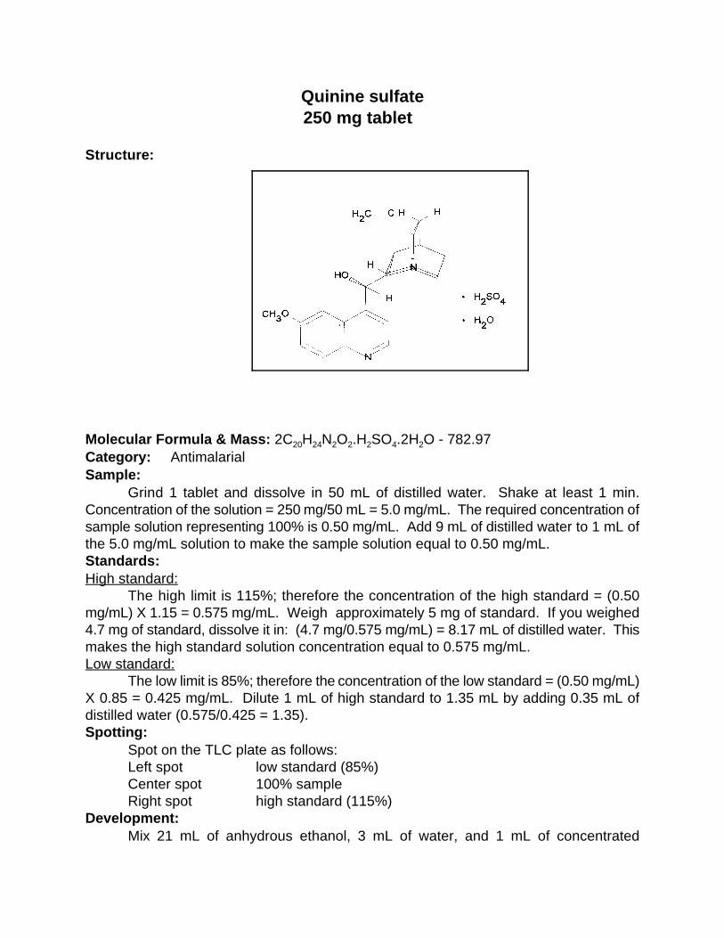
Quinine sulfate250 mg tablet
Structure:
Molecular Formula & Mass: 2C H N O .H SO .2H O - 782.9720 24 2 2 2 4 2
Category: AntimalarialSample:
Grind 1 tablet and dissolve in 50 mL of distilled water. Shake at least 1 min.Concentration of the solution = 250 mg/50 mL = 5.0 mg/mL. The required concentration ofsample solution representing 100% is 0.50 mg/mL. Add 9 mL of distilled water to 1 mL ofthe 5.0 mg/mL solution to make the sample solution equal to 0.50 mg/mL.Standards:High standard:
The high limit is 115%; therefore the concentration of the high standard = (0.50mg/mL) X 1.15 = 0.575 mg/mL. Weigh approximately 5 mg of standard. If you weighed4.7 mg of standard, dissolve it in: (4.7 mg/0.575 mg/mL) = 8.17 mL of distilled water. Thismakes the high standard solution concentration equal to 0.575 mg/mL.Low standard:
The low limit is 85%; therefore the concentration of the low standard = (0.50 mg/mL)X 0.85 = 0.425 mg/mL. Dilute 1 mL of high standard to 1.35 mL by adding 0.35 mL ofdistilled water (0.575/0.425 = 1.35).Spotting:
Spot on the TLC plate as follows: Left spot low standard (85%)
Center spot 100% sampleRight spot high standard (115%)
Development:Mix 21 mL of anhydrous ethanol, 3 mL of water, and 1 mL of concentrated
ammonium hydroxide. Add 24 mL of this mixture to the TLC development bag. Develop untilthe solvent front reaches within 1 cm of the top of the TLC plate.Detection:UV:
Dry the plate and observe under UV light. The maximum is at 235 nm but a shortwavelength UV light (254 nm) will work. Observe the size and intensity of the spots.Iodine stain:
Dip the plate in the iodine-KI solution in the detection bag. Allow the plate to dry andobserve the size and intensity of the spots.
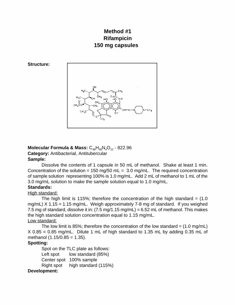
Method #1Rifampicin
150 mg capsules
Structure:
Molecular Formula & Mass: C H N O - 822.9648 58 4 12
Category: Antibacterial, AntitubercularSample:
Dissolve the contents of 1 capsule in 50 mL of methanol. Shake at least 1 min.Concentration of the solution = 150 mg/50 mL = 3.0 mg/mL. The required concentrationof sample solution representing 100% is 1.0 mg/mL. Add 2 mL of methanol to 1 mL of the3.0 mg/mL solution to make the sample solution equal to 1.0 mg/mL.Standards:High standard:
The high limit is 115%; therefore the concentration of the high standard = (1.0mg/mL) X 1.15 = 1.15 mg/mL. Weigh approximately 7-8 mg of standard. If you weighed7.5 mg of standard, dissolve it in: (7.5 mg/1.15 mg/mL) = 6.52 mL of methanol. This makesthe high standard solution concentration equal to 1.15 mg/mL.Low standard:
The low limit is 85%; therefore the concentration of the low standard = (1.0 mg/mL)X 0.85 = 0.85 mg/mL. Dilute 1 mL of high standard to 1.35 mL by adding 0.35 mL ofmethanol (1.15/0.85 = 1.35).Spotting:
Spot on the TLC plate as follows:Left spot low standard (85%)Center spot 100% sampleRight spot high standard (115%)
Development:

Mix 21 mL of methanol, 2 mL of toluene, and 2 mL of acetone. Add this mixture tothe TLC development bag. Develop until the solvent front reaches within 1 cm of the top ofthe TLC plate.Detection:Visible:
Dry the plate and observe under white light. The maximum is at 445 nm. Observethe size and intensity of the spots.

METHOD #2
Rifampicin150 mg capsules
Structure:
Molecular Formula & Mass: C H N O - 822.9643 58 4 12
Category: Antibacterial, (tuberculostatic)
Preparation of the sample solution:The dosage forms of rifampin are supplied in many different quantities of the drug. The
procedure described here is to serve as representative. Weights and volumes need to be adjusted forother dosages. Solutions are prepared by two different methods.Analytical balance available.
Prepare the sample solution by weighing an aliquot of the drug. Follow the proceduredescribed in the previous sections. Determine the weight of the drug in the aliquot and add solventto produce a concentration such as 1mg/mL. The volumes must be measured accurately by using acombination of pipetts plus a 1 mL graduated tuberculin syringe for the fractional volumes. Pipettsare available in 1mL increments up to 10 mL. For example: You weighed 10.5 mg of the drug, thenyou would add 10.5mL of solvent to prepare a solution with a concentration of 1.0 mg/mL. (use a 10mL pipette and the 0.5 mL is measured by a 1mL graduated tuberculin syringe). Analytical balance not available.
Grind 1 tablet to a fine powder in a small polyethylene bag and insert the bag and contents intoa suitable vessel and add 25 mL of methanol. Shake vigorously for at least 1 minute to dissolve thepowder. The concentration of this solution is = 150/25= 6 mg/mL. The required concentration forthe sample solution representing 100 % solution is 1 mg/mL. The concentrated solution must befurther diluted. Take 1 mL of the 6 mg/mL solution and add 5 mL of methanol to prepare the properconcentration of 1 mg/mL.
Preparation of standard solutions:

Preparation of standard solutions:Reference solutions are prepared depending on the availability of reference compounds. The
reference materials may be either in the form of reference tablets or powders of primary/secondarystandards. Reference tablets may be available containing a predetermined weight of the drug whichwhen dissolved in 5 mL of the solvent produces a solution concentration representing 115% of thesample solution. No weighing is required. Weighing is required when the reference compound is notavailable in tablet form. The reference solutions then must be prepared using either primary orsecondary standards.
Preparation of the High Standard: 1. Reference tablet available.The reference tablet for rifampin contains 5.75 mg. Add one reference tablet to a vessel and add 5mL of methanol to prepare a solution having a concentration of 5.75mg/5mL equal to 1.15 mg/mL.This represents 115% of the sample concentration.
2. Reference material available as a powder. Weigh approximately 10 mg of the standard. For example you weighed 9.7 mg, then the volume ofsolvent added would be 9.7 mg/1.15 mg/mL = 8.43 mL of methanol. This will make a finalconcentration of 1.15 mg/mL which will represent the 115% solution.Low standard:
The low limit is 85%; therefore the concentration of the low standard = (1.0 mg/mL)X 0.85 = 0.85 mg/mL. Dilute 1 mL of high standard to 1.35 mL by adding 0.35 mL ofmethanol (1.15/0.85 = 1.35).Spotting:
Spot on the TLC plate as follows:Sample each of the solutions with a 3µL capillary pipette ans spot.Left spot low standard (85%)Center spot 100% sampleRight spot high standard (115%)
Development:Mix 13 mL of methanol, 17 mL of acetone and 1 mL of concentrated ammonium hydroxide. Add 24mL of this mixture to the TLC development bag. Develop until the solvent front reaches within 1 cmof the top of the plate. Detection:Visible and UV:
Dry the plate and observe under white light. The maximum is at 445 nm. Observethe size and intensity of the spots. Rifampin has a strong absorption in the UV at 254 nmallowing excellent detection. The spots are better observed by UV when the stray lights ofthe room are eliminated.
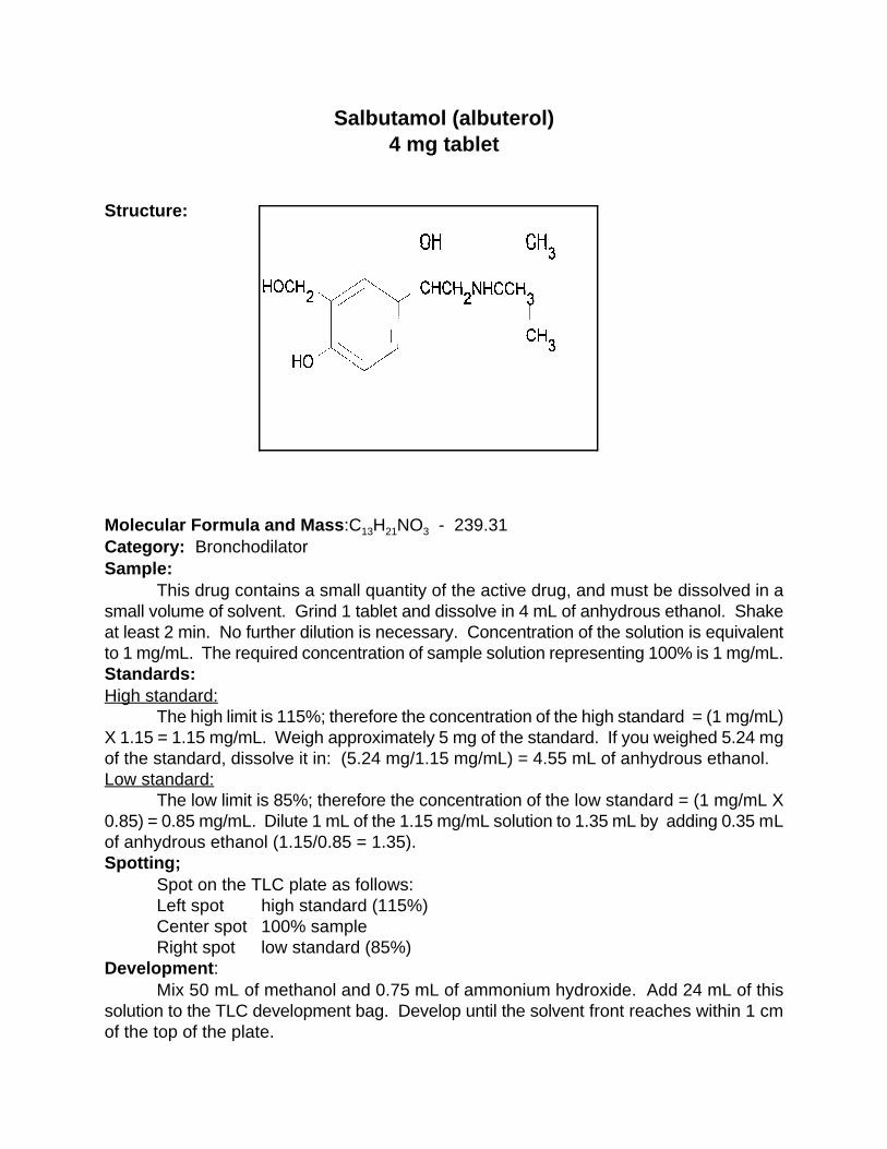
Salbutamol (albuterol)4 mg tablet
Structure:
Molecular Formula and Mass:C H NO - 239.3113 21 3
Category: BronchodilatorSample:
This drug contains a small quantity of the active drug, and must be dissolved in asmall volume of solvent. Grind 1 tablet and dissolve in 4 mL of anhydrous ethanol. Shakeat least 2 min. No further dilution is necessary. Concentration of the solution is equivalentto 1 mg/mL. The required concentration of sample solution representing 100% is 1 mg/mL.Standards:High standard:
The high limit is 115%; therefore the concentration of the high standard = (1 mg/mL)X 1.15 = 1.15 mg/mL. Weigh approximately 5 mg of the standard. If you weighed 5.24 mgof the standard, dissolve it in: (5.24 mg/1.15 mg/mL) = 4.55 mL of anhydrous ethanol.Low standard:
The low limit is 85%; therefore the concentration of the low standard = (1 mg/mL X0.85) = 0.85 mg/mL. Dilute 1 mL of the 1.15 mg/mL solution to 1.35 mL by adding 0.35 mLof anhydrous ethanol (1.15/0.85 = 1.35).Spotting;
Spot on the TLC plate as follows:Left spot high standard (115%)Center spot 100% sample Right spot low standard (85%)
Development:Mix 50 mL of methanol and 0.75 mL of ammonium hydroxide. Add 24 mL of this
solution to the TLC development bag. Develop until the solvent front reaches within 1 cmof the top of the plate.
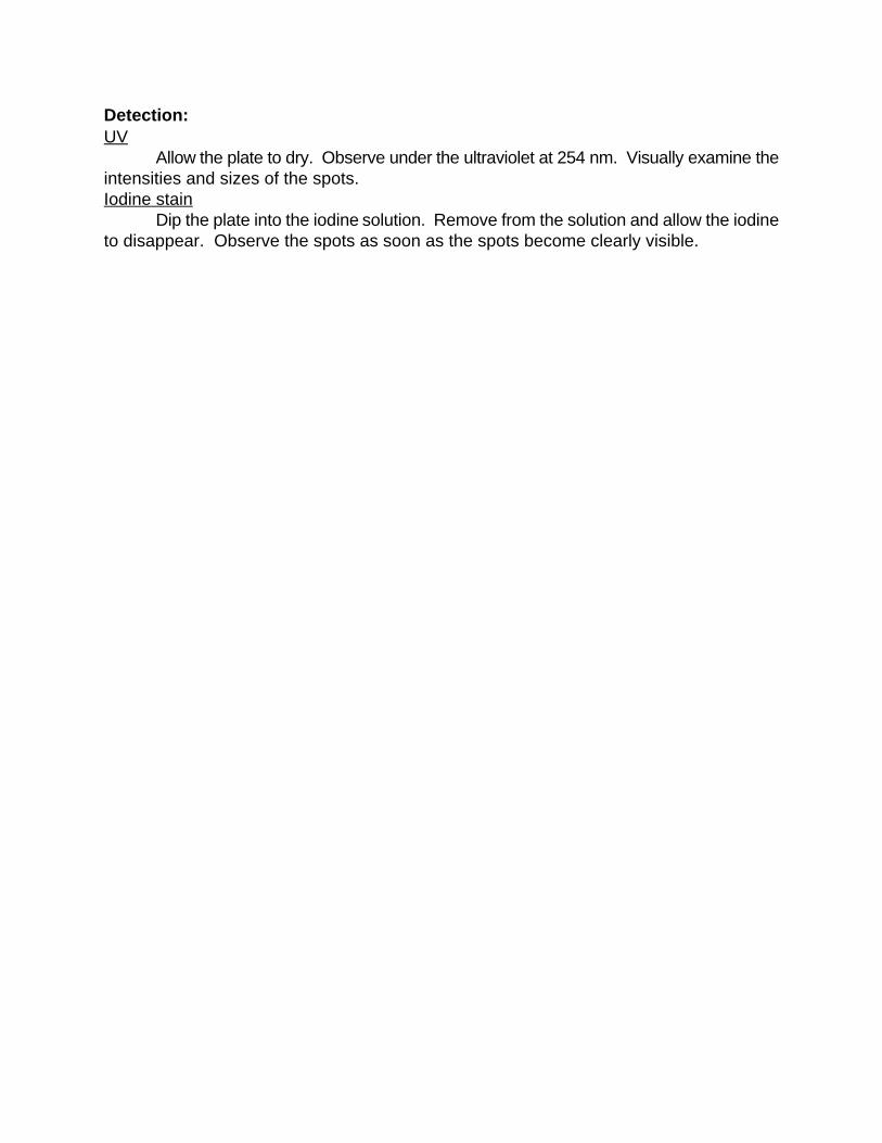
Detection:UV
Allow the plate to dry. Observe under the ultraviolet at 254 nm. Visually examine theintensities and sizes of the spots.Iodine stain
Dip the plate into the iodine solution. Remove from the solution and allow the iodineto disappear. Observe the spots as soon as the spots become clearly visible.

Streptomycin sulfate200 mg/mL injectable
Structure:
Molecular Formula & Mass: (C H N O ) .3 H SO - 1457.3821 39 7 12 2 2 4
Category: Antibacterial (tuberculostatic)Sample:NOTE: Streptomycin must be handled differently from the other TB drugs. The solvents ,developer, and detection are changed.
Streptomycin is an aminoglycoside antibiotic. The standard is in the form of the sulfate andthe potency is quoted as units of streptomycin activity. Thus the standards must be corrected foractivity. The drug is supplied as a 1 gram of the free base in a vial which is reconstituted byadding 5 mL of water to make a solution having a concentration of 200 mg/mL.
Dilute 1 mL of the injectable solution to 40 mL by adding 39 mL of distilled water. Shakeat least 1 min. The sample concentration = (1 mL x 200 mg/mL)/40 mL = 5 mg/mL.Standard:High standard:
The drug is normally supplied as the sulfate, but the content of the drug is based upon thefree base which requires a correction to the weighed quantity. The mass of the sulfate is 1457.38,and the mass of the free base is 1163.14. The correction is the weight of standard multiplied by1163.14/1457.38 = 0.798
The high limit for antibiotics is 120%; therefore the concentration of high standard = (5mg/mL) X 1.20 = 6.0 mg/mL. Weigh approximately 30 mg of sulfate standard. If you weighed29.8 mg of sulfate standard with an activity of 766 µg per mg, dissolve it in: (29.8 mg X0.798)/6.0 mg/mL = 3.96 mL of distilled water. This makes the high standard solutionconcentration equal to 6 mg/mL. Low standard:
The low limit for antibiotics is 85%; therefore the concentration of the low standard = (5mg/mL) X 0.85 = 4.25 mg/mL. The concentration ratio between the high and low standard is6/4.25 =1.41. Dilute 1 mL of high standard to 1.41 mL by adding 0.41 mL of water.
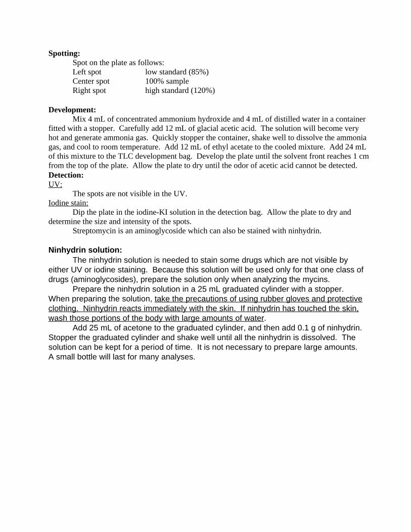
Spotting:Spot on the plate as follows:Left spot low standard (85%)Center spot 100% sampleRight spot high standard (120%)
Development:Mix 4 mL of concentrated ammonium hydroxide and 4 mL of distilled water in a container
fitted with a stopper. Carefully add 12 mL of glacial acetic acid. The solution will become veryhot and generate ammonia gas. Quickly stopper the container, shake well to dissolve the ammoniagas, and cool to room temperature. Add 12 mL of ethyl acetate to the cooled mixture. Add 24 mLof this mixture to the TLC development bag. Develop the plate until the solvent front reaches 1 cmfrom the top of the plate. Allow the plate to dry until the odor of acetic acid cannot be detected.Detection:UV:
The spots are not visible in the UV.Iodine stain:
Dip the plate in the iodine-KI solution in the detection bag. Allow the plate to dry anddetermine the size and intensity of the spots.
Streptomycin is an aminoglycoside which can also be stained with ninhydrin. Ninhydrin solution:
The ninhydrin solution is needed to stain some drugs which are not visible byeither UV or iodine staining. Because this solution will be used only for that one class ofdrugs (aminoglycosides), prepare the solution only when analyzing the mycins.
Prepare the ninhydrin solution in a 25 mL graduated cylinder with a stopper. When preparing the solution, take the precautions of using rubber gloves and protectiveclothing. Ninhydrin reacts immediately with the skin. If ninhydrin has touched the skin,wash those portions of the body with large amounts of water.
Add 25 mL of acetone to the graduated cylinder, and then add 0.1 g of ninhydrin. Stopper the graduated cylinder and shake well until all the ninhydrin is dissolved. Thesolution can be kept for a period of time. It is not necessary to prepare large amounts. A small bottle will last for many analyses.
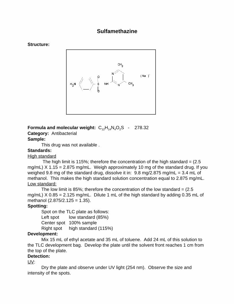
Sulfamethazine
Structure:
Formula and molecular weight: C H N O S - 278.3212 14 4 2
Category: AntibacterialSample:
This drug was not available .Standards:High standard
The high limit is 115%; therefore the concentration of the high standard = (2.5mg/mL) X 1.15 = 2.875 mg/mL. Weigh approximately 10 mg of the standard drug. If youweighed 9.8 mg of the standard drug, dissolve it in: 9.8 mg/2.875 mg/mL = 3.4 mL ofmethanol. This makes the high standard solution concentration equal to 2.875 mg/mL.Low standard:
The low limit is 85%; therefore the concentration of the low standard = (2.5mg/mL) X 0.85 = 2.125 mg/mL. Dilute 1 mL of the high standard by adding 0.35 mL ofmethanol (2.875/2.125 = 1.35).Spotting:
Spot on the TLC plate as follows:Left spot low standard (85%)Center spot 100% sampleRight spot high standard (115%)
Development:Mix 15 mL of ethyl acetate and 35 mL of toluene. Add 24 mL of this solution to
the TLC development bag. Develop the plate until the solvent front reaches 1 cm fromthe top of the plate.Detection:UV:
Dry the plate and observe under UV light (254 nm). Observe the size andintensity of the spots.

Iodine stain:Dip the plate in the iodine-KI solution in the detection bag. Allow to dry until the
spots are clearly visible. Observe the size and intensity of the spots.

Sulfamethoxazole500 mg Tablet
Structure:
Molecular Formula and Mass: C H N O S -253.3110 11 3 3
Category: AntibacterialSample:
This drug is supplied both as a single component and in combination with otherdrugs; therefore the drug content will vary. The sample described here has a drugcontent of 500 mg. Some combinations have a drug content of 400 mg. Theconcentration of the sample, measured by ultraviolet light, is 0.5 mg/mL, but to bemeasured by iodine staining, the concentration must be 5 mg/mL. In all cases, grind 1tablet and dissolve in 50 mL of methanol. Shake at least 5 min. The solution of the 500mg tablet will have a concentration of 10 mg/mL. For the iodine staining, add 1 mL ofmethanol to 1 mL of the 10 mg/mL solution to prepare a 5 mg/mL solution. For UVdetection, add 9 mL of methanol to 1 mL of the 5 mg/mL solution to prepare a 0.5mg/mL solution. Both the 5 mg/mL solution (iodine staining) and the 0.5 mg/mL solution(UV detection) represent a sample concentration of 100%. Always use UV detection if itis available.Standards:High standard:
The high limit is 115%; therefore, the concentration of the high standard = (5mg/mL X 1.15) or (0.5 mg/mL x 1.15). Weigh approximately 10 mg of the standard, andadd sufficient methanol to prepare the 5 mg/mL solution. If you weighed 9.75 mg,dissolve it in: 9.75 mg/(5.0 X 1.15) = 1.7 mL of methanol for the 5 mg/mL solution. Forthe 0.5 mg/mL solution, add 9 mL of methanol to 1 mL of the 5.0 mg/mL solution.Low standard:
The low limit is 85%; therefore the concentration of the low standard = (5 X 0.85)= 4.25 mg/mL. To prepare this concentration, add 0.35 mL of methanol to 1 mL of the
5.75 mg/mL solution (5.75/4.25 = 1.35).Spotting:
Spot on the TLC plate as follows:Left spot low standard (85%)Center spot 100% sampleRight spot high standard (115%)
Development:Mix 9 mL of ethyl acetate and 21 mL of toluene. Add 24 mL of this solution to the
TLC development bag. Develop until the solvent front reaches within 1 cm of the top ofthe plate.Detection:UV:
Dry the plate and observe under UV light (254 nm). Observe the size andintensity of the spots.Iodine stain:
Dip the plate in the iodine-KI solution. Observe the spots as soon as they becomeclearly visible.
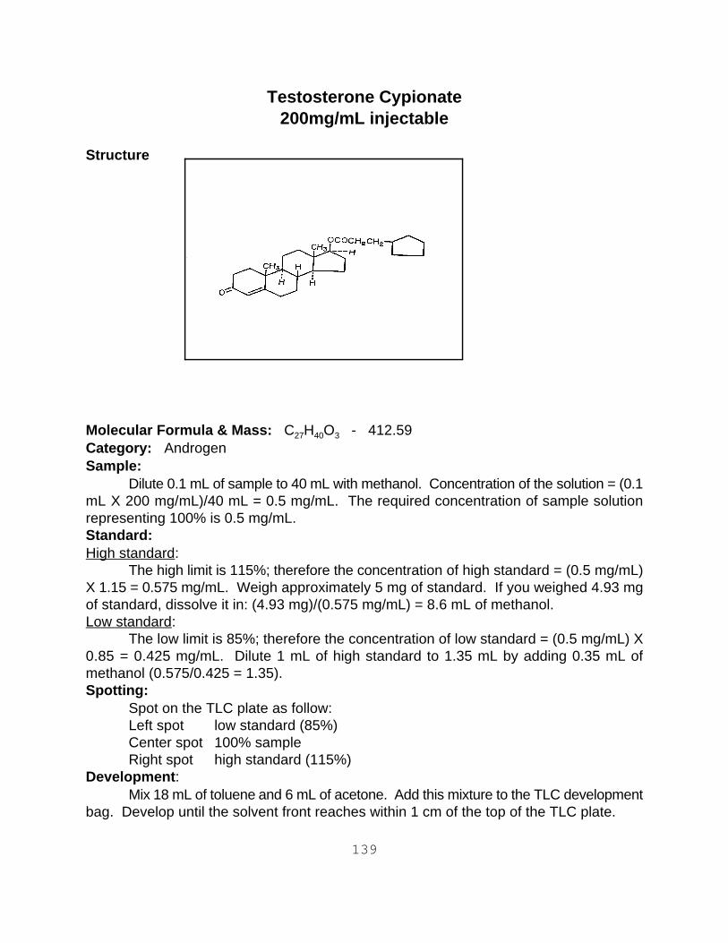
139
Testosterone Cypionate200mg/mL injectable
Structure
Molecular Formula & Mass: C H O - 412.59 27 40 3
Category: Androgen Sample:
Dilute 0.1 mL of sample to 40 mL with methanol. Concentration of the solution = (0.1mL X 200 mg/mL)/40 mL = 0.5 mg/mL. The required concentration of sample solutionrepresenting 100% is 0.5 mg/mL.Standard:High standard:
The high limit is 115%; therefore the concentration of high standard = (0.5 mg/mL)X 1.15 = 0.575 mg/mL. Weigh approximately 5 mg of standard. If you weighed 4.93 mgof standard, dissolve it in: (4.93 mg)/(0.575 mg/mL) = 8.6 mL of methanol.Low standard: The low limit is 85%; therefore the concentration of low standard = (0.5 mg/mL) X0.85 = 0.425 mg/mL. Dilute 1 mL of high standard to 1.35 mL by adding 0.35 mL ofmethanol (0.575/0.425 = 1.35).Spotting:
Spot on the TLC plate as follow:Left spot low standard (85%)Center spot 100% sample Right spot high standard (115%)
Development:Mix 18 mL of toluene and 6 mL of acetone. Add this mixture to the TLC development
bag. Develop until the solvent front reaches within 1 cm of the top of the TLC plate.
140
Detection:UV:
Dry the plate and observe under UV light. The maximum is at 250 nm but a shortwavelength light (254 nm) will work. Observe the size and intensity of the spots.Iodine stain:
Dip the plate in the iodine-KI solution in the detection bag. Allow the plate to dry andobserve the size and intensity of the spots.

141
Tetracyline250 mg and 500 mg tablets
Structure:
Molecular Formula & Mass: C H N O - 444.4322 24 2 8
Category: Antiamebic , antibacterial, antirickettsialSample:
250 mg tablet: Grind 1 tablet and dissolve in 50 mL of methanol to prepare a 5mg/mL solution. The solution needed to represent 100% requires a concentration of 1mg/mL. Dilute 1 mL of the 5 mg/mL solution with 4 mL of methanol for a final concentrationof 1 mg/mL.
500 mg tablet: Grind 1 tablet and dissolve in 50 mL of methanol to prepare a 10mg/mL solution. Dilute 1 mL of the 10 mg/mL solution with 9 mL of methanol for a finalconcentration of 1 mg/mL.Standards:High standard:
The high limit is 120%; therefore the concentration of the high standard = (1.0 mg/mLX 1.20) = 1.20 mg/mL. Weigh approximately 5.0 mg of standard. If you weighed 4.7 mgof standard, dissolve it in: (4.7 mg)/(1.20 mg/mL) = 3.91 mL of methanol. This makes thehigh standard solution concentration equal to 1.20 mg/mL.Low standard:
The low limit is 85%; therefore the concentration of the low standard = (1.0 mg/mL)X 0.85 = 0.85 mg/mL. Dilute 1 mL of the high standard to 1.41 mL by adding 0.41 mL ofmethanol (1.20/0.85 = 1.41).Spotting:
Spot on the TLC plate as follows:Left spot low standard (85%)

141
Center spot 100% sampleRight spot high standard (120%)
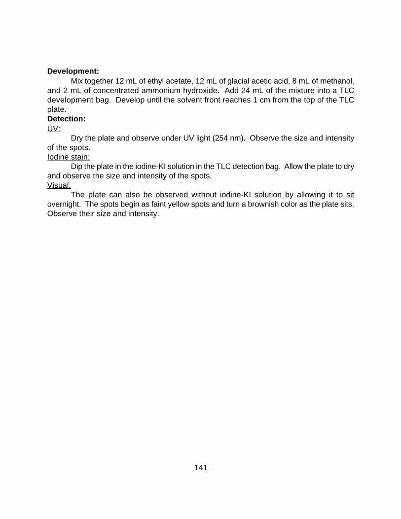
141
Development:Mix together 12 mL of ethyl acetate, 12 mL of glacial acetic acid, 8 mL of methanol,
and 2 mL of concentrated ammonium hydroxide. Add 24 mL of the mixture into a TLCdevelopment bag. Develop until the solvent front reaches 1 cm from the top of the TLCplate.Detection:UV:
Dry the plate and observe under UV light (254 nm). Observe the size and intensityof the spots.Iodine stain:
Dip the plate in the iodine-KI solution in the TLC detection bag. Allow the plate to dryand observe the size and intensity of the spots.Visual:
The plate can also be observed without iodine-KI solution by allowing it to sitovernight. The spots begin as faint yellow spots and turn a brownish color as the plate sits.Observe their size and intensity.
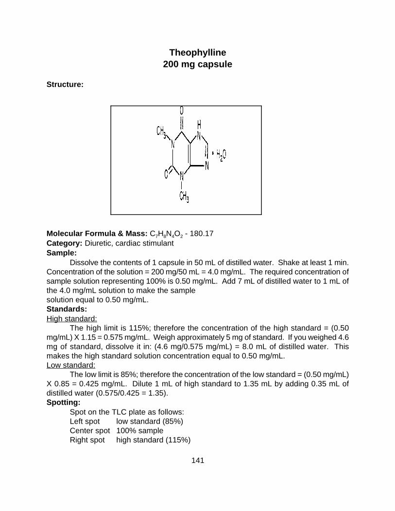
141
Theophylline200 mg capsule
Structure:
Molecular Formula & Mass: C H N O - 180.177 8 4 2
Category: Diuretic, cardiac stimulantSample:
Dissolve the contents of 1 capsule in 50 mL of distilled water. Shake at least 1 min.Concentration of the solution = 200 mg/50 mL = 4.0 mg/mL. The required concentration ofsample solution representing 100% is 0.50 mg/mL. Add 7 mL of distilled water to 1 mL ofthe 4.0 mg/mL solution to make the samplesolution equal to 0.50 mg/mL.Standards:High standard:
The high limit is 115%; therefore the concentration of the high standard = (0.50mg/mL) X 1.15 = 0.575 mg/mL. Weigh approximately 5 mg of standard. If you weighed 4.6mg of standard, dissolve it in: (4.6 mg/0.575 mg/mL) = 8.0 mL of distilled water. Thismakes the high standard solution concentration equal to 0.50 mg/mL.Low standard:
The low limit is 85%; therefore the concentration of the low standard = (0.50 mg/mL)X 0.85 = 0.425 mg/mL. Dilute 1 mL of high standard to 1.35 mL by adding 0.35 mL ofdistilled water (0.575/0.425 = 1.35).Spotting:
Spot on the TLC plate as follows:Left spot low standard (85%)Center spot 100% sampleRight spot high standard (115%)

141
Development:Mix 16 mL of acetone and 8 mL of toluene. Add this mixture to the TLC development
bag. Develop until the solvent front reaches within 1 cm of the top of the TLC plate. Detection:UV:
Dry the plate and observe under UV light. The maximum is at 270 nm but a shortwavelength UV light (254 nm) will work. Observe the size and intensity of the spots.Iodine stain:
Dip the plate in the iodine-KI solution in the detection bag. Allow the plate to dry andobserve the size and intensity of the spots.

Triamcinolone4 mg tablet
Structure:
Molecular Formula and Mass: C H FO - 394.4521 27 6
Category: GlucocorticoidSample:
This drug has a low dosage content of 4 mg. It is necessary to use multiple tabletsin order to increase the volume of solvent so that a suitable sampling can be made. Grindand dissolve 3 tablets in 4 mL of ethanol to prepare a sample solution equivalent to 3mg/mL. Shake well for 2 minutes. Allow the insolubles to settle before sampling, andremove approximately 1mL for the sample spotting.Standards:High standard:
The high standard concentration is 115% of the concentration of the sample whichis equal to 1.15 X 3 = 3.45 mg/mL. Weigh approximately 10 mg and dissolve in the correctvolume of ethanol. For example: You weighed 9.75 mg, then the volume needed is 9.75mg/ 3.45 mg/mL = 2.82 mL of ethanol.Low standard:
The concentration of the low standard is 85% of the concentration of the samplewhich is 0.85 X 3 = 2.55 mg/mL. This solution can be prepared by taking 1 mL of the3.45mg/mL solution and adding 0.35 mL of ethanol.Development:
Mix together 20 mL of toluene, 10 mL of acetone, and 3 mL of ethanol. Add 24 mLof this solution to the TLC development bag. Develop the plate until the solvent frontreaches 1 cm from the top of the plate.Detection:UV:
Dry the plate and observe at 254 nm ultraviolet light. Observe the intensities and thesizes of the spots.Iodine stain:
Dip the plate into the iodine solution, and allow the iodine to fade until the spots areclearly visible.

Triamterene 50mg capsule
Structure
Molecular Formula & Mass: C H N - 253.26 12 11 7
Category: DiureticSample:
Dissolve the drug from 1 capsule in 100 mL of methanol. Concentration of thesolution = 50 mg/100 mL = 0.5 mg/mL. The required concentration of sample solutionrepresenting 100% is 0.5 mg/mL.Standard:High standard:
The high limit is 115%; therefore the concentration of high standard = (0.5 mg/mL)X 1.15 = 0.575 mg/mL. Weigh approximately 5 mg of standard. If you weighed 4.7 mg ofstandard, dissolve it in: (4.7 mg)/(0.575 mg/mL) = 8.2 mL of methanol.Low standard:
The low limit is 85%; therefore the concentration of low standard = (0.5 mg/mL) X0.85 = 0.425 mg/mL. Dilute 1 mL of high standard to 1.35 mL by adding 0.35 mL ofmethanol (0.575/0.425 = 1.35).Spotting:
Spot on the TLC plate as follows:Left spot low standard (85%)Center spot 100% sampleRight spot high standard (115%)
Development:Mix 24 mL of methanol and 0.25 mL of concentrated ammonium hydroxide. Add this
mixture to the TLC development bag. Develop until the solvent front reaches within 1 cm ofthe top of the TLC plate.Detection:UV:

Dry the plate and observe under UV light. The maximum is at 270 nm but a shortwavelength light (254 nm) will work. Observe the size and intensity of the spots.Iodine stain:
Dip the plate in the iodine-KI solution in the detection bag. Allow the plate to dry andobserve the size and intensity of the spots.
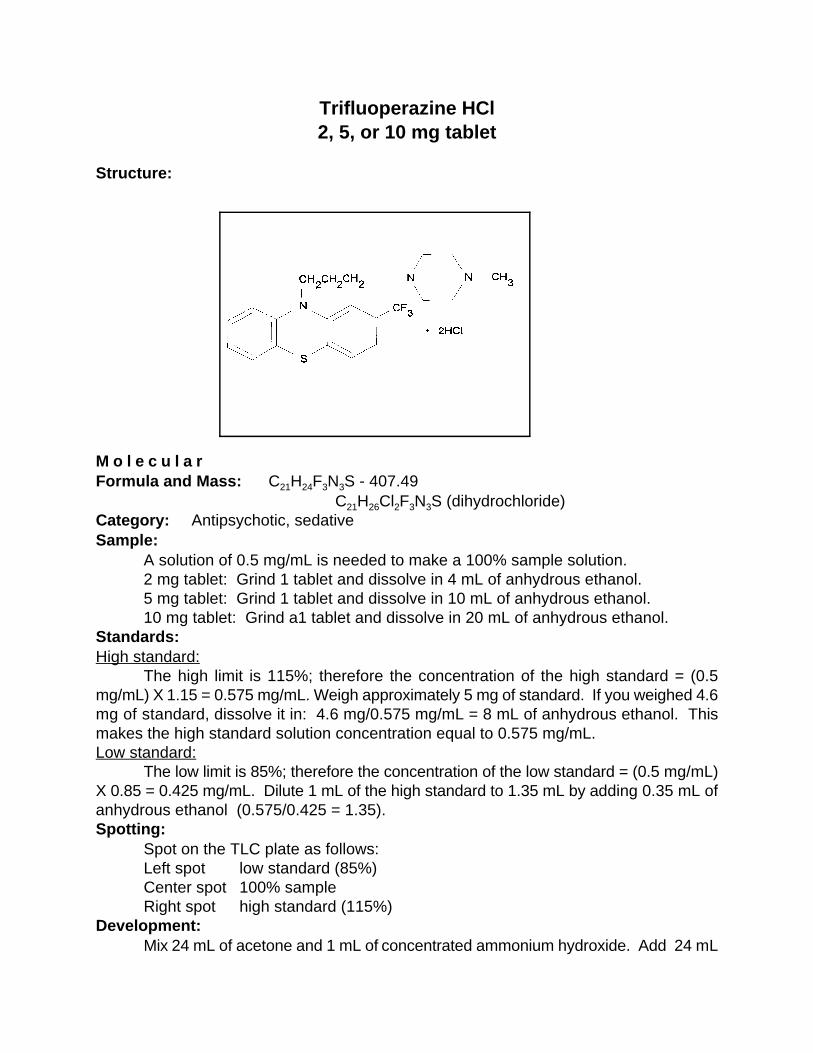
Trifluoperazine HCl2, 5, or 10 mg tablet
Structure:
M o l e c u l a rFormula and Mass: C H F N S - 407.4921 24 3 3
C H Cl F N S (dihydrochloride) 21 26 2 3 3
Category: Antipsychotic, sedativeSample:
A solution of 0.5 mg/mL is needed to make a 100% sample solution. 2 mg tablet: Grind 1 tablet and dissolve in 4 mL of anhydrous ethanol. 5 mg tablet: Grind 1 tablet and dissolve in 10 mL of anhydrous ethanol.10 mg tablet: Grind a1 tablet and dissolve in 20 mL of anhydrous ethanol.
Standards:High standard:
The high limit is 115%; therefore the concentration of the high standard = (0.5mg/mL) X 1.15 = 0.575 mg/mL. Weigh approximately 5 mg of standard. If you weighed 4.6mg of standard, dissolve it in: 4.6 mg/0.575 mg/mL = 8 mL of anhydrous ethanol. Thismakes the high standard solution concentration equal to 0.575 mg/mL.Low standard:
The low limit is 85%; therefore the concentration of the low standard = (0.5 mg/mL)X 0.85 = 0.425 mg/mL. Dilute 1 mL of the high standard to 1.35 mL by adding 0.35 mL ofanhydrous ethanol (0.575/0.425 = 1.35).Spotting:
Spot on the TLC plate as follows:Left spot low standard (85%)Center spot 100% sampleRight spot high standard (115%)
Development:Mix 24 mL of acetone and 1 mL of concentrated ammonium hydroxide. Add 24 mL

of this mixture to the TLC development bag. Develop until the solvent front reaches within1 cm of the top of the TLC plate.Detection:UV:
Dry the plate and observe under UV light (254 nm). Observe the size and intensityof the spots.Iodine stain:
Dip the plate in the iodine-KI solution in the detection bag. Allow the plate to dry andobserve the size and intensity of the spots.
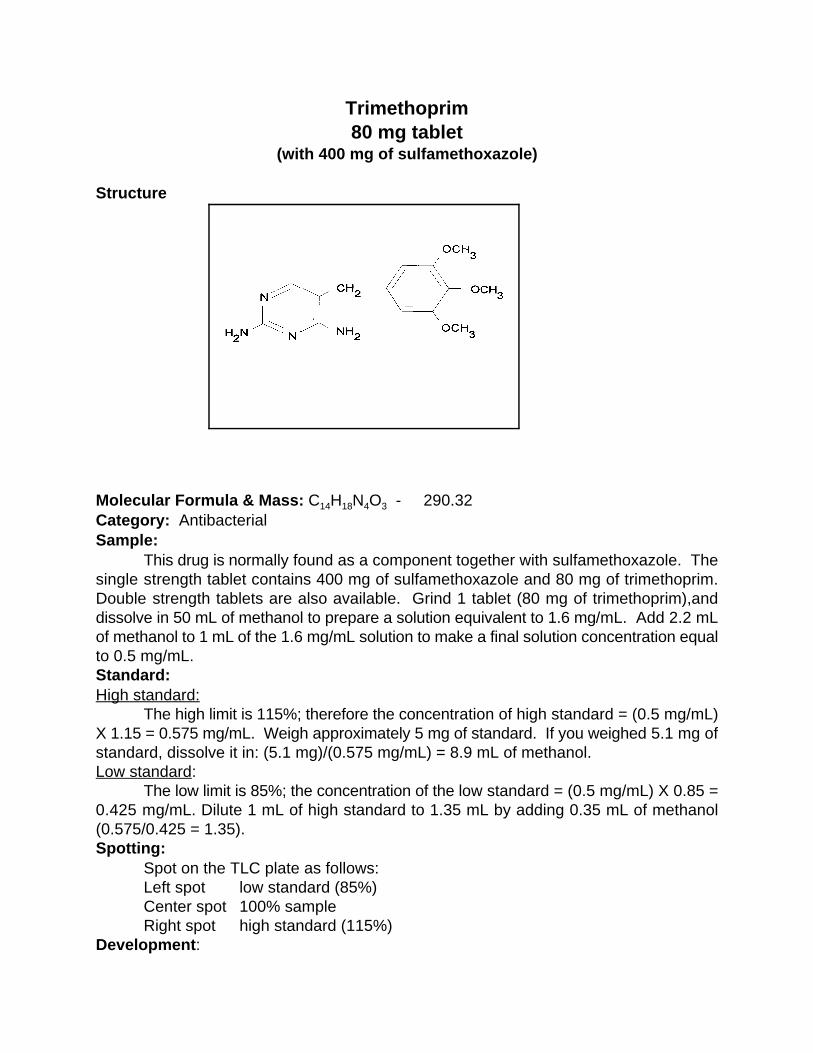
Trimethoprim80 mg tablet
(with 400 mg of sulfamethoxazole)
Structure
Molecular Formula & Mass: C H N O - 290.32 14 18 4 3
Category: AntibacterialSample:
This drug is normally found as a component together with sulfamethoxazole. Thesingle strength tablet contains 400 mg of sulfamethoxazole and 80 mg of trimethoprim.Double strength tablets are also available. Grind 1 tablet (80 mg of trimethoprim),anddissolve in 50 mL of methanol to prepare a solution equivalent to 1.6 mg/mL. Add 2.2 mLof methanol to 1 mL of the 1.6 mg/mL solution to make a final solution concentration equalto 0.5 mg/mL. Standard:High standard:
The high limit is 115%; therefore the concentration of high standard = (0.5 mg/mL)X 1.15 = 0.575 mg/mL. Weigh approximately 5 mg of standard. If you weighed 5.1 mg ofstandard, dissolve it in: (5.1 mg)/(0.575 mg/mL) = 8.9 mL of methanol.Low standard:
The low limit is 85%; the concentration of the low standard = (0.5 mg/mL) X 0.85 =0.425 mg/mL. Dilute 1 mL of high standard to 1.35 mL by adding 0.35 mL of methanol(0.575/0.425 = 1.35).Spotting:
Spot on the TLC plate as follows:Left spot low standard (85%)Center spot 100% sampleRight spot high standard (115%)
Development:

Mix 11mL of acetone, 11 mL of toluene, and 2.5 mL of methanol. Add this mixtureto the TLC development bag. Develop until the solvent front reaches within 1 cm of the topof the TLC plate.Detection:UV:
Dry the plate and observe under UV light. The maximum is at 278 nm but a shortwavelength light (254 nm) will work. Observe the size and intensity of the spots.Iodine stain:
Dip the plate in the iodine-KI solution in the detection bag. Allow the plate to dry andobserve the size and intensity of the spots.

Vitamin A -Retinol7.5 mg capsule
(in cottonseed oil)
Structure
Molecular Formula & Mass: C H O - 286.44 20 30
Category: VitaminSample:
Dissolve 1 capsule (alcohol base) in 15 mL of anhydrous ethanol. The concentrationof solution = (7.5 mg/15 mL) = 0.5 mg/mL. The required concentration of sample solutionrepresenting 100% is 0.5 mg/mL.Standard:High standard:
The high limit is 115%; therefore the concentration of high standard = (0.5 mg/mL)X 1.15 = 0.575 mg/mL. The standard is Vitamin A acetate capsule (1 g of solution = 30.3mg of retinol). Weigh approximately 229 mg of standard solution. If you weighed 229.74 mgof standard solution, calculate as follows: (30.3 mg of retinol X 229.74 g of solution)/1 gsolution = 6.96 mg of retinol. Dissolve it in: (6.69 mg)/(0.575 mg/mL) = 11.63 mL of ethanol.Low standard:
The low limit is 85%; therefore the concentration of low standard = (0.5 mg/mL) X0.85 = 0.425 mg/mL. Dilute 1 mL of high standard to 1.35 mL by adding 0.35 mL of ethanol(0.575/0.425 = 1.35).Spotting:
Spot on the TLC plate as follows:Left spot low standard (85%)Center spot 100% sampleRight spot high standard (115%)
Development:Mix 21 mL of toluene and 3 mL of ethyl acetate. Add this mixture to the TLC
development bag. Develop until the solvent front reaches within 1 cm of the top of the TLCplate.Detection:UV:
Dry the plate and observe under UV light. The maximum is at 325 nm but a shortwavelength light (254 nm) will work. Observe the size and intensity of the spots.Iodine stain:
Dip the plate in the iodine-KI solution in the detection bag. Allow the plate to dry andobserve the size and intensity of the spots.Note:
The cottonseed oil in this drug lowers its solubility. It also makes spotting much moredifficult. The R value for the sample is lower than that of the standard because the standardf
is acetate base but the sample is alcohol base and thus more polar.

Warfarin 2.5 mg tablet
Structure
Molecular formula & mass: C H O - 308.32 19 16 4
Category: AnticoagulantSample:
Grind 4 tablets and dissolve in 10 mL of anhydrous ethanol. Concentration of thesolution = (2.5 mg X 4)/10 mL = 1 mg/mL. The required concentration of the samplesolution representing 100% is 1 mg/mL.Standard:High standard:
The high limit is 115%; therefore the concentration of high standard = (1 mg/mL) X1.15 = 1.15 mg/mL. Weigh approximately 5 mg of standard. If you weighed 5.8 mg ofstandard, dissolve it in: (5.8 mg)/(1.15 mg/mL) = 5 mL of anhydrous ethanol. Low standard:
The low limit is 85%; therefore the concentration of the low standard = (1 mg/mL) X0.85 = 0.85 mg/mL. Dilute 1 mL of high standard to 1.35 mL by adding 0.35 mL of ethanol(1.15/0.85 = 1.35).Spotting:
Spot on the TLC plate as follow:Left spot low standard (85%)Center spot 100% sampleRight spot high standard (115%)
Development:Add 24 mL of 100% ethyl acetate to the TLC development bag. Develop until the
solvent front reaches within 1 cm of the top of the TLC plate.Detection:UV:

Dry the plate and observe under UV light. The maximum is at 310 nm but a shortwavelength light (254 nm) will work. Observe the size and intensity of the spots.Iodine stain:
Dip the plate in the iodine-KI solution in the detection bag. Allow the plate to dry andobserve the size and intensity of the spots. Note:
The sample used was a sodium salt. If this sample is used, it must be taken intoconsideration. For example, if the percent concentration of sample is 94, it needs to beadjusted as follows:(330/308) X 94% = 101%. The final concentration of the sample is 101%.330 g/mol = molecular weight of the sodium salt sample308 g/mol = molecular weight of the pure standard

1. Visiting Scientist, Food and Drug Administration, Division of Testing and Applied AnalyticalDevelopment (DTAAD), 1114 Market Street, Room 1002, St. Louis, MO 63101. Communicationsshould be addressed to Dr. Thomas Layloff, Director, at this address.
2. Shanghai Institute for Drug Control, Republic of China. On technical assignment to DTAAD,St. Louis, MO.
3. Senior Laboratory Officer, Institute of Science and Forensic Medicine, Singapore. Ontechnical assignment to DTAAD, St. Louis, MO.
Simple, At-Site Detection of Diethylene Glycol/Ethylene Glycol Contamination of Glycerin
and Glycerin-based Raw Materials by Thin-Layer Chromatographic
Allen S. Kenyon , Shi Xiaoye , Wang Yan , and Ng Wai Har1 2 2 3
FDA Division of Testing and Applied Analytical Development
St. Louis, MO 63101
and
Robert Prestridge and Kirsten Sharp
Australian Therapeutic Goods Administration
Woden, ACT 2606 Australia
Reprint: Journal of AOAC International Vol.81,No.1,44-50,1998

Abstract
This paper describes a rapid, inexpensive thin-layer chromatographic (TLC) method that
separates diethylene glycol (DEG) from glycerin and other glycols. Studies with
collaborating laboratories of the World Health Organization have shown that about 6%
DEG in glycerin and about 2% DEG in acetaminophen (paracetamol) elixirs may be
detected by direct visual inspection of the developed TLC sheets. Staining the sheet
permits detection of DEG at less than 0.1%. The method costs less than $1.00 per test and
takes 20 minutes with visual inspection, longer when staining is required. The visual
method can be performed without laboratory facilities by personnel having little previous
training. Samples testing positive by the visual method can be submitted to a laboratory for
confirmation and quantitation of DEG.
Introduction
In 1937, 105 Americans died of renal failure when they ingested a sulfanilamide elixir
containing 72% DEG used as a diluent (1). In spite of this famous incident, additional deaths
attributed to DEG poisoning continue to occur (2). The fourth largest outbreak of DEG poisonings
associated with pharmaceutical products occurred in Haiti in 1996, 59 years after the U.S.
sulfanilamide elixir episode, and was linked to the deaths of many children(3,4).
Glycols such as glycerin are present in many formulations of medicinal, cosmetic, and food
products. As a result of world trade, such commodities can become contaminated by DEG or
ethylene glycol and may unwittingly be accepted for use in manufacture of pharmaceuticals.
Investigations of DEG-related poisonings in various countries have repeatedly revealed lack of
testing for contamination and other quality-control measures at ports of entry or during the
pharmaceutical manufacturing process (2-4).
The minimum safe level for humans for ingested DEG is not known, but at a 1997
interagency workshop supported by the U.S. Centers for Disease Control and Prevention (CDC),
Food and Drug Administration (FDA), and the World Health Organization (WHO), a detection
level of 0.1% DEG was selected as adequate for screening raw materials and glycerin-based
elixirs.
Earlier we reported the development of a novel TLC system used for rapid screening
analysis of many drugs(5-7). The purpose of this work was to adapt this method to detect the
presence of DEG in glycerin in raw materials and liquid oral pharmaceuticals.
We required that the method (a) distinguish DEG from related components (e.g., glycerin,
and ethylene and propylene glycols) often present in glycerin and glycerin-based materials and
products; (b) be sufficiently sensitive and reliable to prevent future DEG-related fatalities; (c)
allow detection of gross DEG contamination (about 6% DEG in glycerin and about 2% DEG in
elixirs) through direct visual inspection of the sheet without spraying and at locations without
laboratory facilities, such as at ports of entry; and (d) allow detection of DEG at 0.1% through
spraying or staining the sheet. (Throughout, we use percentage of DEG to refer to the percentage
in the original sample before dilution.)
Separation of glycols by TLC has been reported previously (8), although with developing
solvent systems not used here. Because glycols are not visible by the usual UV illumination
methods, investigators have used a variety of staining sprays to make the spots visible (9).
Experimental
The chemicals and solvents used for the TLC developing solvent (toluene, acetone,
ammonium hydroxide), as well as reference materials of DEG, glycerin, ethylene glycol,
propylene glycol, and sorbitol, were all pure grade as obtained from commercial sources. The
toluene and acetone were chromatographic grade, but reagent grade solvents are also satisfactory .
Samples of contaminated or uncontaminated glycerins and syrups were prepared or
obtained from various sources. Some were representative samples that had been analyzed by the
CDC, Atlanta, GA, and the FDA National Forensic Chemistry Center (NFCC) in Cincinnati, OH,
by gas chromatography (GC) and GC/mass spectrometry (GC/MS).
The rapid screening TLC apparatus used in this work (Figure 1) and its operation have
been previously described (5,6) (US Patent 5,350,510.). :The apparatus was produced by Granite
Engineering. Inc., P.O. Box 1269, Granite City, Illinois 62040, USA. The TLC method described
here is not specific for this apparatus, but was used because of the ease, reliability, cost, and
portability.
Preparation of TLC sheets
Merck plastic-backed silica-coated 20 X 20-cm sheets or equivalent, either with or
without fluorescent indicator, were used; at low DEG concentrations, best results were obtained
with the fluorescent sheets.
Cut the sheet into eight equal parts, each 5 X 10 cm. To prevent edge effects, remove 1 mm
of the coating layer from the sides and the bottom of the sheet by bearing down on a #2 pencil
along a straight edge. Draw the spotting line 2.5 cm from the bottom of the sheet without cutting
into the coating by very lightly applying a pencil tip. Mark the three spotting locations at 1.5, 2.5,
and 3.5 cm from the left edge. Mark the development stopping line by placing a straight edge
across the sheet 1 cm from its top and removing 1 mm of the silica coating by bearing down on a
pencil.
Preparation of solutions
To reduce viscosity, dilute glycerin and other glycols threefold with methanol before
spotting; typically, add 1 mL of methanol to 0.5 mL of the glycol in a 1.5-mL plastic centrifuge tube
graduated at 0.5, 1.0, and 1.5 mL. Prepare lower concentrations by diluting aliquots from the
initial solutions with methanol. Use solutions of the pure glycols as reference materials to assign
the spot positions. The developing solvent is toluene:acetone:5M ammonium hydroxide (5:85:10);
prepare daily.
Spotting of the solutions and TLC sheet development
This TLC system accommodates two sheets, with three samples or reference solutions
spotted on each sheet. Spot the sample or reference solutions at the marked spotting locations with
a 3-µL capillary pipette. Always apply a reference solution of DEG as one of the spots on each
sheet. Allow the spots to dry for approximately 10 minutes.

Clamp the spotted sheets onto the aluminum trays in the system.( The developing solvent
may be reused for additional analysis during the same day by replacing the volume lost in the
saturation pads.) Add 24 mL of the developing solvent to the plastic bag, and insert the assembled
sheets into the polyethylene bag until the large saturation pad just touches the developing solvent
solution. Position the assembly in the bag with a wire rod (fabricated from a coat hanger), which
also serves as a tool for removing the assembly from the bag after development. Clamp the bag
containing the assembly in the system's metal support, which seals the top of the bag to maintain a
uniform environment.
Migration and detecting the spots
Allow 2 minutes for the system to establish equilibrium. Grasp the bottom of the bag and
gently pull it down, forcing the developing solvent solution up onto the TLC sheets to initiate
migration. At this point, the bottoms of the TLC sheets are immersed in the developing solvent to a
depth of 1 cm. Allow the developing solvent to migrate upward until the solvent front reaches the
scored top line on the sheets (approximately 20 minutes). Remove the assembly containing the
sheets from the bag with the positioning rod, and allow the sheets to air dry.
At high concentrations of DEG (6% or more in glycerin, 2% or more in elixirs), the spots
may be visually detected by transmitted daylight; the spots appear dark relative to the sheet
because of the difference in refractive index. At lower concentrations of DEG, the sheets must be
stained to make the spots visible. The Appendix details two methods for staining the developed
sheets.
Results and Discussion
Several developing solvents were considered in this study. We wished to avoid hazardous
solvents such as chloroform or benzene that have been frequently used in TLC developing solvent
systems.
Three different developing solvent mixtures were evaluated: (a) n-butanol saturated with
1.5 M ammonium hydroxide; (b) chloroform:acetone:5M ammonium hydroxide (10:80:10); and (c)
toluene:acetone:5M ammonium hydroxide (5:85:10). The development time for (a) was 75
minutes, whereas (b) and (c) each required 20 minutes. The WHO has banned use of chloroform
in a TLC developing solvent because of its carcinogenic nature; therefore, the chloroform in
developing solvent (b) was replaced by a 1:1 mixture of toluene and acetone to form developing
solvent (c) to achieve approximately the same solubility behavior. Developing solvent (c) proved
to be the most satisfactory because it reduced the migration time in (a) and eliminated toxic
chloroform in (b). The retardation times for developing solvents (b) and (c) were identical.
To achieve detection at 0.1% DEG, we tried several different staining procedures;
however, most required spraying with highly toxic (e.g., benzidine) or costly materials. These
procedures require the use of a well-ventilated hood, a face mask for the operator, and a well-
equipped laboratory, and essentially rule out performing the test in open, nonlaboratory
environments.
Visual observation of the spots in bright transmitted white light was the most simple and
effective detection method when the DEG content was greater than 2% in elixirs or 6% in glycerin.
Figure 2 shows the appearance of the spots on the developed sheets. Circles were drawn around
the spots to show their locations. Sample A was pure glycerin. Sample B contained 4.5%
glycerin and 25.5% DEG; the DEG was readily detected but not glycerin. Sample C contained
18% glycerin and 18% DEG; both DEG and glycerin were easily seen Samples D and E were
syrups containing no drug. Sample D contained 2.2% glycerin and 14% DEG; sample E contained
67% glycerin and no DEG.

Table 1 lists the retardation factors (R s) for the reference glycols using the three solventf
mixtures (a,b andc) , and illustrates that glycerin, DEG, and other glycols can be detected by
matching the R values of sample spots with those of authentic references. No study was made onf
possible variation of R with temperature or humidity.f
The method can also be used to estimate the concentration of DEG contamination in
glycerin or syrup samples. The concentration of DEG may be estimated by spotting solutions
having different concentrations of DEG and directly comparing the intensities of the spots. In this
procedure, the intensity of a sample spot (and hence the amount or concentration of DEG) can be
visually estimated to be between certain limits, bracketed by two reference DEG spots of different
concentration. Detection using only transmitted light worked well with the samples studied, since
the concentration of DEG in the glycerin ranged between 18 and 25%.
Several stains that enhance the detection of glycols(8) have been reported, but most are
toxic and costly, require operation in a hood to protect the analyst from the harmful sprays, and
lack sensitivity. Many of the recommended stains are applicable only for high concentrations of
glycol. Use of these stains was dismissed as impractical after several trials. As recently as 1996,
the Pharmacopeia of India(10) described a staining procedure in which benzidine was used to
detect DEG at the 0.1% level. This method worked well in our hands but is not recommended for
use in open places or without suitable laboratory facilities. Two staining procedures were found
feasible for use in open, nonlaboratory places, namely, (a) iodine vapor/starch spray and (b)
oxidation of the glycols with potassium permanganate. Details of these staining methods are
described in the Appendix.
Figure 3 shows the detection of DEG in glycerin at concentrations from 0.1 to 0.75% as
measured by densitometry at 400 nm after the sheets were stained with iodine vapor and sprayed

Color photo of Fig 5 can be supplied upon request4
with starch. The densitometer was used to obtain performance data and to show that the intensity
of the spots were a function of concentration. The densitometer response for the 0.1%
concentration was low and erratic, and that point is not shown; however, the spot was visible to
the eye. The responses were linear with a correlation coefficient of >0.99, based on average
values obtained in duplicate runs by three analysts. The responses from solutions of higher DEG
concentrations were also linear. The intensities from different sheets could not be compared
because of differences in staining and fading; standards and samples must be run on the same sheet.
An estimate of the concentration can be determined visually without the need for any
instrumentation by spotting solutions of bracketing concentrations of DEG along with the sample.
Children’s acetaminophen elixir obtained from a local pharmacy was spiked with DEG.
Samples of the laboratory-contaminated product were diluted to various concentrations down to
0.1% DEG. The developed spots were oxidized with potassium permanganate. An actual
separation at the level of 0.25% DEG is shown in Figure 5 as yellow spots on a purple
background, demonstrating the ability to separate the sugars (sucrose and sorbitol), the glycols,
and the other components in a final dosage form. The listed major components of the children’s
acetaminophen elixir were citric acid, glycerin, propylene glycol, sucrose/sorbitol, and
acetaminophen; the minor components did not interfere with the separation of the glycols.
Since the size of the spot is proportional to the concentration, at lower concentrations the4
spots can be very small and difficult to detect. The detection limit can be lowered just by
increasing the volume. If the volume of material spotted is increased from 3 to 15 µL, the
detection limit for DEG can be lowered from 0.1% to 0.05%.
The detection levels for contamination of glycerin were different from those for the elixirs
or syrups. Glycerin, because of its high viscosity, required at least a threefold dilution for the
contaminant to separate, which meant that the detection limit must be higher for pure glycerin than
for samples of elixirs or syrups. With care, DEG could be detected in glycerin down to a level of
0.025% with iodine vapor/starch spray and down to 0.01% in elixirs or syrups with permanganate.
Staining by oxidation with permanganate is faster, easier to perform, and more sensitive,
and it eliminates the need for spray equipment and a large glass developing tank, but the spots fade
faster. Selection of the two staining methods was based on low toxicity, low cost, availability of
reagents, and ease of operation. Permanent records of the developed TLC sheets can be made by
photo copy.
A common practice for increasing the sensitivity of TLC is to multiply spots (11) by
applying the spot, allowing it to dry, and spotting additional material on top of the dried material.
By spotting three pipette loadings on a single position, the detection level has effectively been
increased, making the developed spot more visible.
This method will not separate DEG and ethylene glycol, but if both are present in
approximately equal amounts, the developed spots have the contour of a distorted figure eight.
Since both DEG and ethylene glycol are toxic, it does not matter that the pair cannot be
distinguished as long as they can be separated from the other nontoxic glycols.
Testing with even the simple visual method of detection presented here would have been
adequate to detect the contaminated materials that have caused the large numbers of deaths among
children and adults in the last 60 years.
Acknowledgments
The National Center for Environmental Health, Atlanta, GA, and the FDA NFCC kindly
supplied samples and suggestions. The authors thank Karen Wolnik and Kevin Mulligan of the
NFCC and Thomas P. Layloff of the FDA Division of Testing and Applied Analytical
Development (DTAAD), St. Louis, for helpful discussions and review of this work, and Walter L.
Zielinski and William B. Furman, also of DTAAD, for help in preparing technical revisions of this
paper. This assistance and these contributions are gratefully acknowledged.
Special thanks and appreciation are expressed to the following Collaborating Laboratories
of the World Health Organization for their assistance in evaluating the method and their helpful
comments:
Aiache, J.M., Laboratoire de Biopharmacie, Clermont, France.
Török, I., National Institute of Pharmacy, Budapest, Hungary.
Howard, R.K., Therapeutic Goods Administration, Woden, Australia.
Wongpinairat, C., Ministry of Public Health, Thailand
Westermark, M., Centre for Chemical Reference Substances, Stockholm, Sweden.
Lasis,A., Health Protecton Branch, Ontario, Canada.
Lambert, M. J., Health Protection Branch, Quebec, Canada.
The method was independently validated by Virginia Kleekamp and Tom Lavelle,
FDA, DTAAD, St. Louis, MO.
References
(1) Geiling, E. M. K.; Cannon, P. R. JAMA (1938), 111, 919-926; through Ann. Intern. Med.
(1995) 122, 456.
(2) Okuonghae, H.O.; Ighogboja, I.S.; Lawson, J.O.; Nwana, E.J. Ann. Trop. Paediatr.( 1992),
12, 235-238. Hanif, M.; Mobarak, M.R.; Ronan, A.; Rahman, D.; Donovan, J.J.; Bennish,
M.L. Br. Med. J.( 1995), 311, 88-91. Centers for Disease Control and Prevention, Private
communication, February 1997.
(3) Malebranche, R., et al. Morbidity and Mortality Weekly Report (1996), 45, 649-650.
[Ed.: This article has more than two dozen authors.]
(4) WHO Drug Information (1996), 10(2), 84.
(5) Flinn, P. E.; Kenyon, A. S.; Layloff, T. P. J. Liq. Chromatogr. (1992), 15, 1639-1653.
(6) Kenyon, A. S.; Flinn, P. E.; Layloff, T. P. J. AOAC Int. (1995), 78, 41-49.
(7) Kenyon, A. S.; Flinn, P. E.; Layloff, T. P. World Health Organization (1995),
PHARM.290, 110 pp.
(8) Stahl, E. Thin-Layer Chromatography; Springer-Verlag, New York, 1969;
Zweig,G.;Sherma,J.,Handbook of Chromatography, Vol. II; CRC Press, Cleveland, OH,
1974.
(9) Cifonelli, J.A.; Smith, E. Anal. Chem. (1954), 26, 1132; Knappe, E.; Petrie, D.;
Rohdewald, I. Z. Anal. Chem. (1963), 199, 270.
(10) Manager of Publications , Ministry of Health, Delhi ; Pharmacopeia of India, 4th ed.;
(1996), p. 349.
(11) Klaus, R.; Fischer, W. Chromatographia (1987), 23(2), 137-140.
Appendix

Staining Procedures for Detecting Low Levels of DEG on Developed TLC Sheets
Method 1. Iodine/Starch Stain
1. Place an amount (approximately 25 to 50g) of iodine crystals on the bottom of a glass TLC
developing tank that can be sealed with a lid or cap. The tank must be deep enough that the sheet
can hang vertically in the iodine vapor. Suspend the developed sheet in the tank, cover the tank
with the lid, and allow the sheet to be exposed to the vapor for 5 minutes.
2. Remove the sheet and allow it to stand in the open air for 1 hour to remove all the iodine that
has been absorbed by the silica coating on the sheet. If the iodine is not removed at this point, the
detection of low concentrations of DEG will be masked by the background.
3. Prepare a 1% starch solution by adding 1 g of starch to 10 mL of water, making a slurry, adding
90 mL of water, and boiling until the solution becomes clear. (The starch solution will appear
turbid when cooled, but this will not harm the solution.) Spray the sheet with the 1% starch
solution and observe the spots. The method of detecting the spots will depend upon the
concentration level of the DEG. Once the DEG is detected, it will not be necessary to proceed to
another step. Three levels of detection are possible:
Level 1: For DEG levels of 2 to 6% or higher. Observe the sheets visually by transmitted light
immediately after they are dried. The spots appear dark because of the difference in refractive
index. No further staining is necessary. If no spots are detected, proceed to Level 2.
Level 2: For DEG levels of 0.5% or higher. Immerse the sheet in iodine vapor. The iodine-
stained spots will be visible. If no spots are visible, proceed to level 3.

Level 3: For DEG levels from 0.1 to 0.5%. Spray the sheet with a 1% starch solution. If no spots
are observed, the concentration of DEG is below 0.25%.
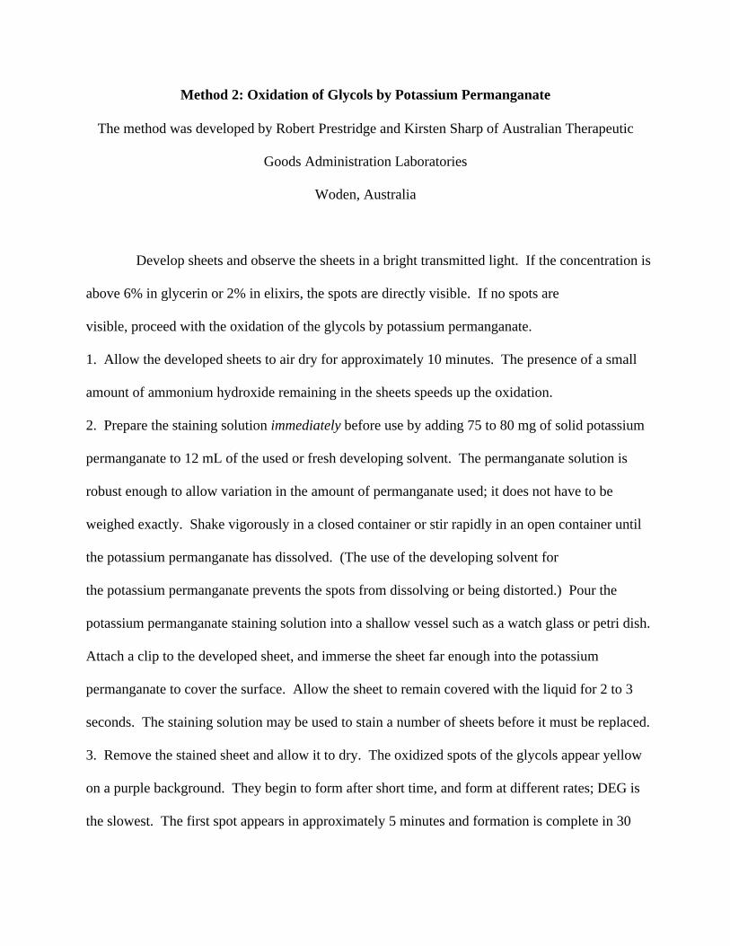
Method 2: Oxidation of Glycols by Potassium Permanganate
The method was developed by Robert Prestridge and Kirsten Sharp of Australian Therapeutic
Goods Administration Laboratories
Woden, Australia
Develop sheets and observe the sheets in a bright transmitted light. If the concentration is
above 6% in glycerin or 2% in elixirs, the spots are directly visible. If no spots are
visible, proceed with the oxidation of the glycols by potassium permanganate.
1. Allow the developed sheets to air dry for approximately 10 minutes. The presence of a small
amount of ammonium hydroxide remaining in the sheets speeds up the oxidation.
2. Prepare the staining solution immediately before use by adding 75 to 80 mg of solid potassium
permanganate to 12 mL of the used or fresh developing solvent. The permanganate solution is
robust enough to allow variation in the amount of permanganate used; it does not have to be
weighed exactly. Shake vigorously in a closed container or stir rapidly in an open container until
the potassium permanganate has dissolved. (The use of the developing solvent for
the potassium permanganate prevents the spots from dissolving or being distorted.) Pour the
potassium permanganate staining solution into a shallow vessel such as a watch glass or petri dish.
Attach a clip to the developed sheet, and immerse the sheet far enough into the potassium
permanganate to cover the surface. Allow the sheet to remain covered with the liquid for 2 to 3
seconds. The staining solution may be used to stain a number of sheets before it must be replaced.
3. Remove the stained sheet and allow it to dry. The oxidized spots of the glycols appear yellow
on a purple background. They begin to form after short time, and form at different rates; DEG is
the slowest. The first spot appears in approximately 5 minutes and formation is complete in 30

minutes. The difference in formation rate may be used to distinguish DEG and ethylene glycol if
present individually.
4. Properly dispose of all used or unused staining solution after treating the samples, since it will
not keep. Keep volumes to a minimum.

Glycols A B & C
Solvent solvents
Glycerin 30 16
Dietylene 52 39
glycol
Ethylene 56 42
glycol
Propylene 64 54
glycol
Sucrose 0 0
Sorbitol 0 0
Table 1. Retardation factors (R ) for Glycolsf

LEGENDS FOR FIGURES:
Figure 1. Rapid screening TLC apparatus.
Figure 2. TLC separation of diols.
Figure 3. Detection of low DEG concentrations.
Figure.4. TLC separation of contaminated and noncontaminated
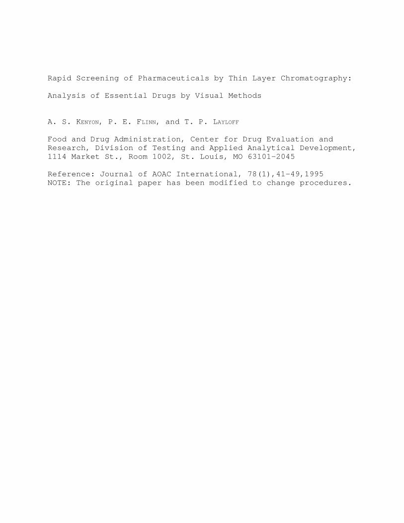
Rapid Screening of Pharmaceuticals by Thin Layer Chromatography:
Analysis of Essential Drugs by Visual Methods
A. S. KENYON, P. E. FLINN, and T. P. LAYLOFF
Food and Drug Administration, Center for Drug Evaluation andResearch, Division of Testing and Applied Analytical Development,1114 Market St., Room 1002, St. Louis, MO 63101-2045
Reference: Journal of AOAC International, 78(1),41-49,1995NOTE: The original paper has been modified to change procedures.

Abstract A method for rapidly screening pharmaceuticals by thin layerchromatography has been designed for use in areas with limitedresources and by operators with limited training. An apparatuswas designed to perform the analysis in a plastic bag underequilibrium conditions. Results can be reproduced by differentoperators and in different locations. The analysis can beperformed without electricity or in a remote area away from alaboratory. The method is low-cost, maintenance-free, fast, andreliable, and it uses limited volumes of solvents. The analyses can be performed without weighing if referencematerials can be supplied in tablet form, provided the drugcontent is listed and only 1 unit is required for each analysis. The solvents, developers, concentrations, and procedures weredeveloped for a list of essential drugs supplied by the WorldHealth Organization. From this list, 3 drugs were selected andprepared in the form of reference tablets. Comparisons with theanalyses of the drugs in standard dosage forms were made by usingreference tablets and primary USP standards. Comparable resultswere obtained, proving the concept that the screening process canbe conducted by using reference tablets and without weighingeither the sample or the reference. The method has beensuccessfully demonstrated and used in Swaziland, in the U. S. Aby high school teachers, and by Saudi Arabia personnel from theMinistry of Health. Personnel can be trained to performscreening analysis of drugs in a short time.

Introduction A simple analytical method for rapid screening of drugs isneeded for use in areas that do not have fully equippedlaboratories. The method should fulfill the following criteria: It must be inexpensive, must require only limited facilities andminimum training, and must be capable of quickly analyzing thedrugs. The analysis must be operable without electricity or alaboratory facility. It must be possible to prepare solutionswithout weighing either the sample or reference, providedreference tablets are available. If the analysis requireshandling organic solvents, it should be possible to perform theprocedure in open air with a minimum amount of solvent and withno hood available. No electronic measurements should be needed,and estimates should be made from visual inspections underdaylight. The TLC method satisfies these requirements.
A previously developed TLC method (1) has been useful forrapidly screening drugs by comparing the sample solution withdifferent concentrations of reference material. The success ofthis system, however, has required that the laboratory haveelectricity and special equipment. Even though this method isfast and economical, and the operator requires little training,it has not been able to meet the needs of locations withoutelectricity and with only limited resources. The work describedhere was directed toward the development of a rapid screeningsystem which could be performed either in a well-equippedlaboratory or in remote areas with or without electricity, and bypersons having limited technical background. Another objectivewas to establish the necessary specifications for the referencetablets. The study was limited to the drugs declared essentialby the World Health Organization and to a few additional ones toshow the general applicability of the method. It must beemphasized that the method described here is intended not as areplacement for other analytical methods but only as a rapidscreening procedure for drugs in areas with limited facilities.
TLC as normally practiced uses thick glass tanks, beakers,or other glass containers for the development of thechromatograms. A general systematic procedure that usesinexpensive, portable, unbreakable equipment would be useful forthe rapid screening of pharmaceuticals (and other drugs) in afield environment. Some of the desirable features of such asystem would include portability, low cost, and simplicity ofoperation with minimal training to achieve acceptable results. The apparatus should be designed so that it can also be usedwhere laboratory facilities are unavailable and no instrumentmaintenance is required.
Flinn et al. (2) described a simple, inexpensive TLC methodin which the rigid, fragile developing chambers were replaced byflexible polyethylene bags for developing the chromatogram, and

tested the analytical capability of the system with the drugtheophylline. A new system was then developed to provide a moreefficient analysis under equilibrium conditions. The completeassembled apparatus (Figure 1), known as "SPEEDY TLC" (3), iscommercially available. The apparatus consists of a rigidaluminum support, 2 aluminum trays for holding the TLC sheets, apolyethylene development bag, and saturator pads. The TLCapparatus allows the analyses to be performed underequilibrium conditions and prevents distortion of the spots. Reproducibility between analysts and laboratories is easilyachieved.
Other containers such as beakers and jars have been used,but none were as successful as the polyethylene bag. The solventlevel can be controlled and the development stage started andstopped as desired by simply manipulating (raising and lowering)the bag while it is in the holder. The entire system can easilybe supplied in kit form so that analyses can be carried outwithout delay. Experimental
Apparatus(a) Development bag.--Fabricate the development bag, 40 cm
long, from large rolls of 0.006-gauge flat polyethylene tubing, 8cm wide (commercially available). Seal each tube with an impulsesealer at a point ca 6 cm from the bottom. (The development bagscan also be supplied prefabricated.)
(b) Saturation pads.--Cut rectangular saturation pads fromheavy filter paper (0.3 mm thick). (The pads are also availablein 20 H 20 cm sheets.) Cut 1 large pad, 14.4 H 7.0 cm, and 2small pads, each 10.0 H 5.7 cm, for each complete assembly. Adjust the pads as necessary if the polyethylene bag hasdifferent dimensions. To establish and maintain equilibrium inthe plastic bag chamber before and during development, clamp thelarger pad between the 2 aluminum TLC support trays so that itextends below the holders and into the solvent. Fit the 2smaller saturation pads between the aluminum tray and the TLCsheet. Insert 1 of these small pads in each tray between theclip and the tray first, and then insert the TLC sheet on top ofeach pad. (Some of the thin layer area that contacts the clipmay be disturbed while the TLC sheet is slid between the clip andsaturation pad, but this does not cause a problem because thedevelopment step is stopped before the solvent front reaches theclip area.)
(c) Thin layer support.--Use plastic sheets coated with alayer of Silica Gel 60 (E. Merck, F-254 or equivalent) 200 :mthick. (The plastic-backed TLC sheets, 20 x 20 cm, are the mostdesirable.) Cut each sheet into 8 equal parts to form smallersheets each measuring 5 H 10 cm.
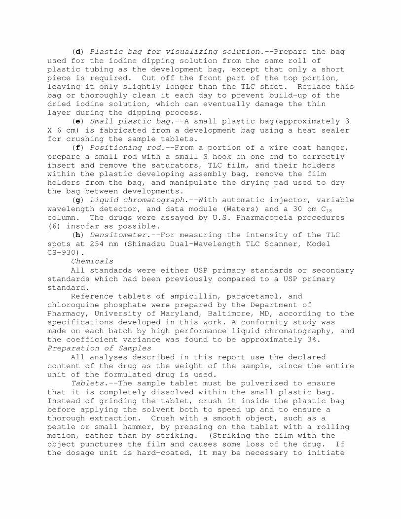
(d) Plastic bag for visualizing solution.--Prepare the bagused for the iodine dipping solution from the same roll ofplastic tubing as the development bag, except that only a shortpiece is required. Cut off the front part of the top portion,leaving it only slightly longer than the TLC sheet. Replace thisbag or thoroughly clean it each day to prevent build-up of thedried iodine solution, which can eventually damage the thinlayer during the dipping process.
(e) Small plastic bag.--A small plastic bag(approximately 3X 6 cm) is fabricated from a development bag using a heat sealerfor crushing the sample tablets.
(f) Positioning rod.--From a portion of a wire coat hanger,prepare a small rod with a small S hook on one end to correctlyinsert and remove the saturators, TLC film, and their holderswithin the plastic developing assembly bag, remove the filmholders from the bag, and manipulate the drying pad used to drythe bag between developments.
(g) Liquid chromatograph.--With automatic injector, variablewavelength detector, and data module (Waters) and a 30 cm C18column. The drugs were assayed by U.S. Pharmacopeia procedures(6) insofar as possible.
(h) Densitometer.--For measuring the intensity of the TLCspots at 254 nm (Shimadzu Dual-Wavelength TLC Scanner, ModelCS-930).
ChemicalsAll standards were either USP primary standards or secondary
standards which had been previously compared to a USP primarystandard.
Reference tablets of ampicillin, paracetamol, andchloroquine phosphate were prepared by the Department ofPharmacy, University of Maryland, Baltimore, MD, according to thespecifications developed in this work. A conformity study wasmade on each batch by high performance liquid chromatography, andthe coefficient variance was found to be approximately 3%.Preparation of Samples
All analyses described in this report use the declaredcontent of the drug as the weight of the sample, since the entireunit of the formulated drug is used.
Tablets.--The sample tablet must be pulverized to ensurethat it is completely dissolved within the small plastic bag. Instead of grinding the tablet, crush it inside the plastic bagbefore applying the solvent both to speed up and to ensure athorough extraction. Crush with a smooth object, such as apestle or small hammer, by pressing on the tablet with a rollingmotion, rather than by striking. (Striking the film with theobject punctures the film and causes some loss of the drug. Ifthe dosage unit is hard-coated, it may be necessary to initiate

the process by striking it once or twice to crack it, but caremust be taken not to puncture the bag.) After grinding thedosage unit, open the bag so that the solvent will have easyaccess to the powder. Drop the bag and its contents into aflask. Perform all manipulations of the bag carefully while itcontains powder, to ensure against loss.
Capsules.--Drugs in capsule form do not need to be crushed,since the contents are already in powder form. To speed thedissolving process, slit the end of the capsule with a sharprazor blade, making the slit large enough to prevent closure fromswelling, which would slow the process when the solvent is added. Some capsules can be separated by gently pulling apart the 2sections and dumping the contents into the flask.
TLC AnalysisWARNING! Take necessary precautions to avoid skin contact
with liquids and solids required for this work and avoidbreathing their vapors! For rapidly screening pharmaceuticalssemiquantitatively, visually compare the intensity of the spotsof the sample with those obtained from solutions of references atdifferent concentrations. To avoid the necessity of weighingwhen the solutions of references are prepared for analysis, useindividual preweighed references furnished prepackaged in tabletform and containing a predetermined amount of the drug. If areference tablet is not available, weigh a primary or secondarystandard.
Note: The quantity of sample is based on the quantity ofdrug declared for that particular dosage unit (tablet, capsule,etc.). The sample and reference solutions are prepared from 1unit of each dosage form. The desired concentration is obtainedby a simple volume adjustment. The sample solution from drugshaving large dosages may be prepared from a fraction of thetablet (if the tablet can be broken into clean equal fractions);however, this could lead to error. An effort was made to keepconstant the volumes used for preparing solutions to reduce theglassware needed. In all cases, the volumes have been kept lowby first preparing a concentrated solution and diluting analiquot as necessary.
Preparation of solutions.--Prepare solutions of thereference tablets and sample in the required solvent. (Somedrugs require a lower or higher concentration, depending on theintensity of the visualized spot.) Prepare the sample solutionby adding 1 entire unit of the dosage to a 100 mL wide-mouthedbottle with cap, then add 50mL of the solvent. Dilute an aliquotof this solution if necessary to make the required concentration. Prepare the reference solutions by dissolving 1 reference tabletto produce the high concentration and diluting a portion of thissolution to give the low concentration reference. The
concentrations of the reference solutions should represent 85 and115% of the expected sample concentration for most drugs (low andhigh concentration limits, respectively; for antibiotics, therespective limits are 85 and 120%). The appropriateconcentrations of the solutions for spotting 3 :L onto the TLCsheet have been experimentally determined.
Preparation of the TLC sheet.--Prepare the TLC sheet asshown in Figure 2. Gently mark a fine line across the bottom ofthe 5 H 10 cm sheet (the vertical position of the film is thelong dimension) 2.5 cm from the bottom edge. Use a dull #2pencil, and do not press hard enough to mar the silica surface. Mark another line across the sheet 1 cm down from the top edge. To limit the maximum migration point, remove the silica coatingfrom this upper line in a zone ca 2 mm wide by applying pressureto the pencil as it is being drawn across the sheet. Remove ca1-2 mm of the silica coating from the side edges of the 5 H 10 cmfilm and ca 1-2 mm from the bottom edge. Completely remove thecoating around the edges, leaving no material, since anyremaining coating or loose powder will transport liquid and causeedge effects which distort the spots. Do not breathe any of thefinely divided silica. Remove loose powder (be careful not toinhale the dust), using a clean, soft brush or weak air jet, orby lightly tapping one edge of the TLC sheet on a hard solidobject. Remove remaining loose powder by rubbing lightly withyour finger, being careful not to remove any of the attached thinlayer. Mark 3 spotting positions on the bottom penciled line at1.5, 2.5, and 3.5 cm from 1 edge of the sheet.
Spotting the film.--Spot 3 :L aliquots of the solutions atpositions on the line drawn at 2.5 cm from the bottom. Spot thesample solution, expected to be equivalent to 100%, in the centerspot position. Spot the 85% reference at the left position andthe 115% reference (120% reference for antibiotics) at the rightposition. Spot the solutions as shown in Figure 2. Allow thespots to dry for 5 min before development to ensure that nosolvent remains.
Development and visualization of spots.--The apparatus has 2plate holders so that duplicate analyses can be performed underidentical conditions. Place 1 of the 2 spotted TLC sheets on topof each saturation pad contained in each of the 2 aluminum trays. Place another saturation pad slightly larger than the above padsbetween the bottoms of the 2 aluminum trays. Then clamp thesetrays together with a small clip. It is not necessary to removethe clip when changing saturation pads or TLC sheets. Slip thesaturation pads and the TLC sheet under the clip, making surethat the TLC sheet is properly aligned and held in position.
Pour 17-20 mL of the developing solution into the plasticbag.

Carefully lower the assembled aluminum holders into the bagwithout getting any solvent on the TLC sheet. The long saturatorpad should dip into the solvent during this operation to saturatethe developing chamber. Clamp the assembled bag in the rigidsupport straight up and down (some room is available on each sideof the bag to make minor adjustments) with no crimping when thebag is properly positioned. Seal the bag with its contents bymaking 1 or 2 folds at the top of the bag and clamping.
Allow 10 min for the system to reach equilibrium, and slowlypull down the bag until the developing solvent reaches 1 cm fromthe bottom of the TLC sheet. The developing solution will beginto migrate and will continue until the upper line on the TLCsheet is reached. Watch the migration of the solvent, and keepthe liquid level constant at the 1 cm mark (lower marking)on the TLC sheet by making minor adjustments of the bag duringthe development time. The solvent migration stops automaticallywhen it reaches the upper marked (scored) line. Do not allow theTLC sheet to stand after the solvent has reached the automaticstop level. Remove the assembled aluminum holders with the TLCsheets from the polyethylene beg.
Allow the TLC sheets to completely dry. The spots from mostdrugs will not be visible under white light, and will requirespecial treatment to make them visible. If a source ofultraviolet radiation is available, always examine the plateunder UV light at 254 nm(electric operated is better thanbattery) before using any other means to detect the spots. Fordrugs that produce light or invisible spots because they haveweak absorbance in UV light, treat the spots by dipping the driedfilm into a visualization bag (similar to the development bagexcept that it is shorter) containing a solution of iodine andacidified potassium iodide (4) prepared as described bySenanayake and Wijesekera(5). The iodine stains the spots,making them visible in white light.Discussion
The World Health Organization through UNICEF identified thefollowing drugs as essential, based upon frequency of usage andtheir effect on improving the quality of life:
1. Amoxicillin, all oral forms2. Ampicillin, all oral and injectable forms3. Benzylpenicillin (Penicillin-G), all injectable forms4. Chloramphenicol, all oral and injectable forms5. Chloroquine diphosphate6. Mebendazole, all oral forms7. Paracetamol (acetaminophen), all oral forms8. Praziquantel, all oral forms9. Quinine sulfate, all oral formsThe following drugs were added to the list to demonstrate
the applicability of the method:

10. Cloxicillin, all forms11. Estradiol cypionate12. Sulfamethoxazole, all forms13. Theobromine or theophylline14. Trifluoperazine HClThe above drugs were used to establish the suitability of
the apparatus for rapid screening apparatus of pharmaceuticals. All analyses were performed either with USP primary standards orwith secondary standards which had been compared previously tothe primary standards. The objective of this portion of the workwas to develop the complete method needed for visual observationby iodine staining when the reference drugs were supplied intablet form. Another goal was to reduce the number of solventsneeded for development.
The formulated drugs in normal dosage forms and contentswere obtained from commercial pharmaceutical suppliers. Theconcentrations needed for the sample and standards were preparedby weighing and diluting aliquots. Table 1 shows the drugcontent of the reference tablets found suitable for viewing whenstained with iodine.
The number of chemicals used as solvents and developers mustbe minimal if a method is to be successful in areas with limitedsupplies. Table 2 shows the minimum number of chemicals neededfor the analyses of the above drugs. This list is notnecessarily complete, but it is sufficient to begin suchanalyses. It will be noted that chloroform has been includedeven though it is carcinogenic; it was used in our well-equippedlaboratory where suitable handling was available. All TLC can beperformed with other development solvents so that chloroform canbe eliminated. Studies are under way to find other solvents byusing the polar series of chemicals to establish a system free ofchloroform.
These chemicals are the most widely used for TLC analyses;however, additional solvents may be necessary for otherpharmaceuticals. All solvents have been found compatible with thepolyethylene bag. Chemicals must be handled properly, and allanalyses must be performed in areas with ventilation, preferablyin a hood with a suitable air flow if one is available.
As emphasized above, the rapid screening of pharmaceuticalsis intended to be used in areas where equipment and training arelimited. If the analyses were performed in well-equippedlaboratories with highly trained personnel, it would only benecessary to indicate the final concentration needed. The methodwas developed to use only 1 unit of a reference tablet and volumeof 4 mL to prepare the highest desired concentration. The volumeof solvents required was kept at a minimum to reduce cost,decrease exposure to chemicals, and decrease waste disposal. Itwas also found that measuring small volumes accurately is

difficult with pipets or limited equipment. A volume of 4 mLwas selected because it could be handled easily with a 5 mLgraduated syringe. Experience showed that pipets are impracticalfor use by the unskilled analyst, whereas graduated syringes areeasy to use. Although the graduated syringe is not as accurateas volumetric glassware, it has sufficient accuracy for this typeof estimation.
Procedures were developed for preparing reference solutionsbased upon the use of weighed standards, since no referencetablets existed at that time. The specifications for a singledosage unit call for the drug content to fall between 85 and115% of the declared content for most drugs and between 85 and120% for the antibiotics. This criterion was used to establishthe suitable conditions for the reference solutions.
The drug contents of the reference tablets were determinedon the basis that a single tablet contained the quantitynecessary to prepare a concentration equivalent to the highestallowable concentration of the sample (115 or 120%). The lowerconcentration of the reference (85% of the sample concentrationin both cases) can always be obtained by diluting an aliquot ofthe high concentration solution with the same volume regardlessof the concentration because the preparation is based on apercentage (1 mL diluted to 1.35 for 115% solutions and 1 mLdiluted to 1.41 for 120% solutions).
Table 3 shows the suggested concentration for the sample,the reference tablet content, and the high concentration of thereference when 1 unit of a reference tablet is dissolved in 4 mLsolvent. The volume to be added can be adjusted when thereference tablets contain a weight other than the suggestedamount.
The sample solutions are prepared from 1 dosage unitdissolved in 50 mL; therefore the same volume of solvent is usedin most cases except for those drugs with a small content. Whennecessary, an aliquot was used to prepare the suggestedconcentration from high dosage drugs. Table 4 shows the drugs,the content of a typical dosage unit, the solvent system, and anyrequired dilution to make the desired final concentration. Thevolume needed will have to be modified when the declared contentof the drug differs from that listed.
Table 5 lists the developers that have been foundsatisfactory. Other developers could have been used. Chloroformhas been used in the developer for several of the drugs as amatter of convenience in developing TLC methods. However,because chloroform is carcinogenic, it may be desirable tosubstitute another solvent from the polar series. Any developingsystem may be used as long as the relative retention lies between0.1 and 0.8. The ability to analyze these drugs visually in whitelight due to a change in intensity of the spots with
concentration was verified by measuring the intensity in the UVat 254 nm with a densitometer. Plots of concentration versusintensity were found to be linear with a correlation of0.99+. The densitometer measurements demonstrated thatdifferences in intensity were sufficient for visual analysis. Because spots vary in size with concentration, size and intensitydifferences can readily be detected visually. All TLC sheetswere dipped into a solution of iodine after the UV measurements,and the intensities were compared visually. Again thedifferences could be seen well enough to decide whether the drugwas within specifications. The results showed that if referencetablets were available, drugs could be rapidly screened with thesame confidence as a comparison with USP standards. The dataestablished the quantity of drug required in each referencetablet and the conditions for analysis.
To be suitable, the reference tablets must be stable over aperiod of time and variation in temperature. In many areas ofthe world, daily temperatures range around 40EC during a largepart of the year. All the drugs listed in Table 1 were testedfor stability over a period of 1 year at 40EC under anhydrousconditions. Since no reference tablets existed at this stage ofthe investigation, formulated drugs in normal dosage forms wereused. The formulated drugs were stored in sealed glass bottlesand in a 40EC oven. Samples were removed at intervals andanalyzed by liquid chromatography using high and lowconcentrations methods to detect possible degradation and assay. USP primary standards were used as references (6). The listeddrugs showed no degradation when not exposed to moisture at thiselevated temperature. Some drugs were in capsules and otherswere in tablet form. It would be expected that drugs would bemore stable in tablet form than powder.
To test the concept of using reference tablets in rapidscreening of drugs by TLC, the following 3 drugs were selectedfrom the essential drug list: ampicillin, chloroquine phosphate,and paracetamol. These drugs were selected because of the broadrange of differences in concentration needed for suitable visualanalysis. The reference tablets were prepared by theDepartment of Pharmacy, University of Maryland. If referencetablets of drugs were available, neither the sample nor referencewould need to be weighed, and the complete analysis could be donein remote areas or away from a laboratory.
The total weight of each of the reference tablets wasselected to be 100 mg for convenient handling. This meant thatdifferent reference tablets would contain a wide range ofexcipients. The excipient content ranged from slightly over 50%to 97+%. Table 6 shows the suggested weight for the active drug,the measured assay, the standard deviation, and the percent ofthe expected assay. The measured content of the active drug and

standard deviation would be supplied to the analyst, who couldmake a small adjustment in the volume of solvent needed toproduce the specified concentration if the measured content wasdifferent from that prescribed. All reference tabletswere formulated to disintegrate quickly in the solvent system toeliminate grinding. The assays listed for the tablets weredetermined by liquid chromatography with USP primary standards asthe references, and enough samples were analyzed to establish areliable standard deviation. The standard USP methods with slightmodifications were used insofar as possible (6). Samples ofampicillin, chloroquine phosphate, and paracetamol in theirstandard dosage forms were analyzed by comparing the results withthe USP primary standards and with the reference tablets. Theanalyzed content was used for the reference tablets, and the USPstandards were weighed on an analytical balance. As an example,Figure 3 shows the comparison of paracetamol with the USPstandards and the reference tablets. Similar data were obtainedwith the 2 other drugs. The least squares fit of the data ispresented as the solid line, which shows that the data from thereference tablets agree with the data from USP standards. Avariance is recognized in the assay content of each dosage formas well as a variance in the reference tablets. The response ofthe intensity of the spots as a function of concentration waschecked by densitometer in the UV at 254 nm. Correlations byleast squares fit of intensity versus concentration for the 3drugs were in the range of 0.99+ for the reference tablets andUSP standards. It was shown that concentration versus responseis linear, which allows an estimation of the drug content. Theresults for 100% of the sample shown in the plot has a variance;therefore if this spot is ignored and a calibration curve isdeveloped with the USP standards, an estimate of theconcentration can be made.
Table 7 shows the estimated content of these 3 drugs. Allare within the specifications, even though the cloroquinephosphate content is low. The low value of chloroquine could bedue to the extraction from the reference tablet, which containsonly 2+% of the active ingredient. The Rf values for the 3 drugsare shown for reference. The data show that reference tabletssupplied with a known content could be used to rapidly screenpharmaceuticals without weighing with a confidence equal toweighing with USP standards. When the spots indicate that sampleis out of specification, the sample must be submitted for furtheranalysis, since this method is intended only as a screeningprocess.
Each reference tablet should be individually packaged in analuminum or plastic wrap to prevent exposure to moisture. Thepackaging for the single tablets would be similar to thatcommonly found in many over-the-counter drugs.

Rapid screening of drugs by the plastic bag method has beentested successfully over a period greater than 1 year inSwaziland, Africa, under the direction of Project HOPE, andpersonnel were trained with minimal effort (7). Over 100separate analyses were performed, and new methodswere developed. The method has been further tested in highschools and incorporated into a teaching module tested bypersonnel from the Ministry of Health, Saudi Arabia. All havereported success with the method for screening.
Conclusions1. The rapid screening method by TLC has been demonstrated
to be useful in remote areas where resources and training arelimited. Operators with limited chemistry backgrounds can betrained quickly to perform analyses successfully. Teachers ofhigh school chemistry have shown that the method can aid inimproving the general chemistry curriculum.
2. Use of reference tablets containing a predetermined drugcontent makes it possible to analyze drugs without weighing orthe use of electricity. Results obtained with reference tabletsare equivalent to those from USP standards for screeningpurposes.
3. A system has been developed which is low-cost and free ofmaintenance, uses small quantities of solvent, requires limitedlaboratory equipment, and helps the environment.
4. The coefficient of variance for visual methods is largerthan that for instrumental analysis which makes the determinationat or near the lower and upper concentration limits unreliable. Samples that show a content near the limits should be analyzed byan official method. The majority of samples will show aconcentration near the middle of the limits, and as such willscreen the drug.References(1) Flinn, P. E., Kenyon, A. S., & Layloff, T. P. World Health
Organization, PHARM/95.290(2) Flinn, P. E., Juhl, Y. H., & Layloff, T. P. (1989) Bull.
World Health Organization 67(5), 555, 1989(3) "Speedy TLC" apparatus available from Granite Engineering,Inc., PO BOx 1269, Granite City, IL 62040, USA(4) Flinn, P. E., Kenyon, A. S., & Layloff, T. P. (1992) J. Liq.
Chromatogr. 15(10), 1639(5) Senanayake, U. M., & Wijesekera, R. O. B. (1968) J.Chromatogr. 32, 75(6) U. S. Pharmacopeia, National Formulary, XXII (1990) U. S.Pharmacopeial Convention, Rockville, MD(7) Kenyon, T. A., Kenyon, A. S., & Sibiya, T. Bull. World Health
Organization, 72(4),615-620,1994.


![UNOFFICIAL/UNAUTHENTICATED TRANSCRIPT 26 July 2019.]€¦ · UNOFFICIAL/UNAUTHENTICATED TRANSCRIPT UNOFFICIAL/UNAUTHENTICATED TRANSCRIPT 24485 [The R.M.C. 803 session was called to](https://img.pdfslide.us/doc/110x75/5f07260f7e708231d41b8c44/unofficialunauthenticated-transcript-26-july-2019-unofficialunauthenticated.jpg)