Embed Size (px)
Citation preview
A comparative ontogenetic study of the tetraodontiform
caudal complex
Peter Konstantinidis1 and G. David Johnson2
1Department of Zoology, The Natural
History Museum, Cromwell Road, London
SW7 5BD, UK; 2Department of Zoology,
Division of Fishes, Smithsonian Institution,
P.O. Box 37012, National Museum of
Natural History, MRC 0159, Washington,
District of Columbia 20013-7012, USA
Keywords:
comparative ontogeny, homology, Tetra-
odontiformes, caudal skeleton, development
Accepted for publication:
2 November 2010
Abstract
Konstantinidis P. and Johnson, G. D. 2012. A comparative ontogenetic study of
the tetraodontiform caudal complex. —Acta Zoologica (Stockholm) 93: 98–114.
Interpretation of the caudal complex of adult Tetraodontiformes has proven
problematic because of the consolidation of the component elements. Here, we
show that an ontogenetic approach offers considerable elucidation of the homol-
ogy of the caudal complex, resulting in a new understanding of the grundplan of
these fishes. The reductions of structures of the caudal complex are interpreted
in a phylogenetic context. The caudal skeleton of larval triacanthodids resembles
that of many adult percomorphs; however, during subsequent development
epural 3 disappears, while epural 2 is reduced so that it can hardly be distin-
guished from the uroneural remnants. Juvenile triacanthids have an epural 2 that
is lost in ontogeny, and the cartilaginous parhypural becomes integrated into the
large hypural plate. In ostraciids and diodontids, the parhypural is absent
throughout development. The hypural plates of adult balistids, monacanthids
and tetraodontids have a conspicuous diastema between the dorsal and ventral
portions. However, in early stages of the former two, the dorsal and ventral por-
tions are continuous in cartilage proximally and remain fused in the adults. In
tetraodontids, the two hypurals are separate from their initial appearance in carti-
lage and never fuse, raising the question of homology of the individual hypurals
among the different families.
Peter Konstantinidis, Department of Zoology, The Natural History Museum,
Cromwell Road, London SW7 5BD, UK. E-mail: Peter.Konstantinidis
@uni-jena.de
‘This type of research [i.e. morphological work] is laborious and
requires specialized training, especially in the dissection and identifi-
cation of minute nubbins of developing cartilage and bone that are
usually overlooked by reasonable people.’
(Leis et al. 1997; in Proceedings of the symposium Fish
Larvae and Systematics: Ontogeny and Relationships).
Introduction
The Tetraodontiformes are a small order of highly derived
teleosts, which comprise nine families with around 350
species (Nelson 2006). Members of the Tetraodontiformes
can be found in all major marine habitats. Representatives of
the families Tetraodontidae, Diodontidae, Balistidae, Mon-
acanthidae and Ostraciidae are mostly coral reef associated
and occur in the Atlantic, Indian and the Pacific Ocean. The
Triacanthidae inhabit the shallow waters over sandy and
muddy bottoms of the Indo-Pacific Ocean. A few members of
the Ostraciidae, Diodontidae inhabit the epipelagic zone and
Triodon macropterus, the sole member of the family Triodonti-
dae the benthos between 10–300 m deep. One species of the
Triacanthodidae (Atrophacanthus japonicus) can also be found
in the bathypelagic zone down to 2000 meters. Members of
the Molidae undertake vertical migrations in the pelagic zone
worldwide. Only representatives of the Tetraodontidae have
invaded freshwaters of South East Asia, Africa and South
America. The diversity of the Tetraodontiformes is also
reflected in the wide size range of members of this order. It
includes one of the largest and most massive of recent teleosts,
the ocean sunfish, Mola mola, with a length of up to 3 m and a
weight of up to 2300 kg, and at the same time one of the
smallest, the dwarf puffer, Carinotetraodon travancoricus, with
a standard length (SL) of around 25 mm.
Our understanding of the inter- and intrarelationship of
Tetraodontiformes is in flux, and many hypotheses have been
published in recent years (Winterbottom 1974; Tyler 1980;
Leis 1984; Rosen 1984; Tyler and Sorbini 1996; Holcroft
2005; Tyler and Holcroft 2007; Alfaro et al. 2007; Yamanoue
Acta Zoologica (Stockholm) 93: 98–114 (January 2012) doi: 10.1111/j.1463-6395.2010.00490.x
� 2010 The Authors
98 Acta Zoologica � 2010 The Royal Swedish Academy of Sciences
et al. 2008). The most comprehensive cladistic analysis was
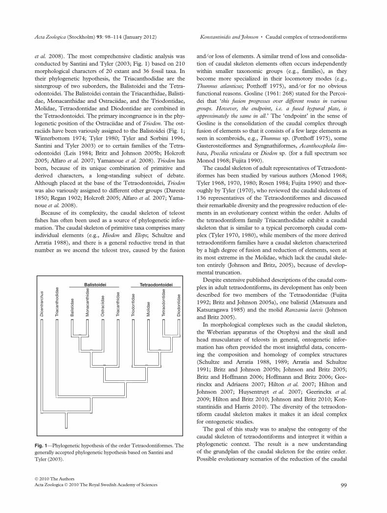
conducted by Santini and Tyler (2003; Fig. 1) based on 210
morphological characters of 20 extant and 36 fossil taxa. In
their phylogenetic hypothesis, the Triacanthodidae are the
sistergroup of two suborders, the Balistoidei and the Tetra-
odontoidei. The Balistoidei contain the Triacanthidae, Balisti-
dae, Monacanthidae and Ostraciidae, and the Triodontidae,
Molidae, Tetraodontidae and Diodontidae are combined in
the Tetraodontoidei. The primary incongruence is in the phy-
logenetic position of the Ostraciidae and of Triodon. The ost-
raciids have been variously assigned to the Balistoidei (Fig. 1;
Winterbottom 1974; Tyler 1980; Tyler and Sorbini 1996,
Santini and Tyler 2003) or to certain families of the Tetra-
odontoidei (Leis 1984; Britz and Johnson 2005b; Holcroft
2005; Alfaro et al. 2007; Yamanoue et al. 2008). Triodon has
been, because of its unique combination of primitive and
derived characters, a long-standing subject of debate.
Although placed at the base of the Tetraodontoidei, Triodon
was also variously assigned to different other groups (Dareste
1850; Regan 1902; Holcroft 2005; Alfaro et al. 2007; Yama-
noue et al. 2008).
Because of its complexity, the caudal skeleton of teleost
fishes has often been used as a source of phylogenetic infor-
mation. The caudal skeleton of primitive taxa comprises many
individual elements (e.g., Hiodon and Elops; Schultze and
Arratia 1988), and there is a general reductive trend in that
number as we ascend the teleost tree, caused by the fusion
and ⁄ or loss of elements. A similar trend of loss and consolida-
tion of caudal skeleton elements often occurs independently
within smaller taxonomic groups (e.g., families), as they
become more specialized in their locomotory modes (e.g.,
Thunnus atlanticus; Potthoff 1975), and ⁄ or for no obvious
functional reasons. Gosline (1961: 268) stated for the Percoi-
dei that ‘this fusion progresses over different routes in various
groups. However, the endpoint, i.e. a fused hypural plate, is
approximately the same in all.’ The ‘endpoint’ in the sense of
Gosline is the consolidation of the caudal complex through
fusion of elements so that it consists of a few large elements as
seen in scombroids, e.g., Thunnus sp. (Potthoff 1975), some
Gasterosteiformes and Syngnathiformes, Acanthocephola lim-
bata, Poecilia reticulata or Diodon sp. (for a full spectrum see
Monod 1968; Fujita 1990).
The caudal skeleton of adult representatives of Tetraodont-
iformes has been studied by various authors (Monod 1968;
Tyler 1968, 1970, 1980; Rosen 1984; Fujita 1990) and thor-
oughly by Tyler (1970), who reviewed the caudal skeletons of
136 representatives of the Tetraodontiformes and discussed
their remarkable diversity and the progressive reduction of ele-
ments in an evolutionary context within the order. Adults of
the tetraodontiform family Triacanthodidae exhibit a caudal
skeleton that is similar to a typical percomorph caudal com-
plex (Tyler 1970, 1980), while members of the more derived
tetraodontiform families have a caudal skeleton characterized
by a high degree of fusion and reduction of elements, seen at
its most extreme in the Molidae, which lack the caudal skele-
ton entirely (Johnson and Britz, 2005), because of develop-
mental truncation.
Despite extensive published descriptions of the caudal com-
plex in adult tetraodontiforms, its development has only been
described for two members of the Tetraodontidae (Fujita
1992; Britz and Johnson 2005a), one balistid (Matsuura and
Katsuragawa 1985) and the molid Ranzania laevis (Johnson
and Britz 2005).
In morphological complexes such as the caudal skeleton,
the Weberian apparatus of the Otophysi and the skull and
head musculature of teleosts in general, ontogenetic infor-
mation has often provided the most insightful data, concern-
ing the composition and homology of complex structures
(Schultze and Arratia 1988, 1989; Arratia and Schultze
1991; Britz and Johnson 2005b; Johnson and Britz 2005;
Britz and Hoffmann 2006; Hoffmann and Britz 2006; Gee-
rinckx and Adriaens 2007; Hilton et al. 2007; Hilton and
Johnson 2007; Huysentruyt et al. 2007; Geerinckx et al.
2009; Hilton and Britz 2010; Johnson and Britz 2010; Kon-
stantinidis and Harris 2010). The diversity of the tetraodon-
tiform caudal skeleton makes it makes it an ideal complex
for ontogenetic studies.
The goal of this study was to analyse the ontogeny of the
caudal skeleton of tetraodontiforms and interpret it within a
phylogenetic context. The result is a new understanding
of the grundplan of the caudal skeleton for the entire order.
Possible evolutionary scenarios of the reduction of the caudal
Dic
entr
arch
us
Tria
cant
hod
idae
Bal
istid
ae
Mon
acan
thid
ae
Ost
raci
idae
Tria
cant
hid
ae
Trio
don
tidae
Mol
idae
Tetr
aod
ontid
ae
Dio
don
tidae
Balistoidei Tetraodontoidei
Fig. 1—Phylogenetic hypothesis of the order Tetraodontiformes. The
generally accepted phylogenetic hypothesis based on Santini and
Tyler (2003).
Acta Zoologica (Stockholm) 93: 98–114 (January 2012) Konstantinidis and Johnson • Caudal complex of tetraodontiforms
� 2010 The Authors
Acta Zoologica � 2010 The Royal Swedish Academy of Sciences 99
fin elements are discussed in the light of the topology of San-
tini and Tyler (2003; Fig. 1).
Material and Methods
Specimens were cleared and double stained (c&s) for bone
and cartilage following Taylor and Van Dyke (1985). For his-
tological transverse sections (10 lm), a specimen of Atroph-
acanthus japonicus (see Material examined) was embedded in
Paraffin and stained by the Azan-Domagk procedure (Romeis
1986).
Photographs of most of the cleared and double-stained
specimens were taken either with a ProgRes C 12 plus digital
camera attached to a Zeiss Tessovar microscope or with a
Zeiss digital camera attached to a Zeiss Discovery V20 dissect-
ing scope. Photographs of the histological sections and the
smaller cleared and double-stained specimens were taken with
a Nikon Coolpix E4500 attached to a Nikon Microscope
Eclipse E600.
For the analysis of the character evolution of the epurals,
uroneurals, parhypural, and the hypural series, a simple
taxa ⁄ character matrix was created and parsimoniously
mapped onto the topology of Santini and Tyler (2003) in
MacClade (Maddison and Maddison 2005).
Institutional abbreviations
AMS, Australian Museum, Sydney; ANSP, Academy of Nat-
ural Science, Philadelphia; BMNH, The Natural History
Museum, London; NSMT, The National Museum of
Science and Nature, Tokyo; SEAMAP, Southeast Area
Monitoring and Assessment Program Ichthyoplankton
Archiving Center, Fish and Wildlife Research Institute;
USNM, National Museum of Natural History, Smithsonian
Institution.
Material examined
Perciformes
Moronidae. Dicentrarchus labrax (Linnaeus), BMNH
2009.3.16.16–24, 28 mm SL, c&s.
Tetraodontiformes
Triacanthodidae. A. japonicus (Kamohara), BMNH
1987.1.23, one specimen, 58 mm SL, c&s; two speci-
mens, uncatalogued (Chiba Institute of Technology),
14.5–18 mm SL, c&s; one specimen, property of the
University of Tuebingen, 15 mm SL, serial sectioned. Hol-
lardia sp. (Poey), uncatalogued, 4.9 mm SL, c&s. Parahol-
lardia sp. (Fraser–Brunner), one specimen, uncatalogued,
3.9 mm notochord length (NL), c&s. Triacanthodes anom-
alus (Temminck & Schlegel), three specimens, ANSP
101257, 54–60 mm SL, c&s. Hollaria hollardi (Poey), one
specimen, USNM 187811, photograph only; Triacanth-
odes ethiops (Alcock), one specimen, USNM 93491,
photograph only.
Triacanthidae. Tripodichthys oxycephalus (Bleeker), two speci-
mens, BMNH 2006.3.280, 16–33 mm SL, c&s. Tripodich-
thys sp. (Tyler), AMS I. 24205–36, 3.9 mm NL, c&s.
Balistidae. Balistapus undulatus (Park), four specimens, uncat-
alogued (NSMT), 3.4 mm NL – 35 mm SL, c&s.
Monacanthidae. Monacanthus ciliatus (Mitchill), one speci-
men, BMNH 1976.6.3, 37 mm SL, c&s. Stephanolepis sp.
(Gill), one specimen, SEAMAP 10741, 5.4 mm SL, c&s;
two specimens, uncatalogued (NSMT), 3.9 mm NL &
14.4 mm SL, c&s.
Ostraciidae. Lactophrys sp. (Swainson), one specimen, SEA-
MAP 25817, 3.5 mm NL, c&s; one specimen, SEAMAP
25776, 4.0 mm SL, c&s; one specimen, SEAMAP 22682,
11.3 mm SL, c&s; one specimen, uncatalogued (SEA-
MAP), 8.0 mm SL, c&s.
Tetraodontidae. Carinotetraodon irrubesco (Tan), uncata-
logued, one specimen, 25 mm SL, c&s; Monotrete suvatii
(Sontirat), uncatalogued, seven specimens, 4.2 mm NL –
16.4 mm SL, c&s. Adult specimens were kept and
spawned in captivity. Larvae were preserved on a daily
basis in 4% formalin and 2 days later transferred into 70%
ethanol.
Diodontidae. Diodon hystrix (Linnaeus), SEAMAP 14506,
5.5 mm SL; SEAMAP 22672, 15 mm SL, c&s.
Figure abbreviations
For the additional cartilages in the caudal skeleton that sup-
port some of the fin rays, the general term distal caudal radial
(adopted from Nybelin 1971) is used.
For the cartilaginous precursor and subsequent ossified ele-
ment, the same abbreviation is used. The abbreviations,
nspu2 and hspu2 apply to both the neural spine and arch and
hemal spine and arch, respectively.
Distal caudal radial dcr
Epural (cartilage) ep
Hemal spine and arch of preural centrum 2 hspu2
Hemal spine and arch of preural centrum 3 hspu3
Hypural (cartilage) hu
Neural spine and arch of preural centrum 2 nspu2
Neural spine and arch of preural centrum 3 nspu3
Parhypural (cartilage) phu
Parhypurapophysis pphu
Preural centrum 2 pu2
Preural centrum 3 pu3
Ural centrum uc
Uroneural un
Terminology of the hypurals
In Teleostei in which the number of hypurals is reduced to
fewer than five, the homology assignment and with that
the terminology of the remaining hypurals can be problematic.
In most previous studies of such taxa (see citations in the
Caudal complex of tetraodontiforms • Konstantinidis and Johnson Acta Zoologica (Stockholm) 93: 98–114 (January 2012)
� 2010 The Authors
100 Acta Zoologica � 2010 The Royal Swedish Academy of Sciences
Discussion), a large hypural plate has been interpreted as a
result of fusion of several hypurals, but it often remains
unclear whether a phylogenetic or ontogenetic fusion has led
to the reduction of hypurals.
Herein, where there is no evidence of ontogenetic fusion,
the terminology (number 1–5) of each hypural plate follows
the hypothesis that hypural elements have been lost rather
than fused to form a compound element. We do this because
it is not possible to test the hypothesis of phylogenetic fusion,
while allowing that such fusion is a possibility (see Discussion
about the homology of the hypurals).
In the text, the term diastema refers to the space that divides
the supports for the upper lobe of the caudal fin from the
lower and is usually located between hypurals 2 and 3 (Fig. 2).
Results – Comparative Ontogeny and Review of the
Literature of the Caudal Complex
Because the quantity and the developmental degree of the
semaphoronts used in this study differ greatly between the
taxa, the results for the Tetraodontiformes are arranged
according to anatomical structures, rather than taxonomically.
The figures, however, are arranged in taxonomic context fol-
lowing the phylogenetic hypothesis of Santini and Tyler
(2003; Fig. 1).
The caudal skeleton of a basal percomorph
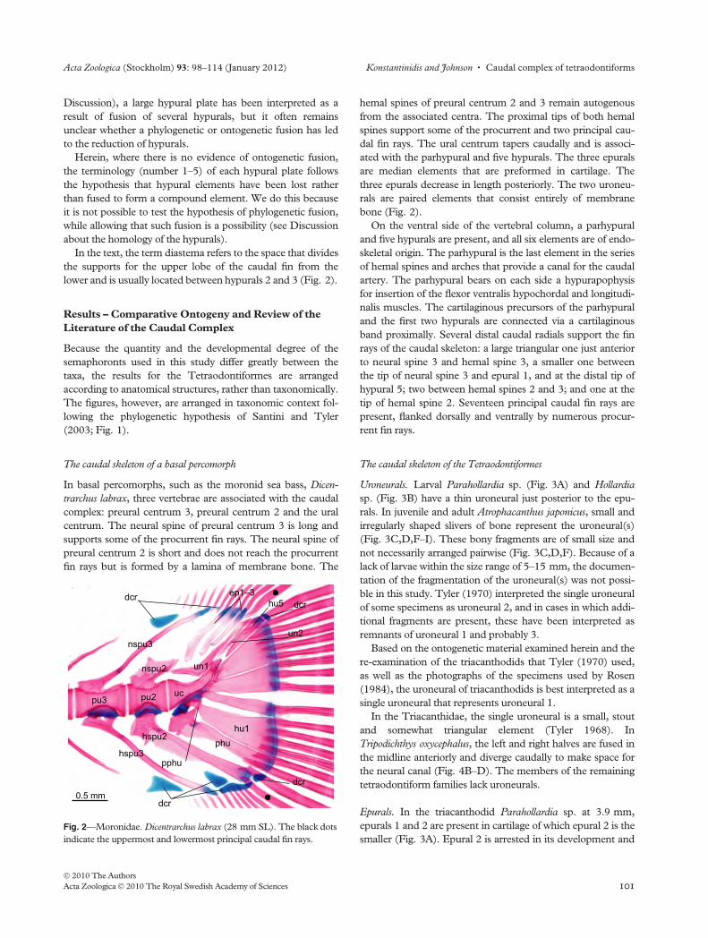
In basal percomorphs, such as the moronid sea bass, Dicen-
trarchus labrax, three vertebrae are associated with the caudal
complex: preural centrum 3, preural centrum 2 and the ural
centrum. The neural spine of preural centrum 3 is long and
supports some of the procurrent fin rays. The neural spine of
preural centrum 2 is short and does not reach the procurrent
fin rays but is formed by a lamina of membrane bone. The
hemal spines of preural centrum 2 and 3 remain autogenous
from the associated centra. The proximal tips of both hemal
spines support some of the procurrent and two principal cau-
dal fin rays. The ural centrum tapers caudally and is associ-
ated with the parhypural and five hypurals. The three epurals
are median elements that are preformed in cartilage. The
three epurals decrease in length posteriorly. The two uroneu-
rals are paired elements that consist entirely of membrane
bone (Fig. 2).
On the ventral side of the vertebral column, a parhypural
and five hypurals are present, and all six elements are of endo-
skeletal origin. The parhypural is the last element in the series
of hemal spines and arches that provide a canal for the caudal
artery. The parhypural bears on each side a hypurapophysis
for insertion of the flexor ventralis hypochordal and longitudi-
nalis muscles. The cartilaginous precursors of the parhypural
and the first two hypurals are connected via a cartilaginous
band proximally. Several distal caudal radials support the fin
rays of the caudal skeleton: a large triangular one just anterior
to neural spine 3 and hemal spine 3, a smaller one between
the tip of neural spine 3 and epural 1, and at the distal tip of
hypural 5; two between hemal spines 2 and 3; and one at the
tip of hemal spine 2. Seventeen principal caudal fin rays are
present, flanked dorsally and ventrally by numerous procur-
rent fin rays.
The caudal skeleton of the Tetraodontiformes
Uroneurals. Larval Parahollardia sp. (Fig. 3A) and Hollardia
sp. (Fig. 3B) have a thin uroneural just posterior to the epu-
rals. In juvenile and adult Atrophacanthus japonicus, small and
irregularly shaped slivers of bone represent the uroneural(s)
(Fig. 3C,D,F–I). These bony fragments are of small size and
not necessarily arranged pairwise (Fig. 3C,D,F). Because of a
lack of larvae within the size range of 5–15 mm, the documen-
tation of the fragmentation of the uroneural(s) was not possi-
ble in this study. Tyler (1970) interpreted the single uroneural
of some specimens as uroneural 2, and in cases in which addi-
tional fragments are present, these have been interpreted as
remnants of uroneural 1 and probably 3.
Based on the ontogenetic material examined herein and the
re-examination of the triacanthodids that Tyler (1970) used,
as well as the photographs of the specimens used by Rosen
(1984), the uroneural of triacanthodids is best interpreted as a
single uroneural that represents uroneural 1.
In the Triacanthidae, the single uroneural is a small, stout
and somewhat triangular element (Tyler 1968). In
Tripodichthys oxycephalus, the left and right halves are fused in
the midline anteriorly and diverge caudally to make space for
the neural canal (Fig. 4B–D). The members of the remaining
tetraodontiform families lack uroneurals.
Epurals. In the triacanthodid Parahollardia sp. at 3.9 mm,
epurals 1 and 2 are present in cartilage of which epural 2 is the
smaller (Fig. 3A). Epural 2 is arrested in its development and
0.5 mm
nspu2
pu2
hspu2
uc
phuhu1
hu5ep1–3
nspu3
dcrdcr
dcr
hspu3
un1
un2
dcr
pu3
pphu
Fig. 2—Moronidae. Dicentrarchus labrax (28 mm SL). The black dots
indicate the uppermost and lowermost principal caudal fin rays.
Acta Zoologica (Stockholm) 93: 98–114 (January 2012) Konstantinidis and Johnson • Caudal complex of tetraodontiforms
� 2010 The Authors
Acta Zoologica � 2010 The Royal Swedish Academy of Sciences 101
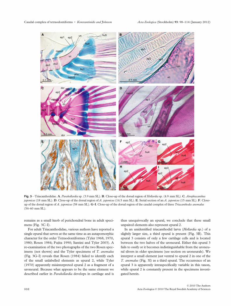
remains as a small knob of perichondral bone in adult speci-
mens (Fig. 3C–I).
For adult Triacanthodidae, various authors have reported a
single epural that serves at the same time as an autapomorphic
character for the order Tetraodontiformes (Tyler 1968, 1970,
1980; Rosen 1984; Fujita 1990; Santini and Tyler 2003). A
re-examination of the two photographs of the two Rosen spec-
imens (not shown) and the Tyler specimens of T. anomalus
(Fig. 3G–I) reveals that Rosen (1984) failed to identify each
of the small unlabelled elements as epural 2, while Tyler
(1970) apparently misinterpreted epural 2 as a fragment of a
uroneural. Because what appears to be the same element we
described earlier in Parahollardia develops in cartilage and is
thus unequivocally an epural, we conclude that these small
unpaired elements also represent epural 2.
In an unidentified triacanthodid larva (Hollardia sp.) of a
slightly larger size, a third epural is present (Fig. 3B). This
epural 3 consists of only a few cartilage cells and is located
between the two halves of the uroneural. Either this epural 3
fails to ossify or it becomes indistinguishable from the uroneu-
ral slivers in older specimens (see section on uroneurals). We
interpret a small element just ventral to epural 2 in one of the
T. anomalus (Fig. 3I) as a third epural. The occurrence of an
epural 3 is apparently intraspecifically variable in this taxon,
while epural 2 is constantly present in the specimens investi-
gated herein.
ep1
ep2
0.1 mm
E
Aep1 ep2
nspu2
pu2
uc
hspu2phu
hu1
hu4
un hu5
0.1 mm
pu3
un
ep1
hu1
hu5
phu
ucpu2
nspu2
hspu2
ep2
dcr
dcr
0.5 mm
C
pu3
ep1
ep2
un
0.1 mm
D
nspu2
B
un
ep3ep2
0.1 mm
ep1
uc
ep2
un
ep2
ep2ep2
ep3
unun
un
1 mm
1 mm
1 mm
1 mm
F G
IH
hu4
hu4
hu4
hu4
dcr
Fig. 3—Triacanthodidae. A. Parahollardia sp. (3.9 mm SL). B. Close-up of the dorsal region of Hollardia sp. (4.9 mm SL). C. Atrophacanthus
japonicus (18 mm SL). D. Close-up of the dorsal region of A. japonicus (14.5 mm SL). E. Serial section of an A. japonicus (15 mm SL). F. Close-
up of the dorsal region of A. japonicus (58 mm SL). G–I. Close-up of the dorsal region of the caudal complex of three Triacanthodes anomalus
(54–60 mm SL).
Caudal complex of tetraodontiforms • Konstantinidis and Johnson Acta Zoologica (Stockholm) 93: 98–114 (January 2012)
� 2010 The Authors
102 Acta Zoologica � 2010 The Royal Swedish Academy of Sciences
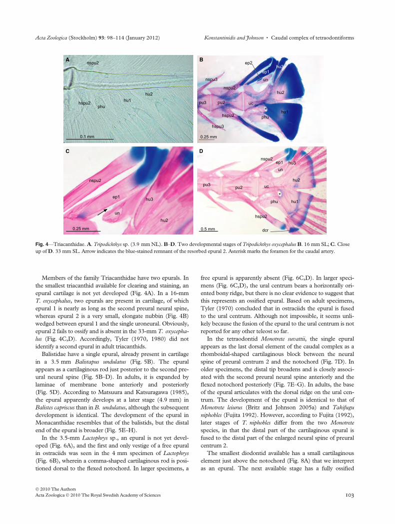
Members of the family Triacanthidae have two epurals. In
the smallest triacanthid available for clearing and staining, an
epural cartilage is not yet developed (Fig. 4A). In a 16-mm
T. oxycephalus, two epurals are present in cartilage, of which
epural 1 is nearly as long as the second preural neural spine,
whereas epural 2 is a very small, elongate nubbin (Fig. 4B)
wedged between epural 1 and the single uroneural. Obviously,
epural 2 fails to ossify and is absent in the 33-mm T. oxycepha-
lus (Fig. 4C,D). Accordingly, Tyler (1970, 1980) did not
identify a second epural in adult triacanthids.
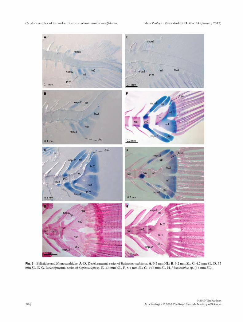
Balistidae have a single epural, already present in cartilage
in a 3.5 mm Balistapus undulatus (Fig. 5B). The epural
appears as a cartilaginous rod just posterior to the second pre-
ural neural spine (Fig. 5B–D). In adults, it is expanded by
laminae of membrane bone anteriorly and posteriorly
(Fig. 5D). According to Matsuura and Katsuragawa (1985),
the epural apparently develops at a later stage (4.9 mm) in
Balistes capriscus than in B. undulatus, although the subsequent
development is identical. The development of the epural in
Monacanthidae resembles that of the balistids, but the distal
end of the epural is broader (Fig. 5E–H).
In the 3.5-mm Lactophrys sp., an epural is not yet devel-
oped (Fig. 6A), and the first and only vestige of a free epural
in ostraciids was seen in the 4 mm specimen of Lactophrys
(Fig. 6B), wherein a comma-shaped cartilaginous rod is posi-
tioned dorsal to the flexed notochord. In larger specimens, a
free epural is apparently absent (Fig. 6C,D). In larger speci-
mens (Fig. 6C,D), the ural centrum bears a horizontally ori-
ented bony ridge, but there is no clear evidence to suggest that
this represents an ossified epural. Based on adult specimens,
Tyler (1970) concluded that in ostraciids the epural is fused
to the ural centrum. Although not impossible, it seems unli-
kely because the fusion of the epural to the ural centrum is not
reported for any other teleost so far.
In the tetraodontid Monotrete suvattii, the single epural
appears as the last dorsal element of the caudal complex as a
rhomboidal-shaped cartilaginous block between the neural
spine of preural centrum 2 and the notochord (Fig. 7D). In
older specimens, the distal tip broadens and is closely associ-
ated with the second preural neural spine anteriorly and the
flexed notochord posteriorly (Fig. 7E–G). In adults, the base
of the epural articulates with the dorsal ridge on the ural cen-
trum. The development of the epural is identical to that of
Monotrete leiurus (Britz and Johnson 2005a) and Takifugu
niphobles (Fujita 1992). However, according to Fujita (1992),
later stages of T. niphobles differ from the two Monotrete
species, in that the distal part of the cartilaginous epural is
fused to the distal part of the enlarged neural spine of preural
centrum 2.
The smallest diodontid available has a small cartilaginous
element just above the notochord (Fig. 8A) that we interpret
as an epural. The next available stage has a fully ossified
A
hu1hu2
phu
nspu2
hspu2
0.1 mm
D
hu2
hu1phu
unep1
nspu2
hspu2
ucpu2
hu3
*
dcr0.5 mm
pu3
C
nspu2
ep1
un
hu3
hu2
0.25 mm
B ep2
ep1hu3
hu2
un
uc
nspu2
0.25 mm
hu1hspu2 phu
pu2
*pu3
nspu3
hspu3
Fig. 4—Triacanthidae. A. Tripodichthys sp. (3.9 mm NL). B–D. Two developmental stages of Tripodichthys oxycephalus B. 16 mm SL; C. Close
up of D. 33 mm SL. Arrow indicates the blue-stained remnant of the resorbed epural 2. Asterisk marks the foramen for the caudal artery.
Acta Zoologica (Stockholm) 93: 98–114 (January 2012) Konstantinidis and Johnson • Caudal complex of tetraodontiforms
� 2010 The Authors
Acta Zoologica � 2010 The Royal Swedish Academy of Sciences 103
0.1 mm
A
hu1hu2
phu
nspu2
hspu2
0.1 mm
E
hu1 hu2
phu
nspu2
hspu2
0.1 mm
B
hu1
hu2
phu
nspu2
hspu2
ep
0.1 mm
C
hu1
hu2
phu
nspu2
hspu2
hu3
uc
pu2
ep
pu3
F
hu1
hu2
phu
nspu2
hspu2
ep
hu3
ucpu2
0.2 mm
pu3
D
phu
nspu2
hspu2
hu3
ucpu2
ep
1 mm
pu3
hu1
hu2
0.5 mm
G
phu
nspu2
hspu2
ucpu2
ep
hu3
pu3
hu2
hu1
H
phu
nspu2
hspu2
ucpu2
ep
1 mm
pu3hu1
hu2
Fig. 5—Balistidae and Monacanthidae. A–D. Developmental series of Balistapus undulatus. A. 3.5 mm NL; B. 3.2 mm SL; C. 4.2 mm SL; D. 35
mm SL. E–G. Developmental series of Stephanolepis sp. E. 3.9 mm NL; F. 5.4 mm SL; G. 14.4 mm SL. H. Monacanthus sp. (37 mm SL).
Caudal complex of tetraodontiforms • Konstantinidis and Johnson Acta Zoologica (Stockholm) 93: 98–114 (January 2012)
� 2010 The Authors
104 Acta Zoologica � 2010 The Royal Swedish Academy of Sciences
hypural plate with a large lamina of membrane bone at its
anterodorsal margin (Fig. 8B), and no real trace of an epural
can be observed. It is not clear whether the epural will be
reduced or incorporated into the caudal complex. Tyler
(1970) assumed that the epural in adult diodontids is fused to
the neural spine of preural centrum 2.
Ural centrum, parhypural and hypurals. As observed by Tyler
(1968, 1970), adult specimens of triacanthodids have an
autogenous parhypural and five individual hypurals, of which
hypural 5 is the smallest. In the 3.9-mm Parahollardia sp., the
parhypural and the five hypurals are already present (Fig. 3A).
The bases of the cartilaginous parhypural, hypural 1 and hyp-
ural 2 are fused to each other, while the cartilaginous precur-
sors of hypurals 3–5 remain separate, even in larger
triacanthodids (Fig. 3A,C). In juvenile and adult A. japonicus,
the ossified parhypural and hypurals 1 and 2 are separate, and
only a remnant of the cartilage remains as evidence of the early
fusion of the three elements (Fig. 3C).
The 3.9 mm Tripodichthys sp. has two cartilaginous hypu-
rals of which hypural 1 is fused with the parhypural proximally
(Fig. 4A). In the triacanthids, the number of hypurals is
reduced to three of which hypural 1 and 2 form a large plate
(Fig. 4D; Tyler 1968, 1970). It is uncertain whether the carti-
laginous hypurals fuse proximally prior to ossification or
remain separate until ossification begins. The diastema at the
posterior margin marks the position where the two hypurals
are fused (Fig. 4D). In the 16-mm T. oxycephalus, the parhyp-
ural is completely fused to the hypural plate which, in turn,
has started to fuse to the ural centrum (Fig. 4B–D). The fora-
men for the caudal artery (Fig. 4B,D) within the lower part of
the hypural plate is the only evidence that a separate parhyp-
ural was present in an earlier stage (Fig. 4D), and Tyler
(1968, 1970) suspected the fusion of the parhypural to the
lower hypural because of the exit of the caudal artery in the
anteroventral part of the hypural plate. An anterior extension
of laminar membrane bone extends the hypural plate antero-
ventrally. A small, third hypural is present just dorsal to the
large hypural plate (Fig. 4B–D).
Two of the three hypurals are already present in a 3.5-mm
B. undulatus (Fig. 5A). At this stage, the parhypural is fore-
shortened and does not enclose the caudal artery. The first
hypural bears a large foramen (Fig. 5A). Before ossification
begins, hypurals 1 and 2 fuse together proximally (Fig. 5B–
D). A small hypural 3 develops after flexion of the notochord
but remains much smaller than the first two hypurals
(Fig. 5C,D). In the 4.2 mm B. undulatus, the cartilaginous
parhypural is connected to hypural 1. In the 4.2-mm speci-
men, the parhypural encloses the caudal artery, but in later
stages the parhypural is again foreshortened (Fig. 5D; Tyler
1970; Matsuura 1979); instead, the ural centrum develops a
ventrally oriented crest of membrane bone that encloses the
caudal artery (Fig. 5D), here referred to as the ‘hemal arch
element’ following the nomenclature introduced by Matsuura
C
A
D
B
ep
hspu2
ucpu2
ucpu2
hspu2
nspu2
0.1 mm 0.1 mm
0.2 mm 0.2 mm
nspu2
hspu2
hu1
**
hu1
hu1 hu1
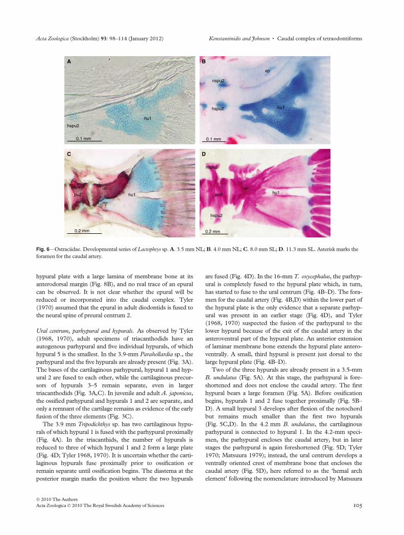
Fig. 6—Ostraciidae. Developmental series of Lactophrys sp. A. 3.5 mm NL; B. 4.0 mm NL; C. 8.0 mm SL; D. 11.3 mm SL. Asterisk marks the
foramen for the caudal artery.
Acta Zoologica (Stockholm) 93: 98–114 (January 2012) Konstantinidis and Johnson • Caudal complex of tetraodontiforms
� 2010 The Authors
Acta Zoologica � 2010 The Royal Swedish Academy of Sciences 105
(1979). The large hypural plate, consisting of two hypurals,
fuses to the ural centrum (Fig. 5D). Matsuura and Katsuraga-
wa (1985) observed four hypural anlagen in larval Balistes
capriscus of which the lower two fuse together, forming the
first hypural of the adults. This is in contrast to our observa-
tion in B. undulatus. However, the foramen we observed in
0.1 mm
nspu2ep
hu2
phuhspu2hu1
D
0.1 mm
hu1
phu
nspu2
hspu2
B
0.1 mm
hu1
phu
nspu2
hspu2
hu2
C
0.1 mm
nspu2
ep
hu2
phuhspu2
hu1
E
0.5 mm
nspu2
hspu2
ucpu2
ep
phu
hu2
hu1
G
pu3
H
uc
hu1
phu
hspu20.5 mm
hu2
pu3
0.2 mm
nspu2
hspu2
ucpu2
ep
phu
hu2
hu1
F
pu3
0.1 mm
hu1
A
phu
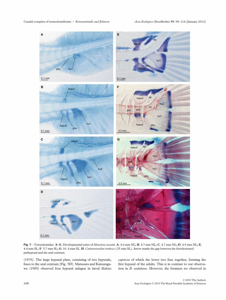
Fig. 7—Tetraodontidae. A–G. Developmental series of Monotrete suvattii. A. 4.6 mm NL; B. 4.7 mm NL; C. 4.7 mm NL; D. 4.9 mm NL; E.
4.4 mm SL; F. 5.7 mm SL; G. 16. 4 mm SL. H. Carinotetraodon irrubesco (25 mm SL). Arrow marks the gap between the foreshortened
parhypural and the ural centrum.
Caudal complex of tetraodontiforms • Konstantinidis and Johnson Acta Zoologica (Stockholm) 93: 98–114 (January 2012)
� 2010 The Authors
106 Acta Zoologica � 2010 The Royal Swedish Academy of Sciences

the smallest B. undulatus (Fig. 5A) might indicate a fusion of
two individual hypurals as described by Matsuura and Katsu-
ragawa (1985) for B. capriscus.
The development of the caudal complex in Stephanolepis
sp. is very similar to that in B. undulatus, but some differences
are notable. The parhypural never encloses the caudal artery
and contact with the cartilaginous hypural 1 is never estab-
lished. Hypural 1 does not bear a foramen at any stage of
ontogeny (Fig. 5E–H). In adult monacanthids, the hemal
arch element is missing or less developed than in the balistids
(Matsuura 1979; Tyler 1980). The caudal skeleton of some
monacanthid genera lacks hypural 3 as, for example, in Mona-
canthus ciliatus (Fig. 5H; Tyler 1970; Matsuura 1979). It is
possible that this difference among the genera might yield
phylogenetic information. As far as is known, the balistids, as
the proposed sistergroup, have a hypural 3, and therefore, the
monacanthid genera that possess hypural 3 probably represent
the plesiomorphic state.
Larvae of Lactophrys sp. smaller than 3.5 mm do not show
any elements of the caudal complex. In the 3.5 mm Lactophrys
sp., the cartilaginous precursor for the hemal arch is present,
and there is an irregular-shaped structure that appears to be
the only element that develops in the region of the parhypural
and hypurals (Fig. 6A). Anteriorly, the hypural plate has an
anterodorsally oriented process (Fig. 6A,B). There is a single
origin of ossification of the hypural plate (Fig. 6C). The ele-
ment becomes larger and foreshadows the shape of a hypural
plate seen in adult specimens (Fig. 6B–D). A foramen present
in the 4 mm specimen (Fig. 6B) is absent in the smaller as
well as in larger specimens. The anterior margin of the carti-
laginous process is connected with the base of the hypural
plate via a lamina of membrane bone, bearing a foramen that
encloses the caudal artery (marked by an asterisk in
Fig. 6C,D). Tyler (1970), based on adult specimens, hypothe-
sized a fusion of the parhypural to the hypural plate, but as
shown here a separate parhypural never develops. In the largest
stage, the hypural plate is fused to the ural centrum (Fig. 6D).
In M. suvattii, the first elements to appear are the parhypur-
al and hypural 1 (Fig. 7A). They are followed by the hemal-
and neural arch of preural centrum 2 in the next larger
specimen (Fig. 7B). At 4.7 mm, the second hypural develops
as a roughly triangular-shaped cartilage (Fig. 7C). In the same
stage, the proximal base of the parhypural, which forms the
hemal canal, curves towards the proximal base of the lower
hypural (Fig. 7C) and fuses with it (Fig. 7D). The ossification
of the hemal canal of the parhypural occurs from both sides
(Fig. 7E,F). A perichondral ossification with its origin at hyp-
ural 1 points ventrad but does not approach the parhypural
ossification (Fig. 7F), leaving a remnant band of cartilage.
The lower hypural fuses to the ural centrum, while the upper
hypural remains separate (Fig. 7F,G).
The development of the parhypural and the hypurals are in
accordance with the developmental sequence of these ele-
ments reported for M. leiurus (Fig. 7; Britz and Johnson
2005a). The first elements that appear are the parhypural and
hypural 1 (Fig. 7A). They are followed by the neural and
hemal spines of preural centrum 2 (Fig. 7B). Hypural 2 is the
last element in the ventral series that develops (Fig. 7C,D). In
contrast to the puffers of the genus Monotrete, Fujita (1992)
noted that in Takifugu niphobles a second hypural appears
before the parhypural. Furthermore, he reported for T. nipho-
bles three separate hypurals, of which the first two fuse to form
a compound element (his ‘hypural 1+2¢).The parhypural of adults of the tetraodontid genus Carino-
tetraodon does not bear a hemal canal; instead, a lamina of
membrane bone projects ventrad and encloses the caudal
artery, similar to the situation in the balistids and monacanth-
ids (Figs 5D,F–H and 7H). The lack of the hemal canal of
the parhypural in Carinotetraodon and the balistid ⁄ monacant-
hid clade is clearly convergent but helps to distinguish puffers
of the genus Carinotetraodon from Monotrete.
In the caudal region of our 5.5 mm Diodon hystrix, a single
element (referred to as the hypural plate) is present and has
already started to ossify from a single ossification centre
anterodorsally (Fig. 8A). The hypural plate does not show
any separation of elements nor a foramen for the caudal
artery. The 15 mm D. hystrix resembles the adult situation
closely, and the ossification of the hypural plate is nearly com-
plete (Fig. 8B). The anteroventral margin of the hypural plate
is extended by a lamina of membrane bone. In front of the
A
ep
nspu2
hspu2
0.1 mm
hu1
Bnspu2
hspu2
ucpu2
0.5 mm
hu1pu3
Fig. 8—Diodontidae. A. and B. Two developmental stages of Diodon hystrix A. 5.5 mm SL; B. 15.0 mm SL.
Acta Zoologica (Stockholm) 93: 98–114 (January 2012) Konstantinidis and Johnson • Caudal complex of tetraodontiforms
� 2010 The Authors
Acta Zoologica � 2010 The Royal Swedish Academy of Sciences 107

hypural plate, an unpaired process projects ventrally and fills
the gap between the hemal spine of preural centrum 2 and the
hypural plate. This ventral outgrowth might become conflu-
ent with the hypural plate because it is not present in larger
specimens. There are no traces of a cartilaginous preformed
parhypural in these two stages. Tyler (1970) noted that in
diodontids the parhypural is either fused to the hypural plate
in Diodon holacanthus or to the hemal spine of preural centrum
2 in Diodon jaculiferus, and Chilomycterus tigrinus. The ontog-
eny of the caudal skeleton of D. hystrix, however, shows no
trace of a parhypural.
Distal caudal radials. Within tetraodontiforms, distal caudal
radials are only present in triacanthodids and triacanthids.
However, distal caudal radials have not been described for
members of these two families so far. Either they were over-
looked because of lack of cartilage staining or they were per-
ceived as the distal tips of the corresponding underlying
elements of the caudal skeleton.
Atrophacanthus japonicus has four distal caudal radials
(Fig. 3C). Ventrally, a large distal caudal radial at the tip of the
hemal spine of preural centrum 2 articulates with the lower-
most caudal fin ray. Dorsally, there are three distal caudal radi-
als, of which two are situated between epural 1 and hypural 5
in the 18 mm specimen and at the distal tip of epural 1 in the
58 mm specimen. In the larger specimen, the distal caudal
radials articulate with the uppermost fin ray (Fig. 3C,F). The
third distal caudal radial is the smallest and is situated on the
tip of hypural 5. The distal caudal radial at the tip of hypural 5
can be homologized with one in D. labrax. The other two in
the dorsal part of the caudal skeleton in A. japonicus are not
present in D. labrax. The large ventral distal caudal radial in
A. japonicus is problematic to homologize with one of the two
ventral distal caudal radial between hemal spine of preural cen-
trum 3 and hemal spine of preural centrum 2 in D. labrax.
In the triacanthid Tripodichthys oxycephalus, the dorsal distal
caudal radials are reduced, and only a single distal caudal
radial is present in the ventral part that articulates with the
lowermost fin ray (Fig. 4D). This distal caudal radial in
T. oxycephalus is homologous with the single ventral one of
A. japonicus.
Neural and hemal spines and arches of preural centrum 2. In the
larval specimen of Parahollardia sp., the cartilaginous neural
and hemal arches of preural centrum 2 are already fully devel-
oped (Fig. 3A). The hemal spine is more massive and slightly
longer than the neural spine. In A. japonicus, the hemal and
neural spines are perichondrally ossified except at their distal
tips. In adult triacanthodids, the neural arch fuses to preural
centrum 2, whereas the hemal arch remains free (Tyler 1970).
The tips of the neural and hemal spines do not reach the fin
rays (Fig. 3C,F).
In the smallest Tripodichthys sp., the cartilaginous precursor
of the hemal spine of preural centrum 2 is already fully grown,
while its associated neural spine has not yet reached its full
length (Fig. 4A). The distal tip of the hemal spine approaches
the most ventral fin ray in triacanthodids (Fig. 4D; Tyler,
1970). In triacanthids, the hemal and neural arches of the first
and second preural centrum are coalesced with their associ-
ated centra (Fig. 4D).
In the smallest Balistapus undulatus, the long hemal spine
on preural centrum 2 articulates with the most anterior fin ray
(Fig. 5A), whereas in adults it loses contact with the most
ventral ray (Fig. 5D; Tyler 1970; Matsuura 1979). Matsuura
and Katsuragawa (1985) described a similar development of
the second preural hemal and neural arches and spines in
B. capriscus. Both spines are extended by laminae of mem-
brane bone in fully developed specimens. The neural arch
fuses to preural centrum 2, whereas the hemal arch does not
(Fig. 5D; Tyler 1970; Matsuura 1979).
In monacanthids, the development of the hemal and neural
arches of preural centrum 2 and their associated spines resem-
ble that described for the balistids (Fig. 5E–H).
The Ostraciidae differ from all other tetraodontiform fami-
lies in having reduced neural and hemal spines on preural cen-
trum 2. The first element to appear in association with preural
centrum 2 is the hemal arch (Fig. 6A). In the larger stage, the
hemal arch has developed (Fig. 6B). In the next larger stage
small, ill-defined neural and hemal spines are present and are
probably not preformed in cartilage (Fig. 6C,D). The hemal
arch remains free from the second preural centrum (Fig. 6D).
According to Klassen (1995), the articulation of the hemal
arch with preural centrum 2 and the length of the hemal spine
have diagnostic potential and can be used for distinguishing
members of the subfamily Aracaninae (long and remains free
from preural centrum 2) and the ostraciine genus Lactophrys
(remains free from preural centrum 2) from all other Ostracii-
nae (short and fused to preural centrum 2).
In the Tetraodontidae, the hemal arch and spine of preural
centrum 2 appear at roughly the same time, although the
neural spine lags a bit behind the hemal spine (Fig. 7B). The
hemal and neural spines of preural centrum 2 become promi-
nent elements of the caudal skeleton. The cartilaginous pre-
cursors of both spines are equal in size until the 4.4 mm
specimen (Fig. 7E). During subsequent development, the
neural spine becomes more massive than the hemal spine
(Fig. 7F,G). The neural arch fuses to the centrum, whereas
the hemal arch remains free. The development of the hemal
and neural arches and spines of preural centrum 2 of Mono-
trete suvattii resembles that of M. leiurus as it was described by
Britz and Johnson (2005a).
In Diodon hystrix, the distal half of the hemal and neural
spine of preural centrum 2 is bent at almost 90� to its base
(Fig. 8B). The neural spine is not expanded as in the tetra-
odontids. In diodontids, the anterior part of the neural arch
and spine is extended by a lamina of membrane bone. Both
the hemal and neural arches fuse to their associated centrum.
Caudal fin rays. The plesiomorphic situation for the perco-
morphs is a complement of 17 fin rays (Fig. 2; Johnson and
Caudal complex of tetraodontiforms • Konstantinidis and Johnson Acta Zoologica (Stockholm) 93: 98–114 (January 2012)
� 2010 The Authors
108 Acta Zoologica � 2010 The Royal Swedish Academy of Sciences
Patterson 1993). Tetraodontiformes have a reduced number
of fin rays. Triacanthodids, triacanthids, balistids, monacanth-
ids and Triodon have 12 caudal fin rays, the highest number
within the order (Figs 3C, 4D and 5D, H; Tyler 1970;
Matsuura 1979), and they are equally distributed over the
upper and lower lobe. Among members of the family Ostracii-
dae, the number of fin rays is variable. Ostraciinae have ten
equally distributed rays (e.g., Lactophrys sp.; Fig. 6D),
whereas members of the Aracaninae have an additional fin ray
associated with the lower lobe (Tyler 1970). Tetraodontids
have consistently 11 caudal fin rays, of which five are associ-
ated with the upper and six with the lower hypural (Fig. 7G)
(Tyler 1970). The nine caudal fin rays in D. hystrix are equally
distributed over the homogenous hypural plate (Fig. 8B). As
far as known, only Chilomycterus reticulatus differs from the
other diodontid species in having ten fin rays (Richards 2006).
Discussion
Before we present our interpretation of the evolution of the
individual structures, we feel it is important to address the
complex issue of the homology of the hypural elements. As
shown in the following paragraphs, there are different inter-
pretations of the evolutionary history of the hypural plates of
the taxa in which fewer than five hypurals are present. The
significance of the full neural spine on preural centrum 2 is
also discussed separately.
Homology of the hypurals
Among the most common reductions in the caudal skeleton
of teleosts is the consolidation of hypurals to one or two
large plates, e.g., myctophids (Fujita 1990), gobiesocids
(Konstantinidis and Conway 2010), scombroids (Potthoff
1975; Fujita 1990), some zeiforms (Tyler et al. 2003), some
labrids (Fujita 1990), gobiids (Fujita 1990) and some carang-
ids (Fujita 1990; Hilton et al. 2010).
The tetraodontiform caudal skeleton shows a wide range of
diversity, from the plesiomorphic condition with five hypurals
to a single large plate in the ostraciids and diodontids (and the
total absence of the caudal skeleton in the molids; Johnson
and Britz 2005). In taxa with a consolidated caudal skeleton
(e.g., to one or two large hypural plates), the identity of the
remaining hypurals is problematic. In this study, the hypurals
are sequentially numbered from the most ventral to the most
dorsal. This is a simple, practical approach and does not auto-
matically imply homology of hypurals among different taxa.
It has been assumed that the consolidated caudal skeleton
evolved either through fusion of individual hypurals into a
compound element or through loss of some of the hypurals.
However, the caudal skeletons of adults cannot be differenti-
ated from each other.
In cases in which an ontogenetic fusion of hypurals has
been documented, e.g., in the blackfin tuna (Potthoff 1975),
the swordfish (Potthoff and Kelley 1982), dolphin fishes
(Potthoff 1980) and some jacks and pompanos (Hilton and
Johnson 2007) the situation is obvious. However, in tetra-
odontiforms, there is no evidence of ontogenetic fusion of
hypurals. Among the taxa of the order with a consolidated
caudal skeleton either an evolutionary fusion of hypurals to a
compound element or a loss of hypurals has to be proposed.
However, a fusion or a loss of hypurals ab initio during evolu-
tion cannot directly be tested, and only indirect aspects such
as topology, shape, relation to other structures, and ⁄ or posi-
tion, etc. (‘principe de connexion’: Geoffroy 1830; ‘Kriterium der
Lage’: Remane 1952; ‘special quality’: Patterson 1982) might
give an indication of the trajectory (either fusion or loss) that
has caused the reduction of hypurals.
Regarding particular cases within the tetraodontiforms,
indirect indicators to assign the homology of hypurals are as
follows:
1 The fusion of the proximal ends of the parhypural to
hypural 1 as well as the cartilaginous connection between
hypural 1 and hypural 2. The early fusion of the cartilagi-
nous hypurals 1 and 2 appears to be highly conserved
among teleosts and can be found throughout teleostean
diversity (Potthoff 1975; Fritzsche and Johnson 1980;
Potthoff 1980; Potthoff et al. 1980; Potthoff and Kelley
1982; Potthoff et al. 1987, 1988; Potthoff and Tellock
1993; Bird and Mabee 2003; Hilton and Johnson 2007)
and is demonstrated herein for the triacanthodids
(Fig. 3A) as well. Fusion at their first appearance has
never been known to occur between hypural 2 and 3 or
any other hypurals.
2 The position of the diastema. In teleosts with a more prim-
itive organization of the caudal fin elements (Figs 1 and 2;
Monod 1968; Fujita 1989), the diastema is always located
between hypurals 2 and 3.
3 The size of the hypurals. In taxa with a reduced number of
hypurals, the remaining hypurals are usually larger, and
that can be interpreted as the result of a fusion of individ-
ual hypurals to a compound element.
The two cartilaginous hypurals in the balistid ⁄ monacanthid
clade (Fig. 5; the triacanthids are uncertain because of an
incomplete ontogenetic series) are connected via a cartilagi-
nous bridge, as is the case for hypural 1 and 2 of many teleo-
sts. This supports the loss of the more dorsally located
hypurals (either hypurals 4–5 in balistids and some mon-
acanthids or 3–5 in all other monacanthids) rather than the
fusion of individual hypurals. However, this evidence is con-
tradicted by the position of the diastema. One has to postulate
a shift of the diastema from the position between hypural 2
and 3 to hypural 1 and 2 and a change in the size of the
remaining hypurals. The situation in the tetraodontids contra-
dicts that found in balistids and monacanthids. In M. suvattii,
the two hypurals are never connected via cartilage at any stage
of development (Fig. 7) and together with the size of the
remaining hypurals support the fusion theory (the ventral
hypural plate of 1 + 2 and the dorsal hypural plate of 3–5).
Acta Zoologica (Stockholm) 93: 98–114 (January 2012) Konstantinidis and Johnson • Caudal complex of tetraodontiforms
� 2010 The Authors
Acta Zoologica � 2010 The Royal Swedish Academy of Sciences 109
Ontogeny as a source to identify homologous structures
fails to be of great use because a fusion ab initio of hypurals
cannot be observed. The problem of the homology of the hyp-
urals in the derived clades of the Tetraodontiformes remains
ambiguous.
A consolidation of the caudal complex is a general theme
within the Teleostei, independent of their phylogenetic rela-
tionships (various citations herein). We believe, based on our
results, that both the phylogenetic fusion and the reduction of
elements are potential programs that have led to an identical
appearance of the caudal skeleton in these teleostean groups.
Therefore, the use of the number of hypurals to reveal homol-
ogous hypurals across taxa is suspicious.
Character evolution of the caudal skeleton
The results of this study are discussed in reference to the phy-
logenetic hypothesis for tetraodontiforms proposed by Santini
and Tyler (2003). Santini and Tyler’s study is based on 210
morphological characters exemplified by 36 fossil and 20
extant taxa. This is, by far, the most comprehensive phyloge-
netic hypothesis published on this group.
Dic
entr
arch
us
Tria
cant
hod
idae
Bal
istid
ae
Mon
acan
thid
ae
Ost
raci
idae
Tria
cant
hid
ae
Trio
don
tidae
Mol
idae
Tetr
aod
ontid
ae
Dio
don
tidae
321
Epurals
A Balistoidei Tetraodontoidei B
Dic
entr
arch
us
Tria
cant
hod
idae
Bal
istid
ae
Mon
acan
thid
ae
Ost
raci
idae
Tria
cant
hid
ae
Trio
don
tidae
Mol
idae
Tetr
aod
ontid
ae
Dio
don
tidae
21Absent
Uroneurals
Balistoidei Tetraodontoidei
Dic
entr
arch
us
Tria
cant
hod
idae
Bal
istid
ae
Mon
acan
thid
ae
Ost
raci
idae
Tria
cant
hid
ae
Trio
don
tidae
Mol
idae
Tetr
aod
ontid
ae
Dio
don
tidae
PresentAbsent
Parhypural
C Balistoidei Tetraodontoidei D
Dic
entr
arch
us
Tria
cant
hod
idae
Bal
istid
ae
Mon
acan
thid
ae
Ost
raci
idae
Tria
cant
hid
ae
Trio
don
tidae
Mol
idae
Tetr
aod
ontid
ae
Dio
don
tidae
5
32
Equivocal
Hypurals
1
4
Balistoidei Tetraodontoidei
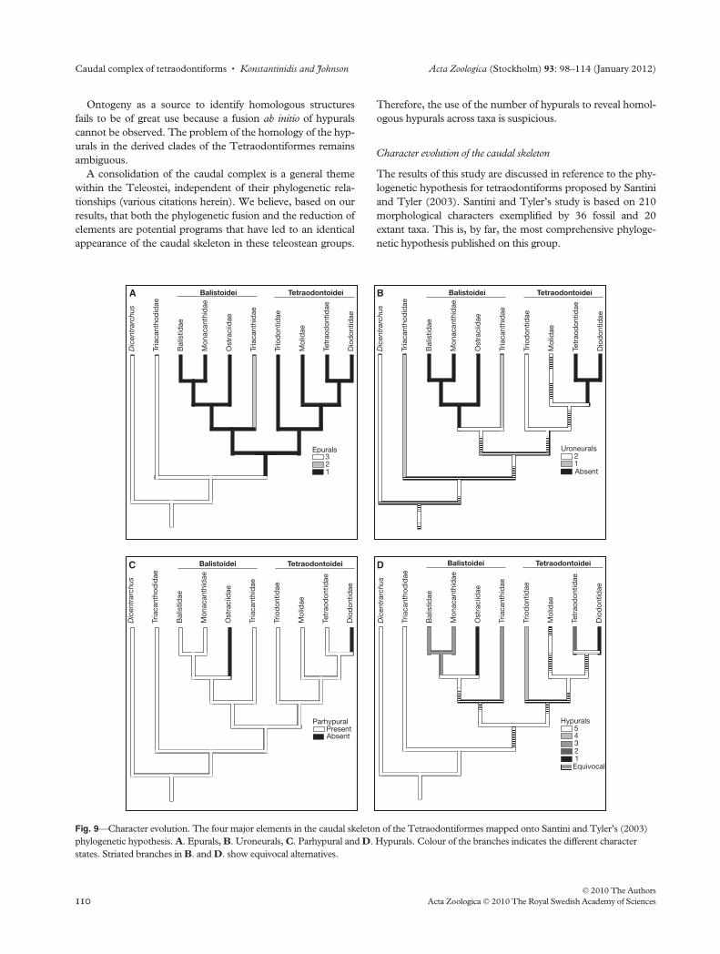
Fig. 9—Character evolution. The four major elements in the caudal skeleton of the Tetraodontiformes mapped onto Santini and Tyler’s (2003)
phylogenetic hypothesis. A. Epurals, B. Uroneurals, C. Parhypural and D. Hypurals. Colour of the branches indicates the different character
states. Striated branches in B. and D. show equivocal alternatives.
Caudal complex of tetraodontiforms • Konstantinidis and Johnson Acta Zoologica (Stockholm) 93: 98–114 (January 2012)
� 2010 The Authors
110 Acta Zoologica � 2010 The Royal Swedish Academy of Sciences
The evolution of the caudal skeleton is reconstructed, and
the character states for the epurals, uroneurals, the parhypural
and the hypurals are mapped parsimoniously at nodes onto
Santini and Tyler’s phylogenetic hypothesis (the elongated
neural spine of preural centrum 2 is discussed but not mapped
onto the phylogenetic tree). The characters (number of epu-
rals, number of uroneurals, presence or absence of a parhyp-
ural and number of hypurals) are treated as independent
evolutionary events and therefore mapped separately. The
absence of the caudal complex in the Molidae is most likely
not the result of a subsequent loss of individual elements,
rather of a single event. Herein, we treated the situation in
Ranzania as not applicable in the analyses of the character
evolution. For a detailed anatomical analysis of the ontogeny
of the clavus, see Johnson and Britz (2005).
Extended neural spine of preural centrum 2. A long neural spine
on preural centrum 2 characterizes all tetraodontiforms but
the ostraciids. A small neural spine on preural centrum 2 is
primitive for teleosts and resembles the plesiomorphic situa-
tion for derived clades, such as the Acanthomorpha and Perc-
omorpha as well (Patterson 1968; Rosen and Patterson
1969).
Although there is no consensus concerning the relationship
among the tetraodontiform families (Winterbottom 1974;
Tyler 1980; Leis 1984; Rosen 1984; Santini and Tyler 2003;
Holcroft 2004; Alfaro et al. 2007; Yamanoue et al. 2008), it is
unlikely that the ostraciids represent the most basal taxon.
The small preural neural spine on preural centrum 2 of the
ostraciids is therefore secondary, and the long neural spine of
preural centrum 2 represents the plesiomorphic character
state.
Among other characters, Rosen (1984) and Tyler et al.
(2003) designated a fully developed neural spine of preural
centrum 2, among other characters, as a synapomorphy for
the Tetraodontiformes and Zeiformes. However, even though
the sistergroup relationship of the tetraodontiforms is ambigu-
ous (Wiley and Johnson 2010), the fully developed second
preural neural spine is treated here as an autapomorphy of the
tetraodontiforms.
Epurals. The most parsimonious reconstruction of the evolu-
tion of the epurals requires only two steps (characters are
unordered) to describe the evolution of the epurals. Three
epurals are present in the triacanthodids and the outgroup.
The first step implies a reduction of two epurals, i.e. from
three epurals directly to one on the branch leading to the com-
mon ancestor of the Balistoidei ⁄ Tetraodontoidei. The third
step involves a reversal of a rudimentary epural 2 in the branch
leading to the triacanthids that is reduced in the adult again.
Although one step longer, the reconstruction with characters
ordered (e.g., from three epurals to two epurals, from two to
one and one to absence) provides an alternative explanation:
the triacanthids, with two epurals, represent the subsequent
step of the reduction of the epurals and an independent loss of
epural 2 occurs in the Balistoidei above the triacanthids and in
the Tetraodontoidei. Although less parsimonious, it seems
more plausible that two epurals are present at the base of the
Balistoidei, and the reduction to a single epural appears within
the Balistoidei (ostraciids, balistids and monacanthids) and
convergently at the base of the Tetraodontoidei (Fig. 9A).
Uroneurals. The most parsimonious reconstruction requires
four steps and produces 21 equally parsimonious possibilities
to explain the evolution of the uroneurals. According to Tyler
(1970, 1980), Triodon bears two uroneurals, which represents
the most plesiomorphic state within the order and resembles
the situation of the outgroup. One uroneural is present in the
triacanthodids and triacanthids, whereas all other taxa lack a
uroneural. The distribution of the uroneurals makes it difficult
to interpret; according to the character optimization, Triodon
either retains a second uroneural while the triacanthodids
have independently lost a second uroneural or, which is unli-
kely, Triodon gains independently a second uroneural
(Fig. 9B).
Parhypural. The most parsimonious reconstruction requires
two steps and produces a single parsimonious option to
express the evolution of the parhypural. The parhypural is
reduced two times independently (excluding the Molidae) in
the Ostraciidae and Diodontidae (Fig. 9C).
Hypurals. The most parsimonious reconstruction requires
five steps and produces 32 equally parsimonious explanations
for the evolution of the hypurals. Five hypurals is the primitive
condition of the Tetraodontiformes. It is most likely that at
the base of the Balistoidei, the hypurals are reduced to three
(this is the present condition in triacanthids, balistids and
some monacanthids) and then further reduced to a single
large element in the Ostraciidae. The Tetraodontoidei show a
wide variety of hypural reduction as well. According to Tyler
(1970, 1980), Triodon, as the basal member of the Tetraodon-
toidei, has four hypurals. The subsequent step requires the
loss of two hypurals and leads to the situation of the Tetra-
odontidae with two large hypural plates. The reduction to one
hypural plate in the Diodontidae appears convergently with
the Ostraciidae (Fig. 9D).
Conclusions
Ontogenetic information reveals that the caudal skeleton of
the Triacanthodidae is more plesiomorphic than previously
reported. It is actually more comparable to a generalized per-
comorph, such as the common sea bass, Dicentrarchus labrax.
Nonetheless, the tetraodontiform caudal skeleton bauplan is
derived in several features, namely the long neural spine of
preural centrum 2 (vs. short), a single uroneural (vs. two) and
the lack of procurrent caudal fin rays.
Comparative morphological and molecular studies have
failed to fully resolve the interrelationships of the families and
Acta Zoologica (Stockholm) 93: 98–114 (January 2012) Konstantinidis and Johnson • Caudal complex of tetraodontiforms
� 2010 The Authors
Acta Zoologica � 2010 The Royal Swedish Academy of Sciences 111
conclusively identify the sistergroup of the Tetraodontiformes.
Despite the fact that ontogenetic information cannot discrimi-
nate between phylogenetic fusion and loss of bony elements,
we have demonstrated here that it is a critical source of infor-
mation for the elucidation of homology in the composition of
complex structures, such as the caudal skeleton. An ontoge-
netic approach is fundamental for and often the only morpho-
logical pathway towards new insight into and solution to
longstanding systematic problems, such as those presented by
the complex evolutionary history of the Tetraodontiformes,
one of the most comprehensively studied groups of acanth-
omorph fishes.
Acknowledgements
The authors are grateful to Carola Zimmermann, Sebastien
Lavoue, Lukas Ruber, Oliver Crimmen, James Maclaine and
Jim Tyler for their helpful advice. We are also very grateful to
Timo Moritz, Eric Hilton and Jim Tyler for their constructive
comments. For the supply of larval and adult material, the
authors are also indebted to Karsten Hartel from the MCZ,
Keiichi Matsuura from the NSMT, Mark McGrouther and
Jeff Leis from the AMS, and Mark Sabaj from the ANSP. To
include, histological sections were only possible because of
Monika Meinert from the University of Tuebingen. We are
thankful to Ralf Britz, NHM, who initiated the project, and
helped to obtain necessary stages of M. suvattii. PK is also
very grateful to Prof Wolfgang Maier from the University of
Tuebingen and to Prof Phil Rainbow from the Natural
History Museum in London. The project is part of a Ph.D.
thesis, which is funded by the Department of Zoology of the
Natural History Museum, London.
References
Alfaro, M. E., Santini, F. and Brock, C. D. 2007. Do reefs drive
diversification in marine teleosts? Evidence from the pufferfish
and their allies (Order Tetraodontiformes). – Evolution 61:
2104–2126.
Arratia, G. and Schultze, H. P. 1991. Palatoquadrate and its ossifica-
tions: development and homology within osteichthyans. – Journal of
Morphology 208: 1–81.
Bird, N. C. and Mabee, P. M. 2003. Developmental morphology of
the axial skeleton of the zebrafish, Danio rerio (Ostariophysi: Cyp-
rinidae). – Developmental Dynamics 228: 337–357.
Britz, R. and Hoffmann, M. 2006. Ontogeny and homology of the
claustra in otophysan Ostariophysi (Teleostei). – Journal of Morphol-
ogy 267: 909–923.
Britz, R. and Johnson, G. D. 2005a. Leis’ conundrum: homology of
the clavus of the ocean sunfishes. 1. Ontogeny of the median fins
and axial skeleton of Monotrete leiurus (Teleostei, Tetraodontifor-
mes, Tetraodontidae). – Journal of Morphology 266: 1–10.
Britz, R. and Johnson, G. D. 2005b. Occipito-vertebral fusion in
ocean sunfishes (Teleostei: Tetraodontiformes: Molidae) and its
phylogenetic implications. – Journal of Morphology 266: 74–79.
Dareste, C. 1850. Recherches sur la classification des poissons de l’or-
dre des Plectognathes. – Annales des Sciences Naturelles. B. Zoologie
3: 105–133.
Fritzsche, R. A. and Johnson, G. D. 1980. Early osteological develop-
ment of white perch and striped bass with emphasis on identifica-
tion of their larvae. – Transaction of the American Fisheries Society
109: 387–406.
Fujita, K. 1989. Nomenclature of cartilaginous elements in the caudal
skeleton of teleostean fishes. – Japanese Journal of Ichthyology 36:
22–29.
Fujita, K.. 1990. The Caudal Skeleton of Teleostean Fishes. Tokai
University Press, Tokyo.
Fujita, K. 1992 Development of the caudal skeleton in the tetraodon-
tid fish, Takifugu niphobles. – Japanese Journal of Ichthyology 38:
438–440.
Geerinckx, T. and Adriaens, D. 2007. Ontogeny of the intermandibu-
lar and hyoid musculature in the suckermouth armoured catfish
Ancistrus cf. triradiatus (Loricariidae, Siluriformes). – Animal Biology
57: 339–357.
Geerinckx, T., Huysentruyt, F. and Adriaens, D. 2009. Ontogeny of
the jaw and maxillary barbel musculature in the armoured catfish
families Loricariidae and Callichthyidae (Loricarioidea, Silurifor-
mes), with a discussion on muscle homologies. – Zoological Journal
of the Linnean Society 155: 76–96.
Geoffroy, S.-H. E. 1830. Principes de Philosophie Zoologique. Pitcher
and Didier, Rosseau Paris.
Gosline, W. A. 1961. The perciform caudal skeleton. – Copeia 1961:
265–270.
Hilton, E. J. and Britz, R. 2010. The caudal skeleton of osteo-
glossomorph fishes, revisited: comparisons, homologies, and char-
acter. In: Nelson, J. S., Schultze, H. P. and Wilson, M. V. H.
(Eds): Mesozoic Fishes 4 – Homology and Phylogeny, pp. 219–237.
Dr. Friedrich Pfeil, Munchen.
Hilton, E. J. and Johnson, G. D. 2007. When two equals three: devel-
opmental osteology and homology of the caudal skeleton in caran-
gid fishes (Perciformes : Carangidae). – Evolution & Development 9:
178–189.
Hilton, E. J., Britz, R., Johnson, G. D. and Forey, P. L. 2007.
Clarification of the occipito-vertebral region of Arapaima gigas
(Osteoglossomorpha: Osteoglossidae) through developmental oste-
ology. – Copeia 2007: 218–224.
Hilton, E. J., Johnson, G. D. and Smith-Vaniz, W. F. 2010. Osteology
and systematics of Parastromateus niger (Perciformes: Carangidae),
with comments on the carangid dorsal Gill-Arch skeleton. – Copeia
2: 312–333.
Hoffmann, M. and Britz, R. 2006. Ontogeny and homology of the
neural complex of otophysan Ostariophysi. – Zoological Journal of
the Linnean Society 147: 301–330.
Holcroft, N. I. 2004. A molecular test of alternative hypotheses of tet-
raodontiform (Acanthomorpha: Tetraodontiformes) sister group
relationships using data from the RAG1 gene. – Molecular Phyloge-
netics and Evolution 32: 749–760.
Holcroft, N. I. 2005. A molecular analysis of the interrelationships of
tetraodontiform fishes (Acanthomorpha: Tetraodontiformes). –
Molecular Phylogenetics and Evolution 34: 525–544.
Huysentruyt, F., Geerinckx, T. and Adriaens, D. 2007. A descriptive
myology of Corydoras aeneus (Gill, 1858) (Siluriformes: Callichthyi-
dae), with a brief discussion on adductor mandibulae homologies. –
Animal Biology 57: 433–452.
Johnson, G. D. and Britz, R. 2005. Leis’ conundrum: homology of
the clavus of the ocean sunfishes. 2. Ontogeny of the median fins
and axial skeleton of Ranzania laevis (Teleostei, Tetraodontiformes,
Molidae). – Journal of Morphology 266: 11–21.
Johnson, G. D. and Britz, R. 2010. Occipito-vertebral fusion in actin-
opterygians: conjecture, myth and reality. Part 2: Teleosts. In: Nel-
son, J. S., Schultze, H. P. and Wilson, M. V. H. (Eds): Mesozoic
Caudal complex of tetraodontiforms • Konstantinidis and Johnson Acta Zoologica (Stockholm) 93: 98–114 (January 2012)
� 2010 The Authors
112 Acta Zoologica � 2010 The Royal Swedish Academy of Sciences
Fishes 4 – Homology and Phylogeny, pp. 95–110. Dr. Friedrich Pfeil,
Munchen.
Johnson, G. D. and Patterson, C. 1993. Percomorph phylogeny – a
survey of acanthomorphs and a new proposal. – Bulletin of Marine
Science 52: 554–626.
Klassen, G. J. 1995. Phylogeny and biogeography of the Ostraciinae
(Tetraodontiformes, Ostraciidae). – Bulletin of Marine Science 57:
393–441.
Konstantinidis, P. and Conway, K. W. 2010. The median-fin skel-
eton of the Eastern Atlantic and Mediterranean Clingfishes Lep-
adogaster lepadogaster (Bonnaterre) and Gouania wildenowi (Risso)
(Teleostei: Gobiesocidae). – Journal of Morphology 271: 215–
224.
Konstantinidis, P. and Harris, M. P. 2010. Same but different: ontog-
eny and evolution of the musculus adductor mandibulae in the Tet-
raodontiformes. – Journal of Experimental Zoology Part B. in press.
Leis, J. M. 1984. Tetraodontiformes: Relationships. In: Moser, H.
G., Richards, W. J., Cohen, D. M., Fahay, M. P., Kendall, A. W. J.
and Richardson, S. L. (Eds): Ontogeny and Systematics of Fishes, pp.
459–463. Allen Press, Lawrence, KS.
Leis, J. M., Olney, J. E. and Okiyama, M. 1997. Proceedings of the
symposium fish larvae and systematics: ontogeny and relationships.
The International Larval Fish Conference, June 16–30, 1995 held
in Sydney, Australia, at the 19th Annual Meeting of the Early Life
History Section of the American Fisheries Society. – Bulletin of Mar-
ine Science 60: 1–212.
Maddison, D. R. and Maddison, W. P. 2005. MacClade 4: Analysis of
Phylogeny and Character Evolution, Version 4.08. Sinauer, Sunder-
land, MA.
Matsuura, K. 1979. Phylogeny of the superfamily balistoidea pisces
Tetraodontiformes. – Memoirs of the Faculty of Fisheries Hokkaido
University 26: 49–170.
Matsuura, Y. and Katsuragawa, M. 1985. Osteological development
of fins and their supports of larval grey triggerfish, Balistes capriscus.
– Japanese Journal of Ichthyology 31: 411–421.
Monod, T. 1968. Le complex urophore des poissons ‘teleosteens’. –
Memoires Institut France Afrique Noire 81: 1–705.
Nelson, J. S. 2006. Fishes of the World, 4th edn, pp. 1–601. Wiley &
Sons, New York.
Nybelin, O. 1971. On the caudal skeleton in Elops with remarks on
other teleostean fishes. – Acta Regiae Societatis Scientiarum et Littera-
rum Gothoburgensis. Zoologica 7: 1–78.
Patterson, C. 1968. The caudal skeleton in Lower Liassic pholido-
phorid fishes. – Bulletin of the British Museum (Natural History) Geol-
ogy 16: 201–239.
Patterson, C. 1982. Morphological Characters and Homology. In:
Joysey, K. A. and Friday, A. E. (Eds): Problems of the Phylogenetic
Reconstruction, pp. 21–74. Academic Press, London.
Potthoff, T. 1975. Development and structure of caudal complex,
vertebral column, and Pterygiophores in Blackfin Tuna (Thunnus
atlanticus, Pisces, Scombridae). – Bulletin of Marine Science 25:
205–231.
Potthoff, T. 1980. Development and structure of fins and fin supports
in dolphin fishes Coryphaena hippurus and Coryphaena equiselis (Co-
ryphaenidae). – Fishery Bulletin 78: 277–312.
Potthoff, T. and Kelley, S. 1982. Development of the vertebral col-
umn, fins and fin supports, branchiostegal rays, and squamation in
the Swordfish, Xiphias gladius. – Fishery Bulletin 80: 161–186.
Potthoff, T. and Tellock, J. A. 1993. Osteological development of the
snook, Centropomus undecimalis (Teleostei, Centropomidae). – Bul-
letin of Marine Science 52: 669–716.
Potthoff, T., Richards, J. W. and Ueyanagi, S. 1980. Development of
Scombrolabrax heterolepis (Pisces, Scombrolabracidae) and
comments on familial relationships. – Bulletin of Marine Science 30:
329–357.
Potthoff, T., Kelley, S., Saksena, V., Moe, M. and Young, F. 1987.
Description of larval and juvenile yellowtail damselfish, Microspath-
odon chrysurus, Pomacentridae, and their osteological development.
– Bulletin of Marine Science 40: 330–375.
Potthoff, T., Kelley, S. and Collins, L. A. 1988. Osteological develop-
ment of the Red Snapper, Lutjanus campechanus (Lutjanidae). –
Bulletin of Marine Science 43: 1–40.
Regan, C. T. 1902. On the classification of the fishes of the suborder
Plectognathi; with notes and description of new species from speci-
mens in the British Museum collection. – Proceedings of the Zoologi-
cal Society of London 2: 284–305.
Remane, A. 1952. Die Grundlagen des natuerlichen Systems, der verglei-
chenden Anatomie und Phylogenetik. Geest & Port, Leipzig.
Richards, W. J. 2006. Early stages of Atlantic fishes: an identification
guide for the western central North Atlantic. In: Richards, W. J.
(Ed.): Early Stages of Atlantic Fishes: an Identification Guide for the
Western Central North Atlantic, 2, pp. i–vi, 1337–2640. CRC Tyler
and Francis, Boca Raton.
Romeis, B. 1986. Mikroskopische Technik. Urban & Schwarzenberg.
Muenchen.
Rosen, D. 1984. Zeiformes as primitive plectognath fishes. – American
Museum Novitates 2782: 1–45.
Rosen, D. E. and Patterson, C. 1969. The structure and relationship
of the paracanthopterygian fishes. – Bulletin of the American Museum
of Natural History 141: 361–469.
Santini, F. and Tyler, J. C. 2003. A phylogeny of the families of fossil
and extant tetraodontiform fishes (Acanthomorpha, Tetraodonti-
formes), Upper Cretaceous to recent. – Zoological Journal of the Lin-
nean Society 139: 565–617.
Schultze, H. P. and Arratia, G. 1988. Reevaluation of the caudal skel-
eton of some actinopterygian fishes .2. Hiodon, Elops, and Albula.
– Journal of Morphology 195: 257–303.
Schultze, H. P. and Arratia, G. 1989. The composition of the caudal
skeleton of Teleosts (Actinopterygii, Osteichthyes). – Zoological
Journal of the Linnean Society 97: 189–231.
Taylor, W. R. and Van Dyke, C. C. 1985. Revised procedures for
staining and clearing small fishes and other vertebrates for bone
and cartilage study. – Cybium 9: 107–120.
Tyler, J. C. 1968. A monograph on plectognath fishes of the super-
family Triacanthoidea. – Monographs of the Academy of Natural Sci-
ences of Philadelphia 16: 1–364.
Tyler, J. C. 1970. The progressive reduction in number of elements
supporting the caudal fin of fishes of the order Plectognathi. –
Proceedings of the Academy of Natural Sciences of Philadelphia 122:
1–85.
Tyler, J. C. 1980. Osteology, phylogeny, and higher classification of
the fishes of the order Plectognathi (Tetraodontiformes). – NOAA
Technical Report NMFS Circular 434: 1–422.
Tyler, J. C. and Sorbini, L. 1996. New superfamily and three new
families of tetraodontiform fishes from the Upper Cretaceous: the
earliest and most morphologically primitive plectognaths. – Smithso-
nian Contributions to Paleobiology 82: i–iii, 1–59.
Tyler, J. C. and Holcroft, N. 2007. Tetraodontiformes - triggerfishes,
boxfishes, puffers (fugu), molas and allies. DOI: http://tolweb.org/
Tetraodontiformes/52153/2007.02.19.
Tyler, J. C., O’Toole, B. and Winterbottom, R. 2003. Phylogeny of
the genera and families of zeiform fishes, with comments on their
relationships with tetraodontiform and caproids. – Smithsonian
Contributions to Zoology 618: 1–110.
Wiley, E. O. and Johnson, G. D. 2010. A teleost classification based
on monophyletic groups. In: Nelson, J. S., Schultze, H. P. and
Acta Zoologica (Stockholm) 93: 98–114 (January 2012) Konstantinidis and Johnson • Caudal complex of tetraodontiforms
� 2010 The Authors
Acta Zoologica � 2010 The Royal Swedish Academy of Sciences 113
Wilson, M. V. H. (Eds): Mesozoic Fishes 4 – Homology and Phylog-
eny, pp. 123–182. Dr. Friedrich Pfeil, Munchen.
Winterbottom, R. 1974. The familial phylogeny of the Tetraodonti-
formes (Acanthopterygii: Pisces) as evidenced by their comparative
myology. – Smithsonian Contributions to Zoology 155: 1–201.
Yamanoue, Y., Miya, M., Matsuura, K., Katoh, M., Sakai, H. and
Nishida, M. 2008. A new perspective on phylogeny and evolution
of tetraodontiform fishes (Pisces: Acanthopterygii) based on whole
mitochondrial genome sequences: basal ecological diversification? –
BMC Evolutionary Biology 8: 1–14.
Caudal complex of tetraodontiforms • Konstantinidis and Johnson Acta Zoologica (Stockholm) 93: 98–114 (January 2012)
� 2010 The Authors
114 Acta Zoologica � 2010 The Royal Swedish Academy of Sciences