Embed Size (px)
Citation preview
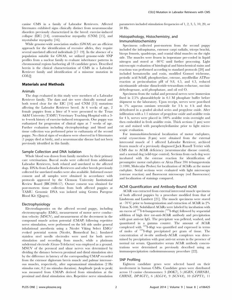
A COLQ Missense Mutation in Labrador RetrieversHaving Congenital Myasthenic SyndromeCaitlin J. Rinz1, Jonathan Levine2, Katie M. Minor3, Hammon D. Humphries4, Renee Lara5,
Alison N. Starr-Moss1, Ling T. Guo4, D. Colette Williams6, G. Diane Shelton4*, Leigh Anne Clark1*
1 Department of Genetics and Biochemistry, College of Agriculture, Forestry, and Life Sciences, Clemson University, Clemson, South Carolina, United States of America,
2 Department of Small Animal Clinical Sciences, College of Veterinary Medicine and Biomedical Sciences, Texas A&M University, College Station, Texas, United States of
America, 3 Department of Veterinary and Biomedical Sciences, College of Veterinary Medicine, University of Minnesota, St. Paul, Minnesota, United States of America,
4 Department of Pathology, School of Medicine, University of California San Diego, La Jolla, California, United States of America, 5 Kingdom Animal Hospital, Bryan, Texas,
United States of America, 6 R. Prichard Veterinary Medical Teaching Hospital, University of California Davis, Davis, California, United States of America
Abstract
Congenital myasthenic syndromes (CMSs) are heterogeneous neuromuscular disorders characterized by skeletal muscleweakness caused by disruption of signal transmission across the neuromuscular junction (NMJ). CMSs are rarelyencountered in veterinary medicine, and causative mutations have only been identified in Old Danish Pointing Dogs andBrahman cattle to date. Herein, we characterize a novel CMS in 2 Labrador Retriever littermates with an early onset ofmarked generalized muscle weakness. Because the sire and dam share 2 recent common ancestors, CMS is likely the resultof recessive alleles inherited identical by descent (IBD). Genome-wide SNP profiles generated from the Illumina HD array for9 nuclear family members were used to determine genomic inheritance patterns in chromosomal regions encompassing 18functional candidate genes. SNP haplotypes spanning 3 genes were consistent with autosomal recessive transmission, andmicrosatellite data showed that only the segment encompassing COLQ was inherited IBD. COLQ encodes the collagenoustail of acetylcholinesterase, the enzyme responsible for termination of signal transduction in the NMJ. Sequences from COLQrevealed a variant in exon 14 (c.1010T.C) that results in the substitution of a conserved amino acid (I337T) within the C-terminal domain. Both affected puppies were homozygous for this variant, and 16 relatives were heterozygous, while 288unrelated Labrador Retrievers and 112 dogs of other breeds were wild-type. A recent study in which 2 human CMS patientswere found to be homozygous for an identical COLQ mutation (c.1010T.C; I337T) provides further evidence that thismutation is pathogenic. This report describes the first COLQ mutation in canine CMS and demonstrates the utility of SNPprofiles from nuclear family members for the identification of private mutations.
Citation: Rinz CJ, Levine J, Minor KM, Humphries HD, Lara R, et al. (2014) A COLQ Missense Mutation in Labrador Retrievers Having Congenital MyasthenicSyndrome. PLoS ONE 9(8): e106425. doi:10.1371/journal.pone.0106425
Editor: Claire Wade, University of Sydney, Australia
Received May 23, 2014; Accepted July 29, 2014; Published August 28, 2014
Copyright: � 2014 Rinz et al. This is an open-access article distributed under the terms of the Creative Commons Attribution License, which permits unrestricteduse, distribution, and reproduction in any medium, provided the original author and source are credited.
Data Availability: The authors confirm that all data underlying the findings are fully available without restriction. All relevant data are within the paper and itsSupporting Information files.
Funding: Research reported in this publication was supported by the National Institute of Arthritis and Musculoskeletal and Skin Diseases of the NationalInstitutes of Health under Award Number R15AR062868 (LAC) and the Canine Health Foundation under Award Number 01311 (ANSM). The content is solely theresponsibility of the authors and does not necessarily represent the official views of the National Institutes of Health or Canine Health Foundation. The fundershad no role in study design, data collection and analysis, decision to publish, or preparation of the manuscript.
Competing Interests: The authors have declared that no competing interests exist.
* Email: [email protected] (GDS); [email protected] (LAC)
Introduction
Skeletal muscle contraction is stimulated by the emission of the
neurotransmitter acetylcholine (ACh) by the motor neuron, and
terminated by acetylcholinesterase (AChE) in the neuromuscular
junction (NMJ). Disruption of signal transmission within the NMJ
resulting from presynaptic, synaptic, or post-synaptic defects
causes congenital myasthenic syndromes (CMSs), heterogeneous
neuromuscular disorders characterized by skeletal muscle weak-
ness and fatigue. Mutations causing CMSs in humans have been
identified in 18 genes to date, with a majority of cases attributed to
CHRNE, COLQ, RAPSN, and DOK7 [1]. Mutations are
predominantly autosomal recessive, and often act in compound
heterozygosity [2–5].
Naturally-occurring CMSs are rarely described in veterinary
medicine; when they do occur, they are usually in animals between
6 to 12 weeks of age, appear to be familial, and are characterized
by severe generalized skeletal muscle weakness. The first report of
canine CMS was in the Jack Russell Terrier in 1974 [6]. Since that
time, acetylcholine receptor (AChR) deficiency has been con-
firmed in Jack Russell Terriers [7], as well as Smooth Fox Terriers
[8]. CMS has also been clinically described in families of Springer
Spaniels [9], Miniature Smooth-Haired Dachshunds [10], and
Old Danish Pointing Dogs [11]. Characterization of CMS at the
molecular level has only been achieved in Old Danish Pointing
Dogs (missense mutation in CHAT) [12], and in young Brahman
cows (deletion in CHRNE) [13].
Diagnosis of CMS is challenging and relies on clinical
evaluation, morphological studies of muscle and peripheral nerves,
electrodiagnostic studies, the absence of serum antibodies against
muscle AChRs, demonstration of AChR deficiency, and most
recently, molecular genetic studies. We have identified a novel
PLOS ONE | www.plosone.org 1 August 2014 | Volume 9 | Issue 8 | e106425
canine CMS in a family of Labrador Retrievers. Affected
littermates exhibited signs clinically distinct from neuromuscular
disorders previously characterized in the breed: exercise-induced
collapse (EIC) [14], centronuclear myopathy (CNM) [15], and
myotubular myopathy [16].
While genome-wide association studies (GWAS) are an efficient
approach for the identification of recessive alleles, they require
several unrelated affected individuals [17–19]. In the absence of a
population suitable for GWAS, we utilized genome-wide SNP
profiles from a nuclear family to evaluate inheritance patterns in
chromosomal regions harboring all 18 candidate genes. Described
herein is the clinical characterization of CMS in a Labrador
Retriever family and identification of a missense mutation in
COLQ.
Materials and Methods
AnimalsThe dogs evaluated in this study were members of a Labrador
Retriever family. The dam and sire were clinically normal and
both tested clear for the EIC [14] and CNM [15] mutations
affecting the Labrador Retriever breed. At 6 weeks of age, 2
female puppies from a litter of 9 were presented to the Texas
A&M University (TAMU) Veterinary Teaching Hospital with a 3-
to 4-week history of exercise-induced tetraparesis. One puppy was
euthanized for progression of clinical signs at 7 weeks of age.
Further evaluation including electrophysiology and blood and
tissue collection was performed prior to euthanasia of the second
puppy. No clinical signs of weakness were observed in 6 littermates
(1 puppy died at birth), and a neuromuscular disease had not been
previously identified in this family.
Sample Collection and DNA IsolationWhole blood was drawn from family members by their primary
care veterinarians. Buccal swabs were collected from additional
Labrador Retrievers, both related and unrelated to the affected
dogs. DNAs from Labrador Retrievers and other breeds previously
collected for unrelated studies were also available. Informed owner
consent and all samples were obtained in accordance with
protocols approved by the Clemson University Institutional
Review Board (IBC2008-17). Owner consent was obtained for
post-mortem tissue collection from both affected puppies at
TAMU. Genomic DNA was isolated using Gentra Puregene
Blood Kit (Qiagen).
ElectrophysiologyElectrodiagnostics on the affected second puppy, including
electromyography (EMG), measurement of motor nerve conduc-
tion velocity (MNCV), and measurement of the decrement in the
compound muscle action potential (CMAP) following repetitive
nerve stimulation, were performed on the left side under general
inhalational anesthesia using a Nicolet Viking Select EMG/
evoked potential system (Nicolet, Biomedical Inc.). Insulated
stainless steel needle electrodes were used for both nerve
stimulation and recording from muscle, while a platinum
subdermal electrode (Grass-Telefactor) was employed as a ground.
MNCV of the peroneal and ulnar nerves was determined by
dividing the distance between proximal and distal stimulation sites
by the difference in latency of the corresponding CMAP recorded
from the extensor digitorum brevis muscle and palmar interosse-
ous muscles, respectively, after supramaximal stimulation (2 Hz
stimulus rate, 0.2 ms stimulus duration). Amplitude (peak to peak)
was measured from CMAPs derived from stimulation at the
proximal and distal stimulation sites. Repetitive nerve stimulation
parameters included stimulation frequencies of 1, 2, 3, 5, 10, 20, or
50 Hz.
Histopathology, Histochemistry, andImmunohistochemistry
Specimens collected post-mortem from the second puppy
included the infraspinatus, extensor carpi radialis, triceps brachii,
biceps femoris, quadriceps, and cranial tibial muscles on the right
side. The muscles were frozen in isopentane pre-cooled in liquid
nitrogen and stored at –80uC until further processing. Light
microscopic evaluation of histological and histochemical stains and
reactions was performed according to standard protocols [20] and
included hematoxylin and eosin, modified Gomori trichrome,
periodic acid Schiff, phosphorylase, esterase, myofibrillar ATPase
reaction at preincubation pH of 9.8, 4.5, and 4.3, reduced
nicotinamide adenine dinucleotide-tetrazolium reductase, succinic
dehydrogenase, acid phosphatase, and oil red O.
Specimens from the radial and peroneal nerves were immersion
fixed in 2.5% glutaraldehyde in 0.1 M phosphate buffer before
shipment to the laboratory. Upon receipt, nerves were post-fixed
in 1% aqueous osmium tetroxide for 3 h to 4 h and then
dehydrated in a graded alcohol series and propylene oxide. After
infiltration with a 1:1 mixture of propylene oxide and araldite resin
for 4 h, nerves were placed in 100% araldite resin overnight and
then embedded in fresh araldite resin. Thick sections (1 mm) were
cut and stained with paraphenylediamine prior to light micro-
scopic evaluation.
For immunohistochemical localization of motor end-plates,
serial cryosections (8 mm) were obtained from the external
intercostal muscle of 1 affected Labrador Retriever, archived
frozen muscle of a previously diagnosed Jack Russell Terrier with
CMS due to AChR deficiency (neuromuscular disease control),
and a normal dog (wild-type control). Sections from each dog were
incubated with the esterase reaction for identification of
presumptive motor end-plates or Alexa Fluor 594 a-bungarotoxin
(1:1000, Molecular Probes) for localization of AChRs at the motor
end-plate. Serial sections were evaluated with light microscopy
(esterase reaction) and fluorescent microscopy (red fluorescence)
and localization of stainings compared.
AChR Quantification and Antibody-Bound AChRAChR was extracted from external intercostal muscle specimens
of both affected puppies by a procedure modified from that of
Lindstrom and Lambert [21]. The muscle specimens were stored
at –70uC prior to homogenization and extraction of AChR in 2%
Triton X-100. Solubilized AChRs were labeled by incubation with
an excess of 125I-a-bungarotoxin (125I-abgt) followed by sequential
addition of high titer rat-anti-AChR antibody and precipitation
with goat anti-rat IgG. The precipitate was pelleted, washed, and
quantitated in a gamma counter. The amount of AChR
complexed with 125I-abgt was quantified and expressed in terms
of moles of 125I-abgt precipitated per gram of tissue. The
concentration of in-situ antibody-AChR complexes was deter-
mined by precipitation with goat anti-rat serum in the presence of
normal rat serum. Quantitative serum AChR antibody concen-
trations were determined as previously described using an
immunoprecipitation radioimmunoassay procedure [22].
SNP ProfilingEighteen candidate genes were selected based on their
involvement in human CMSs. Candidate genes were distributed
across 13 canine chromosomes: 3 (DOK7), 5 (AGRN, CHRNB1,
CHRNE, DPAGT1), 6 (ALG14), 9 (SCN4A), 10 (GFPT1), 11
COLQ Mutation in Labrador Retrievers with CMS
PLOS ONE | www.plosone.org 2 August 2014 | Volume 9 | Issue 8 | e106425
(ALG2, MUSK), 13 (PLEC1), 18 (RAPSN), 20 (LAMB2), 23
(COLQ), 25 (CHRNG, CHRND), 28 (CHAT), and 36 (CHRNA1)
[1]. The Illumina CanineHD Infinium BeadChip was used to
profile 173,662 SNPs representing all chromosomes for 9 nuclear
family members: 7 littermates and both parents (Figure 1). Arrays
were processed according to manufacturer’s protocols. Allele
frequencies were calculated using a case/control analysis con-
ducted with PLINK [23]. Chromosomal regions harboring
candidate genes were evaluated for SNP haplotypes with
frequencies of 1.0 in the CMS cases and between 0.14 and 0.50
in the controls. Individual genotypes in regions fitting these
parameters were then manually examined to ensure that no
unaffected littermates were homozygous for the allele present in
the affected dogs.
Microsatellite AnalysisPCR for amplification of individual microsatellite markers was
performed using materials and thermal cycling parameters
previously described [24,25]. Products were resolved with
GeneScan 600 LIZ size standard (Applied Biosystems) on an
ABI 3730XL DNA Analyzer (Applied Biosystems). GeneMapper
Software version 4.0 (Applied Biosystems) was used to visualize the
microsatellite signals.
Sanger SequencingPrimers were designed to amplify all coding regions and splice
sites of CHRNG, CHRND, and COLQ. Products were amplified
using ReddyMix Master Mix (Thermo Scientific) with 0.4 mM of
each primer and 50 ng of DNA. Products were purified using the
Qiax II Gel Extraction Kit (Qiagen) or an ExoSAP master mix
consisting of 0.5 units of Exonuclease I (New England BioLabs)
and 0.25 units of SAP (Promega). Primer sequences and
purification methods for each amplicon are given in Table S1.
Sequencing products were resolved on an ABI 3730XL DNA
Analyzer (Applied Biosystems).
Restriction Enzyme DigestDetection of the COLQ variant was accomplished using the
endonuclease BtsI, which recognizes the following sequence and
cutsite (‘): 59 …NN‘CACTGC…39. The digestion was performed
using BtsI CutSmart (New England BioLabs) with 2.5 mL 10X
buffer, 0.40 mL BtsI, 1 mg DNA, and water adjusted accordingly
for a total reaction volume of 25 mL. Products were resolved on a
2% agarose gel.
Results
Clinical DescriptionNeurological examination was consistent with a generalized
neuromuscular disease with marked short-strided tetraparesis that
worsened with exercise. Postural reactions were preserved with the
exception of hopping which was diminished in all limbs when the
puppies were made to bear full weight. Spinal reflexes including
the patellar, cranial tibial, and flexor withdrawals were reduced in
all limbs. A pyridostigmine bromide challenge resulted in
worsening of muscle weakness.
ElectrophysiologyEMG was performed on the left side of the body and included
the supraspinatus, infraspinatus, triceps, biceps, extensor carpi
radialis, superficial and deep digital flexor, interosseous, biceps
femoris, rectus femoris, cranial tibial, and gastrocnemius muscles.
Abnormal EMG activity was not identified in any muscle group.
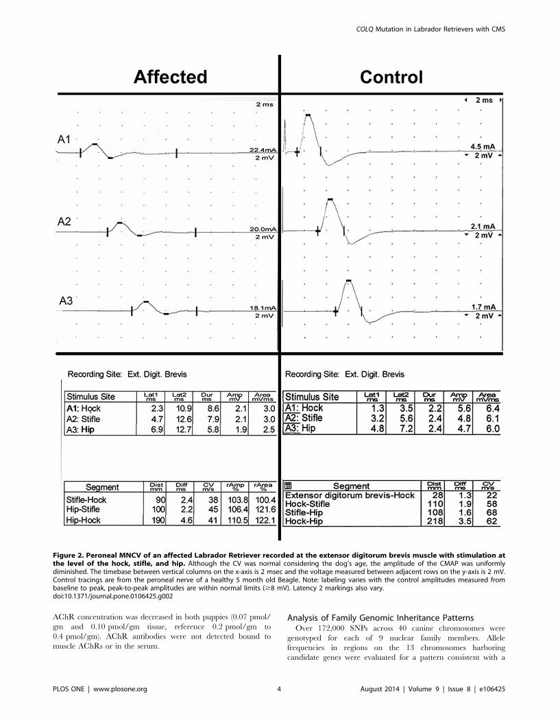
Peroneal and ulnar MNCV was likely age appropriate (41 m/sec
and 31 m/sec, respectively; reported values for normal dogs 1 to 6
months of age 32 m/sec to 55 m/sec and 25 m/sec to 48 m/sec)
[26,27]. The peroneal and ulnar nerve CMAP amplitudes were
reduced at all stimulus sites (1.9 mV to 2.1 mV and 2.6 mV to
2.9 mV, respectively, reference $8 mV, peak-to-peak) (Figure 2)
[27]. F-waves were not recordable upon stimulation of either the
peroneal or ulnar nerve. Repetitive stimulation of the peroneal
nerve revealed a decrement in the CMAP amplitude of 22% at
2 Hz, 33% at 5 Hz, and 35% at 50 Hz when the first and third
CMAPs were compared (Figure 3). Decrements greater than 10%
are considered significant.
Histopathology, Histochemistry, andImmunohistochemistry
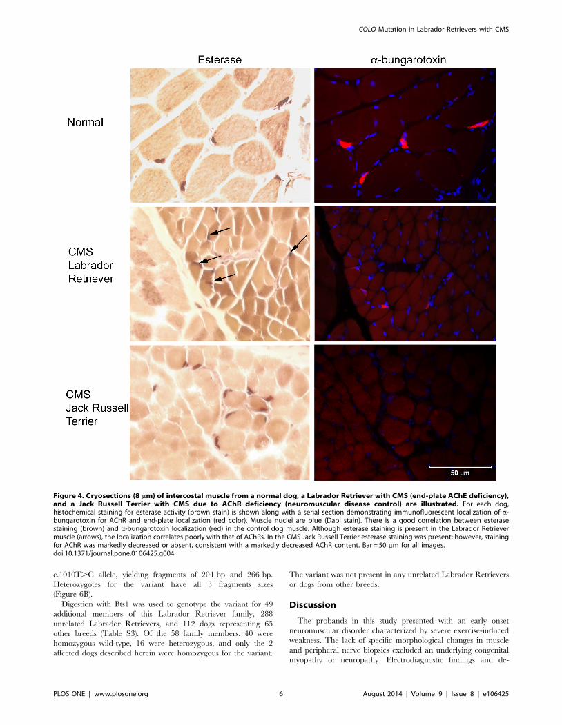
Myofiber size was appropriate in all muscles evaluated and no
specific abnormalities were identified, making a congenital
myopathy unlikely. Multifocal areas of esterase reactivity were
identified, but it could not be determined from this reaction if
staining correlated with localization of motor end-plates. In the
Labrador Retriever with CMS, esterase staining was evident in
multiple locations but did not fully correlate with AChR labeling
(Figure 4). There was a good correlation with esterase staining and
AChR labeling in the normal dog. In the Jack Russell Terrier with
CMS (neuromuscular disease control), several end-plates were
stained with esterase; however, no AChRs were labeled in the
serial muscle section, consistent with the marked decrease in
muscle AChRs described in this breed. There was no evidence of
axonal degeneration, demyelination, or abnormalities of support-
ing structures in the peripheral nerve evaluations, excluding a
congenital polyneuropathy.
AChR Quantification and Antibody-Bound AChRThe concentration of AChR was determined from external
intercostal muscle samples collected origin to insertion following
euthanasia of both affected Labrador Retriever puppies. The
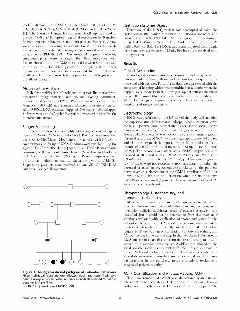
Figure 1. Multigenerational pedigree of Labrador Retrievers.Filled individual icons denote affected dogs and semi-filled iconsdenote obligate carriers. Asterisks mark individuals selected for whole-genome SNP profiling.doi:10.1371/journal.pone.0106425.g001
COLQ Mutation in Labrador Retrievers with CMS
PLOS ONE | www.plosone.org 3 August 2014 | Volume 9 | Issue 8 | e106425
AChR concentration was decreased in both puppies (0.07 pmol/
gm and 0.10 pmol/gm tissue, reference 0.2 pmol/gm to
0.4 pmol/gm). AChR antibodies were not detected bound to
muscle AChRs or in the serum.
Analysis of Family Genomic Inheritance PatternsOver 172,000 SNPs across 40 canine chromosomes were
genotyped for each of 9 nuclear family members. Allele
frequencies in regions on the 13 chromosomes harboring
candidate genes were evaluated for a pattern consistent with a
Figure 2. Peroneal MNCV of an affected Labrador Retriever recorded at the extensor digitorum brevis muscle with stimulation atthe level of the hock, stifle, and hip. Although the CV was normal considering the dog’s age, the amplitude of the CMAP was uniformlydiminished. The timebase between vertical columns on the x-axis is 2 msec and the voltage measured between adjacent rows on the y-axis is 2 mV.Control tracings are from the peroneal nerve of a healthy 5 month old Beagle. Note: labeling varies with the control amplitudes measured frombaseline to peak, peak-to-peak amplitudes are within normal limits ($8 mV). Latency 2 markings also vary.doi:10.1371/journal.pone.0106425.g002
COLQ Mutation in Labrador Retrievers with CMS
PLOS ONE | www.plosone.org 4 August 2014 | Volume 9 | Issue 8 | e106425

recessive trait (Table S2). SNP haplotypes on both chromosomes
23 and 25 were homozygous in the cases and had frequencies of
0.36 and 0.29, respectively, in the unaffected individuals.
Chromosome 23 harbors COLQ, while the region on chromosome
25 includes both CHRNG and CHRND.
Because the sire and dam share 2 recent common ancestors, we
hypothesized that the causative mutation was inherited identical
by descent (IBD). To determine whether the aforementioned
segments of chromosomes 23 and 25 were inherited IBD, we
genotyped polymorphic microsatellite markers in each region.
Genotypes revealed homozygosity in the affected dogs for markers
flanking COLQ, providing evidence that the segment on chromo-
some 23 is IBD (Figure 5). Affected dogs were heterozygous for
haplotypes encompassing CHRNG and CHRND, suggesting that
the chromosome 25 segment was not inherited from a recent
common ancestor.
Sequencing of CHRNG, CHRND, and COLQNo nonsynonymous variants were identified in CHRNG. A
single nonsynonymous variant in exon 4 of CHRND did not
segregate with a recessive phenotype. Sequence data for COLQrevealed 3 nonsynonymous variants, only 1 of which segregated
with a recessive trait. The exon 14 variant, c.1010T.C, predicts
the substitution of isoleucine with threonine at residue 337
(Figure 6A).
Screening of c.1010T.C VariantPCR amplicons from the COLQ exon 14 primer set are 470 bp
in size. BtsI cleaves the amplicon only in individuals having the
Figure 3. Repetitive stimulation of the peroneal motor nerve of an affected Labrador Retriever at 2 Hz (A), 5 Hz (B), and 50 Hz (C).Decrement of the CMAP was observed at all tested frequencies. Sweep speed and sensitivity settings are identical to those in Figure 2. Controltracings are from the peroneal nerve of a healthy 5 month old Beagle with no decrement seen at low frequency stimulation and normalpseudofaciliation (CMAP gets taller and narrower) with tetanic stimulation.doi:10.1371/journal.pone.0106425.g003
COLQ Mutation in Labrador Retrievers with CMS
PLOS ONE | www.plosone.org 5 August 2014 | Volume 9 | Issue 8 | e106425
c.1010T.C allele, yielding fragments of 204 bp and 266 bp.
Heterozygotes for the variant have all 3 fragments sizes
(Figure 6B).
Digestion with Bts1 was used to genotype the variant for 49
additional members of this Labrador Retriever family, 288
unrelated Labrador Retrievers, and 112 dogs representing 65
other breeds (Table S3). Of the 58 family members, 40 were
homozygous wild-type, 16 were heterozygous, and only the 2
affected dogs described herein were homozygous for the variant.
The variant was not present in any unrelated Labrador Retrievers
or dogs from other breeds.
Discussion
The probands in this study presented with an early onset
neuromuscular disorder characterized by severe exercise-induced
weakness. The lack of specific morphological changes in muscle
and peripheral nerve biopsies excluded an underlying congenital
myopathy or neuropathy. Electrodiagnostic findings and de-
Figure 4. Cryosections (8 mm) of intercostal muscle from a normal dog, a Labrador Retriever with CMS (end-plate AChE deficiency),and a Jack Russell Terrier with CMS due to AChR deficiency (neuromuscular disease control) are illustrated. For each dog,histochemical staining for esterase activity (brown stain) is shown along with a serial section demonstrating immunofluorescent localization of a-bungarotoxin for AChR and end-plate localization (red color). Muscle nuclei are blue (Dapi stain). There is a good correlation between esterasestaining (brown) and a-bungarotoxin localization (red) in the control dog muscle. Although esterase staining is present in the Labrador Retrievermuscle (arrows), the localization correlates poorly with that of AChRs. In the CMS Jack Russell Terrier esterase staining was present; however, stainingfor AChR was markedly decreased or absent, consistent with a markedly decreased AChR content. Bar = 50 mm for all images.doi:10.1371/journal.pone.0106425.g004
COLQ Mutation in Labrador Retrievers with CMS
PLOS ONE | www.plosone.org 6 August 2014 | Volume 9 | Issue 8 | e106425
Figure 5. Microsatellite and SNP haplotypes (color-coded bars below individuals) are shown for 3 candidate genes. Positions (in Mb)are according to CanFam 2. Filled individual icons denote affected dogs and semi-filled icons denote obligate carriers. Chromosome 23 haplotypes(blue) are inherited IBD in both affected dogs.doi:10.1371/journal.pone.0106425.g005
Figure 6. (A) Sequence from the 59 end of the C-terminal domain of ColQ in mammals. Identical residues are denoted by an asterisk,conserved substitutions by a colon, and semi-conserved substitutions by a period. Residues altered in human CMS cases are highlighted in yellow[4,30,32,37]. (B) BtsI digest results for the Labrador Retriever family. PCR amplicons from COLQ exon 14 are 470 bp in size and cleaved into 204 and266 bp fragments in the presence of c.1010T.C. Three clinically normal littermates were identified as carriers, denoted by semi-filled icons.doi:10.1371/journal.pone.0106425.g006
COLQ Mutation in Labrador Retrievers with CMS
PLOS ONE | www.plosone.org 7 August 2014 | Volume 9 | Issue 8 | e106425
creased AChR concentration in the muscle indicated a disorder of
neuromuscular transmission. The autoimmune disease myasthenia
gravis was eliminated based on the early age of onset and an
absence of AChR antibodies in serum and AChR-bound
antibodies in the muscle. The clinical diagnosis in the Labrador
Retrievers was CMS.
While clinical signs and electrophysiological findings are
generally similar between presynaptic, synaptic, and postsynaptic
forms of CMS, a notable observation in the affected puppies was a
worsening of the phenotype upon administration of an AChE
inhibitor. This response indicates desensitization of the AChRs
from overexposure to ACh and is consistent with a synaptic form
of CMS referred to as end-plate AChE deficiency (EAD) [5]. EAD
accounts for 10% to 15% of all human cases of CMSs and is
always caused by mutations in COLQ [5]. COLQ encodes a
collagen strand that homotrimerizes to form the tail subunit of
asymmetric AChE. ColQ anchors AChE to the basal lamina
where the enzyme hydrolyzes ACh, thereby limiting the length of
the synaptic response [28]. In the absence of ColQ, ACh
accumulates, causing prolonged muscle contraction and eventually
the desensitization of AChR [29].
Through the examination of SNP allele frequencies in the
Labrador Retriever family, we identified 2 chromosomes harbor-
ing CMS candidate genes that showed an inheritance pattern
consistent with autosomal recessive transmission. Whereas human
forms of CMS are often caused by compound heterozygosity, low
levels of genetic diversity within purebred dog populations make
simple recessive alleles more common. Linebreeding in this
Labrador Retriever family makes it likely that the sire and dam
inherited the mutation from a common ancestor and that the
affected puppies are homozygous for a chromosome segment
transmitted IBD. Analysis of polymorphic microsatellites showed
that the regions flanking COLQ are IBD, whereas those flanking
the other 2 identified candidate genes are not.
Sequencing of COLQ in the Labrador Retrievers revealed a
missense mutation that predicts the replacement of a conserved
hydrophobic isoleucine with a hydrophilic threonine in the C-
terminal domain. ColQ has 3 domains: an N-terminal proline-rich
attachment domain (PRAD), a collagenic central domain, and a
C-terminal domain. The PRAD serves to attach the ColQ strand
to an AChE tetramer. The collagen domain assembles the triple
helix, while the C-terminal domain is involved in both the
formation of the triple helix [30] and anchoring of the structure to
the basal lamina [30,31].
In humans, mutations responsible for EAD have been identified
in each domain of COLQ and have different functional
consequences depending on their location [4]. In the C-terminus,
missense mutations in residues ranging from positions 342 to 452
are thought to inhibit the attachment of ColQ to the basal lamina
of the muscle cell [30–33]. Some C-terminus mutations (e.g.,
V322D) may prevent the formation of the ColQ triple helix [32].
In the affected Labrador Retrievers, localization of the esterase
reaction showed a poor correlation between AChE and AChR.
This finding suggests improper anchoring of ColQ to the basal
lamina, or mislocalization. Insufficient muscle samples prevented
us from conducting a sedimentation profile of AChE to determine
the exact consequence of the I337T mutation identified.
Linebreeding practices expedite the appearance of recessive
diseases in purebred dog populations. The availability of genetic
tests for the detection of carrier dogs allows for selective breeding
to prevent widespread dissemination of the deleterious allele to the
breed while maintaining genetic diversity. Because only 2 affected
littermates were available for study herein, GWAS techniques
could not be applied. The analysis of chromosomal inheritance
patterns indicated a single functional and positional candidate
gene and led to the discovery of the COLQ c.1010T.C mutation;
however, our approach does not exclude the possibility that
another mutation exists in a novel CMS gene.
While this manuscript was in revision, Matlik et al. reported that
an identical mutation (c.1010T.C; I337T) was homozygous in 2
human CMS patients with EAD [34]. The affected children were
first cousins from consanguineous relationships; both sets of
parents were heterozygous for the mutation [34]. The substitution
was the only variation identified in COLQ and was determined to
be pathogenic through a prediction program [34]. Although
uncommon, identical changes at the DNA level between humans
and dogs with similar phenotypes have been previously identified
[35,36]. The identification of c.1010T.C in humans and dogs
diagnosed with CMS strongly supports the causality of the
mutation and shows that conservation of residue 337 is critical
for proper function of ColQ.
Supporting Information
Table S1 Primers (59-39) for amplification of CHRNG,CHRND, and COLQ. Primers were designed to amplify exons
and splice sites. ExoSAP indicates the use of the ExoSAP protocol
and Gel X indicates the use of the gel extraction protocol for post-
PCR clean-up.
(PDF)
Table S2 Candidate regions based on allele frequencies.
Genomic regions known to harbor CMS candidate genes were
screened for case allele frequencies of 1.0 and control allele
frequencies of between 0.14 and 0.50. CHR = chromosome
number; SNP = SNP name; BP = chromosome position; A1 =
allele 1; F_A = frequency of allele 1 in affected dogs (cases);
F_U = frequency of allele 1 in unaffected dogs (controls); A2 =
allele 2.
(PDF)
Table S3 Dogs screened for the COLQ 14 variant. Digestion
with Bts1 was used to genotype the 2 affected dogs, 56 other
members of the Labrador Retriever pedigree, 288 unrelated
Labrador Retrievers, and 112 dogs representing 65 other breeds.
(PDF)
Acknowledgments
The authors would like to thank the dog owners and veterinarians who
contributed DNA samples for this study.
Author Contributions
Conceived and designed the experiments: GDS LAC. Performed the
experiments: CJR JL HDH LTG DCW. Analyzed the data: CJR JL GDS
LAC. Contributed reagents/materials/analysis tools: KMM RL ANSM.
Contributed to the writing of the manuscript: CJR ANSM GDS LAC.
References
1. Hantaı D, Nicole S, Eymard B (2013) Congenital myasthenic syndromes: an
update. Curr Opin Neurol 26: 561–8.
2. Ohno K, Brengman J, Tsujino A, Engel AG (1998) Human endplate
acetylcholinesterase deficiency caused by mutations in the collagen-like tail
COLQ Mutation in Labrador Retrievers with CMS
PLOS ONE | www.plosone.org 8 August 2014 | Volume 9 | Issue 8 | e106425
subunit (ColQ) of the asymmetric enzyme. Proc Natl Acad Sci USA 95: 9654–
9659.3. Ishigaki K, Nicolle D, Krejci E, Leroy J-P, Koenig J, et al. (2003) Two novel
mutations in the COLQ gene cause endplate acetylcholinesterase deficiency.
Neuromuscul Disord 13: 236–244.4. Mihaylova V, Muller JS, Vilchez JJ, Salih MA, Kabiraj MM, et al. (2008)
Clinical and molecular genetic findings in COLQ-mutant congenital myasthenicsyndromes. Brain 131: 747–759.
5. Abicht A, Muller JS, Lochmuller H (2012) Congenital Myasthenic Syndromes.
GeneReviews Available: http://www.ncbi.nlm.nih.gov/books/NBK1168/. Ac-cessed 27 February 2014.
6. Palmer AC, Barker J (1974) Myasthenia in the dog. Vet Rec 95: 452–454.7. Oda K, Lambert EH, Lennon VA, Palmer AC (1984) Congenital canine
myasthenia gravis: I. Deficient junctional acetylcholine receptors. Muscle Nerve7: 705–716.
8. Miller LM, Lennon VA, Lambert EH, Reed SM, Hegreberg GA, et al. (1983)
Congenital myasthenia in 13 smooth fox terriers. J Am Vet Med Assoc 182:694–697.
9. Johnson RP, Watson AD, Smith J, Cooper BJ (1975) Myasthenia in SpringerSpaniel littermates. J Small Anim Pract 16: 641–647.
10. Dickinson PJ, Sturges BK, Shelton GD, LeCouteur RA (2005) Congenital
Myasthenia Gravis in Smooth-Haired Miniature Dachshund Dogs. J Vet InternMed 19: 920–923.
11. Flagstad A, Trojaborg W, Gammeltoft S (1989) Congenital myasthenicsyndrome in the dog breed Gammel Dansk Hønsehund: clinical, electrophys-
iological, pharmacological and immunological comparison with acquiredmyasthenia gravis. Acta Vet Scand 30: 89–102.
12. Proschowsky HF, Flagstad A, Cirera S, Joergensen CB, Fredholm M (2007)
Identification of a Mutation in the CHAT Gene of Old Danish Pointing DogsAffected with Congenital Myasthenic Syndrome. J Hered 98: 539–543.
13. Kraner S, Sieb JP, Thompson PN, Steinlein OK (2002) Congenital myastheniain Brahman calves caused by homozygosity for a CHRNE truncating mutation.
Neurogenetics 4: 87–91.
14. Patterson EE, Minor KM, Tchernatynskaia AV, Taylor SM, Shelton GD, et al.(2008) A canine DNM1 mutation is highly associated with the syndrome of
exercised-induced collapse. Nat Genet 40: 1235–1239.15. Pele M, Tiret L, Kessler J-L, Blot S, Panthier J-J (2005) SINE exonic insertion in
the PTPLA gene leads to multiple splicing defects and segregates with theautosomal recessive centronuclear myopathy in dogs. Hum Mol Genet 14:
1417–1427.
16. Beggs AH, Bohm J, Snead E, Kozlowski M, Maurer M, et al. (2010). MTM1mutation associated with X-linked myotubular myopathy in Labrador retrievers.
Proc Natl Acad Sci U S A 107: 14697–14702.17. Gill JL, Tsai KL, Krey C, Noorai RE, Vanbellinghen J-F, et al. (2012) A canine
BCAN microdeletion associated with episodic falling syndrome. Neurobiol Dis
45: 130–136.18. Safra N, Bassuk AG, Ferguson PJ, Aguilar M, Coulson RL, et al. (2013)
Genome-Wide Association Mapping in Dogs Enables Identification of theHomeobox Gene, NKX2–8, as a Genetic Component of Neural Tube Defects
in Humans. PLoS Genet 9: e1003646.19. Vernau KM, Runstadler JA, Brown EA, Cameron JM, Huson HJ, et al. (2013)
Genome-Wide Association Analysis Identifies a Mutation in the Thiamine
Transporter 2 (SLC19A3) Gene Associated with Alaskan Husky Encephalop-athy. PLoS One 8: e57195.
20. Dubowitz V, Sewry CA (2007). Histological and histochemical stains and
reactions. In: Dubowitz V, Sewry CA, editors. Muscle Biopsy: A Practical
Approach, 3rd ed. London: Saunders Elsevier: pp. 21–39.
21. Lindstrom JM, Lambert EH (1978) Content of acetylcholine receptor and
antibodies bound to receptor in myasthenia gravis, experimental autoimmune
myasthenia gravis, and Eaton-Lambert syndrome. Neurology 28: 130–138.
22. Lindstrom J, Shelton D, Fujii Y (1988) Myasthenia gravis. Adv Immunol 42:
233–284.
23. Purcell S, Neale B, Todd-Brown K, Thomas L, Ferreira MA, et al. (2007)
PLINK: a tool set for whole-genome association and population-based linkage
analyses. Am J Hum Genet 81: 559–575.
24. Cargill EJ, Clark LA, Steiner JM, Murphy KE (2002) Multiplexing of canine
microsatellite markers for whole-genome screens. Genomics 80: 250–3.
25. Clark LA, Tsai KL, Steiner JM, Williams DA, Guerra T, et al. (2004)
Chromosome-specific microsatellite multiplex sets for linkage studies in the
domestic dog. Genomics 84: 550–554.
26. Swallow JS, Griffiths IR (1977) Age related changes in the motor nerve
conduction velocity in dogs. Res Vet Sci 23: 29–32.
27. Sims MH, Redding RW (1980) Maturation of nerve conduction velocity and the
evoked muscle potential in the dog. Am J Vet Res 41: 1247–52.
28. Katz B, Miledi R (1973) The Binding of Acetylcholine to Receptors and its
Removal from the Synaptic Cleft. J Physiol 231: 549–574.
29. Engel AG, Lambert EH, Gomez MR (1977) A New Myasthenic Syndrome with
End-Plate Acetylcholinesterase Deficiency, Small Nerve Terminals, and
Reduced Acetylcholine Release. Ann Neurol 1: 315–330.
30. Ohno K, Engel AG, Brengman JM, Shen X-M, Heidenreich F, et al. (2000) The
Spectrum of Mutations Causing End-Plate Acetylcholinesterase Deficiency. Ann
Neurol 47: 162–170.
31. Kimbell LM, Ohno K, Engel AG, Rotundo RL (2004) C-terminal and Heparin-
binding Domains of Collagenic Tail Subunit Are Both Essential for Anchoring
Acetylcholinesterase at the Synapse. J Biol Chem 279: 10997–11005.
32. Nakata T, Ito M, Azuma Y, Otsuka K, Noguchi Y, et al. (2013) Mutations in the
C-Terminal Domain of ColQ in Endplate Acetylcholinesterase Deficiency
Compromise ColQ–MuSK Interaction. Hum Mutat 00: 1–8.
33. Arredondo J, Lara M, Ng F, Gochez DA, Lee DC, et al. (2014) COOHterminal
collagen Q (COLQ) mutants causing human deficiency of endplate acetylcho-
linesterase impair the interaction of ColQ with proteins of the basal lamina.
Hum Genet 133: 599–616.
34. Matlik HN, Milhem RM, Saadeldin IY, Al-Jaibeji HS, Al-Gazali L, et al. (2014)
Clinical and Molecular Analysis of a Novel COLQ Missense Mutation Causing
Congenital Myasthenic Syndrome in a Syrian Family. Pediatr Neurol 51: 165–
169.
35. Zangerl B, Goldstein O, Philp AR, Lindauer SJ, Pearce-Kelling SE, et al. (2006)
Identical mutation in a novel retinal gene causes progressive rod-cone
degeneration in dogs and retinitis pigmentosa in humans. Genomics 88: 551–
563.
36. Seppala EH, Reuser AJ, Lohi H (2013) A nonsense mutation in the acid a-
glucosidase gene causes Pompe disease in Finnish and Swedish Lapphunds.
PLoS One 8: e56825.
37. Wargon I, Richard P, Kuntzer T, Sternberg D, Nafissi S, et al. (2012) Long-term
follow-up of patients with congenital myasthenic syndrome caused by COLQ
mutations. Neuromuscul Disord 22: 318–324.
COLQ Mutation in Labrador Retrievers with CMS
PLOS ONE | www.plosone.org 9 August 2014 | Volume 9 | Issue 8 | e106425