Embed Size (px)
Citation preview
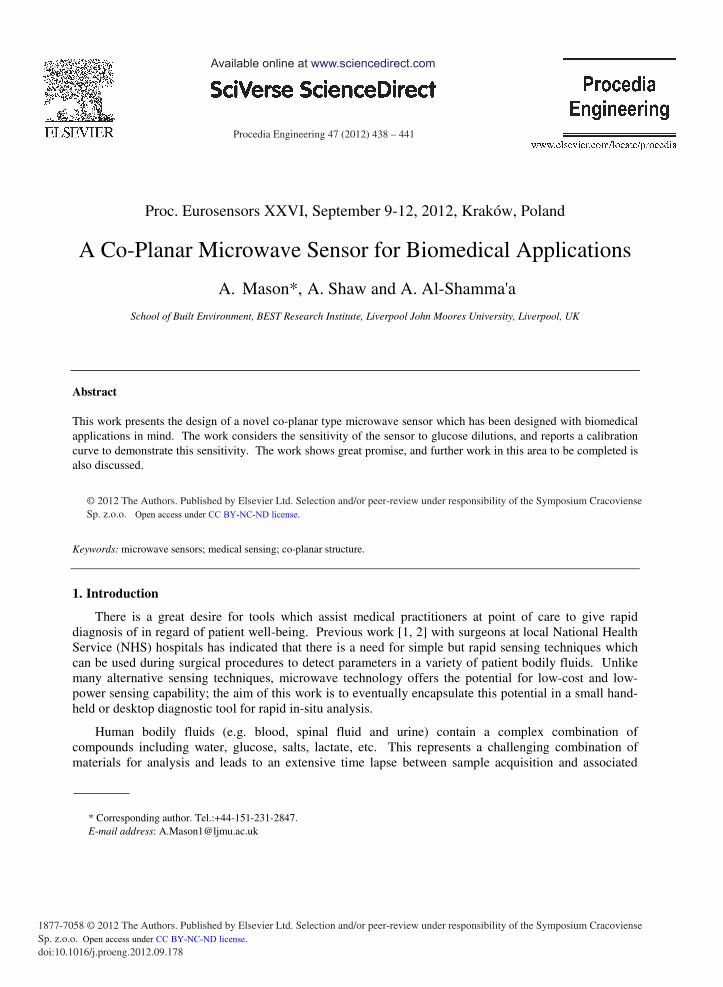
Procedia Engineering 47 ( 2012 ) 438 – 441
1877-7058 © 2012 The Authors. Published by Elsevier Ltd. Selection and/or peer-review under responsibility of the Symposium Cracoviense Sp. z.o.o.doi: 10.1016/j.proeng.2012.09.178
Proc. Eurosensors XXVI, September 9-12, 2012, Kraków, Poland
A Co-Planar Microwave Sensor for Biomedical Applications
A. Mason*,aA. Shaw and A. Al-Shamma'a
School of Built Environment, BEST Research Institute, Liverpool John Moores University, Liverpool, UK
Abstract
This work presents the design of a novel co-planar type microwave sensor which has been designed with biomedical applications in mind. The work considers the sensitivity of the sensor to glucose dilutions, and reports a calibration curve to demonstrate this sensitivity. The work shows great promise, and further work in this area to be completed is also discussed.
© 2012 Published by Elsevier Ltd.
Keywords: microwave sensors; medical sensing; co-planar structure.
1. Introduction
There is a great desire for tools which assist medical practitioners at point of care to give rapid diagnosis of in regard of patient well-being. Previous work [1, 2] with surgeons at local National Health Service (NHS) hospitals has indicated that there is a need for simple but rapid sensing techniques which can be used during surgical procedures to detect parameters in a variety of patient bodily fluids. Unlike many alternative sensing techniques, microwave technology offers the potential for low-cost and low-power sensing capability; the aim of this work is to eventually encapsulate this potential in a small hand-held or desktop diagnostic tool for rapid in-situ analysis.
Human bodily fluids (e.g. blood, spinal fluid and urine) contain a complex combination of compounds including water, glucose, salts, lactate, etc. This represents a challenging combination of materials for analysis and leads to an extensive time lapse between sample acquisition and associated
* Corresponding author. Tel.:+44-151-231-2847. E-mail address: [email protected]
Available online at www.sciencedirect.com
© 2012 The Authors. Published by Elsevier Ltd. Selection and/or peer-review under responsibility of the Symposium Cracoviense Sp. z.o.o. Open access under CC BY-NC-ND license.
Open access under CC BY-NC-ND license.
439 A. Mason et al. / Procedia Engineering 47 ( 2012 ) 438 – 441
diagnosis. Optical techniques [1-3] are commonly employed for biomedical applications to assist when time is a critical factor. However these methods can be bulky and expensive to implement and it is often there is still the requirement for an experienced operator to take time to consider the meaning of results obtained. Obviously this is inappropriate during a surgical procedure as time is often critical from the perspectives of patient wellbeing and hospital efficiency.
Microwave sensing is a developing technology which has shown vast potential in a number of industrial and medical areas [4-6]. This is a result of the technique being robust, requiring low power and having good depth of penetration in respect of analyte materials. Also, of particular interest in medical application is the non-ionising nature of microwave radiation which practitioners see as a significant benefit over existing technologies, namely x-ray imaging. As a continuation of previous work [7, 8] this paper introduces a novel co-planar sensor design which can be placed in-line with a system designed to draw bodily fluid. This paper briefly reports on the design of the sensor and discusses a calibration curve determined through experimental measurement.
2. Co-Planar Sensor
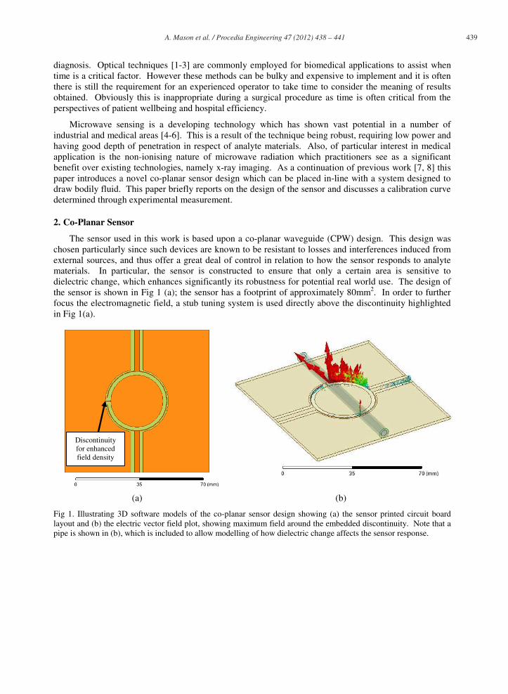
The sensor used in this work is based upon a co-planar waveguide (CPW) design. This design was chosen particularly since such devices are known to be resistant to losses and interferences induced from external sources, and thus offer a great deal of control in relation to how the sensor responds to analyte materials. In particular, the sensor is constructed to ensure that only a certain area is sensitive to dielectric change, which enhances significantly its robustness for potential real world use. The design of the sensor is shown in Fig 1 (a); the sensor has a footprint of approximately 80mm2. In order to further focus the electromagnetic field, a stub tuning system is used directly above the discontinuity highlighted in Fig 1(a).
(a) (b)
Fig 1. Illustrating 3D software models of the co-planar sensor design showing (a) the sensor printed circuit board layout and (b) the electric vector field plot, showing maximum field around the embedded discontinuity. Note that a pipe is shown in (b), which is included to allow modelling of how dielectric change affects the sensor response.
Discontinuity for enhanced field density
440 A. Mason et al. / Procedia Engineering 47 ( 2012 ) 438 – 441
3. Experimental Procedure
The experimental procedure was devised to prove the ability of the sensor to detect varying concentrations of analyte material. Of particular interest in this work was relatively small quantities of analyte to give a reasonable representation of the real world.
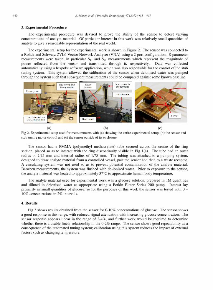
The experimental setup for the experimental work is shown in Figure 2. The sensor was connected to a Rohde and Schwarz ZVL6 Vector Network Analyser (VNA) using a 2-port configuration. S-parameter measurements were taken, in particular S11 and S21 measurements which represent the magnitude of power reflected from the sensor and transmitted through it, respectively. Data was collected automatically using a bespoke software application, which was also responsible for the control of the stub tuning system. This system allowed the calibration of the sensor when deionised water was pumped through the system such that subsequent measurements could be compared against some known baseline.
(a) (b) (c) Fig 2. Experimental setup used for measurements with (a) showing the entire experimental setup, (b) the sensor and stub tuning motor control and (c) the sensor outside of its enclosure.
The sensor had a PMMA (polymethyl methacrylate) tube secured across the centre of the ring section, placed so as to interact with the ring discontinuity visible in Fig 1(a). The tube had an outer radius of 2.75 mm and internal radius of 1.75 mm. The tubing was attached to a pumping system, designed to draw analyte material from a controlled vessel, past the sensor and then to a waste receptor. A circulating system was not used so as to prevent potential contamination of the analyte material. Between measurements, the system was flushed with de-ionised water. Prior to exposure to the sensor, the analyte material was heated to approximately 37°C to approximate human body temperature.
The analyte material used for experimental work was a glucose solution, prepared in 1M quantities and diluted in deionised water as appropriate using a Perkin Elmer Series 200 pump. Interest lay primarily in small quantities of glucose, so for the purposes of this work the sensor was tested with 0 – 10% concentrations in 2% intervals.
4. Results
Fig 3 shows results obtained from the sensor for 0-10% concentrations of glucose. The sensor shows a good response in this range, with reduced signal attenuation with increasing glucose concentration. The sensor response appears linear in the range of 2-4%, and further work would be required to determine whether there is a usable linear relationship in the 0-2% range. The sensor shows good repeatability as a consequence of the automated tuning system; calibration using this system reduces the impact of external factors such as changing temperature.

441 A. Mason et al. / Procedia Engineering 47 ( 2012 ) 438 – 441
5. Conclusion
This paper reports on the implementation of a novel co-planar microwave sensor with possible application in biomedical situations where the sensor could be placed in-line with tubes used to pump materials of interest. On-going work [7, 8] considers the usefulness of the sensor for monitoring whole blood in addition to other bodily fluids such as cerebrospinal fluid. Furthermore, the sensor could find application in other areas, where real-time non-contact sensing of some analyte could be beneficial to improve process yields or general efficiency. The work thus far has shown considerable promise however, and has resulted in an international patent [9] being recently granted.
References
[1] V. Mishra, et al., "Fiber grating sensors in medicine: Current and emerging applications," Sensors and Actuators A: Physical, vol. 167, pp. 279-290, 2011.
[2] E. Thrush, et al., "Monolithically integrated semiconductor fluorescence sensor for microfluidic applications," Sensors and Actuators B: Chemical, vol. 105, pp. 393-399, 2005.
[3] C. Connolly, "A review of medical microscopy techniques," Sensor Review, vol. 25, pp. 252 - 258, 2005. [4] J. Choi, et al., "Microwave Detection of Metastasized Breast Cancer Cells in the Lymph Node; Potential
Application for Sentinel Lymphadenectomy," Breast Cancer Research and Treatment, vol. 86, pp. 107-115, 2004.
[5] O. Korostynska, et al., "Glucose monitoring using electromagnetic waves and microsensor with interdigitated electrodes," in Sensors Applications Symposium, 2009. SAS 2009. IEEE, 2009, pp. 34-37.
[6] S. R. Wylie, et al., "RF sensor for multiphase flow measurement through an oil pipeline," Measurement Science and Technology, vol. 17, p. 2141, 2006.
[7] J. H. Goh, et al., "Lactate Detection Using Microwave Spectroscopy for In-Situ Medical Applications," International Journal on Smart Sensing and Intelligent Systems, vol. 4, pp. 338-352, September 2011.
[8] J. H. Goh, et al., "Lactate Detection Using a Microwave Cavity Sensor for Biomedical Applications," presented at the Fifth International Conference on Sensing Technology, Palmerston North, New Zealand, 2011.
[9] A. I. Al-Shamma’a, et al., "NON-INVASIVE MONITORING DEVICE," 2010.
(a) (b)
Fig 3. Results showing (a) the captured spectrum between 3610 and 3630 MHz, and (b) the resulting attenuation linked calibration curve.