Embed Size (px)
Citation preview

CLINICAL RESEARCH www.jasn.org
A Clinicopathologic Study of ThromboticMicroangiopathy in IgA Nephropathy
Khalil El Karoui,*† Gary S. Hill,* Alexandre Karras,‡ Christian Jacquot,‡ Luc Moulonguet,§
Olivier Kourilsky,| Véronique Frémeaux-Bacchi,¶ Michel Delahousse,** Jean-Paul DuongVan Huyen,* Alexandre Loupy,* Patrick Bruneval,* and Dominique Nochy*
*Department of Pathology, Hôpital Européen Georges Pompidou, Paris, France; †Institut National de la Santé et de laRecherche Médicale INSERM U845, Hôpital Necker-Enfants Malades, Paris, France; ‡Department of Nephrology,Hôpital Européen Georges Pompidou, Paris, France; §Department of Nephrology, Hôpital Ambroise Paré, BoulogneBillancourt, France; |Department of Nephrology, Hôpital Sud Francilien, Evry, France; ¶Department of Immunology,Hôpital Européen Georges Pompidou, Paris, France; and **Department of Nephrology, Hôpital Foch, Suresnes, France
ABSTRACTThrombotic microangiopathy (TMA) occurs in IgA nephropathy, but its clinical significance is not welldescribed. We retrospectively examined a series of 128 patients diagnosed with IgA nephropathybetween 2002 and 2008 who had a mean follow-up of 44627 months. In our series, 53% presented withlesions of TMA, acute or organized, in arteries and/or arterioles. Among patients with TMA, 4% werenormotensive, 25% had controlled hypertension, and 71% had uncontrolled hypertension. Of those withuncontrolled hypertension, 26% had malignant hypertension. Histologically, the group with TMA had asignificantly greater percentage of sclerotic glomeruli and worse tubulointerstitial fibrosis than those ofthe group without TMA. However, a significant minority of patients had near-normal histology, with min-imal tubular atrophy (20%) and/or ,20% interstitial fibrosis (24%). TMA rarely occurred in the absence ofsignificant proteinuria. During follow-up, a doubling of serum creatinine or ESRD occurred in all patientswith laboratory evidence of TMA, in 42% of those with morphologic evidence but no laboratory evidenceof TMA, and in 11% of those without TMA. In summary, lesions of TMA are frequent in IgA nephropathyand may occur in normotensive patients with near-normal renal histology. Although the pathophysiologicmechanisms involved remain undetermined, the current study rules out severe hypertension or advancedrenal disease as sole causes.
J Am Soc Nephrol 23: 137–148, 2012. doi: 10.1681/ASN.2010111130
Thrombotic microangiopathy (TMA) is a hetero-geneous disorder characterized by platelet thrombiin arterioles and capillaries and on occasion inarteries.1,2 Renal histopathologic lesions in TMAtend to take one of two broad forms with consider-able overlap: (1) predominant arteriolar, and lesserarterial, involvement, with thrombi and fibrinoidnecrosis, particularly in thrombotic thrombocyto-penic purpura, malignant hypertension (MHT),and scleroderma; or (2) glomerular involvement,with capillary thrombi, capillary loops with doublecontours due to mesangial interposition, and vari-able mesangiolysis,3 the latter most frequently seenin the hemolytic–uremic syndromes. These mor-phologic lesions occur in a number of other clinical
settings as well, including anti-phospholipid anti-body syndrome, or as a side effect of various phar-macologic agents, and are often associated withpoor renal prognosis.1–3
In immunoglobulin A nephropathy (IgAN), themost common form of primary glomerular disease
Received November 3, 2010. Accepted July 22, 2011.
Published online ahead of print. Publication date available atwww.jasn.org.
Correspondence: Dr. Khalil El Karoui, Department of Pathology,Hôpital Européen Georges Pompidou, 21, rue Leblanc 75015,Paris, France. Email: [email protected]
Copyright © 2012 by the American Society of Nephrology
J Am Soc Nephrol 23: 137–148, 2012 ISSN : 1046-6673/2301-137 137

worldwide,4 it has long been recognizedthat intrarenal arterial and arteriolar le-sions, such as arteriolar wall thickeningand hyalin changes,may be a prominent fea-ture.5,6 Further, TMA has been described inIgAN in a recent study7 and attributed bythe authors to severe or malignant hyper-tension. However, a large-scale clinicopath-ologic analysis focused onTMA in IgANhasnot been performed. This report describesthe prevalence, associated clinical features,and outcome of histologic TMA lesionsfound in a retrospective survey of IgAN.
RESULTS
This study included 128 patients, with malespredominating (69.5%). Among them, 118(92.2%) were Caucasians, and 10 (7.8%)were Asians.Mean age was 38.7 years (range,18–78 years). Mean proteinuria was 2.47 g/d(25th to 75th percentile: 0.8–3.00 g/d), andmean estimated GFR (eGFR) was 51.2ml/min per 1.73m2 (25th to 75th percentile,29–76 ml/min per 1.73 m2). All patients ex-cept one (who was pregnant and presentedwithout TMA) received angiotensin-converting enzyme inhib-itors, angiotensin II receptor antagonists, or both, in case ofhypertension or persistent proteinuria. Only one patient, inthe non-TMA group, had received corticosteroid therapy priorto diagnosis, and none had steroid therapy subsequent to diag-nosis. No patient had other immunosuppressive therapy, eitherprior to or subsequent to diagnosis.
Mean follow-up was 44 months (25th to 75th percentile,23–60 months); for those who went to ESRD, mean time fromdiagnosis to dialysis was 15 months (25th to 75th percentile,1–29 months).
TMA Is a Common Feature of IgANAmong our patients, 68 (53.1%) presented with acute ororganized TMA lesions. There were no significant differencesin age or sex between the patients with and without TMA.Clinical and biological characteristics of TMA patients aresummarized in Table 1. Hypertension was present in 71.0%and 23.3% of patients in the TMA and the non-TMA groups,respectively (P=0.00). MHT was noted in 26% of patients inthe TMA group; no patient in the non-TMA group presentedwith MHT. Neurologic symptoms were absent (except in pa-tients with MHT). Compared with patients in the non-TMAgroup, patients with TMA had significantly higher protein-uria, lower serum albumin, higher serum creatinine, andlower eGFR at the time of the biopsy (Table 1). No possiblecause for TMA (such as radiotherapy, Shiga toxin–producingbacteria infection, or drug-induced TMA) was documented
in any patient. Among the 52 patients tested, there wasno difference between the groups for the presence of anti-cardiolipin antibody (26.5% versus 28.5%, P,0.10) or its titerwhen present. Only 1 of the 39 patients tested had lupus an-ticoagulant (although that patient indeed had TMA). Simi-larly, of the 47 patients tested, only 3 patients (2 with TMAand 1 without TMA) had anti-b2 glycoprotein (anti-b2GP1)antibody. It is evident, then, that none of these factors plays amajor role in TMA. Eleven (8%) patients with TMA had com-plete complement assays and genetic screening for comple-ment regulatory protein gene mutations; none presented suchmutations.
Notably, 20 patients presentedwith TMA lesions (includingacute lesions) either without associated hypertension ornormotensive under treatment (Table 2). Of note, most(73.9%) patients from the TMA group did not have MHT atthe time of biopsy or in their medical history.
Comparisons of Patients According to the Degreeof HypertensionComparisons were made between completely normotensivepatients, patients normotensive under treatment, hypertensivepatients, and those with MHT; the clinical data and themorphologic parameters are presented in Table 2. Among the63 normotensive patients, 44 (69.8%) were treated with one ormore antihypertensive agents. MHTwas found in 18 (14.1%)patients who, compared with patients with less severe hyper-tension, presented with much more advanced renal
Table 1. Comparison of clinical data between patients with and without TMA
Parameter TMA No TMA P Value
Number of patients 68 60Age (years) 39.7 (29–49) 36.2 (26–44) .0.10Male 49 of 68 = 72.1% 41 of 60 = 68.3% .0.10Initial systolic BP (mmHg) 161 (135–177) 130 (120–140) 0.00Initial diastolic BP (mmHg) 94 (80–107) 77 (67–85) 0.000002Percentage hypertensive 48 of 68 = 70.6% 14 of 60 = 23.3% 0.00Percentage MHT 18 of 68 = 26% 0 of 60 = 0 0.00Final systolic BP (mmHg) 131 (120–144) 122 (110–130) 0.004Final diastolic BP (mmHg) 81 (72–87) 74 (69–80) .0.10a
Number of antihypertensive agents 2.8 (2–4) 1.4 (1–2) 0.00Proteinuria Dx (g/day) 3.37 (1.2–4.0) 1.33 (0.40–1.85) 0.00Macroscopic hematuria 11 of 59 = 18.6% 22 of 46 = 47.8% 0.0014Serum albumin (g/L) 34.6 (30–39) 39.8 (36–43) 0.0001Initial SCr (mmol/L) 368 (140–412) 123 (80–130) 0.00eGFR (ml/min per 1.73 m2) 34 (14–50) 73 (51–90) 0.00Final SCr (mmol/L) 340 (123–569) 156 (81–119) 0.00Final eGFR (ml/min per 1.73 m2) 23 (0–49) 69 (50–96) 0.00Bad outcomeb 34 of 68 = 50.0% 6 of 53 = 11.3% 0.00RRT 30 of 68 = 44.1% 5 of 53 = 9.4% 0.00Laboratory evidence of TMA 8 of 65 = 12.3% 0 of 55 = 0 0.01Family history 5 of 53 = 9.4% 7 of 39 = 17.9% .0.10
Values expressed as mean (25th to 75th percentile) or percentages. P values calculated byMann–Whitney U test or Fisher’s exact test as appropriate. BP, blood pressure; Dx, diagnosis.aValue of .0.10 after Holm–Bonferroni correction to minimize type 1 error (a=0.05).bBad outcome defined as doubling of initial SCr or need for dialysis.
138 Journal of the American Society of Nephrology J Am Soc Nephrol 23: 137–148, 2012
CLINICAL RESEARCH www.jasn.org

Table
2.Clin
ical
andmorpho
logic
differen
cesbetwee
npatientswithno
rmoten
sion
,mod
eratehy
pertension,
andMHTat
thetimeof
diagno
sis
Param
eter
Norm
otens
ive,
NoAntihyp
ertens
ors
PValue
Norm
otensive
on
Antihyp
ertens
ors
PValue
Hyp
ertens
ion
PValue
MHT
PValue
(Versu
sNorm
otens
ive
witho
utTrea
tmen
t)
Num
ber
ofpatients
1944
4718
SystolicBPDx(m
mHg)
119(114
–12
7)0.01
126(120
–13
3)0.00
162(150
–17
0)0.00
119
3(164
–22
0)0.00
Diastolic
BPDx(m
mHg)
72(62–
80)
.0.10
73(66–
80)
0.00
95(88–
100)
0.00
211
1(97–
125)
0.00
0001
SCrD
x(mmol/L)
113(79–
141)
.0.10
173(81–
163)
0.00
121
5(126
–13
6)0.00
0173
9(248
–13
16)
0.00
0003
eGFR
Dx(m
l/min
per
1.73
m2)
77(68–
89)
.0.10
63(44–
89)
0.00
244
.3(26.4–
55.6)
0.00
001
15.8
(4.3–20
.0)
0.00
0001
Proteinuria
Dx(g/day)
1.01
(0.11–
1.90
).0.10
1.42
(0.66–
2.00
)0.00
043.34
(1.10–
4.05
).0.10
4.21
(2.00–
6.40
)0.00
004
Laboratoryev
iden
ceof
TMA
0of
16=0
.0.10
1of
41=2.4%
.0.10
2of
45=4.5%
0.01
5of
18=27
.7%
0.03
TMA arteria
lacute,
with
fibrin
0of
19=0
.0.10
2of
44=4.5%
.0.10
6of
47=12
.8%
.0.10
6of
18=33
.3%
0.00
1organ
ized
0of
19=0
.0.10
3of
44=6.8%
0.03
11of
47=23
.4%
.0.10
6of
18=33
.3%
0.00
1arterio
lar
acute,
with
fibrin
1of
19=5.2%
.0.10
5of
44=11
.3%
.0.10
9of
47=19
.1%
.0.10
4of
18=22
.2%
.0.10
organ
ized
3of
19=15
.8%
.0.10
13of
44=29
.5%
0.02
25of
47=53
.2%
.0.10
11of
18=61
.1%
0.00
5Any
TMA(acu
teor
organ
ized
,arterialo
rarterio
lar)
3of
19=15
.8%
0.07
17of
44=38
.6%
0.01
31of
47=65
.9%
0.00
418
of18
=10
0%0.00
Bad
outcom
ea1of
19=5.2%
.0.10
8of
44=18
.2%
0.04
17of
45=37
.8%
0.00
214
of17
=82
.3%
0.00
RRT
0of
19=0
0.05
8of
44=18
.2%
.0.10
13of
45=28
.9%
0.00
0214
of17
=82
.3%
0.00
Immed
iate
RRTb
0of
19=0
.0.10
4of
44=9.1%
.0.10
3of
45=6.7%
0.00
029of
17=57
.9%
0.00
03
Value
sex
pressed
asmea
n(25thto
75th
perce
ntile
)orp
erce
ntag
es.P
values
calculated
byMan
n–Whitney
Utest
orFisher’sex
acttest
asap
propria
te.B
P,blood
pressure;
Dx,
diagno
sis.
a Bad
outcom
eisdefi
nedas
dou
blin
gof
SCror
need
ford
ialysis.
bIm
med
iate
RRTisdefi
nedbyRR
Tinitiation,3months
afterb
iopsy.
J Am Soc Nephrol 23: 137–148, 2012 Thrombotic Microangiopathy in IgA Nephropathy 139
www.jasn.org CLINICAL RESEARCH

insufficiency and with much lower eGFR, 58% of them requir-ing renal replacement therapy from the outset compared with7% with lesser hypertension (P=0.00; Table 2). They also hadgreater proteinuria (Table 2). Importantly, there was no dif-ference in the frequency of anti-cardiolipin antibodies be-tween the four groups of patients. As might be anticipated,MHT biopsies disclosed greater interstitial fibrosis, greaterpercentage of sclerotic glomeruli, worse glomerular extracap-illary proliferation, and more frequent TMA than biopsies ofhypertensive patients without MHT (Supplemental Table 1).All the MHT patients (100%) presented with TMA lesionsversus 65.9% of hypertensive patients (without MHT) and31.7% of normotensive patients overall (15.8% of entirelynormotensive patients and 38.6% of patients normotensiveon antihypertensive therapy) (P=0.004 and P=0.0004, respec-tively; Table 2).
Histologic Findings in Patients with or withoutIgAN-Associated TMATMAwas nearly exclusively arterial and arteriolar in location(Figure 1). Only two cases (one in the original series and one inthe supplemental cases stained for CD61 [see later]) had glo-merular fibrin thrombi. There was no evidence in any case forglomerular capillary endothelial swelling, double contours, ormesangiolysis. The fresh fibrinous vascular thrombi (Figures2–4) were characterized by the presence of fibrinous material(staining bright reddish on trichrome stain as performed inour laboratory using acetic acid–formol–absolute alcohol(AFA) fixative, as opposed to the blue staining of the hyalindeposits of hyalin arteriolosclerosis) and dilation with markeddistension and smoothing out of the internal elastic lamina(Figures 1 and 2). Chronic lesions were basically organizedthrombi with small recanalized vascular channels and
reduction or obliteration of the lumen (Figures 1 and 5–7),sometimes having an “onion-skin” appearance. The organizedfibrous tissue was generally oriented in the long axis of thelumen but lacked the lamellar quality of the fibroelastotic le-sions of arteriosclerosis. Focal myocyte necrosis was seen, usu-ally in association with thrombi but sometimes separately(Figure 8).
IgAN-associated TMA was associated with more severeother vascular lesions, both in terms of reduction of lumen(arterial intimal sclerosis and arteriolar lumen reduction) andsmooth muscle hypertrophy (Supplemental Figures 1–6).
Figure 1. Acute TMA in artery. Fresh TMA in segment of in-terlobular artery (arrow, upper right). Adjacent section showsmarked intimal fibroplasias and mild medial hypertrophy. Patientwith malignant hypertension. Masson’s trichrome, original mag-nification 3450.
Figure 2. Acute TMA in afferent arteriole. Lesion markedly dis-tends internal elastica, with early thrombus degeneration andmarkedly reduced slitlike lumen. Note normal surrounding pa-renchyma; probable tip lesion in glomerulus. Patient normotensiveon antihypertensive agents. Masson’s trichrome, original magni-fication 3350.
Figure 3. Organizing TMA in afferent arteriole. Residual red-staining fibrinoidmaterial, with portion of foam cell visible. Internalelastica is maximally stretched. Glomerulus and parenchymalargely intact. Patient normotensive, without antihypertensiveagents. Masson’s trichrome, original magnification 3400.
140 Journal of the American Society of Nephrology J Am Soc Nephrol 23: 137–148, 2012
CLINICAL RESEARCH www.jasn.org

These differences between TMA and non-TMA cases weremaintained when patients were divided into normotensiveand hypertensive groups, although not all differences re-mained significant (Table 3) (patients with MHT were notincluded in this comparison because all had TMA). Consistentwith these changes, hyperplasia of the juxtaglomerular appa-ratus was more frequent in patients with TMA (P=0.04).
In general, the biopsies with IgAN-associated TMA showedmore extensive damage in terms of percentage of scleroticglomeruli and tubulointerstitial damage (Supplemental Table 2).
The ensemble of cases was also evaluated in terms of theOxford Classification (Supplemental Table 2). As anticipated,all of the parameters were more frequent/worse among thepatients with TMA than among those without.
Immunohistochemical StudiesStaining using anti-CD61, an antiplatelet antibody, wasperformed for 12 recent cases of IgAN not included in theearlier main series reported here. All had evidence of eitheracute and/or organized TMA on routine Masson stain. Ofthese, 10 showed at least focal positivity on staining for CD61.
Arteries and ArteriolesIn acute lesions, although sometimes platelet-rich thrombicompletely filled the lumen (Figure 9A), typically plateletswere present in fewer numbers, admixed in varying degreeswith other elements (Figure 9B and Supplemental Figures 7and 8), and might be present in one section of the lumen andabsent in an adjacent one (Supplemental Figure 9). There fre-quently was staining for platelets in the media of arteries with
Figure 4. Organizing TMA in afferent arteriole. Residual fibri-noid material surrounding markedly narrowed lumen (arrow). Pa-tient hypertensive. Masson’s trichrome, original magnification3500.
Figure 5. Organized TMA in afferent arteriole. The lumen iscompletely obliterated (arrow) in one segment of arteriole, al-though adjacent arteriolar segment appears normal. Glomeruluslargely normal, save for capsular adhesions, as in parenchyma.Patient normotensive on antihypertensive agents. Masson’s tri-chrome, original magnification 3450.
Figure 6. Organized arterial TMA in advanced disease. Multiplesegments with narrowing to near-complete obliteration of lumenare seen (arrows). Patient with malignant hypertension. Masson’strichrome, original magnification 3400.
Figure 7. Multiple arteries with advanced organized TMA. Inmost instances, the lumen is completely obliterated. Media of allarteries show significant hypertrophy, particularly that of the larg-est artery (upper left). Patient hypertensive. Masson’s trichrome,original magnification 3500.
J Am Soc Nephrol 23: 137–148, 2012 Thrombotic Microangiopathy in IgA Nephropathy 141
www.jasn.org CLINICAL RESEARCH

acute lesions (Figure 9B and Supplemental Figure 8). Plateletsprogressively disappeared from the intima and media as le-sions advanced (Figure 9C) and were generally entirely absentin organized TMA (Figure 9D).
GlomeruliOne of the 12 cases had glomerular thrombi, recognizable onCD61 staining (Figure 9E). Another case showed platelets atthe site of a presumptive area of fibrinoid necrosis (Figure 9G).The corresponding glomerulus was not identifiable on theinitial Masson stain, but another glomerulus from the samecase showed clear fibrinoid necrosis (Figure 9H). In addition,several cases had isolated platelets or platelet aggregates inglomerular capillary lumens in a minority of glomeruli (Fig-ure 9F), but these lesions were not recognizable by routinemicroscopy.
Veins and Peritubular CapillariesCD61 staining permitted detectionof rare capillary and venouslesions unapparent on routine Masson stain. Some of theserepresented definite venous thrombi (Supplemental Figure10). Others may simply represent platelet aggregates (Supple-mental Figures 11 and 12).
TMA Associated with IgAN May Occur in Early and/orMild CasesBecause TMA in IgAN has previously been reported pre-dominantly in patients with MHT,7 it is important to pointout that TMA may occur in early/mild cases: 33% (23 of69 cases) with systolic blood pressures #140 mmHg;52% in patients with diastolic pressures #90 mmHg;19% with serum creatinine (SCr) #120 mmol/L; 16% witheGFR .60 ml/min per 1.73 m2. In morphologic terms,16 (23.2%) of the cases of TMA occurred in patients withminimal to mild interstitial fibrosis/tubular atrophy (Oxfordclass 0),8,9 with 4 (5.8%) cases showing only acute TMA,5 (7.2%) cases only organized TMA, and 7 (10.1%) casesshowing both.
Conversely, however, IgAN-associated TMA rarely oc-curred in the absence of significant proteinuria, only 4.6%of cases having ,0.5 g/24 h versus 34.5% of cases withoutTMA (P=0.001). Similarly, TMA was tightly associated withthe presence of glomerular lesions, only two (2.9%) cases hav-ing entirely normal glomeruli by light microscopy versus 14(24.1%) cases among the non-TMA cases (P=0.0004). How-ever, glomerular lesions, although present in 97% of TMAbiopsies, were not necessarily severe in a given biopsy.
TMA Is Associated with Bad OutcomeTable 4 presents a univariate analysis of the various clinical andvascular parameters relatable to IgAN-associated TMA withbad outcome. Among the vascular parameters, all showed sig-nificant associations with bad outcome except hyalin arterio-lar deposits. As anticipated, both fibrinoid and organizedTMAwere strongly associated with bad outcome.
Table 3. Relationship between nonspecific lesions of arteries/arterioles and TMA in normotensive and hypertensive patients(MHT excluded)
Normotensive Patients Hypertensive Patients
TMA Present TMA Absent P Value TMA Present TMA Absent P Value
Number of patients 20 43 30 13Lesionarteriosclerosis (global) 2.21 (2–3) 1.14 (0–2) 0.0004 2.57 (2–3) 1.69 (1–2) 0.003arterial intimal sclerosis 1.20 (0–2) 0.54 (0–1) 0.01 1.85 (1–2.5) 0.71 (0–1) 0.001arterial S/M hypertrophy 0.60 (0–1) 0.39 (0–1) .0.10 0.85 (0–2) 0.20 (0.–0.25) 0.02arterial S/M hyalin deposits 0.30 (0–1) 0.15 (0–0) .0.10 0.58 (0–1) 0.08 (0–0) 0.01arteriolar lumen caliber 2.25 (2–3) 2.77 (3–3) 0.0002 2.25 (2–3) 2.76 (3–3) 0.01arteriolar S/M hypertrophy 0.1 (0–0) 0.01 (0–0) .0.10 0.62 (0–1) 0.23 (0–0) 0.02arteriolar hyalin deposits 1.20 (0.5–2) 0.51 (0–1) 0.01 0.93 (0–2) 1.31 (0–2) .0.10
MHT cases could not be broken down in this fashion because all patients had some form of TMA. Values expressed as mean (25th to 75th percentile). P valuescalculated by Mann–Whitney U test. S/M, smooth muscle.
Figure 8. Medial myocyte necrosis. Arrows point to several myo-cytes with necrosis. These appear here in isolation but were morefrequently found as part of TMA. Patient normotensive, withoutantihypertensive agents. Masson’s trichrome, original magnifica-tion 3450.
142 Journal of the American Society of Nephrology J Am Soc Nephrol 23: 137–148, 2012
CLINICAL RESEARCH www.jasn.org

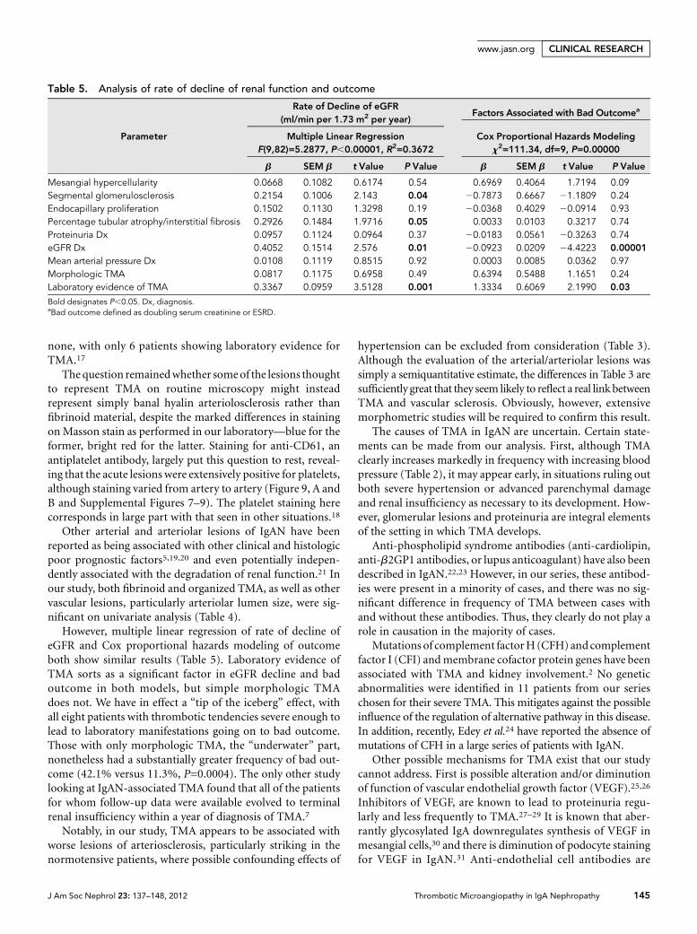
The contribution of TMA (comparedwith theOxford criteria) to decline of eGFRand tobadoutcomewas analyzedbymultiplelinear regression and by Cox proportionalhazardsmodeling, respectively (Table 5). Po-tential confounding factors such as meanarterial pressure, SCr, and proteinuria atthe diagnosis were included. When this wasdone, the eGFR at diagnosis sorted as sig-nificant. Similarly, laboratory evidence forTMA sorted as significantly associated withdecline of eGFR (P=0.001). However, thesimple morphologic presence of TMA didnot sort as significant.
Renal survival was 52.2% at 44 monthsamong the TMA patients versus 93.5%among those without TMA (P=0.00001).However, a more telling separation comesfrom dividing the cases with morphologiclesions of TMA only compared with thoseTMA patients who had, in addition, labo-ratory evidence of TMA. All eight of thelatter patients had a bad outcome within6months of presentation, with a highly sig-nificant difference between this group andthose with morphologic lesions only(P=0.0002; Figure 10).
DISCUSSION
TMA was a frequently identified lesion inthis study of IgAN in adults, being foundin slightly more than one-half (53.1%) ofour patients. This high frequency is in partattributable to the fact that our patients asa group had rather advanced disease.
A very high percentage of patients wereeither frankly hypertensive (48.4%) ornormotensive on antihypertensive treat-ment (34.4%), with 18 (14%) patientspresenting with MHT. This frequency ofhypertension is substantially higher thanthat in other series8,9 and is attributableto an active hypertension clinic in ourinstitution from which many patientswere drawn. This biased recruitment of pa-tients accounts in large part for the muchpoorer survivals (80% of MHT patientswent to ESRD). Because our data revealthat IgAN-associated TMA increasesmarkedly in frequency with increasinghypertension (Table 2), this accounts inlarge part for the very high incidence ofTMA in our series.
Figure 9. Immunohistochemical studies using anti-CD61 antibody. (A) CD61-positiveThrombi. These thrombi in an artery and arteriolar branch appear composed nearly entirelyof platelets. Anti-CD61, original magnification 3400. (B) Arterial and arteriolar thrombi.Platelets constitute roughly half of the thrombus in the artery (left) and are absent from thelumen of the arteriole on the right, but are present in themedia (arrow). Anti-CD61, originalmagnification3400. (C)More advanced TMA. RareCD61-positiveplatelets (arrows) remainin the intima of this advanced TMA, as well as in a glomerulus with near-total sclerosis. Anti-CD61, original magnification 3350. (D) Organized TMA. This artery with advanced orga-nized TMA is CD61 negative. Arrow indicates internal elastica for orientation. Anti-CD61,originalmagnification3500. (E) Glomeruluswith capillary thrombus. A capillary thrombus ispresent, confined to the capillary lumen, along with isolated granules in other capillaries(arrows). Anti-CD61, original magnification3400. (F) Numerous CD61-positive glomerularcapillary granules. These granules, representing isolated platelets or platelet aggregates(arrows), are locatedmostly in dilated capillaries filledwith red blood cells. Such aggregatesare not evident on routine microscopy. Anti-CD61, original magnification 3400. (G) Prob-ableglomerularfibrinoidnecrosis.Numerouspositivegranules surrounda centralmass, andthe glomerular basementmembrane is not recognizable. Anti-CD61, originalmagnification3500. (H) Glomerular fibrinoid necrosis. Fibrinoid necrosis (arrow) in another glomerulusfrom the same case as in (G). Masson’s trichrome, original magnification 3350.
J Am Soc Nephrol 23: 137–148, 2012 Thrombotic Microangiopathy in IgA Nephropathy 143
www.jasn.org CLINICAL RESEARCH

Even taking the increased severity in our patients intoaccount, however, it is evident that the incidence of TMA inIgAN generally is substantially higher than has previously beenappreciated, as numerous examples occurred in patients whowere either entirely normotensive or normotensive undertherapywithnormal/near-normal renal function. (In addition,we believe that our use of AFA fixative and the trichrome stainfacilitates the search for these lesions, which may be in-conspicuous on other stains.)
The only other study looking specifically at TMA in IgAN7
found MHT in 6 of 10 patients studied, with severe hyperten-sion in another 3 patients, and favored the hypothesis that theTMAwas the consequence of the MHT, the MHT itself beingthe consequence of advanced parenchymal lesions. This was aplausible theory for patient sample of that study, particularlygiven that MHT occurs in 7%–15% of IgAN.7,10,11 The as-sociation between MHT and TMA is well recognized, both in
spontaneous12,13 and drug-inducedMHT,13,14 the assumption being that theTMA is due to pressure-induced endo-thelial disruption.13 (In support of thepressure-induced mechanism for TMA,TMA has only been described in severe/malignant hypertension, not in mild tomoderate essential hypertension.) How-ever, our series essentially refutes the hy-pothesis that the TMA in IgAN is due toMHT. The frequency of TMAdid indeed in-creasemarkedly in frequencywith increasingblood pressure, leading to the conclusionthat increasing blood pressure is a major ag-gravating factor. But it seems unlikely to bethe sole cause of IgAN-associated TMA, as20 of 69 (29%) cases occurred in patientswith systolic pressures ,140 mmHg at thetime of biopsy, levels at which TMA has notbeen described in essential hypertension.
Nor did IgAN-associated TMA necessar-ily develop in a setting of advanced paren-chymal lesions, 19% occurring in patientswith an SCr ,120 mmol/L and 23.9% oc-curring in patients with minimal to mild(Oxford class 0) interstitial fibrosis/tubularatrophy. It thus appears clear that TMA canprecede the development of glomeruloscle-rosis and interstitial fibrosis rather thanbeing a consequence of it, a sequence thathas been suggested by others for vascularlesions in general in IgAN.5 Thus, neitherhypertension nor advanced parenchymallesions are necessary prerequisites to thedevelopment of TMA.
By contrast, the appearance of TMA inthe biopsy did appear to be tightly linked tothe presence of glomerular lesions and
proteinuria. Only three (4.6%) TMA cases had proteinuria,0.5 g/24 h as opposed to 19 (34.5%) cases without TMA.Further, the frequency of TMA increased with increasing pro-teinuria, from13.6% for cases,0.5 g/24 h to 80% for cases.3.0g/24 h (P=0.00). Similar considerations held for the associationof TMA with overt glomerular lesions. Only two (2.9%) TMAbiopsies had normal glomeruli by light microscopy comparedwith 14 (24.1%) biopsies without TMA (P=0.0004). Any expla-nation of the mechanism(s) of TMA in IgAN must take its as-sociation with glomerular lesions and proteinuria into account.
Although CD61 staining revealed glomerular lesions to beslightly more extensive than appreciated on routinemicroscopy,IgAN-associated TMA remains a primarily arterial/arteriolarlesion. In this regard, it resembles scleroderma,15,16 and par-ticularly, the kidney of malignant hypertension. A recent re-port of 21 patients with MHT-associated TMA found arterialor arteriolar lesions in all but glomerular thromboses in
Table 4. Univariate analysis of clinical and vascular factors associated with badoutcome
Bad OutcomeaPreserved
RenalFunction
P Value
Number of patients 40 81Clinical parameterssystolic Dx (mmHg) 167 (140–197) 139 (120–150) 0.000002diastolic Dx (mmHg) 97 (80–115) 82 (70–90) 0.0002HBP Dx 37 of 40 = 92.5% 55 of 81 = 67.9% 0.003systolic BP, end 136 (124–154) 123 (111–130) 0.000001MHT 14 of 40 = 35% 3 of 81 = 3.7% 0.00proteinuria Dx (g/day) 3.99 (1.81–5.30) 1.81 (0.49–2.52) 0.000001proteinuria, end (g/day) 3.32 (1.80–4.92) 0.94 (0.10–1.07) 0.0001serum albumin (g/L) 32.661.08 39.160.64 0.000004SCr Dx (mmol/L) 537 (190–759) 125 (89–161) 0.00eGFR Dx (ml/min per 1.73 m2) 22 (6–34) 66 (44–83) 0.00laboratory evidence of TMA 8 of 38 = 21.1% 0 of 78 = 0 0.00
Vascular parameters (arbitrary units)arteriosclerosis, global 2.34 (2–3) 1.72 (1–2) 0.01arterial intimal sclerosis 1.59 (1–3) 0.99 (0–2) 0.01arterial S/M hypertrophy 0.99 (0–2) 0.52 (0–1) 0.002arterial hyalin deposits 0.42 (0–1) 0.24 (0–0) 0.06arteriolar lumen caliber 2.04 (1.5–2.0) 2.60 (2–3) 0.000001arteriolar S/M hypertrophy 0.67 (0–1) 0.38 (0–1) 0.003arteriolar hyalin deposits 0.82 (0–1) 0.81 (0–1.5) .0.10
TMA by vessel sizearterial fibrinoid TMA 11 of 40 = 27.5% 4 of 81 = 4.9% 0.0004arterial organized TMA 10 of 40 = 25% 11 of 81 = 13.6% .0.10arteriolar fibrinoid TMA 10 of 40 = 25% 9 of 81 = 11.1% 0.05arteriolar organized TMA 25 of 40 = 62.5% 26 of 81 = 32.1% 0.0014
TMA: all vesselsfibrinoid TMA 22 of 40 = 55% 19 of 81 = 23.5% 0.001organized TMA 29 of 40 = 72.5% 28 of 81 = 34.6% 0.0001any TMA 34 of 40 = 85% 34 of 81 = 42% 0.00
Values expressed as mean (25th to 75th percentile) or percentages. P values calculated byMann–Whitney U test or Fisher’s exact test as appropriate. Dx, diagnosis, HBP, high BP; MHT,malignant hypertension; S/M, smooth muscle.aBad outcome defined as doubling of SCr or need for dialysis.
144 Journal of the American Society of Nephrology J Am Soc Nephrol 23: 137–148, 2012
CLINICAL RESEARCH www.jasn.org
none, with only 6 patients showing laboratory evidence forTMA.17
Thequestion remainedwhether someof the lesions thoughtto represent TMA on routine microscopy might insteadrepresent simply banal hyalin arteriolosclerosis rather thanfibrinoid material, despite the marked differences in stainingonMasson stain as performed in our laboratory—blue for theformer, bright red for the latter. Staining for anti-CD61, anantiplatelet antibody, largely put this question to rest, reveal-ing that the acute lesions were extensively positive for platelets,although staining varied from artery to artery (Figure 9, A andB and Supplemental Figures 7–9). The platelet staining herecorresponds in large part with that seen in other situations.18
Other arterial and arteriolar lesions of IgAN have beenreported as being associated with other clinical and histologicpoor prognostic factors5,19,20 and even potentially indepen-dently associated with the degradation of renal function.21 Inour study, both fibrinoid and organized TMA, as well as othervascular lesions, particularly arteriolar lumen size, were sig-nificant on univariate analysis (Table 4).
However, multiple linear regression of rate of decline ofeGFR and Cox proportional hazards modeling of outcomeboth show similar results (Table 5). Laboratory evidence ofTMA sorts as a significant factor in eGFR decline and badoutcome in both models, but simple morphologic TMAdoes not. We have in effect a “tip of the iceberg” effect, withall eight patients with thrombotic tendencies severe enough tolead to laboratory manifestations going on to bad outcome.Those with only morphologic TMA, the “underwater” part,nonetheless had a substantially greater frequency of bad out-come (42.1% versus 11.3%, P=0.0004). The only other studylooking at IgAN-associated TMA found that all of the patientsfor whom follow-up data were available evolved to terminalrenal insufficiency within a year of diagnosis of TMA.7
Notably, in our study, TMA appears to be associated withworse lesions of arteriosclerosis, particularly striking in thenormotensive patients, where possible confounding effects of
hypertension can be excluded from consideration (Table 3).Although the evaluation of the arterial/arteriolar lesions wassimply a semiquantitative estimate, the differences in Table 3 aresufficiently great that they seem likely to reflect a real link betweenTMA and vascular sclerosis. Obviously, however, extensivemorphometric studies will be required to confirm this result.
The causes of TMA in IgAN are uncertain. Certain state-ments can be made from our analysis. First, although TMAclearly increases markedly in frequency with increasing bloodpressure (Table 2), it may appear early, in situations ruling outboth severe hypertension or advanced parenchymal damageand renal insufficiency as necessary to its development. How-ever, glomerular lesions and proteinuria are integral elementsof the setting in which TMA develops.
Anti-phospholipid syndrome antibodies (anti-cardiolipin,anti-b2GP1 antibodies, or lupus anticoagulant) have also beendescribed in IgAN.22,23 However, in our series, these antibod-ies were present in a minority of cases, and there was no sig-nificant difference in frequency of TMA between cases withand without these antibodies. Thus, they clearly do not play arole in causation in the majority of cases.
Mutations of complement factorH (CFH) and complementfactor I (CFI) andmembrane cofactor protein genes have beenassociated with TMA and kidney involvement.2 No geneticabnormalities were identified in 11 patients from our serieschosen for their severe TMA. This mitigates against the possibleinfluence of the regulation of alternative pathway in this disease.In addition, recently, Edey et al.24 have reported the absence ofmutations of CFH in a large series of patients with IgAN.
Other possible mechanisms for TMA exist that our studycannot address. First is possible alteration and/or diminutionof function of vascular endothelial growth factor (VEGF).25,26
Inhibitors of VEGF, are known to lead to proteinuria regu-larly and less frequently to TMA.27–29 It is known that aber-rantly glycosylated IgA downregulates synthesis of VEGF inmesangial cells,30 and there is diminution of podocyte stainingfor VEGF in IgAN.31 Anti-endothelial cell antibodies are
Table 5. Analysis of rate of decline of renal function and outcome
Parameter
Rate of Decline of eGFR(ml/min per 1.73 m2 per year)
Factors Associated with Bad Outcomea
Multiple Linear RegressionF(9,82)=5.2877, P,0.00001, R2=0.3672
Cox Proportional Hazards Modelingx2=111.34, df=9, P=0.00000
b SEM b t Value P Value b SEM b t Value P Value
Mesangial hypercellularity 0.0668 0.1082 0.6174 0.54 0.6969 0.4064 1.7194 0.09Segmental glomerulosclerosis 0.2154 0.1006 2.143 0.04 20.7873 0.6667 21.1809 0.24Endocapillary proliferation 0.1502 0.1130 1.3298 0.19 20.0368 0.4029 20.0914 0.93Percentage tubular atrophy/interstitial fibrosis 0.2926 0.1484 1.9716 0.05 0.0033 0.0103 0.3217 0.74Proteinuria Dx 0.0957 0.1124 0.0964 0.37 20.0183 0.0561 20.3263 0.74eGFR Dx 0.4052 0.1514 2.576 0.01 20.0923 0.0209 24.4223 0.00001Mean arterial pressure Dx 0.0108 0.1119 0.8515 0.92 0.0003 0.0085 0.0362 0.97Morphologic TMA 0.0817 0.1175 0.6958 0.49 0.6394 0.5488 1.1651 0.24Laboratory evidence of TMA 0.3367 0.0959 3.5128 0.001 1.3334 0.6069 2.1990 0.03
Bold designates P,0.05. Dx, diagnosis.aBad outcome defined as doubling serum creatinine or ESRD.
J Am Soc Nephrol 23: 137–148, 2012 Thrombotic Microangiopathy in IgA Nephropathy 145
www.jasn.org CLINICAL RESEARCH

another possibility to be considered. A study from the 1980sfound a 32% incidence of anti–endothelial cell antibodies inIgAN compared with 4% in controls and 9% in other glomer-ular diseases. Little had been done since in this area until arecent study32 found anti–endothelial cell antibodies in 34 of75 (45.3%) patients with IgAN (24 of the 34 having MHT)compared with 3 of 19 patients with primary MHT (P=0.02).
This study has several limitations. It is retrospective andobservational and will need to be validated with a prospectivecohort of patients. Further, the evaluation of prognosis wasrendered difficult by the variable nature of the treatment received.
In conclusion, we have shown in this study that the lesionsof TMA are frequent and severe in IgAN and have a poorprognosis. They increase in frequency with both increasingblood pressure and proteinuria. Lesions of TMA are partic-ularly associated with MHT, but their frequent presence inpatients who are normotensive either naturally or underantihypertensive therapy indicates that they are not theresult of the MHT. The causative factors responsible for thisTMA remain to be determined. We believe that these lesionsshould be systematically sought on renal biopsy, so that the TMAmay be addressed therapeutically, with the future goal being tooptimize treatment for this lesion when it occurs in IgAN.
CONCISE METHODS
PatientsAll the adult (.18 years) patients diagnosed with IgAN from January
2002 to January 2008 at the Pathology Department of the Hôpital
Européen Georges Pompidou (Paris, France)
were enrolled in this study. These biopsies
came from four different medical centers. The
diagnosis was based on the presence of predom-
inant IgA and C3 deposits in the mesangium.
Patients with SLE, Henoch–Schönlein purpura,
chronic liver disease, or HIV infection were ex-
cluded, as well as patients whose renal biopsy
specimen contained less than eight glomeruli.
Clinical and laboratory data including age, gen-
der, blood pressure, number of antihypertensive
agents used, immunosuppressive therapy, pro-
teinuria, hematuria, familial history of IgAN,
SCr, and presence of anti-cardiolipin antibody
and lupus anticoagulant (defined by spontane-
ously prolonged activated partial thromboplastin
time and abnormal specific lupus anticoagulant
test). Anti-b2GP1 antibodieswere collected at the
time of renal biopsy and at the end of follow-up
(or institution of renal replacement therapy).
Seven patients were lost to follow-up shortly
after biopsy and were not included in the out-
come analysis. The following definitions were
used. (1) Normotension: Systolic blood pres-
sure ,140 mmHg and diastolic blood pressure
,90 mmHg. (2) Hypertension: Systolic blood pressure.140 mmHg
or diastolic blood pressure.90 mmHg, or the need for antihyperten-
sive medication to maintain pressures below these levels (this latter
group considered separately in some analyses). (3) MHT: Marked
elevation of blood pressure (mean in this study, 193/111 mmHg),
obligatorily associated with central nervous system symptoms, such
as blurred vision, headaches, nausea, vomiting, or papilledema. (4)
Laboratory evidence of TMA: Association of anemia and/or throm-
bocytopenia, lowhaptoglobin, presence of schizocytes, elevated lactate
dehydrogenase. (5) Bad outcome: Persistent doubling of SCr or re-
quirement for renal replacement therapy (RRT). The glomerular fil-
tration rate was estimated (eGFR) with the simplified modification of
diet in renal disease formula.33
Renal HistopathologyThe renal biopsies were processed for light microscopy and direct
immunofluorescence. Tissue for histology was fixed in AFA and
processed and stained by standard methods. Six-micrometer sections
were stained for immunofluorescence study with FITC-conjugated
antibodies specific for human IgG, IgM, IgA, C1q, C3, k and l light
chains, and fibrinogen (DAKO, Carpinteria, CA). All biopsy slides
were re-reviewed by two senior pathologists (D. Nochy and G.S. Hill)
without knowledge of clinical outcomes. The biopsies were graded ac-
cording to the Oxford classification of IgAN.8,9 TMA lesions were de-
scribed as (1) “acute,” with fibrin deposits, or (2) as “organized,” with
evident fibrosis and recanalization and narrowing of the lumen at the
arterial and arteriolar levels. TMA lesions were also classified according
to location: arterial, arteriolar, or glomerular. The severity of interstitial
cell infiltration and tubular atrophy was semiquantitatively scored on a
scale of 0–4+. Interstitial fibrosis was also estimated as a percentage of
Figure 10. Survival from renal bad outcome. Three groups are compared: those withno TMA versus those with only morphologic TMA and those having morphologic TMAwith recognizable laboratory manifestations. Significance of differences in survival be-tween groups was calculated by log-rank test.
146 Journal of the American Society of Nephrology J Am Soc Nephrol 23: 137–148, 2012
CLINICAL RESEARCH www.jasn.org
the renal parenchyma involved. In a separate analysis performed by
one pathologist (G. S. Hill), arteries and arterioles were evaluated
semiquantitatively for global estimation of arteriosclerosis on a scale
of 0–4+ (0, no lesions; 1+, minimal recognizable intimal sclerosis
with or without mild recognizable medial fibrosis; 2+, intimal scle-
rosis with ,25% luminal occlusion with or without mild medial
fibrosis; 3+, intimal fibrosis with,50% lumenal occlusion with def-
inite medial fibrosis and smooth muscle atrophy; 4+, advanced le-
sions with.50% luminal occlusion-markedmedial lesions); arterial
intimal sclerosis on a scale of 0–4+ (0, none; 1+, recognizable intimal
sclerosis but no luminal compromise; 2+, intimal sclerosis with
,25% luminal occlusion; 3+, 25%–50% occlusion; 4+, .50% occlu-
sion); smooth muscle hypertrophy on a scale of 0–2+ (0, absent; 1,
recognizable, minimal to mild; 2, moderate to severe); size of arteriolar
lumen on a scale of 0–4+ (0, total occlusion; 1, marked narrowing; 2,
definite narrowing; 3, normal diameter; 4, dilated); and hyalin de-
posits in arteries and arterioles on a scale of 0–2+ (0, absent; 1,
present, small, nonocclusive of lumen; 2, present, extensive, and/or
impinging on lumen).
Immunohistochemical StudiesTwelve recent cases of IgAN, not included in the original series, all
having either acutefibrinoid and/or organizedTMAwere stainedwith
anti-CD61, an anti-platelet antibody (Y2/51; DAKO). Three cases of
TMA of other causes (cocaine-induced, hemolytic–uremic syn-
drome) and five cases of IgAN without TMAwere used as confirma-
tory positive and negative controls, respectively.
Complement Assays and Genetic ScreeningAnalyses were performed using EDTA plasma samples at the
immunology laboratory of the Hôpital Européen Georges Pompidou.
Plasma concentrations of CFH and CFIweremeasured by ELISA, and
concentrations of C4, C3, and complement factor B were deter-
mined by nephelometry (Dade Behring, Deerfield, IL). Membrane
expression of CD46 was analyzed on granulocytes from patients us-
ing phycoerythrin-conjugated antibodies (clone MEM258; Serotec,
Oxford, UK). All CFH, membrane cofactor protein, and CFI exons
were sequenced as previously described.34
Statistical AnalysesResults were expressed as numerical values and percentages for
categorical variables. Continuous variables are expressed as mean
(25th to 75th percentiles) because the majority had non-Gaussian
distribution. Comparisons were based on Fisher’s exact test for cat-
egorical data and the t test for normally distributed continuous data.
For non-Gaussian–distributed parameters, we used the nonparamet-
ric Mann–Whitney U test to compare continuous variables and the
Wilcoxon test to compare two paired groups. The associations of the
Oxford criteria with decline in eGFR were evaluated by standard mul-
tiple linear regression analysis and with outcome by Cox proportional
hazards modeling. P,0.05 was regarded as statistically significant.
DISCLOSURESNone.
REFERENCES
1. Tsai HM: Advances in the pathogenesis, diagnosis, and treatment ofthrombotic thrombocytopenic purpura. J Am Soc Nephrol 14: 1072–1081, 2003
2. Noris M, Remuzzi G: Atypical hemolytic-uremic syndrome. N Engl J
Med 361: 1676–1687, 20093. Droz D, Nochy D, Noël LH, Heudes D, Nabarra B, Hill GS: Thrombotic
microangiopathies: renal and extrarenal lesions. Adv Nephrol Necker
Hosp 30: 235–259, 20004. Donadio JV, Grande JP: IgA nephropathy.NEngl JMed 347: 738–748,
20025. Wu J, Chen X, Xie Y, Yamanaka N, Shi S, Wu D, Liu S, Cai G: Charac-
teristics and risk factors of intrarenal arterial lesions in patients with IgAnephropathy. Nephrol Dial Transplant 20: 719–727, 2005
6. Feiner HD, Cabili S, Baldwin DS, Schacht RG, Gallo GR: Intrarenalvascular sclerosis in IgA nephropathy. Clin Nephrol 18: 183–192, 1982
7. Chang A, Kowalewska J, Smith KD, Nicosia RF, Alpers CE: A clinico-pathologic study of thrombotic microangiopathy in the setting of IgAnephropathy. Clin Nephrol 66: 397–404, 2006
8. Working Group of the International IgA Nephropathy Network and theRenal Pathology Society; Cattran DC, Coppo R, Cook HT, Feehally J,Roberts IS, et al: The Oxford classification of IgA nephropathy: ratio-nale, clinicopathological correlations, and classification. Kidney Int 76:534–545, 2009
9. Working Group of the International IgA Nephropathy Network and theRenal Pathology Society, Roberts IS, Cook HT, Troyanov S, Alpers CE,AmoreA, et al: TheOxford classification of IgA nephropathy: pathologydefinitions, correlations, and reproducibility. Kidney Int 76: 546–556,2009
10. Subías R, Botey A, Darnell A, Montoliu J, Revert L: Malignant or ac-celerated hypertension in IgA nephropathy. Clin Nephrol 27: 1–7,1987
11. Chen Y, Tang Z, Yang G, Shen S, Yu Y, Zeng C, Chen H, Liu ZH, Li LS:Malignant hypertension in patients with idiopathic IgA nephropathy.Kidney Blood Press Res 28: 251–258, 2005
12. van den Born BJ, Honnebier UP, Koopmans RP, van Montfrans GA:Microangiopathic hemolysis and renal failure in malignant hyperten-sion. Hypertension 45: 246–251, 2005
13. Vaughan CJ, Delanty N: Hypertensive emergencies. Lancet 356: 411–417, 2000
14. Bakir AA, Dunea G: Drugs of abuse and renal disease. Curr Opin
Nephrol Hypertens 5: 122–126, 199615. D’Angelo WA, Fries JF, Masi AT, Shulman LE: Pathologic observations
in systemic sclerosis (scleroderma). A study of fifty-eight autopsy casesand fifty-eight matched controls. Am J Med 46: 428–440, 1969
16. Traub YM, Shapiro AP, Rodnan GP, Medsger TA, McDonald RH Jr,Steen VD, Osial TA Jr, Tolchin SF: Hypertension and renal failure(scleroderma renal crisis) in progressive systemic sclerosis. Reviewof a 25-year experience with 68 cases. Medicine (Baltimore) 62: 335–352, 1983
17. Zhang B, Xing C, Yu X, Sun B, Zhao X, Qian J: Renal thrombotic mi-croangiopathies induced by severe hypertension. Hypertens Res 31:479–483, 2008
18. Galindo M, Gonzalo E, Martinez-Vidal MP, Montes S, Redondo N,Santiago B, Loza E, Pablos JL: Immunohistochemical detection of in-travascular platelet microthrombi in patients with lupus nephritis andanti-phospholipid antibodies. Rheumatology (Oxford) 48: 1003–1007,2009
19. Daniel L, Saingra Y, Giorgi R, Bouvier C, Pellissier JF, Berland Y: Tubularlesions determine prognosis of IgA nephropathy. Am J Kidney Dis 35:13–20, 2000
20. Katafuchi R, Vamvakas E, Neelakantappa K, Baldwin DS, Gallo GR:Microvascular disease and the progression of IgA nephropathy. Am J
Kidney Dis 15: 72–79, 1990
J Am Soc Nephrol 23: 137–148, 2012 Thrombotic Microangiopathy in IgA Nephropathy 147
www.jasn.org CLINICAL RESEARCH
21. Rauta V, Finne P, Fagerudd J, Rosenlöf K, Törnroth T, Grönhagen-RiskaC: Factors associated with progression of IgA nephropathy are relatedto renal function—a model for estimating risk of progression in milddisease. Clin Nephrol 58: 85–94, 2002
22. Sinniah R, Gan HC, Yoon KH: Primary antiphospholipid antibody syn-drome and mesangial IgA glomerulonephritis. Am J Nephrol 21: 134–140, 2001
23. Silva MF, Pimentel FL, Faria MS, Carvalho-Costa AE, Nunes JP: IgAnephropathy and antiphospholipid syndrome.Nephron 83: 95–96, 1999
24. Edey M, Strain L, Ward R, Ahmed S, Thomas T, Goodship TH: Is com-plement factor H a susceptibility factor for IgA nephropathy? Mol Im-munol 46: 1405–1408, 2009
25. Sartelet H, Toupance O, Lorenzato M, Fadel F, Noel LH, Lagonotte E,Birembaut P, Chanard J, Rieu P: Sirolimus-induced thrombotic micro-angiopathy is associated with decreased expression of vascular endo-thelial growth factor in kidneys. Am J Transplant 5: 2441–2447, 2005
26. El Karoui K, Vuiblet V, Dion D, Izzedine H, Guitard J, Frimat L,Delahousse M, Remy P, Boffa JJ, Pillebout E, Galicier L, Noël LH,Daugas E: Renal involvement in Castleman disease. Nephrol DialTransplant 26: 599–609, 2011.
27. Frangié C, Lefaucheur C, Medioni J, Jacquot C, Hill GS, Nochy D:Renal thrombotic microangiopathy caused by anti-VEGF-antibodytreatment for metastatic renal-cell carcinoma. Lancet Oncol 8: 177–178, 2007
28. Eremina V, Jefferson JA, Kowalewska J, Hochster H, HaasM,WeisstuchJ, Richardson C, Kopp JB, Kabir MG, Backx PH, Gerber HP, Ferrara N,Barisoni L, Alpers CE, Quaggin SE: VEGF inhibition and renal throm-botic microangiopathy. N Engl J Med 358: 1129–1136, 2008
29. Nochy D, Lefaucheur C, Hill G: VEGF inhibition and renal thromboticmicroangiopathy. N Engl J Med 359: 206, author reply 206–207,2008
30. Amore A, Conti G, Cirina P, Peruzzi L, Alpa M, Bussolino F, Coppo R:Aberrantly glycosylated IgAmolecules downregulate the synthesis andsecretion of vascular endothelial growth factor in human mesangialcells. Am J Kidney Dis 36: 1242–1252, 2000
31. Hill GS, Karoui KE, Karras A,Mandet C, Duong Van Huyen JP, Nochy D,Bruneval P: Focal segmental glomerulosclerosis plays a major role inthe progression of IgA nephropathy. I. Immunohistochemical studies.Kidney Int 79: 635–642, 2011
32. Jiang L, Zhang JJ, Lv JC, Liu G, ZouWZ, Zhao MH, Zhang H: Malignanthypertension in IgA nephropathy was not associated with backgroundpathological phenotypes of glomerular lesions. Nephrol Dial Trans-plant 23: 3921–3927, 2008
33. Levey AS, Bosch JP, Lewis JB, Greene T, Rogers N, Roth D; Modifica-tion of Diet in Renal Disease Study Group: A more accurate method toestimate glomerular filtration rate from serum creatinine: a new pre-diction equation. Ann Intern Med 130: 461–470, 1999
34. Le Quintrec M, Lionet A, Kamar N, Karras A, Barbier S, Buchler M,Fakhouri F, Provost F, Fridman WH, Thervet E, Legendre C, Zuber J,Frémeaux-Bacchi V: Complement mutation-associated de novothrombotic microangiopathy following kidney transplantation. Am JTransplant 8: 1694–1701, 2008
This article contains supplemental material online at http://jasn.asnjournals.org/lookup/suppl/doi:10.1681/ASN.2010111130/-/DCSupplemental.
148 Journal of the American Society of Nephrology J Am Soc Nephrol 23: 137–148, 2012
CLINICAL RESEARCH www.jasn.org