Embed Size (px)
Citation preview
97 International Journal of Scientific Study | January 2018 | Vol 5 | Issue 10
A Clinical Analysis of Eagle’s Syndrome by its Clinical Features and Therapeutic Response to Surgical Treatment
R T Abdul Salam
Associate Professor, Department of ENT, Government Medical College, Kozhikode, Kerala, India
the condition has been named Eagle’s syndrome in his honor. Later, it has also been called as stylalgia, stylohyoid syndrome, styloid syndrome, “elongated styloid process syndrome,” “styloid process-carotid artery syndrome,” or “styloid process neuralgia.”[3] Eagle reported several cases of cervicopharyngeal symptoms associated with a radiographic diagnosis of an elongated, ossified styloid process occurring a few months post-tonsillectomy.[3] He described the symptoms as nagging or aching sensation in the throat, similar to chronic pharyngitis, and pain spreading to the ear and the mastoid region, difficulty in swallowing, and the sensation of a foreign object lodged in the throat.[4] It may develop inflammatory changes or impinge on the adjacent arteries or sensory nerve endings,
INTRODUCTION
Eagle’s syndrome was first documented by Eagle an otorhinolaryngologist.[1] Over a 20-year period, Eagle reported over 200 cases and explained that the normal styloid process is approximately 2.5–3.0 cm in length.[2] Due to his special interest in this syndrome,
Original Article
AbstractBackground: Elongated styloid process is a relatively common disorder and is frequently misdiagnosed. Elongated styloid process with or without calcified stylohyoid ligament causes recurrent throat pain, foreign body sensation throat, dysphagia cervicofacial pain, and referred otalgia. Eagle’s syndrome is defined as the symptomatic elongation of the styloid process or mineralization (ossification or calcification) of the stylohyoid ligament complex. Diagnosis is based on history, production of symptoms on palpation of tonsillar fossa with a palpable styloid process, and relief of symptoms with injection of lignocaine into the tonsillar fossa. It is confirmed by radiology.
Aim of the study: The aim of this study is to evaluate the clinical, diagnostic, and therapeutic aspects of elongated styloid process.
Materials and Methods: A total of 42 patients with symptoms of elongated styloid process were included and confirmed by computed tomography (CT) scan and intraoral palpation method. All the patients were thoroughly investigated and the medical and surgical treatment efficacy was evaluated.
Observations and Results: Age of the 42 patients varied from 23 to 68 years. The mean age was 35.65 ± 3.10 years. The most commonly affected age group was 30–39 years. Of the total 42 patients, 18 were males and 24 were females.
Conclusions: Eagle’s syndrome is a relatively common disorder that is frequently misdiagnosed. Patients with vague cervico facial pain, throat pain, foreign body sensation throat, referred otalgia, etc., should be palpated for elongated styloid process and investigated for the same. This condition has a female preponderance, and the most commonly affected age group is 30–39 years. CT scan styloid remains a radiological investigation to diagnose elongated styloid process with or without calcified stylohyoid ligament. Medical treatment will not give a long-standing symptomatic relief in elongated styloid process.
Key words: Eagle’s syndrome, Elongated styloid process, Stylalgia, Neuralgia
Access this article online
www.ijss-sn.com
Month of Submission : 11-2017 Month of Peer Review : 12-2017 Month of Acceptance : 12-2017 Month of Publishing : 01-2018
Corresponding Author: Dr. R T Abdul Salam, Department of ENT, Government Medical College, Kozhikode, Kerala, India. E-mail: [email protected]
Print ISSN: 2321-6379Online ISSN: 2321-595X
DOI: 10.17354/ijss/2018/20
Salam: Study on Clinical Features and Therapeutic Response to Surgery in Eagle’s Syndrome
98International Journal of Scientific Study | January 2018 | Vol 5 | Issue 10
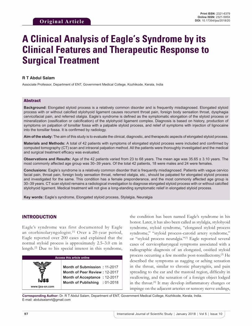
leading to the symptoms described. It had been estimated that between 2% and 4% of the general population presents radiographic evidence of an ossified portion of the stylohyoid chain.[5] The majority of these patients are asymptomatic. The cartilaginous element of second arch (Reichert’s cartilage) gives rise to styloid process, stylohyoid ligament, lesser cornua, and probably the cranial rim of body of hyoid bone [Figure 1].
The clinical signs associated with a symptomatic styloid process were systematized by Eagle in a series of articles (1937, 1948, 1949, 1958).[1-3] This syndrome was first documented by Eagle over a 20-year period. Loeser and Cardwell[6] mentioned that the pain may also radiate to cheek, eyes, and forehead. They excised the tip of styloid process by an external approach with good results. Marchetti[7] of Padua described a case of ossification in stylohyoid ligament. The length of the styloid process has been studied by Wang et al.,[8] Thot et al.,[9] Basekim et al.,[10] and Jung et al.,[11] from radiographs or three-dimension computed tomography (CT). Stylagia[12] published a series of 52 cases, and according to him, the condition is more common than was generally thought. Christiansen et al.[13] discussed in detail “styloid process neuralgia myth or fact,” and from their series, they believed that there was justification for the concepts that “elongated styloid process can produce pain” which is relieved by amputation of the tip. The length of styloid processes is variable. Kaufmann et al.[14] reported that 30 mm is the upper limit for normal styloid process. Moffat et al.[15] performed a cadaver study on styloid process and reported that the normal length is between 1.52 and 4.7 cm. Monsour and Young[16] concluded that the diagnosis of elongated styloid process could be made whenever the styloid process was longer than 40 mm. Palpation of the styloid process in the tonsillar fossa and radiological evaluation of the styloid process help in the diagnosis of this syndrome. Russell[17] in 1977 reported that infiltration of 2% lignocaine at the
area superficial to the prominence produced by elongated styloid process will relieve the pain and patients reported temporary symptomatic relief. Radiological diagnosis remains the most definitive diagnostic criteria in elongated styloid process. In the literature, various radiological views have been discussed and analyzed by various authors. Brat[18] suggested that transoral view is the best to detect this syndrome. Goldstein and Scopp[19] in 1973 defined some diagnostic criteria based on panoramic X-rays. Three-dimensional (3D) CT is a valuable diagnostic imaging tool in patients with Eagle’s syndrome that allows clinicians to evaluate the styloid process in spatial geometry, make accurate length measurements, and explain the problem in detail to patients. For Arellano,[20] Basekim et al.,[10] and Heitz et al.,[21] CT is the most effective method for evaluating the styloid process length and morphological alterations, since this examination provides two-dimensional and 3D reconstructions of the whole temporomandibular joint.
Type of StudyThis was a prospective, cross-sectional, and analytical study.
Period of StudyThe study duration was between June 2009 and October 20011.
Institute of StudyThis study was conducted at Government Medical College, Kozhikode, Kerala.
MATERIALS AND METHODS
A total of 42 outpatients who presented with nagging throat pain, cervicofacial pain, foreign body sensation throat, and referred otalgia that were found to be having elongated styloid process with or without calcified stylohyoid ligament were included in this study. An ethical committee clearance certificate was obtained and a committee approved consent letter was used.
Inclusion Criteria(1) Patients of all age groups were included. (2) Patients with confirmed elongated styloid process on CT scan were included. (3) Patients willing to undergo either surgery or medical treatment were included. (4) Patients with symptoms of throat pain, cervicofacial pain, foreign body sensation throat, and referred otalgia were included. (5) Patients with unilateral or bilateral symptoms were included.
Exclusion Criteria(1) Patients with previous history of tonsillectomy were excluded. (2) Patients with diseases of the spine and Figure 1: Four divisions of Reichert’s cartilage
Salam: Study on Clinical Features and Therapeutic Response to Surgery in Eagle’s Syndrome
99 International Journal of Scientific Study | January 2018 | Vol 5 | Issue 10
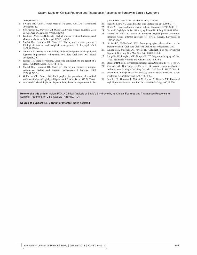
neuralgic pain in other parts of the body were excluded. (3) Patients with diabetes mellitus, hypertension, and malignant diseases of the oral or pharyngeal cavity were excluded. A thorough clinical history was taken. Various radiological views for styloid process are compared to select a diagnostic tool. Various medical and surgical treatments with the incidence of complications are also analyzed in the present study. All the patients in the study group underwent clinical examination palpation of tonsillar fossa. Apart from routine blood, urine, blood sugar, and renal function tests, radiological investigations were also done to diagnose elongated styloid process. Radiological evaluation of cervical spine and barium swallow studies were done in selected patients to rule out cervical spine disorders and pharyngoesophageal causes, respectively (Figure 5). All the data were analyzed using standard statistical methods.
Medical TreatmentOf 42 patients diagnosed, 16 patients were not willing for surgery. Hence, medical treatment was tried in these patients with temporary and variable results. Temporary symptomatic relief was obtained in 14 patients with carbamazepine 100–200 mg once/twice daily, amitriptyline 10–50 mg once/twice daily, and weeks to month’s diclofenac sodium 50 mg twice/thrice daily for a short course.
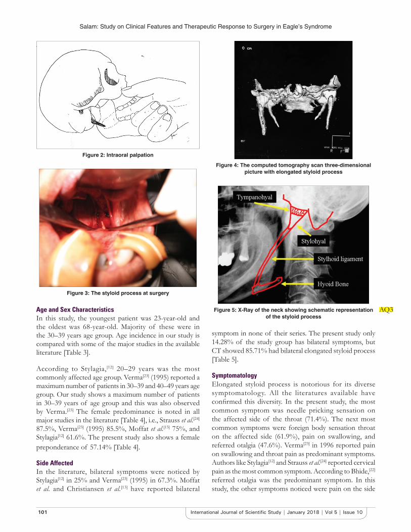
Surgical TreatmentSurgical treatment of elongated styloid process was the main mode of treatment. Bilateral excision was done in 19 patients and unilateral excision in 7 patients. In all these patients, styloid process was palpable per orally. CT scan showed elongated styloid process. Surgery was done under general anesthesia using nasotracheal intubation except in 3 patients where nasotracheal intubation was difficult. Hence, orotracheal intubation was done. Patient was placed in Rose’s position as in tonsillectomy. Mouth was kept open using Boyle–Davis mouth gag, and tonsillectomy was done by classical dissection and snare method. Superior constrictor was separated at the point where the styloid process was prominent and palpable. The tip of styloid process was then delivered. The periosteum was incised at the tip, elevated, and reflected back. The styloid process was broken and removed with a bone nibbler or Luc’s forceps [Figure 2]. After excision, the superior constrictor muscle was closed with catgut sutures and bleeding points were ligated if any. Postoperatively, oral feeding in the form of cold liquid was started after 6 h as in tonsillectomy. Parental antibiotics in the form of ampicillin and gentamicin were given for 2 days. Patients were discharged after 2 days with oral antibiotics and followed up for 1 year at 1 week, 3 weeks, 2 months 6 months, and 1 year.
OBSERVATIONS AND RESULTS
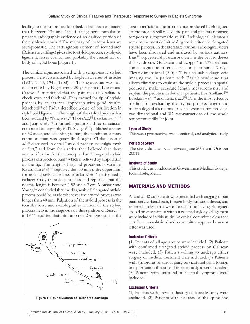
Age and Sex DistributionAge of the patients varied from 23 to 68 years. The mean age was 35.65 ± 3.10 years. The most commonly affected age group was 30–39 years. Of the total 42 patients, 18 were males and 24 were females. The most commonly affected are middle-aged females. The age and sex distribution is presented in Table 1 and Charts 1-3.
Side AffectedBy symptomatology, the condition was unilateral in 36 patients and bilateral only in 6 patients. Intraoral palpation detected palpable styloid process affected side in 30 patients and both sides 12 patients.
Table 1: Age and sex distributionAge in years Number of cases Sex
Male Female20–29 6 1 530–39 17 8 940–49 15 6 950–59 3 2 160–69 1 1 070–79 0 0 080–89 0 0 0
42 18 24
Chart 1: Age and sex distribution
Chart 2: Sex incidence
Salam: Study on Clinical Features and Therapeutic Response to Surgery in Eagle’s Syndrome
100International Journal of Scientific Study | January 2018 | Vol 5 | Issue 10
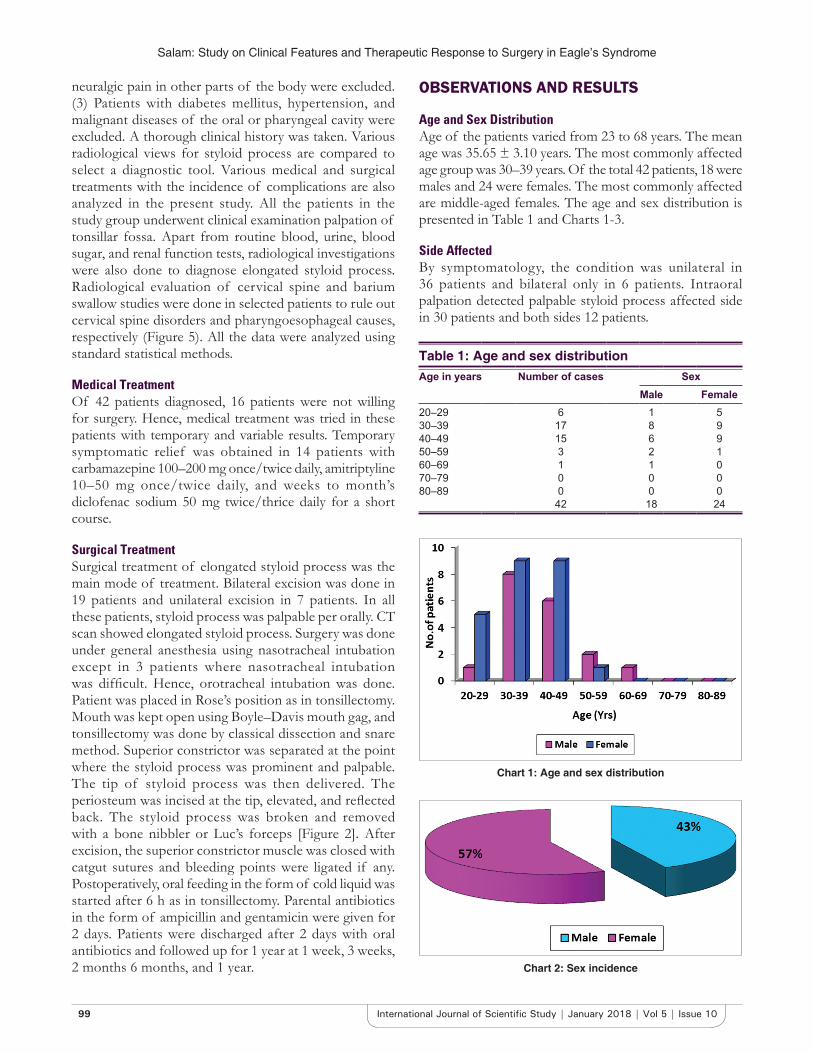
SymptomatologyMajority of patients reported vague and multiple symptoms and the most common symptom was needle pricking sensation on the affected side of the throat.
71.4 patients complained needle pricking sensation on the affected side of the throat. 61.9% of the study group showed foreign body sensation throat on the affected side, and 47.6% complained of aggravating of the symptoms on swallowing and referred pain in the ipsilateral ear. Pain in the temporomandibular area was reported by 28.6%. Other symptoms include pain on changing the head posture 9.5% and headache 14.3%. In 14.3% of the study group, the symptoms were bilateral. Radiation of pain to the surrounding area was complained by 80.95% Table 2.
Intraoral PalpationOn intraoral palpation, 71.4% were unilateral and 28.6% were bilateral. In all patients, same symptoms were reproduced on intraoral palpation of the elongated styloid process. Elongated styloid process was graded according to the tip of styloid process to the tonsillar fossa. In 4 patients (14.3%), the tip of styloid process was palpable in the upper pole of tonsillar fossa (Grade 1) on the affected side. In 23 patients, the tip was palpable in the middle of tonsillar fossa (Grade 2), and in 15 patients, the tip was palpable
in the lower pole of the tonsillar fossa (Grade 3). Among these, two patients had the tip projecting to the tongue base.
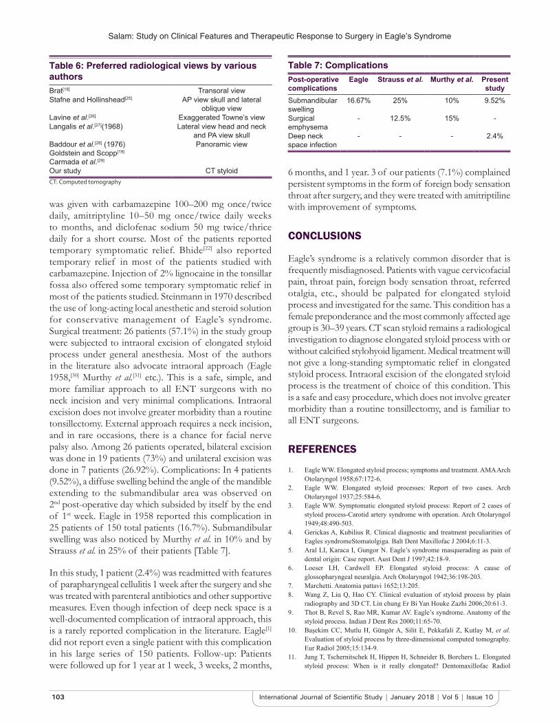
CT ScanWe compared the commonly used radiological studies such as X-ray lateral view of head and neck for styloid, x-ray skull AP view with open mouth, and orthopantomogram with CT styloid. It was found that CT scan is a very useful diagnostic tool. Unilateral elongated styloid process was visualized in 10 patients (23.8%) and bilateral in 32 patients (76.2%). Of the total 42 patients, 18 patients (43%) had calcified stylohyoid ligament [Figures 3 and 4]. In unilateral cases, calcified stylohyoid ligament was seen in 2 patients, and in bilateral cases, calcified stylohyoid ligament was seen in 16 patients. For calcified stylohyoid ligament, there was no sex predilection. Majority of patients (89%) with calcified stylohyoid ligament were above 40 years.
TreatmentOf 42 patients diagnosed, 26 patients underwent intraoral excision of elongated styloid process under general anesthesia. 16 patients were not willing for the surgery and these patients were treated with drugs such as carbamazepine, amitriptyline, and diclofenac sodium. Bilateral excision was done in 19 patients and unilateral excision in 7 patients.
ComplicationsFour patients had diffuse swelling behind the angle of mandible extending to submandibular region noticed on the 2nd post-operative day which subsided by itself by the end of 1st week. 1 patient was readmitted with features of parapharyngeal cellulitis 1 week after the surgery, and she was treated with parental antibiotics and other supportive measures.
Follow-upPatients were followed up at intervals of 1 week, 3 weeks, 2 months, 6 months, and 1 year to know the recurrence or persistence of the symptoms after surgery. On follow-up, 3 patients complained of persistent foreign body sensation throat and they were put on amitriptyline with the improvement of symptoms.
DISCUSSION
Patients who attended ear, nose, and throat (ENT) outpatient department at medical college hospital, Calicut, between June 2009 and October 2011, with nagging throat pain, cervicofacial pain, and foreign body sensation throat were analyzed. Of these, 42 patients were found to be having elongated styloid process. In this study, clinical, diagnostic, and therapeutic parameters of elongated styloid process are discussed.
Chart 3: Side affected by symptomatology, palpation, and radiology
Table 2: Incidence of various symptomsSymptoms Number of patients (%)Needle pricking sensation on the affected side of throat
30 (71.4)
Foreign body sensation throat 26 (61.9)Pain on swallowing 20 (47.6)Pain on the affected side of neck
14 (33.3)
Pain in the temporomandibular joint area
12 (28.6)
Pain on changing the head posture
4 (9.5)
Headache 6 (14.3)
Salam: Study on Clinical Features and Therapeutic Response to Surgery in Eagle’s Syndrome
101 International Journal of Scientific Study | January 2018 | Vol 5 | Issue 10
Age and Sex CharacteristicsIn this study, the youngest patient was 23-year-old and the oldest was 68-year-old. Majority of these were in the 30–39 years age group. Age incidence in our study is compared with some of the major studies in the available literature [Table 3].
According to Stylagia,[12] 20–29 years was the most commonly affected age group. Verma[23] (1995) reported a maximum number of patients in 30–39 and 40–49 years age group. Our study shows a maximum number of patients in 30–39 years of age group and this was also observed by Verma.[23] The female predominance is noted in all major studies in the literature [Table 4], i.e., Strauss et al.[24] 87.5%, Verma[23] (1995) 85.5%, Moffat et al.[15] 75%, and Stylagia[12] 61.6%. The present study also shows a female preponderance of 57.14% [Table 4].
Side AffectedIn the literature, bilateral symptoms were noticed by Stylagia[12] in 25% and Verma[23] (1995) in 67.3%. Moffat et al. and Christiansen et al.[13] have reported bilateral
symptom in none of their series. The present study only 14.28% of the study group has bilateral symptoms, but CT showed 85.71% had bilateral elongated styloid process [Table 5].
SymptomatologyElongated styloid process is notorious for its diverse symptomatology. All the literatures available have confirmed this diversity. In the present study, the most common symptom was needle pricking sensation on the affected side of the throat (71.4%). The next most common symptoms were foreign body sensation throat on the affected side (61.9%), pain on swallowing, and referred otalgia (47.6%). Verma[23] in 1996 reported pain on swallowing and throat pain as predominant symptoms. Authors like Stylagia[12] and Strauss et al.[24] reported cervical pain as the most common symptom. According to Bhide,[22] referred otalgia was the predominant symptom. In this study, the other symptoms noticed were pain on the side
Figure 2: Intraoral palpation
Figure 3: The styloid process at surgery
Figure 4: The computed tomography scan three-dimensional picture with elongated styloid process
Figure 5: X-Ray of the neck showing schematic representation of the styloid process
AQ3

Salam: Study on Clinical Features and Therapeutic Response to Surgery in Eagle’s Syndrome
102International Journal of Scientific Study | January 2018 | Vol 5 | Issue 10
of neck (33.3%), pain in the temporomandibular joint area (28.6%), and pain on changing the head posture (9.5%). A small number of patients also reported other symptom like headache.
Clinical ExaminationApart from other routine general and ENT examination, palpation of the tonsillar fossa for elongated styloid process was a very useful diagnostic method. According to Verma,[23] elongated styloid process was graded according to the relation of the tip of the styloid process and the tonsillar fossa.1. Grade I: The tip of the styloid process palpable in the
upper pole.2. Grade II: The tip of the styloid process palpable in
the middle of the tonsillar fossa.3. Grade III: The tip of the styloid process palpable in
the lower pole and tongue base.In our study group: 4 patients - Grade I 23 patients - Grade II
15 patients - Grade IIIPalpation of the tip of the styloid process in the tonsillar fossa produced aggravation of characteristic symptoms in all these patients.
Radiological StudyEven though the radiological study remains the most helpful diagnostic method in Eagle’s syndrome, a single definite radiological view is still not advocated in available literature. Various authors have suggested different radiological views [Table 6].
Orthopantomogram is considered as a very useful view by many authors like Goldstein and Scopp,[19] Baddour,[28] and Carmada et al.[29] Exaggerated Towne’s view was
suggested by Lavine et al.,[26] in 1968. In our study, we compared lateral view of head and neck, lateral oblique view, posteroanterior view of skull, orthopantomogram, and CT styloid in a few of our patients. It was found that CT styloid was very useful diagnostic tool and was taken in all our patients. In the study, the longest styloid process measured radiologically by CT was 6.2 cm. CT scan styloid process: In this study, unilateral elongated styloid process was found in 10 patients (23.8%) and bilateral in 32 patients (76.2%). Of the total 42 patients, 18 patients (43%) had calcified stylohyoid ligament. In unilateral cases, calcified stylohyoid ligament was seen in 2 patients (20%), and in bilateral cases, bilateral calcified stylohyoid ligaments were seen in 16 patients (50%). For calcified styloid ligament, there was no sex predilection. Majority of patients (89%) with calcified stylohyoid ligament was above 40 years and increased predilection for calcification of stylohyoid ligament was seen in cases with bilateral elongated styloid process. On comparing the signs and symptoms of patients with elongated styloid process with calcified stylohyoid ligament and elongated styloid process without stylohyoid ligament, it is seen that there is no difference in the symptoms in these two groups. Hence, stylohyoid ligament calcification perse associated with elongated styloid process is not producing any additional symptoms when compared with isolated elongated styloid process without stylohyoid ligament calcification. In this study, we recommend CT scan styloid process as the effective radiological investigation to diagnose elongated styloid process with or without calcified stylohyoid ligament. Diagnostic role of CT scan in Eagle’s syndrome was stressed by Baddour[28] and Carmada et al.[29] Treatment: Conservative treatment: In the study group of 42 patients, 16 patients (42.9%) were not willing for surgery. In these patients, medical treatment
Table 3: Age and sex characteristicsAuthors Total patients Age in years
10–19 20–29 30–39 40–49 50–59 60–69 70–79 80–89Stylagia[12] 52 7 20 12 8 5 0 0 0Bhide[22] 41 0 0 18 13 10 0 0 0Verma[23] (1995) 55 0 0 15 20 20 0 0 0Present study 42 0 6 17 15 3 1 0 0
Table 4: Sex incidenceAuthors Total number of
patientsMales (%) Females (%)
Stylagia[12] 52 20 (38.4) 32 (61.6)Moffat et al.[15] 4 1 (25) 3 (75)Bhide[22] 41 18 (43.9) 23 (56.1)Strauss et al.[24] 8 1 (12.5) 7 (87.5)Verma[23] (1995) 55 8 (14.5) 47 (85.5)Our study 42 18 24 (57.14)
Table 5: Side affectedAuthors Number of
patientsUnilateral symptoms
(%)
Bilateral symptoms
(%)Stylagia[12] 55 39 (75) 13 (25)Moffat et al.[18] (1977) 4 4 (100) 0Christiansen et al.[13] 5 5 (100) 0Strauss et al. 8 7 (87.5) 1 (12.5)Verma[23] (1995) 55 18 (32.7) 37 (67.3)Our study 42 (85.71%) 36 6 (14.28)
Salam: Study on Clinical Features and Therapeutic Response to Surgery in Eagle’s Syndrome
103 International Journal of Scientific Study | January 2018 | Vol 5 | Issue 10
was given with carbamazepine 100–200 mg once/twice daily, amitriptyline 10–50 mg once/twice daily weeks to months, and diclofenac sodium 50 mg twice/thrice daily for a short course. Most of the patients reported temporary symptomatic relief. Bhide[22] also reported temporary relief in most of the patients studied with carbamazepine. Injection of 2% lignocaine in the tonsillar fossa also offered some temporary symptomatic relief in most of the patients studied. Steinmann in 1970 described the use of long-acting local anesthetic and steroid solution for conservative management of Eagle’s syndrome. Surgical treatment: 26 patients (57.1%) in the study group were subjected to intraoral excision of elongated styloid process under general anesthesia. Most of the authors in the literature also advocate intraoral approach (Eagle 1958,[30] Murthy et al.[31] etc.). This is a safe, simple, and more familiar approach to all ENT surgeons with no neck incision and very minimal complications. Intraoral excision does not involve greater morbidity than a routine tonsillectomy. External approach requires a neck incision, and in rare occasions, there is a chance for facial nerve palsy also. Among 26 patients operated, bilateral excision was done in 19 patients (73%) and unilateral excision was done in 7 patients (26.92%). Complications: In 4 patients (9.52%), a diffuse swelling behind the angle of the mandible extending to the submandibular area was observed on 2nd post-operative day which subsided by itself by the end of 1st week. Eagle in 1958 reported this complication in 25 patients of 150 total patients (16.7%). Submandibular swelling was also noticed by Murthy et al. in 10% and by Strauss et al. in 25% of their patients [Table 7].
In this study, 1 patient (2.4%) was readmitted with features of parapharyngeal cellulitis 1 week after the surgery and she was treated with parenteral antibiotics and other supportive measures. Even though infection of deep neck space is a well-documented complication of intraoral approach, this is a rarely reported complication in the literature. Eagle[1] did not report even a single patient with this complication in his large series of 150 patients. Follow-up: Patients were followed up for 1 year at 1 week, 3 weeks, 2 months,
6 months, and 1 year. 3 of our patients (7.1%) complained persistent symptoms in the form of foreign body sensation throat after surgery, and they were treated with amitriptiline with improvement of symptoms.
CONCLUSIONS
Eagle’s syndrome is a relatively common disorder that is frequently misdiagnosed. Patients with vague cervicofacial pain, throat pain, foreign body sensation throat, referred otalgia, etc., should be palpated for elongated styloid process and investigated for the same. This condition has a female preponderance and the most commonly affected age group is 30–39 years. CT scan styloid remains a radiological investigation to diagnose elongated styloid process with or without calcified stylohyoid ligament. Medical treatment will not give a long-standing symptomatic relief in elongated styloid process. Intraoral excision of the elongated styloid process is the treatment of choice of this condition. This is a safe and easy procedure, which does not involve greater morbidity than a routine tonsillectomy, and is familiar to all ENT surgeons.
REFERENCES
1. Eagle WW. Elongated styloid process; symptoms and treatment. AMA Arch Otolaryngol 1958;67:172-6.
2. Eagle WW. Elongated styloid processes: Report of two cases. Arch Otolaryngol 1937;25:584-6.
3. Eagle WW. Symptomatic elongated styloid process: Report of 2 cases of styloid process-Carotid artery syndrome with operation. Arch Otolaryngol 1949;48:490-503.
4. Gerickas A, Kubilius R. Clinical diagnostic and treatment peculiarities of Eagles syndromeStomatolgiga. Balt Dent Maxillofac J 2004;6:11-3.
5. Aral LI, Karaca I, Gungor N. Eagle’s syndrome masquerading as pain of dental origin: Case report. Aust Dent J 1997;42:18-9.
6. Loeser LH, Cardwell EP. Elongated styloid process: A cause of glossopharyngeal neuralgia. Arch Otolaryngol 1942;36:198-203.
7. Marchetti. Anatomia pattavi 1652;13:205.8. Wang Z, Liu Q, Hao CY. Clinical evaluation of styloid process by plain
radiography and 3D CT. Lin chung Er Bi Yan Houke Zazhi 2006;20:61-3.9. Thot B, Revel S, Rao MR, Kumar AV. Eagle’s syndrome. Anatomy of the
styloid process. Indian J Dent Res 2000;11:65-70.10. BaşekimCC,MutluH,GüngörA,SilitE,PekkafaliZ,KutlayM,et al.
Evaluation of styloid process by three-dimensional computed tomography. Eur Radiol 2005;15:134-9.
11. Jung T, Tschernitschek H, Hippen H, Schneider B, Borchers L. Elongated styloid process: When is it really elongated? Dentomaxillofac Radiol
Table 6: Preferred radiological views by various authorsBrat[18] Transoral viewStafne and Hollinshead[25] AP view skull and lateral
oblique viewLavine et al.[26] Exaggerated Towne’s viewLangalis et al.[27](1968) Lateral view head and neck
and PA view skullBaddour et al.[28] (1976)Goldstein and Scopp[19]
Carmada et al.[29]
Panoramic view
Our study CT styloidCT: Computed tomography
Table 7: ComplicationsPost-operative complications
Eagle Strauss et al. Murthy et al. Present study
Submandibular swelling
16.67% 25% 10% 9.52%
Surgical emphysema
- 12.5% 15% -
Deep neck space infection
- - - 2.4%
Salam: Study on Clinical Features and Therapeutic Response to Surgery in Eagle’s Syndrome
104International Journal of Scientific Study | January 2018 | Vol 5 | Issue 10
2004;33:119-24.12. Stylagia HR. Clinical experiences of 52 cases. Acta Oto (Stockholm)
1967;24:49-53.13. Christiansen TA, Meyeroff WI, Quick CA. Styloid process neuralgia-Myth
or fact. Arch Otolaryngol 1975;101:120-2.14. Kaufman SM, Elzay RP, Irish EF. Styloid process variation. Radiologic and
clinical study. Arch Otolaryngol 1970;91:460-3.15. Moffat DA, Ramsden RT, Shaw HJ. The styloid process syndrome:
Etiological factors and surgical management. J Laryngol Otol 1977;91:279-94.
16. Monsour PA, Young WG. Variability of the styloid process and stylohyoid ligament in panoramic radiographs. Oral Surg Oral Med Oral Pathol 1986;61:522-6.
17. Russell TE. Eagle’s syndrome. Diagnostic considerations and report of a case. J Am Dentl Assoc 1977;94:548-50.
18. Moffat DA, Ramsden RT, Shaw HJ. The styloid process syndrome: Aetiological factors and surgical management. J Laryngol Otol 1977;91:279-94.
19. Goldstein GR, Scopp IW. Radiographic interpretations of calcifiedstylomandibulra and stylohyoid ligaments. J Prosthet Dent 1973;30:330-4.
20. ArellanoJC.Metadologia,nodiagnosisthere,disfincia,temperomandibular
joint. J Bran Oclus ATM Dor Orofac 2002; 2: 78-86.21. Heitz C, Rocha JR, Souza PH. Rev Bras Protese Implant 1999;6:31-7.22. Bhide A. Hyoid syndrome a review. Indian J Otolaryngol 1985;37:141-3.23. Verma R. Stylalgia. Indian J Otolaryngol Head Neck Surg 1996;48:312-4.24. Strauss M, Zohar Y, Laurian N. Elongated styloid process syndrome:
Intraoral versus external approach for styloid surgery. Laryngoscope 1985;95:976-9.
25. Stafne EC, Hollinshead WH. Roentgenographic observations on the stylohyoid chain. Oral Surg Oral Med Oral Pathol 1962;15:1195-200.
26. Lavine MH, Stoopack JC, Jerrold TL. Calcification of the stylohyoidligament. Oral Surg Oral Med Oral Path 1968;25:55-8.
27. Langalis RP, Langland OE, Nortje CJ. CT Diagnostic Imaging of Jaw. 1st ed. Baltimore: Williams and Wilkins; 1995. p. 620-2.
28. Baddour HM. Eagle’s syndrome, report of a case. Oral Surg 1978;46:486-94.29. Carmada AJ, Deschamps C, Forest D. Stylohyoid chain ossification:
A discussion of etiology. Oral Surg Oral Med Oral Pathol 1989;67:508-14.30. Eagle WW. Elongated styloid process; further observations and a new
syndrome. Arch Otolaryngol 1948;47:630-40.31. Murthy PS, Hazarika P, Mathai M, Kumar A, Kamath MP. Elongated
styloid process-An overview. Int J Oral Maxillofac Surg 1990;19:230-1.
How to cite this article: Salam RTA. A Clinical Analysis of Eagle’s Syndrome by its Clinical Features and Therapeutic Response to Surgical Treatment. Int J Sci Stud 2017;5(10)97-104.
Source of Support: Nil, Conflict of Interest: None declared.