Embed Size (px)
Citation preview

A CASE STUDY ON THE OSTEOPATHIC
APPROACH TO CHRONIC LOW BACK
PAIN AND RADICULOPATHY:
What are the influencing factors and does an osteopathic
treatment have a positive influence on symptoms and
compliance of the patient in a short period of time?
Master Thesis zur Erlangung des Grades
Master of Science in Osteopathie
an der Donau Universität Krems
niedergelegt
an der Wiener Schule für Osteopathie
von Astrid Schmied
Wien, Dezember 2006
Betreut von Kathie Musil

EIDESSTATTLICHE ERKLÄRUNG
Hiermit versichere ich, die vorgelegte Materthese selbständig verfasst zu haben.
Alle Stellen, die wörtlich oder sinngemäß aus veröffentlichten oder nicht
veröffentlichten Arbeiten anderer übernommen wurden, wurden als solche
gekennzeichnet. Sämtliche Quellen und Hilfsmittel, die ich für die Arbeit genutzt
habe sind angegeben. Die Arbeit hat mich gleichem Inhalt noch keiner anderen
Prüfungsbehörde vorgelegen.
------------------------ --------------------------------------- Datum Unterschrift

DANKSAGUNG Hiermit möchte ich bei all jenen bedanken die mich in der Fertigstellung meiner
Masterthese so tatkräftig unterstützt haben.
Allen voran danke ich meiner Betreuerin Kathie Musil, die mir auch in Krisensituationen kompetent und aufopfernd zur Seite stand.
Einen besonderen Dank möchte ich meinem Unterstützer in osteopathischen Fragen Dr. Christian Wutzl, D.O. aussprechen, der trotz seines umfangreichen
beruflichen Engagements ein ständiger und sehr wertvoller Mentor in meiner osteopathischen Entwicklung ist, meine Behandlungen supervidiert und mir die
Praxisräumlichkeiten dafür zu Verfügung gestellt hat. Danke!!
In diesem Rahmen liegt es mir am Herzen, auch meine Praxiskollegen, das
gesamte Team des Ganzheitsmedizinischen Therapiezentrums MEDICUS, zu erwähnen, die für mich sowohl fachlich als auch im ganz besonderen menschlich
unbezahlbar sind.
Ich möchte hier auch die Gelegenheit nutzen ich mich bei meiner Familie, vor allem meiner Mutter, und meinen Freunden bedanken, die mich alle die Jahre durch meine Osteopathieausbildung hindurch unterstützt, motiviert und an mich geglaubt haben. Danke auch an meine „Asyl-Eltern“ Sybille und Hans, die mir
über fast die gesamten 6 Jahre der Ausbildungsseminare in Wien so liebevoll Unterschlupf und Verpflegung gewährt haben.
Last but not least gilt mein herzlicher Dank Gisela für die bereitwillige und
liebevolle Unterstützung in allen Belangen und meinem Freund Marcus, der mich trotz meiner Tendenz unangenehme Dinge hinauszuschieben, immer wieder mit Motivation versorgt hat und mir in der Fertigstellung der These mit Rat und Tat
zur Seite stand.

CCOONNTTEENNTTSS
4
ILLUSTRATION INDEX................................................................ 6
1 INTRODUCTION ..................................................................... 7
2 FUNDAMENTALS ................................................................... 9
2.1 BASIC ANATOMY OF THE LUMBAR SPINE .............................................. 9 2.1.1 Vertebrae...................................................................................... 9 2.1.2 Discus......................................................................................... 10 2.1.3 Ligaments of the spine ................................................................ 10 2.1.4 Muscles....................................................................................... 12 2.1.5 Fascia......................................................................................... 12 2.1.6 Neural structures ........................................................................ 13 2.1.7 Vascular supply .......................................................................... 14 2.1.8 Lymphatic drainage .................................................................... 15
2.2 FUNCTIONAL ANATOMY, BIOMECHANICAL CONSIDERATIONS AND THEIR OSTEOPATHIC RELEVANCE.................................................................. 16
2.2.1 The pelvis ................................................................................... 16 2.2.2 The foot ...................................................................................... 18 2.2.3 The fascial system....................................................................... 19 2.2.4 Attachments of the spinal dura.................................................... 20
3 DEFINITION OF LOW BACK PAIN ................................... 22
3.1 DEFINITION ....................................................................................... 22 3.1.1 Radiculopathy............................................................................. 22 3.1.2 Non-specific low back pain ......................................................... 22
3.2 THE MENTAL FACTOR ......................................................................... 24 3.3 LOW BACK PAIN- A COMPLEX CLINICAL PICTURE ................................. 25
4 PRINCIPLES OF OSTEOPATHY ........................................ 27
4.1 THE FIVE PRINCIPLES.......................................................................... 27 4.2 DYSFUNCTION.................................................................................... 29 4.3 THE SOMATIC DYSFUNCTION............................................................... 29 4.4 THE OSTEOPATHIC DIAGNOSIS ............................................................ 30 4.5 OSTEOPATHIC TECHNIQUES ................................................................ 30
4.5.1 Direct and indirect techniques .................................................... 31 4.5.2 Structural osteopathy .................................................................. 31 4.5.3 Visceral osteopathy.................................................................... 33 4.5.4 Craniosacral osteopathy ............................................................. 33
5 MANAGEMENT OF LBP IN OSTEOPATHY................... 34
5.1 EXPERT INTERVIEW WITH AN OSTEOPATH........................................... 34
6 METHOD................................................................................. 37
6.1 LIFE QUALITY SCORES ....................................................................... 37 6.1.1 SF-36® Health survey (German version) .................................... 37 6.1.2 Owestry-disability-index (ODI), German version ......................... 38
6.2 PAIN DIARY....................................................................................... 39 6.3 OSTEOPATHIC ASSESSMENT AND TREATMENT..................................... 39
CCOONNTTEENNTTSS
5
7 CASE PRESENTATION ........................................................ 42
7.1 THE PATIENTS CASE HISTORY............................................................. 42 7.1.1 Anamnesis .................................................................................. 42 7.1.2 Medical history........................................................................... 44 7.1.3 Aim and expectations of the patient............................................. 46
7.2 THE OSTEOPATHIC TREATMENT.......................................................... 46 7.2.1 First appointment – October 2nd.................................................. 46 7.2.2 Second appointment – October 10th............................................. 50 7.2.3 Third appointment – October 18th ............................................... 51 7.2.4 Fourth appointment – October 24th ............................................. 53 7.2.5. Fifth appointment – November 14th ............................................. 54 7.2.6 Final appointment – December 18th ............................................ 56
8 RESULTS................................................................................. 58
8.1 SF-36 SCORE..................................................................................... 58 8.1.1 SF-36 total results....................................................................... 58 8.1.2 Component summary score ......................................................... 60
8.2 ODI SCORE ....................................................................................... 61 8.3 VAS – PAIN SCORES .......................................................................... 62 8.4 PAIN DIARY....................................................................................... 63
9 DISCUSSION........................................................................... 65
LITERATURE ............................................................................... 67
APPENDIX..................................................................................... 71
IILLLLUUSSTTRRAATTIIOONN IINNDDEEXX
6
ILLUSTRATION INDEX
Figures Fig. 1 Artery system of the lumbar spine .......................................................... 15 Fig. 2 Ligaments of the pelvis ......................................................................... 17 Fig. 3 Ligaments of the pelvis ......................................................................... 18 Fig. 4 Scheme of the myofascial chains and horizontal diaphragms ................. 19 Fig. 5 Fascial connections of the lower extremity ............................................ 20 Fig. 6 Scheme of the treatment process ............................................................ 41 Fig. 7 Scheme of treatment 1............................................................................ 49 Fig. 8 Scheme of treatment 2............................................................................ 51 Fig. 9 Scheme of treatment 3............................................................................ 52 Fig. 10 Scheme of treatment 4............................................................................ 54 Fig. 11 Scheme of treatment 5............................................................................ 55 Tables Tab. 1 Sf-36® health status scores ................................................................... 58 Tab. 2 Component summary scores of PCS and MCS ...................................... 60 Tab. 3 ODI score............................................................................................. 61 Tab. 4 specified pain locations and number of occurrence ................................ 63 Diagrams Diag. 1 SF-36® scale scores ............................................................................ 59 Diag. 2 Range of the bodily pain scores ( 0 = poor, 100 = good) ...................... 59 Diag. 3 ODI score ............................................................................................ 61 Diag. 4 VAS scores.......................................................................................... 62
IINNTTRROODDUUCCTTIIOONN
7
1 INTRODUCTION
From my experience, low back pain in general is probably one of the most
common reasons for patients to consult an osteopath. One of the reasons might be
the large variety of causes and their complex correlation that lead to pain and
limited range of movement in the lumbar spine.
A clear diagnosis leading to a specific therapy in conventional medicine can rarely
be stated and most patients are diagnosed with unspecific low back pain where an
exact patho-anatomical diagnosis not possible. This leads to a huge number of
new therapy forms and minimal invasive techniques of which most are not proved
to efficient.1
For patients with low back pain the diagnosis discus prolaps, disc herniation, or
disc protrusion seems like the worst-case. Reasons for that might be a high
incidence of discopathies in today’s society (possibly caused by the invention and
numerous implementation of CT and MRI scan) and their, from the patients’ point
of view, associated consequences like having to undergo surgery and a complete
change of lifestyle. 2
Working as a physiotherapist and osteopath, dealing with patients suffering from
low back pain is a daily issue and rarely solved easily and in a short period of
time, often resulting from a comprehensive medical history and the state of
chronification. I did experience positive effects of my osteopathic treatment in
patients with disc defects and recurrent chronic low back pain after surgical
interventions but in my daily practise it is often not possible to keep track of the
treatment outcome in between the appointments. Further on I think that the
patient’s lacking physical „consciousness“ and a missing self-reflection on the
1 Gerdesmeye1 L., Haake M., Goebel1 M., Wagner K.: Der Rückenschmerz ; Notfall & Hausarztmedizin 2004; 30: 319– 324 2 Hildebrandt J.: Gibt es einen unspezifischen Rückenschmerz?; Z Orthop 2004; 142; 139- 145
IINNTTRROODDUUCCTTIIOONN
8
negatively influencing factors concerning his health often affects the success of
the osteopathic treatment.
The aim of my thesis on the basis of a case study is, to present the osteopathic
approach to chronic low back pain with subsisting disc defects and radiculopathy
and to find out which parameters have the largest influence on the outcome of an
osteopathic treatment. Further information I was interested in is whether a positive
trend of symptomatic improvement can be perceived in a quite short period of
time and if the osteopathic work has an impact on the patient’s self-reflection and
compliance.
I chose the method of a single-subject design selecting a patient with persisting
chronic low back pain and a complex medical history including disc herniation
and surgical intervention who has not had osteopathic treatment before.
FFUUNNDDAAMMEENNTTAALLSS
9
2 FUNDAMENTALS
The basic anatomy in this section is reduced to the lumbar spine although the
osteopathic approach postulates the consideration of all anatomical structures in
the body. In order to provide the anatomical and physiological background to the
osteopathic principles and approaches I will refer to important anatomical and
physiological facts in detail in section 2.2 (Functional anatomy, physiology and
pathology of the spine) and the case study section.
2.1 Basic Anatomy of the Lumbar spine
2.1.1 Vertebrae
The lumbar spine consists of normally five lumbar vertebrae. Each of them has an
anterior vertebral body, which consists of a very compact anular epiphysis
surrounding the spongiosa, a dorsal vertebral arch, whose pedicles and laminae
are relatively short but strong and extend into an almost horizontal quadrangular
spinosous process. The vertebral foramen lying within the arch is triangularly
shaped. The transverse process is small and thin. The angle of the inferior border
may represent the tip of a costal element and the lateral end the tip of the true
transverse process. The inferior articular process, with vertical convex articular
facets, faces anterolaterally. The superior articular process, with vertical concave
articular facets facing posteriormedially, has a rough mamillary process on its
posterior border. The superior vertebral notch lies in between the vertebral body
and the superior articular process, building, together with the inferior vertebral
notch of the articulating upper vertebrae, the intervertebral foramen: the passage
for the radix, the nerve root of each segment.3, 4
3 Gray’s Anatomy: the anatomical basis of clinical practice; Ed.-in-chief: Standring S.: ElsevierChurchill Livingston 2005 4 Platzer, W.: Taschenatles der Anatomie: Bewegungsapparat; Thieme Verlag 1991
FFUUNNDDAAMMEENNTTAALLSS
10
2.1.2 Discus
The intervertebral disc lies in between two corpuses and consists of the anulus
fibrosus, a ring containing collagene fibres on the outside and mostly fibrous
cartilage on the inside, which centres the nucleus pulposus. 5
Absorbing compression and shock forces and the disc allows motion in the spine.
It retains tension in all ligaments of the vertebral bodies and increases the stability
of the spine. Both longitudinal ligaments, which also support the discus’ position,
build a functional unit together with the discus, being tightly connected with the
posterior longitudinal ligament and in lose contact with the anterior longitudinal
ligament, and are referred to as intervertebral symphysis. 6, 7
2.1.3 Ligaments of the spine
Anterior/posterior longitudinal ligament
The anterior longitudinal ligament (ALL) and posterior longitudinal ligament
(PLL) run anteriorly respectively posteriorly along the surfaces of the vertebral
bodies. The ALL broadens towards the lumbar spine and attaches on anterior
surface of the vertebral bodies. The PLL divides into a superficial layer, which
originates at the body of the second cervical spine (axis) as a prolongation of the
membrana tectoria and reaches down to the intervertebral disc between the third
lumbar vertebrae (L3) and L4, and a deep part depicting prolongation of the
cruciform ligament of the atlas reaching down the sacral canal. In adults the
lumbar PLL is fused to the annulus fibrosus of the vertebral disc and gives room
for veins coming out of the vertebral bodies.8
5 van den Berg, F.: Angewandte Physiologie: Band 1-Das Bindegewebe des Bewegungsapparates verstehen und
beeinflussen; Thieme Verlag1999 6 van den Berg, F.: Angewandte Physiologie: Band 1-Das Bindegewebe des Bewegungsapparates verstehen und
beeinflussen; Thieme Verlag 1999 7 Platzer, W.: Taschenatlas der Anatomie: Bewegungsapparat; Thieme Verlag 1991 8 Platzer, W.: Taschenatlas der Anatomie: Bewegungsapparat; Thieme Verlag 1991
FFUUNNDDAAMMEENNTTAALLSS
11
Ligamenta flava
The ligamenta flava attach on the vertebral arches and consist of elastic fibres,
delimiting the intervertebral foramen medially and dorsally. They extend from the
facet joint capsules to the spines posteriorly where the ligaments from both sides
unite partially, leaving spaces in between for veins connecting the internal and
posterior external vertebral venous plexus. They limit the flexion and assist
erecting the spine from a flexed position.9 10
Interspinous ligaments
The short interspinous ligaments connect the spinous processes, attaching along
each spine from the root to the apex. In the lumbar region they are broader and
thicker than at other levels of the spine.11
Supraspinous ligament
The supraspinous ligament connects the tips of the spinous processes from C7 to
the sacrum, consisting of superficial fibres, extending over three to four, and deep
fibres extending over two to three vertebraes. It is only lightly attached to the
spines at the levels of L3-5 but thicker and broader in the lumbar spine and fuses
with neighbouring lumbar fascia.12
Iliolumbal ligament
The iliolumbal ligament attach on the transverse process of L4 and L5. The
posterior part of the ligament connects L4 with the posterior border of the iliac
crest. The posterior part runs form L5 to the anterior border of the iliac crest, the
linea terminalis of the ilium and has connections to the quadratus lumborum
muscle. 13
9 Platzer, W.: Taschenatlas der Anatomie: Bewegungsapparat; Thieme Verlag 1991 10 Gray’s Anatomy: the anatomical basis of clinical practice; Ed.-in-chief: Standring S.: ElsevierChurchill Livingston 2005 11 Gray’s Anatomy: the anatomical basis of clinical practice; Ed.-in-chief: Standring S.: ElsevierChurchill Livingston 2005 12 Gray’s Anatomy: the anatomical basis of clinical practice; Ed.-in-chief: Standring S.: ElsevierChurchill Livingston 2005 13 Gray’s Anatomy: the anatomical basis of clinical practice; Ed.-in-chief: Standring S.: ElsevierChurchill Livingston 2005
FFUUNNDDAAMMEENNTTAALLSS
12
2.1.4 Muscles
The back muscle complex attaching on the lumbar spine can be primarily consist
of a series of layers. Only the deeper back muscles are true intrinsic and
characterized by the innervation of the posterior rami of the spinal nerves-
therefore termed as erector spinae. Those intrinsic muscles can be also divided
into deep, sometimes termed as erector spinae (Gray’s 2005)and superficial
layers, sometimes termed as transversospinalis (Gray’s 2005).The superficial
muscle group consists of the iliocostalis, longissimus and in some literature also
spinalis (Gray’s 2005) , which act in extending and laterally flexing the vertebral
column. The deeper muscle group consists of an oblique and a straight system.
Oblique muscles are the semispinalis, multifidus and rotator muscles and act
single-side innervated rotating and innervated on both sides extending. The
straight muscles are the interspinales, intertransversarii and spinalis and act
innervated on one side as side benders and innervated on both sides as extensors. 14
Important muscles, concerning the biomechanics of the spine and low back pain,
attaching on the lumbar spine are also the psoas major flexing the hips
respectively erecting the trunk from a lying position and slightly helping in
sidebending the vertebral column, the quadratus lumborum lowering the 12th rib
and sidebending the trunk, the posterior inferior serratus lowering the ribs and not
less important the posterior part of the diaphragm.15
2.1.5 Fascia
The fascial layers in the lumbar region consist of the thoarcolumbar fascia and the
continous prevertebral plane. The thoracolumbar fascia sourrounds the whole
intrinsic muscle group and consist of three layers. Posteriorly it is attached to the
spines of the lumbar and sacral vertebrae and the supraspinous ligaments whereas
the middle layer is attached to the tips of the transvers processes of the lumbar
14 Platzer, W.: Taschenatlas der Anatomie: Bewegungsapparat; Thieme Verlag 1991 15 Platzer, W.: Taschenatlas der Anatomie: Bewegungsapparat; Thieme Verlag 1991
FFUUNNDDAAMMEENNTTAALLSS
13
vertebrae, the intertransvers ligaments, the iliac crest and the lower border of the
twelfth rib and the lumbocostal ligament. The anterior part of the thoracolumbar
fascia is attached to the anterior surfaces of the lumbar transverse processes
behind the psoas major, the iliolumbar ligament and the iliac crest and covers the
quadratus lumborum. Since the all fascial layers build a system it is important to
mention the psoas, iliac, renal and lateroconal fascia which are closely related and
attached to the lumbar vertebrae.16
2.1.6 Neural structures
The spinal cord (medulla) with all its nerve and blood supply is covered by the
dura mater arachnoid and pia mater which extend form the foramen magnum to
the second sacral vertebra where it extends to the as a fine cord, the filum
terminale, and finally fuses with the posterior periosteum of the first coccygeal
segment. The spinal dura mater builds the epidural space together with the tissues
of the vertebral canal. The outer layer of the arachnoid mater is closely applied to
the inner dura mater and encloses the spinal cord with the and its nerve roots to
the point where they pass through the intervertebral foramina. The subarachnoid
space cointains intermediate layers of the arachnoid and the cerebrospinal fluid
(CSF) which is built by the choroids plexuses in the lateral, third and fourth
ventricles in the brain and flows from the ventricular system down into the
subarachnoid space and along the spinal cord. The closest layer to the spinal cord
itself is the pia mater. Whereas the medulla ends at he level of L2 in the conus
medullaris and is continued by the filum terminale, the nerve roots of each
segment below run within the cover of all three mater layers to the segment where
they pass through the intervertebral foramina. From there on the nerves are
covered with the epineurium, which fuses with the dural sheaths.17
The peripheral nerve branches of the lumbar spine, building the lumbar plexus,
are: iliohypogastric and ilioinguinal (L1), genitofemoral (L1/2), lateral femoral
cutaneus (L2/3), femoral and obturator (L2-4 dorsal and ventral) and the 16 Gray’s Anatomy: the anatomical basis of clinical practice; Ed.-in-chief: Standring S.: ElsevierChurchill Livingston 2005 17 Gray’s Anatomy: the anatomical basis of clinical practice; Ed.-in-chief: Standring S.: ElsevierChurchill Livingston 2005
FFUUNNDDAAMMEENNTTAALLSS
14
accessory obturator nerve (L2/3).18 The sacral plexus is built of: the superior
gluteal (L4-S1), inferior gluteal (L5-S2), posterior femoral cutaneous (S1-
3),perforating cutaneous (S2-3),sciatic (L4-S2), common peroneal (L4-S1), tibial
(L5-S1) and pudendal nerve (S2-S4). 19
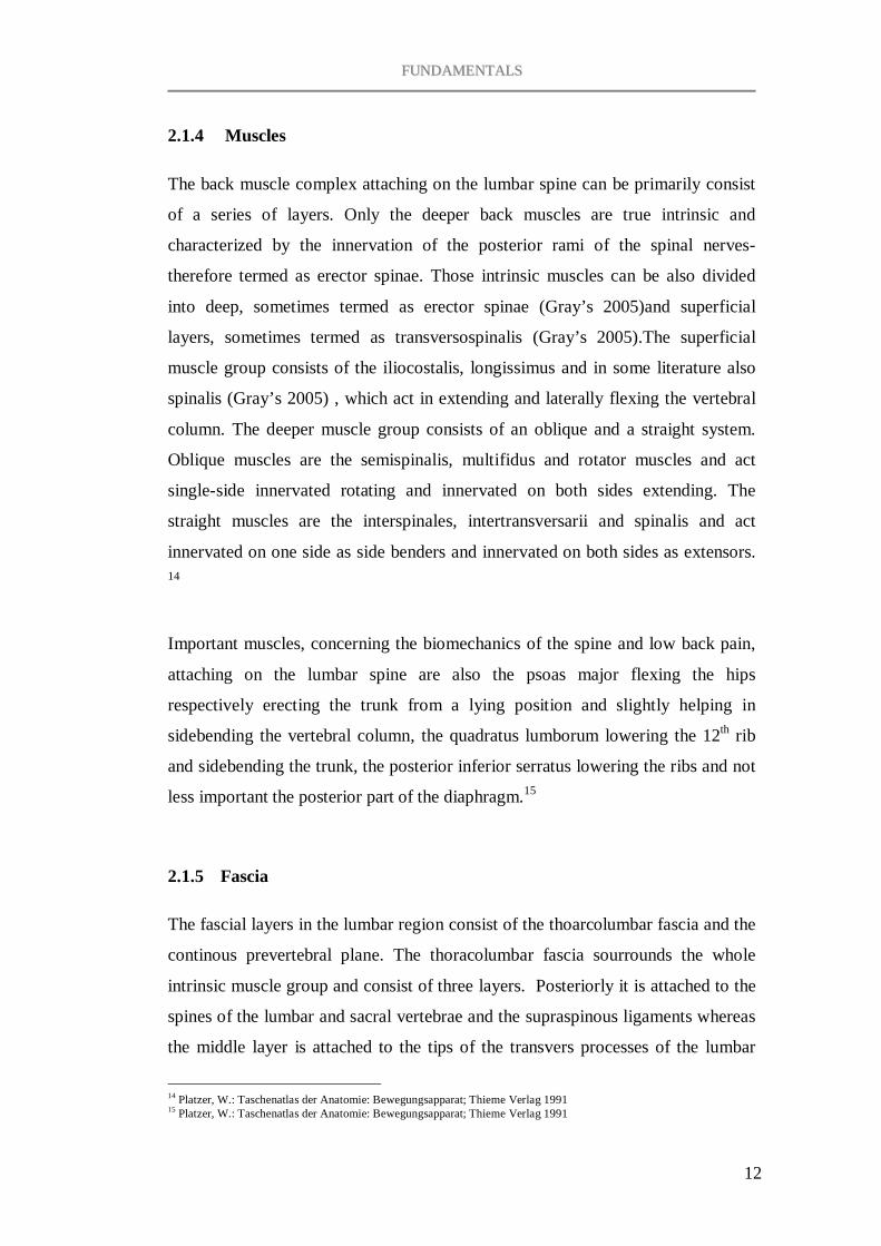
2.1.7 Vascular supply
Concerning the osteopathic approach the arterial supply and venous and lymphatic
drainage are a very important factor since it influences the quality of the tissues
and the potency of the body to self-healing.
Arteries
The arterial supply of the lumbar vertebral column is provided by paired branches
of the aorta, the lumbar arteries, passing around the vertebral bodies, first giving
off periosteal and equatorial branches to the body itself, then continuing into a
major dorsal branch and giving off a spinal branch (the spinal artery) which enters
the intervertebral foramen (Fig.1). The ongoing dorsal branch supplies the facet
joints, the posterior surface of the laminae and the overlying muscles and skin of
the back. The spinal branch divides again into a postcentral, prelaminar and
radicular branch of which the first mainly supplies the vertebral bodies and
periphery of the intervertebral disc. Postcentral branches of adjacent levels
anastomose beneath the PLL and supply the anterior epidural tissues and dura.
The prelaminar branches build a posterior anastomotic plexus on the wall of the
vertebral canal and supply the majority of the vertebral arch, the posterior epidural
tissues and dura, and the ligamnetum flavum. The radicular branches supply the
nerve roots and the spinal cord.20
Veins
Equally to the artery systems the vessels of the venous plexus of the vertebral
column anastomose segmentally and longitudinally to build the anterior and
18 Gray’s Anatomy: the anatomical basis of clinical practice; Ed.-in-chief: Standring S.: ElsevierChurchill Livingston 2005 19 Gray’s Anatomy: the anatomical basis of clinical practice; Ed.-in-chief: Standring S.: ElsevierChurchill Livingston 2005 20 Gray’s Anatomy: the anatomical basis of clinical practice; Ed.-in-chief: Standring S.: ElsevierChurchill Livingston 2005
FFUUNNDDAAMMEENNTTAALLSS
15
posterior external vertebral plexus and the anterior and posterior internal vertebral
plexus and finally drain, as well as the spinal cord, to the intervertebral veins
which accompany the nerve roots through the intervertebral foramina. The lumbar
veins either meet with ascending lumbar veins in front of the transverse processes
or, running around the vertebral bodies, directly end into the inferior vena cava.21
Fig. 1 Artery system of the lumbar spine22
2.1.8 Lymphatic drainage
Most of the lymphatic vessels of the lumbar vertebral column follow the arteries
and drain to the lateral aortic and retro-aortic nodes. The sacral part drains to the
lateral sacral and internal iliac nodes. The lymphatic system upwards the vertebral
column continues in the thoracic duct extending from the level of the second
lumbar vertebra to the base of the neck. At the first and second lumbar level lies
the confluence of the lymph- the cysterna chyli at the level of the thoracolumbar
vertebrae.23
21 Gray’s Anatomy: the anatomical basis of clinical practice; Ed.-in-chief: Standring, S.: ElsevierChurchill Livingston 2005 22 figure taken out of: van den Berg, F.: Angewandte Physiologie: Band 1-Das Bindegewebe des Bewegungsapparates
verstehen und beeinflussen; Thieme Verlag 1999: 116 23 Gray’s Anatomy: the anatomical basis of clinical practice; Ed.-in-chief: Standring, S.: ElsevierChurchill Livingston 2005
FFUUNNDDAAMMEENNTTAALLSS
16
2.2 Functional anatomy, biomechanical considerations and their
osteopathic relevance
The possible movements of the spine are flexion, extension, sidebending and
rotation. The largest range of motion in the lumbar spine is by far
flexion/extension (60°/35° according to Allbrook and David) and sidebending
(20° according to Tanz). The rotatory movement in this region is clearly the
smallest with only 5° (Gregerson and Lucas).
Within the lumbar spine, the largest amplitude of flexion and extension is found in
the segments of L4/L5 and L5/S1 24, causing a maximum of pressure and strain
forces onto the discs, which might be on of the reasons for the high incidence of
discopathies in this region.
In the function of the spine Schmorl differentiates between a passive segment and
moving segment built by the vertebra. The intervertebral discus, the
intervertebrale foramen, the intervertebral joints, the ligamenta flava and the
interspinal ligaments build the moving segment. The vertebra can be seen as a
lever with the intervertebral joint building its centre of rotation. In this function
the pedicle build the link between the anterior and posterior pillar. This system
allows absorbing and transferring the axial pressure forces, which are directly and
passively absorbed by the discus and indirectly and actively absorbed by the
intrinsic back muscles.25
2.2.1 The pelvis
The pelvis consists of the sacrum and the two iliac bones, each of them
articulating with the sacrum in the sacro-iliac joint (SI), and should be seen as a
part of spine. The symphysis builds the anterior articulation of the pubic bones.
The two iliac bones are considered to be functionally assigned to the lower
extremities whereas the sacrum builds the prolongation of the spine.
24 White A A, Panjabi M.M.:Clinical biomechanics of the spine. JB Lippincott 1978, Philadelphia 25 Schmorl G.: Zur pathologischen Anatomie der Lendenbandscheiben; Klin. Wschr. 2 (1932), Über Verlagerungen von Bandscheibengewebe und ihre Folgen.; Langenbecks Arch. Klein. Chir. 172(1932)
FFUUNNDDAAMMEENNTTAALLSS
17
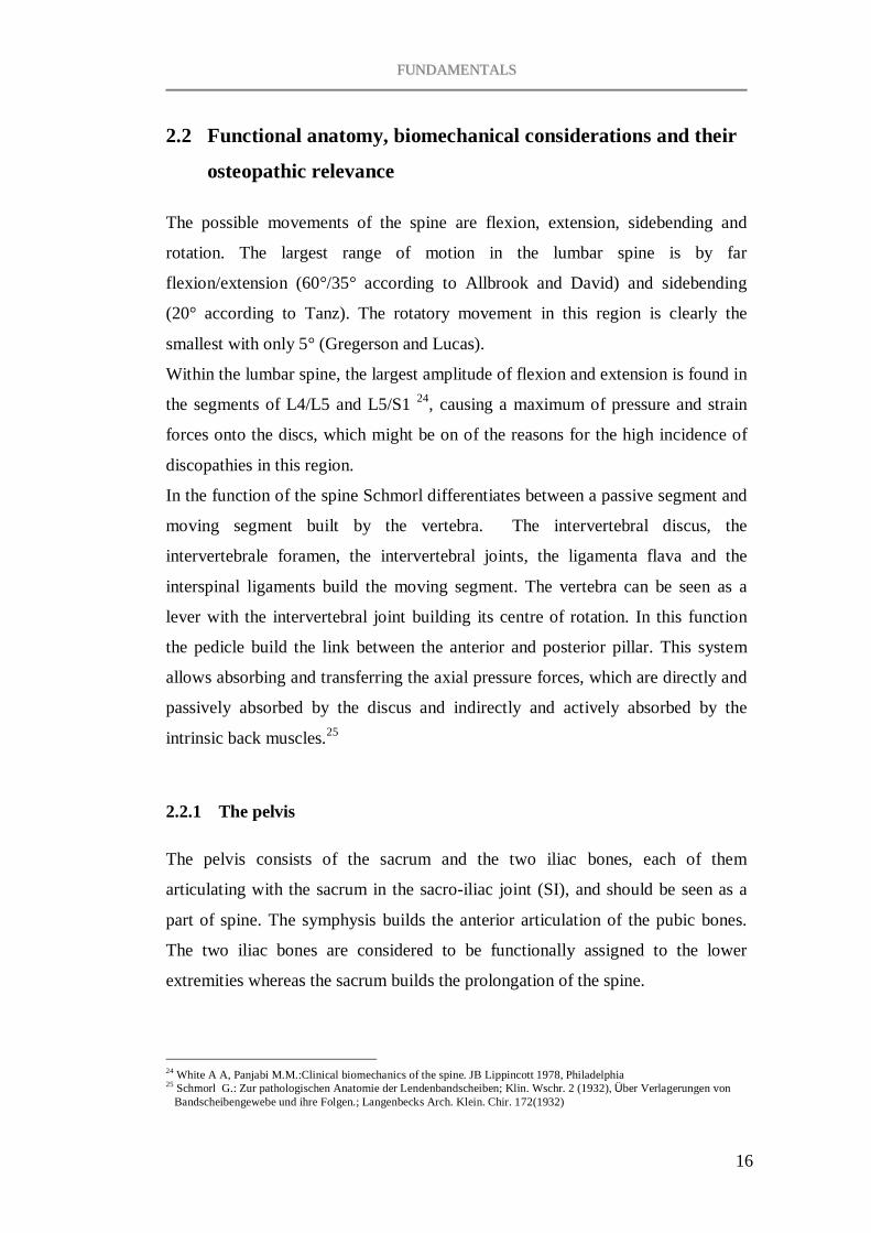
The whole pelvic ring is a strongly ligament-stabilised structure, inducing the
importance of these structures in treating lumbar and pelvic dysfunctions (Fig. 2).
Although the strong ligamentous attachment in the SI joint and the symphysis
does not allow a large range of movement, the mobility in these joints is
functional vital for the whole pelvis and lumbar spine region. The terms nutation /
contra-nutation name the anterior / posterior movement of the base of the sacrum
versus the ilium and imply functionally the sacro-iliac movement and possible
dyfunctions according to Greenman, in the sense of a single / bilateral nutation
anterior / posterior or torsion anterior / posterior. The movements of the ilium are
anterior / posterior rotation around a transversal axis and internal / external
rotation around a transversal axis implying possible dysfunctions of the ileum
according to Greenman in the sense of an ileum anterior / posterior, inflare /
outflare and shear dysfunctions (ilium superior / inferior).26
Fig. 2 Ligaments of the pelvis 27
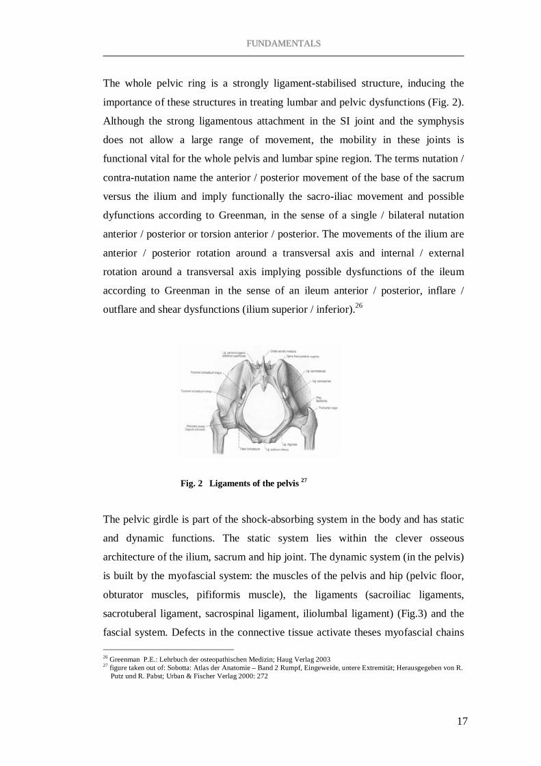
The pelvic girdle is part of the shock-absorbing system in the body and has static
and dynamic functions. The static system lies within the clever osseous
architecture of the ilium, sacrum and hip joint. The dynamic system (in the pelvis)
is built by the myofascial system: the muscles of the pelvis and hip (pelvic floor,
obturator muscles, pififormis muscle), the ligaments (sacroiliac ligaments,
sacrotuberal ligament, sacrospinal ligament, iliolumbal ligament) (Fig.3) and the
fascial system. Defects in the connective tissue activate theses myofascial chains 26 Greenman P.E.: Lehrbuch der osteopathischen Medizin; Haug Verlag 2003 27 figure taken out of: Sobotta: Atlas der Anatomie – Band 2 Rumpf, Eingeweide, untere Extremität; Herausgegeben von R.
Putz und R. Pabst; Urban & Fischer Verlag 2000: 272

FFUUNNDDAAMMEENNTTAALLSS
18
through neuronal signals to protect the injured tissue with the aim to balance the
body.28
Fig. 3 Ligaments of the pelvis 29
2.2.2 The foot
Regarding biomechanical influence factors of the lumbar spine it is indispensable
to take the lower limb into count. I think, the fact that that myofascial chains are
the basis for all static and dynamic functions in the body marks the importance of
assessing all anatomical structures involved, which is in the case of lumbar pain
also the lower limb.
In static and dynamic, the foot is the first structure to be confronted with the
mission to reduce on coming shock forces and dispense it onto the whole body, so
that, provided the fact that the myofascial system is in balance, the stress onto a
single region is diminished. The foot arch is held up by the plantar aponeurosis,
ligaments and muscles and demands a 30
My conclusion is that in this sense a good function of the foot arch predetermines
the balance of the knee, hip, pelvis and therefore also the lumbar spine.
28 Meert G.F.: Das Becken aus osteopathischer Sicht; Urban & Fischer Verlag 2003 29 figure taken out of: Sobotta: Atlas der Anatomie – Band 2 Rumpf, Eingeweide, untere Extremität; Herausgegeben von R.
Putz und R. Pabst; Urban & Fischer Verlag 2000: 272 30 Meert G.F.: Das Becken aus osteopathischer Sicht; Urban & Fischer Verlag 2003
FFUUNNDDAAMMEENNTTAALLSS
19
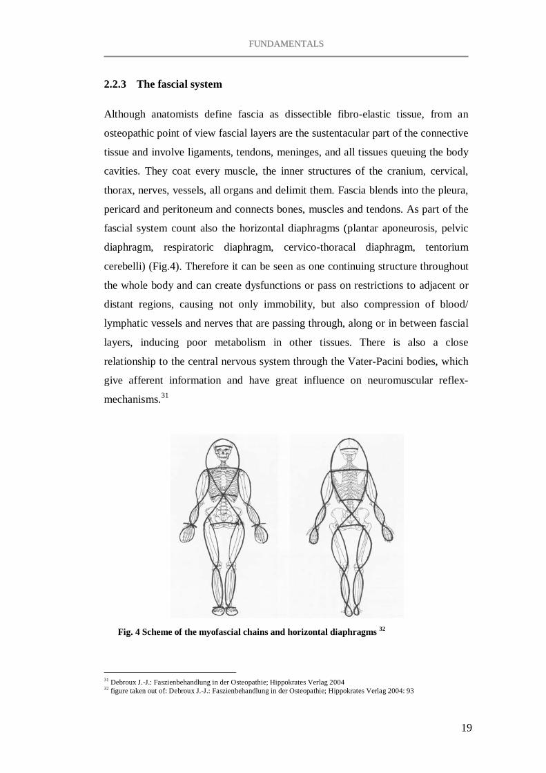
2.2.3 The fascial system
Although anatomists define fascia as dissectible fibro-elastic tissue, from an
osteopathic point of view fascial layers are the sustentacular part of the connective
tissue and involve ligaments, tendons, meninges, and all tissues queuing the body
cavities. They coat every muscle, the inner structures of the cranium, cervical,
thorax, nerves, vessels, all organs and delimit them. Fascia blends into the pleura,
pericard and peritoneum and connects bones, muscles and tendons. As part of the
fascial system count also the horizontal diaphragms (plantar aponeurosis, pelvic
diaphragm, respiratoric diaphragm, cervico-thoracal diaphragm, tentorium
cerebelli) (Fig.4). Therefore it can be seen as one continuing structure throughout
the whole body and can create dysfunctions or pass on restrictions to adjacent or
distant regions, causing not only immobility, but also compression of blood/
lymphatic vessels and nerves that are passing through, along or in between fascial
layers, inducing poor metabolism in other tissues. There is also a close
relationship to the central nervous system through the Vater-Pacini bodies, which
give afferent information and have great influence on neuromuscular reflex-
mechanisms.31
Fig. 4 Scheme of the myofascial chains and horizontal diaphragms 32
31 Debroux J.-J.: Faszienbehandlung in der Osteopathie; Hippokrates Verlag 2004 32 figure taken out of: Debroux J.-J.: Faszienbehandlung in der Osteopathie; Hippokrates Verlag 2004: 93
FFUUNNDDAAMMEENNTTAALLSS
20
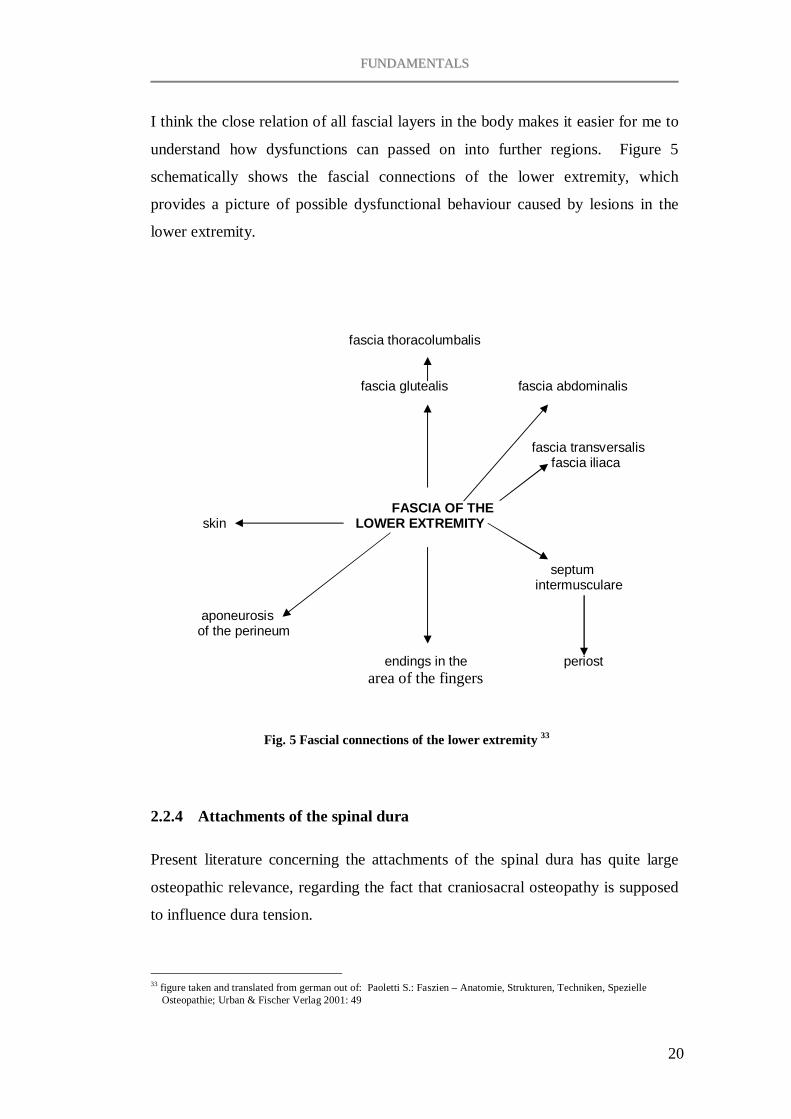
I think the close relation of all fascial layers in the body makes it easier for me to
understand how dysfunctions can passed on into further regions. Figure 5
schematically shows the fascial connections of the lower extremity, which
provides a picture of possible dysfunctional behaviour caused by lesions in the
lower extremity.
fascia thoracolumbalis
fascia glutealis fascia abdominalis
fascia transversalis
fascia iliaca
FASCIA OF THE
skin LOWER EXTREMITY
septum intermusculare
aponeurosis of the perineum endings in the periost
area of the fingers
Fig. 5 Fascial connections of the lower extremity 33
2.2.4 Attachments of the spinal dura
Present literature concerning the attachments of the spinal dura has quite large
osteopathic relevance, regarding the fact that craniosacral osteopathy is supposed
to influence dura tension.
33 figure taken and translated from german out of: Paoletti S.: Faszien – Anatomie, Strukturen, Techniken, Spezielle
Osteopathie; Urban & Fischer Verlag 2001: 49
FFUUNNDDAAMMEENNTTAALLSS
21
The ligamentum nuchae is part of the functional region of the cervical spine and is
an intermuscular septum, which runs from the external occipital protuberance to
the spinous process of C7.34 Although Platzer mentions a continuation of the
ligamentum nuchae into the thoracic and lumbar spine by the supraspinous and
intraspinous ligaments Gray’s Anatomy rules out the structural distinction.
The more interesting aspect concerning the lumbar spine is probably the fact that
Dean and Mitchell (2002) found direct fibrous attachments between the
ligamentum nuchae und the spinal dura on the atlanto-occipital and atlanto-axial
level.35
Interesting, concerning dural fixations and their potential role in low back pain,
are Trolard’s ligament, a link between the dura mater and the posterior
longitudinal ligament on lumbar and sacral levels, Hoffman’s ligamenta dorso-
lateralia, linking the anterior radix and the anterior vertebral canal, and the
opercula of Forestier, covering the intervertebral foramina laterally from the
spinal ganglion and having contact with laminae and disci, rarely found in the
newer literature but verified by Van Dun and Girardin (2006). They concluded
that ‘the attachments perceived between spinal dura and its surroundings may be
considered to be the remains of an original unitary tissue, which will differentiate
into full-grown ligamentous structures according to the unique functional heritage
of the individual’.36
34 Gray’s Anatomy: the anatomical basis of clinical practice; Ed.-in-chief: Standring S.: ElsevierChurchill Livingston 2005 35 Dean N.A., Mitchell B.S.: Anatomic relation between the nuchal ligament (ligamentum nuchae) and the spinal dura
mater in the craniocervical region; Clinical Anatomy 2002May; 15 (3): 182-185 36 van Dun Patrick L.S. Girardin M.R.G.: Embryological study of the spinal dura and its attachment into the vertebral
canal; International Journal of Osteopathic Medicine Vol.9; Issue 3; Sept. 2006: 85-93

DDEEFFIINNIITTIIOONN OOFF LLOOWW BBAACCKK PPAAIINN
22
3 DEFINITION OF LOW BACK PAIN
3.1 Definition
In modern medical literature low back pain is defined in a non-specific and
specific and an acute and chronic form. Whereas the specific low back pain
(SLBP) is defined by a morphologically detectable aetiology (i.e. vertebral
fractures, tumours, disc herniations, spinal stenosis, spondylolisthesis or
inflammatory processes), the non-specific low back pain (NLBP) covers the large
group of LBP syndromes without clear aetiology. Acute low back pain is defined
by less than 4 weeks and chronic low back pain by persisting symptoms over a
time frame of more than 3 months.37
3.1.1 Radiculopathy
Radicular low back pain is defined as nerve-root compression with irradiation into
the pelvis/leg. Further clinical signs are the loss of sensibility in the corresponding
dermatome, paresis of the characteristic muscle of the affected segment, reflex
deficits, a positive Lasegue-sign and pain triggering through compression,
sneezing and coughing. The differentiation to a pseudo-radiculopathy lies mainly
in the neurogenic deficits.38
3.1.2 Non-specific low back pain
Scientists still debate about the term ‘unspecific low back pain’ because it only
describes the fact that, at this stage, most of the back pain syndromes cannot
clearly be assigned to a certain structures and therefore a specific therapy is
precluded. Therefore a new trend in medical science, represented for example by
Bodguk and Aprill, is to assign this group to a specified source of pain, like
37 Weiland W., Wessel K.: Therapie des Rückenschmerzes- Was ist durch Studien belegt?; Fortschr Neurol Psychiat 2004;
72: 344-350 38 Gerdesmeye1 L., Haake M., Goebel1 M., Wagner K.: Der Rückenschmerz ; Notfall & Hausarztmedizin 2004; 30: 319-
324

DDEEFFIINNIITTIIOONN OOFF LLOOWW BBAACCKK PPAAIINN
23
discogenic pain, facet syndrome and sacroiliac joint pain, using higher
differentiated diagnostic methods.39
Discogenic pain is defined as a nociceptive pain syndrome with its source in the
outer part of the annulus of the intervertebral disc- an internal disc disruption
(IDD). The MRI scan in this case shows a so-called high-intensity zone (HIZ) in
the dorsal annulus, which is of unknown content and is speculated to be degraded
nucleus material or an inflammatory process in an annulus fissure.40
Studies have shown that discogenic pain is quite common (40%) but degeneration
of the discus is apparently more of genetic origin than of hard physical work or a
constant sitting-position at work. HIZ are also frequent in persons without back
pain and do not state whether the disc causes pain.41
The facet syndrome is defined as pain caused by the facets of the zygapophysial
joints through an incarcerated or stretched capsule, an inflammation of the capsule
or synovia, a subluxation of the joint, restricted range of movement caused by
muscle-hypertension and degenerative changes. However, like most of all other
unspecific low back pain syndromes a clear assignment of certain back pain
symptoms to these joints is not possible. Degenerative changes in the joint
frequently occur in elderly people but are often also verifiable in asymptomatic
patients and clinical consequences out of an evidence of a facet joint related pain
syndrome are rare.42
Sacro-iliac joint pain is quite common cause for back pain although there are no
valid and reliable clinical tests and anamnestic indication for existing SIJ pain.
The distinct diagnosis can only be through radiological-controlled blocks of the
joint and are also quite successful for therapeutic intervention, as is in this case
also manual therapy.43
39 Hildebrandt J.: Gibt es einen unspezifischen Rückenschmerz?; Z Orthop 2004; 142: 139-145 40 Kniesel B.: Diskogene lumbale Rückenschmerzen; Z Orhop 2004; 142: 709-715 41 Hildebrandt J.: Gibt es einen unspezifischen Rückenschmerz?; Z Orthop 2004; 142: 139-145 42 Hildebrandt J.: Gibt es einen unspezifischen Rückenschmerz?; Z Orthop 2004; 142: 139-145 43 Hildebrandt J.: Gibt es einen unspezifischen Rückenschmerz?; Z Orthop 2004; 142: 139-145

DDEEFFIINNIITTIIOONN OOFF LLOOWW BBAACCKK PPAAIINN
24
Nachemson, Waddell and Bigos represent the majority of scientists opposing the
this structural specifications, alluding to their opinion that in most cases a clear
differentiation is neither possible nor useful and being labelled with a damaged
structure can negatively influence the patient handling his situation. These
strongly structure orientated diagnoses often rather describe radiological findings
and therefore, especially if they do not imply any therapeutic or prognostic
consequences, bear the risk that the patient gets caught in sorrow and grieve
concerning his personal and professional future.44
In my opinion, to label low back pain by assigning it to a defect structure does
only make sense if it entails a clear therapeutic intervention with a good outcome
on a long term base and although patients mostly ask for a diagnose that names
their problem, their only intent is to have someone finding a way to treat the
structure causing the pain. In my osteopathic work a structure-defined diagnose
according to radiological findings, like degeneration of the vertebrae or disc, is an
important factor concerning containdications for certain osteopathic techniques
but does not automatically indicate a certain treatment procedure, because even if
it really is the pain causing structure (which is according to the mentioned studies
above often not proved) the arising question is: what caused the degeneration and
how can I help the patients body getting back into balance to cope with the
degenerated structure?
3.2 The mental factor
In the anamnesis of low back pain patients, my questions concerning their private
and professional situation and triggering events of acute pain phases often refer to
a significant correlation between emotional stress and a worsening of the patients
physical condition which gives reason to conclude that poor mental condition
negatively influences the physical state of the patient or even the other way
around, a chronic physical handicap impairs the mental state.
44 Hildebrandt J.: Gibt es einen unspezifischen Rückenschmerz?; Z Orthop 2004; 142: 139-145

DDEEFFIINNIITTIIOONN OOFF LLOOWW BBAACCKK PPAAIINN
25
Studies have shown that in patients with chronic low back pain the fear of pain
triggers a high incidence of motion and weight loading avoidance resulting in a
constant progress of negative conditioning and over a long term leading to
immobilisation.45 The patients are then caught in a pathological behaviour pattern
concerning their problems and the body’s attempt of self-healing fails. The
importance of psychological factors and their influence on chronic pain has been a
frequent issue in studies in the last years. Turk and Okifuji46 point out the
importance especially the patient’s appraisal of his symptoms and the ability of a
self-management concerning the pain.
3.3 Low back pain- a complex clinical picture
The reason for the fact that low back pain still brings up so many controversial
opinions about the sources of pain in conventional medicine might lie in the
complexity of this clinical picture. From my point of view the structural findings
described in literature are often the result of many influencing factors onto the
patients body over a long period of time.
Acute pain very often disappears after a few days or weeks and is, if treated at all,
mostly quite responsive to conservative therapy like pain medication and physical
applications.47
In my experience, patients with chronic low back pain have a long history of acute
episodes, which they only mention when they are explicitly asked for because a
connection between these episodes in a time frame of years and the chronic pain
now is often not taken into consideration.
Even in patients with a lumbar disc herniation and neurogenic symptoms,
undergoing invasive nerve-root decompression, the rate of relapse
(postnucleotomy-syndrome) is, with 10-30% (Fritsch et al. 1996), high. Zöllner et
al. have shown that a nucleotomy has an influence on the biomechanical
45 Weiland W., Wessel K.: Therapie des Rückenschmerzes- Was ist durch Studien belegt?; Fortschr Neurol Psychiat 2004;
72: 344-350 46 Turk D.C., Okifuji A.: Psychological factors in chronic pain: evolution and revolution; J Consult Clin Psychol.; Jun
2002; 70 (3): 678-90 47 Weiland W., Wessel K.: Therapie des Rückenschmerzes- Was ist durch Studien belegt?; Fortschr Neurol Psychiat 2004;
72: 344-350
DDEEFFIINNIITTIIOONN OOFF LLOOWW BBAACCKK PPAAIINN
26
behaviour of the lumbar motion segment in the sense of an increased range of
motion, possibly leading to instability of the segment.48
Defined risk factors for chronic pain are a higher age, negative attitude of the
patient to his disease, professional overload, poor bodily condition and the fact
that the multi-causal genesis is often ignored from the physician.49
I think these facts already imply that low back pain is of a complex functional
genesis and even if there is a defect stucture involved, from my osteopathic point
of view the key to help the patient lies within treating the whole system including
functional imbalances on structual and fluidic levels and certainly paying attention
to his emotional and professional situation.
48 Zöllner et al.: Der Einfluss einer Nukleotomieauf die biomechanischenEigenschaftendes lumbalen Bewegungssegmentes;
Zentralbl Neurochir 2000; 61: 138-142 49 Schumacher M.: Schmerztherapie der Wirbelsäule; Radiologie up2date 2002; 2: 263-278

TTHHEE PPRRIINNCCIIPPLLEESS OOFF OOSSTTEEOOPPAATTHHYY
27
4 PRINCIPLES OF OSTEOPATHY
The history of osteopathy goes back to Andrew Taylor Still in the 19th century
who found the American School of Osteopathy in Kirksville. His autobiography
tells us how osteopathy began and how his philosophy of osteopathy evolved out
of the political and medical circumstances in the time back then. Still lost his
belief in the effectiveness of medicine and drugs due to the loss of his children in
a meningitis epidemic. Out of his grieve he tried to find a way to understand how
the human body functions and which fundamentals build base to keep them in
balance and help the body to heal itself. Stills considerations and observations
formed the principles of osteopathy, which, even after the development of
osteopathy all over the world, all cultural and historical changes, the huge
progress of medicine, are still inevitable. The circumstances under which Still
lived might have changed but the human body’s own concept to self-healing has
not.50, 51
Swope (1938) outlined the basic principle of osteopathy as so: ‘A diagnosis is an
opinion, that is the result of a comparison of abnormal findings with in literature
described symptoms in the therapists’ mind. The osteopath should never be
satisfied with a named disease. He should be interested in the patient’s whole
picture of health. The main principle of osteopathy does not lie within the
treatment of symptoms that are based on a pathological state. We try to find their
cause.’52
4.1 The five principles
For me these principles are, even though the time and medical circumstances that
influenced their development have changed dramatically, still appropriate and the
fact that medicine at the time back then was lacking of technical and 50 Still, A.T.: Das große Still-Kompendium; Jolandos Verlag 2002 51 Liem T., Dobler T.K.: Leitfaden der Osteopathie – Parietale Techniken; Urban & Fischer Verlag 2002 52 I translatet it from the german quote in: Liem T., Dobler T.K.: Leitfaden der Osteopathie – Parietale Techniken; Urban
& Fischer Verlag 2002, 75
TTHHEE PPRRIINNCCIIPPLLEESS OOFF OOSSTTEEOOPPAATTHHYY
28
pharmaceutical opportunities saved the holistic appraisal of physiological and
patho-physiological processes, which I think provides, in combination with the
results of modern science, a good therapeutic outcome.
The five principles of osteopathy 53 54 according to A.T. Still are:
First principle: Structure and function
It bases on the fact that the structural conditions of tissue govern its function and
the function of tissue governs their structure, meaning a healthy structure fulfils
all functions that it is designed for and all functions are only as good as the
structure is. For example, the functions of a joint are based on its shape and the
tissue it is surrounded with. If the structure in the joint changes the functions will
change. Or if the functions are not used to its whole extend over a period of time,
the joint structures will change.
Second principle: Self-Healing
Osteopathy believes in the natural power of the body to overcome diseases. If for
some reason the body cannot get back into balance, the osteopath tries to support
the patients’ self-healing power by finding and resolving the restriction that blocks
the healing process or to help the body adapting to new structural conditions if a
tissue defect is irreversible.
Third principle: The body as a unit
The body must be seen as a unit and cannot be subdivided. Although dividing the
body into sections simplifies the analysis of structures and function, so done in
allopathy, the complexity and correlation of bodily functions as a whole gets lost.
For example, a disease or dysfunction is the weakest link and often resolves out of
an imbalance in the body that started somewhere else. If we only try to treat the
obvious dysfunction we might neglect the chance to find the source of the
problem.
53 Still A.T.: Das große Still-Kompendium; Jolandos- Verlag, 2002 54 Liem T. Dobler T.K.: Leitfaden der Osteopathie – Parietale Techniken; Urban & Fischer Verlag 2002
TTHHEE PPRRIINNCCIIPPLLEESS OOFF OOSSTTEEOOPPAATTHHYY
29
Fourth principle: Rule of the artery
The rule of the artery can also be altered in ‘ life is motion’ meaning the vital fluid
of our body is the blood. Poor blood flow causes stagnation and fermentation in
the tissues and therefore weak structure. Improving the blood circulation brings
the body back to his self-healing power.
Fifth principle: The patient, not the disease
The patient has to be seen as individual who impersonates all his genetic inherent
and his life story. An osteopath should rather concentrate on the nature of the
invalid and his functional behaviour than onto the attempt to label the patient
regarding his symptoms. Disease is the bodies attempt to adapt in order to survive,
therefore the osteopath must comprehend its total function.
4.2 Dysfunction
A dysfunction is defined as functional disorder developing out of anatomical or
structural changes. 55
4.3 The somatic dysfunction
According to Willard the somatic dysfunction (osteopathic lesion) is a term in
osteopathy, used to describe a structural significant finding, a palpable,
pathological change in the tissue quality, representing pathologies in the
neuromuscular system but also in the visceral organs. Characteristics are:
T.A.R.T. - tenderness, asymmetry, restricted range of motion, tissue texture
changes and P.R.A.T. – pain, restricted range of motion, alignment, tissue texture
changes.56
55 Liem T., Dobler T.K.: Leitfaden der Osteopathie – Parietale Techniken; Urban & Fischer Verlag 2002 56 Liem T., Dobler T.K.: Leitfaden der Osteopathie – Parietale Techniken; Urban & Fischer Verlag 2002

TTHHEE PPRRIINNCCIIPPLLEESS OOFF OOSSTTEEOOPPAATTHHYY
30
The osteopathic lesion is, from my point of view either of physiologic or
unphysiologic origin. For me a physiologic osteopathic lesion is a dysfunction
defined by imbalance of the tissue whereas the unphysiologic osteopathic lesion
involves pathologies that cannot be changed through osteopathic techniques,
which does not mean that that it precludes a treatment in order to help the body to
cope in a better way with this pathology.
4.4 The osteopathic diagnosis
The osteopathic treatment is based on a comprehensive examination and holistic
interpretation of the patient’s situation and condition. The anamnesis, medical
history, test results and radiological findings have to be taken into count for a
clear differential diagnosis and to preclude life-threatening processes or tissue
defects in the body which contraindicate certain osteopathic actions. Dysfunctions
and lesions can be found through observation, palpation, active and passive
movement, global and specific testing in certain regions or joints, global listening
from the feet, head and sacrum in the levels of bones, membranes and fluids.57
For me personally, the osteopathic diagnosis is a ‘snapshot’ of the patient in the
moment of his visit. It states his physiologic condition at this very moment and his
potency to cope with all environmental influences and functional imbalances
affecting his health.
4.5 Osteopathic techniques
Out of Stills philosophy and its resulting principles and their experience
osteopaths all over the world developed numerous models to explain lesion
patterns and find techniques to treat them. Some of them are disproved by
57 Liem T., Dobler T.K.: Leitfaden der Osteopathie – Parietale Techniken; Urban & Fischer Verlag 2002
TTHHEE PPRRIINNCCIIPPLLEESS OOFF OOSSTTEEOOPPAATTHHYY
31
biomechanical studies and therefore osteopaths are often criticized to cling to their
myths in osteopathic treatment.58
In this section I will give a quick view on osteopathic treatment methods in order
to understand the intention of the treatment actions in the case presentation.
4.5.1 Direct and indirect techniques
Dysfunctions in general can be treated with direct and indirect techniques. Direct
means a correction into the direction of the restriction whereas in indirect
techniques the osteopath follows the tissues into the direction of the least
resistance until the body releases this pattern.59
4.5.2 Structural osteopathy
Joint mobilisation (direct)
The aim of this technique is to regain the full range of movement by a slow and
repetitive mobilisation of the restricted joint into the restricted direction of the
motion and thereby to improve the circulatory processes of blood and lymph
system and reprogram the proprioceptors in the joint and surrounding tissues.60
HVLA (direct)
High velocity low amplitude thrusts have the purpose to regain full range of
movement in the joint, to normalise the muscles-tonus by reconstituting the
physiological activity of the proprioceptors and to improve the intra- and extra
vascular fluid transport, with a short specific impulse into the restricted
direction.61
58 Liem T., Dobler T.K.: Leitfaden der Osteopathie – Parietale Techniken; Urban & Fischer Verlag 2002 59 Liem T., Dobler T.K.: Leitfaden der Osteopathie – Parietale Techniken; Urban & Fischer Verlag 2002 60 Greenman .P..E: Lehrbuch der osteopathischen Medizin; Haug Verlag 2003 61 Greenman P.E.: Lehrbuch der osteopathischen Medizin; Haug Verlag 2003
TTHHEE PPRRIINNCCIIPPLLEESS OOFF OOSSTTEEOOPPAATTHHYY
32
Recoil Techniques (direct)
In recoil techniques the osteopath gives a short and quick impulse onto the area of
the most resistance with compression and vibrations.62
MET (direct)
The Muscle energy techniques of Mitchell63 aim at a rebalancing of the muscular
system by applying post-isometric relaxation, isotonic contraction or isolytic
contraction techniques.
Strain-counterstrain (indirect)
Jones (1981) found out that a restricted joint can be released by passively leading
it to a pain-free position and maintaining this position for 90 seconds before the
osteopath brings it back into neutral. The base model for this treatment is also a
reprogramming of proprioceptors. Tender points in the muscles serve the
diagnostic and treatment-control.64
Functional release technique (indirect)
The base of this technique goes back to Still and was later formed into a concept
by Bowles and Johnston. The aim is to reach a release in the treated lesion
through inducing motion and reacting on the body’s’ resistance with a change of
the direction of the movement to ease the tension until all restrictions are
loosened.65
Fascial and ligamentous release techniques
W.G. Sutherland’s concept of direct and indirect techniques to treat dysfunctions
that are mainly of traumatic origin is based on ligamentous, articular release
(indirect) and myofascial release (direct). The principle is to give
compression/decompression into the joint or fascia until the joint/fascia is easy to
move, exaggerating the distortion to the point of the least resistance of the tissue 62 Debroux J.-J.: Faszienbehandlung in der Osteopathie; Hippokrates Verlag 2004 63 Mitchell F.: Handbuch der MuskelEnergieTechnik Band 1; Hippokrates Verlag 2004 64 Jones L.H.: Strain-counterstrain; Urban & Fischer Verlag 2001 65 Liem T., Dobler T.K.: Leitfaden der Osteopathie – Parietale Techniken; Urban & Fischer Verlag 2002

TTHHEE PPRRIINNCCIIPPLLEESS OOFF OOSSTTEEOOPPAATTHHYY
33
and then, holding this position, balancing the bones, membranes and fluids until
the cranial rhythm (tide) returns to the traumatised region.66
4.5.3 Visceral osteopathy
The aim is to treat the mobility and motility of the abdominal organs and its
surrounding tissue, especially the abdominal fascial system- the suspension of the
organs. Hereby direct and indirect techniques are required to improve the
circulatory quality in the abdominal tissues and consequently the function of the
organ.67
4.5.4 Craniosacral osteopathy
The principles of the craniosacral osteopathy are the motility of the brain and
spinal cord, fluctuation of the cerebrospinal liquor, mobility of intracranial and
intraspinal membranes, mobility of the cranial bones and the involuntary mobility
of the sacrum. The aim of these techniques is a balance of the primary respiratory
mechanism, whose physiologically cycle is 6-10 (Sutherland) times per minute,
and release dysfunctions in the cranial bones, membranes and fluids and so
revitalise the system.68 In this case study the midline plays a major role in the
assessment and treatment of the patient. James S. Jealous defines it as a bioelectric
potency, developing out of the chorda dorsalis, building a primary line of
orientation for the orientation of structure and function in an organism.69 In
practise it is a good way for me to locate the point of imbalance in the body and
can be transferred to structural, fluidic, metabolic and energetic levels to balance
the organism and support the patients potency of self-healing.
66 Speece C.A., Crow W.T., Simmons S.L.: Osteopathische Körpertechniken nach W.G. Sutherland; Hippokrates Verlag
2003 67 Liem T., Dobler T.K.: Leitfaden der Osteopathie – Parietale Techniken; Urban & Fischer Verlag 2002 68 Liem T.: Kranisosakrale Osteopathie; Hippokrates Verlag 2001 69 Liem T.: Morphodynamik in der Osteopathie; Hippokrates Verlag 2006

MMAANNAAGGEEMMEENNTT OOFF LLOOWW BBAACCKK PPAAIINN IINN OOSSTTEEOOPPAATTHHYY
34
5 MANAGEMENT OF LBP IN OSTEOPATHY
The management of low back pain (LBP) in osteopathy cannot be stated generally
since the approach to every patient is individual. However I think, according to
the principles of osteopathy the multi-causality of low back pain, as described in
section 3.2, is probably the key to a successful treatment. Handling psychological
factors like professional and/or private stress, the personal attitude to the body’s
reaction on stress and the compliance of the patient in therapeutic actions are for
me just as important as dealing with the structural and circulatory lesions.
5.1 Expert interview with an osteopath
For an expert opinion concerning low back pain in general and in particular as
presented in this case study, I took an interview with Dr. Christian Wutzl, D.O.,
an experienced osteopath and physician, to get a statement on his personal point
of view on this topic out of his experience (original interview in German see
appendix)
1. How long have you been working as an osteopath and how did you get to
become an osteopath?
I finished courses for kinesiology (Physioenergetik) and A.O.R.T. (Jones techniques) and then i started the course for osteopathy, finishing in 1997 at the WSO.
2. You are also a medical practitioner. Do you mainly work osteopathic or do you apply other therapeutic interventions as well? If so, what are they?
Personally I only work as an osteopath but in my practise holopathy, which is based on the energetic measurement and therapy on electro-accupunctur-points, is offered as well.
MMAANNAAGGEEMMEENNTT OOFF LLOOWW BBAACCKK PPAAIINN IINN OOSSTTEEOOPPAATTHHYY
35
3. Due to the low number of studies proving the efficiency of osteopathic treatments, osteopathy is quite often contentious in medical spheres. What is your opinion about it?
I personally do not care about it at all, since the reputation of osteopathy among all techniques in holistic medicine is excellent.
4. Concerning the treatment of chronic therapy-resistant low back pain in general: Do you think that an osteopathic treatment can help the patient and why?
Provided the fact that we are dealing with a ‘physiologic’ lesion (= osteopathic lesion), osteopathy is the treatment of choice because it involves not only structural but also functional techniques (key word: ‘fluid in the fluid’). For ‘unphysiologic’ lesions (i.e. listhesis) the prognosis is markedly adverse.
5. In your experience, what are the most common mistakes in an osteopathic treatment of patients with low back pain?
When the treatment is ‘narrowly’ fixated on the structural level.
6. You have supervised the treatments of this case study. How would you estimate the presupposition of the patient for a successful treatment?
Good, in combination with ‘discipline’, meaning physiotherapeutic exercises and prevention of one-sided physical load. The problem lies within the duration of the pain (negative conditioning) and the lack of discipline (relapsing into old patterns for example through the moving).
7. From your point of view, what are the main causes for the symptoms of
the patient?
A poor midline and the inability to equalize the imbalance of the shifted weight-bearing axis on a long-term basis like for example in the case of the foot lesion (!!!).
8. What status in the osteopathic treatment do you think has the existing disc lesion and ensued back surgery?
The ‘unphysiologic’ lesion can be quite good balanced through fluid techniques. From my point of view it is not superficial.

MMAANNAAGGEEMMEENNTT OOFF LLOOWW BBAACCKK PPAAIINN IINN OOSSTTEEOOPPAATTHHYY
36
9. What do you think are the factors influencing the symptoms of the patient the most in her daily life?
The mental conditioning on the lesion and the physical overload.
10. In your opinion, was the success of the osteopathic treatment satisfying within this period of time?
Yes, because many levels were treated.
11. How would you estimate the patient’s prognosis and do you think that an osteopathic treatment can lead the patient to be free of pain?
In order to reach this the patient would have to be focused on the ‘potency’. Otherwise ‘the half glass stays half-empty instead of half-full’.

MMEETTHHOODD
37
6 METHOD
This thesis is based on a single subject design. One patient with persisting low
back pain was evaluated and treated osteopathicly. The time frame of the whole
treatment process was 2 ½ months. Number and interval where not set in advance
to keep up the normal procedure of an osteopathic treatment process. The
treatment outcome was recorded with two life quality scores and a for this study
designed pain diary including a visual analogue scale.
In order to maintain the standard and reduce the error source due to lacking
experienced, all treatments were supervised by the experienced osteopath Dr.
Christian Wutzl D.O..
6.1 Life quality scores
As an outcome measurement of an osteopathic treatment in this case study the
most reliable and effective way was to use a life quality score to capture the
symptomatic changes in between the treatments.
Two validated and reliable scores were chosen to present the evaluation of the
patients’ life quality and changes in between the osteopathic treatment. The
patient completed both prior to each appointment.
6.1.1 SF-36® Health survey (German version)
The SF36® is a 36-item questionnaire valid70, 71, 72, 73 and reliable74, 75, 76 to
measure health status (Ware and Sherbourne 1992).
70 Brazier J. E., Harper R., Jones,N.M.B., O’Cathain A., Usherwood T., Westlake, J. : Validating the SF-36 Health survey
questionnaire: new outcome measure for primary care; British Medical Journal 1992; 305: 160-164 71 Anderson C., Laubscher S., Burns R: Validation of the short form 36 (SF-36) health survey questionnaire among stroke
patients; Stroke 1996; 27 (10): 1812 –1816 72 Ware J. E.: The MOS 36-Item Short Form Health Survey (SF-36). In Sederer, L. I & Dickey, B (1996). Outcomes
Assessment in Clinical Practice; Baltimore: Williams and Wilkins 73 Ware J. E, (1993) SF-36 Health Survey: Manual and Interpretation Guide. Boston: The Health Institute, New England
Medical Center

MMEETTHHOODD
38
The 36 items give an algorithm score in 8 scales in numbers from 0 (poor) to 100
(good). The scales are:
PF = physical functioning (10 items)
RP = role limitations due to physical problems (4 items)
BP = bodily pain (2 items)
GH = general medical health (6 items)
VT = vitality (4 items)
SF = social functioning (2items)
RE = role limitations due to emotional problems (3 items)
MH = mental health (5 items)
A second score, the component summary score, is built by summarizing the scales
concerning the physical condition (PF, RP, BP and GH) = Physical component
summary (PCS), and concerning the mental condition (VT, SF, RE and MH) =
Mental Component Summary (MCS).
6.1.2 Owestry-disability-index (ODI), German version
The ODI (German translation) is a valid, condition-specific questionnaire,
recommended for use with back pain patients.77
In 10 sections pain intensity, personal care, lifting, walking, sitting, standing,
sleeping, sex life, social life and travelling are interrogated. Each section has 6
answering possibilities, which are rated 0 (no pain/vitiation) to 5 (maximum
pain/vitiation). The score ranges from 0% (no pain/derogation) to 100%
(maximum pain/vitiation). The ODI score is calculated as so: the sum of all scores
74 Ware J. E and Sherbourne C. D . The MOS 36-item short form health survey (SF-36): I. Conceptual framework and item
selection; Medical Care 1992 Jun; 30: 473-483 75 Scott K. M., Tobias M. I., Sarfati D., Haslett S: SF-36 health survey reliability, validity and norms for New Zealand
Australian and New Zealand Journal of Public Health 1999; 23:, 401-406 76 Brazier, J. E., Harper, R., Jones, N.M.B., O’Cathain, A., Usherwood, T., & Westlake, J.: Validating the SF-36 Health
survey questionnaire: new outcome measure for primary care. British Medical Journal 1992; 3 05: 160-164 77 Mannion A.F. et al: Development of a German version of the Oswestry Disability Index. Part 1: cross-cultural
adaptation, reliability, and validity; Eur Spine J. 2006 Jan;15 (1): 55-65

MMEETTHHOODD
39
(0-5 in each section) builds the total score and is then divided by: 5 x the number
of completed sections and then multiplied by 100 to receive a percentage.78
6.2 Pain diary
In addition to the acknowledged scores, a pain diary was created to observe the
patient’s daily state of health. Therefore the diary was structured into 4 sections:
night before, morning, afternoon and evening. In each section any observed
physical discomfort was recorded, defining the location and type of sensation and
activities that have taken place beforehand. The pain intensity of each day and
section was recorded using the visual analogue scale (VAS 79). The VAS asses the
momentary state of pain on a line 10 cm long with only two definitions at the
beginning and the end of the line: no pain and most possible pain. The non-scaled
form of the VAS was used to prevent the patient from being visually affected by
scaling. The pain extend was captured in a millimetre measured value.
The diary was continued daily and started 6 weeks before the first appointment in
order to capture the changes during the period of regular treatments.
6.3 Osteopathic assessment and treatment
The osteopathic evaluation and treatment was run like in my daily osteopathic
practise. A set of evaluation factors was build and observed throughout the whole
treating period.
The first evaluation was more comprehensive in the anamnesis and global testing.
First evaluation factors where:
Anamnesis 78 www.orthosurg.org.uk/odi/ 79 De Boer A.G.E.M. et al: Is a single-item visual analogue scale as valid, reliable and responsive as multi-item scales in
measuring quality of life?; Biomedical and Life Sciences, 2004 Mar; 13 (2): 311-320
MMEETTHHOODD
40
Observation of the patient in standing position, active and passive
movement of the spine (bending forward, backward, side-bending, rotation)
in standing and sitting position
Palpation/scanning of the tissue texture along the spine
Global listening and craniosacral assessment of the midline, sacrum and
cranium (on fluid, membranous and structural level)
Global testing of joint motion, viscera and soft tissue such as fascia and
muscle
Specific testing of joint motion in all dysfunction-related joints (spine,
pelvis, foot, hip, knee and ankle)
Safety tests (Lasegue, Valsalva, vertebral artery test, palpation of the
abdominal aorta)
Before each following treatment the patient was assessed through observation of
the spontaneous movement and positioning, a short anamnesis on changes since
the last treatment or occurring symptoms, global listening, assessing the midline
and specific testing in the designated dysfunction-area. Then a treatment plan was
set and recorded with all parameters that would be worked on and that changed
within this session. All new necessary treatment-steps that resulted out of tissue
changes within the treatment where recorded as well. At the end the new status
was compared to the beginning and a new plan and interval for the next session
was set. The following appointment was again started with an assessment of the
patient and comparing the new status after the treatment-interval with the plan
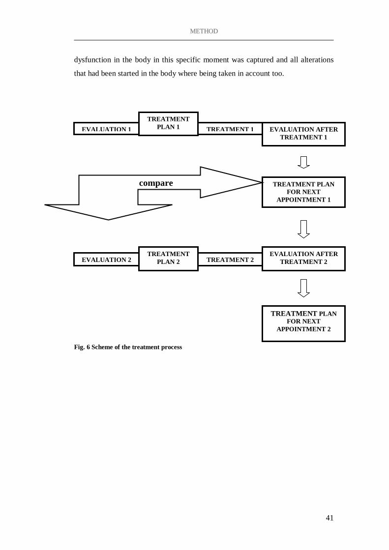
made at the end of the last treatment (Fig.2). In this way the most conspicuous
MMEETTHHOODD
41
TREATMENT PLAN 1 TREATMENT 1 EVALUATION AFTER
TREATMENT 1
TREATMENT PLAN FOR NEXT
APPOINTMENT 1
EVALUATION 1
EVALUATION 2 TREATMENT
PLAN 2 TREATMENT 2EVALUATION AFTER
TREATMENT 2
TREATMENT PLAN FOR NEXT
APPOINTMENT 2
dysfunction in the body in this specific moment was captured and all alterations
that had been started in the body where being taken in account too.
Fig. 6 Scheme of the treatment process
compare
CCAASSEE PPRREESSEENNTTAATTIIOONN
42
7 CASE PRESENTATION
7.1 The patients case history
7.1.1 Anamnesis
Date of Birth: 26.07.1951
Sex: female
Family status: divorced, 2 sons (34 and 30)
Profession: retired since 1 year, worked as a shop assistant
1.What is your reason for an osteopathic treatment?
Lumbar pain and giving way symptoms in the left leg which have led to tumbles,
without obvious reason, in the last couple of months
2.When did those complaints start?
Pain in the lumbar and cervical spine have been present on and off during the last
couple of years. It came to a peak end of 2005.
3.What kind of pain do you have?
Lumbar pain, occurring especially after static behaviour like sitting and standing
for a while but also after a long walk including irradiations into the left leg and
sharp, tearing pain from the lumbar spine irradiating in a girdle sensation to the
stomach.
4. Do you have any other pain or discomfort?
Yes. Pain in the cervical spine over years and the left foot is swollen most of the
time and feels different than the other.

CCAASSEE PPRREESSEENNTTAATTIIOONN
43
5. What kind of treatments did you have for these problems?
I had physical therapy for the cervical and lumbar spine a few times and pain
medication. In autumn last year a periradicular infiltration in my lower lumbar
spine was done twice. I felt a little bit better after the first one but the pain came
back so I had a second one which did not bring any relieve, so that end of last
December I decided to have the surgery done. After that I felt quite good for a
while. I had physical therapy in the beginning but could not quite keep up the
daily training. And then the symptoms started again and I fell twice within 2
weeks in spring 2006 because my left leg gave way. And now I am afraid that I
will get back to the point where the pain gets unbearable.
6. Did you have any traumas?
I had my leg ankle broken twice. Once 16 years ago and the second time 2 ½
years ago, both without surgery.
7. Does your left leg still give you trouble?
Yes it is swollen quite a lot- mostly in the calf down into the foot
8. What kind of surgeries did you have done?
Appendectomy seven years ago, ovariectomy left 16 years ago shortly after my
foot fracture, a laryngeal tumour and vein stripping in both legs.
9. How many pregnancies did you have?
I had 3 pregnancies. I gave birth to two children and had one abort.
10. Did you have any problems during your pregnancies or delivering?
I didn’t have problems during my pregnancies. My first son was delivered with a
forceps. My second son came in breech presentation but I did not have any major
problems.
CCAASSEE PPRREESSEENNTTAATTIIOONN
44
11. How was your cycle?
I did not have any problems when I was young. When I had my ovariectomy the
surgeon found out that my second ovary didn’t work and I was prescribed
hormones that I took for 10 years.
12. Do you take any medication right now?
Yes I am on medication easing my high blood pressure and my asthma.
13. How is your digestion?
I did have diarrhoea on and off and had enteroscopy and gastroscopy without any
significant result.
14. Do you smoke and how much?
Yes 20-30 cigarettes a day.
7.1.2 Medical history
18.11.1999 MRI cervical spine: Chondrosis C5/C6 paramedian left with
prolapse and stenosis of the foramen. Slight changes on the right
side.
4.03.2001 MRI lumbar spine: malpositioning of the lumbar spine, slight
osteochondrosis L3 to L5 with moderate median to paramedian
left pointed protrusions. In these segments the radix is already
affected in
22.06.2002 radical recurrent varices surgery with crossectomy, right
proximal stripping v.saphena magna, left distal stripping
v.saphena magna, ligation of insufficient perforans veins both
sides
CCAASSEE PPRREESSEENNTTAATTIIOONN
45
17.02.2003 duplex-sonography of the big cervical arteries: 3mm plaque on
the right bifurcation, all other vessel parts show regular stream
14.06.2004 abruptio ossea apices mall. lat. sin., contusion mall.med.sin –
lower leg scotchcast
22.02.2005-24.02.2005 final report stationary examinations because of
retrosternal pain with irradiation to both shoulders paired with anxieties and state
of tension, all cardiovascular tests negative, recommendation of an orthopaedic
evaluation of the cervical and thoracic spine
09.08.2004 densitometry: moderate diminution of bone density in the
lumbar spine and femoral shaft
09.08.2005 duplex-sonography of the big cervical arteries: no significant
changes in comparison to the assessment in 02.2003
17.08.2004 cystectomy right larynx
06.09.2005 esophago-gastro-duodenoscopy: small esophageal hernia,
mucosa of the stomach shows patchy erythema, subtle
granulation and pointy exudates in the antrum, 3mm ulcus
prepylory, obvious signs of an acute duodenitis
06.09.2005 colonoscopy: hemorrhoids gradeII, proctopolypus
05.10.2005 CT-controlled periradicular infiltration: radix L4 left
21.11.2005 CT-controlled periradicular infiltration: radix L4 left
27.12.2005 microsurgical discusextraction- sequestrectomy: L4/5 left
24.03.2006 epigastric sonography: spleen cysts, no pathological findings
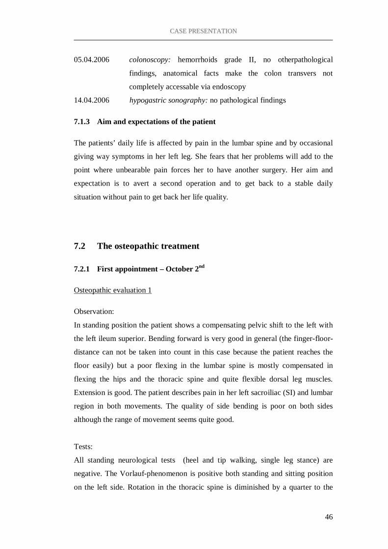
CCAASSEE PPRREESSEENNTTAATTIIOONN
46
05.04.2006 colonoscopy: hemorrhoids grade II, no otherpathological
findings, anatomical facts make the colon transvers not
completely accessable via endoscopy
14.04.2006 hypogastric sonography: no pathological findings
7.1.3 Aim and expectations of the patient
The patients’ daily life is affected by pain in the lumbar spine and by occasional
giving way symptoms in her left leg. She fears that her problems will add to the
point where unbearable pain forces her to have another surgery. Her aim and
expectation is to avert a second operation and to get back to a stable daily
situation without pain to get back her life quality.
7.2 The osteopathic treatment
7.2.1 First appointment – October 2nd
Osteopathic evaluation 1
Observation:
In standing position the patient shows a compensating pelvic shift to the left with
the left ileum superior. Bending forward is very good in general (the finger-floor-
distance can not be taken into count in this case because the patient reaches the
floor easily) but a poor flexing in the lumbar spine is mostly compensated in
flexing the hips and the thoracic spine and quite flexible dorsal leg muscles.
Extension is good. The patient describes pain in her left sacroiliac (SI) and lumbar
region in both movements. The quality of side bending is poor on both sides
although the range of movement seems quite good.
Tests:
All standing neurological tests (heel and tip walking, single leg stance) are
negative. The Vorlauf-phenomenon is positive both standing and sitting position
on the left side. Rotation in the thoracic spine is diminished by a quarter to the
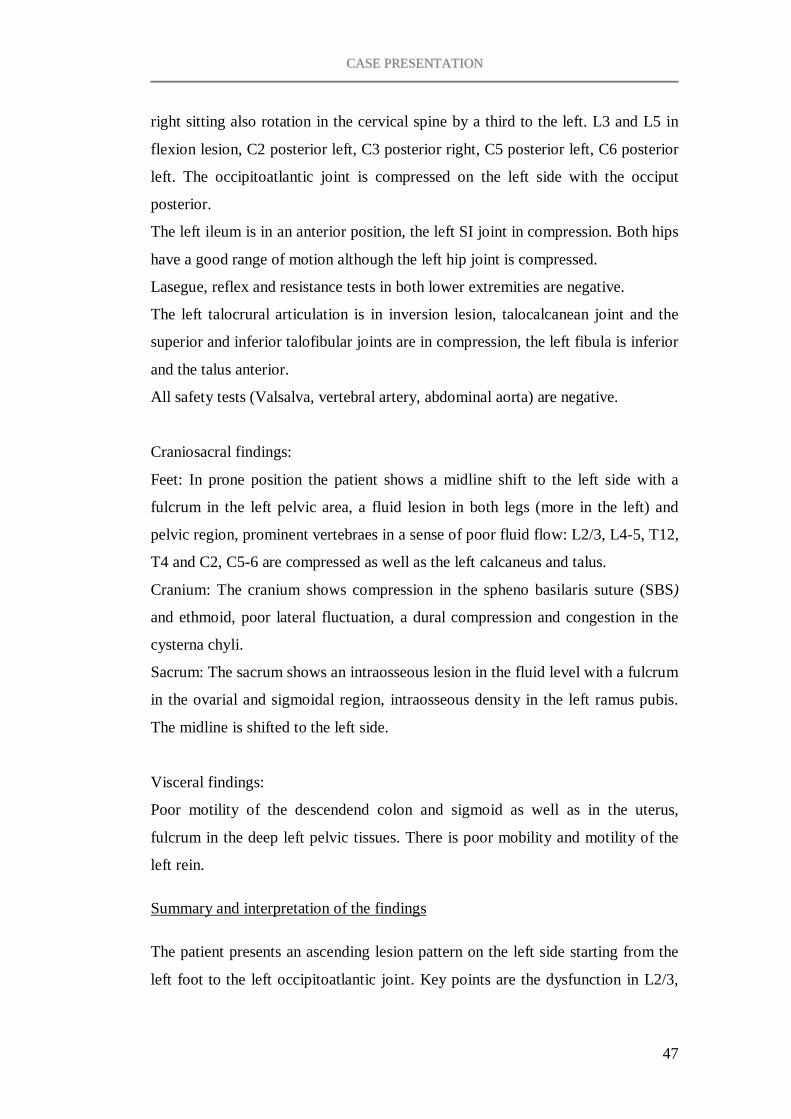
CCAASSEE PPRREESSEENNTTAATTIIOONN
47
right sitting also rotation in the cervical spine by a third to the left. L3 and L5 in
flexion lesion, C2 posterior left, C3 posterior right, C5 posterior left, C6 posterior
left. The occipitoatlantic joint is compressed on the left side with the occiput
posterior.
The left ileum is in an anterior position, the left SI joint in compression. Both hips
have a good range of motion although the left hip joint is compressed.
Lasegue, reflex and resistance tests in both lower extremities are negative.
The left talocrural articulation is in inversion lesion, talocalcanean joint and the
superior and inferior talofibular joints are in compression, the left fibula is inferior
and the talus anterior.
All safety tests (Valsalva, vertebral artery, abdominal aorta) are negative.
Craniosacral findings:
Feet: In prone position the patient shows a midline shift to the left side with a
fulcrum in the left pelvic area, a fluid lesion in both legs (more in the left) and
pelvic region, prominent vertebraes in a sense of poor fluid flow: L2/3, L4-5, T12,
T4 and C2, C5-6 are compressed as well as the left calcaneus and talus.
Cranium: The cranium shows compression in the spheno basilaris suture (SBS)
and ethmoid, poor lateral fluctuation, a dural compression and congestion in the
cysterna chyli.
Sacrum: The sacrum shows an intraosseous lesion in the fluid level with a fulcrum
in the ovarial and sigmoidal region, intraosseous density in the left ramus pubis.
The midline is shifted to the left side.
Visceral findings:
Poor motility of the descendend colon and sigmoid as well as in the uterus,
fulcrum in the deep left pelvic tissues. There is poor mobility and motility of the
left rein.
Summary and interpretation of the findings
The patient presents an ascending lesion pattern on the left side starting from the
left foot to the left occipitoatlantic joint. Key points are the dysfunction in L2/3,

CCAASSEE PPRREESSEENNTTAATTIIOONN
48
Th10-12, the descendend colon and the left pelvic region. Important relations of
vertebrae and innervations of abdominal organs in this case are the sympathic
innervation of the lower extremity (Th 10- L2). Related organs of the restricted
vertebrae are: Th12 (peritoneum, small intestine, lymphatic circulation and reins),
L2 (bladder, colon) and L3 (ovaries, uterus). The restricted vertebral segments
match with the visceral findings.
From this first evaluation and the medical history and anamnesis a complex lesion
pattern of the whole left side is quite prominent. The injury of the left foot and
ankle 20 years ago seems to have played a major roll in the development of the
present imbalance of the body. It will be vital to treat the foot and ankle joints and
to balance the weight-bearing axis of the whole left leg in order to change the
massive overload on the lumbar segments and tissues. The visceral findings
(colon left and rein) and peritoneal tension in the left pelvis might be partly origin
of the presented lesion pattern as well as the restricted scar of the ovariectomy
(fascial lesion pattern).
My impression of the patient is that mental stress in combination with physical
overload and the metabolic situation (smoking, nutrition) also seem to affect the
patients’ overall condition.
Treatment Plan 1
All of the evaluated dysfunctions in this moment show a highly fluid-related
lesion pattern. So the first step will be to balance all fluids through craniosacral
techniques, which are probably partly a compensation for structural lesions
especially of the left foot and ankle. The sacrum will be the first region to start
with because all dysfunctions seem to be related to the left pelvic area. Further on
it will be necessary to work on all joints in the left foot, tibia and fibula on a fluid,
interosseous and membranous level (structural techniques onto the joints seem to
be to early at this stage considering the long maintained lesion pattern and its
resulting compensations) and balancing the SBS.

CCAASSEE PPRREESSEENNTTAATTIIOONN
49
Treatment 1 (Fig.7)
Starting the treatment with the sacrum (1) induced balancing the SBS (2) to
stabilize the fluids. After that the ankle was treated with balanced ligamentous
techniques, the restriction in the fibula was treated using a recoil technique onto
the superior and inferior joints and an interosseous release technique as well as a
deep fascial release of the interosseous membrane (3).
After the treatment the midline was much more integrated and the cervical
dysfunctions diminished.
Treatment plan for the next appointment
The next appointment will be in about one week to prevent the patient falling back
into her old lesion pattern. The focus will be onto the treatment of the descendend
colon, left pelvic region, and the foot. The SBS and occipitoatlantic joint will be
under survey.
Fig. 7 Scheme of treatment 1

CCAASSEE PPRREESSEENNTTAATTIIOONN
50
7.2.2 Second appointment – October 10th
Osteopathic evaluation 2
The overall impression is that the result of the first treatment has been maintained.
The dysfunction in the left ileum and pelvic (restricted SI joint, compressed ileum,
compressed pubis, tension in the peritoneum and the ovarial region) area is
accented. A slight compression in the SBS is palpable. The patient reports a
tearing sensation in the left wrist joint but does not show any restrictions.
Treatment plan 2
The plan for today is to start again with balancing the SBS, then work on the
pelvis starting with a fluid technique on the sacrum, balancing the left pelvis and
again working on the foot.
Treatment 2 (Fig.8)
The SBS reacts much faster than a week ago loosening the compression,
presenting a compression of the dura and ethmoid, not fully releasing in the
treatment (1). The balancing of the SI joint through the sacrum and ileum results
in an accented fascial dysfunction around the scar of the ovariectomy and
therefore followed by an indirect fascial technique on he scar, which takes quite a
long time to release (2). After working on the peritoneum und the colon
descendens and a MET on the psoas and the left ileum (posteriorising,) the fluid
system in the pelvis seems to improve (2). The treatment of the foot and ankle is
focused on the calcaneus and talus with interosseous techniques (3). A re-
evaluation of the whole system indicates a decompression of Th12 (4) through a
HVLA technique, also giving input onto the cysterna chyli. A global fluid
balancing closes this session.
CCAASSEE PPRREESSEENNTTAATTIIOONN
51
Fig. 8 Scheme of treatment 2
Treatment plan for the next appointment
The focus will be on the cysterna chyli and sacral plexus, foot and ankle and the
scar.
7.2.3 Third appointment – October 18th
Osteopathic evaluation 3
The reported pain in the left wrist joint are gone since the last treatment. In the
global evaluation the restricted region in the left pelvis is still prominent as is the
fascial dysfunction around the scar. The restriction in the left ankle and foot has
improved as well as the fluidal situation. The state global fluid circulatory is quite
well. The SBS is quite well balanced.

CCAASSEE PPRREESSEENNTTAATTIIOONN
52
Treatment plan 3
Today’s focus will be again on the left pelvic region, scar and on the foot/ankle as
well as on the alignment of the left leg in sense of the weight bearing axis. The
next appointment should be scheduled in about one week.
Treatment 3 (Fig.9)
The treatment begins again with the sacrum and then balancing the ileum and the
sacrum to release the pelvic tension, then a fascial release of the scar, which reacts
very quick (1). The work on the weight bearing of the leg starts with myofascial
realease techniques on the upper leg (hereby especially the region of the adductor
muscles) (2), lower leg (3) and foot (4), and the thoraco-lumbar fascia. The
system seems to integrate the structural changes very well. The treatment finishes
with the global fluid balancing.
Fig. 9 Scheme of treatment 3

CCAASSEE PPRREESSEENNTTAATTIIOONN
53
Treatment plan for next appointment
Working on the foot and ankle structurally including the knee and hip and a new
appointment in one week is scheduled.
7.2.4 Fourth appointment – October 24th
Osteopathic evaluation 4
The patient reports of the diagnosis of a beginning cataract in the left eye but
doesn’t seem to upset about it. In general her bodily condition is much better on a
global fluid circulatory level as well as in the standing position, where the weight
bearing is much more centred, the pelvic shift decreased and the active movement
in the lumbar spine seems to be smoother.
Treatment Plan 4
Structural release techniques on the foot, ankle, knee and hip and the next
appointment in about one week are scheduled.
Treatment 4 (Fig.10)
This treatment is started with a HVLA technique to release the compression of the
talocrural articulation and to bring the talus back into its position (1) and a
myofascial release of the plantar fascia (2). The knee reacts quite well on a global
dynamic fascia technique. Then a myofascial technique to decompress the hip is
applied (3) and supported by balancing the scarum and ileum (4). The treatment
finishes with a craniosacral treatment with focus on a centralising of the midline
and improving the circulation around the cysterna chyli (5).
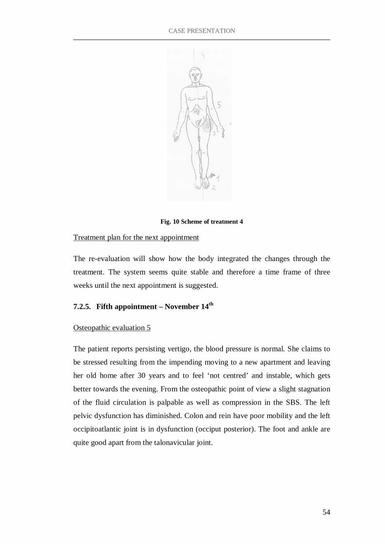
CCAASSEE PPRREESSEENNTTAATTIIOONN
54
Fig. 10 Scheme of treatment 4
Treatment plan for the next appointment
The re-evaluation will show how the body integrated the changes through the
treatment. The system seems quite stable and therefore a time frame of three
weeks until the next appointment is suggested.
7.2.5. Fifth appointment – November 14th
Osteopathic evaluation 5
The patient reports persisting vertigo, the blood pressure is normal. She claims to
be stressed resulting from the impending moving to a new apartment and leaving
her old home after 30 years and to feel ‘not centred’ and instable, which gets
better towards the evening. From the osteopathic point of view a slight stagnation
of the fluid circulation is palpable as well as compression in the SBS. The left
pelvic dysfunction has diminished. Colon and rein have poor mobility and the left
occipitoatlantic joint is in dysfunction (occiput posterior). The foot and ankle are
quite good apart from the talonavicular joint.
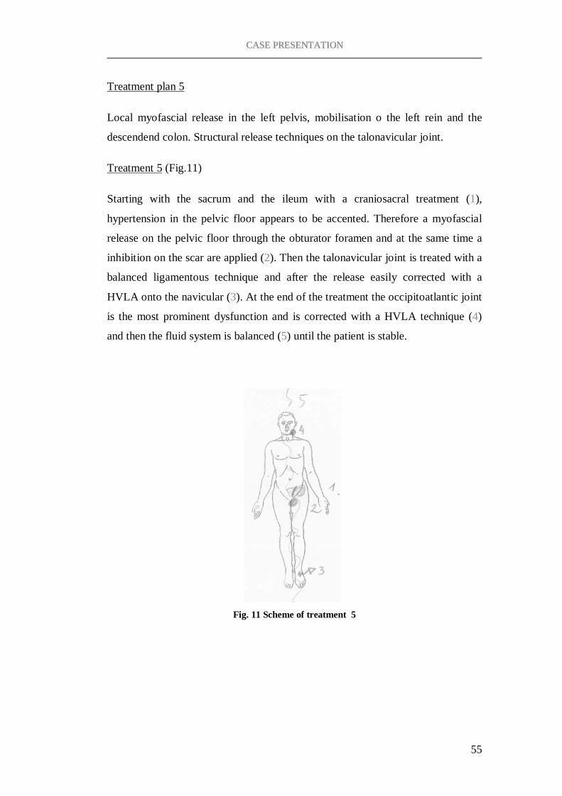
CCAASSEE PPRREESSEENNTTAATTIIOONN
55
Treatment plan 5
Local myofascial release in the left pelvis, mobilisation o the left rein and the
descendend colon. Structural release techniques on the talonavicular joint.
Treatment 5 (Fig.11)
Starting with the sacrum and the ileum with a craniosacral treatment (1),
hypertension in the pelvic floor appears to be accented. Therefore a myofascial
release on the pelvic floor through the obturator foramen and at the same time a
inhibition on the scar are applied (2). Then the talonavicular joint is treated with a
balanced ligamentous technique and after the release easily corrected with a
HVLA onto the navicular (3). At the end of the treatment the occipitoatlantic joint
is the most prominent dysfunction and is corrected with a HVLA technique (4)
and then the fluid system is balanced (5) until the patient is stable.
Fig. 11 Scheme of treatment 5
CCAASSEE PPRREESSEENNTTAATTIIOONN
56
Treatment plan for the next appointment
The plan for the next treatment will be developed out of the findings at the
beginning of the next appointment, which will be scheduled in about 3-4 weeks.
7.2.6 Final appointment – December 18th
The scheduled appointment on December 5th was cancelled deferred by the patient
due to being busy moving although the lifting of heavy boxes and the physical and
psychological stress has kicked the patient back into massive pain. A final
evaluation and interview was then held 6 weeks after the last treatment.
Final evaluation
Due to the patients moving to a new flat the patient is physically exhausted and
complains about pain in the back, arms and neck. From an osteopathic point of
view the patient has fallen back into her old pattern but is still more balanced than
before the first appointment. Structural dysfunctions are found in the
talocalcanean joint, the sacrum and ileum, the SI joint, TH12, TH4, C6/7, C2/3,
and the occipitoatlantic joint left. From a craniosacral point of view the dura is
strongly compressed but reacts quite good on a fluid balancing. The midline is
slightly shifted to the left, as is the weight-bearing axis in the left leg.
Final interview with the patient
- How did you experience the osteopathic treatments?
I think it is very good. I felt comfortable in the treatment and have trust in your
techniques. And although you have explained what you are doing it is impossible
for me to understand how those techniques are able to improve my physical
problems. I was quite hard for me to lie still when I cannot feel a technique that
you are applying.
- Do you think the treatment helped your symptoms to get better?

CCAASSEE PPRREESSEENNTTAATTIIOONN
57
At the moment I cannot really say anything about it because the moving really did
damage to my physical condition and again raised the level of pain. Before that I
realized small improvements but I am not able to state whether this was because
of the treatment or not.
- What do you think causes your recurrent physical problems?
I think it is mostly physical overload and during the time I had to fill in the pain
diary I realised that mental stress might worsen the symptoms as well.
- Would you consider further osteopathic treatment and why?
Yes, because I have tried so many ways to get better and nothing really helped. I
tried physiotherapy too but have to admit that I stopped doing the exercise
program at home because the exercises were too complicated and I didn’t really
know what they were aiming at. I think the osteopathic treatment and a few
exercises that are not too complicated would help me a lot.

RREESSUULLTTSS
58
8 RESULTS
8.1 SF-36 score
All SF-36 results are presented in numbers from 0 (poor) to 100 (good).
8.1.1 SF-36 total results
The health status scores of each testing, subdivided in the eight scales, are
presented in Tab. 1. The first testing was done before the first osteopathic
treatment so it represents the initial condition at the beginning of the treatment
process.
Tab. 1 Sf-36® health status scores
The scores in average show a light increase towards the fourth testing (24.10. =
before the third treatment) followed by a significant drop in the fifth testing
(14.11.) as displayed in Diag.1.
Social functioning, and role limitations emotional seem to be quite steady at a
high of 100 points, with the exception of the fifth testing. Role limitations due to
physical problems are steadily at a high of 100 points and only drop at the last
testing. Vitality rises is highest at the third and fourth testing but drops
significantly in the last two tests. The bodily pain scale shows an improvement of
23 points (or 23%) between the first and fourth testing and drops then back to the
initial score (Diag.2). General medical health and physical functioning fluctuate
steadily in a range of 12 and 10 points.
SF-36® Health status scales 01.10. 10.10. 18.10. 24.10. 14.11. 18.12. Physical functioning (PF) 75 70 75 85 75 80
Role limitations - physical (RP) 100 100 100 100 100 75 Bodily pain (BP) 51 51 62 74 51 51
General medical health (GH) 87 75 82 82 75 87 Vitality (VT) 70 60 75 75 50 35
Social functioning (SF) 100 100 100 100 87,5 100 Role limitations - emotional (RE) 100 100 100 100 66,7 100
Mental health (MH) 92 84 88 84 64 80
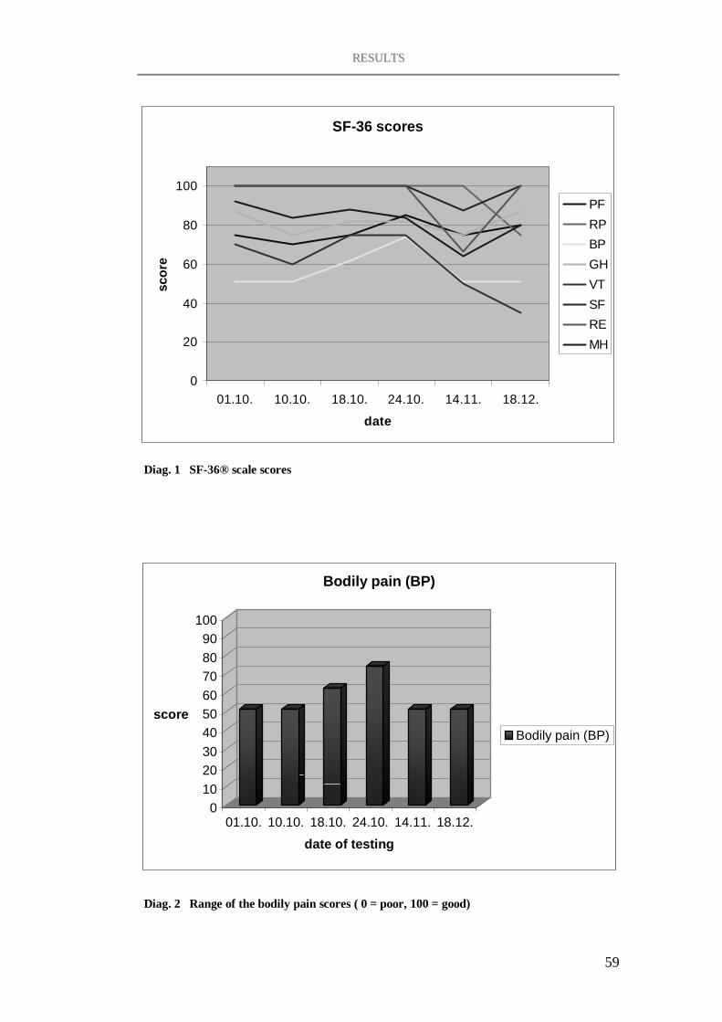
RREESSUULLTTSS
59
SF-36 scores
0
20
40
60
80
100
01.10. 10.10. 18.10. 24.10. 14.11. 18.12.
date
scor
e
PFRPBPGHVTSFREMH
Diag. 1 SF-36® scale scores
0102030405060708090
100
score
01.10. 10.10. 18.10. 24.10. 14.11. 18.12.
date of testing
Bodily pain (BP)
Bodily pain (BP)
Diag. 2 Range of the bodily pain scores ( 0 = poor, 100 = good)
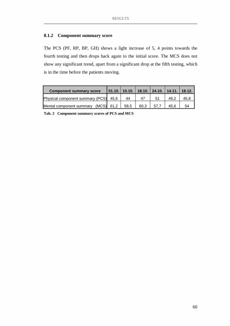
RREESSUULLTTSS
60
8.1.2 Component summary score
The PCS (PF, RP, BP, GH) shows a light increase of 5, 4 points towards the
fourth testing and then drops back again to the initial score. The MCS does not
show any significant trend, apart from a significant drop at the fifth testing, which
is in the time before the patients moving.
Component summary score 01.10. 10.10. 18.10. 24.10. 14.11. 18.12.
Physical component summary (PCS) 45,6 44 47 51 49,2 45,8
Mental component summary (MCS) 61,2 58,5 60,3 57,7 45,6 54
Tab. 2 Component summary scores of PCS and MCS
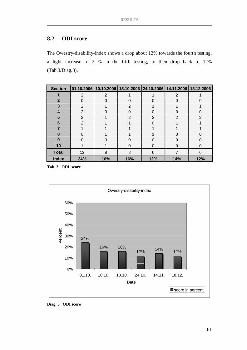
RREESSUULLTTSS
61
8.2 ODI score
The Owestry-disability-index shows a drop about 12% towards the fourth testing,
a light increase of 2 % in the fifth testing, to then drop back to 12%
(Tab.3/Diag.3).
Section 01.10.2006 10.10.2006 18.10.2006 24.10.2006 14.11.2006 18.12.20061 2 2 1 1 2 1 2 0 0 0 0 0 0 3 2 1 2 1 1 1 4 2 0 0 0 0 0 5 2 1 2 2 2 2 6 2 1 1 0 1 1 7 1 1 1 1 1 1 8 0 1 1 1 0 0 9 0 0 0 0 0 0
10 1 1 0 0 0 0 Total 12 8 8 6 7 6 Index 24% 16% 16% 12% 14% 12%
Tab. 3 ODI score
Owestry-disability-index
24%
16% 16%12% 14% 12%
0%
10%
20%
30%
40%
50%
60%
01.10. 10.10. 18.10. 24.10. 14.11. 18.12.
Date
Perc
ent
score in percent
Diag. 3 ODI score

RREESSUULLTTSS
62
8.3 VAS – pain scores
The overall trend of the results of the VAS-scores shows a significant, steady drop
of daily reported pain from the first until a day before the third treatment. The two
days after the first treatment show an increase of 1,15. After the fifth treatment the
score drops again to zero before exploding to 8,9 and above, around the time of
the patients increased physical and mental stress due to moving.
The daily VAS scores were measured by hand on the non-scaled line of 10
centimetres with a degree of precision of 0,05 cm. The days when the patient
forgot to mark the pain on the VAS where left blank but the dates where taken
into the diagram (Diag.4). In these periods the line in the diagram is broken.
The dates of the osteopathic treatments are highlighted in pink.
Visual analogue scale (VAS)
Date of appointment
0123456789
10
15.8
29.8
12.9
26.9
10.10
24.10 7.1
121
.11 5.12
Date
VAS
scor
e
Diag. 4 VAS scores
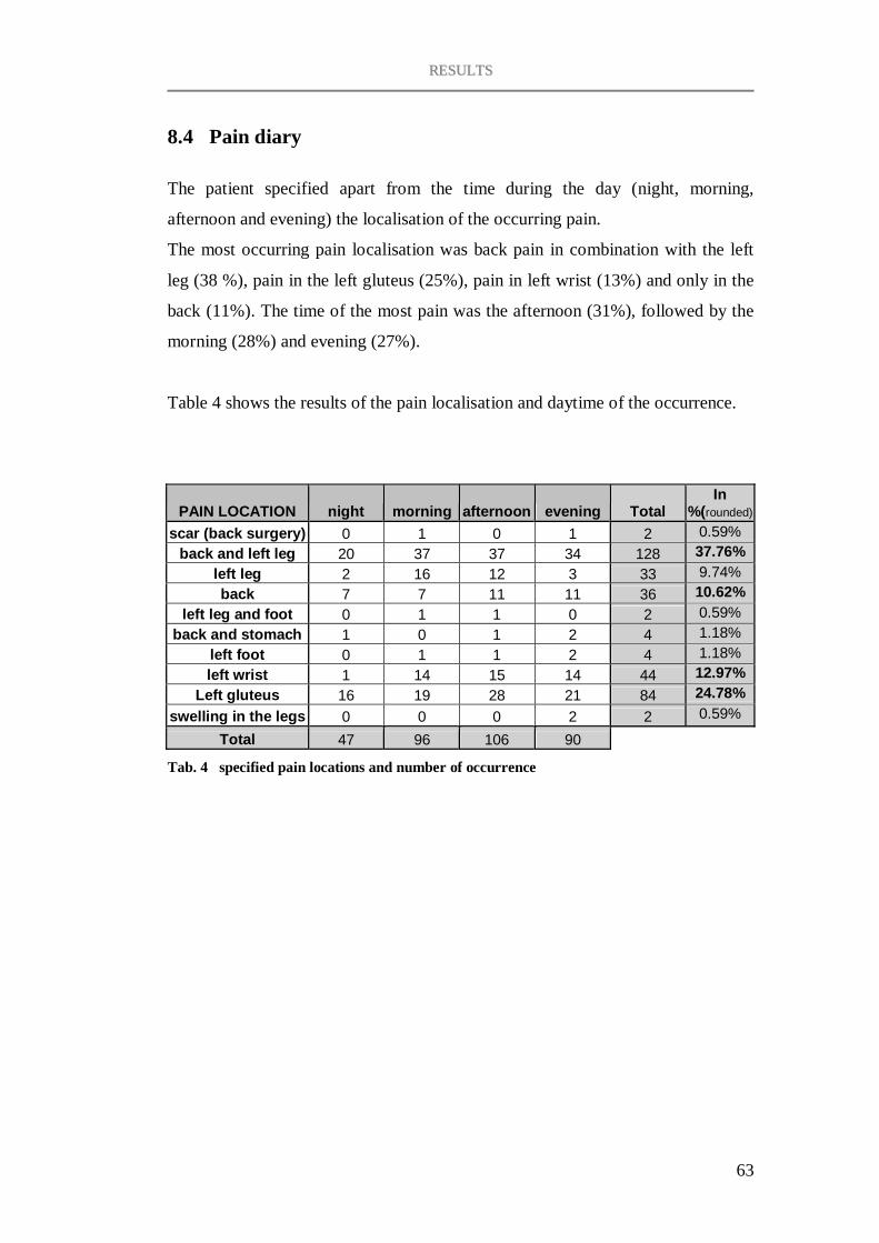
RREESSUULLTTSS
63
8.4 Pain diary
The patient specified apart from the time during the day (night, morning,
afternoon and evening) the localisation of the occurring pain.
The most occurring pain localisation was back pain in combination with the left
leg (38 %), pain in the left gluteus (25%), pain in left wrist (13%) and only in the
back (11%). The time of the most pain was the afternoon (31%), followed by the
morning (28%) and evening (27%).
Table 4 shows the results of the pain localisation and daytime of the occurrence.
PAIN LOCATION night morning afternoon evening Total In
%(rounded)
scar (back surgery) 0 1 0 1 2 0.59% back and left leg 20 37 37 34 128 37.76%
left leg 2 16 12 3 33 9.74% back 7 7 11 11 36 10.62%
left leg and foot 0 1 1 0 2 0.59% back and stomach 1 0 1 2 4 1.18%
left foot 0 1 1 2 4 1.18% left wrist 1 14 15 14 44 12.97%
Left gluteus 16 19 28 21 84 24.78% swelling in the legs 0 0 0 2 2 0.59%
Total 47 96 106 90
Tab. 4 specified pain locations and number of occurrence

DDIISSCCUUSSSSIIOONN
65
9 DISCUSSION
A case study must not lead to the conclusion that the results are representative for
other similar cases but I think it can give important information about the effects
of a treatment and the factors influencing it.
It was important to me to keep the design of the treatment process as realistic as
possible in order to capture the influencing factors on to the patient and the
osteopathic management of the problem. This gave an interesting view on the
effect of the patient’s compliance, time management and coping with her problem
within a therapy-process. The results show a significant worsening of the
symptoms in times of mental stress and physical overload and a lack of
compliance in the sense of forgetting to fill in the pain diary and postponing
appointments in periods of stress, which points out the importance of the patient’s
compliance for the success of the treatment.
Overall the results of this case study show a positive trend of improvement
concerning the patient’s pain syndrome. From an osteopathic point of view
positive changes in the patient’s tissue quality (especially in the foot and ankle),
vitality and balance after two treatments can be stated, which leads to the
conclusion that an osteopathic treatment in this case is indicated.
The final interview with the patient gave me the impression that she did develop
more understanding for the nature of her problem and her body’s reaction on
physical and mental overload which is inevitable for the accomplishment of her
chronic pain syndrome.
Concerning the method, I have to say that the SF-36®, which I chose for the fact
that the average appointment interval in my daily practise is 4 to 6 weeks, was not
representative in this case study because it is designed for the use of a 4 weeks
interval and might therefore not me sensitive enough for physical and mental
changes within a shorter period of time. Therefore, although it only contains 10
DDIISSCCUUSSSSIIOONN
66
questions, the more valuable life quality score was the ODI. I think it the best
impression of all affecters and the state of health in the patient’s life gave the pain
diary and especially the VAS. For a more objective presentation of the progress it
might have been interesting to take pictures of the standing position and especially
the swelling and colouring of the left foot and ankle of the patient.
Concluding, I think that this case study objectifies my daily experience in
practising osteopathy and dealing with patients with chronic back pain syndromes.
In this case, according to the results of the study, the chances for a successful
osteopathic treatment seem good and although this is not transferable to other
cases, I was able to draw interesting information about the osteopathic treatment
process out of this study.
LLIITTEERRAATTUURREE
67
LITERATURE
Anderson C., Laubscher S., Burns R: Validation of the short form 36 (SF-36)
health survey questionnaire among stroke patients; Stroke 1996; 27 (10): 1812 -
1816
Brazier J. E., Harper R., Jones,N.M.B., O’Cathain A., Usherwood T., Westlake,
J. : Validating the SF-36 Health survey questionnaire: new outcome measure for
primary care; British Medical Journal 1992; 305: 160-164
Dean N.A., Mitchell B.S.: Anatomic relation between the nuchal ligament
(ligamentum nuchae) and the spinal dura mater in the craniocervical region;
Clinical Anatomy 2002May; 15 (3): 182-185
De Boer A.G.E.M. et al: Is a single-item visual analogue scale as valid, reliable
and responsive as multi-item scales in measuring quality of life?; Biomedical and
Life Sciences, Volume 13, Number 2 / March, 2004
Debroux J.-J.: Faszienbehandlung in der Osteopathie; Hippokrates Verlag 2004
Gerdesmeyer L., Haake M., Goebel1 M., Wagner K.: Der Rückenschmerz ;
Notfall & Hausarztmedizin 2004; 30: 319– 324
Gray’s Anatomy: the anatomical basis of clinical practice; Ed.-in-chief: Standring
S.: ElsevierChurchill Livingston 2005
Greenman P.E.: Lehrbuch der osteopathischen Medizin; Haug Verlag 2003
Hildebrandt J.: Gibt es einen unspezifischen Rückenschmerz?; Z Orthop 2004;
142; 139- 145
Jones L.H.: Strain-counterstrain; Urban & Fischer Verlag 2001

LLIITTEERRAATTUURREE
68
Kniesel B.: Diskogene lumbale Rückenschmerzen; Z Orhop 2004; 142: 709-715
Liem T., Dobler T.K.: Leitfaden der Osteopathie – Parietale Techniken; Urban &
Fischer Verlag 2002
Liem T.: Kranisosakrale Osteopathie; Hippokrates Verlag 2001
Liem T.: Morphodynamik in der Osteopathie; Hippokrates Verlag 2006
Mannion A.F. et al: Development of a German version of the Oswestry Disability
Index. Part 1: cross-cultural adaptation, reliability, and validity; Eur Spine J. 2006
Jan;15 (1): 55-65
Meert G.F.: Das Becken aus osteopathischer Sicht; Urban & Fischer Verlag 2003
Mitchell F.: Handbuch der MuskelEnergieTechnik Band 1; Hippokrates Verlag
2004
Paoletti S.: Faszien – Anatomie, Strukturen, Techniken, Spezielle Osteopathie;
Urban & Fischer Verlag 2001
Platzer, W.: Taschenatles der Anatomie: Bewegungsapparat; Thieme Verlag 1991
Schmorl G.: Zur pathologischen Anatomie der Lendenbandscheiben; Klin.
Wschr. 2 (1932), Über Verlagerungen von Bandscheibengewebe und ihre Folgen.;
Langenbecks Arch. Klein. Chir. 172(1932)
Schumacher M.: Schmerztherapie der Wirbelsäule; Radiologie up2date 2002; 2:
263-278
LLIITTEERRAATTUURREE
69
Scott K. M., Tobias M. I., Sarfati D., Haslett S: SF-36 health survey reliability,
validity and norms for New Zealand Australian and New Zealand Journal of
Public Health 1999; 23:, 401-406
Sobotta: Atlas der Anatomie – Band 2 Rumpf, Eingeweide, untere Extremität;
Herausgegeben von R. Putz und R. Pabst; Urban & Fischer Verlag 2000
Speece C.A., Crow W.T., Simmons S.L.: Osteopathische Körpertechniken nach
W.G. Sutherland; Hippokrates Verlag 2003
Still, A.T.: Das große Still-Kompendium; Jolandos Verlag 2002
Turk D.C., Okifuji A.: Psychological factors in chronic pain: evolution and
revolution; J Consult Clin Psychol.; Jun 2002; 70 (3): 678-90
Van den Berg, F.: Angewandte Physiologie: Band 1-Das Bindegewebe des
Bewegungsapparates verstehen und beeinflussen; Thieme Verlag1999
Van Dun Patrick L.S. Girardin M.R.G.: Embryological study of the spinal dura
and its attachment into the vertebral canal; International Journal of Osteopathic
Medicine Vol.9; Issue 3; Sept. 2006: 85-93
Ware J. E, (1993) SF-36 Health Survey: Manual and Interpretation Guide. Boston:
The Health Institute, New England Medical Center
Ware J.E.Jr, Sherbourne C.D.: The MOS 36-item short-form health survey (SF-
36). I. Conceptual framework and item selection; Medical Care1992Jun; 30 (6):
473-483
Ware J. E.: The MOS 36-Item Short Form Health Survey (SF-36). In Sederer, L.
I & Dickey, B (1996). Outcomes Assessment in Clinical Practice; Baltimore:
Williams and Wilkins

LLIITTEERRAATTUURREE
70
Weiland W., Wessel K.: Therapie des Rückenschmerzes- Was ist durch Studien
belegt?; Fortschr Neurol Psychiat 2004; 72: 344-350
White A A, Panjabi M.M.:Clinical biomechanics of the spine. JB Lippincott 1978,
Philadelphia
www.orthosurg.org.uk/odi/
Zöllner et al.: Der Einfluss einer Nukleotomie auf die
biomechanischenEigenschaftendes lumbalen Bewegungssegmentes; Zentralbl
Neurochir 2000; 61: 138-142

APPENDIX

German ODI Translation
Bitte füllen Sie diesen Fragebogen aus. Er soll uns darüber informieren, wie Ihre Rücken- (oder Bein-) Probleme Ihre Fähigkeit beeinflussen, den Alltag zu bewältigen. Wir bitten Sie, jeden Abschnitt zu beantworten. Kreuzen Sie in jedem Abschnitt nur die Aussage an, die Sie heute am besten beschreibt.
Abschnitt 1: Schmerzstärke
• Ich habe momentan keine Schmerzen • Die Schmerzen sind momentan sehr schwach • Die Schmerzen sind momentan mässig • Die Schmerzen sind momentan ziemlich stark • Die Schmerzen sind momentan sehr stark • Die Schmerzen sind momentan so schlimm wie nur vorstellbar
Abschnitt 2: Körperpflege (Waschen, Anziehen etc.)
• Ich kann meine Körperpflege normal durchführen, ohne dass die Schmerzen dadurch stärker werden
• Ich kann meine Körperpflege normal durchführen, aber es ist schmerzhaft • Meine Körperpflege durchzuführen ist schmerzhaft, und ich bin langsam
und vorsichtig • Ich brauche bei der Körperpflege etwas Hilfe, bewältige das meiste aber
selbst • Ich brauche täglich Hilfe bei den meisten Aspekten der Körperpflege • Ich kann mich nicht selbst anziehen, wasche mich mit Mühe und bleibe im
Bett
Abschnitt 3: Heben
• Ich kann schwere Gegenstände heben, ohne dass die Schmerzen dadurch stärker werden
• Ich kann schwere Gegenstände heben, aber die Schmerzen werden dadurch stärker
• Schmerzen hindern mich daran, schwere Gegenstände vom Boden zu heben, aber es geht, wenn sie geeignet stehen (z.B. auf einem Tisch)
• Schmerzen hindern mich daran, schwere Gegenstände zu heben, aber ich kann leichte bis mittelschwere Gegenstände heben, wenn sie geeignet stehen
• Ich kann nur sehr leichte Gegenstände heben • Ich kann überhaupt nichts heben oder tragen
Abschnitt 4: Gehen
• Schmerzen hindern mich nicht daran, so weit zu gehen, wie ich möchte • Schmerzen hindern mich daran, mehr als 1-2 km zu gehen • Schmerzen hindern mich daran, mehr als 0.5 km zu gehen • Schmerzen hindern mich daran, mehr als 100 m zu gehen • Ich kann nur mit einem Stock oder Krücken gehen • Ich bin die meiste Zeit im Bett und muss mich zur Toilette schleppen
Abschnitt 5: Sitzen
• Ich kann auf jedem Stuhl so lange sitzen wie ich möchte • Ich kann auf meinem Lieblingsstuhl so lange sitzen wie ich möchte • Schmerzen hindern mich daran, länger als 1 Stunde zu sitzen • Schmerzen hindern mich daran, länger als eine halbe Stunde zu sitzen • Schmerzen hindern mich daran, länger als 10 Minuten zu sitzen • Schmerzen hindern mich daran, überhaupt zu sitzen
Abschnitt 6: Stehen
• Ich kann so lange stehen wie ich möchte, ohne dass die Schmerzen dadurch stärker werden
• Ich kann so lange stehen wie ich möchte, aber die Schmerzen werden dadurch stärker
• Schmerzen hindern mich daran, länger als 1 Stunde zu stehen • Schmerzen hindern mich daran, länger als eine halbe Stunde zu stehen • Schmerzen hindern mich daran, länger als 10 Minuten zu stehen • Schmerzen hindern mich daran, überhaupt zu stehen
Abschnitt 7: Schlafen
• Mein Schlaf ist nie durch Schmerzen gestört • Mein Schlaf ist gelegentlich durch Schmerzen gestört • Ich schlafe auf Grund von Schmerzen weniger als 6 Stunden • Ich schlafe auf Grund von Schmerzen weniger als 4 Stunden • Ich schlafe auf Grund von Schmerzen weniger als 2 Stunden • Schmerzen hindern mich daran, überhaupt zu schlafen
Abschnitt 8: Sexualleben (falls zutreffend)
• Mein Sexualleben ist normal, und die Schmerzen werden dadurch nicht stärker
• Mein Sexualleben ist normal, aber die Schmerzen werden dadurch stärker • Mein Sexualleben ist nahezu normal, aber sehr schmerzhaft • Mein Sexualleben ist durch Schmerzen stark eingeschränkt

• Ich habe auf Grund von Schmerzen fast kein Sexualleben • Schmerzen verhindern jegliches Sexualleben
Abschnitt 9: Sozialleben
• Mein Sozialleben ist normal, und die Schmerzen werden dadurch nicht stärker
• Mein Sozialleben ist normal, aber die Schmerzen werden dadurch stärker • Schmerzen haben keinen wesentlichen Einfluss auf mein Sozialleben,
ausser dass sie meine eher aktiven Interessen, z.B. Sport einschränken • Schmerzen schränken mein Sozialleben ein, und ich gehe nicht mehr so oft
aus • Schmerzen schränken mein Sozialleben auf mein Zuhause ein • Ich habe auf Grund von Schmerzen kein Sozialleben
Abschnitt 10: Reisen
• Ich kann überallhin reisen, und die Schmerzen werden dadurch nicht stärker
• Ich kann überallhin reisen, aber die Schmerzen werden dadurch stärker • Trotz starker Schmerzen kann ich länger als 2 Stunden unterwegs sein • Ich kann auf Grund von Schmerzen höchstens 1 Stunde unterwegs sein • Ich kann auf Grund von Schmerzen nur kurze notwendige Fahrten unter 30
Minuten machen • Schmerzen hindern mich daran, Fahrten zu machen, ausser zur
medizinischen Behandlung
Source: http://www.orthosurg.org.uk/odi/ Translation by:
PD Dr Anne F Mannion Schulthess Klinik Lengghalde 2 8008 Zürich Switzerland Tel: 00 41 1 385 7584 Fax: 00 41 1 385 7590 E-mail: [email protected] Web: www.schulthess-clinic.ch
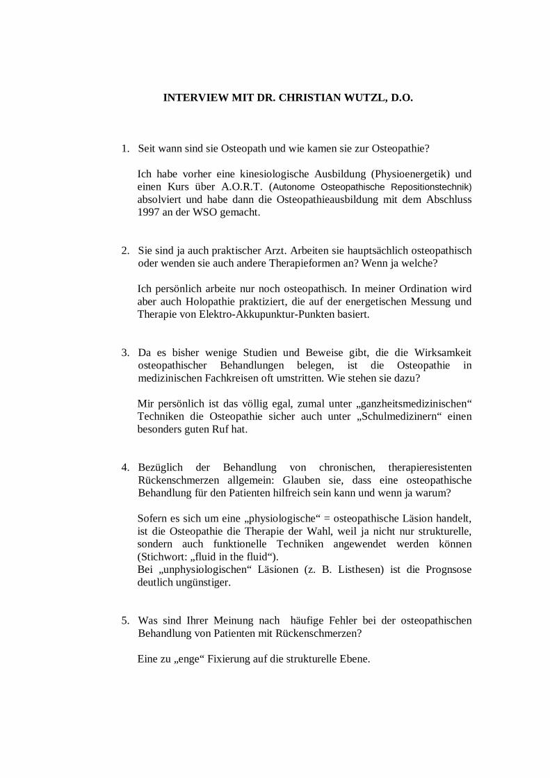
INTERVIEW MIT DR. CHRISTIAN WUTZL, D.O.
1. Seit wann sind sie Osteopath und wie kamen sie zur Osteopathie?
Ich habe vorher eine kinesiologische Ausbildung (Physioenergetik) und einen Kurs über A.O.R.T. (Autonome Osteopathische Repositionstechnik) absolviert und habe dann die Osteopathieausbildung mit dem Abschluss 1997 an der WSO gemacht.
2. Sie sind ja auch praktischer Arzt. Arbeiten sie hauptsächlich osteopathisch oder wenden sie auch andere Therapieformen an? Wenn ja welche?
Ich persönlich arbeite nur noch osteopathisch. In meiner Ordination wird aber auch Holopathie praktiziert, die auf der energetischen Messung und Therapie von Elektro-Akkupunktur-Punkten basiert.
3. Da es bisher wenige Studien und Beweise gibt, die die Wirksamkeit osteopathischer Behandlungen belegen, ist die Osteopathie in medizinischen Fachkreisen oft umstritten. Wie stehen sie dazu?
Mir persönlich ist das völlig egal, zumal unter „ganzheitsmedizinischen“ Techniken die Osteopathie sicher auch unter „Schulmedizinern“ einen besonders guten Ruf hat.
4. Bezüglich der Behandlung von chronischen, therapieresistenten Rückenschmerzen allgemein: Glauben sie, dass eine osteopathische Behandlung für den Patienten hilfreich sein kann und wenn ja warum?
Sofern es sich um eine „physiologische“ = osteopathische Läsion handelt, ist die Osteopathie die Therapie der Wahl, weil ja nicht nur strukturelle, sondern auch funktionelle Techniken angewendet werden können (Stichwort: „fluid in the fluid“). Bei „unphysiologischen“ Läsionen (z. B. Listhesen) ist die Prognsose deutlich ungünstiger.
5. Was sind Ihrer Meinung nach häufige Fehler bei der osteopathischen
Behandlung von Patienten mit Rückenschmerzen?
Eine zu „enge“ Fixierung auf die strukturelle Ebene.
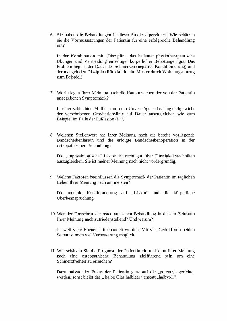
6. Sie haben die Behandlungen in dieser Studie supervidiert. Wie schätzen sie die Vorraussetzungen der Patientin für eine erfolgreiche Behandlung ein?
In der Kombination mit „Disziplin“, das bedeutet physiotherapeutische Übungen und Vermeidung einseitiger körperlicher Belastungen gut. Das Problem liegt in der Dauer der Schmerzen (negative Konditionierung) und der mangelnden Disziplin (Rückfall in alte Muster durch Wohnungsumzug zum Beispiel)
7. Worin lagen Ihrer Meinung nach die Hauptursachen der von der Patientin angegebenen Symptomatik?
In einer schlechten Midline und dem Unvermögen, das Ungleichgewicht der verschobenen Gravitationslinie auf Dauer auszugleichen wie zum Beispiel im Falle der Fußläsion (!!!!).
8. Welchen Stellenwert hat Ihrer Meinung nach die bereits vorliegende Bandscheibenläsion und die erfolgte Bandscheibenoperation in der osteopathischen Behandlung?
Die „unphysiologische“ Läsion ist recht gut über Flüssigkeitstechniken auszugleichen. Sie ist meiner Meinung nach nicht vordergründig.
9. Welche Faktoren beeinflussen die Symptomatik der Patientin im täglichen Leben Ihrer Meinung nach am meisten?
Die mentale Konditionierung auf „Läsion“ und die körperliche Überbeanspruchung.
10. War der Fortschritt der osteopathischen Behandlung in diesem Zeitraum Ihrer Meinung nach zufriedenstellend? Und warum?
Ja, weil viele Ebenen mitbehandelt wurden. Mit viel Geduld von beiden Seiten ist noch viel Verbesserung möglich.
11. Wie schätzen Sie die Prognose der Patientin ein und kann Ihrer Meinung nach eine osteopathische Behandlung zielführend sein um eine Schmerzfreiheit zu erreichen?
Dazu müsste der Fokus der Patientin ganz auf die „potency“ gerichtet werden, sonst bleibt das „ halbe Glas halbleer“ anstatt „halbvoll“.


![+XPEROGW 8QLYHUVLWlW] X% HUOLQ - rewi.hu-berlin.de · einschlägige Art. 11 Abs. 1 zu jenen Vorschriften gehört, die in Bedeutung und Tragweite dem korrespondierenden Artikel der](https://img.pdfslide.us/doc/110x75/5d5b0a6288c99336108bcf5d/xperogw-8qlyhuvlwlw-x-huolq-rewihu-einschlaegige-art-11-abs-1-zu-jenen.jpg)