Embed Size (px)
Citation preview
Received 03/25/2019 Review began 04/01/2019 Review ended 04/03/2019 Published 04/03/2019
© Copyright 2019Ravindran et al. This is an openaccess article distributed under theterms of the Creative CommonsAttribution License CC-BY 3.0., whichpermits unrestricted use, distribution,and reproduction in any medium,provided the original author andsource are credited.
Laser-assisted Excision of GingivalOvergrowth in an Endodontic Perforation:A Case ReportDeepak M. Ravindran , Ram Sabarish , Devi Arul , Supraja Ajit , Dhivya M. Harini
1. Periodontology, Sri Ramachandra Medical College and Research Institute, Chennai, IND
Corresponding author: Deepak M. Ravindran, [email protected] Disclosures can be found in Additional Information at the end of the article
AbstractWith the increase in preventive and restorative dentistry, there is also an increase in theiatrogenic conditions that occur in modern dental practice. The goal of modern dentistry is toprovide patients with a holistic solution by providing functional restoration. This case reportwill highlight one such case where a tooth was diagnosed as having a gingival overgrowththrough a perforation during prior endodontic treatment. Proper diagnosis and treatmentplanning helped restore a tooth that would have been lost.
Categories: MiscellaneousKeywords: gingival polyp, gingival enlargement, diode laser, mineral trioxide aggregate (mta),endodontic perforations
IntroductionThe aim of dentistry today is to salvage any tooth destroyed by dental caries, periodontalbreakdown, or trauma. Modern technology allows the clinician to achieve that. However, thereare times when overzealous treatment and the lack of skill and practice of the clinician causesmore harm than good. One such iatrogenic complication in dentistry is endodontic perforationsseen during root canal therapy. Accidental perforations are a serious complication ofconservative therapy and can cause pain, abscesses, fistulae, and possible proliferation ofgingival epithelium, especially if it occurs in the crestal area [1]. With a thorough diagnosis,treatment planning, and the use of modern restorative materials, a good clinical outcome canbe achieved.
Case PresentationA 33-year-old woman reported to the Department of Periodontology, Sri Ramachandra DentalCollege, Sri Ramachandra Institute of Higher Education & Research (DU), with the chiefcomplaint of a missing tooth and the inability to masticate. She had no relevant medical historyand had a history of undergoing orthodontic treatment, extraction of the lower left first molar,and endodontic treatment done three years back in relation to the left lower second molar. Thepatient had presented with fair oral hygiene.
On clinical examination, a gingival growth was present on the floor of the endodonticallytreated tooth (#37), as seen in Figure 1.
1 1 1 1 1
Open Access CaseReport DOI: 10.7759/cureus.4378
How to cite this articleRavindran D M, Sabarish R, Arul D, et al. (April 03, 2019) Laser-assisted Excision of Gingival Overgrowthin an Endodontic Perforation: A Case Report. Cureus 11(4): e4378. DOI 10.7759/cureus.4378
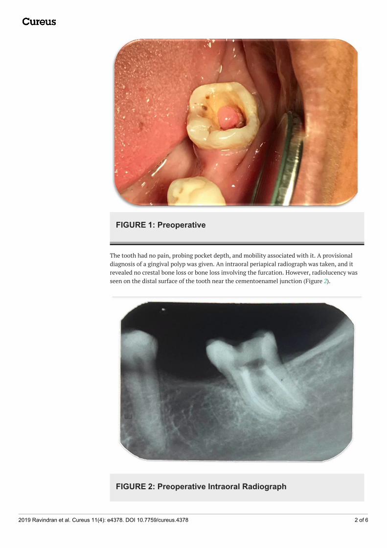
FIGURE 1: Preoperative
The tooth had no pain, probing pocket depth, and mobility associated with it. A provisionaldiagnosis of a gingival polyp was given. An intraoral periapical radiograph was taken, and itrevealed no crestal bone loss or bone loss involving the furcation. However, radiolucency wasseen on the distal surface of the tooth near the cementoenamel junction (Figure 2).
FIGURE 2: Preoperative Intraoral Radiograph
2019 Ravindran et al. Cureus 11(4): e4378. DOI 10.7759/cureus.4378 2 of 6
To substantiate the radiographic and clinical features and to establish the pathway of the polyp,a periodontal probe was inserted horizontally from the lingual aspect of the tooth and a smallperforation was noticed on the distolingual aspect of 37. Furthermore, a Gutta Percha Point wasinserted from the distolingual aspect and a pathway was established from the lingual marginalgingiva to the floor of the cavity.
A final diagnosis of gingival enlargement - gingival overgrowth due to accidental perforationwas established. Treatment options included the extraction of the said tooth or thepreservation of the tooth with a combination of periodontal procedures and endodonticmaterials, though it had a questionable prognosis. Both options were explained to the patientand the patient was willing to save the natural tooth, as she had extracted the tooth mesial to it.
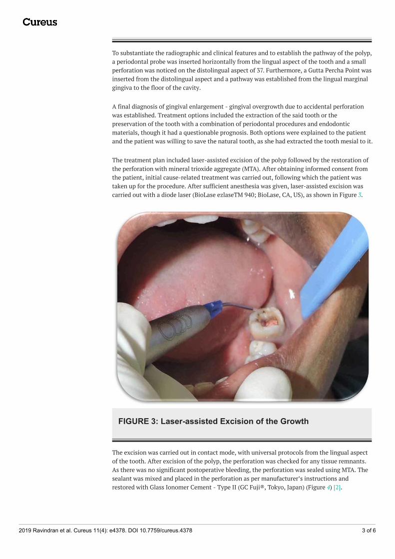
The treatment plan included laser-assisted excision of the polyp followed by the restoration ofthe perforation with mineral trioxide aggregate (MTA). After obtaining informed consent fromthe patient, initial cause-related treatment was carried out, following which the patient wastaken up for the procedure. After sufficient anesthesia was given, laser-assisted excision wascarried out with a diode laser (BioLase ezlaseTM 940; BioLase, CA, US), as shown in Figure 3.
FIGURE 3: Laser-assisted Excision of the Growth
The excision was carried out in contact mode, with universal protocols from the lingual aspectof the tooth. After excision of the polyp, the perforation was checked for any tissue remnants.As there was no significant postoperative bleeding, the perforation was sealed using MTA. Thesealant was mixed and placed in the perforation as per manufacturer’s instructions andrestored with Glass Ionomer Cement - Type II (GC Fuji®, Tokyo, Japan) (Figure 4) [2].
2019 Ravindran et al. Cureus 11(4): e4378. DOI 10.7759/cureus.4378 3 of 6
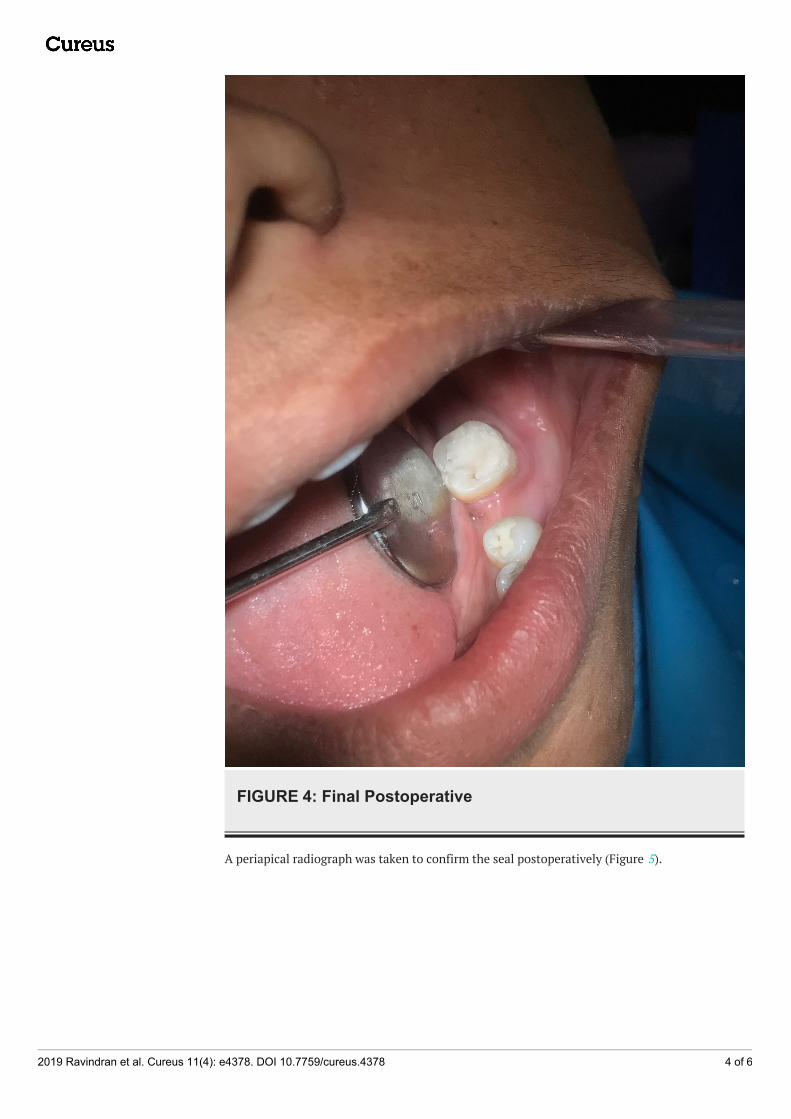
FIGURE 4: Final Postoperative
A periapical radiograph was taken to confirm the seal postoperatively (Figure 5).
2019 Ravindran et al. Cureus 11(4): e4378. DOI 10.7759/cureus.4378 4 of 6
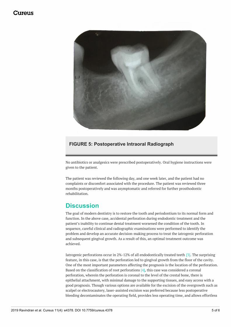
FIGURE 5: Postoperative Intraoral Radiograph
No antibiotics or analgesics were prescribed postoperatively. Oral hygiene instructions weregiven to the patient.
The patient was reviewed the following day, and one week later, and the patient had nocomplaints or discomfort associated with the procedure. The patient was reviewed threemonths postoperatively and was asymptomatic and referred for further prosthodonticrehabilitation.
DiscussionThe goal of modern dentistry is to restore the tooth and periodontium to its normal form andfunction. In the above case, accidental perforation during endodontic treatment and thepatient’s inability to continue dental treatment worsened the condition of the tooth. Insequence, careful clinical and radiographic examinations were performed to identify theproblem and develop an accurate decision-making process to treat the iatrogenic perforationand subsequent gingival growth. As a result of this, an optimal treatment outcome wasachieved.
Iatrogenic perforations occur in 2%-12% of all endodontically treated teeth [3]. The surprisingfeature, in this case, is that the perforation led to gingival growth from the floor of the cavity.One of the most important parameters affecting the prognosis is the location of the perforation.Based on the classification of root perforations [4], this case was considered a coronalperforation, wherein the perforation is coronal to the level of the crestal bone, there isepithelial attachment, with minimal damage to the supporting tissues, and easy access with agood prognosis. Though various options are available for the excision of the overgrowth such asscalpel or electrocautery, laser-assisted excision was preferred because less postoperativebleeding decontaminates the operating field, provides less operating time, and allows effortless
2019 Ravindran et al. Cureus 11(4): e4378. DOI 10.7759/cureus.4378 5 of 6
excision [5-6].
Mineral trioxide aggregate (MTA) is a unique and versatile material in today’s dental practice.MTA has shown excellent results in pulp capping, pulpotomy, periapical surgery, as well asan excellent potential for apexogenesis and apexification [7]. MTA was chosen as the sealant forthe perforation because of its superior marginal adaptation and sealing ability, higherregenerative potential, and because it allows the growth of periodontal ligament on its surface[8].
ConclusionsContemporary dental treatment must result in true oral health, incorporating comfort,function, and aesthetics. Clinical and radiographic evaluation, along with modern treatmentmodalities, helped conserve the tooth and provide a stable and functional result.
Additional InformationDisclosuresHuman subjects: Consent was obtained by all participants in this study. Conflicts of interest:In compliance with the ICMJE uniform disclosure form, all authors declare the following:Payment/services info: All authors have declared that no financial support was received fromany organization for the submitted work. Financial relationships: All authors have declaredthat they have no financial relationships at present or within the previous three years with anyorganizations that might have an interest in the submitted work. Other relationships: Allauthors have declared that there are no other relationships or activities that could appear tohave influenced the submitted work.
References1. Parolia A, Gait TC, Porto IC, Mala K: Endo-perio lesion: a dilemma from 19 th until 21 st
century. J Interdiscip Dentistry. 2013, 3:2-11.2. Dotto RF, Barbosa AN, Dotto SR, Hermes CR: Sealing of root perforation with glass ionomer
cement: a case report. Stomatos. 2014, 20:38.3. Kaushik A, Talwar S, Yadav S, Chaudhary S, Nawal RR: Management of iatrogenic root
perforation with pulp canal obliteration. Saudi Endod J. 2014, 4:141-144.4. Tsesis I, Zvi Fuss Z: Diagnosis and treatment of accidental root perforations. Endod Topics.
2006, 13:95-107.5. Butchi B: Laser assisted excision of gingival polyp in a pediatric patient: report of a case .
European J Biomed Pharm Sci. 2015, 4:600-607.6. Pradeep K, Mishra A, Kalakonda B, Swapna LA, Bagalkotkar A, Mach D: Fibroepithelial polyp
excision with laser and scalpel: a comparative evaluation. Int J Curr Microbiol App Sci. 2014,3:1057-1062.
7. Macwan C, Deshpande A: Mineral trioxide aggregate (MTA) in dentistry: a review of literature .J Oral Res Rev. 2014, 6:71-74.
8. Kaur M, Singh H, Dhillon JS, Batra M, Saini M: MTA versus biodentine: review of literaturewith a comparative analysis. J Clin Diagn Res. 2017, 11:01-05.10.7860/JCDR/2017/25840.10374
2019 Ravindran et al. Cureus 11(4): e4378. DOI 10.7759/cureus.4378 6 of 6