Embed Size (px)
Citation preview
CASE REPORT PEER REVIEWED | OPEN ACCESS
www.edoriumjournals.com
International Journal of Case Reports and Images (IJCRI)International Journal of Case Reports and Images (IJCRI) is an international, peer reviewed, monthly, open access, online journal, publishing high-quality, articles in all areas of basic medical sciences and clinical specialties.
Aim of IJCRI is to encourage the publication of new information by providing a platform for reporting of unique, unusual and rare cases which enhance understanding of disease process, its diagnosis, management and clinico-pathologic correlations.
IJCRI publishes Review Articles, Case Series, Case Reports, Case in Images, Clinical Images and Letters to Editor.
Website: www.ijcasereportsandimages.com
A case report of breast implant-associated anaplastic large cell lymphoma: The good, the bad, and the ugly
Alya Binmahfouz, Karin Steinke
ABSTRACT
Introduction: Primary lymphoma of the breast is very rare, comprising 0.5% breast neoplasms. The majority of breast lymphomas are of B cell type, postulated to originate from intramammary lymph nodes or breast lymphatics. Moreover, a still unresolved entity known as breast implant-associated anaplastic large T cell lymphoma (BIA-ALCL) has emerged over the last 20 years, its incidence believed to be on the rise, as the prevalence of women with breast implants is increasing. Case Report: We report an extremely uncommon presentation of BIA-ALCL in a 55-year-old lady who presented 23 years post-bilateral cosmetic breast implants with a locally aggressive mass in the left breast. The mass invaded the chest wall and was associated with left axillary and internal mammary lymph nodes. We review the medical imaging and histopathologic findings of this mass and briefly discuss the different presentations and recommended treatment options. Conclusion: This report reinforces the importance of understanding the possible inherent complications and variable clinical presentations associated with breast implants, in order to assist with early recognition and prompt management of this recently emerging, potentially fatal disease.
(This page in not part of the published article.)
International Journal of Case Reports and Images, Vol. 7 No. 8, August 2016. ISSN – [0976-3198]
Int J Case Rep Images 2016;7(8):537–541. www.ijcasereportsandimages.com
Binmahfouz et al. 537
CASE REPORT OPEN ACCESS
A case report of breast implant-associated anaplastic large cell lymphoma: The good, the bad, and the ugly
Alya Binmahfouz, Karin Steinke
AbstrAct
Introduction: Primary lymphoma of the breast is very rare, comprising 0.5% breast neoplasms. the majority of breast lymphomas are of b cell type, postulated to originate from intramammary lymph nodes or breast lymphatics. Moreover, a still unresolved entity known as breast implant-associated anaplastic large t cell lymphoma (bIA-ALcL) has emerged over the last 20 years, its incidence believed to be on the rise, as the prevalence of women with breast implants is increasing. case report: We report an extremely uncommon presentation of bIA-ALcL in a 55-year-old lady who presented 23 years post-bilateral cosmetic breast implants with a locally aggressive mass in the left breast. the mass invaded the chest wall and was associated with left axillary and internal mammary lymph nodes. We review the medical imaging and histopathologic findings of this mass and briefly discuss the different presentations and recommended treatment options. conclusion: this report
Alya Binmahfouz1,2, Karin Steinke3
Affiliations: 1MD, Radiology Fellow in Women’s Imaging, Department of Medical Imaging, Royal Brisbane and Women’s Hospital, Brisbane, QLD, Australia; 2Department of Medical Imaging, King Abdul-Aziz University Hospital, Jeddah, Saudi Arabia; 3MD. PhD, Associate Professor, Department of Medical Imaging, Royal Brisbane and Women’s Hospital, University of Queensland, School of Medicine, Brisbane, QLD, Australia.Corresponding Author: Karin Steinke, Department of Medical Imaging, Royal Brisbane and Women’s Hospital, University of Queensland, School of Medicine, Brisbane, QLD, Australia; Email: [email protected]
Received: 05 March 2016Accepted: 26 May 2016Published: 01 August 2016
reinforces the importance of understanding the possible inherent complications and variable clinical presentations associated with breast implants, in order to assist with early recognition and prompt management of this recently emerging, potentially fatal disease.
Keywords: Anaplastic t cell breast lymphoma, breast implant, Implant associated breast lym-phoma, Implant complication
How to cite this article
Binmahfouz A, Steinke K. A case report of breast implant-associated anaplastic large cell lymphoma: The good, the bad, and the ugly. Int J Case Rep Images 2016;7(8):537–541.
Article ID: Z01201608CR10683AB
*********
doi:10.5348/ijcri-201695-CR-10683
INtrODUctION
Primary lymphoma of the breast is very rare, comprising 0.5% breast neoplasms, most of them being of B cell type, postulated to originate from intramammary lymph nodes or breast lymphatics [1]. Moreover, a still unresolved entity, known as breast implant-associated anaplastic large cell lymphoma (BIA-ALCL) has emerged over the past 20 years [2], with its incidence is believed to be on the rise, as is the prevalence of women with breast implants.
CASE REPORT PEER REviEwEd | OPEN ACCESS
International Journal of Case Reports and Images, Vol. 7 No. 8, August 2016. ISSN – [0976-3198]
Int J Case Rep Images 2016;7(8):537–541. www.ijcasereportsandimages.com
Binmahfouz et al. 538
cAsE rEPOrt
A 55-year-old female presented to her general physician complaining of left breast swelling, pain and tenderness, first noticed five months prior. No nipple changes or discharge were reported. She has had a bilateral augmentation mammoplasty 23 years prior for cosmetic reasons. She was married, a smoker, a social drinker and had three children. She did not take regular medications, and had no family or personal history of breast cancer or lymphoma.
On examination, the breasts were asymmetric; the left breast was grossly enlarged and tender to palpation with overlying erythema. No nipple retraction, skin ulceration or axillary lymph nodes were noted. The right breast and axilla were unremarkable.
Her most recent mammogram and breast ultrasound scan done 1 year before presentation were unremarkable, apart from bilateral subpectoral intact saline implants.
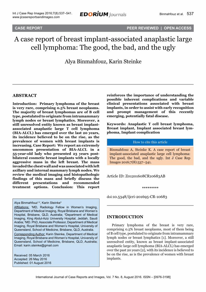
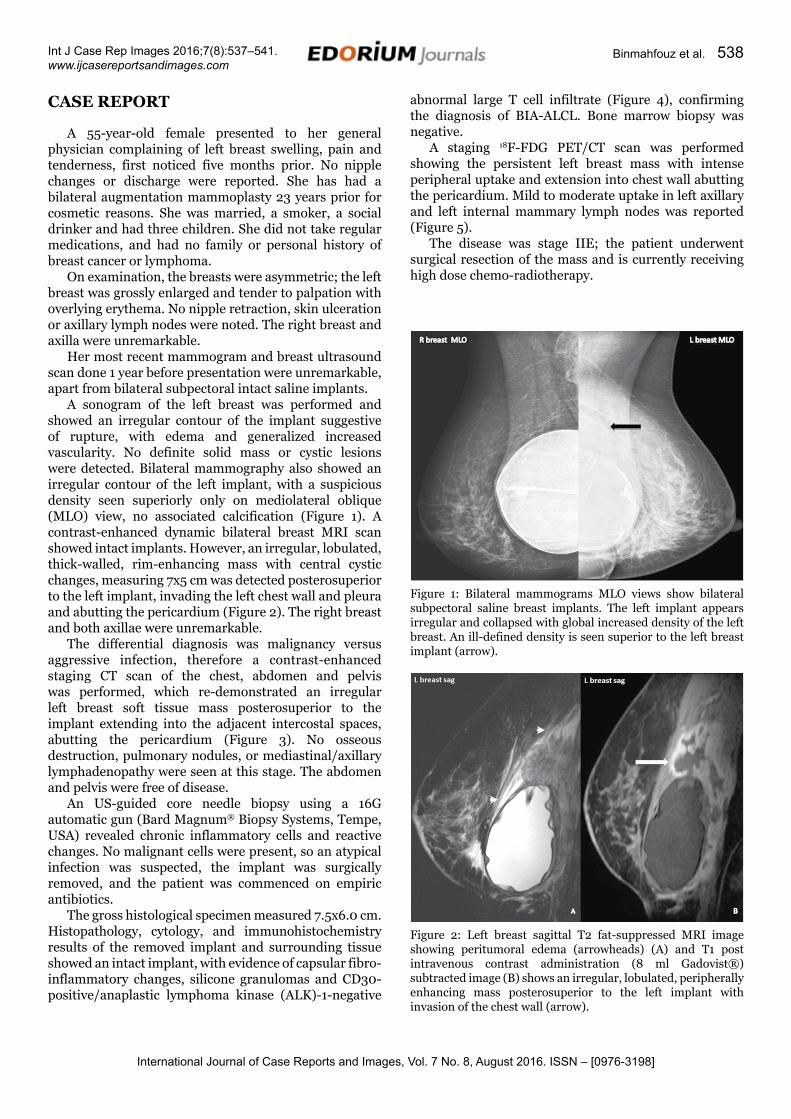
A sonogram of the left breast was performed and showed an irregular contour of the implant suggestive of rupture, with edema and generalized increased vascularity. No definite solid mass or cystic lesions were detected. Bilateral mammography also showed an irregular contour of the left implant, with a suspicious density seen superiorly only on mediolateral oblique (MLO) view, no associated calcification (Figure 1). A contrast-enhanced dynamic bilateral breast MRI scan showed intact implants. However, an irregular, lobulated, thick-walled, rim-enhancing mass with central cystic changes, measuring 7x5 cm was detected posterosuperior to the left implant, invading the left chest wall and pleura and abutting the pericardium (Figure 2). The right breast and both axillae were unremarkable.
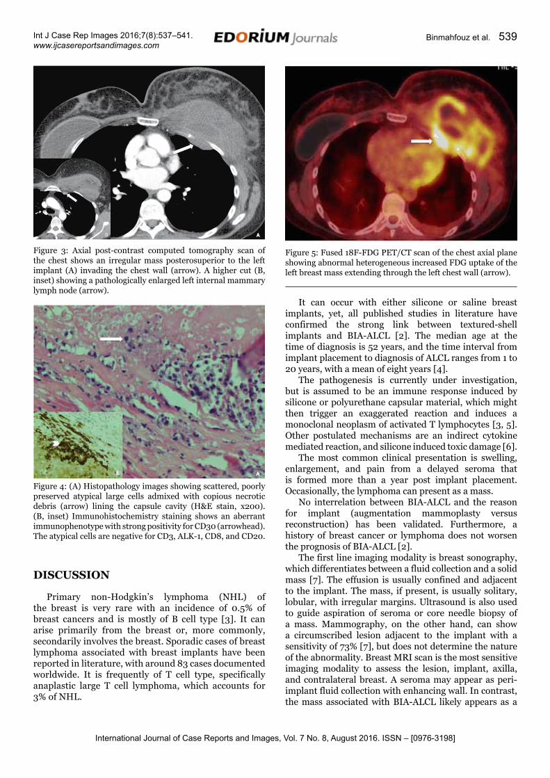
The differential diagnosis was malignancy versus aggressive infection, therefore a contrast-enhanced staging CT scan of the chest, abdomen and pelvis was performed, which re-demonstrated an irregular left breast soft tissue mass posterosuperior to the implant extending into the adjacent intercostal spaces, abutting the pericardium (Figure 3). No osseous destruction, pulmonary nodules, or mediastinal/axillary lymphadenopathy were seen at this stage. The abdomen and pelvis were free of disease.
An US-guided core needle biopsy using a 16G automatic gun (Bard Magnum® Biopsy Systems, Tempe, USA) revealed chronic inflammatory cells and reactive changes. No malignant cells were present, so an atypical infection was suspected, the implant was surgically removed, and the patient was commenced on empiric antibiotics.
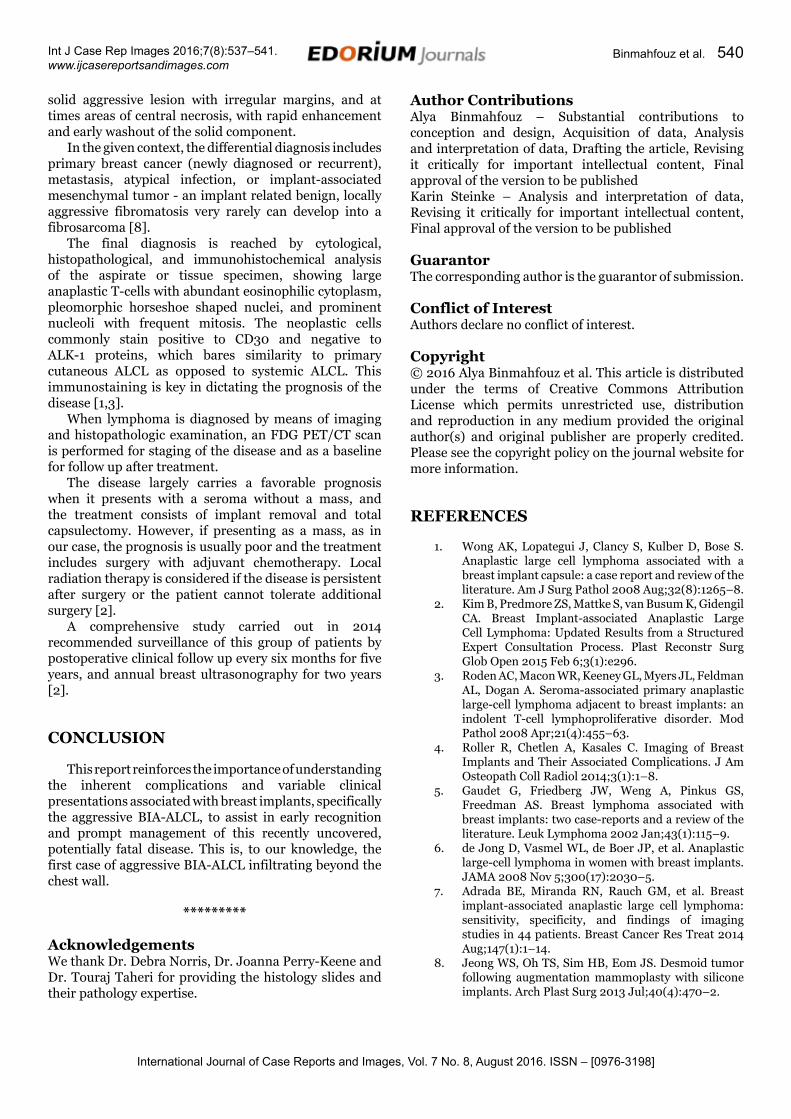
The gross histological specimen measured 7.5x6.0 cm. Histopathology, cytology, and immunohistochemistry results of the removed implant and surrounding tissue showed an intact implant, with evidence of capsular fibro-inflammatory changes, silicone granulomas and CD30-positive/anaplastic lymphoma kinase (ALK)-1-negative
abnormal large T cell infiltrate (Figure 4), confirming the diagnosis of BIA-ALCL. Bone marrow biopsy was negative.
A staging 18F-FDG PET/CT scan was performed showing the persistent left breast mass with intense peripheral uptake and extension into chest wall abutting the pericardium. Mild to moderate uptake in left axillary and left internal mammary lymph nodes was reported (Figure 5).
The disease was stage IIE; the patient underwent surgical resection of the mass and is currently receiving high dose chemo-radiotherapy.
Figure 1: Bilateral mammograms MLO views show bilateral subpectoral saline breast implants. The left implant appears irregular and collapsed with global increased density of the left breast. An ill-defined density is seen superior to the left breast implant (arrow).
Figure 2: Left breast sagittal T2 fat-suppressed MRI image showing peritumoral edema (arrowheads) (A) and T1 post intravenous contrast administration (8 ml Gadovist®) subtracted image (B) shows an irregular, lobulated, peripherally enhancing mass posterosuperior to the left implant with invasion of the chest wall (arrow).
International Journal of Case Reports and Images, Vol. 7 No. 8, August 2016. ISSN – [0976-3198]
Int J Case Rep Images 2016;7(8):537–541. www.ijcasereportsandimages.com
Binmahfouz et al. 539
DIscUssION
Primary non-Hodgkin’s lymphoma (NHL) of the breast is very rare with an incidence of 0.5% of breast cancers and is mostly of B cell type [3]. It can arise primarily from the breast or, more commonly, secondarily involves the breast. Sporadic cases of breast lymphoma associated with breast implants have been reported in literature, with around 83 cases documented worldwide. It is frequently of T cell type, specifically anaplastic large T cell lymphoma, which accounts for 3% of NHL.
It can occur with either silicone or saline breast implants, yet, all published studies in literature have confirmed the strong link between textured-shell implants and BIA-ALCL [2]. The median age at the time of diagnosis is 52 years, and the time interval from implant placement to diagnosis of ALCL ranges from 1 to 20 years, with a mean of eight years [4].
The pathogenesis is currently under investigation, but is assumed to be an immune response induced by silicone or polyurethane capsular material, which might then trigger an exaggerated reaction and induces a monoclonal neoplasm of activated T lymphocytes [3, 5]. Other postulated mechanisms are an indirect cytokine mediated reaction, and silicone induced toxic damage [6].
The most common clinical presentation is swelling, enlargement, and pain from a delayed seroma that is formed more than a year post implant placement. Occasionally, the lymphoma can present as a mass.
No interrelation between BIA-ALCL and the reason for implant (augmentation mammoplasty versus reconstruction) has been validated. Furthermore, a history of breast cancer or lymphoma does not worsen the prognosis of BIA-ALCL [2].
The first line imaging modality is breast sonography, which differentiates between a fluid collection and a solid mass [7]. The effusion is usually confined and adjacent to the implant. The mass, if present, is usually solitary, lobular, with irregular margins. Ultrasound is also used to guide aspiration of seroma or core needle biopsy of a mass. Mammography, on the other hand, can show a circumscribed lesion adjacent to the implant with a sensitivity of 73% [7], but does not determine the nature of the abnormality. Breast MRI scan is the most sensitive imaging modality to assess the lesion, implant, axilla, and contralateral breast. A seroma may appear as peri-implant fluid collection with enhancing wall. In contrast, the mass associated with BIA-ALCL likely appears as a
Figure 3: Axial post-contrast computed tomography scan of the chest shows an irregular mass posterosuperior to the left implant (A) invading the chest wall (arrow). A higher cut (B, inset) showing a pathologically enlarged left internal mammary lymph node (arrow).
Figure 4: (A) Histopathology images showing scattered, poorly preserved atypical large cells admixed with copious necrotic debris (arrow) lining the capsule cavity (H&E stain, x200). (B, inset) Immunohistochemistry staining shows an aberrant immunophenotype with strong positivity for CD30 (arrowhead). The atypical cells are negative for CD3, ALK-1, CD8, and CD20.
Figure 5: Fused 18F-FDG PET/CT scan of the chest axial plane showing abnormal heterogeneous increased FDG uptake of the left breast mass extending through the left chest wall (arrow).
International Journal of Case Reports and Images, Vol. 7 No. 8, August 2016. ISSN – [0976-3198]
Int J Case Rep Images 2016;7(8):537–541. www.ijcasereportsandimages.com
Binmahfouz et al. 540
solid aggressive lesion with irregular margins, and at times areas of central necrosis, with rapid enhancement and early washout of the solid component.
In the given context, the differential diagnosis includes primary breast cancer (newly diagnosed or recurrent), metastasis, atypical infection, or implant-associated mesenchymal tumor - an implant related benign, locally aggressive fibromatosis very rarely can develop into a fibrosarcoma [8].
The final diagnosis is reached by cytological, histopathological, and immunohistochemical analysis of the aspirate or tissue specimen, showing large anaplastic T-cells with abundant eosinophilic cytoplasm, pleomorphic horseshoe shaped nuclei, and prominent nucleoli with frequent mitosis. The neoplastic cells commonly stain positive to CD30 and negative to ALK-1 proteins, which bares similarity to primary cutaneous ALCL as opposed to systemic ALCL. This immunostaining is key in dictating the prognosis of the disease [1,3].
When lymphoma is diagnosed by means of imaging and histopathologic examination, an FDG PET/CT scan is performed for staging of the disease and as a baseline for follow up after treatment.
The disease largely carries a favorable prognosis when it presents with a seroma without a mass, and the treatment consists of implant removal and total capsulectomy. However, if presenting as a mass, as in our case, the prognosis is usually poor and the treatment includes surgery with adjuvant chemotherapy. Local radiation therapy is considered if the disease is persistent after surgery or the patient cannot tolerate additional surgery [2].
A comprehensive study carried out in 2014 recommended surveillance of this group of patients by postoperative clinical follow up every six months for five years, and annual breast ultrasonography for two years [2].
cONcLUsION
This report reinforces the importance of understanding the inherent complications and variable clinical presentations associated with breast implants, specifically the aggressive BIA-ALCL, to assist in early recognition and prompt management of this recently uncovered, potentially fatal disease. This is, to our knowledge, the first case of aggressive BIA-ALCL infiltrating beyond the chest wall.
*********
AcknowledgementsWe thank Dr. Debra Norris, Dr. Joanna Perry-Keene and Dr. Touraj Taheri for providing the histology slides and their pathology expertise.
Author contributionsAlya Binmahfouz – Substantial contributions to conception and design, Acquisition of data, Analysis and interpretation of data, Drafting the article, Revising it critically for important intellectual content, Final approval of the version to be publishedKarin Steinke – Analysis and interpretation of data, Revising it critically for important intellectual content, Final approval of the version to be published
GuarantorThe corresponding author is the guarantor of submission.
conflict of InterestAuthors declare no conflict of interest.
copyright© 2016 Alya Binmahfouz et al. This article is distributed under the terms of Creative Commons Attribution License which permits unrestricted use, distribution and reproduction in any medium provided the original author(s) and original publisher are properly credited. Please see the copyright policy on the journal website for more information.
rEFErENcEs
1. Wong AK, Lopategui J, Clancy S, Kulber D, Bose S. Anaplastic large cell lymphoma associated with a breast implant capsule: a case report and review of the literature. Am J Surg Pathol 2008 Aug;32(8):1265–8.
2. Kim B, Predmore ZS, Mattke S, van Busum K, Gidengil CA. Breast Implant-associated Anaplastic Large Cell Lymphoma: Updated Results from a Structured Expert Consultation Process. Plast Reconstr Surg Glob Open 2015 Feb 6;3(1):e296.
3. Roden AC, Macon WR, Keeney GL, Myers JL, Feldman AL, Dogan A. Seroma-associated primary anaplastic large-cell lymphoma adjacent to breast implants: an indolent T-cell lymphoproliferative disorder. Mod Pathol 2008 Apr;21(4):455–63.
4. Roller R, Chetlen A, Kasales C. Imaging of Breast Implants and Their Associated Complications. J Am Osteopath Coll Radiol 2014;3(1):1–8.
5. Gaudet G, Friedberg JW, Weng A, Pinkus GS, Freedman AS. Breast lymphoma associated with breast implants: two case-reports and a review of the literature. Leuk Lymphoma 2002 Jan;43(1):115–9.
6. de Jong D, Vasmel WL, de Boer JP, et al. Anaplastic large-cell lymphoma in women with breast implants. JAMA 2008 Nov 5;300(17):2030–5.
7. Adrada BE, Miranda RN, Rauch GM, et al. Breast implant-associated anaplastic large cell lymphoma: sensitivity, specificity, and findings of imaging studies in 44 patients. Breast Cancer Res Treat 2014 Aug;147(1):1–14.
8. Jeong WS, Oh TS, Sim HB, Eom JS. Desmoid tumor following augmentation mammoplasty with silicone implants. Arch Plast Surg 2013 Jul;40(4):470–2.
International Journal of Case Reports and Images, Vol. 7 No. 8, August 2016. ISSN – [0976-3198]
Int J Case Rep Images 2016;7(8):537–541. www.ijcasereportsandimages.com
Binmahfouz et al. 541
ABOUT THE AUTHORS
Article citation: Binmahfouz A, Steinke K. A case report of breast implant-associated anaplastic large cell lymphoma: The good, the bad, and the ugly. Int J Case Rep Images 2016;7(8):537–541.
Alya binmahfouz is Radiology Fellow in women’s imaging at the Royal Brisbane and Women’s Hospital, Brisbane, Queensland, Australia. She earned MBBS degree from King Abdulaziz University, Jeddah, Saudi Arabia and The Saudi Board In Radiology from The Saudi Commission For Health Specialties. She has published a few research papers in National and International academic journals. Her research interests include Women’s Imaging and Intervention.
Karin steinke is Senior Staff Specialist at The Royal Brisbane and Women’s Hospital, Brisbane, Queensland, Australia. She has published over 60 research papers in national and international academic journals and authored two book chapters. Her research interests include lung and breast imaging and intervention.
Access full text article onother devices
Access PDF of article onother devices

EDORIUM JOURNALS AN INTRODUCTION
Edorium Journals: On Web
About Edorium JournalsEdorium Journals is a publisher of high-quality, open ac-cess, international scholarly journals covering subjects in basic sciences and clinical specialties and subspecialties.
Edorium Journals www.edoriumjournals.com
Edorium Journals et al.
Edorium Journals: An introduction
Edorium Journals Team
But why should you publish with Edorium Journals?In less than 10 words - we give you what no one does.
Vision of being the bestWe have the vision of making our journals the best and the most authoritative journals in their respective special-ties. We are working towards this goal every day of every week of every month of every year.
Exceptional servicesWe care for you, your work and your time. Our efficient, personalized and courteous services are a testimony to this.
Editorial ReviewAll manuscripts submitted to Edorium Journals undergo pre-processing review, first editorial review, peer review, second editorial review and finally third editorial review.
Peer ReviewAll manuscripts submitted to Edorium Journals undergo anonymous, double-blind, external peer review.
Early View versionEarly View version of your manuscript will be published in the journal within 72 hours of final acceptance.
Manuscript statusFrom submission to publication of your article you will get regular updates (minimum six times) about status of your manuscripts directly in your email.
Our Commitment
Favored Author programOne email is all it takes to become our favored author. You will not only get fee waivers but also get information and insights about scholarly publishing.
Institutional Membership programJoin our Institutional Memberships program and help scholars from your institute make their research accessi-ble to all and save thousands of dollars in fees make their research accessible to all.
Our presenceWe have some of the best designed publication formats. Our websites are very user friendly and enable you to do your work very easily with no hassle.
Something more...We request you to have a look at our website to know more about us and our services.
We welcome you to interact with us, share with us, join us and of course publish with us.
Browse Journals
CONNECT WITH US
Invitation for article submissionWe sincerely invite you to submit your valuable research for publication to Edorium Journals.
Six weeksYou will get first decision on your manuscript within six weeks (42 days) of submission. If we fail to honor this by even one day, we will publish your manuscript free of charge.*
Four weeksAfter we receive page proofs, your manuscript will be published in the journal within four weeks (31 days). If we fail to honor this by even one day, we will pub-lish your manuscript free of charge and refund you the full article publication charges you paid for your manuscript.*
This page is not a part of the published article. This page is an introduction to Edorium Journals and the publication services.
* Terms and condition apply. Please see Edorium Journals website for more information.