Embed Size (px)
Citation preview
+ + + + + + + + + + + + + + + + + + + + + + + + + + + + + + + + + + + + + + + + + + + + + + + + + + + + + + + + + + + ++ + + + + + + + + + + + + + + + + + + + + + + + + + + + + + + + + + + + + + + + + + + + + + + + + + + + + + + + + + + ++ + + + + + + + + + + + + + + + + + + + + + + + + + + + + + + + + + + + + + + ++ + + + + + + + + + + + + + + + + + + ++ + + + + + + + + + + + + + + + + + + + + + + + + + + + + + + + + + + + + + + ++ + + + + + + + + + + + + + + + + + + ++ + + + + + + + + + + + + + + + + + + ++ + + + + + + + + + + + + + + + + + + ++ + + + + + + + + + + + + + + + + + + ++ + + + + + + + + + + + + + + + + + + ++ + + + + + + + + + + + + + + + + + + +
Cancer Res Treat. 2012;44(4):267-270
pISSN 1598-2998, eISSN 2005-9256
http://dx.doi.org/10.4143/crt.2012.44.4.267
Hak Jin Kim,MD1
Mi Hyang Kwak,MD, PhD1
Sun-Young Kong, MD, PhD2
Moon-Woo Seong,MD, PhD2
Han-Sung Kang, MD, PhD3
Keun Seok Lee, MD3
Jungsil Ro, MD, PhD3
Departments of 1Cardiology and 2Laboratory Medicine,3Center for Breast Cancer,National Cancer Center, Goyang, Korea
Correspondence: Jungsil Ro, MD, PhD
Center for Breast Cancer,
National Cancer Center, 111 Jungbalsan-ro,
Ilsandong-gu, Goyang 410-769, Korea
Tel: 82-31-920-1610
Fax: 82-31-903-0455
E-mail: [email protected]
Received April 8, 2011
Accepted June 2, 2011
Pulmonary tumor thrombotic microangiopathy (PTTM) is a rare, malignancy-related complication that causes marked pulmonary hypertension, right heart failure, and death.We report on a patient with locally advanced breast cancer whose course was complicatedby fatal PTTM based on clinical and laboratory findings.
Key words
Breast neoplasms, Pulmonary hypertension,Pulmonary tumor thrombotic microangiopathy
Case Report Open Access
A Case of Locally Advanced Breast Cancer Complicated by PulmonaryTumor Thrombotic Microangiopathy
│ http://www.cancerresearchandtreatment.org ││ http://www.e-crt.org │
267Copyright ⓒ 2012 by the Korean Cancer AssociationThis is an Open-Access article distributed under the terms of the Creative Commons Attribution Non-Commercial License (http://creativecommons.org/
licenses/by-nc/3.0/)which permits unrestricted non-commercial use, distribution, and reproduction in any medium, provided the original work is properly cited.
I n t r o d u c t i o n
Pulmonary tumor thrombotic microangiopathy (PTTM) is an
uncommon, fatal, malignancy-related complication causing marked
pulmonary hypertension, right heart failure, and death. It is diag-
nosed primarily during autopsy, and is rarely diagnosed antemortem.
Tumor emboli in the vasculature with consequent local activation
of coagulation and by widespread fibrocellular intimal proliferation
of small pulmonary arteries and arterioles, leading to increased
vascular resistance, resulting in marked pulmonary hypertension
[1,2]. In addition, PTTM also shows hemodynamic effects similar
to those observed with microangiopathic hemolytic anemia
(MAHA) and disseminated intravascular coagulation (DIC). Thus
far, the majority of reported underlying malignancies have been
adenocarcinomas of gastrointestinal origin [1].
We report on a 30-year-old female patient with locally advanced
breast cancer who presented with acute onset of rapidly progressive
dyspnea, culminating in cardiovascular collapse from right heart
failure.
Cancer Res Treat. 2012;44(4):267-270
268 CANCER RESEARCH AND TREATMENT
C a s e R e p o r t
In May 2009, a 30-year -old woman came to the emergency room
(ER) with a five-day history of acute progressive dyspnea on
exertion (NYHA class III). In 2007, ductal carcinoma in situ was
diagnosed by biopsy during pregnancy. In 2008, after a full-term
delivery, findings on fluorodeoxyglucose positron emission tomog-
raphy computed tomography (CT) and ultrasound of the right breast
revealed diffuse microcalcification and skin thickening of the right
breast, and metastatic axillary lymph nodes. The patient refused
further management and was lost to follow up until the ER visit.
On admission, the patient’s blood pressure was 126/76 mm Hg,
heart rate was 120 beats per min, respiratory rate was 18 breaths per
minute, and body temperature was 36.7°C. A chest radiograph
showed that the lung field was clear. Results of arterial blood gas
analysis in room air indicated hypoxemia: pH 7.446, pCO2 28.2 mm
Hg, pO2 44.3 mm Hg, HCO3 19.1 mmol/L, SaO2 76.8%. D-dimers
were elevated to 2.59 μg/mL (normal, <0.39 μg/mL) with an
elevated troponin I level to 0.24 ng/mL (normal, <0.04 ng/mL) and
a brain natriuretic peptide of 774 pg/mL (normal, <100 pg/mL).
Diffuse enlargement of the right breast with skin thickening and
many enlarged axillary lymph nodes consistent with locally
advanced breast cancer was observed on CT.
On the second day of admission, the patient was consulted with
the cardiology department for possible preoperative cardiac evalu-
ation. An electrocardiogram showed an S1Q3T3 pattern with
inverted or flattened T waves in leads V1 through V4. A transtho-
racic echocardiogram showed normal left ventricular systolic
function with right ventricular enlargement and free wall hypokine-
sia sparing the apex. In addition, the echocardiogram showed typical
findings of acute pulmonary thromboembolism with a D-shaped left
ventricle, moderate tricuspid regurgitation, and moderate pulmonary
hypertension with an estimated right ventricular systolic pressure of
61 mm Hg.
Other laboratory tests showed the following results: white blood
cell 7,900/mm3 with normal differential counts, hemoglobin15.4
g/dL, platelets 144,000/mm3, alanine aminotransferase 50 IU/L (nor-
mal, 0 to 40 IU/L), aspartate aminotransferase 218 IU/L (normal, 0
to 40 IU/L), total bilirubin 1.4 mg/dL (normal, 0.2 to 1.2 mg/dL),
C-reactive protein 0.81 mg/dL (normal, 0 to 0.30 mg/dL), prothrom-
bin time (PT) international normalized ratio (INR) of 1.36 (normal,
0.8 to 1.2), activated partial thromboplastin time (aPTT) of 45.7
seconds (normal, 27.0 to 45.0 seconds), fibrinogen of 196 mg/dL
(normal, 200 to 400 mg/dL), and fibrin degradation products (FDP)
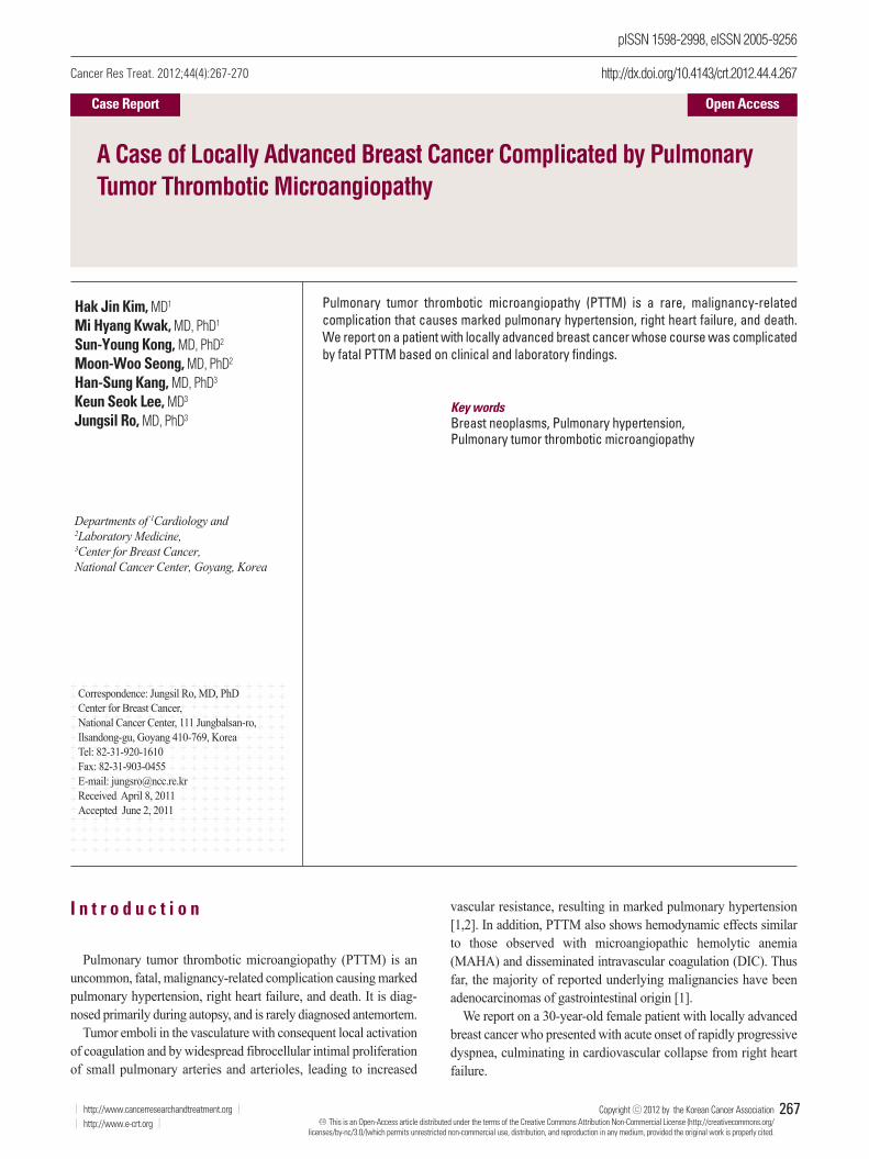
of 1 : 2 positive (normal, negative). A peripheral blood smear showed
increased numbers of schistocytes and reticulocytes consistent with
MAHA (Fig. 1).
We measured serum vascular endothelial growth factor (VEGF)
and interleukin 6 (IL-6) in view of preexisting information on VEGF,
a critical angiogenic molecule and IL-6, a multifunctional cytokine
promoting tumor growth. Serum VEGF levels were 26.9 pg/mL on
the second day in the hospital and 9.5 pg/mL on the third day in the
hospital (normal, 88.7 to 1,048.7 pg/mL); IL-6 levels were 50.3
pg/mL and 25.6 pg/mL on the second and third days, respectively
(normal range, 0.4 to 8.6 pg/mL).
Anticoagulation therapy with enoxaparin was startedunder the
clinical diagnosis of submassive acute pulmonary thromboem-
bolism. While no evidence of pulmonary thromboembolism was
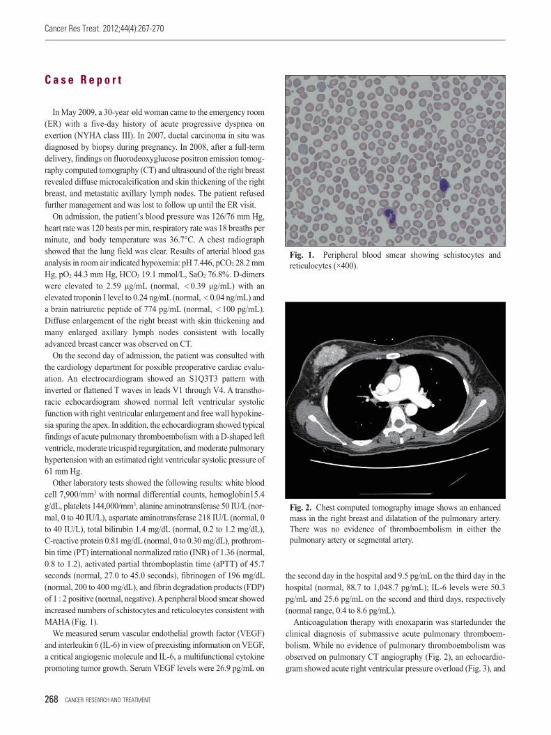
observed on pulmonary CT angiography (Fig. 2), an echocardio-
gram showed acute right ventricular pressure overload (Fig. 3), and
Fig. 1. Peripheral blood smear showing schistocytes and
reticulocytes (×400).
Fig. 2. Chest computed tomography image shows an enhanced
mass in the right breast and dilatation of the pulmonary artery.
There was no evidence of thromboembolism in either the
pulmonary artery or segmental artery.
Hak Jin Kim, Pulmonary Tumor Thrombotic Microangiopathyin Breast Cancer
VOLUME 44 NUMBER 4 DECEMBER 2012 269
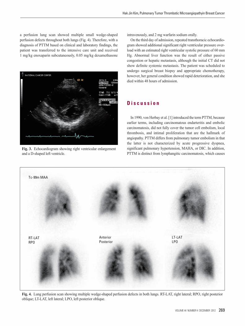
a perfusion lung scan showed multiple small wedge-shaped
perfusion defects throughout both lungs (Fig. 4). Therefore, with a
diagnosis of PTTM based on clinical and laboratory findings, the
patient was transferred to the intensive care unit and received
1 mg/kg enoxaparin subcutaneously, 0.05 mg/kg dexamethasone
intravenously, and 2 mg warfarin sodium orally.
On the third day of admission, repeated transthoracic echocardio-
gram showed additional significant right ventricular pressure over-
load with an estimated right ventricular systolic pressure of 60 mm
Hg. Abnormal liver function was the result of either passive
congestion or hepatic metastasis, although the initial CT did not
show definite systemic metastasis. The patient was scheduled to
undergo surgical breast biopsy and appropriate chemotherapy,
however, her general condition showed rapid deterioration, and she
died within 48 hours of admission.
D i s c u s s i o n
In 1990, von Herbay et al. [1] introduced the term PTTM, because
earlier terms, including carcinomatous endarteritis and embolic
carcinomatosis, did not fully cover the tumor cell embolism, local
thrombosis, and intimal proliferation that are the hallmark of
angiopathy. PTTM differs from pulmonary tumor embolism in that
the latter is not characterized by acute progressive dyspnea,
significant pulmonary hypertension, MAHA, or DIC. In addition,
PTTM is distinct from lymphangitic carcinomatosis, which causesFig. 3. Echocardiogram showing right ventricular enlargement
and a D-shaped left ventricle.
Fig. 4. Lung perfusion scan showing multiple wedge-shaped perfusion defects in both lungs. RT-LAT, right lateral; RPO, right posterior
oblique; LT-LAT, left lateral; LPO, left posterior oblique.
Tc-99m MAA
RT-LATRPO
LT-LATLPO
AnteriorPosterior
Cancer Res Treat. 2012;44(4):267-270
270 CANCER RESEARCH AND TREATMENT
1. von Herbay A, Illes A, Waldherr R, Otto HF. Pulmonary tumor thrombotic microangiopa-thy with pulmonary hypertension. Cancer. 1990;66:587-92.
2. Pinckard JK, Wick MR. Tumor-related thrombotic pulmonary microangiopathy: reviewof pathologic findings and pathophysiologic mechanisms. Ann Diagn Pathol. 2000;4:154-7.
3. Case records of the Massachusetts General Hospital. Weekly clinicopathological exercises. Case 19-1995. A 55 year-old woman with acute respiratory failure and radiographically clear lungs. N Engl J Med. 1995;332:1700-7.
4. Chinen K, Fujino T, Horita A, Sakamoto A, Fujioka Y. Pulmonary tumor thrombotic microangiopathy caused by an ovarian cancer expressing tissue factor and vascular endothelial growth factor. Pathol Res Pract. 2009;205:63-8.
5. Chinen K, Kazumoto T, Ohkura Y, Matsubara O, Tsuchiya E. Pulmonary tumor thrombotic microangiopathy caused by a gastric carcinoma expressing vascular endothelial growth factor and tissue factor. Pathol Int. 2005;55:27-31.
6. Takahashi F, Kumasaka T, Nagaoka T, Wakiya M, Fujii H, Shimizu K, et al. Osteopontin
expression in pulmonary tumor thrombotic microangiopathy caused by gastric carcinoma. Pathol Int. 2009;59:752-6.
7. Babar SI, Sobonya RE, Snyder LS. Pulmonary microvascular cytology for the diagnosisof pulmonary tumor embolism. West J Med. 1998;168:47-50.
8. Miyano S, Izumi S, Takeda Y, Tokuhara M, Mochizuki M, Matsubara O, et al. Pulmonarytumor thrombotic microangiopathy. J Clin Oncol. 2007;25:597-9.
9. Bachelot T, Ray-Coquard I, Menetrier-Caux C, Rastkha M, Duc A, Blay JY. Prognosticvalue of serum levels of interleukin 6 and of serum and plasma levels of vascular endothelial growth factor in hormone-refractory metastatic breast cancer patients. Br JCancer. 2003;88:1721-6.
10. Sakashita N, Yokose C, Fujii K, Matsumoto M, Ohnishi K, Takeya M. Pulmonary tumorthrombotic microangiopathy resulting from metastatic signet ring cell carcinoma of thestomach. Pathol Int. 2007;57:383-7.
11. Keenan NG, Nicholson AG, Oldershaw PJ. Fatal acute pulmonary hypertension causedby pulmonary tumour thrombotic microangiopathy. Int J Cardiol. 2008;124:e11-3.
R e f e r e n c e s
less acute symptoms and is accompanied by abnormal findings on
chest CT [3]. In a study of 630 carcinoma autopsies, 21 cases (3.3%)
were diagnosed with PTTM. Of these, 19 cases (90.5%) had
adenocarcinomas of various organs, including stomach, lung, breast,
colon, pancreas, liver, bladder, and prostate. While stomach cancer
was the most common, there were two cases of breast cancer [1].
The diagnosis of PTTM was based on clinical features and labora-
tory findings including peripheral blood smear showing MAHA,
prolonged PT and aPTT, low fibrinogen levels, and positive FDP
consistent with DIC.
Although the underlying molecular mechanism of PTTM remains
uncertain, platelet-derived growth factor, VEGF, and osteopontin
may play a role in the pathogenesis of PTTM [4-6].
Due to the extremely rapid progression of PTTM, almost all
reported patients died within one week of onset of dyspnea [2,5].
Thus far, no predisposing factors have been identified. Therefore,
in order to make an early diagnosis and administer therapeutic
intervention, it is important for clinicians to be keenly aware of the
disease entity. In some cases, aggressive bronchoscopic biopsy,
transbronchial lung biopsy, and pulmonary microvascular cytology
by a wedged pulmonary artery catheter have been suggested as tools
for use in antemortem diagnosis of PTTM [3,7]. However, these
procedures may not be applicable to all patients who are already
experiencing severe and progressive dyspnea.
Since MAHA and DIC are part of the clinical spectrum as seen
in the current patient, peripheral blood smear and DIC tests are
warranted. Miyano et al. [8] reported a markedly elevated serum
VEGF level in a gastric cancer with PTTM, which normalized after
chemotherapy. Although serum VEGF level was not elevated in the
current case, IL-6 was significantly elevated. While we do not know
the exact implication in relationship with PTTM, IL-6 level was
reportedly correlated with poor survival in metastatic breast cancer
[9].
Since no effective management for PTTM is identified todate,
chemotherapy, corticosteroids, and anticoagulation could be applied
as reported in a gastric cancer patient with PTTM [9]. Type 2A
serotonin receptor antagonists may also be useful by suppressing
intimal proliferation [10], and new drugs for treatment of pulmonary
arterial hypertension, such as endothelin antagonists, prostacyclin
analogues, and phosphodiesterase type 5 inhibitors may be benefi-
cial [11].
C o n f l i c t s o f I n t e r e s t
Conflicts of interest relevant to this article was not reported.