Embed Size (px)
Citation preview
Brief Report
Vol. 29, No. 3, 2017 383
Received April 11, 2016, Revised July 13, 2016, Accepted for publication July 20, 2016
Corresponding author: Kyung Ho Lee, Department of Dermatology, Bucheon St. Mary’s Hospital, College of Medicine, The Catholic University of Korea, 327 Sosa-ro, Wonmi-gu, Bucheon 14647, Korea. Tel: 82-32-340- 2115, Fax: 82-32-340-2118, E-mail: [email protected]
This is an Open Access article distributed under the terms of the Creative Commons Attribution Non-Commercial License (http://creativecommons.org/licenses/by-nc/4.0) which permits unrestricted non-commercial use, distribution, and reproduction in any medium, provided the original work is properly cited.
Copyright © The Korean Dermatological Association and The Korean Society for Investigative Dermatology
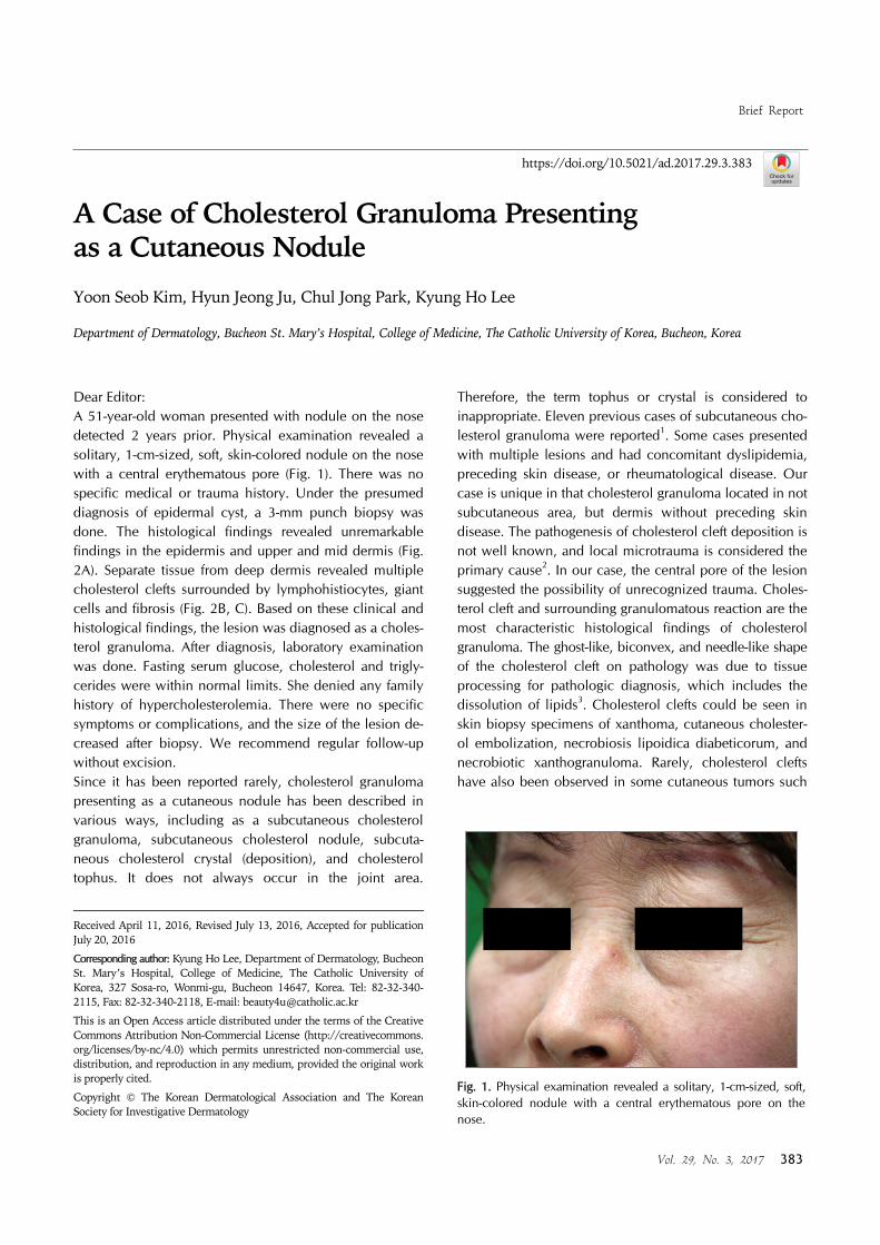
Fig. 1. Physical examination revealed a solitary, 1-cm-sized, soft, skin-colored nodule with a central erythematous pore on the nose.
https://doi.org/10.5021/ad.2017.29.3.383
A Case of Cholesterol Granuloma Presenting as a Cutaneous Nodule
Yoon Seob Kim, Hyun Jeong Ju, Chul Jong Park, Kyung Ho Lee
Department of Dermatology, Bucheon St. Mary’s Hospital, College of Medicine, The Catholic University of Korea, Bucheon, Korea
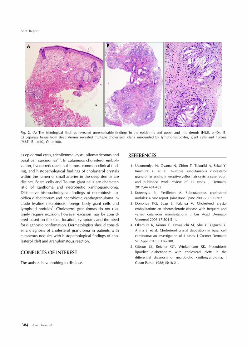
Dear Editor:A 51-year-old woman presented with nodule on the nose detected 2 years prior. Physical examination revealed a solitary, 1-cm-sized, soft, skin-colored nodule on the nose with a central erythematous pore (Fig. 1). There was no specific medical or trauma history. Under the presumed diagnosis of epidermal cyst, a 3-mm punch biopsy was done. The histological findings revealed unremarkable findings in the epidermis and upper and mid dermis (Fig. 2A). Separate tissue from deep dermis revealed multiple cholesterol clefts surrounded by lymphohistiocytes, giant cells and fibrosis (Fig. 2B, C). Based on these clinical and histological findings, the lesion was diagnosed as a choles-terol granuloma. After diagnosis, laboratory examination was done. Fasting serum glucose, cholesterol and trigly-cerides were within normal limits. She denied any family history of hypercholesterolemia. There were no specific symptoms or complications, and the size of the lesion de-creased after biopsy. We recommend regular follow-up without excision. Since it has been reported rarely, cholesterol granuloma presenting as a cutaneous nodule has been described in various ways, including as a subcutaneous cholesterol granuloma, subcutaneous cholesterol nodule, subcuta-neous cholesterol crystal (deposition), and cholesterol tophus. It does not always occur in the joint area.
Therefore, the term tophus or crystal is considered to inappropriate. Eleven previous cases of subcutaneous cho-lesterol granuloma were reported1. Some cases presented with multiple lesions and had concomitant dyslipidemia, preceding skin disease, or rheumatological disease. Our case is unique in that cholesterol granuloma located in not subcutaneous area, but dermis without preceding skin disease. The pathogenesis of cholesterol cleft deposition is not well known, and local microtrauma is considered the primary cause2. In our case, the central pore of the lesion suggested the possibility of unrecognized trauma. Choles-terol cleft and surrounding granulomatous reaction are the most characteristic histological findings of cholesterol granuloma. The ghost-like, biconvex, and needle-like shape of the cholesterol cleft on pathology was due to tissue processing for pathologic diagnosis, which includes the dissolution of lipids3. Cholesterol clefts could be seen in skin biopsy specimens of xanthoma, cutaneous cholester-ol embolization, necrobiosis lipoidica diabeticorum, and necrobiotic xanthogranuloma. Rarely, cholesterol clefts have also been observed in some cutaneous tumors such
Brief Report
384 Ann Dermatol
Fig. 2. (A) The histological findings revealed unremarkable findings in the epidermis and upper and mid dermis (H&E, ×40). (B, C) Separate tissue from deep dermis revealed multiple cholesterol clefts surrounded by lymphohistiocytes, giant cells and fibrosis (H&E; B: ×40, C: ×100).
as epidermal cysts, trichilemmal cysts, pilomatricomas and basal cell carcinomas3,4. In cutaneous cholesterol emboli-zation, livedo reticularis is the most common clinical find-ing, and histopathological findings of cholesterol crystals within the lumen of small arteries in the deep dermis are distinct. Foam cells and Touton giant cells are character-istic of xanthoma and necrobiotic xanthogranuloma. Distinctive histopathological findings of necrobiosis lip-oidica diabeticorum and necrobiotic xanthogranuloma in-clude hyaline necrobiosis, foreign body giant cells and lymphoid nodules5. Cholesterol granulomas do not rou-tinely require excision, however excision may be consid-ered based on the size, location, symptoms and the need for diagnostic confirmation. Dermatologists should consid-er a diagnosis of cholesterol granuloma in patients with cutaneous nodules with histopathological findings of cho-lesterol cleft and granulomatous reaction.
CONFLICTS OF INTEREST
The authors have nothing to disclose.
REFERENCES
1. Utsunomiya N, Oyama N, Chino T, Tokuriki A, Sakai Y,
Imamura Y, et al. Multiple subcutaneous cholesterol
granulomas arising in eruptive vellus hair cysts: a case report and published work review of 11 cases. J Dermatol
2017;44:481-482.
2. Kotevoglu N, Yesilleten A. Subcutaneous cholesterol nodules: a case report. Joint Bone Spine 2003;70:300-302.
3. Donohue KG, Saap L, Falanga V. Cholesterol crystal
embolization: an atherosclerotic disease with frequent and varied cutaneous manifestations. J Eur Acad Dermatol
Venereol 2003;17:504-511.
4. Okamura K, Konno T, Kawaguchi M, Abe Y, Yaguchi Y, Ajima S, et al. Cholesterol crystal deposition in basal cell
carcinoma: an investigation of 4 cases. J Cosmet Dermatol
Sci Appl 2015;5:176-180.5. Gibson LE, Reizner GT, Winkelmann RK. Necrobiosis
lipoidica diabeticorum with cholesterol clefts in the
differential diagnosis of necrobiotic xanthogranuloma. J Cutan Pathol 1988;15:18-21.


![Annals of Clinical Case Reports Case Report - anncaserep.com · pyogenic granuloma was described [5]. The Term Pyogenic granuloma is a misnomer because the The Term Pyogenic granuloma](https://img.pdfslide.us/doc/110x75/5d0a41bb88c993cf0c8b7f5f/annals-of-clinical-case-reports-case-report-pyogenic-granuloma-was-described.jpg)