Embed Size (px)
Citation preview
British Journal ofOphthalmology, 1982, 66, 530-535
A case of macular subretinal neovascularisation inchronic uveitis probably caused by sarcoidosisH. A. L. F. HOOGSTEDE AND A. C. COPPER
From the Royal Netherlands Eye Hospital, Utrecht, The Netherlands
SUMMARY A 29-year-old man with a 7-year history of bilateral chronic uveitis developed subretinalneovascularisation in the macular area of his right eye. There was a history suggesting sarcoidosis.Long-term therapy with systemic corticosteroids decreased the activity of the uveitis, and thesubretinal neovascularisation changed into a cystic scar. After the systemic corticosteroids werediscontinued there was a recurrence of the uveitis but not of the subretinal neovascularisation.
Subretinal neovascularisation in young people hasbeen reported in many diseases.,In the absence ofinflammatory signs in aqueous and vitreous or otherspecific ocular abnormalities it may be related toinfection with Histoplasma capsulatwn,' but the sameclinical picture has been observed in populations inwhich histoplasmosis is relatively rare.23Some specific causes of uveitis can be complicated
by subretinal neovascularisation, but only a few'reports have been delivered of subretinal neo-vascularisation in chronic aspecific uveitis.45
Case report
A man aged 29 years noted a sudden decrease ofvision of his right eye in July 1979. Since 1972 he wasknown to be suffering from a bilateral chroniciridocyclitiswithout involvementofthe posterior pole.
Medical examination in 1972 showed enlargedlymph nodes at the lung hila on chest x-ray. Becauseof a negative Mantoux reaction these changes werethought to be due to sarcoidosis. Seven months laterthe enlargement of the lymph nodes spontaneouslyregressed. No other abnormalities were found.
In July 1979 the corrected visuafacuity in the righteye was 0-16 and in the left eye 0 9. Intraocularpressure measured by applanation tonometry was 13mmHg. The aqueous of both eyes showed flare andcells, and there were some old synechiae of the iris tothe lens. The lenses were clear. The vitreous showed a
Correspondence to Dr H. A. L. F. Hoogstede, KoninklijkNederlands Gasthuis voor Ooglijders, F.C. Dondersstraat 65,Utrecht, The Netherlands.
small number of cells and somewhat more flare in theright than in the left eye.
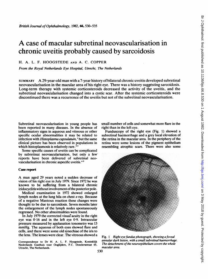
Funduscopy of the right eye (Fig. 1) showed asubretinal haemorrhage and a grey local elevation ofthe retina in the macular area. In the periphery of theretina were some lesions of the pigment epitheliumresembling atrophic scars. There were also some
530
Fig. 1 Right eyefundus photograph, showing afovealannular dark lesion, with a small subretinal haemorrhage.The detachment ofthe neuroepithelium covers the wholemacular area.
on 3 May 2018 by guest. P
rotected by copyright.http://bjo.bm
j.com/
Br J O
phthalmol: first published as 10.1136/bjo.66.8.530 on 1 A
ugust 1982. Dow
nloaded from
A case of macular subretinal neovascularisation in chronic uveitis probably caused by sarcoidosis
7
.7.
Fig. 2a Fig. 2b
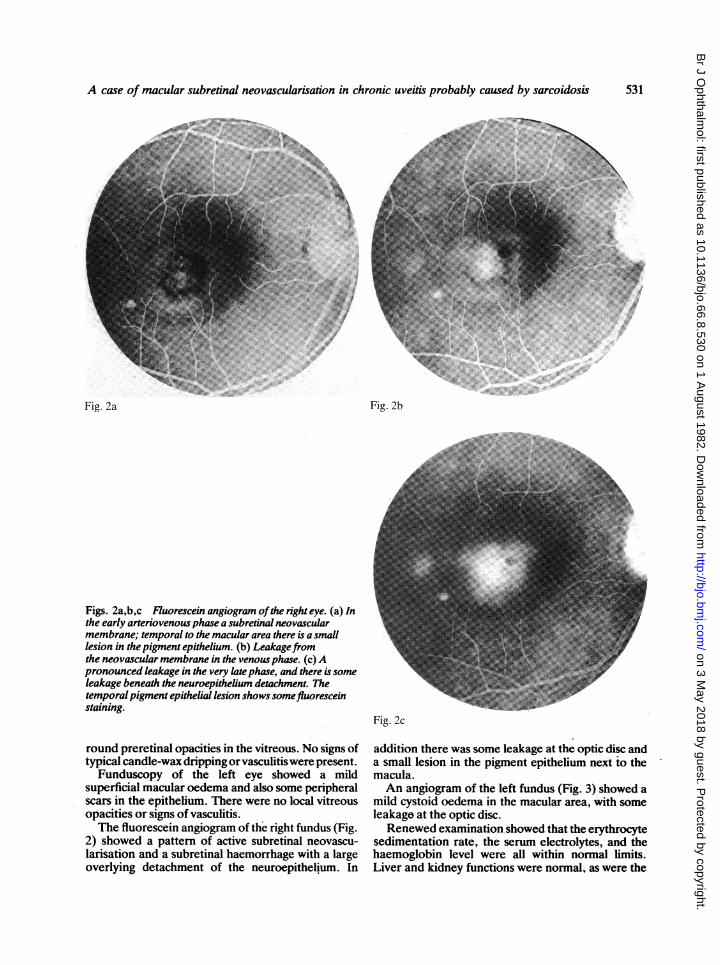
Figs. 2a,b,c Fluorescein angiogram ofthe right eye. (a) Inthe early arteriovenous phase a subretinal neovascularmembrane; temporal to the macular area there is a smalllesion in the pigment epithelium. (b) Leakagefromthe neovascular membrane in the venous phase. (c) Apronounced leakage in the very late phase, and there is someleakage beneath the neuroepithelium detachment. Thetemporalpigment epithelial lesion shows somefluoresceinstaining.
round preretinal opacities in the vitreous. No signs oftypical candle-wax dripping or vasculitis were present.Funduscopy of the left eye showed a mild
superficial macular oedema and also some peripheralscars in the epithelium. There were no local vitreousopacities or signs of vasculitis.The fluorescein angiogram of the right fundus (Fig.
2) showed a pattern of active subretinal neovascu-larisation and a subretinal haemorrhage with a largeoverlying detachment of the neuroepithelium. In
Fig. 2c
addition there was some leakage at the optic disc anda small lesion in the pigment epithelium next to themacula.An angiogram of the left fundus (Fig. 3) showed a
mild cystoid oedema in the macular area, with someleakage at the optic disc.Renewed examination,showed that the erythrocyte
sedimentation rate, the serum electrolytes, and thehaemoglobin level were all within normal limits.Liver and kidney functions were normal, as were the
531
on 3 May 2018 by guest. P
rotected by copyright.http://bjo.bm
j.com/
Br J O
phthalmol: first published as 10.1136/bjo.66.8.530 on 1 A
ugust 1982. Dow
nloaded from
H. A. L. F. Hoogstede and A. C. Copper
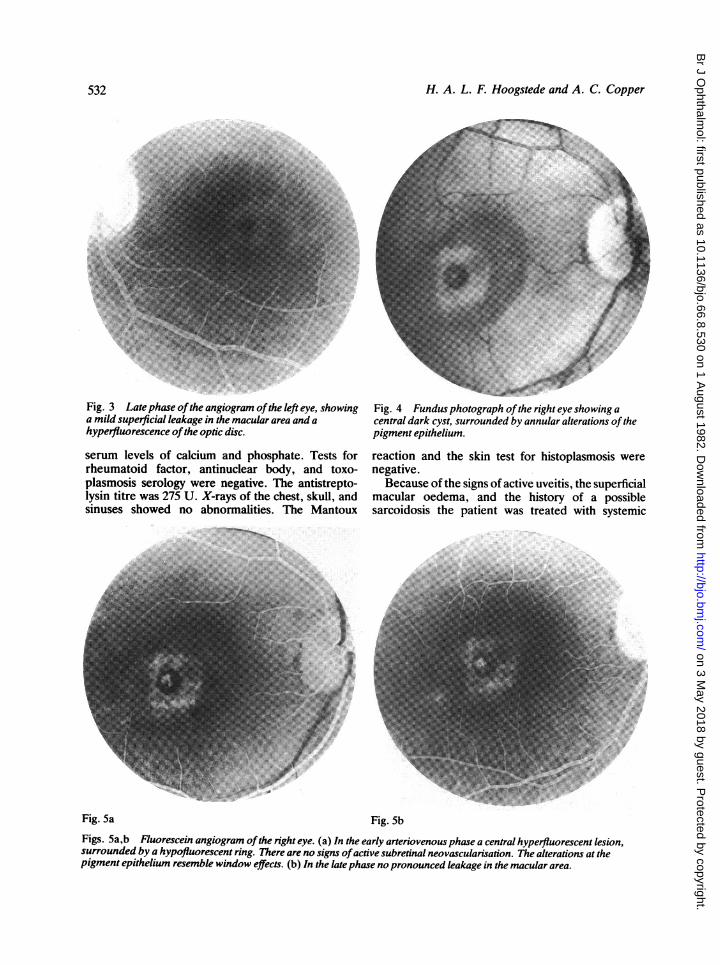
Fig. 3 Late phase ofthe angiogram ofthe left eye, showinga mild superficial leakage in the macular area and ahyperfluorescence ofthe optic disc.
serum levels of calcium and phosphate. Tests forrheumatoid factor, antinuclear body, and toxo-plasmosis serology were negative. The antistrepto-lysin titre was 275 U. X-rays of the chest, skull, andsinuses showed no abnormalities. The Mantoux
Fig. 4 Fundus photograph ofthe right eye showing acentral dark cyst, surrounded by annular alterations ofthepigment epithelium.
reaction and the skin test for histoplasmosis werenegative.
Because of the signs of active uveitis, the superficialmacular oedema, and the history of a possiblesarcoidosis the patient was treated with systemic
Fig. 5a Fig. 5bFigs. 5a,b Fluorescein angiogram ofthe right eye. (a) In the early arteriovenous phase a central hyperfluorescent lesion,surrounded by a hypofluorescent ring. There are no signs ofactive subretinal neovascularisation. The alterations at thepigment epithelium resemble window effects. (b) In the late phase no pronounced leakage in the macular area.
532
-
on 3 May 2018 by guest. P
rotected by copyright.http://bjo.bm
j.com/
Br J O
phthalmol: first published as 10.1136/bjo.66.8.530 on 1 A
ugust 1982. Dow
nloaded from
A case of macular subretinal neovascularisation in chronic uveitis probably caused by sarcoidosis
Fig. 6a Fig. 6b
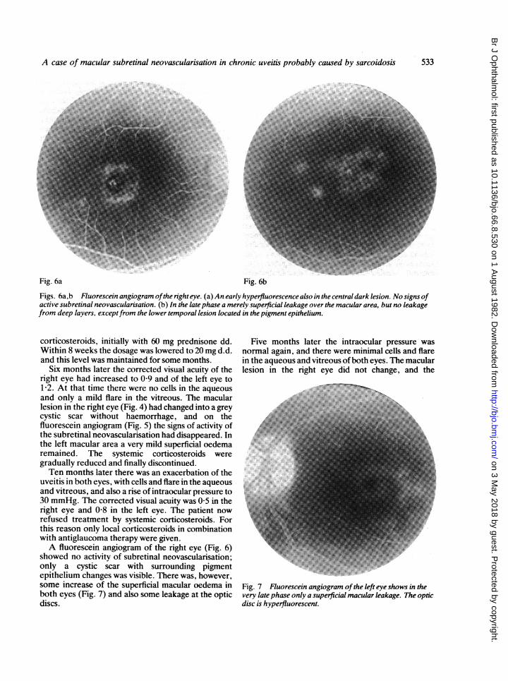
Figs. 6a,b Fluorescein angiogram ofthe right eye. (a) An early hyperfiuorescence also in the central dark lesion. No signs ofactive subretinal neovascularisation. (b) In the late phase a merely superficial leakage over the macular area, but no leakagefrom deep layers, exceptfrom the lower temporal lesion located in the pigment epithelium.
corticosteroids, initially with 60 mg prednisone dd. Five months later the intraocular pressure wa!Within 8 weeks the dosage was lowered to 20 mg d.d. normal again, and there were minimal cells and flareand this level was maintained for some months. in the aqueous and vitreous of both eyes. The maculai
Six months later the corrected visual acuity of the lesion in the right eye did not change, and theright eye had increased to 0 9 and of the left eye to1-2. At that time there were no cells in the aqueousand only a mild flare in the vitreous. The macular
the subretinal neovascularisation had disappeared. In oX40F! fthe left macular area a very mild superficialoedema°l -remained. The systemic corticosteroids were S l l - Egradually reduced and finally discontinued. -1Ten months later there was an exacerbation of the
uveitis in both eyes, wit hcells and flare inthe aqueousand vitreous, and also a rise of intraocular pressure to30 mmHg. The corrected visual acuity was 05 in thellright eye and 08 in the left eye. The patient norefused treatment by systemic corticosteroids. For - in2this reason only local corticosteroids in combinationll 1l| @Fwith antiglaucoma therapy were given._ l !
Afluorescein angiogram ofg.5 the right eye (Ftig.t 6)shoednaciiyosubretinal neovascularisationa iapae.I
ron ied . ctie sc seith surrounding pigment
epithelium changes was visible. There was, however,some increase of the superficial macular oedema in Fig. 7 Fluorescein angiogram ofthe eft eye shows in theboth eyes (Fig. 7) and also some leakage at the optic very late phase only a superficial macular leakage. Theoptndiscs. disc is hyperfluorescent.
S
ir
533
I I
c
on 3 May 2018 by guest. P
rotected by copyright.http://bjo.bm
j.com/
Br J O
phthalmol: first published as 10.1136/bjo.66.8.530 on 1 A
ugust 1982. Dow
nloaded from
H. A. L. F. Hoogstede and A. C. Copper
corrected visual acuity had now increased to 0O8 in theright and 0-9 in the left eye. Only local corticosteroidswere given, and up to September 1981 the findings didnot change.
Discsion
The occurrence of subretinal neovascularisation isprobablyrelatedtochangesinthepigmentepithelium-Bruch's membrane-choriocapillaris complex. Thenature of the primary stimulating factors is notknown. Ischaemia of the choriocapillaris (especiallyin the macular area), changes and breaks in Bruch'smembrane, detachment of the pigment epithelium,haemorrhage, or other vasoproliferative factorscould play a role in the stimulation of new vesselgrowth from the choroid into the subpigmentepithelial space. Generally, subretinal neovascular-isation occurs in the absence of inflammatory signs,such as in senile disciform macular degeneration,6angioid streaks occurring in pseudoxanthomaelasticum, Ehlers-Danlos disease, Paget's disease ofthe bone, sickle-cell disease, the hereditary form ofsenile elastosis and acromegaly,'7 myopia,8vitteliform macular degeneration,9 hereditarydrusen,'° Sorsby's pseudoinflammatory dystrophy,'"fundus flavimaculatus,'2 traumatic choroidalrupture,6 focal macular choroidopathy,3 idiopathicsubretinal neovascularisation,'3 optic drusen,'4papilloedema by pseudotumor cerebri,'5 followinglaser therapy,' and in association with choroidal naeviand tumours.7On the other hand it is known that subretinal neo-
vascularisation occurs in mainly 'inflammatory'diseases such as Behcet's disease,"6 toxoplasmosis,'718toxocara,'9 serpiginous choroiditis,20 chronicuveitis,45 Harada's disease,21 presumed sarcoidosis,22rubella retinopathy,23 and presumed histoplasmosis.'
Fluorescein angiography is indispensable fordetecting subretinal neovascularisation in the earlystages. In the later stages, when there are subretinalhaemorrhages or a more pronounced disciformlesion, an angiogram is not always necessary.
Subretinal neovascularisation is not a wellrecognised complication in chronic uveitis. This couldbe partly explained by the fact that some complica-tions in chronic uveitis make it difficult to obtain agood fluorescein angiogram-for example, posteriorsynechiae of' the iris, resulting in insufficientmydriasis, complicating cataract or vitreous opacities.Fundus changes have often been described in
sarcoidosis, including neovascularisation of the retinaand optic disc.24 Subretinal neovascularisation inpatients with presumed sarcoidosis was recentlyreported.22 In our patient a history of sarcoidosis wassuggested but was not proved histologically.
Our case supports the concept that the appearanceof subretinal neovascularisation in chronic uveitis isnot mere coincidence. The disturbance of the pig-ment epithelium-Bruch's membrane-choriocapillariscomplex in the macular area caused by uveitis maygive rise to the formation of subretinal neovascular-isation. Ophthalmoscopically this can develop into alesion with the aspect of the 'focal macularchoroidopathy,' 'presumed histoplasmuosis,' and thelike.
If special attention is given to this aspect-especially to the minor forms-more cases of thistype of subretinal neovascularisation in chronicuveitis may be found.
We thank Ms H. J. H. Schenk and Mr P. van Nigtevecht, who madethe photographs.
References
I Krill AE, Archer D. Choroidal neovascularisation in multifocal(presumed histoplasmine) choroiditis. Arch Ophthalmol 1970;84:595-604.
2 Braunstein RA, Rosen DA, Bird AC. Ocular histoplasmosissyndrome in the United Kingdom. Br J Ophthalnol 1974; 58:893-8.
3 Craandijk A. Focal macular choroidopathy. Doc Ophthalnol1979; 48: 1-103.
4 Schwartz PL, Gragoudas EW, Lapus JV. Peripapillary subretinalneovascularisation in chronic uveitis. Arch Ophthabnol 1978; 96:836-8.
5 Augsburger JJ, Benson WE. Subretinal neovascularisation inchronic uveitis. Albrecht von Graefes Arch Klin Ophthabnol1980; 215: 43-51.
6 Ryan SJR, Rainer NM, Maunenee AE. The disciform response:an historical perspective. Albrecht von Graefes Arch KlinOphthalmol 1980; 215: 1-20.
7 Gass JDM. Pathogenesis of disciform detachment of the neuro-epithelium. Am J Ophthalmol 1967; 63: 573-711.
8 Hotchkiss ML, Stuart LF. Pathologic myopia and choroidal neo-vascularisation. AmJ Ophthalmol 1981; 91: 177-83.
9 Deutman AF. Unexpected findings in hereditary maculardystrophy. Doc Ophthabmol, Proceedings series, newdevelopments in ophthalnology 1976; 7: 281-312.
10 Gass JDM. Stereoscopic Atlas of Macular Diseases. St Louis:Mosby, 1969:13.
11 Ashton N, Sorsby A. Fundus dystrophy with unusual features: ahistorical study. BrJ Ophthalmol 1951; 35: 751-64.
12 Klein R, Lewis RA, Meyers SM, Myers FL. Subretinal neo-vascularisation associated with fundus flavimaculatus. ArchOphthalmol 1978; 96: 2054-67.
13 Cleasby GW. Idiopathic focal subretinal neovascularisation. AmJ Ophthalmol 1976; 81: 590-6.
14 Yanuzzi LA, Gitter KA, Schatz H. The Macula. Baltimore:Williams and Wilkins, 1979: 288-9.
15 Jamison RR. Subretinal neovascularisation and papilledemaassociated with pseudotumor cerebri. AmJ Ophthalmol 1978; 85:78-81.
16 Michelson JB, Michelson PE, ChisariFV. Subretinal neovascularmembrane and disciform scar in Behget's disease. Am JOphthalmol 1980; 96i 182-5.
17 Wilierson D jr, Aaberg TM, Reeser F, Meredith TA. Unusualocular presentation of acute toxoplasnosis. Br J Ophthalmol1979; 61: 693-8.
534
on 3 May 2018 by guest. P
rotected by copyright.http://bjo.bm
j.com/
Br J O
phthalmol: first published as 10.1136/bjo.66.8.530 on 1 A
ugust 1982. Dow
nloaded from
A case of macular subrefinal neovascularisation in chronic uveifis probably caused by sarcoidos's 535
18 Fine SL, Owens SL, Haller JA, Knox DL, Patz A. Choroidal 21 Snyder DA, Tessler HH. Vogt-Koyanagi-Haradasyndrome. Amneovascularisation as late complication of ocular toxoplasmosis. J Ophthabnol 1980;W 69-75.AmJ Ophthabnol 1980; 91: 318-22. 22 Gragoudas ES, Regan CDJ. Peripallary subretinal neo-
19 Gass JDM. Stereoscopic Adas of Macular Diseases, Diagnosis vascularisation in presumed sarcoidosis. Arch Ophdhalmno 1981;and Treatment. St Louis: Mosby, 1977: 108. - 1194-7.
23 Deutman AF, Sanderson Grizzard W. Rubela retinopathy and20 Chisholm JH, Gass JDM, Hutton WL. The late stage of subretinal neovascularization. AmJOphtahlol 1978;85:82-7.
serpiginous (geographic) choroiditis. Am JOphthanol 1976;82: 24 Spalton DJ, Sanders MD. Fundus changes in histolgically343-51. confirmed sarcoidosis. BrJOphdtalmol 1981; 65: 348-58.
on 3 May 2018 by guest. P
rotected by copyright.http://bjo.bm
j.com/
Br J O
phthalmol: first published as 10.1136/bjo.66.8.530 on 1 A
ugust 1982. Dow
nloaded from