Embed Size (px)
Citation preview
A Cascade of Sequentially Expressed Sucrose Transporters inthe Seed Coat and Endosperm Provides Nutrition for theArabidopsis EmbryoOPEN
Li-Qing Chen,a,1 I Winnie Lin,a,b Xiao-Qing Qu,a Davide Sosso,a Heather E. McFarlane,c Alejandra Londoño,a
A. Lacey Samuels,c and Wolf B. Frommera,1
a Department of Plant Biology, Carnegie Institution for Science, Stanford, California 94305bDepartment of Biology, Stanford University, Stanford, California 94305cDepartment of Botany, University of British Columbia, Vancouver, British Columbia V6T 1Z4, Canada
ORCID ID: 0000-0002-0964-5388 (L.-Q.C.)
Developing plant embryos depend on nutrition from maternal tissues via the seed coat and endosperm, but the mechanisms thatsupply nutrients to plant embryos have remained elusive. Sucrose, the major transport form of carbohydrate in plants, is deliveredvia the phloem to the maternal seed coat and then secreted from the seed coat to feed the embryo. Here, we show that seedfilling in Arabidopsis thaliana requires the three sucrose transporters SWEET11, 12, and 15. SWEET11, 12, and 15 exhibit specificspatiotemporal expression patterns in developing seeds, but only a sweet11;12;15 triple mutant showed severe seed defects,which include retarded embryo development, reduced seed weight, and reduced starch and lipid content, causing a “wrinkled”seed phenotype. In sweet11;12;15 triple mutants, starch accumulated in the seed coat but not the embryo, implicating SWEET-mediated sucrose efflux in the transfer of sugars from seed coat to embryo. This cascade of sequentially expressed SWEETsprovides the feeding pathway for the plant embryo, an important feature for yield potential.
INTRODUCTION
Developing embryos of Metazoa and plants have to be nurtured bymaternal tissues: the placenta and umbilical cord in mammals andthe seed coat and endosperm in plants. Glucose transporters of theGLUT (SLC2) and SGLT (SLC5) families are likely involved in sup-plying glucose to mammalian embryos (Illsley, 2000; Baumannet al., 2002; Kevorkova et al., 2007), although direct evidence, e.g.,from the analysis of mutants, is lacking. The mechanisms for nu-trition of plant embryos have also remained elusive. Although seedscan undergo greening, embryo development depends on the supplyof photoassimilates from maternal tissues, particularly photosyn-thetic source leaves. Sucrose is the major long-distance transportform of sugars delivered from photosynthetic tissues to the growthand storage organs, including seeds of many plants; green siliquewalls may also contribute to some extent. Importantly, sucrosecreates the driving force for long-distance translocation of all othercompounds in the phloem. Sucrose is imported into the developingembryo by plasma membrane SUT sucrose/proton cotransporters(Patrick and Offler, 1995; Baud et al., 2005; Zhang et al., 2007).
In Arabidopsis thaliana, sugars are delivered to the maternalseed coat via the funicular phloem, which is symplasmicallyconnected to the outer integument (Stadler et al., 2005). Sugarsare released from the outer integument to the inner integument,
and potentially the suspensor, followed by secretion into theseed apoplasm by yet unknown membrane transport mecha-nisms. How sucrose is released from maternal tissues (seed coat)to support filial tissues (embryo) also remains unclear, except forthe contribution of a subset of transporters of the SUT sucrose/H+
cotransporter family. Evidence from studies of pea (Pisumsativum) and bean (Phaseolus vulgaris) seed coats implicated SUFtransporters, which appear to have lost proton coupling to act asuniporters, in sucrose efflux from seed coat (Ritchie et al., 2003;Zhou et al., 2007). These uncoupled SUFs appear to have evolvedfrom recent gene duplications and likely represent a specific ad-aptation in legumes to sustain the large seeds in some of theselegumes, yet they have not been found in other plants, such asArabidopsis or maize (Zea mays) (Zhou et al., 2007).The recently identified SWEET sugar transporters of eukaryotes
have seven transmembrane domains and function predominantlyin cellular efflux (Chen et al., 2010, 2012; Xu et al., 2014). TheSWEET family is divided into four clades. Clade III SWEETs appearto transport sucrose in a pH-independent manner and are typicallyinvolved in cellular efflux processes (Chen et al., 2010, 2012; Linet al., 2014). Two members of this clade, SWEET11 and 12, ap-pear to localize to the plasma membrane of phloem parenchymacells and export sucrose from these cells into the phloem’sapoplasm in preparation for phloem loading. A third member, thenectary-specific SWEET9, was shown to be essential for nectarsecretion in angiosperms (Lin et al., 2014). Rice SWEET11, 13, and14 and cassava (Manihot esculenta) SWEET10a play importantroles in pathogen susceptibility, possibly supplying nutrients topathogens (Chen et al., 2012; Streubel et al., 2013; Cohn et al.,2014). Analysis of microarrays generated by laser capture micro-dissection of tissues in the seed of Arabidopsis indicated thatseveral Clade III SWEETs were expressed in developing seeds
1Address correspondence to [email protected] or [email protected] author responsible for distribution of materials integral to the findingspresented in this article in accordance with the policy described in theInstructions for Authors (www.plantcell.org) is: Wolf B. Frommer ([email protected]).OPENArticles can be viewed online without a subscription.www.plantcell.org/cgi/doi/10.1105/tpc.114.134585
The Plant Cell, Vol. 27: 607–619, March 2015, www.plantcell.org ã 2015 American Society of Plant Biologists. All rights reserved.
(Figure 1A; Supplemental Figure 1) (Dean et al., 2011; Belmonteet al., 2013). We thus hypothesized that one or several Clade IIISWEETs may serve as the elusive sucrose carriers for efflux fromthe integument into the apoplasm, as well as from the endospermto support growth and development of the embryo.
Here, we show that SWEET11, 12, and 15 are expressed inparticular tissues of the seeds during development. We dem-onstrate that a sweet11;12;15 triple mutant shows retardedembryo development, reduced seed weight and lipid content,
and “wrinkled” seeds. The seed coat of sweet11;12;15 mutantsaccumulated more starch, while the embryos had reduced starchcontent compared with the wild type. These findings, together withresults from reciprocal crosses showing that the phenotypes ofsweet11;12;15 are mainly maternally controlled, indicate thatSWEETs are responsible for sugar efflux from the maternal seedcoat. Differential expression in seed coat and endosperm indicatesthat the path of sugar from the phloem embedded in the funiculusto the embryo involves a developmentally controlled multistep
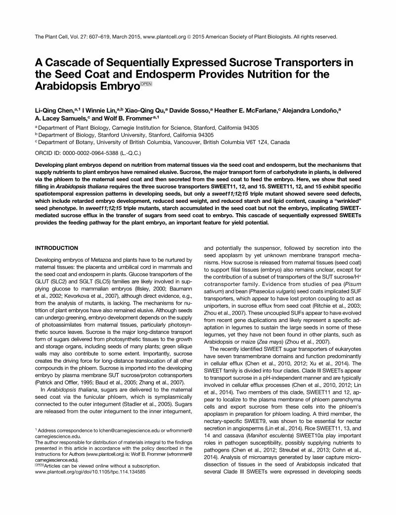
Figure 1. Expression and Localization of SWEET11, 12, and 15 in Seeds.
(A) Tissue-specific SWEET gene expression from microarray analysis (Dean et al., 2011) and protein accumulation as assessed by translational SWEET-GFP fusions during seed development. Representative images of developing seeds are shown above the panels. MSC, micropylar end of seed coat;MCE, micropylar endosperm; OI, outer integument; S, suspensor.(B) Confocal images of eGFP fluorescence in transgenic Arabidopsis seeds expressing translational SWEET11-, 12-, or 15-eGFP fusions under controlof their native promoters. The white arrow points to red autofluorescence of the cotyledon. The blue arrow points to red propidium iodide staining of cellwalls. The red arrow points to the suspensor. Bar = 50 mm.
608 The Plant Cell
process with several apoplasmic transport steps mediated bySWEET11, 12, and 15.
RESULTS
Expression of SWEET11, 12, and 15 in Seeds
SWEETs are prime candidates to play roles in sugar secretion frommaternal tissues during seed development. To explore whetherSWEETs may function in supplying the developing embryo withsucrose, we analyzed the expression of SWEET genes in Arabi-dopsis seeds from two sets of microarray data with cell-type ortissue-specific resolution (Dean et al., 2011; Belmonte et al., 2013).Microarray data analyses indicate that SWEET11, 12, and 15 maybe candidates for sucrose efflux from seed coat, as well as effluxfrom the endosperm to ultimately supply the developing embryowith phloem-derived sugar (Figure 1A; Supplemental Figure 1) (Chenet al., 2010; Dean et al., 2011; Belmonte et al., 2013). SWEET11transcripts accumulated primarily in the endosperm and seedcoat during the linear cotyledon stage and the maturation greenstage. SWEET12 transcripts were most abundant in the seedcoat at the same stages (i.e., linear cotyledon and maturationgreen stage) and appeared in the suspensor and at the micropylarend of the seed coat at the globular stage. SWEET15 transcriptswere detected in the endosperm during the globular and maturationgreen stages. Only weak expression of SWEET15 was found inseeds at the preglobular stage, but SWEET15 expression in seedcoat became dominant during the linear cotyledon and maturationstages. Despite partial overlap of SWEET11, 12, and 15 expression inseveral subregions during multiple stages, each gene appeared to beregulated differentially during development (Figure 1A; SupplementalFigure 1A), implying a complex, multistep feeding pattern.
To explore the tissue and cellular distribution of SWEET11, 12,and 15 at the protein level, Arabidopsis seeds from plants ex-pressing native promoter-driven translational SWEET11-, 12-, or15-eGFP (enhanced green fluorescent protein) fusions were ex-amined using confocal microscopy. Strong SWEET11-eGFP fluo-rescence was observed in the endosperm at the linear cotyledonstage, while weaker fluorescence was also detected in the chalazalseed coat (Figure 1B; Supplemental Figure 2 and SupplementalMovies 1 and 2). Transmission electron microscopy (TEM) immu-nocytochemistry also supported plasma membrane localization ofSWEET11 (Supplemental Figure 2) (Chen et al., 2012). Confocalmicroscopy of SWEET12-eGFP-expressing seeds detected strongfluorescence at the micropylar end of the seed coat from the pre-globular stage throughout the maturation stage and in the sus-pensor at the globular stage (Figures 1A and 1B). Accumulation ofSWEET15-eGFP was detected in the epidermal cells of the seedcoat at the preglobular stage, which then faded from globular stageto heart stage, but appeared bright again from the linear cotyledonstage throughout the maturation stage, as well as in the micropylarendosperm layer closest to the embryo at the linear cotyledonstage (Figure 1B; Supplemental Figure 2). In seed coat cells,SWEET15-eGFP was detectable both at the plasma membraneand in intracellular puncta, which likely are Golgi bodies, as indicatedby TEM (Figure 1B; Supplemental Figure 2). The localization patternof the three transporters did not perfectly match the microarray ex-pression profiles, potentially implying posttranscriptional regulation.
Overall, each SWEET appears to exhibit a specific spatiotemporalexpression and distribution pattern in the developing seed, es-pecially in the micropylar end of the seed coat, the micropylarendosperm, and the suspensor, intimating diversified roles indifferent cell types over time, as well as partially redundant rolesduring seed development.
Tissue-Specific Expression of SWEET15
In addition to their presence in seed tissues, we found SWEET11 and12 proteins in the vascular tissue of rosette leaves (Chen et al., 2012).SWEET15, also named SAG29 (SENESCENCE-ASSOCIATEDGENE29), was localized at the plasma membrane of Arabidopsisprotoplasts (Seo et al., 2011). SWEET15/SAG29 expression wasused as a senescence marker because the transcript had beenfound to increase during senescence (Quirino et al., 1999; Seo et al.,2011). To evaluate the temporal and spatial expression pattern ofSWEET15 at the protein level, a histochemical analysis usinga translational SWEET15-GUS (b-glucuronidase) fusion driven fromits native promoter was performed. In young plants (before bolting),GUS activity was below the detection level except for very weakstaining in the petioles (Figure 2A). The 38-d-old plants (2 d afterbolting) and 44-d-old plants (8 d after bolting) showed high GUSactivity in young buds of the inflorescence, which declined duringmaturation. However, only low GUS activity was observed insenescent leaves (Figures 2B to 2E). Consistent with the SWEET15-eGFP data shown above, GUS activity was also detected in theseed (Figure 2F). Thus, while SWEET15 gene expression appears tobe highly induced during senescence, there is little evidence forSWEET15 protein accumulation in leaves at the stages used herefor analyzing the role of SWEETs in seed filling, intimating thatSWEET15 is not likely to make a major contribution to phloem ex-port of sucrose for a major part of the seed filling phase.
SWEET11, 12, and 15 Transport Sucrose in Xenopuslaevis Oocytes
To test whether SWEET15 functions as a sucrose transporter,we expressed SWEET15 in X. laevis oocytes and measured[14C]-sucrose uptake. As one might expect from its phylo-genetic proximity to the sucrose transporting SWEET11 and12 (Chen et al., 2012), SWEET15 also functions as a sucrosetransporter (Supplemental Figure 3).
Delayed Embryo Development in a sweet11;12;15 Mutant
We hypothesized that if less sugar was released from maternaltissues of sweet mutants, less sugar would be available to filialtissues (embryo), possibly leading to defects in embryo growthand development. Therefore, we tested embryo growth of singlesweet11, 12, and 15 mutants. All three single sweet mutants ap-peared normal; only embryos of sweet12 were slightly smallercompared with the wild type (Figure 3A). Similarly, the doublemutants sweet11;12, sweet11;15, and sweet12;15 were charac-terized by a more severe reduction in embryo size relative to thesweet12 single mutant (Figure 3B). By contrast, the triple mutant ofsweet11;12;15 showed a significant delay in embryo developmentcompared with the double mutants, at least during the periodbetween 3 and 13 d postanthesis (DPA) (Figures 3C and 4).
Wrinkled Seeds of a SWEET Mutant 609

Although the difference in embryo size between the wild type andthe triple mutant gradually diminished after 8 DPA, in terms ofdevelopmental milestones, e.g., greening (Figures 3C [at 13 DPA]and 4), they remained smaller in size even at the mature stage.The defect in embryo size was rescued by complementation withany one of SWEET11, 12, or 15 (as translational eGFP fusions)expressed from their own native promoters (Supplemental Figure4), strongly supporting the hypothesis that in the triple mutant,retarded embryo growth is caused by loss of SWEET function.The result also demonstrates that the SWEET-GFP fusions werefunctional in planta. It is worth noting that SWEET11 and 12 arekey for phloem loading (Chen et al., 2012) and may thus affectdelivery of sucrose from the leaves, thereby further limiting seedfilling. However, SWEET15, which is unlikely to make a major con-tribution to phloem loading, can mostly rescue the delayed growthof sweet11;12;15 triple mutant embryos.To analyze how embryogenesis is affected in the triple
sweet11;12;15 mutant, we compared embryo development atdifferent stages by differential interference contrast (DIC) micro-scopy of cleared seeds (Figures 4A and 4B) and quantified embryosize (Figure 4C). Differences between embryos of sweet11;12;15and the wild type became apparent at the transition from globularto heart stage (4 DPA) in young siliques (Figures 4A and 4B). Themaximal difference was observed around 8 DPA, at a time whenSWEET11, 12, and 15 proteins accumulated to high levels (Figure1; Supplemental Figure 2). The delay in development was clearlyidentifiable when comparing mutant to the wild type; i.e., at 6 DPA,the wild type reached the linear cotyledon stage, while the triplemutant had reached the heart stage. Transporter-mediatedsucrose export from the endosperm is supported by the ob-servation that fluorescence derived from SWEET fusions ac-cumulated in the endosperm, a finding in line with publishedmicroarray data (Belmonte et al., 2013) (Figure 1B; SupplementalFigure 2).To test whether the delay in embryo growth in sweet11;12;15 is
maternally controlled, we performed reciprocal crosses between thewild type and sweet11;12;15. A dramatic reduction in sweet11;12;15embryo size was observed only when sweet11;12;15 was used asthe maternal parent (Figure 4D). Inheritance of integuments is ma-ternally controlled and inheritance of endosperm is maternal in part(Berger et al., 2006); thus, the sequential expression of SWEETsin the seed coat and endosperm is in agreement with the resultsof reciprocal crosses. In conclusion, SWEET11, 12, and 15 playkey roles on the maternal side and are necessary for securingefficient sugar supply for seed development.
Role of SWEETs in Determining Yield
To characterize the effect of changes in SWEET activity on yield,we measured the dry seed weight in triple mutants. In four in-dependent experiments, the yield (seed weight per plant) ofthe sweet11;12;15 mutant was reduced by ;43%, while that ofsweet11;12 was reduced by ;23% (Figure 5A). Reduced seedweight could be caused by smaller seeds, fewer seeds, or fewersiliques per plant. To differentiate between these possibilities, wemeasured the dry seed weight from a defined number of siliquesand found a decrease in seed weight of sweet11;12 by 17% andsweet11;12;15 by 34% (Figure 5B). The lower seed weight was
Figure 2. Spatial and Temporal Expression of SWEET15.
(A) to (C) Distribution of GUS activity in whole plants at different ages: (A)25 d old, (B) 38 d old, and (C) 44 d old.(D) High GUS activities detected in the inflorescence.(E) and (F) GUS activity (E) in leaves at different developmental stagesand in seed (F). Two individual transgenic lines were tested. One of thelines giving stronger GUS activity is shown here. GC, green cauline leaf;GR, green rosette leaf and seed; PS, partially senescent leaf; LS, latestage senescent leaf.
610 The Plant Cell

not caused by fewer seeds per silique, since there was no differencein the number of seeds per silique when comparing sweet11;12 andsweet11;12;15 with Columbia-0 (Col-0) (Figure 5C). Accordingly, itwas deduced that the number of siliques was reduced on averageby ;7% in sweet11;12 and 14% in sweet11;12;15.
Oil and protein are the major components of dry Arabidopsisseeds, each contributing ;40% to the seed mass (Baud et al.,2002). Seed dry mass closely follows the increase in oil duringdevelopment (Baud et al., 2003). In seeds, sucrose is the primarysource of acetyl-CoA, which serves as the precursor for the lipidbiosynthesis (Rawsthorne, 2002). As one may have expected,seed lipid content (fatty acid methyl esters [FAMEs]) was re-duced by 34% in sweet11;12 and 71% in sweet11;12;15.
Scanning electron microscopy was used to ascertain possiblemorphological alterations in the seed. Seeds of the triple mutanthad a deformed surface, phenotypically similar to the wrinkledwri1 mutants, which are defective in an APETALA2/ethylene-responsive element binding protein transcription factor involved inseed storage metabolism (Focks and Benning, 1998; Cernac andBenning, 2004). The wrinkled seed phenotype was not simply theresult of lower polysaccharide levels in the seed epidermis be-cause seeds hydrated in Ruthenium Red did not show a mucilageextrusion defect (Supplemental Figure 5B). The wrinkled pheno-type in sweet11;12;15 is consistent with incompletely filled seedscontaining smaller embryos (Figures 3A to 3C).
Rescue of Impaired sweet11;12;15 Seedling and EmbryoGrowth by Sucrose
Early postgerminative growth critically depends on sufficientamounts of nutrients stored in the seed. In the case of Arabidopsis,
nutrients are transiently stored as starch, which in later developmentis converted to oil (Kelly et al., 2011; Theodoulou and Eastmond,2012). To determine whether early postgerminative growth ofsweet11;12;15 is impaired, we analyzed root growth. When ger-minated on sugar-free media, sweet11;12;15 exhibited reducedroot length that could be ameliorated by external sucrose addition(Figure 6A). Reduced root growth, although less severe, wasobserved in the sweet11;12 double mutant (Figure 6A) (Chenet al., 2012).Sucrose is used as a carbon and energy source for in vitro
growth of immature embryos (Raghavan, 2003). To test whetherexogenously supplied sucrose can promote embryo growth of thesweet11;12;15 mutant, in vitro embryo culture was performed.Embryos dissected from sweet11;12;15 mutants and Col-0 at 3.5to ;4 DPA failed to grow in vitro in the absence of sucrose whencultured for additional 5 to ;5.5 days (9 DPA; Figure 6B). Supply ofsucrose accelerated embryo growth of sweet11;12;15 mutants andCol-0, and the area of sweet11;12;15 embryos reached 48% that ofCol-0 embryos. However, in vivo, the area of embryos dissectedfrom sweet11;12;15 plants was only 24% that of embryos fromCol-0 plants (Figures 6B and 6C). The reason why sweet11;12;15embryos are still smaller than Col-0 embryos when cultured invitro could be due to the differences already present at the timeof culture initiation (3.5 to ;4 DPA). The difference is shown inFigure 3C: The embryo area of sweet11;12;15 was 65 and 45%of that of Col-0 at 3 and 4 DPA, respectively. Thus, embryos ofthe triple mutant grew faster in vitro when supplied with su-crose compared with in vivo, directly supporting the notion thatthe observed embryonic phenotype in the triple mutant iscaused by reduced release of sucrose from seed coat andendosperm.
Relative Contribution of SWEET11, 12, and 15 to Seed Fillingin Seeds versus Leaves
Reduced carbohydrate export from leaves is expected to negativelyimpact embryo development (Andriotis et al., 2012). SWEET11 and12 are not only expressed in seeds, but also in leaves, as shownpreviously (Chen et al., 2012). To determine whether leaf exportcontributes in a major way to the mutant phenotype, we suppliedsugar to wild-type and mutant flowers cultured in vitro (Figure 7).Similarly sized flowers of sweet11;12;15 and Col-0 right after anthesiswere cultured vertically with the pedicel inserted into Murashige andSkoog (MS)-agar medium with or without 3% sucrose. After 9 d ofculture in a light/dark cycle, siliques developed from wild-typeflowers cultured in the absence of sugars produced a few seeds,some of which contained a normal sized embryo (Figures 7A and7B). Siliques from triple mutant flowers produced only a fewseeds, and those that were produced did not develop beyond theheart stage (Figures 7C and 7D). In contrast, siliques cultured insucrose-supplemented medium were able to produce seed fromboth wild-type and mutant flowers, although a significant numberof aborted seeds was observed. Importantly, embryos from wild-type siliques were on average much larger compared with theones from mutant siliques, in spite of the large variation of theembryos in size (Figures 7E to 7I). These results, which eliminatepotential effects of the mutations of phloem delivery from leaves,are consistent with the results from intact plants, strongly
Figure 3. SWEET11, 12, and 15 Are Required for Embryo Development.
Embryo phenotype of sweet single mutants at 8 DPA (A), double mutantsat 8 DPA (B), and triple mutants at 8 and 13 DPA (C). Bars = 0.2 mm.
Wrinkled Seeds of a SWEET Mutant 611

supporting a role of SWEET11, 12, and 15 in embryo sustenancewithin the seed itself (Figure 3C).
Changes in Starch Accumulation in Seed Coats andEmbryos of the Triple Mutant
To investigate the role of the three SWEETs in sugar release frommaternal tissues of the seed itself, we tested the effect of themutations on starch accumulation. We hypothesized that if re-duced efflux from maternal leaves was the primary cause fordelayed embryo development, one would predict reduced sugarlevels in the seed coat and embryo, whereas if reduced effluxfrom the maternal seed coat was the primary cause, sugar mightaccumulate in the seed coat. Starch accumulation was analyzedin sweet11;12;15 seeds and embryos using Lugol’s iodine, whichstains starch blue/black. Starch was barely detectable insweet11;12;15 embryos and dramatically reduced relative to wild-type seeds at 9, 10, and 11 DPA, indicative of reduced delivery ofsucrose to mutant embryos (Figure 8). Interestingly, starch in themutant stained blue (Figures 8C and 8D). Consistent with theoptical analysis of the starch staining, enzymatic quantitation ofstarch showed that the seed coat of sweet11;12;15 mutants ac-cumulated up to 5 times more starch compared with Col-0.Embryos of sweet11;12;15 mutants accumulated up to 19 timesless starch relative to Col-0 at 10 DPA (Figure 8E; SupplementalFigure 6). The accumulation of starch in the seed coat implicates
a block in sugar release from the seed coat as a key step in seedfilling. Consistent with the elevated abundance of carbohydratesin mutant seed coats, mucilage extrusion did not appear to beaffected negatively in the mutant, as one might have expectedif sugar supply from the phloem was the main cause for the seedphenotype (Supplemental Figure 5).
DISCUSSION
Embryo development is fully dependent on an adequate supply ofnutrients frommaternal tissues. Significant progress has been madein understanding the translocation from source tissues toward thevarious sinks (Lalonde et al., 2004; Chen et al., 2012); however,the mechanism of sucrose efflux from the maternal seed coat andthe endosperm (partially maternal and partially paternal) and theexact path have remained elusive. Here, we identified SWEET15,as well as SWEET11 and 12, as key players in seed coat efflux.Studies of developing bean seeds suggested that ;50% of
sucrose efflux from the seed coat is mediated by sucrose/protonantiporters driven by the proton-motive force (Fieuw and Patrick,1993; Walker et al., 1995), while the remaining activity was attrib-uted to mechanisms independent of the proton gradient (Walkeret al., 1995). In legume seeds, results from inhibitor studies led tothe proposal that nonselective passive pores are responsible forsucrose efflux (van Dongen et al., 2001; Ritchie et al., 2003). Neitherthe predicted antiporters nor the nonselective pores have been
Figure 4. Comparison of the Developmental Stages of Embryos in Col and sweet11;12;15 and Determination of the Maternal Contribution to Embryo Growth.
(A) and (B) Images of cleared seeds taken by DIC microscopy. Col-0 (A) and sweet11;12;15 (B) seeds at similar stages of embryogenesis. Bar = 0.1mm.(C) Embryo area measured at the corresponding stages from images in (A) and (B) (mean 6 SD, 3 DPA, n $ 167; 4 DPA, n $ 61; 5 to 8 DPA, n $ 18).(D) Embryo area measured at 9 d after pollination from the indicated reciprocal crosses (mean 6 SD, n $ 13).
612 The Plant Cell

identified at the molecular level to date. Uniporters play critical rolesin intercellular transport in both yeast and human; it is thereforeconceivable that plants also use a uniport mechanism for sucroseefflux from the seed coat. Members of SWEET family were char-acterized as proton-independent sugar transporters. Interestingly,
the human SWEET1 (SLC50A1) is found in decidual cells of theplacenta (Chen et al., 2010) (Human Protein Atlas, http://www.proteinatlas.org/ENSG00000169241-SLC50A1/tissue). Decidualcells accumulate glycogen and are thought to be involved inembryo nutrition, therefore potentially implicating these sugar effluxtransporters in human embryo development. Here, we identifySWEET11, 12, and 15 as being key to sucrose efflux frommaternal tissues of Arabidopsis seeds and as necessary forfilial tissue growth.Developing Arabidopsis seeds have at least three apoplasmic
borders that require membrane transporters: (1) between the outerintegument (a symplasmic extension of the phloem) and the innerintegument; (2) between the inner integument and the endosperm;and (3) between the endosperm and the embryo (Stadler et al.,2005). Based on expression patterns, all three SWEETs contributein cascade-like patterns to distinct steps in seed filling. Moreover,we identified localization patterns that may implicate new domainsinvolved in seed filling, particularly the micropylar end of seed coat,the micropylar endosperm, and the suspensor.
The Role of SWEETs in Specific Expression Regions
The specific spatiotemporal distribution of SWEET11, 12, and 15proteins at the micropylar end of the seed coat, the micropylarendosperm, and the suspensor intimates specific roles forSWEET11, 12, and 15, in addition to their redundant functionsduring seed development.SWEET15, which accumulated in the outer integument, is likely
responsible for mediating sucrose efflux from the outer integumentinto the apoplasm. Interestingly, strong SWEET12-GFP fluores-cence was observed specifically at the micropylar end. This is ex-actly the location where symplasmic GFP movement was reducedor absent relative to the rest of the seed coat (Stadler et al., 2005),indicating that symplasmic connectivity of the outer integument isreduced or interrupted at the micropylar end. It is therefore pro-posed that sucrose carriers are responsible for sucrose transportat the micropylar end, which based on the high accumulation ofSWEET12 in this region is likely mediated by SWEET12.The suspensor is thought to be a site where, during the early
stages of embryo development, nutrients and growth factors aretransferred to the embryo proper (Kawashima and Goldberg,2010). However, nutrient transfer across the suspensor has notbeen demonstrated experimentally. The suspensor is symplasmi-cally connected to the embryo during the globular stage (Stadleret al., 2005). However, the symplasmic connectivity betweensuspensor and embryo hypophysis is reduced during the transitionfrom the globular stage to the heart stage at which the suspensorstarts to degenerate (Stadler et al., 2005). The initiation ofSWEET12 expression in the suspensor at the globular stage maycoincide with the time when symplasmic connections betweensuspensor and embryo are blocked. The endosperm, which isdivided into three distinct domains, micropylar, peripheral, andchalazal endosperm, is an essential part of the seed that sustainsembryo development and stores reserves. Experiments in which[14C]-sucrose was supplied to intact oilseed rape (Brassica napus)siliques support the hypothesis that import of sugars into de-veloping embryos occurs via the micropylar rather than the cha-lazal endosperm (Morley-Smith et al., 2008). The localization of
Figure 5. Characterization of Other Seed Development-Related Phe-notypes of sweet11;12;15.
(A) Comparison of dry seed weight per plant from Col-0, sweet11;12, andsweet11;12;15 (mean6 SD, n$ 14 plants, from four independent experiments).(B) Comparison of dry seedweights of 20 siliques fromCol-0, sweet11;12, andsweet11;12;15 (mean6 SD, n$ 13 plants, from four independent experiments).(C) Comparison of number of seeds from each silique from Col-0,sweet11;12, and sweet11;12;15 (mean 6 SD, n = 30, from three plants).Two biological repeats were done.(D) Total fatty acid content (measured as FAME) of dry seeds from Col-0,sweet11;12, and sweet11;12;15 (mean 6 SD, n = 14 plants, from fourindependent experiments).(E) and (F) Scanning electron microscopy images of dry seeds fromCol-0 (E) and from sweet11;12;15 (F).
Wrinkled Seeds of a SWEET Mutant 613

SWEET11 and 15 in the micropylar endosperm indicates roles insucrose transfer from the endosperm.
Overall, SWEET11, 12, and 15 coexpress in several subregionsand in several stages; nevertheless, each of them also showeddistinct expression patterns during development. The temporal andspatial profiles observed at both the transcriptional and trans-lational levels indicate a cascade of sucrose efflux steps that beginsat the outer integument, passes first through the inner integument,then through the endosperm, and involves a number of particularsteps at the micropylar end, the micropylar endosperm, and thesuspensor, before it reaches the embryo proper. The expressionpatterns of SWEET11, 12, and 15 overlap partially and show very
specific patterns (e.g., in the suspensor and different domains ofseed coat and endosperm) that change during development(Figures 2 and 9; Supplemental Figures 1 and 2), indicating theexistence of several alternate routes for sucrose import, whichshift during seed development.
The Seed Phenotype in the sweet11;12;15 Mutant IsPredominantly Caused by Defects in Seed Import
Carbohydrate imported from leaves is critical for the growth ofyoung reproductive tissues, especially at night, as shown in ele-gant studies using the high-starch mutant sex1 (starch excess1)
Figure 6. Effect of Sucrose on Early Root Growth in Seedlings and on in Vitro-Grown Embryos.
(A) Comparison of root growth of Col-0, sweet11;12, and sweet11;12;15 seedlings grown on sugar-free and sugar-supplied media.(B) Embryos dissected from seeds at 3.5 DPA grown in vitro for 5.5 d with and without supply of sucrose.(C) Embryo size as approximated by area measurements of embryo from in vitro culture with 5% sucrose and dissected seed of intact plant at 9 DPA.Area of sweet11;12;15 is normalized to Col-0 (mean 6 SD, n $ 20, from three independent experiments).
Figure 7. Analysis of Silique and Embryo Growth for in Vitro-Cultured Flowers in Medium with or without Sucrose.
(A) to (D) Comparison of silique development ([A] and [C]) and embryo growth ([B] and [D]) from Col-0 ([A] and [B]) and sweet11;12;15 ([C] and [D])cultured for 9 d in sugar-free medium.(E) to (H) Comparison of silique development ([E] and [G]) and embryo growth ([F] and [H]) from Col-0 ([E] and [F]) and sweet11;12;15 ([G] and [H])cultured for 9 d in medium with 3% sucrose.(B), (D), (F), and (H) Embryos were dissected from three siliques cleared with 0.2 NaOH and 1% SDS solution by gently pressing cover slide. Yellowcoloration was observed in Col-0, while sweet11;12;15 showed reduced coloration. Black bar = 5 mm and white bar = 0.5 mm.(I) Embryo area was measured for data shown in (B), (D), (F), and (H) (mean 6 SD, n = 7 [B], n = 24 [F], and n = 21 [H]). None of embryos ofsweet11;12;15 cultured in medium without sucrose could be measured due to abortion. The data shown are from a single experiment. Two independentexperiments showed similar results.
614 The Plant Cell

(Andriotis et al., 2012). Similar to the sweet11;12;15 triple mutant,embryo development is significantly delayed in sex1. The delay insex1 growth is caused by low carbohydrate availability during thenight (Andriotis et al., 2012). SWEET11 and 12 were found to beexpressed both in leaves (Chen et al., 2012) and in seeds; thus, theobserved seed phenotype is likely due to a combination of re-duced supply from leaves and reduced import into filial tissues.Similarly, one could hypothesize that SWEET15 could contributeto phloem loading particularly during senescence, although anal-ysis of translational GUS fusions indicates that SWEET15 levelsare not elevated at the seed filling stage at least in the early phase.Here, we decoupled the contribution of SWEETs in leaves fromtheir role in seed nutrition by culturing flowers axenically (Figure 7).We found that seeds were able to develop to maturation stages inwild-type siliques when sucrose was supplied in the medium tomimic the phloem supply. However, embryos from sweet11;12;15remained much smaller compared with the wild type, while de-veloped in the same conditions. These data strongly support thenotion that the mutant effect on embryo growth is mainly due tolimited availability of sucrose to the embryo proper. Comparison ofsiliques grown in sugar-free medium further indicates that photo-synthesis of silique walls is insufficient to support seed growth andthat normal seed development, as one may have expected, reliesto a large extent on supply from leaves. The fact that only a fewembryos developed beyond the bent cotyledon stage furthersupports the assumption that sucrose produced in the siliques isjust enough to feed a few seeds when sucrose export from theseed coat is functional, as in Col-0, in contrast to sweet11;12;15.
Further evidence for a major contribution of the three SWEETs toseed filling stems from the observation that starch accumulatedmore specifically in the seed coat of the sweet11;12;15 mutant(Figure 8). If leaf supply would be the dominant factor, one wouldhave expected the opposite, i.e., reduced starch levels in bothseed coat and embryo.
Functional Model of SWEETs in Sucrose Translocationin Seeds
Based on the results presented here, we propose a functionalmodel for sucrose translocation from the maternal phloem towardthe developing embryo (Figure 9). During early developmentalstages, sucrose is unloaded into the postphloem unloadingdomain via the vascular bundle of the funiculus. Then, sucrosemoves toward the micropylar end of the seed coat within thesymplasmically connected outer integument. Subsequently,sucrose appears to be released into the apoplasm by the actionof SWEET15. Within the micropylar end, symplasmic loadingswitches to apoplasmic loading with the appearance of SWEET12
Figure 8. Analysis of Starch Accumulation in Embryos and Seed Coats.
(A) and (B) Comparison of starch accumulation in sweet11;12;15 andCol-0 at 9, 10, and 11 DPA.(C) and (D) Cross section of seeds stained with Lugol’s iodine solution at9 DPA.(E) Enzymatic quantification of starch content of seed coats and em-bryos from Col-0 or sweet11;12;15 at 10 DPA (mean 6 SD, n = 4).
Figure 9. Model for Multistep Sequential Apoplasmic Transport Stepsduring Seed Development Initiated by SWEET Sucrose Transporters.
Sucrose, which arrives in the seed coat via the funicular phloem, canenter the outer integument of the seed coat likely through plasmodes-mata. SWEET15, expressed in the outer integument (OI), exports sucroseinto the apoplasm (AP). Possibly, SWEET11 is involved in the release ofsucrose from the inner integument (II). SWEET12 appears to play a role intransport of sucrose out of cells at the micropylar end of the seed coat(MSC) and SWEET11 and 15 from the micropylar endosperm (MCE) tosupply the embryo proper (EM).
Wrinkled Seeds of a SWEET Mutant 615
at the globular stage (Werner et al., 2011). The presence ofSWEET12 protein in the micropylar end of seed coat implies a rolefor SWEET12 in mediating sucrose transport to and within thissubregion of the seed coat. Unloaded sucrose is used for starchaccumulation in the seed coat and cell wall biosynthesis in theendosperm (Fallahi et al., 2008). Some of the sucrose may be di-rectly delivered to the embryo from the seed coat via the suspensor,a hypothesis compatible with SWEET12 expression in the suspen-sor during early seed development. However, cellular hexose importmay dominate over sucrose import during the early stages of seeddevelopment (Baud et al., 2002), consistent with high acid invertaseactivity (Hill et al., 2003). High hexose accumulation is also in linewith detected expression of glucose/proton symporters in the seedcoat during early seed development (Supplemental Figure 7A). It isworth noting that most of the sugars in the central vacuole in theendosperm of oilseed rape are hexoses, and the bulk of the sugarpool may, at early developmental stages, not be exchangeable withthat of phloem-derived sugars (Morley-Smith et al., 2008). Howsucrose is exported into the apoplasm during the stages whenSWEETs expression is hardly detected in seed coat remains anopen question. Yet to be identified sucrose/proton antiporters maybe responsible for this function (Fieuw and Patrick, 1993; Walkeret al., 1995). The existence of additional transport mechanismscould be one of the reasons why sweet11;12;15 triple mutants arenot lethal. During later developmental stages, sucrose is mainlyexported from the outer integument by SWEET15 and likely bySWEET11 from the inner integument. SUC5 may be responsible foruptake of sucrose into the endosperm from the apoplasm betweenthe seed coat and the endosperm (Baud et al., 2005). SUC5 couldalso act at the interface between the endosperm and embryo, sinceit is expressed in the endosperm and specifically the epidermis ofthe outer surface of the cotyledons (Supplemental Figure 7B)(Pommerrenig et al., 2013). Data from a SUC9 translational fusionto GUS indicated that SUC9 protein was also present in the em-bryo (Sivitz et al., 2007), although no transcriptional GUS activitywas detected in seeds (Sauer et al., 2004). All these reports are inagreement with the observation that in vitro dissected embryosfrom the globular stage can reach the mature stage when grown inmedia supplemented with sucrose.
Together, our data demonstrate a critical role of SWEETs insucrose release from the seed coat and multiple sites in theseed to supply the developing embryo. It will be interestingto determine whether human SWEET1 plays an orthologousfunction in the placenta. It also will be interesting to explore theparallels and potential convergent evolution of embryo nutri-tion in plants and animals, particularly the role of SWEETs inthese processes.
METHODS
Plasmid Constructs
Constructs for Expression in Xenopus laevis Oocytes
Oocyte expression construct for SWEET12 has been described pre-viously (Chen et al., 2010). The open reading frame of Arabidopsis thalianaSWEET15 (with stop codon) in vector pDONR221-f1 was transferred tothe oocyte expression vector pOO2-GW as described previously for otherSWEETs (Chen et al., 2010).
eGFP Fusion Constructs under Native Promoters
For analyzing the expression of SWEET12 and 15, GFP fusions were con-structed. The fragment of SWEET12 comprising a 1887-bp SWEET12 pro-moter sequence and 1858 bp of the coding region up to but not including thestop codon was amplified with the forward primer AtSWT12KpnIF containinga KpnI restriction site and the reverse primer AtSWT12PstIR containing a PstIrestriction site and subcloned into the eGFP fusion vector pGTKan3 (Kasarasand Kunze, 2010) via KpnI and PstI restriction sites. A similar strategy wasapplied for SWEET15. The 3784-bp fragment of SWEET15 was amplified byprimers AtSWT15KpnIF and AtSWT15PstIR and ligated into pGTKan3 byKpnI and PstI restriction sites. The translational GUS fusion construct wasgenerated by amplifying the identical sequence used for GFP fusion constructusing primers AtSWT15attB1 and AtSWT15attB2 and then cloning into donorvector pDONR221-f1 followedby recombination reaction intoGateway vectorpGWB3 (Nakagawa et al., 2007) by LR reaction as described before (Chenet al., 2012). Construction of pSWEET11:SWEET11-eGFP has been de-scribed previously (Chen et al., 2012). Primer sequences are provided inSupplemental Table 1.
Plasmids for Complementation of Mutants
For complementation of the sweet11;12:15 triple mutant, the AgrobacteriumtumefaciensGV3101 transformants carrying constructspSWEET11:SWEET11-eGFP, pSWEET12:SWEET12-eGFP, and pSWEET15:SWEET15-eGFP wereused to transform sweet11;12:15 triple mutants.
Tracer Uptake in X. laevis Oocytes
Linearization of the plasmids in pOO2 vector, capped cRNA synthesis, X.laevis oocyte isolation and cRNA injection, [14C]-labeled sugar uptake,and efflux were performed as described before (Chen et al., 2010).
Plant Material and Growth Conditions
Plants were grown under light (150 to 170 mE m22 s21 with 16 h light/8 hdark) photoperiod conditions. Only primary shoots and the first secondaryshoots were used. To collect siliques of desired developmental stages,individual flowers were tagged with colored strings on the day of flowering(referred to as 0 DPA) between 11 AM and 1 PM. Tagging plants started withinthe first week of plant bolting.
Seeds of transposon line SM_3_14944 for SWEET15 were orderedfrom ABRC. In order to get triple mutant, sweet15 was crossed withsweet11;12. Homozygous sweet11;12;15 was selected by genotyping.
For seedling growth analysis, seeds were sown on half-strength MSmedium with or without sucrose (as indicated) and then kept at 4°C for 3d before being transferred to a growth chamber and positioned vertically(16-h-light period). At indicated days after transfer, seedlings were digi-tally photographed.
Arabidopsis wild-type Col-0 and sweet11;12;15 triple mutants weretransformed by the floral dipmethod (Davis et al., 2009). Transgenic seedlingswere selected on media with kanamycin for pSWEET11:SWEET11-eGFP,pSWEET12:SWEET12-eGFP, and pSWEET15:SWEET11-eGFP.
Genotyping and Transcript Analysis of T-DNA Mutants
Genomic DNA was extracted from Arabidopsis Col-0 and F3 segregatinglines after crossing sweet15 (SM_3_14944) with sweet11;12 (Salk_073269and Salk_031696) and was used as template for genotyping. Primers usedfor sweet11 and sweet12 genotyping were as described (Chen et al., 2012).Primers specific to SWEET15 sequence flanking the transposon elementwere AtSWT15LP and AtSWT15RP. The left border primer of transposonelement was Spm32. PCR was performed as described on the websitehttp://signal.salk.edu/database/T-DNA/SM.435.pdf.
616 The Plant Cell
Total RNA was extracted from leaves of Arabidopsis from Col-0, controls,and insertion lines using a Spectrum plant total RNA kit (Sigma-Aldrich;#120M6117). First-strand cDNAwas synthesized using oligo(dT) andM-MuLVreverse transcriptase following the instructions of the supplier (Fermentas).Primers for the full-lengthopen reading frameofSWEET15 (AtSWT15attB1andAtSWT15attB2) were used for RT-PCR to determine the expression levels.ACTIN2 (primers AtACT2F and AtACT2R) served as reference gene (Chenet al., 2010). Primer sequences are provided in Supplemental Table 1.
Starch Staining
After being harvested in the late afternoon, siliques were placed in 80% (v/v)ethanol plus 5% (v/v) formic acid and incubated in a 60°C oven overnight,then washed once with 70% ethanol. Then, siliques were stained in Lugol’siodine solution for 5 min and washed twice in water.
GUS Histochemical Analysis
GUS staining was performed immediately after samples were collected atthe different ages, following the procedures described previously (Chen et al.,2012). To avoid overlooking the expression of SWEET15 in other tissues,staining was extended to 24 h. A flatbed scanner was used to documentthe results.
Lipid Analysis
Lipid content of dry seeds was analyzed following the published procedureswith minor changes (Focks and Benning, 1998). Seeds from mutants andCol-0 plants, carefully planted andmanaged together, were harvested on thesame day. Dry seeds were counted and prepared in 8-mL glass reactiontubes. Three seeds from each background were used for the first biologicalrepeat and 20 seeds for the next two biological repeats. At least fourtechnical repeats were included in each experiment. One hundredmicrolitersof 50 µg/mL pentadecanoic acid (C15:0) was used as an internal standard.Samples were incubated in 1 mL of 1 N HCl in methanol at 80°C for 90 min.Tubes were allowed to cool to room temperature. FAMEs were extracted in1 mL hexane following the addition of 1 mL 0.9% (w/v) NaCl. The hexanephasewas transferred to a gas chromatography vial after vigorous vortexingand centrifuged for 3 min at 3000 rpm. Samples were analyzed using a gaschromatography-flame ionization detector.
Seed Coat and Embryo Starch Measurement
Starch content was measured enzymatically following published procedureswith some changes (Andriotis et al., 2010). Thirty embryos or seed coats werepooled from one plant for one analysis. In detail, 10 embryos or seed coatswere rapidly dissected from seeds of each freshly harvested siliques andtransferred to 300 mL iced-cold 0.7 M perchloric acid. Three siliques werecollected from each individual plant. In total, four individual sweet11;12;15 orCol-0 plants were used. Sixteen samples were pooled, homogenized, andcentrifugedat 11,000g for 45minat 4°C. Sedimentswerewashedonce in 1mLdistilled water and twice in 1 mL 80% (v:v) ethanol, then vacuum dried. Driedsedimentswere resuspended in 40mL distilledwater by boiling for 6min twice.A starch measurement kit (R-Biopharm) was used to quantify starch content.Briefly, starch was hydrolyzed to D-glucose by amyloglucosidase. D-glucosewas enzymatically assayed in the presence of hexokinase and glucose 6-phosphate dehydrogenasebydetermining the amount ofNADPH (absorbanceat 340 nm). We note that the seed dissection cannot fully separate embryo,endosperm, and seed coat; thus, we cannot exclude cross-contamination.
In Vitro Embryo Culture
The procedure was slightly modified based on the published protocols(Sauer and Friml, 2008). Embryos were dissected gently from 3.5 to 4 DPAsiliques using sterile forceps. Half of undamaged embryos from one silique
were immediately transferred to in vitro culture medium plates with 5%sucrose under 70% ethanol cleaned stereo microscope. The other half fromthe same silique was transferred to a plate without sucrose. Embryos weredissected carefully from seeds and then the rest of the debris from the seedswas removed from the plates. Five to 10 siliques were used for each ex-periment. Plates were placed in a 22°C growth chamber with 150 to 170 mEm22 s21 light and 16-h-light/8-h-dark diurnal cycle. Two sheets of plainwhitepaper were used to shield the excess light. After 5 d of culture, embryoswereimaged using a Leica MZ125 stereomicroscope.
In Vitro Flower Culture
On the day of anthesis, similarly sized flowers were cut and immediatelytransferred to 0.025% (v/v) sodium hypochlorite for 8 min. The pedicel wasinserted in half-strength MSmediumwith 0.3% (w/v) Phytagel, 4 mM L-Gln,1 mM L-Asn, and 3% (w/v) sucrose or without sugar. Flowers were culturedfor 9 d in a 22°C growth chamberwith 130 to 150mEm22 s21 light and16-h-light/8-h-dark photoperiod conditions.
Microscopy
For all experiments, siliques were freshly harvested.
Phenotype Observation and Documentation
For phenotype observation, siliques were opened on a microscope slideright after collection. Seeds were mounted in water, and gentle pressurewas applied to the cover slip on the seeds to release the embryos. Af-terwards, nondamaged embryos were documented under a Leica MZ125stereomicroscope.
Clearing Seeds and Imaging
For determination of embryo developmental stages, siliques were fixed inethanol:acetic acid (9:1) overnight and washed with 90 and 70% ethanol.Siliques were clearedwith chloral hydrate:glycerol:water (8:1:2, w:v:v) solutionup to overnight (Yadegari et al., 1994). After being dissected from siliques,seedsweremounted in clearing solution under amicroscope with DIC optics.Afterwards, embryo area was measured using Fiji software as needed.
Scanning Electron Microscopy
Dry seeds from Col-0 and sweet11;12;15 were prepared. Scanningelectron microscopy was performed using FBI Quanta 200 at an accel-erating voltage of 30 kV.
Cryofixation and Immuno-TEM
Seeds were staged according to DPA (Western et al., 2000). The 7 and 10DPA seeds were high-pressure frozen in 1-hexadecene in B-type sampleholders (Ted Pella) using a Leica HPM-100. Samples were freeze substitutedin 0.1%uranyl acetate, 0.25%glutaraldehyde, and 8%dimethoxypropane inacetone for 5 d and then brought to room temperature and infiltrated with LRwhite resin (London Resin Company) over 4 d. Immunolabeling was per-formed as described before (McFarlane et al., 2008) using 1/100 anti-GFP(Invitrogen A6455) and 1/100goat-anti-rabbit conjugated to 10-nmgold (TedPella). Samples were viewed using a Hitachi 7600 transmission electronmicroscope at 80-kV accelerating voltage with an AMT Advantage CCDcamera (Hamamatsu ORCA).
Confocal Laser Scanning Microscopy
Fluorescence imaging of seed was performed on a Leica TCS SP5 mi-croscope. eGFP was visualized by standard procedures as describedbefore (Chen et al., 2012). For cell wall staining, fresh siliques were in-cubated with 0.5% propidium iodide in 1.5-mL centrifuge tubes for 10 minat room temperature and were washed twice with water. eGFP was
Wrinkled Seeds of a SWEET Mutant 617

excited by a 488-nm argon laser and detected from 497 to 526 nm. Pro-pidium iodidewas excitedby 488-nm light anddetected from595 to 640 nm(Stadler et al., 2005). A water-corrected 203 or glycerol-corrected 633objective was used. Image analysis was performed using Fiji software.
Accession Numbers
Sequence data from this article can be found in the Arabidopsis GenomeInitiative or GenBank/EMBL databases under the following accessionnumbers: SWEET11 (At3g48740), SWEET12 (At5g23660), SWEET15(At5g13170), SUC1 (At1g71880), SUC2 (At1g22710), SUC3 (At2g02860),SUC4 (At1g09960), SUC5 (At1g71890), SUC9 (At5g06170), STP1(At1g11260), STP4 (At3g19930), STP5 (St1g34580), STP7 (At4g02050),STP8 (At5g26250), STP12 (At4g21480), STP14 (At1g77210), and humanSWEET1 (ENSG00000169241).
Supplemental Data
Supplemental Figure 1. Tissue-specific expression of SWEET tran-scripts in developing seeds of Arabidopsis.
Supplemental Figure 2. Localization of SWEETs indicated by eGFPfluorescence and by immunogold particles.
Supplemental Figure 3. SWEET15 functions as a sucrose transporter.
Supplemental Figure 4. Complementation of the triple sweet11;12;15mutant by individual SWEET genes.
Supplemental Figure 5. Phenotypes of sweet11;12;15 mutant seeds.
Supplemental Figure 6. Starch content measurement.
Supplemental Figure 7. STP and SUT/SUC sugar transporterexpression profiles in developing seed.
Supplemental Table 1. Primer sequences.
Supplemental Movie 1. Localization of SWEET11 during late seeddevelopment (mature green stage).
Supplemental Movie 2. Localization of SWEET11 during late seeddevelopment (mature green stage).
ACKNOWLEDGMENTS
We thank Heather Cartwright for confocal imaging advice. We thankDavid Ehrhardt, Alexander Jones, and Lily Cheung for critical readingof the article. We also thank Xiaobo Li for the help with lipid contentmeasurement. Technical assistance of the UBC Bioimaging Facility isgratefully acknowledged. This work was made possible by grants toW.B.F. from the Department of Energy (DE-FG02- 04ER15542) andfrom the Carnegie Institution of Canada to W.B.F. and A.L.S.
AUTHOR CONTRIBUTIONS
W.B.F., L.-Q.C., and A.L.S. conceived and designed the experiments.L.-Q.C., I.W.L., X.-Q.Q., D.S., H.E.M., and A.L. performed the experiments.W.B.F. and L.-Q.C., analyzed the data. L.-Q.C. andW.B.F. wrote the article.
Received November 24, 2014; revised February 6, 2015; acceptedFebruary 26, 2015; published March 20, 2015.
REFERENCES
Andriotis, V.M., Pike, M.J., Kular, B., Rawsthorne, S., and Smith,A.M. (2010). Starch turnover in developing oilseed embryos. NewPhytol. 187: 791–804.
Andriotis, V.M., Pike, M.J., Schwarz, S.L., Rawsthorne, S., Wang,T.L., and Smith, A.M. (2012). Altered starch turnover in the maternalplant has major effects on Arabidopsis fruit growth and seed compo-sition. Plant Physiol. 160: 1175–1186.
Baud, S., Boutin, J.-P., Miquel, M., Lepiniec, L., and Rochat, C.(2002). An integrated overview of seed development in Arabidopsisthaliana ecotype WS. Plant Physiol. Biochem. 40: 151–160.
Baud, S., Wuillème, S., Lemoine, R., Kronenberger, J., Caboche,M., Lepiniec, L., and Rochat, C. (2005). The AtSUC5 sucrosetransporter specifically expressed in the endosperm is involved inearly seed development in Arabidopsis. Plant J. 43: 824–836.
Baud, S., Guyon, V., Kronenberger, J., Wuillème, S., Miquel, M.,Caboche, M., Lepiniec, L., and Rochat, C. (2003). Multifunctionalacetyl-CoA carboxylase 1 is essential for very long chain fatty acidelongation and embryo development in Arabidopsis. Plant J. 33: 75–86.
Baumann, M.U., Deborde, S., and Illsley, N.P. (2002). Placentalglucose transfer and fetal growth. Endocrine 19: 13–22.
Belmonte, M.F., et al. (2013). Comprehensive developmental profilesof gene activity in regions and subregions of the Arabidopsis seed.Proc. Natl. Acad. Sci. USA 110: E435–E444.
Berger, F., Grini, P.E., and Schnittger, A. (2006). Endosperm: anintegrator of seed growth and development. Curr. Opin. Plant Biol.9: 664–670.
Cernac, A., and Benning, C. (2004). WRINKLED1 encodes an AP2/EREB domain protein involved in the control of storage compoundbiosynthesis in Arabidopsis. Plant J. 40: 575–585.
Chen, L.Q., Qu, X.Q., Hou, B.H., Sosso, D., Osorio, S., Fernie, A.R.,and Frommer, W.B. (2012). Sucrose efflux mediated by SWEETproteins as a key step for phloem transport. Science 335: 207–211.
Chen, L.Q., et al. (2010). Sugar transporters for intercellular exchangeand nutrition of pathogens. Nature 468: 527–532.
Cohn, M., Bart, R.S., Shybut, M., Dahlbeck, D., Gomez, M., Morbitzer,R., Hou, B.H., Frommer, W.B., Lahaye, T., and Staskawicz, B.J.(2014). Xanthomonas axonopodis Virulence Is Promoted by a Tran-scription Activator-Like Effector-Mediated Induction of a SWEET SugarTransporter in Cassava. Mol. Plant Microbe Interact. 27: 1186–1198.
Davis, A.M., Hall, A., Millar, A.J., Darrah, C., and Davis, S.J. (2009).Protocol: Streamlined sub-protocols for floral-dip transformationand selection of transformants in Arabidopsis thaliana. Plant Methods5: 3.
Dean, G., Cao, Y., Xiang, D., Provart, N.J., Ramsay, L., Ahad, A.,White, R., Selvaraj, G., Datla, R., and Haughn, G. (2011). Analysisof gene expression patterns during seed coat development in Arabi-dopsis. Mol. Plant 4: 1074–1091.
Fallahi, H., Scofield, G.N., Badger, M.R., Chow, W.S., Furbank,R.T., and Ruan, Y.-L. (2008). Localization of sucrose synthase indeveloping seed and siliques of Arabidopsis thaliana reveals diverseroles for SUS during development. J. Exp. Bot. 59: 3283–3295.
Fieuw, S., and Patrick, J. (1993). Mechanism of photosynthate effluxfrom Vicia faba L. seed coats. J. Exp. Bot. 44: 65–74.
Focks, N., and Benning, C. (1998). wrinkled1: A novel, low-seed-oilmutant of Arabidopsis with a deficiency in the seed-specific regu-lation of carbohydrate metabolism. Plant Physiol. 118: 91–101.
Hill, L.M., Morley-Smith, E.R., and Rawsthorne, S. (2003). Metab-olism of sugars in the endosperm of developing seeds of oilseedrape. Plant Physiol. 131: 228–236.
Illsley, N.P. (2000). Glucose transporters in the human placenta.Placenta 21: 14–22.
Kasaras, A., and Kunze, R. (2010). Expression, localisation andphylogeny of a novel family of plant-specific membrane proteins.Plant Biol. (Stuttg.) 12 (suppl. 1): 140–152.
Kawashima, T., and Goldberg, R.B. (2010). The suspensor: not justsuspending the embryo. Trends Plant Sci. 15: 23–30.
618 The Plant Cell

Kelly, A.A., Quettier, A.L., Shaw, E., and Eastmond, P.J. (2011).Seed storage oil mobilization is important but not essential forgermination or seedling establishment in Arabidopsis. Plant Physiol.157: 866–875.
Kevorkova, O., Ethier-Chiasson, M., and Lafond, J. (2007). Differ-ential expression of glucose transporters in rabbit placenta: effectof hypercholesterolemia in dams. Biol. Reprod. 76: 487–495.
Lalonde, S., Wipf, D., and Frommer, W.B. (2004). Transport mech-anisms for organic forms of carbon and nitrogen between sourceand sink. Annu. Rev. Plant Biol. 55: 341–372.
Lin, I.W., et al. (2014). Nectar secretion requires sucrose phosphatesynthases and the sugar transporter SWEET9. Nature 508: 546–549.
McFarlane, H.E., Young, R.E., Wasteneys, G.O., and Samuels, A.L.(2008). Cortical microtubules mark the mucilage secretion domainof the plasma membrane in Arabidopsis seed coat cells. Planta 227:1363–1375.
Morley-Smith, E.R., Pike, M.J., Findlay, K., Köckenberger, W., Hill,L.M., Smith, A.M., and Rawsthorne, S. (2008). The transport ofsugars to developing embryos is not via the bulk endosperm inoilseed rape seeds. Plant Physiol. 147: 2121–2130.
Nakagawa, T., Kurose, T., Hino, T., Tanaka, K., Kawamukai, M.,Niwa, Y., Toyooka, K., Matsuoka, K., Jinbo, T., and Kimura, T.(2007). Development of series of gateway binary vectors, pGWBs,for realizing efficient construction of fusion genes for plant trans-formation. J. Biosci. Bioeng. 104: 34–41.
Patrick, J.W., and Offler, C.E. (1995). Post-sieve element transport ofsucrose in developing seeds. Aust. J. Plant Physiol. 22: 681–702.
Pommerrenig, B., Popko, J., Heilmann, M., Schulmeister, S.,Dietel, K., Schmitt, B., Stadler, R., Feussner, I., and Sauer, N.(2013). SUCROSE TRANSPORTER 5 supplies Arabidopsis embryos withbiotin and affects triacylglycerol accumulation. Plant J. 73: 392–404.
Quirino, B.F., Normanly, J., and Amasino, R.M. (1999). Diverserange of gene activity during Arabidopsis thaliana leaf senescenceincludes pathogen-independent induction of defense-related genes.Plant Mol. Biol. 40: 267–278.
Raghavan, V. (2003). One hundred years of zygotic embryo cultureinvestigations. In Vitro Cell. Dev. Biol. Plant 39: 437–442.
Rawsthorne, S. (2002). Carbon flux and fatty acid synthesis in plants.Prog. Lipid Res. 41: 182–196.
Ritchie, R.J., Fieuw-Makaroff, S., and Patrick, J.W. (2003). Sugarretrieval by coats of developing seeds of Phaseolus vulgaris L. andVicia faba L. Plant Cell Physiol. 44: 163–172.
Sauer, M., and Friml, J. (2008). In vitro culture of Arabidopsis em-bryos. Methods Mol. Biol. 427: 71–76.
Sauer, N., Ludwig, A., Knoblauch, A., Rothe, P., Gahrtz, M., andKlebl, F. (2004). AtSUC8 and AtSUC9 encode functional sucrosetransporters, but the closely related AtSUC6 and AtSUC7 genesencode aberrant proteins in different Arabidopsis ecotypes. Plant J.40: 120–130.
Seo, P.J., Park, J.M., Kang, S.K., Kim, S.G., and Park, C.M. (2011).An Arabidopsis senescence-associated protein SAG29 regulatescell viability under high salinity. Planta 233: 189–200.
Sivitz, A.B., Reinders, A., Johnson, M.E., Krentz, A.D., Grof, C.P.,Perroux, J.M., and Ward, J.M. (2007). Arabidopsis sucrose trans-porter AtSUC9. High-affinity transport activity, intragenic control of ex-pression, and early flowering mutant phenotype. Plant Physiol. 143:188–198.
Stadler, R., Lauterbach, C., and Sauer, N. (2005). Cell-to-cellmovement of green fluorescent protein reveals post-phloem trans-port in the outer integument and identifies symplastic domains inArabidopsis seeds and embryos. Plant Physiol. 139: 701–712.
Streubel, J., Pesce, C., Hutin, M., Koebnik, R., Boch, J., andSzurek, B. (2013). Five phylogenetically close rice SWEET genesconfer TAL effector-mediated susceptibility to Xanthomonas oryzaepv. oryzae. New Phytol. 200: 808–819.
Theodoulou, F.L., and Eastmond, P.J. (2012). Seed storage oilcatabolism: a story of give and take. Curr. Opin. Plant Biol. 15:322–328.
van Dongen, J.T., Laan, R.G., Wouterlood, M., and Borstlap, A.C.(2001). Electrodiffusional uptake of organic cations by pea seedcoats. Further evidence for poorly selective pores in the plasmamembrane of seed coat parenchyma cells. Plant Physiol. 126:1688–1697.
Walker, N., Patrick, J., Zhang, W.-H., and Fieuw, S. (1995). Efflux ofphotosynthate and acid from developing seed coats of Phaseolusvulgaris L.: a chemiosmotic analysis of pump-driven efflux. J. Exp.Bot. 46: 539–549.
Werner, D., Gerlitz, N., and Stadler, R. (2011). A dual switch inphloem unloading during ovule development in Arabidopsis. Pro-toplasma 248: 225–235.
Western, T.L., Skinner, D.J., and Haughn, G.W. (2000). Differentia-tion of mucilage secretory cells of the Arabidopsis seed coat. PlantPhysiol. 122: 345–356.
Xu, Y., Tao, Y., Cheung, L.S., Fan, C., Chen, L.Q., Xu, S., Perry, K.,Frommer, W.B., and Feng, L. (2014). Structures of bacterial ho-mologues of SWEET transporters in two distinct conformations.Nature 515: 448–452.
Yadegari, R., Paiva, G., Laux, T., Koltunow, A.M., Apuya, N.,Zimmerman, J.L., Fischer, R.L., Harada, J.J., and Goldberg,R.B. (1994). Cell differentiation and morphogenesis are uncoupledin Arabidopsis raspberry embryos. Plant Cell 6: 1713–1729.
Zhang, W.-H., Zhou, Y., Dibley, K.E., Tyerman, S.D., Furbank, R.T.,and Patrick, J.W. (2007). Review: Nutrient loading of developingseeds. Funct. Plant Biol. 34: 314–331.
Zhou, Y., Qu, H., Dibley, K.E., Offler, C.E., and Patrick, J.W. (2007).A suite of sucrose transporters expressed in coats of developinglegume seeds includes novel pH-independent facilitators. Plant J.49: 750–764.
Wrinkled Seeds of a SWEET Mutant 619

DOI 10.1105/tpc.114.134585; originally published online March 20, 2015; 2015;27;607-619Plant Cell
A. Lacey Samuels and Wolf B. FrommerLi-Qing Chen, I Winnie Lin, Xiao-Qing Qu, Davide Sosso, Heather E. McFarlane, Alejandra Londoño,
Provides Nutrition for the Arabidopsis EmbryoA Cascade of Sequentially Expressed Sucrose Transporters in the Seed Coat and Endosperm
This information is current as of July 14, 2018
Supplemental Data /content/suppl/2015/03/10/tpc.114.134585.DC1.html
References /content/27/3/607.full.html#ref-list-1
This article cites 49 articles, 13 of which can be accessed free at:
Permissions https://www.copyright.com/ccc/openurl.do?sid=pd_hw1532298X&issn=1532298X&WT.mc_id=pd_hw1532298X
eTOCs http://www.plantcell.org/cgi/alerts/ctmain
Sign up for eTOCs at:
CiteTrack Alerts http://www.plantcell.org/cgi/alerts/ctmain
Sign up for CiteTrack Alerts at:
Subscription Information http://www.aspb.org/publications/subscriptions.cfm
is available at:Plant Physiology and The Plant CellSubscription Information for
ADVANCING THE SCIENCE OF PLANT BIOLOGY © American Society of Plant Biologists